Article Text

Abstract
Objective Left atrial appendage (LAA) thrombus has heretofore been considered a contraindication to percutaneous LAA closure (LAAC). Data regarding its management are very limited. The aim of this study was to analyse the medical and invasive treatment of patients referred for LAAC in the presence of LAA thrombus.
Methods This multicentre observational registry included 126 consecutive patients referred for LAAC with LAA thrombus on preprocedural imaging. Treatment strategies included intensification of antithrombotic therapy (IAT) or direct LAAC. The primary and secondary endpoints were a composite of bleeding, stroke and death at 18 months, and procedural success, respectively.
Results IAT was the preferred strategy in 57.9% of patients, with total thrombus resolution observed in 60.3% and 75.3% after initial and subsequent IAT, respectively. Bleeding complications and stroke during IAT occurred in 9.6% and 2.9%, respectively, compared with 3.8% bleeding and no embolic events in the direct LAAC group before the procedure. Procedural success was 90.5% (96.2% vs 86.3% in direct LAAC and IAT group, respectively, p=0.072), without cases of in-hospital thromboembolic complications. The primary endpoint occurred in 29.3% and device-related thrombosis was found in 12.8%, without significant difference according to treatment strategy. Bleeding complications at 18 months occurred in 22.5% vs 10.5% in the IAT and direct LAAC group, respectively (p=0.102).
Conclusion In the presence of LAA thrombus, IAT was the initial management strategy in half of our cohort, with initial thrombus resolution in 60% of these, but with a relatively high bleeding rate (~10%). Direct LAAC was feasible, with high procedural success and absence of periprocedural embolic complications. However, a high rate of device-related thrombosis was detected during follow-up.
- atrial fibrillation
- stroke
Data availability statement
Data are available upon reasonable request. All data relevant to the study are included in the article or uploaded as supplementary information. DATA AVAILABILITY STATEMENT: Data are available upon reasonable request.
This is an open access article distributed in accordance with the Creative Commons Attribution Non Commercial (CC BY-NC 4.0) license, which permits others to distribute, remix, adapt, build upon this work non-commercially, and license their derivative works on different terms, provided the original work is properly cited, appropriate credit is given, any changes made indicated, and the use is non-commercial. See: http://creativecommons.org/licenses/by-nc/4.0/.
Statistics from Altmetric.com
Introduction
Atrial fibrillation is the most common cardiac arrhythmia and a well-established risk factor for thromboembolic complications.1 The presence of left atrial thrombus in patients with non-rheumatic atrial fibrillation is a relatively common finding (17%) during transoesophageal echocardiogram (TEE), cardiac surgery or at autopsy, and the major site of thrombus formation is the left atrial appendage (LAA) in up to ~91%.2 Oral anticoagulation (OAC) is the established treatment for stroke prevention in patients with atrial fibrillation. Transcatheter LAA closure (LAAC) has emerged as an alternative to OAC, especially in those who are at high risk of major bleeding.3 4 Previously, the presence of LAA thrombus was considered a contraindication to LAAC.
Intensification of antithrombotic therapy (IAT) is the most common strategy when LAA thrombus has been diagnosed. However, increased bleeding risk and incomplete thrombus resolution despite IAT may favour an off-label LAAC, although in the presence of thrombus.5 6 Data from case reports and small case series have suggested the feasibility of LAAC in this context.7 However, no specific data comparing a strategy of IAT versus LAAC in this scenario exist. The aim of this study, therefore, was to describe the chosen management of patients referred for LAAC and found to have LAA thrombus, and to assess the safety and efficacy of the adopted strategy, specifically comparing direct LAAC with IAT.
Methods
Study design and patient population
This was a multicentre retrospective registry from 21 centres, including consecutive patients with non-valvular atrial fibrillation referred for LAAC from August 2009 to March 2021 found to have an LAA thrombus on preprocedural imaging. Patients undergoing direct LAAC or deferred LAAC after IAT were included. The indication for LAAC was assessed at each centre. Data were collected in a dedicated database in accordance with the ethics committee of each participating centre. All patients provided informed consent for the procedures.
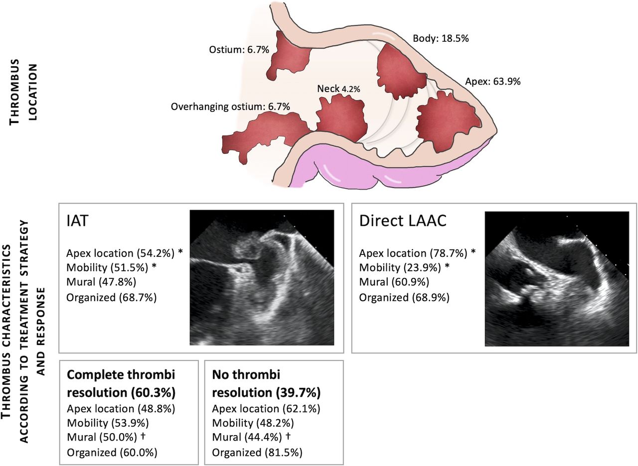
Baseline clinical characteristics, antithrombotic therapy and indication for LAAC at the time of LAA thrombus diagnosis were recorded. Preprocedural images, either by TEE or CT, were analysed by local physicians with experience in LAAC and cardiac imaging. Thrombus location was defined by its most proximal position within the LAA and divided into five categories (apex, body, neck, ostium and overhanging ostium). Mural thrombus was defined as only one surface exposed to the blood pool, flat and parallel to the endocardial surface, while protruding thrombus had more than one surface exposed and protruded into the LAA cavity. Thrombus of high echogenicity and clear borders was classified as organised thrombus.
Two treatment strategies were adopted in patients with LAA thrombus: deferred LAAC after IAT or direct LAAC. IAT was classified into (1) parenteral anticoagulation (either unfractionated heparin or low molecular weight heparin (LMWH)); (2) addition, change or intensification of OAC, involving direct OAC and vitamin K antagonist (VKA); and (3) addition of antiplatelet therapy. LAAC was performed using either standard deployment techniques or the no-touch technique. The no-touch technique involves the avoidance of contrast injections or the use of only small volume, subselective injections from outside the LAA orifice,8 9 avoiding guidewire and catheter manipulation within the LAA by loading the closure device with the delivery sheath in the left superior pulmonary vein (LSPV),10 11 and careful retraction of the delivery sheath from the LSPV into the LAA with the device partially unsheathed into its atraumatic ‘ball’ structure.8 9 The procedure, therefore, is performed without engaging the LAA with the delivery sheath.6 This technique is feasible with the Amplatzer cardiac plug and Amulet (Abbott Laboratories), LAmbre (Lifetech Scientific), Ultraseal (Cardia) and Watchman FLX (Boston Scientific), but not with Watchman (Boston Scientific) device. Embolic protection devices (EPD) were used at the operator’s discretion.
Device success was defined as deployment of the occluder in the correct position. Procedural success was defined as exclusion of the LAA without device-related complications, leak >5 mm or procedural-related complications (including stroke, systemic embolism, transient ischaemic attack, pericardial effusion and bleeding).12 Clinical follow-up was performed according to each centre’s standard of care, and data regarding vital status, bleeding and thromboembolic complications and device-related thrombosis (DRT) were collected at last follow-up.
The primary endpoint was a composite of major adverse events, including death, bleeding and stroke at 18-month follow-up. The secondary endpoint was procedural success rate. This research was done without patient involvement. Patients were not invited to comment on the study design and were not consulted to develop patient relevant outcomes or interpret the results.
Statistical analysis
Categorical variables were summarised as n (percentage) and compared using χ2 test or Fisher’s exact test as appropriate. Continuous variables were summarised as mean (SD) or median (IQR: 25th–75th percentile) and compared using two-sided Student’s t-test or Mann-Whitney U test according to their distribution. Assessment of normality for continuous data was performed using the Shapiro-Wilk test. Survival analyses were performed using Kaplan-Meier survival function. Time zero was considered to be the first diagnosis of LAA thrombus during work-up for LAAC. Survival comparisons were performed using log-rank test. P values less than 0.05 were considered statistically significant. All data were analysed using Stata V.14.
Results
From 3222 patients undergoing LAAC in 21 institutions, 126 (3.9%) patients with LAA thrombus on preprocedural imaging were included. The indication for LAAC and baseline antithrombotic treatment are presented in online supplemental figure 1. Thrombus location and dimensions by TEE (85.7%) or CT (14.3%) are presented in figure 1 and table 1.
Supplemental material

Thrombus location in patients referred for left atrial appendage closure. *P<0.05, †P=0.063. IAT, intensification of antithrombotic therapy; LAAC, left atrial appendage closure.
Baseline characteristics and preprocedural imaging findings according to treatment strategy
Therapeutic strategy: IAT or direct LAAC
Patient flow according to therapeutic approach, thrombus resolution and use of EPD is shown in figure 2. Seventy-three patients (57.9%) underwent IAT followed by deferred LAAC, while 53 (42.1%) underwent direct LAAC. Patients undergoing direct LAAC were older, with a higher prevalence of diabetes and higher bleeding risk (hypertension, abnormal renal/liver function, stroke, bleeding history or predisposition, labile international normalized ratio, elderly, drugs/alcohol concomitantly (HAS-BLED) score: 3.6±1.4 vs 3.0±1.4, p=0.014) (table 1). LAA apical thrombus was more frequent in patients undergoing direct LAAC (54.2% vs 78.7%, p=0.006) and mobile thrombus was more common in the IAT group (51.5% vs 23.9%, p=0.003) (figure 1).
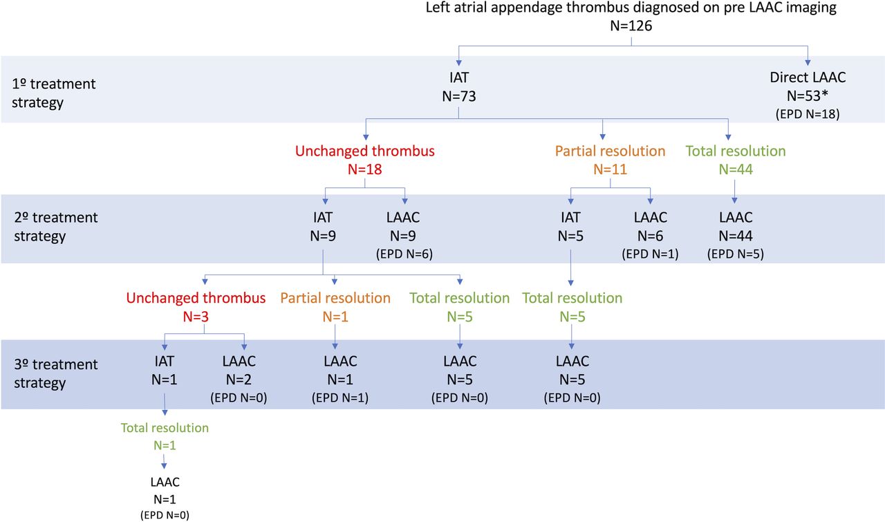
Flow of patients according to treatment strategy. *Six patients showed no thrombus during LAAC imaging. EPD, embolic protection device; IAT, intensification of antithrombotic therapy; LAAC, left atrial appendage closure.
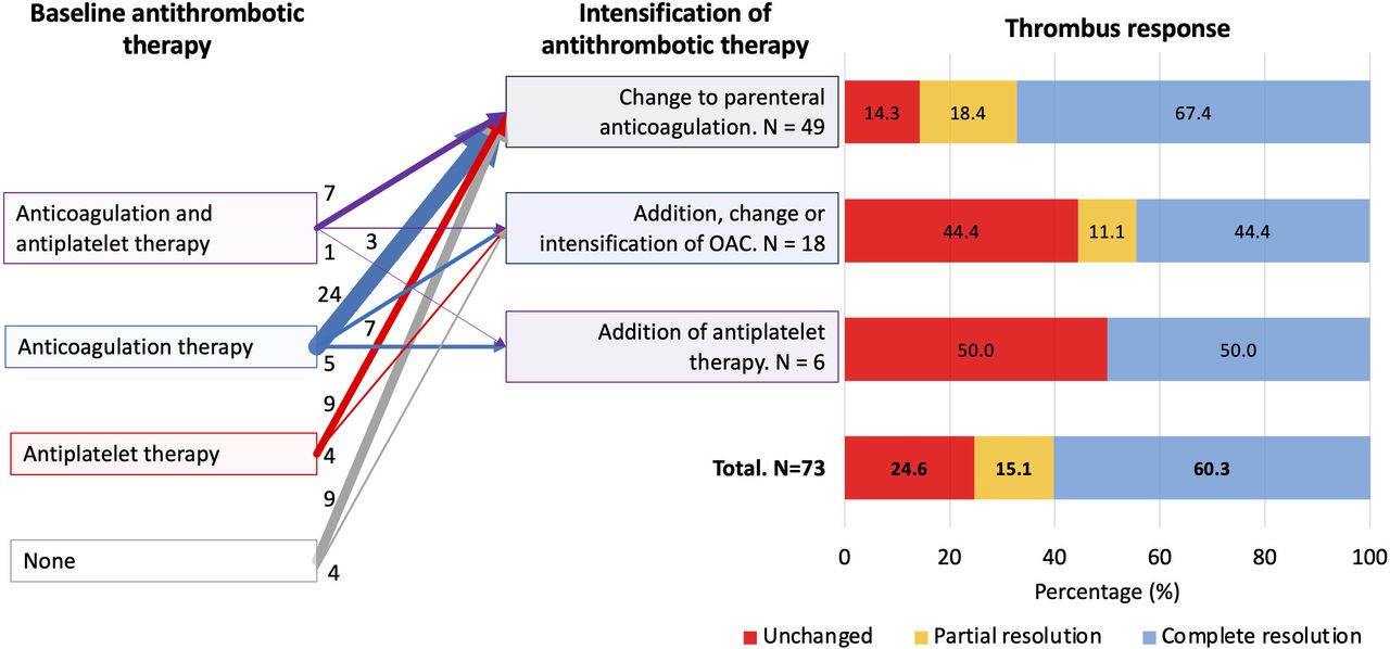
Among patients undergoing IAT, total thrombus resolution was achieved in 60.3% (n=44/73) after 57 days (IQR: 36–103) from IAT to first thrombus re-evaluation (figure 3). In those with persistent thrombus, two further lines of IAT were used, resulting in total thrombus resolution in 55 patients (75.3%) at a median of 63 days (IQR 42–156) (figure 2). Parenteral anticoagulation was the most common IAT strategy (n=49, 67.1%), achieving total thrombus resolution in 33 patients (67.4%). During IAT, there were 7 (9.6%) bleeding events in 6 patients (8.2%): gastrointestinal (n=3), intracranial (n=1), haematuria (n=1), spontaneous intramuscular haematoma (n=1) and epistaxis (n=1). IAT strategy at the time of bleeding was LMWH (n=2), LMWH plus single antiplatelet therapy (n=1), unfractionated heparin (n=1), direct OAC (n=1) and VKA dose intensification (n=1). Additionally, two patients experienced an ischaemic stroke in the IAT arm, one of whom was on LMWH plus dual antiplatelet therapy (DAPT) and the other was on LMWH.

Antithrombotic therapy at baseline, first line of treatment intensification and response of thrombus resolution. OAC, oral anticoagulation.
Among the 53 patients allocated to direct LAAC, 2 (3.8%) suffered gastrointestinal bleeding before undergoing LAAC (one on VKA and the other on LMWH as baseline therapy, which were continued until LAAC). Additionally, six patients (11.3%) having direct LAAC had total thrombus resolution on intraprocedural TEE.
The time from thrombus detection to LAAC was 63.5 days (20–126) in the overall cohort. This period was longer in the IAT arm compared with those who underwent direct LAAC (88 days (50–182) vs 20 days (1–77), p<0.001).
LAAC and in-hospital outcomes
Procedural details according to treatment strategy are summarised in table 2. The most commonly used device was the Amplatzer cardiac plug or Amulet (74.4%), particularly in the group undergoing direct LAAC (83.0% vs 69.9%, p=0.091). No-touch technique and EPD were more frequently used in the direct LAAC group (64.2% vs 20.6%, p<0.001; and 34.0% vs 17.8%, p=0.038, respectively). Embolic material was present in the EPD filters in 19.4% of cases, all of whom had identifiable LAA thrombus during the procedure.
Procedural details according to treatment strategy
In-hospital outcomes and medical therapy at discharge are presented in table 3. In the overall cohort, device and procedural success were achieved in 98.4% and 90.5%, respectively. Two unsuccessful deployments occurred in the IAT group due to complex LAA anatomy. Procedural success was 86.3% in the IAT group and 96.2% in the direct LAAC group (p=0.072). This difference was mainly driven by a lower rate of bleeding, particularly pericardial effusion, and higher rate of complete LAA occlusion in the direct LAAC group. There were no cases of intraprocedural stroke, systemic embolism or death. One patient in the IAT group had a transient ischaemic attack (0.8%) on the day of the procedure. Approximately half (49.6%) were discharged on DAPT. There was no statistical difference between groups in terms of in-hospital outcomes and the choice of medical therapy at discharge. Clinical characteristics, procedural aspects and in-hospital complications according to the presence of thrombus at the time of LAAC procedure are shown in online supplemental tables 1–3.
In-hospital and mid-term outcomes according to treatment strategy
Clinical and imaging follow-up
Follow-up post-LAAC was available in 124 patients (98.4%). The median time of follow-up was 22.1 months (IQR: 13.2–40.5). The primary endpoint of major adverse events at 18 months occurred in 29.3% (95% CI 21.7% to 38.8%) in the overall cohort, 31.5% (95% CI 21.7% to 44.4%) in those with IAT and 26.1% (95% CI 15.7% to 41.6%) of those undergoing direct LAAC (p=0.365; figure 4A). Bleeding complications occurred in 17.4% (95% CI 11.6% to 25.8%) at 18-month follow-up: 22.5% (95% CI 14.1% to 34.7%) in the IAT group and 10.5% (95% CI 4.5% to 23.6%) in the direct LAAC group (p=0.102; figure 4B).

Kaplan-Meier survival estimates for MAE, bleeding and stroke. Plot of survival functions for (A) MAE (composite of bleeding, stroke and death), (B) bleeding and (C) stroke at 18-month follow-up according to the treatment strategy. Median and IQR for time from thrombus diagnosis to LAAC in each group are represented as reference lines on the x axis. IAT, intensification of antithrombotic therapy; LAAC, left atrial appendage closure; MAE, major adverse event.
Six patients suffered a stroke during follow-up. Two presented during IAT and prior to LAAC. Four presented after LAAC: two in the IAT group at 2.6-month and 2.9-month follow-up and two in the direct LAAC group at 10.8-month and 12.7-month follow-up (figure 4C). Eighteen-month all-cause mortality was 11.6% (95% CI 6.7% to 19.7%), 9.0% (95% CI 3.8% to 20.7%) in the IAT group and 15.4% (95% CI 7.6% to 29.7%) in the direct LAAC cohort (p=0.260). Follow-up imaging was performed in 102 (80.9%) patients (90 by TEE, 11 by CT and 1 by both) within a median time of 2.9 months (IQR: 1.9–9.1). Thirteen patients (12.8%) had DRT, without difference between strategies (13.6% and 11.6% in the IAT and direct LAAC group, respectively, p=0.999). A trend towards a lower rate of DRT was observed in patients with OAC at discharge (4%), compared with those without OAC (14.7%, p=0.155).
Discussion
The present study is the first multicentre registry to analyse both medical and interventional management and outcomes in patients referred for percutaneous LAAC and found to have LAA thrombus. The main findings were the following: (1) although ~62% were taking OAC at baseline, the initial management in over half of patients was IAT (58%), with direct LAAC being employed in 42%; (2) first IAT (predominantly parenteral anticoagulation) resulted in complete LAA thrombus resolution in 60%, with subsequent further IAT increasing this to 75%; (3) patients undergoing direct LAAC more frequently had apical and immobile LAA thrombi with high procedural success rate (96%), no increased procedural complications and no periprocedural embolic events; (4) the primary endpoint of major adverse events at 18 months was 29.3%, with no difference between strategies and DRT occurring in 12.8%; and (5) bleeding complications tended to be higher in the IAT group compared with the direct LAAC group despite a lower estimated bleeding risk.
Medical management of LAA thrombus
The prevalence of LAA thrombus is relatively frequent in non-anticoagulated (9%–22%)13 and anticoagulated (up to 8.3%)14–16 patients with atrial fibrillation. A number of clinical (as incorporated into the congestive heart failure, hypertension, age ≥75 years, diabetes mellitus, stroke, vascular disease, age 65–74 years, sex category (CHA2DS2VASc) scoring system), anatomical (left atrial size) and functional (spontaneous echo contrast and LAA exit velocity) factors have been associated with LAA thrombus.13 Multilobed LAAs have been shown to have a higher risk of thrombus.17 Cauliflower LAA morphology, which is associated with lower velocity and shear strain rate within the LAA,18 is associated with a higher risk of stroke,19 while chicken-wing morphology19 and a shorter distance from LAA ostium to the first bend20 have been shown to have a lower risk of stroke. This association may be due to differences in fluid dynamics within the LAA, conferring a greater risk of thrombus formation.21 In patients referred for ablation of atrial fibrillation and found to have LAA thrombus, current recommendations suggest the use of full-dose LMWH followed by VKA, aiming for an international normalized ratio (INR) between 2.5 and 3.5 prior to carrying out the procedure.16 However, this approach is not always feasible in patients referred for LAAC due to their high bleeding risk. Of the patients in our study, 58% underwent IAT, with the remaining undergoing direct LAAC, reflecting the clinical dilemma and the paucity of data in this challenging situation. However, the relatively high rates of bleeding complications (9.6%) and stroke (2.7%) raise some concerns with the IAT strategy. A careful, individualised assessment of patients’ clinical and anatomical characteristics should be performed to weigh the bleeding risk with IAT versus a direct LAAC in the presence of LAA thrombus.
Direct LAAC with LAA thrombus
The presence of atrial thrombi has been an exclusion criterion in randomised LAAC clinical trials to date22–25 due to the risk of periprocedural embolic events on manipulation of the LAA. However, an absolute contraindication for anticoagulation (nearly 30% in our cohort) or failure of IAT to resolve the thrombus (40% in our cohort) has resulted in many operators performing percutaneous LAAC in the presence of thrombus, with many taking steps to minimise potential embolisation.
Data regarding LAAC in the presence of thrombus are restricted to case reports and one retrospective multicentre registry of 28 patients.6 10 26 27 Recently, a systematic review has collected all 58 published cases showing 100% successful device implantation. In this study, lobe-and-disc devices were more frequently used (76%),EPD was used in 29% patients, and no periprocedural complications were reported. Despite these promising results, publication bias might be of concern since a considerable amount of the data came from case reports. Our study included 126 consecutive patients with LAA thrombus, analysing both medical and invasive approaches and their outcomes. Our results support the feasibility and safety of direct LAAC in selected patients, with procedural success being 96% and no cases of systemic embolism observed.
The no-touch technique was employed in 39% of our cohort. Furthermore, these cases should be referred to experienced operators to reduce manipulation of the LAA and limit the need for device recapture and repositioning. Lobe-and-disc devices, such as the Amulet, have a shorter length and the possibility of a shallow deployment, making these devices more appropriate for patients with LAA thrombus. The new-generation Watchman FLX with the possibility of a ball-advancing technique for device implantation may also be effective in trapping thrombus distally. Future studies will have to determine the feasibility and efficacy of this device in a larger cohort.
Acute brain lesions detected by MRI occur in up to 48% after LAAC.28 EPD might reduce the incidence of these lesions, as has been shown in transcatheter aortic valve replacement.29 In the presence of LAA thrombus, the use of these devices may be particularly appropriate; however, in our study, EPD was used in only 34% of patients with LAA thrombus undergoing direct LAAC, a percentage slightly higher than previous reports.7 Unfortunately, the impact of EPD in subclinical acute cerebral lesion could not be determined in our study as brain imaging was not routinely performed. Interestingly, macroscopic embolic material was captured in 19.4% cases, which theoretically at least may have prevented a number of cerebral embolisation events. Their use might even be justified in patients with thrombus resolution, who may still harbour thrombus of smaller size than the spatial resolution of the imaging technique used (1 mm and 0.5 mm for TEE and CT, respectively) or thrombus within LAA trabeculae. EPDs are a common adjunct in many cardiac procedures and could be considered when planning LAAC in patients with evidence of thrombus at any time. Nevertheless, additional data, including randomised studies, are needed to provide a stronger recommendation.
Postprocedural antithrombotic therapy is an additional unanswered question in patients who undergo LAAC, and more so in those with LAA thrombus. Previous studies suggest >50% are discharged on anticoagulation.7 In our study, antiplatelet therapy was the most frequent treatment (68%). However, ~25% were maintained on anticoagulant therapy. Although peripheral embolism was uncommon, DRT occurred in 13% of patients at 3-month follow-up. This rate is higher compared with previous LAAC randomised trials and registries, which have reported rates between 1.5% and 5.7% at 2-month and 18-month follow-up, respectively.30 The high incidence of DRT probably reflects a prothrombotic environment in this population. LAA exclusion with systems such as LARIAT might be an alternative since an endocardial device is not implanted, avoiding the risk of DRT. However, advancement of an endocardial guidewire into the LAA apex is needed during the procedure, representing a limitation for patients with residual thrombus. The clinical implications of DRT justify further studies to confirm our findings and analyse its impact and management in this particular clinical scenario.
Study limitations
This study had the limitations inherent to a non-randomised, observational study, without external adjudication of events. The therapeutic approach (IAT vs direct LAAC), antithrombotic therapy, modifications in LAAC technique and the use of EPD were left to clinicians’ and operators’ discretion, representing real-world practice. Therefore, there is a potential selection bias between treatment groups, and we cannot rule out the possibility that direct LAAC was performed in patients with more favourable anatomy. Imaging follow-up was not available for all patients. Because of the low incidence of procedural complications, predictors of these complications could not be analysed. Additionally, there were no data available for patients with LAA thrombus who ultimately did not undergo LAAC procedure.
Conclusions
Percutaneous LAAC in the presence of LAA thrombus as an initial or deferred strategy is feasible and safe, with high procedural success and low rates of periprocedural complications. Intensification of antithrombotic treatment for thrombus resolution was the initial strategy in half of the population, with a relatively high rate of bleeding events before LAAC and at 18-month follow-up. Device thrombosis remains a concern during follow-up and further work is required to determine the optimal treatment strategy following LAAC.
Key messages
What is already known on this subject?
The management of patients with atrial fibrillation referred for left atrial appendage closure (LAAC) who are found to have left atrial appendage (LAA) thrombus is challenging.
No specific data exist regarding the safety and efficacy of different management options for these patients with high risk of bleeding.
What might this study add?
In this multicentre registry comparing management and outcomes of patients with LAA thrombus referred for percutaneous LAAC, intensification of antithrombotic therapy ultimately resulted in complete thrombus resolution in 75% cases after several lines of antithrombotic treatment; however, a relatively high bleeding rate was observed before LAAC.
Patients undergoing direct LAAC had higher bleeding risk and more frequently apically located and immobile thrombi.
In these selected patients, direct LAAC was feasible with a high procedural success rate and absence of embolic complications.
Both approaches had a high rate of device-related thrombosis during follow-up.
How might this impact on clinical practice?
Direct LAAC might be considered as treatment strategy in selected patients with an indication for LAAC and evidence of LAA thrombus.
Experienced operators, a modified procedural technique and use of embolic protection device should be considered in this scenario.
Data availability statement
Data are available upon reasonable request. All data relevant to the study are included in the article or uploaded as supplementary information. DATA AVAILABILITY STATEMENT: Data are available upon reasonable request.
Ethics statements
Patient consent for publication
References
Supplementary materials
Supplementary Data
This web only file has been produced by the BMJ Publishing Group from an electronic file supplied by the author(s) and has not been edited for content.
Footnotes
Twitter @ConteTirado, @icruzgonzalez, @JS, @saia_francesco, @analaffond, @josecaferca, @ignamatsant, @luisnombela
LM and GT-C contributed equally.
Contributors The authors of this paper specifically contributed to the following aspects: conception and design or analysis and interpretation of data: LM, GT, LNF; drafting of the manuscript or revising it critically for important intellectual content: all authors; and final approval of the manuscript submitted: all authors. LNF is responsible for the overall content of the study as guarantor.
Funding This study was supported by Fundación Interhospitalaria para la Investigación Cardiovascular (FIC Foundation) via an unrestricted grant from Abbott.
Competing interests Relationship with industry: LNF has served as a proctor for Abbott and has received speaker honoraria from Edwards Lifesciences and Boston Scientific. All other authors have reported that they have no relationships relevant to the content of this paper to disclose.
Provenance and peer review Not commissioned; internally peer reviewed.
Supplemental material This content has been supplied by the author(s). It has not been vetted by BMJ Publishing Group Limited (BMJ) and may not have been peer-reviewed. Any opinions or recommendations discussed are solely those of the author(s) and are not endorsed by BMJ. BMJ disclaims all liability and responsibility arising from any reliance placed on the content. Where the content includes any translated material, BMJ does not warrant the accuracy and reliability of the translations (including but not limited to local regulations, clinical guidelines, terminology, drug names and drug dosages), and is not responsible for any error and/or omissions arising from translation and adaptation or otherwise.