Review

Contribution of Topical Antioxidants to Maintain Healthy Skin—A Review

by
Tércio Elyan Azevedo Martins
1,2,†,
Claudinéia Aparecida Sales de Oliveira Pinto
2,*,†,
Andressa Costa de Oliveira
2,
Maria Valéria Robles Velasco
2,
Arilmí Rosa Gorriti Guitiérrez
3,
Martha Francisca Cosquillo Rafael
3,
Jossimar Paúl Huamaní Tarazona
3 and
Mónica Guadalupe Retuerto-Figueroa
3
1
Aesthetics and Cosmetics Course, Paulista University, São Paulo 04709-11, Brazil
2
Department of Pharmacy, School of Pharmaceutical Science, University of São Paulo, São Paulo 05508-000, Brazil
3
Faculty of Pharmacy and Biochemistry, Major National University of São Marcos, Lima 15081, Peru
*
Author to whom correspondence should be addressed.
The contributions of these authors were equal.
Sci. Pharm. 2020, 88(2), 27; https://doi.org/10.3390/scipharm88020027
Submission received: 29 February 2020 / Revised: 6 April 2020 / Accepted: 15 April 2020 / Published: 3 June 2020
(This article belongs to the Special Issue Functional Ingredients in Topical and Transdermal Delivery Systems)

Abstract

:
The skin is constantly exposed to various environmental stresses, in particular to the damage caused by pollution and ultraviolet radiation (UV), and as a consequence, the horny extract can be negatively impacted by the harmful influence of some of its surface components. The mechanisms involved in the degradation processes promoted by UV radiation are driven by the direct absorption of radiation via cellular chromophores, the formation of excited states and the consequent chemical reactions, or even by the photosensitization mechanisms, in which UV light is absorbed by the sensitizers that are excited and their reactions promote the formation of reactive oxygen species (ROS). The mechanisms of polluting agents are not yet fully understood, however, they indicate that one of the main mechanisms involved is oxidative stress by lipid peroxidation, with the ability to promote damage to the composition of sebum, the quality of the stratum corneum and also, promote aging skin. Recent studies demonstrate the potential of antioxidant agents, with an emphasis on products of natural origin, which try to promote the maintenance of the physiological balance of the skin.

Graphical Abstract

1. Introduction

The skin is exposed to several types of environmental stress, of different natures and intensities and there is a lack of data that associate air pollution and skin diseases. In general, the few clinical data suggest the worsening of atopic dermatitis by pollution, with the suspicion that ultrafine particles may exacerbate the inflammatory process, damaging its surface or more deeply the skin, thus inducing dermatological diseases [1,2].
With the harmful action of pollution, it is of utmost importance that the skin, more specifically the stratum corneum, remains integral, so that it can exercise its protective barrier function. The barrier function aims to defend against environmental agents and external contaminants, avoiding the absorption process and the toxic and harmful effects [3,4].
In order to ensure an adequate protection barrier, skin integrity is essential, however, some factors may facilitate the permeation/penetration process such as location, anatomy, age, skin hydration and physicochemical characteristics of the contaminants. Thus, children and the elderly are more exposed to aggressive agents, considering the increased absorption of contaminants [5,6].
Thus, the association of modern lifestyle, the quality of the air increasingly contaminated by smoke residues (from vehicles, industry, cigarette) and the encouragement of the practice of outdoor recreational activities without proper protection, can promote severe damage to health and especially to the skin and some of its attachments. The harmful damage caused to the skin by exposure to sunlight and pollution, can be extremely harmful, promoting cutaneous and systemic immunosuppression, formation of inflammation processes, oxidative stress and metabolic deficiencies, in addition to promoting cellular damage to genetic material, favoring the skin cancer formation [7,8,9].
The association of polluting agents composed of particulate matter (PM) that may contain polyaromatic hydrocarbons (PAH) and toxic gases such as CO2, CO, SO2, NO, NO2 and other nitrogen oxides (NOx) and the cumulative action of UV radiation on the skin without protection, they promote the formation of chemical and morphological reactions, being able to form reactive oxygen species (ROS’s), histochemical alterations of different severities, skin thickening and rectification of the dermoepidermal junction [7,9].
UV radiation can be absorbed by a wide variety of skin molecules, however, with absorption, the molecules can undergo chemical changes, asdeoxyribonucleic acid (DNA) is one of the molecules that has the greatest ability to absorb UV radiation and, consequently, undergo neoplastic mutations and transformations. Another harmful action attributed to UV radiation is the ability to activate the components of the skin’s immune system, promoting an inflammatory response through several mechanisms. In addition to the skin, UV radiation can affect other parts of the body, such as the eyes, causing loss of vision, photoconjunctivitis and cataracts [10].
The mechanisms involved in the cutaneous effects of the pollutants and UV radiation are uncertain, however, it is assumed that exposure can affect the composition of sebum and the quality of the stratum corneum, in addition to intensifying the signs of skin aging, with the formation of spots pigments and wrinkles. Understanding the involved processes is an important step to minimize the damage to skin. Several in vitro and ex vivo studies are described in the literature. Human cell culture demonstrated sensitivity to the toxic effects of pollutants present in the air; one of the important processes involved in the damage caused to the skin by pollution and UV radiation is peroxidation. Studies about antioxidants have been increasing over the last few years. The use of these substances could be useful to improve skin conditions against pollution and UV radiation. For this reason, specific studies should be a trend in the coming years [5,9,11,12].

2. Aggressive Skin Agents

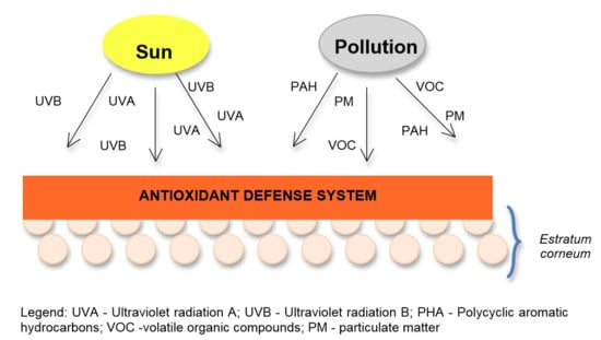
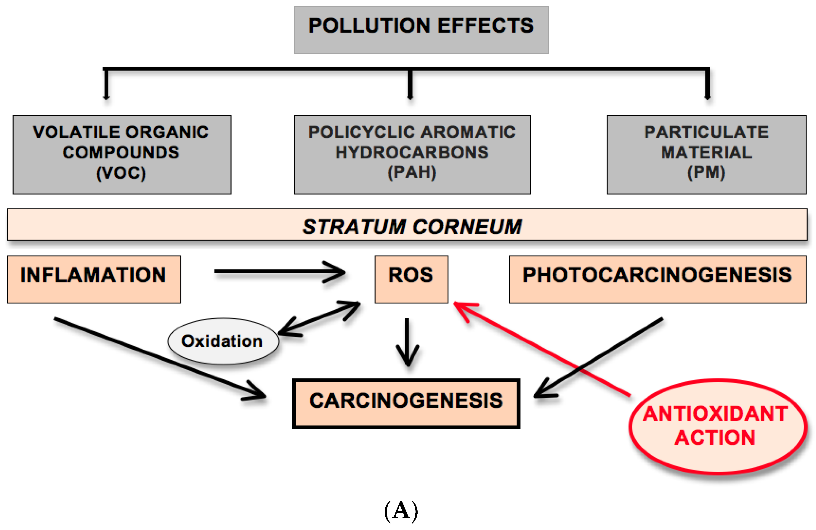
2.1. Pollution Effects

Studies regarding air pollution and its effects on the health of the population show that, even when the pollutants are below the determined levels, they are capable of causing harmful effects on the health of the population [13,14].
Polycyclic aromatic hydrocarbons (PAHs), volatile organic compounds (VOCs) and particulate matter (PM) are the most significant atmospheric pollutants, with the ability to cause damage to the skin in daily activities. VOCs have the ability to increase the inflammatory reaction, induce the formation of cancer lesions and atopic dermatitis in animal models and in vitro, however, the action of these harmful agents can be amplified with the interaction of UV light and cause oxidative damage to the skin [15].
The main cause of PAH is the smoke that comes from the combustion of organic matter, such as burning wood or oil, and has the ability to generate ROS’s, in addition to inducing the proliferation of melanocytes. When associated with UV radiation, they promote the synthesis of melanin, resulting in the formation of environmentally induced lentigines, in addition to intensifying the development of skin cancer (papilloma, carcinoma and squamous cell carcinoma). Importantly, the diet is the main source of contamination by PAH, mainly through the consumption of smoked meat [6,9].
The main air pollutant is the PM, normally produced by factories, power plants, incinerators, cars, the construction industry and fires, however, its physical and chemical characteristics depend on the places where it is produced (rural, urban areas or industrial environment) as well as the station from which it is collected. Normally, PM shows variability in terms of size and can be classified as ultrafine (<100 nm), fine (approximately 2.5 μm) and coarse (approximately 10 μm) particles, with reduced size particles being more harmful [16,17]. However, the transcutaneous penetration of PM is possibly limited, as studies exclude the bioavailability of particles larger than 20 nm [18,19].
In general, the ultrafine particles, thin and PAH, due to their size, are able to interact with the skin more effectively, causing toxicological effects by presenting pro-oxidant, mutagenic and carcinogenic activity; and in specific cases, some of these substances react with sunlight, exerting phototoxicity [5,6].
Contamination agents can reach the human body through penetration/permeationthe stratum corneum orsystemic circulation. Transcutaneous penetration involves the passage of particles through the sebum and several layers of the stratum before reaching the first living keratinocytes [20].
For the protection of the skin from polluting agents it is necessary to constantly use cosmetics that work as a protective agent by forming a barrier, maintaining the functions and the physiological balance of the skin. The substances responsible for the anti-pollution action are products capable of forming a film and promoting an antioxidant activity, improving the function of the skin’s natural barrier, physically protecting the skin and eliminating oxidative pollutants. However, there are other ways to protect the skin from pollution, such as the use of cleaning products that remove excess pollutants adhered to the stratum corneum; sunscreens that spread and/or bounce off UV radiation and prevent the formation of photoreactive compounds; emollient agents that have the function of maintaining and restoring the function of the skin barrier, in addition to preventing excessive removal of the hydrolipidic mantle [5,21].
The active substances used in cosmetics can play several roles in preventing the action of pollution agents, by blocking the signaling pathways of the pro-inflammatory membrane such as Keap1-Nfr2 and IKB-NFKB; causing an immediate reduction in short-term health damage caused by inflammation factors, such as ciclooxigenase (COX) and interleukins; increasing cell differentiation like NOTCH1 and WNT, and improving the metabolism of xenobiotics like AhR [22,23].
Cosmetics with the ability to combat the effect of pollution, exercise their function by improving the structure and function of the skin barrier, supplying topical antioxidants and a consequent decrease in oxidation stress and inflammation processes, reducing pollutant deposits on the skin through the use of cleaning and exfoliation products, protecting the skin from UV radiation that can aggravate the effect of other environmental polluting factors, regulating melanogenesis and removing fibrous protein synthesis in the dermis [11].
The first mechanism involved is oxidative stress, which occurs primarily due to the constant exposure of the stratum corneum and hydrolipid mantle to the air, since ozone is a potent inducer of squalene peroxidation, obtaining reactive aldehydes (malondialdehyde and 4 HNE (4-hydroxy-2-neonal) that react with proteins by forming a complex with amino acid residues, parallel to the carbonylation produced by the direct oxidation of ROS. Thus, the reduction of antioxidant substances in the skin (vitamins E and C and glutathione), promote damage to the structure of the horny extract and its physiological functions. The PM associated with UVA radiation also has the ability to promote an increase in oxidative stress in the superficial layer of the skin [6,24].
As for skin oxidative stress in vivo, the simple presence of PM is able to affect the deeper layers of the epidermis and dermis, since ozone is extremely reactive and can diffuse through the skin layers and can catalyze the production of ROS, for direct redox cycling (HPA-quinones) or indirectly, due to mitochondrial damage. However, the literature does not show results of the influence of the presence of high PM concentration isolated on the production of ROS in the skin [24,25].
The inflammatory process promoted by exposure to pollution is the most established mechanism, since several inflammatory markers have been identified in the blood of smokers or in individuals exposed to air pollution. Studies show that UV radiation promotes squalene peroxidation and triggers a global pro-inflammatory response in primary keratinocyte cultures [26]. PAHs, when activated through the UVA, could produce singlet oxygen and may be involved in the formation of inflammatory cutaneous skin diseases, which can be aggravated at maximum levels of pollution. Thus, the sebum affected by the oxidation promoted by ozone, and by the association UVA/PAH, could act as an exogenous sensitizer in aggravating atopy. In addition, squalene peroxidation can promote the formation of mediators in the development of acne [27].
The third mechanism involved in the action of pollution on the skin is metabolic stress, which aims to increase the hydrophilicity of xenobiotics, in order to facilitate their excretion. This process is controlled by the AhR (aryl hydrocarbon receptor) which stimulates the expression of detoxification of cytochrome P450 by means of the monooxygenase enzymes CYP1A1 and CYP1B1, however, in the skin the AhR is expressed in all cell types and is clearly a factor that connects skin and the environment [6].
The enzymes CYP1A1 and CYP1B1 have the ability to reduce the hydrophobicity of chemicals; however, this biochemical process is often capable of generating potent and toxic intermediates (epoxides or quinones), which destabilize redox homeostasis, thus producing ROS or even consuming the glutathione necessary for its detoxification. Pollutants such as PAHs can activate AhR, contributing to mutagenesis and carcinogenesis, especially in the lungs. AhR is also involved in skin diseases, including chloracne, accelerated aging, skin inflammation and skin carcinogenesis [28].
Hair and scalp are also affected by excessive exposure to environmental pollutants (UV radiation, PAHs, nitrogen oxides, ozone and cigarette smoke, among others), resulting in an opaque, dry and lifeless appearance [5]. To obtain a potential anti-pollution effect, hair cosmetics must form films and anti-adhesives. Plant extracts also have a potential anti-pollution effect because they reduce ROS levels and promote cell autophagy when damaged [29].
In an in vitro investigation of the cutaneous impact of pollution combined with exposure to sunlight, pollutants (PAH, PM or PM extract) irradiated with UVA were associated with a significant phototoxic effect equal to or greater than that produced by d-UV (daily ultraviolet), and the result suggested that exposure to this photo-pollution stress may impair skin homeostasis and aggravate skin damage induced by sunlight [9].

2.2. Solar Radiation

Solar radiation is composed of a continuous electromagnetic spectrum of radiation, divided according to its wavelength (λ): infrared (>780 nm), visible (780–400 nm) and ultraviolet radiation (UV) (400–100 nm). UV radiation, in turn, is divided into UVC (100–290 nm), UVB (290–320 nm), UVA II (320–340 nm) and UVA I (340–400 nm). However, only a some of these radiation types, more specifically ionizing radiation and the most energetic fraction of non-ionizing radiation, have the ability to reach the earth and penetrate the skin tissue [30,31].
Energetically, solar radiation and λ are inversely proportional, and UV rays have a lower λ, however, they are the main factor responsible for photochemical reactions in the skin. UVC radiation has greater energy and penetrating power, however, it does not reach the earth’s surface, being blocked by the ozone layer, however, UVA and UVB radiation, reach the skin in different shapes and depths [32,33].
The UVB band is considered less energetic, reaching only the epidermis, however, it presents several potentially harmful effects such as the ability to form erythema, inflammation, thickening of the stratum corneum, and immunosuppression by the formation of reactive oxygen species(ROS), thus causing direct damage to the cellular genetic material and increasing the carcinogenic potential, by inducing pyridine dimers in the nucleotide sequence responsible for the mutations [5,34].
Currently, three mechanisms involved in the action of UVB radiation are described: altering the modulation and production of inflammation mediators (interleukin 10 (IL-10), interleukin 1 (IL-1), interferon-gamma (INF-g) and tumor necrosis (TNF-a)), modifying the entire inflammatory cascade; inducing the action of keratinocytes in the production of prostaglandin E, promoting a decrease in the presentation of adhesion molecules of the ICAM-1 type; forming DNA by-products, dimers and alkylations in the double strand of DNA, generating uncontrolled cell production, culminating in the formation of apoptosis and uncontrolled cell division and its neoplastic effects [35,36].
UVA radiation reaches the skin tissue more deeply, being responsible for physiological and pathological damage, either early or late, promoting photoaging for damage caused to collagen and elastin fibers, formation of ROS and wrinkles, in addition to potentiating neoplastic damages of UVB radiation [37,38].
It is relevant to present the beneficial effects of UV rays on health, such as body heating, stimulation of blood circulation and treatment of diseases such as rickets, jaundice, multiple sclerosis and asthma. A prominent role of UV radiation in health is the synthesis of cholecalciferol (vitamin D3), directly involved with calcium and phosphorus homeostasis, bone metabolism and the immune system. UV radiation can also be used daily in the form of phototherapy, which associated with medications, promote improvement in dermatological diseases such as psoriasis and eczema [7,39].

3. Skin Peroxidation Process

The ROS induced oxidative damage in biological systems can involve either the addition of oxygen, removal of hydrogen or removal of electrons. The removal of electrons is the most common while removal of hydrogen is moderately involved, and addition of oxygen is not so common [39]. The main ROS are divided into two groups, the radical ones: hydroxyl (HO), superoxide(O2•−), peroxyl (ROO) and alkoxyl (RO); and non-radicals: oxygen, hydrogen peroxide and hypochlorous acid. Among the NRs are nitric oxide (NO), nitrous oxide (N2O3), nitrous acid (HNO2), nitrites (NO2), nitrates (NO3) and peroxynitrites (ONOO). While some of them can be highly reactive in the body attacking lipids, proteins and DNA, others are reactive only with lipids. There are still some that are not very reactive, but despite that they can generate harmful species of reactive oxygen and nitrogen, which are known to play an important role in the defense against infection, cell signaling, apoptosis, besides ageing [40,41].
The HO radical is formed in the body mainly by two mechanisms: reaction of H2O2 with transition metals and water homolysis by exposure to ionizing radiation. The incidence of ultraviolet radiation and X-rays can produce the HO radical in skin cells. The intensive and frequent attack of this radical can originate mutations in the DNA and, consequently, lead to the development of cancer in humans (Equation (1)). H2O2 is practically harmless but, due to the presence of transition metals in the cells, the HO radical is generated inside it (Equation (2)). H2O2 plays an important role in oxidative stress as it can easily cross cell membranes and generate the hydroxyl radical. It only oxidizes proteins that have methionine residues or very reactive thiol groups like GSH. In the body human, the most important transition metals for this reaction to occur are Cu1+, Fe2+ and Mn(n+). In this system, the importance of iron is more pronounced due to its greater bioavailability and most of the time it is complexed with transport proteins as such as transferrin and stored in ferritin and hemosiderin. The reaction of Fe2+ with H2O2 (fenton reaction) can be represented in a simplified way (Equation (3)) or in a more complex way (Equation (4)) [40,41,42].
H 2 O UV HO + H
M + n +   H 2   O 2   M ( n + 1 ) + +   HO + HO
Fe 2 + +   H 2   O 2 Fe 3 + +   HO + HO
Fe 2 + +   H 2   O 2   HO FeOH 3 + (   or   Fe   O 2 +   H ) Fe 3 + +   HO
The hydroxyl radical causes damage to DNA, RNA, proteins, lipids and cell membranes in the nucleus and mitochondrial. The reaction occurs in the mitochondria by the oxidation of the semiquinone from the ubiquinone and the flavin of the NADPH dehydrogenase; in the endoplasmic reticulum from cytochrome P450 and from the flavoprotein NADPH-cytochrome P450 reductase; in the cytosol by the enzyme xanthine oxidase. There are also reactions of generation of hydrogen peroxides in mitochondria, endoplasmic reticulum, peroxymos and cytosol, and the formation by enzymatic reactions of cyclooxygenase, lipoxygenase, aldehyde oxidase, catecholamine auto oxidation, ferredoxinflavine and reactions catalyzed by metals transition like iron and copper [41,43]. The OH radical is one of the most potent oxidants, with the ability to cross membranes and react with unsaturated lipids and DN [41].
Lipid peroxidation can be defined as an oxidative deterioration of polyunsaturated lipids occurring in three stages: initiation, propagation and termination [40,43]. The oxidation lipids by reactive oxygen initiates a cascade of reactions that leads to the formation of triplet excited carbonyls formed by a process under which oxidants such as free radicals or non radical species attack lipids containing carbon–carbon double bond(s), especially polyunsaturated fatty acids (PUFAs) that involve hydrogen abstraction from a carbon, with oxygen insertion resulting in lipid peroxyl radicals and hydroperoxides. The PUFAs are excellent substrates for lipid peroxidation because of the presence of active bis-allylic methylene groups. The carbon–hydrogen bonds on these activated methylene units have lower bond dissociation energies, and these hydrogen atoms are more easily abstracted in radical reactions [40,44,45].
The skin peroxidation is an important process in human aging and involves a complex regulation of the antioxidant defense system. Extrinsic aging processes is driven to a large extent by oxidative stress caused by UV irradiation, xenobiotics and pollution. The body produces several enzymes, including superoxide dismutase, catalase, glutathione peroxidase, and glutathione reductase that neutralize many types of free radicals [40,46,47].
The most harmful form of oxygen to the body is singlet oxygen (1O2) which reacts with some classes of biomolecules and, in general, these reactions are of the eno (Equation (5)) and diene type (Diels–Alder reactions) (Equation (6)). The most reactive natural compounds against 1O2 are carotenoids, due to the multiple conjugated unsaturation. The reaction with fatty acids occurs by incorporating oxygen into the chain with consequent migration of the double bond, forming hydroperoxide acids [41,42,48].
Scipharm 88 00027 i001
Scipharm 88 00027 i002
The superoxide anion radical (O2•−) is inactive but generates hydrogen peroxide (Equation (7)). It is also a weak base whose conjugated acid, the hydroperoxide radical (HOO), is more reactive (Equation (8)). The superoxide anion radical (O2•−) participates in the production of HO radical, through the reduction of Fe (III) chelates, forming Fe2+ (Haber–Weiss reaction) (Equations (9) and (10)). The anion radical O2•− can release Fe2+ from storage proteins and iron-sulfoproteins, such as ferritin and aconitase, respectively. The anion radical O2•− also reacts with the radical HO producing singlet oxygen 1O2 (Equation (11)) and with nitric oxide (NO) producing peroxynitrite (ONOO) (Equation (12)) [41,42,48].
2 O 2 + 2 H + H 2 O 2 + O 2
O 2 + H +   H O O
Fe 3 + + O 2   [ Fe 3 + O 2 Fe 2 + O 2 ]   Fe 2 + + O 2
O 2 + H 2 O 2 Fe H O + HO + O 2
O 2 +   OH   1 O 2 + HO
NO + O 2 ONOO
The superoxide anion radical is a weak bactericide, capable of inactivating iron-sulfur proteins from bacteria, but products that have strong antimicrobial activity, such as hypochlorous acid (HOCl), hydrogen peroxide (H2O2) and peroxynitrite (ONOO) which are the main products responsible for fighting foreign bodies. In addition, the superoxide anion radical works as a molecular signal, as it oxidizes –SH groups in disulfide bonds (Equation (13)), being possible to activate and deactivate enzymes containing methionine [41,42,48].
2 O 2 + 2 RSH 2 HOO + RSSR
Nitric oxide does not directly attack DNA, but when reacting with the superoxide anion radical O2•−, it generates peroxynitrite, which forms agents capable of nitrating aromatic amino acids, such as tyrosine, generating nitrotyrosine and DNA bases [41,42].
There are differences between dermis and epidermis peroxidation, in dermis there are the reduction of collagen, elastic fibers, proteoglycans and hyaluronic acid, the result is fine wrinkles. Dermis antioxidant defense system involves, fundamentally, the combat against metalloproteinases activation and elastase, collagenase and hyaluronidase production. In addition, there is a correlation between the antioxidant power and inhibitory enzymatic activity [46,47]. In the epidermis, basically, the corneal layer is composed of a fusion of keratinized cells, compressed and surrounded by the extracellular lipid matrix. This lipid matrix is composed of mixture of long-chain ceramide, cholesterol and free fatty acids in an almost equal molar ratio (1:1:1). The anti-oxidative mechanisms in epidermis are more highly remarked than in the dermis, there is an epidermal concentration gradient in the case of antioxidants, especially the low molecular-weight ones. The low molecular weight antioxidants act in several ways on the skin tissue, either by chelating metallic ions or by direct neutralization of free radicals. Glutathione, uric acid, ubiquinol, vitamin C and E are detectable in the stratum corneum, but their concentration increases abruptly toward deeper cell-layers of the stratum corneum, after this, the concentration of enzymes and low molecular-weight antioxidants decline towards the stratum basale [46,49]. The antioxidant gradient capacity of keratinocytes is associated with decreasing carcinogenesis risk, because it prevents the survival of mutated stem cells. Besides this, the protection of suprabasal cells is essential for maintenance of skin integrity [50]. In fact, the multiple protection mechanisms of keratinocytes against reactive oxygen are the first skin defense line by damage induced by UV radiation, xenobiotics, and pollutants. In the stratum corneum several cornified envelope proteins, including, loricrin, involucrin, filaggrin and small proline-rich proteins (SPRRs) acts as antioxidants together with low molecular-weight antioxidants [40].
Healthy humans often present controlled oxidative stress in fluids and tissues. The primary biomarkers of lipid peroxidation products found are the lipid hydroperoxides. Among the many different aldehydes which can be formed as secondary products during lipid peroxidation, malondialdehyde (MDA), propanal, hexanal, and 4 hydroxynonenal (4-HNE) and hydrocarbons, such as ethane and ethylene have been extensively studied [40,44].
The skin lipid peroxidation is particularly important for health care, especially in terms of lipidic composition to the stratum corneum (long-chain ceramide, cholesterol and free fatty acids). Under medium or high lipid peroxidation rates the extent of oxidative damage overwhelms repair capacity and induces cell apoptosis or necrosis programmed cell death; processes that lead to molecular cell damage which may facilitate development of various pathological states and accelerated aging [51].
Currently, the stratum corneum oxidative stress behavior has being evaluated by tape stripping or skin model system and measured by 2,2-Diphenyl-1-picrylhydrazyl (DPPH) or thiobarbituric acid reactive species (TBARS) methods. Knowing how the stratum corneum reacts to antioxidant substances will lead to the development of more effective skin care products. Presently products like sunscreen, anti-pollution, anti-aging and antioxidants cosmetics claim the action antioxidant, but it is important to understand how these products act in stratum corneum to help in the protection and maintenance of skin quality [44].

4. Transcutaneous Penetration and Antioxidants Products

Transcutaneous penetration involves the passage of particles through the hydrolipidic mantle and the horny layer, before they reach keratinocytes. This process can be very limited in intact and healthy skin, since particles larger than 20 nm do not present cutaneous bioavailability, except for damaged skin. Intact human skin has an efficient barrier function against the permeation and penetration of exogenous agents, however, this function can become an obstacle to the activity of topically applied substances [6,20].
The penetration and cutaneous permeation studies of antioxidant activities may present as the first limiting factor of the outermost layer of the epidermis, the stratum corneum, as it presents fully keratinized cells (corneocytes), anucleate, stratified and hydrophilic and lipophilic areas. The term penetration consists of the passage of the active principle (s) only from the stratum corneum, while the term permeation consists of the passage of the active (s) through the epidermis, reaching the dermis [52,53].
Antioxidants have been used in topical products for different purposes, but the main function is to create better conditions for healthy skin. Nature provides an abundant source of antioxidant substances and natural phenolic compounds, which demonstrate a topical photoprotective action, as well as antioxidant potential, anti-aging, moisturizing, anti-pollutant, which is beneficial to the skin [54,55].
The penetration of antioxidant substances in the skin can be an important factor for its performance as a skin protector against UV radiation and pollution. The performance of products with antioxidant action combined with the synergistic action with photoprotectors indicate candidates with high potential for maintaining healthy skin.
Traditional antioxidants substances have been evaluated to skin protection. The suppression of erythema, induced by UV radiation, has been an important parameter for evaluating the action of these antioxidants, but is not a direct method to evaluate the cutaneous oxidation suppression.
In the skin, l-ascorbic acid (vitamin C) is the main antioxidant of the fluid phase, protecting the watery part. Topical l-ascorbic acid protects skin against erythema produced by UVB and UVA irradiation by a mechanism unrelated to the absorption of the UV radiation. Moreover, it can provide protection against UVB-induced immunosuppression and systemic tolerance to contact allergens [56]. α-Tocopherol in human skin inhibits the production of prostaglandin E2, prevents sunburn cell formation, ultraviolet (UVB) induced lipid oxidation and edema [57]. Glutathione, vitamin E and ubiquinone (Coenzyme Q10) are the important lipophilic antioxidants that protect the compartment of intracellular membranes [58]. Selenium, an essential trace element in humans and animals, is required by the intracellular antioxidant enzymes glutathione (GSH) peroxidase and thioredoxin reductase. Carotenoids like Lutein (L) and zeaxanthin isomers (Zi) may inhibit peroxidation lipids of membrane and quench singlet oxygen. Lutein is the main carotenoid present in skin cells [59]. Uric acid is an important endogen antioxidant in the serum, representing a final product in the metabolism of the purines and acts as a potent free radical scavenger, inhibiting lipid peroxidation [60]. The damage of UVA radiation has been linked to the release of ‘‘free’’ iron into cells via ferritin proteolysis, and this can be an important factor in UVA-induced skin damage. Thus, the ferritin can regulate the lipid peroxidation [61,62]. Genistein is the most potent isoflavone, isolated from soy, which exhibits anticarcinogenic and antioxidant properties, because it inhibits the production of interleukin 6 (IL-6) and mitogen-activated protein kinase (MAPK). In addition, genistein regulates and mitigates the inflammatory damage caused by UVB in the skin [63]. Lycopene is considered the antioxidant carotenoid with the greatest capacity to inhibit oxygen, the antioxidant activity with twice as much as beta-carotene and up to 100 times as much as vitamin E, in addition to chemo preventive action [64].
The antioxidant substances were evaluated in combination with each other for presenting synergy and better results in the face of oxidative stress. The combination of vitamin C with different antioxidants deserves to be highlighted in this sense, since the scientific literature presents several studies in this direction.
Vitamin C and E antioxidant synergism in the skin is known and was evaluated in aqueous solution of 15% l-ascorbic acid and α-tocopherol 1% applied in Yorkshire pig skin. UV solar-simulated radiation was used to induce skin damage. The combination of L-ascorbic acid and α-tocopherol provided progressive protection against erythema and sunburned cells. The authors suggest that the combination of these two antioxidants may provide a useful supplementation to sun protection against photocarcinogenesis and damage to photoaging produced by the sun. UV irradiation-depleted antioxidants ubiquinol and vitamin E were more photosensitive, whereas L-ascorbic acid was relatively resistant. After oxidation, the lipophilic antioxidants were regenerated by L-ascorbic acid, which in turn was regenerated by glutathione [56]. In addition, vitamin C was evaluated with different antioxidants like vitamin E ferulic acid, ubiquinone (coenzyme Q-10), idebenone and kinetin. Relevant results, with a significant reduction in erythema, were observed in the combination of vitamin C, vitamin E and ferulic acid [65].
Lycopene antioxidant studies, in vivo, ex vivo and in vitro demonstrate antioxidant activity, however, studies in biological systems are scarce [66,67]. The association of lycopene with vitamins C and E and with polyphenols has been evaluated by several authors. The association of lycopene with vitamins C and E, demonstrates that lycopene stabilizes the other substances. The protection of these mixtures may be related to the specific location of different antioxidants in the cell membranes and also in different wavelengths of absorption the context of photodamage in the skin. Recent formulations of antioxidant mixtures in the development of nutritional products have favored their health benefits [68,69]. The antioxidant power of lycopene is due to the high number of double bonds conjugated in its structure and by its lipophilicity. At low oxygen tension, it can also eliminate peroxyl radicals, thereby inhibiting the lipid peroxidation process. There are currently some mechanisms involved in the antioxidant activity of lycopene such as radical addition, electron transfer to the radical and hydrogen abstraction, thus it is considered the best antioxidant based on hydrogen transfer reactions. Lycopene microemulsion enhanced penetration and antioxidant activity in porcine ear skin [70].
Nowadays substances obtained from plants have received attention for presenting promising results.
The effective antioxidant properties of two isoflavones have been evaluated, genistein and daidzein, used individually or in combination, in protecting against inflammation and DNA damage induced by UVB radiation, using 3D reconstituted human skin models. This study revealed that soy extract in vitro reduced the UV-induced expression of cyclooxygenase-2 (COX-2), which suggests that it exerts anti-inflammatory activity, and inhibits angiogenesis and tumor progression. When genistein and daidzein were administered in combination, they exerted a synergistic photoprotective effect greater than the effect obtained with each isoflavone alone. The authors concluded that genistein and daidzein may be good ingredients for protective agents against UV-induced photodamage, and in particular, they may improve sunscreen protection [71].
HelichrysumOdoratissimum (L.) Sweet, (family Asteraceae) is a strong-smelling aromatic plant widely used in perfumes and insect repellents. The plant is a source of biologically active compounds such as flavonols and chalcones [72]. A clinical trial of sun protection factor was conducted to determine whether the extract of this plant had SPF-increasing properties, since it is known for its strong antioxidant activity. The extract was added to sunscreen standard SPF 15 to determine whether the extract could boost the SPF of the reference standard and was subsequently applied to a minimum of ten healthy human volunteers and determined the sun protection factor in vivo. The extract boosted the SPF of the reference standard to 32.4. The authors suggest that this response is due to the high antioxidant content which, in turn, was able to reduce UV-induced erythema [73].
The grape marc extract guaranteed a superior performance in sunscreens. Hubner et al. [74], analyzed the photoprotective efficacy and photostability containing grape marc extract Vitisvinifera L. with UV filters butyl methoxydibenzoylmethane, ethylhexyl methoxycinnamate and ethylhexyl dimethyl 4-aminobenzoic, in which a broad spectrum of protection was found after irradiation and an 81% increase in the in vitro sun protection factor (SPF), suggesting synergism between the active molecules. High in vitro antioxidant activity was detected in formulations containing grape marc extract Vitisvinifera L. due to the presence of phenolic substances, using the DPPH method.
In another study, carried out with Asian bamboos, which are already widely used in cosmetics and pharmaceutical products, due to their potent antioxidant activity related to the rich phenolic content, Wróblewska et al., carried out a comparative test with the crude extracts of Brazilian species of bamboo, in which Brazilian species had a higher phenolic content than Asian ones, according DPPH method, in vitro, of the formulation. Brazilian species provided a promising source of biologically active agents, increasing the effectiveness and photostability of UV sunscreens in vitro studies [55].
Balboa et al. [75] evaluated the performance of natural extracts obtained from plant and macroalgal biomass: concentrates of wine by-products, chestnut self-hydrolysis extract, Sargassummuticum ethanol extracts, Sargassummuticum self-hydrolysis extract; and compared it with commercial antioxidants. The incorporation of these 0.4%–0.5% natural extracts in oil-in-water emulsions reduced lipid oxidation during storage, showing antioxidant activity in vitro, TOTOX methodof formulation and it is safe for topical use based on the absence of irritating effects on reconstructed human tissues of the skin to 0.1%.Thus, such natural extracts are able to be added at higher levels in cosmetic preparations compared to synthetic antioxidants, in addition to offering environmental and economic benefits due to being obtained from renewable residual sources. They have been used as a rat red blood cells model to investigate oxidative damage in biomembranes. The antioxidant activity was evaluated by two methods, 2,2-Azobis (2-methylpropionamidine) dihydrochloride (APPH) and the thiobarbituric acid reactive substances (TBARS). The results were different in the antioxidant activity in these two methods. The authors explain the differences by the hemolytic mechanism used. The better results for the rat red blood cells model were obtained from chestnut burs.
Rosado et al. [76] for example, evaluated the effectiveness and safety in vitro and in vivo of topical formulations of sunscreen containing caffeine 2.5% w/w as an adjunct to UV filters. The authors observed that the association of caffeine contributed to an increase of ±25% in anti-UVB protection in vivo, and concluded that this asset can be considered a new alternative adjuvant in the formulation of sunscreens, since the application was well tolerated on the skin, in addition to increasing the photoprotective activity. The authors suggest that the evaluated antioxidant may improve the photoprotective performance induced by its antioxidant activity. In vivo studies on the antioxidant action of caffeine associated with sunscreens may contribute to the data obtained in this study.
Trommer and Neubert [77], in the screening of some antioxidant compounds for topical administration, used lipid skin model systems and the TBARS method to quantify oxidative stress. The results were the amantadine, bufexamac, tryptophan, melatonin, propranolol and hyaluronic acid were found to act in an antioxidative way whereas for ascorbic acid pro-oxidative effects were determined. Buckwheat extract significantly reduced the level of irradiation induced lipid peroxidation as well as the extracts of St. John’s Wort, melissa and sage. The resistant starch novelose 330 and the samples of locust bean gum from a swing mill grinding series showed lipid protection after UV irradiation in the polysaccharide test rows.
In addition to these studies, Baby et al. [78] evaluated the in vitro cutaneous permeation and retention of rutin using snake skin seedlings of Crotalusdurissus as an alternative biomembrane model, in which the cutaneous retention of rutin in the biomembrane model was verified, however, there was no evidence of favoring the passage of the active substance to the receptor phase through the stratum corneum of C. durissus. This result is extremely relevant for the addition of this substance in photoprotective products, since the photo protectors must act on the surface of the skin and must not penetrate.
Subsequently, other researchers studied the anticancer activity of rutin and its combination with ionic liquids in kidney cells. In conclusion, rutin exhibited cytotoxic effects on 786-O cancer cells and, at a concentration of 50 µM, can be safely used against renal cell carcinoma, as no significant effect was observed at this concentration allowing controlled delivery [79]. The number of new cases of non-melanoma skin cancers is around 1,42,056 million globally each year and there are 287,723 diagnoses of melanoma skin cancers. The number of non-melanoma deaths in 2018 was 65.155 people of both sexes, of all ages, and 60,712 melanoma deaths were reported [80]. Studies on the antioxidant action of rutin on the skin associated with filters is a promising field.
In view of these aggravating factors, photoprotective strategies against photocarcinogenesis and photoaging should be studied. Some groups of researchers are investigating the addition of natural phenolics from terrestrial sources, including flavonoids, benzoic acids and cinnamic acids, which have antioxidants, cardioprotective, neuroprotective, anticancer, anti-inflammation, anti-aging and antimicrobial properties in cosmetic products and especially in sunscreens, in order to assess an improvement in the sun protection factor (SPF) and prevent inflammatory reactions in the skin [75,81].
The tape stripping technique was applied to evaluate ex vivo antioxidant activity of ferulic acid and rutin-loaded ethosomes. Formulations were applied over the volar forearm of the subjects. After that, the tape stripping technique was performed, and the tapes were exposed in solar simulator. An organic solvent was used to extract the stratum corneum. The antioxidant activity was measured by DPPH. The data showed that there was no tendency to increase the antioxidant activity on the skin after treatment with the formulation containing ferulic acid or rutin-loaded ethosomes, even though the in vitro assay of samples, without stratum corneum, demonstrated an affirmative antioxidant action [81,82]. Gonçalves [83], evaluated the efficacy of UVB sunscreens by quantifying the lipid peroxidation from the stratum corneum removed by tape stripping, measured by TBARs method. The efficacy, compared to the inhibition of lipid peroxidation, was similar in all samples, with or without UVB filters. This result was a screening about antioxidant activity in the stratum corneum and indicated a course for further studies.
Figure 1 show the potential effect of the antioxidant action on the stratum corneum and Table 1 shows an overview of the main active compounds and their mechanisms of action as protective agents of antioxidant activity.

5. Conclusions

Lipid peroxidation is a natural process in biological systems, but oxidative stress can be harmful to humans, including healthy skin maintenance. Healthy skin has the capacity to balance peroxidation, but in adverse situations, such as under pollution and UV radiation, it can be unbalanced. Antioxidant substances are great allies to facilitate a return to the equilibrium situation and studies on the performance of antioxidants in the stratum corneum are currently highlighted as this barrier is the first limit for extrinsic damage. Multipurpose treatment strategies combining physical and biological defense against contemporary pollution agents and UV radiation are more effective in terms of both short and long-term damage to the skin.

Author Contributions

Conceptualization, T.E.A.M., C.A.S.d.O.P. and M.G.R.-F.; Formal analysis, A.R.G.G. and J.P.H.T.; Funding acquisition, A.R.G.G., J.P.H.T., M.F.C.R. and M.G.R.-F.; Investigation, J.P.H.T. and M.F.C.R.; Project administration, M.V.R.V., A.R.G.G. and M.G.R.-F.; Resources, M.G.R.-F.; Supervision, M.V.R.V. and M.F.C.R.; Visualization, M.V.R.V. and M.G.R.-F.; Writing—original draft, T.E.A.M., C.A.S.d.O.P. and A.C.d.O.; Writing—review & editing, T.E.A.M., C.A.S.d.O.P. and A.C.d.O. All authors have read and agreed to the published version of the manuscript.

Acknowledgments

The authors would like to express their appreciation to Conselho Nacional de Desenvolvimento Científico e Tecnológico (CNPq).

Conflicts of Interest

The authors declare no conflict of interest.

References

  1. Song, S.; Lee, K.; Lee, Y.-M.; Lee, J.-H.; Lee, S.I.; Yu, S.-D.; Paek, M. Acute health effects of urban fine and ultrafine particles on children with atopic dermatitis. Environ. Res. 2011, 111, 394–399. [Google Scholar] [CrossRef] [PubMed]
  2. Ahn, K. The role of air pollutants in atopic dermatitis. J. Allergy Clin. Immunol. 2014, 134, 993–999. [Google Scholar] [CrossRef] [PubMed]
  3. Kular, J.K.; Basu, S.; Sharma, R.I. The extracellular matrix: Structure, composition, age-related differences, tools for analysis and applications for tissue engineering. J. Tissue Eng. 2014, 5, 1–17. [Google Scholar] [CrossRef] [PubMed]
  4. Kamble, P.; Sadarani, B.; Majumdar, A.; Bhullar, S. Nanofiber based drug delivery systems for skin: A promising therapeutic approach. J. Drug Deliv. Sci. Technol. 2017, 41, 124–133. [Google Scholar] [CrossRef]
  5. Velasco, M.V.R.; Sauce, R.; Oliveira, C.; Pinto, C.A.D.O.; Martinez, R.M.; Baah, S.; Almeida, T.S.; Rosado, C.; Baby, A.R. Active ingredients, mechanisms of action and efficacy tests of antipollution cosmetic and personal care products. Braz. J. Pharm. Sci. 2018, 54, 54. [Google Scholar] [CrossRef]
  6. Marrot, L. Pollution and Sun Exposure: A Deleterious Synergy. Mechanisms and Opportunities for Skin Protection. Curr. Med. Chem. 2019, 25, 5469–5486. [Google Scholar] [CrossRef] [PubMed]
  7. Balogh, T.S.; Velasco, M.V.R.; Pedriali, C.; Kaneko, T.M.; Baby, A.R. Proteção à radiação ultravioleta: Recursos disponíveis na atualidade em fotoproteção. Bras. De Derm. 2011, 86, 732–742. [Google Scholar] [CrossRef] [Green Version]
  8. American Cancer Society (ACS). Learn About Cancer. What Causes Cancer. Sun and UV Exposure. Skin Cancer Prevention and Early Detection. What Is Ultraviolet (UV) Radiation? 2013. Available online: https://www.cdc.gov/cancer/skin/basic_info/what-is-skin-cancer.htm (accessed on 2 January 2020).
  9. Soeur, J.; Belaïdi, J.-P.; Chollet, C.; Denat, L.; Dimitrov, A.; Jones, C.; Perez, P.; Zanini, M.; Zobiri, O.; Mezzache, S.; et al. Photo-pollution stress in skin: Traces of pollutants (PAH and particulate matter) impair redox homeostasis in keratinocytes exposed to UVA1. J. Derm. Sci. 2017, 86, 162–169. [Google Scholar] [CrossRef]
  10. Maverakis, E.; Miyamura, Y.; Bowen, M.P.; Correa, G.; Ono, Y.; Goodarzi, H. Light, including ultraviolet. J. Autoimmun. 2009, 34, J247–J257. [Google Scholar] [CrossRef] [Green Version]
  11. Valacchi, G.; Sticozzi, C.; Pecorelli, A.; Cervellati, F.; Cervellati, C.; Maioli, E. Cutaneous responses to environmental stressors. Ann. N. Y. Acad. Sci. 2012, 1271, 75–81. [Google Scholar] [CrossRef]
  12. Portugal-Cohen, M.; Oron, M.; Cohen, D.; Ma’or, Z. Antipollution skin protection—A new paradigm and its demonstration on two active compounds. Clin. Cosmet. Investig. Dermatol. 2017, 10, 185–193. [Google Scholar] [CrossRef] [PubMed] [Green Version]
  13. Martins, L.C.; Latorre, M.D.R.D.D.O.; Cardoso, M.R.A.; Gonçalves, F.L.T.; Saldiva, P.H.; Braga, A.L.F. Poluição atmosférica e atendimentos por pneumonia e gripe em São Paulo, Brasil. Rev. Saúde Púb. 2002, 36, 88–94. [Google Scholar] [CrossRef] [PubMed] [Green Version]
  14. Nascimento, L.F.C.; Gavinier, S.S. Air pollutants and hospital admissions due to stroke. Ambient. Agua Interdiscip. J. Appl. Sci. 2014, 9, 390–401. [Google Scholar] [CrossRef] [Green Version]
  15. Wisthaler, A.; Weschler, C.J. Reactions of ozone with human skin lipids: Sources of carbonyls, dicarbonyls, and hydroxycarbonyls in indoor air. Proc. Natl. Acad. Sci. USA 2010, 107, 6568–6575. [Google Scholar] [CrossRef] [PubMed] [Green Version]
  16. Kim, K.E.; Cho, D.; Park, H. Air pollution and skin diseases: Adverse effects of airborne particulate matter on various skin diseases. Life Sci. 2016, 152, 126–134. [Google Scholar] [CrossRef]
  17. Falcon-Rodriguez, C.I.; Osornio-Vargas, A.R.; Sada-Ovalle, I.; Segura-Medina, P. Aeroparticles, Composition, and Lung Diseases. Front. Immunol. 2016, 7, 89. [Google Scholar] [CrossRef] [Green Version]
  18. Prow, T.W.; Grice, J.E.; Lin, L.L.; Faye, R.; Butler, M.; Becker, W.; Wurm, E.M.; Yoong, C.; Robertson, T.A.; Soyer, H.P.; et al. Nanoparticles and microparticles for skin drug delivery. Adv. Drug Deliv. Rev. 2011, 63, 470–491. [Google Scholar] [CrossRef]
  19. Filon, F.L.; Mauro, M.; Adami, G.; Bovenzi, M.; Crosera, M. Nanoparticles skin absorption: New aspects for a safety profile evaluation. Regul. Toxicol. Pharm. 2015, 72, 310–322. [Google Scholar] [CrossRef]
  20. Baby, A.R. Avaliação In Vitro da Permeabilidade Cutânea da Rutina em Emulsões Cosméticas. Ph.D. Thesis, Universidade de Sao Paulo, Agencia USP de Gestao da Informacao Academica (AGUIA), São Paulo, Brazil, 10 September 2007. [Google Scholar]
  21. Krutmann, J.; Liu, W.; Li, L.; Pan, X.; Crawford, M.; Sore, G.; Seite, S. Pollution and skin: From epidemiological and mechanistic studies to clinical implications. J. Derm. Sci. 2014, 76, 163–168. [Google Scholar] [CrossRef]
  22. Tanaka, K.; Asamitsu, K.; Uranishi, H.; Iddamalgoda, A.; Ito, K.; Kojima, H.; Okamoto, T. Protecting Skin Photoaging by NF-κB Inhibitor. Curr. Drug Metab. 2010, 11, 431–435. [Google Scholar] [CrossRef]
  23. Jux, B.; Kadow, S.; Luecke, S.; Rannug, A.; Krutmann, J.; Esser, C. The Aryl Hydrocarbon Receptor Mediates UVB Radiation–Induced Skin Tanning. J. Investig. Derm. 2011, 131, 203–210. [Google Scholar] [CrossRef] [PubMed] [Green Version]
  24. Pham, D.-M.; Boussouira, B.; Moyal, D.; Nguyen, Q. Oxidization of squalene, a human skin lipid: A new and reliable marker of environmental pollution studies. Int. J. Cosmet. Sci. 2015, 37, 357–365. [Google Scholar] [CrossRef] [PubMed]
  25. Sousa, E.T.; Lopes, W.A.; De Andrade, J.B. Sources, formation, reactivity and determination of quinones in the atmosphere. Quím. N. 2016, 39, 486–495. [Google Scholar] [CrossRef]
  26. Kostyuk, V.A.; Potapovich, A.; Stancato, A.; DeLuca, C.; Lulli, D.; Pastore, S.; Korkina, L. Photo-Oxidation Products of Skin Surface Squalene Mediate Metabolic and Inflammatory Responses to Solar UV in Human Keratinocytes. PLoS ONE 2012, 7, e44472. [Google Scholar] [CrossRef] [PubMed]
  27. Boussouira, B.; Pham, D.M. Squalene and Skin Barrier Function: From Molecular Target to Biomarker of Environmental Exposure. Ski. Stress Response Pathw. 2016, 29–48. [Google Scholar] [CrossRef]
  28. Nakamura, M.; Ueda, Y.; Hayashi, M.; Kato, H.; Furuhashi, T.; Morita, A. Tobacco smoke-induced skin pigmentation is mediated by the aryl hydrocarbon receptor. Exp. Derm. 2013, 22, 556–558. [Google Scholar] [CrossRef] [PubMed]
  29. Hocquaux, M.; Loing, E.; Bedos, P. Compounds, Use Thereof in Cosmetic and Cosmeceutic Applications, and Compositions Comprising Same. U.S. Patent 9,115,176, 25 August 2015. [Google Scholar]
  30. Rodrigues, N.D.N.; Staniforth, M.; Stavros, V.G. Photophysics of sunscreen molecules in the gas phase: A stepwise approach towards understanding and developing next-generation sunscreens. Proc. R. Soc. Lond. 2016, 472, 20160677. [Google Scholar] [CrossRef] [PubMed]
  31. Nascimento, L.F.D.; Dos Santos, E.P.; De Aguiar, A.P. Organic Sunscreens. Research, Innovation and the Organic Synthesis Importance. Rev. Virtual Quím. 2014, 6, 190–223. [Google Scholar] [CrossRef]
  32. Gueymard, C.A. Parameterized transmittance model for direct beam and circumsolar spectral irradiance. Sol. Energy 2001, 71, 325–346. [Google Scholar] [CrossRef]
  33. Oliveira, C.; Peres, D.D.; Rugno, C.M.; Kojima, M.; Pinto, C.A.S.D.O.; Consiglieri, V.O.; Kaneko, T.M.; Rosado, C.; Mota, J.; Velasco, M.V.R.; et al. Functional photostability and cutaneous compatibility of bioactive UVA sun care products. J. Photochem. Photobiol. B Biol. 2015, 148, 154–159. [Google Scholar] [CrossRef]
  34. Velasco, M.V.R.; Balogh, T.S.; Pedriali, C.A.; Sarruf, F.D.; Pinto, C.A.S.O.; Baby, A.R. Novas metodologias analíticas para avaliação da eficácia fotoprotetora (in vitro)—Revisão. J. Basic Appl. Pharm. Sci. 2011, 32, 27–34. [Google Scholar]
  35. Bezerra, S.M.D.F.M.D.C.; Sotto, M.N.; Orii, N.M.; Alves, C.; Duarte, A.J.D.S. Efeitos da radiação solar crônica prolongada sobre o sistema imunológico de pescadores profissionais em Recife (PE), Brasil. Bras. Derm. 2011, 86, 222–233. [Google Scholar] [CrossRef] [PubMed] [Green Version]
  36. Popim, R.C.; Corrente, J.E.; Marino, J.A.G.; De Souza, C.A. Câncer de pele: Uso de medidas preventivas e perfil demográfico de um grupo de risco na cidade de Botucatu. Ciência Saúde Coletiva 2008, 13, 1331–1336. [Google Scholar] [CrossRef] [PubMed] [Green Version]
  37. Velasco, M.V.R.; Balogh, T.S.; Pedriali, C.A.; Sarruf, F.D.; Pinto, C.A.S.O.; Kaneko, T.M.; Baby, A.R. Associação da rutina com p-Metoxicinamato de Octila e Benzofenona-3: Avaliação In vitro da eficácia fotoprotetora por espectrofotometria de refletância. Lat. Am. J. Pharm. 2007, 27, 23–27. [Google Scholar]
  38. Svobodová, A.R.; Walterova, D.; Vostálová, J. Ultraviolet light induced alteration to the skin. Biomed. Pap. 2006, 150, 25–38. [Google Scholar] [CrossRef] [PubMed] [Green Version]
  39. Pinto, C.A.S.O. Influência da Rutina na Fotoestabilização da Avobenzona (Filtro UVA) e Do r-Metoxicinamato de Octila (Filtro UVB). Ph.D. Thesis, Faculty of Pharmaceutical Sciences, University of São Paulo, São Paulo, Brazil, 2014. [Google Scholar]
  40. Repetto, M.; Semprine, J.; Boveris, A. Lipid Peroxidation: Chemical Mechanism, Biological Implications and Analytical Determination. In Lipid Peroxidation; IntechOpen: London, UK, 2012; pp. 1–28. [Google Scholar]
  41. Barreiros, A.L.B.S.; David, J.M.; David, J.P. Estresse oxidativo: Relação entre geração de espécies reativas e defesa do organismo. Química Nova 2006, 29, 113–123. [Google Scholar] [CrossRef] [Green Version]
  42. Kohen, R.; Nyska, A. Invited Review: Oxidation of Biological Systems: Oxidative Stress Phenomena, Antioxidants, Redox Reactions, and Methods for Their Quantification. Toxicol. Pathol. 2002, 30, 620–650. [Google Scholar] [CrossRef] [Green Version]
  43. Barbosa, K.B.F.; Costa, N.M.B.; Alfenas, R.D.C.G.; De Paula, S.O.; Minim, V.P.R.; Bressan, J. Estresse oxidativo: Conceito, implicações e fatores modulatórios. Rev. Nutr. 2010, 23, 629–643. [Google Scholar] [CrossRef] [Green Version]
  44. Ayala, A.; Muñoz, M.F.; Argüelles, S. Lipid Peroxidation: Production, Metabolism, and Signaling Mechanisms of Malondialdehyde and 4-Hydroxy-2-Nonenal. Oxidative Med. Cell. Longev. 2014, 2014, 1–31. [Google Scholar] [CrossRef]
  45. Pospíšil, P.; Prasad, A.; Rác, M. Mechanism of the Formation of Electronically Excited Species by Oxidative Metabolic Processes: Role of Reactive Oxygen Species. Biomolecules 2019, 9, 258. [Google Scholar] [CrossRef] [Green Version]
  46. Silva, S.A.M.E.; Michniak-Kohn, B.; Leonardi, G.R. An overview about oxidation in clinical practice of skin aging. Anais Bras. Derm. 2017, 92, 367–374. [Google Scholar] [CrossRef] [Green Version]
  47. Rinnerthaler, M.; Bischof, J.; Streubel, M.K.; Trost, A.; Richter, K. Oxidative Stress in Aging Human Skin. Biomolecules 2015, 5, 545–589. [Google Scholar] [CrossRef] [Green Version]
  48. Taube, H. Mechanisms of Oxidation with Oxygen. J. Gen. Physiol. 1965, 49, 29–50. [Google Scholar] [CrossRef] [PubMed]
  49. Yadav, D.K.; Kumar, S.; Choi, E.-H.; Chaudhary, S.; Kim, M.-H. Molecular dynamic simulations of oxidized skin lipid bilayer and permeability of reactive oxygen species. Sci. Rep. 2019, 9, 4496. [Google Scholar] [CrossRef] [Green Version]
  50. Schäfer, M.; Werner, S.; Sch, M. The cornified envelope: A first line of defense against reactive oxygen species. J. Investig. Derm. 2011, 131, 1409–1411. [Google Scholar] [CrossRef] [Green Version]
  51. Van Smeden, J.; Bouwstra, J. Stratum Corneum Lipids: Their Role for the Skin Barrier Function in Healthy Subjects and Atopic Dermatitis Patients. Telemed. Teledermatol. 2016, 49, 8–26. [Google Scholar] [CrossRef]
  52. Haque, T.; Talukder, M.U. Chemical Enhancer: A Simplistic Way to Modulate Barrier Function of the Stratum Corneum. Adv. Pharm. Bull. 2018, 8, 169–179. [Google Scholar] [CrossRef] [PubMed] [Green Version]
  53. Osborne, D.W.; Musakhanian, J. Skin Penetration and Permeation Properties of Transcutol®—Neat or Diluted Mixtures. AAPS Pharmscitech. 2018, 19, 3512–3533. [Google Scholar] [CrossRef] [PubMed]
  54. Saija, A. In vitro and in vivo evaluation of caffeic and ferulic acids as topical photoprotective agents. Int. J. Pharm. 2000, 199, 39–47. [Google Scholar] [CrossRef]
  55. Wróblewska, K.B.; Baby, A.R.; Guaratini, M.T.G.; Moreno, P.R.H. In vitro antioxidant and photoprotective activity of five native Brazilian bamboo species. Ind. Crop. Prod. 2019, 130, 208–215. [Google Scholar] [CrossRef]
  56. Lin, J.-Y.; Selim, M.; Shea, C.R.; Grichnik, J.M.; Omar, M.M.; Monteiro-Riviere, N.A.; Pinnell, S.R. UV photoprotection by combination topical antioxidants vitamin C and vitamin E. J. Am. Acad. Derm. 2003, 48, 866–874. [Google Scholar] [CrossRef] [PubMed] [Green Version]
  57. Keen, M.A.; Hassan, I. Vitamin E in dermatology. Indian Derm. Online J. 2016, 7, 311–315. [Google Scholar] [CrossRef] [PubMed]
  58. Pinnell, S.R.; Yang, H.; Omar, M.; Monteiro-Riviere, N.A.; DeBuys, H.V.; Walker, L.C.; Wang, Y.; Levine, M. Topical L-ascorbic acid: Percutaneous absorption studies. Derm. Surg. 2001, 27, 137–142. [Google Scholar] [CrossRef]
  59. Juturu, V.; Bowman, J.P.; Deshpande, J. Overall skin tone and skin-lightening-improving effects with oral supplementation of lutein and zeaxanthin isomers: A double-blind, placebo-controlled clinical trial. Clin. Cosmet. Investig. Derm. 2016, 9, 325–332. [Google Scholar] [CrossRef] [Green Version]
  60. Freire, S.T.; Gomes, H.C.; Ferreira, L.M.; Percario, S. Uric acid as a monitor of oxidative stress in a random skin flap in rats. Acta Cir. Bras. 2003, 18, 18. [Google Scholar] [CrossRef] [Green Version]
  61. Tišma, V.S.; Basta-Juzbasic, A.; Jaganjac, M.; Brcic, L.; Dobrić, I.; Lipozencic, J.; Tatzber, F.; Zarkovic, N.; Poljak-Blaži, M. Oxidative stress and ferritin expression in the skin of patients with rosacea. J. Am. Acad. Derm. 2009, 60, 270–276. [Google Scholar] [CrossRef]
  62. Torti, F.M.; Torti, S.V. Regulation of ferritin genes and protein. Blood 2002, 99, 3505–3516. [Google Scholar] [CrossRef] [Green Version]
  63. Tuong, W.; Kuo, S.; Sivamani, R. Botanicals and Cosmeceuticals for Sun Protection. Cosmeceutic. Act. Cosmet. 2015, 269–280. [Google Scholar] [CrossRef]
  64. Lopes, L.B.; VanDeWall, H.; Li, H.T.; Venugopal, V.; Li, H.K.; Naydin, S.; Hosmer, J.; Levendusky, M.; Zheng, H.; Bentley, M.V.L.; et al. Topical Delivery of Lycopene using Microemulsions: Enhanced Skin Penetration and Tissue Antioxidant Activity. J. Pharm. Sci. 2010, 99, 1346–1357. [Google Scholar] [CrossRef]
  65. Tournas, J.A.; Lin, F.-H.; Burch, J.A.; Selim, M.A.; Monteiro-Riviere, N.A.; Zielinski, J.E.; Pinnell, S.R. Ubiquinone, Idebenone, and Kinetin Provide Ineffective Photoprotection to Skin when Compared to a Topical Antioxidant Combination of Vitamins C and E with Ferulic Acid. J. Investig. Derm. 2006, 126, 1185–1187. [Google Scholar] [CrossRef]
  66. Darvin, M.E.; Gersonde, I.; Albrecht, H.; Gonchukov, S.A.; Sterry, W.; Lademann, J. Determination of beta carotene and lycopene concentrations in human skin using resonance Raman spectroscopy. Laser Phys. 2005, 2, 295–299. [Google Scholar]
  67. Mein, J.R.; Lian, F.; Wang, X.-D. Biological activity of lycopene metabolites: Implications for cancer prevention. Nutr. Rev. 2008, 66, 667–683. [Google Scholar] [CrossRef] [PubMed]
  68. Kong, K.W.; Khoo, H.E.; Prasad, K.N.; Ismail, A.; Tan, C.; Rajab, N.F. Revealing the Power of the Natural Red Pigment Lycopene. Molecules 2010, 15, 959–987. [Google Scholar] [CrossRef] [PubMed] [Green Version]
  69. Castro, I.A.; Barros, S.M.; Marquez, U.L.; Motizuki, M.; Sawada, T.H. Optimization of the antioxidant capacity of a mixture of carotenoids and α-tocopherol in the development of a nutritional supplement. Food Res. Int. 2005, 38, 861–866. [Google Scholar] [CrossRef]
  70. Stahl, W.; Sies, H. Carotenoids and flavonoids contribute to nutritional protection against skin damage from sunlight. Mol. Biotechnol. 2007, 37, 26–30. [Google Scholar] [CrossRef]
  71. Iovine, B.; Iannella, M.L.; Gasparri, F.; Monfrecola, G.; Bevilacqua, M.A. Synergic Effect of Genistein and Daidzein on UVB-Induced DNA Damage: An Effective Photoprotective Combination. J. Biomed. Biotechnol. 2011, 2011, 1–8. [Google Scholar] [CrossRef] [Green Version]
  72. Lawal, O.; Ogunwande, I.; Kasali, A.A.; Opoku, A.R.; Oyedeji, A.O. Chemical Composition, Antibacterial and Cytotoxic Activities of Essential Oil from the Leaves of Helichrysum odoratissimum grown in South Africa. J. Essent. Oil Bear. Plants 2015, 18, 236–241. [Google Scholar] [CrossRef]
  73. Twilley, D.; Lall, N. Extracts and Composition of Helichrysumodoratissimum for Preventing and Treating Skin Cancer. U.S. Patent WO 2015049666 A1, 9 April 2015. [Google Scholar]
  74. Hubner, A.; Sobreira, F.; Neto, A.V.; Pinto, C.A.S.O.; Dario, M.F.; Díaz, I.E.C.; Lourenço, F.R.; Rosado, C.; Baby, A.R.; Bacchi, E.M. The synergistic behavior of antioxidant phenolic compounds obtained from winemaking waste´s valorization, increased the efficacy of a sunscreen system. Antioxidants 2019, 8, 530. [Google Scholar] [CrossRef] [Green Version]
  75. Balboa, E.; Soto, M.L.; Nogueira-Librelotto, D.R.; González-López, N.; Conde, E.; Moure, A.; Vinardell, M.P.; Mitjans, M.; Domínguez, H. Potential of antioxidant extracts produced by aqueous processing of renewable resources for the formulation of cosmetics. Ind. Crop. Prod. 2014, 58, 104–110. [Google Scholar] [CrossRef] [Green Version]
  76. Rosado, C.; Tokunaga, V.K.; Sauce, R.; De Oliveira, C.A.; Sarruf, F.D.; Parise-Filho, R.; Maurício, E.; De Almeida, T.S.; Velasco, M.V.R.; Baby, A.R. Another Reason for Using Caffeine in Dermocosmetics: Sunscreen Adjuvant. Front. Physiol. 2019, 10, 519. [Google Scholar] [CrossRef] [Green Version]
  77. Trommer, H.; Neubert, R.H. Screening for new antioxidative compounds for topical administration using skin lipid model systems. J. Pharm. Pharm. Sci. 2005, 8, 494–506. [Google Scholar] [PubMed]
  78. Baby, A.R.; Haroutiounian-Filho, C.A.; Sarruf, F.D.; Tavante-Júnior, C.R.; Pinto, C.A.S.D.O.; Zague, V.; Arêas, E.P.G.; Kaneko, T.M.; Velasco, M.V.R. Estabilidade e estudo de penetração cutânea in vitro da rutina veiculada em uma emulsão cosmética através de um modelo de biomembrana alternativo. Rev. Bras. De Ciências Farm. 2008, 44, 233–248. [Google Scholar] [CrossRef]
  79. Caparica, R.; Júlio, A.; Araújo, M.E.M.; Baby, A.R.; Fonte, P.; Costa, J.G.; De Almeida, T.S. Anticancer Activity of Rutin and Its Combination with Ionic Liquids on Renal Cells. Biomolecules 2020, 10, 233. [Google Scholar] [CrossRef] [PubMed] [Green Version]
  80. World Health Organization (WHO). Global Health Observatory; World Health Organization: Geneva, Switzerland, 2018. [Google Scholar]
  81. Peres, D.A.; Sarruf, F.D.; Oliveira, C.A.; Velasco, M.V.R.; Baby, A.R. Ferulic acid photoprotectivepropeties in association with UV filters: Multifuncional sunscreen with improved SPF and UVA-PF. J. Photochem. Photobiol. B Biol. 2018, 185, 46–49. [Google Scholar] [CrossRef]
  82. Candido, T.M.; De Oliveira, C.A.; Ariede, M.B.; Velasco, M.V.R.; Rosado, C.; Baby, A.R. Safety and Antioxidant Efficacy Profiles of Rutin-Loaded Ethosomes for Topical Application. AAPS Pharmscitech. 2018, 19, 1773–1780. [Google Scholar] [CrossRef]
  83. Gonçalves, P.V. Avaliação Ex Vivo da Inibição da Peroxidação Lipídica do Estrato Córneo Promovida por Filtro UVB. Master’s Thesis, Faculty of Pharmaceutical Sciences, University of São Paulo, São Paulo, Brazil, 20 March 2019. [Google Scholar]
Figure 1. (A) Potential effect of the antioxidant action on the stratum corneum against polluting agents. ROS: Reactive Oxygen Species. (B) Potential effect of the antioxidant action on the stratum corneum against ultraviolet radiation (UV). UVA: Ultraviolet A; UVB: Ultraviolet B; ROS: Reactive Oxygen Species; DNA: Deoxyribonucleic Acid.
Scipharm 88 00027 g001aScipharm 88 00027 g001b
Table 1. Summary of antioxidant substances and their chemical structures, main active compounds, main effects and evaluation methods.
NameStructural FormulasTypes of Compound(s)Main Effect(s)MethodRefs.
Ascorbic Acid Scipharm 88 00027 i003Water soluble phenolicProtection skin against erythema produced by UVB and UVA irradiationEx vivosolar simulator[56]
Vitamin E (α-tocopherol) Scipharm 88 00027 i004Fat soluble phenolicPrevents UVB radiationEx vivosolar simulator[56]
Helichrysumodoratissimum (L.) SweetN/AFlavonoid, chalconesIncreases SPF and reduces UV-induced erythemaIn vivo-SANS 1557:2013 and ISO 24444:2010.[72,73]
Genistein Scipharm 88 00027 i005IsoflavoneAgents against UV-induced photodamageIn vitroHuman skin grown in 3D.[63]
Lycopene Scipharm 88 00027 i006Carotenoid microemulsion increased lycopene penetration and
antioxidant activity
Ex vivoporcineearskin[64]
Vitisvinifera L. Scipharm 88 00027 i007PhenolicIncreased SPF value and broad protection spectrumIn vitro-diffuse reflectance spectrophotometry, DPPH[74]
Extracts of Brazilian species of bambooN/APhenolicIncreased efficiency and photo-stability of UV sunscreensIn vitro-diffuse reflectance spectrophotometry, DPPH[55]
Buckwheat extractN/AFlavonoids, flavones, phytosterolsLipid ProtectionIn vivoTBARS[77]
Rutin Scipharm 88 00027 i008Polyphenol, flavonoidPrevent UV irradiation induced oxidative stressIn vivo[79]
Caffeine Scipharm 88 00027 i009Phenolic±25% increase in anti-UVB protection in vivoIn vivo-International SPF Test Method (Cosmetics Europe, 2006).
In vitro-diffuse reflectance spectrophotometers
[76]
N/A: not applicable.

