Review and Classification of Emotion Recognition Based on EEG Brain-Computer Interface System Research: A Systematic Review


Abstract
:1. Introduction
2. Research Methodology
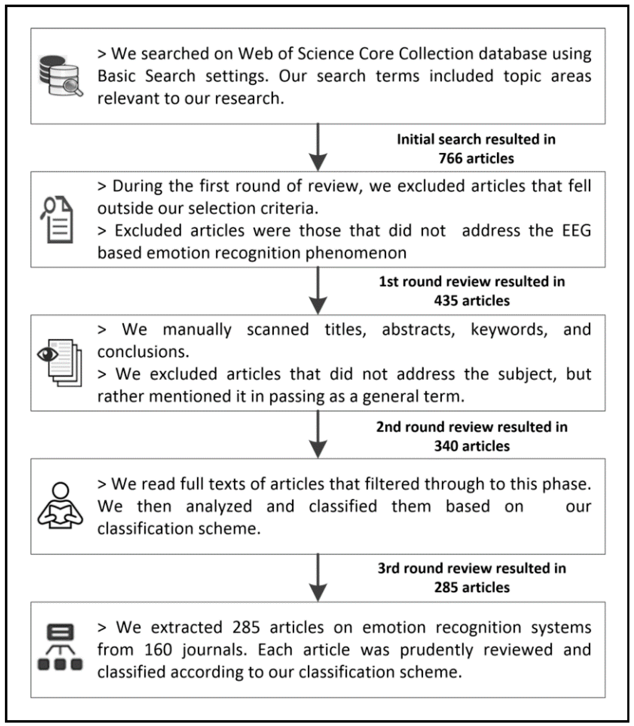
2.1. Data Sources and Procedures for the Extraction of Articles
2.2. Selection Criteria
- Articles must address one of the Gartner Hype Cycle 2016 trending research topics. To meet this criterion, they must be relatively current. In this regard, we chose articles that were published between 2005 and 2016. This 11-year period could be considered to correspond to the main research period of emotion recognition systems.
- We excluded meeting abstracts, book chapters, conference proceedings, workshop descriptions, masters and doctoral dissertations, and non-English articles. Notably, the number of conference papers in this domain was 322, the number of book chapters was 3, and the number of meeting abstracts was 6.
- We also ensured that only peer-reviewed journal articles were included. The logic behind this is that practitioners and academics frequently use journals to both obtain and spread research findings. Thus, journal articles contain the highest level of research.
2.3. Filtering/Reviewing Process
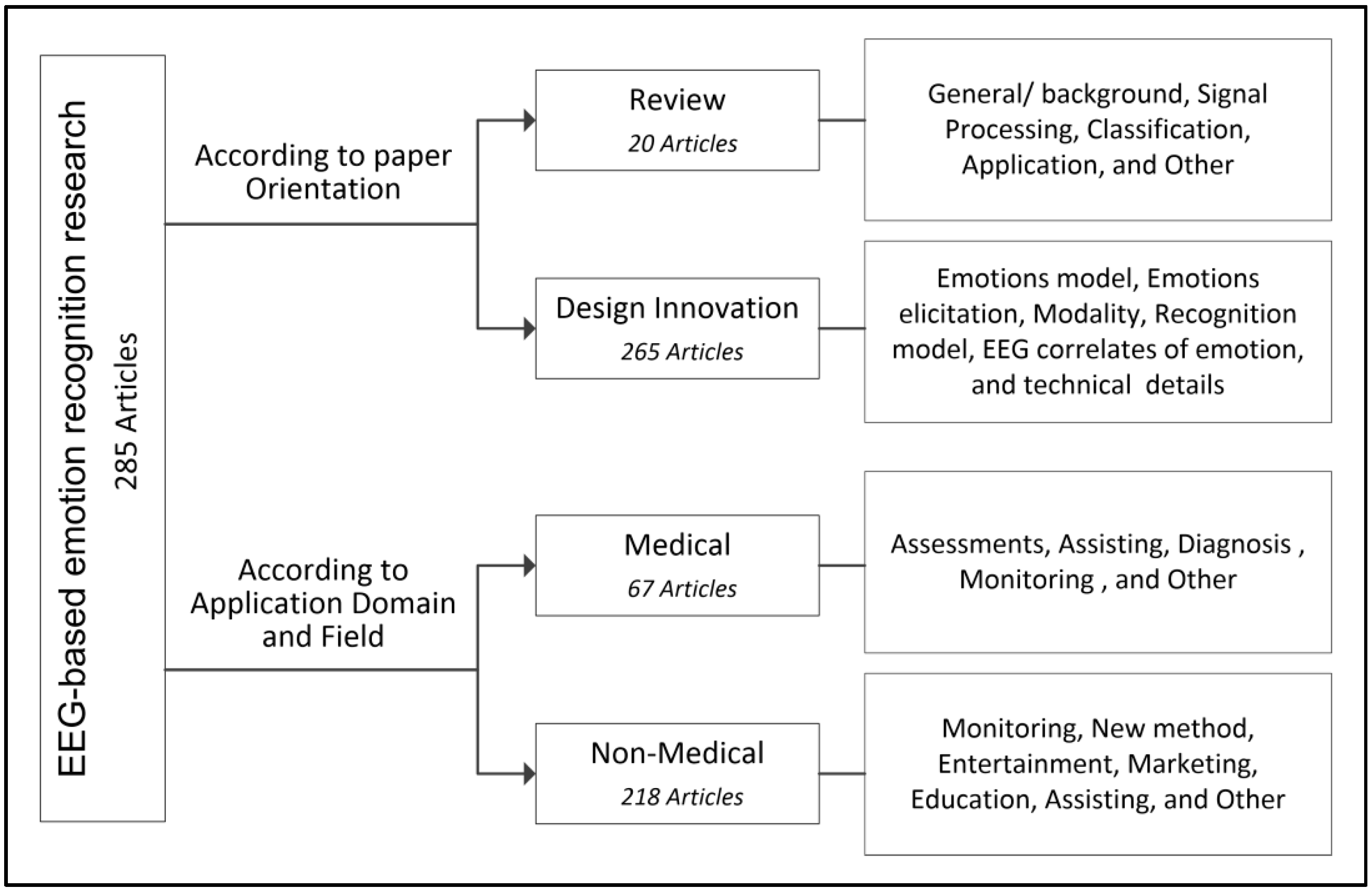
3. Classification Method
3.1. Application Domain and Field
3.1.1. Medical Context
3.1.2. Non-Medical Context
4. Results and Discussion
4.1. Classification of Articles by Paper Orientation
4.1.1. Review Paper
4.1.2. Design Innovation (Experimental) Paper
4.2. Classification of Articles by Application Domain and Field
- the paper discussed a medical condition (disorder/disease), such as a psychiatric or neurological case;
- the participants of the experiment were patients or it involved two groups: one consisting of healthy people, the other of patients;
- the experiment was conducted in a clinical setting; and/or,
- the paper was directed toward the medical community and suggested a new method for assistance, enhancement, monitoring, or diagnosis using emotion-based recognition.
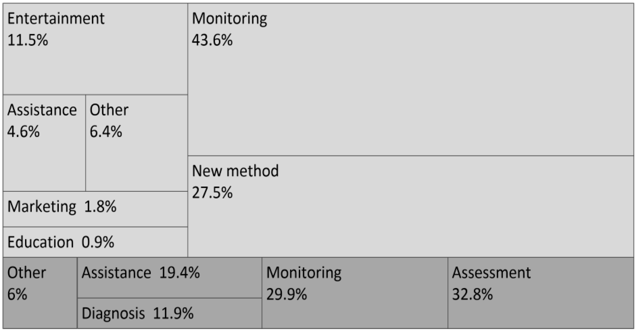
4.2.1. Medical Applications
4.2.2. Non-Medical Applications
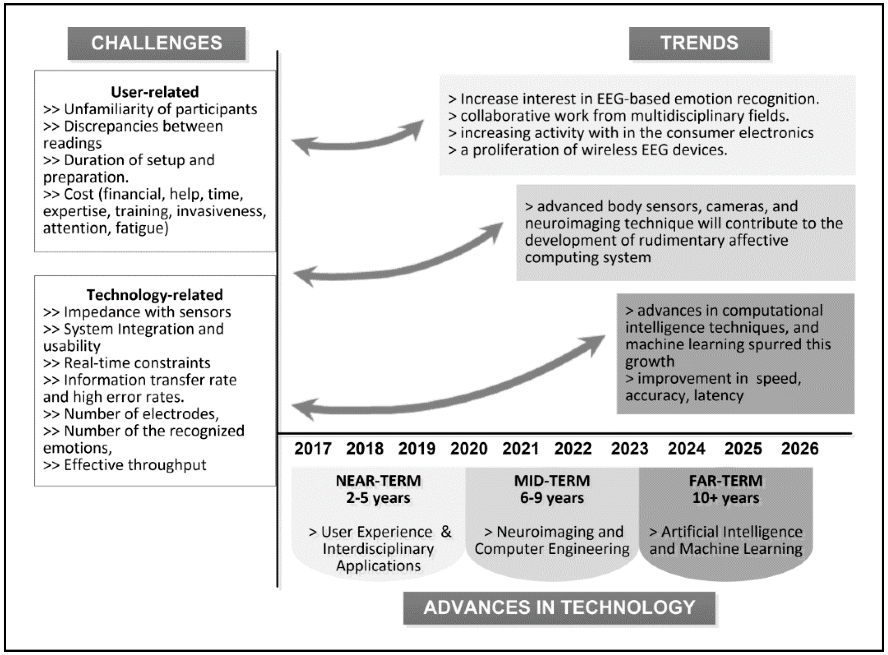
5. Challenges and Future Directions
6. Conclusions
Acknowledgments
Author Contributions
Conflicts of interest
Appendix A

| Range | Field/Domain | Number of Publications |
|---|---|---|
| >100 | Neurosciences | 202 |
| 10–100 | Psychology Experimental | 64 |
| Psychology | 62 | |
| Psychiatry | 44 | |
| Clinical Neurology | 43 | |
| Physiology | 40 | |
| Computer Science Artificial Intelligence | 37 | |
| Behavioral Sciences | 34 | |
| Psychology Biological | 25 | |
| Neuroimaging | 24 | |
| Radiology Nuclear Medicine Medical Imaging, Multidisciplinary Sciences | 23 | |
| Psychology Multidisciplinary | 18 | |
| Computer Science Cybernetics | 17 | |
| Psychology Developmental, Engineering Biomedical | 14 | |
| Engineering Electrical Electronic | 13 | |
| Computer Science Information Systems | 10 | |
| 1–9 | Mathematical Computational Biology, Computer Science Interdisciplinary Applications | 9 |
| Rehabilitation | 7 | |
| Psychology Clinical | 6 | |
| Robotics, Psychology Social, Pharmacology Pharmacy, Medical Informatics, Audiology Speech-Language Pathology | 5 | |
| Operations Research Management Science, Medicine Research Experimental, Linguistics, Ergonomics, Computer Science Theory Methods, Computer Science Software Engineering | 4 | |
| Public Environmental Occupational Health, Pediatrics, Instruments Instrumentation, Engineering Multidisciplinary, Education Special | 3 | |
| Telecommunications, Medical Laboratory Technology, Electrochemistry, Computer Science Hardware Architecture, Chemistry Analytical, Biology, Automation Control Systems, Anesthesiology | 2 | |
| Substance Abuse, Sport Sciences, Social Issues, Psychology Psychoanalysis, Optics Otorhinolaryngology, Medicine General Internal, Materials Science Multidisciplinary, Materials Science Biomaterials, Integrative Complementary Medicine, Ethics, Endocrinology Metabolism, Geriatrics Gerontology, Genetics Heredity, Imaging Science Photographic Technology, Health Care Sciences Services, Education Educational Research, Biotechnology Applied Microbiology, Chemistry Medicinal, Acoustics | 1 |
Appendix B
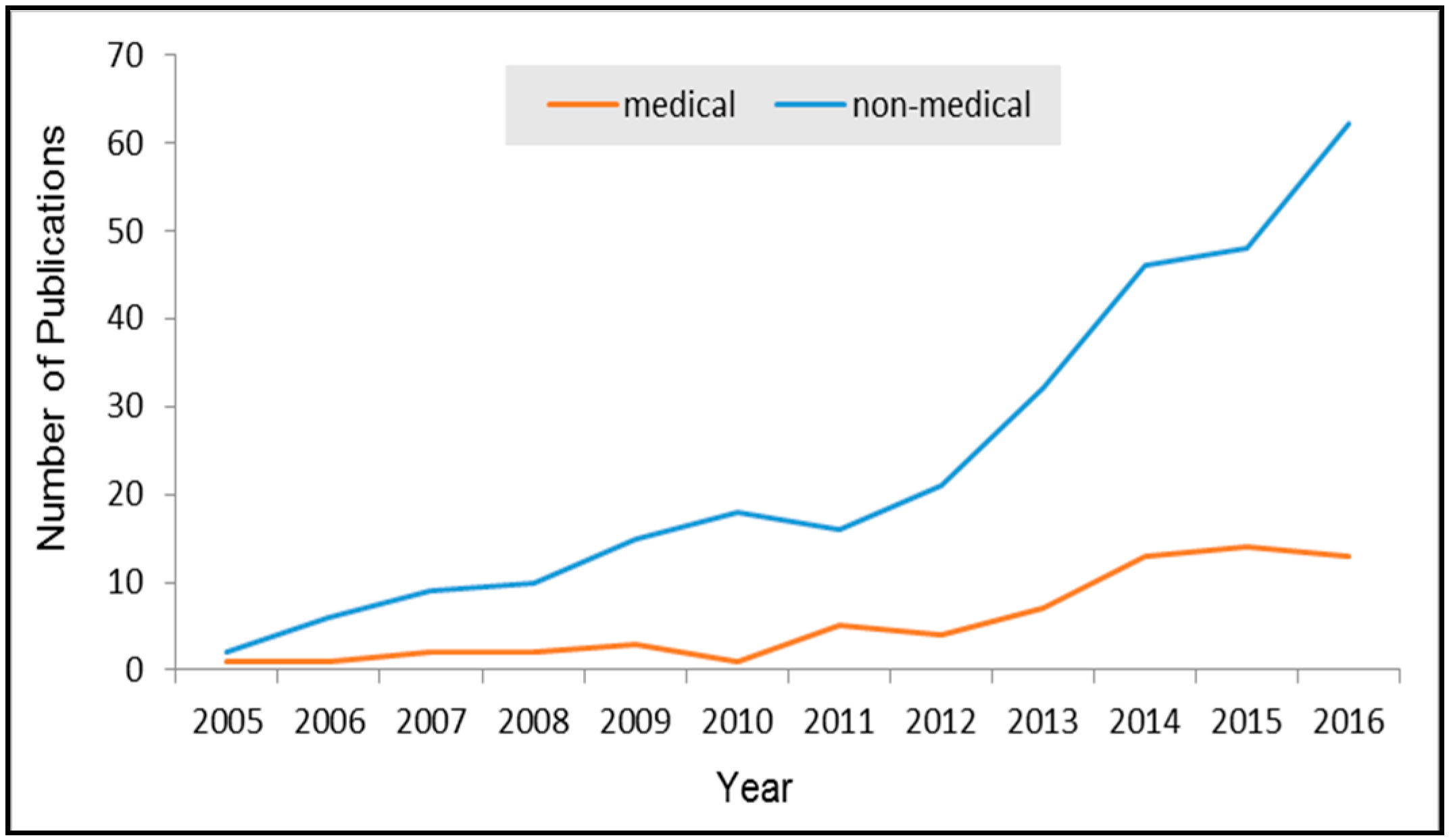
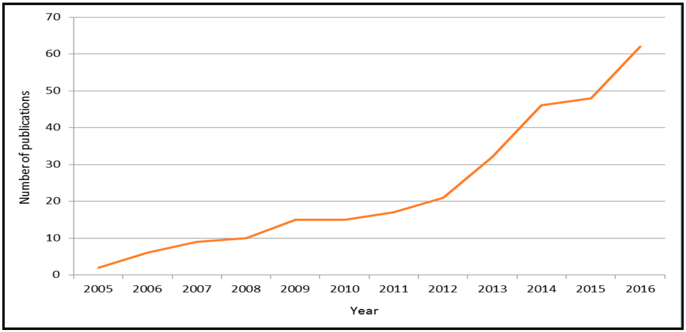
Appendix B.1. Trends in Number of Publications for EEG-Based Emotion Recognition

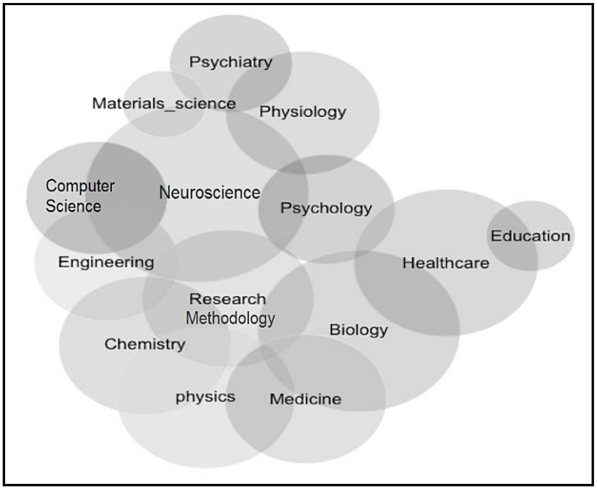
Appendix B.2. Classification of Articles by Research Area

Appendix B.3. Classification of Articles by Online Database and Journal
| Online Database | Number of Articles | Timeframe | Online database | Numbe of Articles | Timeframe |
|---|---|---|---|---|---|
| Science Direct | 103 | 2005–2016 | Wiley Online Library | 12 | 2007–2015 |
| Springer Link | 27 | 2006–2016 | Plos.org | 10 | 2012–2016 |
| IEEE Xplore | 23 | 2006–2016 | World Scientific | 8 | 2010–2016 |
| Frontiers | 22 | 2010–2016 | Hindawi | 6 | 2013–2016 |
| Taylor & Francis | 16 | 2006–2016 | Oxford | 6 | 2008–2016 |
| Journal (Impact Factor IF, Citation C) | Number of Articles |
|---|---|
| PLOS ONE (IF: 2.806, C: 188) | 10 |
| International Journal of Psychophysiology (IF: 2.582, C: 360), IEEE Transactions on Affective Computing (IF: 3.149, C: 1593) | 9 |
| Frontiers in Human Neuroscience (IF: 3.209, C: 73), Neuroimage (IF: 5.835, C: 283), Frontiers in Psychology (IF: 2.323, C: 71) | 7 |
| Neurocomputing (IF: 3.317, C: 185), Neuropsychologia (IF: 3.197, C: 321), Social Neuroscience (IF: 2.255, C: 185) | 6 |
| Frontiers in Neuroscience (IF: 3.566, C: 102), Neuroscience Letters (IF: 2.180, C: 139), Clinical Neurophysiology (IF: 3.866, C: 149) | 5 |
| IEEE Transactions on Information Technology in Biomedicine (IF: 2.493, C: 558), Sensors (IF: 2.677, C: 30), Social Cognitive and Affective Neuroscience (IF: 3.937, C: 99), Biological Psychology (IF: 3.070, C: 40), Brain and Cognition (IF: 2.432, C: 194), Schizophrenia Research (IF: 3.986, C: 227) | 4 |
| IEEE Transactions on Autonomous Mental Development (IF: 1.638, C: 49), Journal of Visualized Experiments (IF: 1.325, C: 5), Behavioural Brain Research (IF: 3.002, C: 39), Computers in Human Behavior (IF: 3.435, C: 31), Psychiatry Research (IF: 2.528, C: 18), Cognitive Affective & Behavioral Neuroscience (IF: 3.263, C: 42), Cognitive Neurodynamics (IF: 1.828, C: 17), Journal of Neural Transmission (IF: 2.392, C: 86) | 3 |
References
- Pun, T.; Alecu, T.L.; Chanel, G.; Kronegg, J.; Voloshynovskiy, S. Brain-computer interaction research at the computer vision and multimedia laboratory, university of geneva. IEEE Trans. Neural Syst. Rehabil. Eng. 2006, 14, 210–213. [Google Scholar] [CrossRef] [PubMed]
- Esfahani, E.T.; Sundararajan, V. Using brain-computer interfaces to detect human satisfaction in human-robot interaction. Int. J. Humanoid Robot. 2011, 8, 87–101. [Google Scholar] [CrossRef]
- Schupp, H.T.; Flaisch, T.; Stockburger, J.; Junghofer, M. Emotion and attention: Event-related brain potential studies. Prog. Brain Res. 2006, 156, 31–51. [Google Scholar] [PubMed]
- Chien, V.S.C.; Tsai, A.C.; Yang, H.H.; Tseng, Y.L.; Savostyanov, A.N.; Liou, M. Conscious and non-conscious representations of emotional faces in asperger’s syndrome. JOVE J. Vis. Exp. 2016. [Google Scholar] [CrossRef] [PubMed]
- Csukly, G.; Stefanics, G.; Komlosi, S.; Czigler, I.; Czobor, P. Event-related theta synchronization predicts deficit in facial affect recognition in schizophrenia. J. Abnorm. Psychol. 2014, 123, 178–189. [Google Scholar] [CrossRef] [PubMed]
- Friedrich, E.V.C.; Sivanathan, A.; Lim, T.; Suttie, N.; Louchart, S.; Pillen, S.; Pineda, J.A. An effective neurofeedback intervention to improve social interactions in children with autism spectrum disorder. J. Autism Dev. Disord. 2015, 45, 4084–4100. [Google Scholar] [CrossRef] [PubMed]
- Liberati, G.; Federici, S.; Pasqualotto, E. Extracting neurophysiological signals reflecting users’ emotional and affective responses to bci use: A systematic literature review. Neurorehabilitation 2015, 37, 341–358. [Google Scholar] [CrossRef] [PubMed]
- Verma, G.K.; Tiwary, U.S. Multimodal fusion framework: A multiresolution approach for emotion classification and recognition from physiological signals. Neuroimage 2014, 102, 162–172. [Google Scholar] [CrossRef] [PubMed]
- Rule, N.O.; Freeman, J.B.; Ambady, N. Culture in social neuroscience: A review. Soc. Neurosci. 2013, 8, 3–10. [Google Scholar] [CrossRef] [PubMed]
- Keysers, C.; Fadiga, L. The mirror neuron system: New frontiers. Soc. Neurosci. 2008, 3, 193–198. [Google Scholar] [CrossRef] [PubMed]
- Grossmann, T.; Johnson, M.H. The development of the social brain in human infancy. Eur. J. Neurosci. 2007, 25, 909–919. [Google Scholar] [CrossRef] [PubMed]
- Muthukumaraswamy, S.D.; Johnson, B.W. A dual mechanism neural framework for social understanding. Philos. Psychol. 2007, 20, 43–63. [Google Scholar] [CrossRef]
- Alotaiby, T.; Abd El-Samie, F.E.; Alshebeili, S.A.; Ahmad, I. A review of channel selection algorithms for EEG signal processing. EURASIP J. Adv. Signal Process. 2015. [Google Scholar] [CrossRef]
- Jenke, R.; Peer, A.; Buss, M. Feature extraction and selection for emotion recognition from EEG. IEEE Trans. Affect. Comput. 2014, 5, 327–339. [Google Scholar] [CrossRef]
- Knyazev, G.G. Motivation, emotion, and their inhibitory control mirrored in brain oscillations. Neurosci. Biobehav. Rev. 2007, 31, 377–395. [Google Scholar] [CrossRef] [PubMed]
- Kim, M.K.; Kim, M.; Oh, E.; Kim, S.P. A review on the computational methods for emotional state estimation from the human EEG. Comput. Math. Methods Med. 2013, 2013. [Google Scholar] [CrossRef] [PubMed]
- Isaac, C.; Januel, D. Neural correlates of cognitive improvements following cognitive remediation in schizophrenia: A systematic review of randomized trials. Socioaffect. Neurosci. Psychol. 2016, 6, 30054. [Google Scholar] [CrossRef] [PubMed]
- Campos, C.; Santos, S.; Gagen, E.; Machado, S.; Rocha, S.; Kurtz, M.M.; Rocha, N.B. Neuroplastic changes following social cognition training in schizophrenia: A systematic review. Neuropsychol. Rev. 2016, 26, 310–328. [Google Scholar] [CrossRef] [PubMed]
- Harrison, A.H.; Connolly, J.F. Finding a way in: A review and practical evaluation of fmri and EEG for detection and assessment in disorders of consciousness. Neurosci. Biobehav. Rev. 2013, 37, 1403–1419. [Google Scholar] [CrossRef] [PubMed]
- Acharya, U.R.; Sudarshan, V.K.; Adeli, H.; Santhosh, J.; Koh, J.E.W.; Adeli, A. Computer-aided diagnosis of depression using EEG signals. Eur. Neurol. 2015, 73, 329–336. [Google Scholar] [CrossRef] [PubMed]
- Bhat, S.; Acharya, U.R.; Adeli, H.; Bairy, G.M.; Adeli, A. Automated diagnosis of autism: In search of a mathematical marker. Rev. Neurosci. 2014, 25, 851–861. [Google Scholar] [CrossRef] [PubMed]
- Bontchev, B. Adaptation in affective video games: A literature review. Cybern. Inf. Technol. 2016, 16, 3–34. [Google Scholar] [CrossRef]
- Reyes-Munoz, A.; Domingo, M.C.; Lopez-Trinidad, M.A.; Delgado, J.L. Integration of body sensor networks and vehicular ad-hoc networks for traffic safety. Sensors 2016, 16. [Google Scholar] [CrossRef] [PubMed] [Green Version]
- Gruzelier, J.H. EEG-neurofeedback for optimising performance. I: A review of cognitive and affective outcome in healthy participants. Neurosci. Biobehav. Rev. 2014, 44, 124–141. [Google Scholar] [CrossRef] [PubMed]
- Harmon-Jones, E.; Amodio, D.M.; Harmon-Jones, C. Action-based model of dissonance: A review, integration, and expansion of conceptions of cognitive conflict. Adv. Exp. Soc. Psychol. 2009, 41, 119–166. [Google Scholar]
- Kroupi, E.; Vesin, J.M.; Ebrahimi, T. Subject-independent odor pleasantness classification using brain and peripheral signals. IEEE Trans. Affect. Comput. 2016, 7, 422–434. [Google Scholar] [CrossRef]
- Keuper, K.; Zwitserlood, P.; Rehbein, M.A.; Eden, A.S.; Laeger, I.; Junghofer, M.; Zwanzger, P.; Dobel, C. Early prefrontal brain responses to the hedonic quality of emotional words—A simultaneous EEG and MEG study. PLoS ONE 2013, 8. [Google Scholar] [CrossRef] [PubMed] [Green Version]
- Balconi, M.; Cobelli, C. Motivational mechanisms (bas) and prefrontal cortical activation contribute to recognition memory for emotional words. Rtms effect on performance and EEG (alpha band) measures. Brain Lang. 2014, 137, 77–85. [Google Scholar] [CrossRef] [PubMed]
- Briesemeister, B.B.; Kuchinke, L.; Jacobs, A.M. Emotion word recognition: Discrete information effects first, continuous later? Brain Res. 2014, 1564, 62–71. [Google Scholar] [CrossRef] [PubMed]
- Kamp, S.M.; Potts, G.F.; Donchin, E. On the roles of distinctiveness and semantic expectancies in episodic encoding of emotional words. Psychophysiology 2015, 52, 1599–1609. [Google Scholar] [CrossRef] [PubMed]
- Mueller, C.J.; Kuchinke, L. Individual differences in emotion word processing: A diffusion model analysis. Cogn. Affect. Behav. Neurosci. 2016, 16, 489–501. [Google Scholar] [CrossRef] [PubMed]
- Imbir, K.K.; Spustek, T.; Zygierewicz, J. Effects of valence and origin of emotions in word processing evidenced by event related potential correlates in a lexical decision task. Front. Psychol. 2016, 7. [Google Scholar] [CrossRef] [PubMed]
- Novosel, A.; Lackner, N.; Unterrainer, H.F.; Dunitz-Scheer, M.; Scheer, P.J.Z.; Wallner-Liebmann, S.J.; Neuper, C. Motivational processing of food cues in anorexia nervosa: A pilot study. Eat. Weight Disord. Stud. Anorex. Bulim. Obes. 2014, 19, 169–175. [Google Scholar] [CrossRef] [PubMed]
- Chanel, G.; Rebetez, C.; Betrancourt, M.; Pun, T. Emotion assessment from physiological signals for adaptation of game difficulty. IEEE Trans. Syst. Man Cybern. Part A Syst. Hum. 2011, 41, 1052–1063. [Google Scholar] [CrossRef]
- Tzieropoulos, H.; de Peralta, R.G.; Bossaerts, P.; Andino, S.L.G. The impact of disappointment in decision making: Inter-individual differences and electrical neuroimaging. Front. Hum. Neurosci. 2011, 4. [Google Scholar] [CrossRef] [PubMed]
- Spape, M.M.; Kivikangas, J.M.; Jarvela, S.; Kosunen, I.; Jacucci, G.; Ravaja, N. Keep your opponents close: Social context affects EEG and femg linkage in a turn-based computer game. PLoS ONE 2013, 8. [Google Scholar] [CrossRef] [PubMed]
- Mothes, H.; Enge, S.; Strobel, A. The interplay between feedback-related negativity and individual differences in altruistic punishment: An EEG study. Cogn. Affect. Behav. Neurosci. 2016, 16, 276–288. [Google Scholar] [CrossRef] [PubMed]
- Charland, P.; Leger, P.M.; Senecal, S.; Courtemanche, F.; Mercier, J.; Skelling, Y.; Labonte-Lemoyne, E. Assessing the multiple dimensions of engagement to characterize learning: A neurophysiological perspective. JOVE J. Vis. Exp. 2015. [Google Scholar] [CrossRef] [PubMed]
- Lopez-Gil, J.M.; Virgili-Goma, J.; Gil, R.; Garcia, R. Method for improving EEG based emotion recognition by combining it with synchronized biometric and eye tracking technologies in a non-invasive and low cost way. Front. Comput. Neurosci. 2016, 10. [Google Scholar] [CrossRef]
- Abdur-Rahim, J.; Morales, Y.; Gupta, P.; Umata, I.; Watanabe, A.; Even, J.; Suyama, T.; Ishii, S. Multi-sensor based state prediction for personal mobility vehicles. PLoS ONE 2016, 11. [Google Scholar] [CrossRef] [PubMed]
- Huang, X.H.; Kortelainen, J.; Zhao, G.Y.; Li, X.B.; Moilanen, A.; Seppanen, T.; Pietikainen, M. Multi-modal emotion analysis from facial expressions and electroencephalogram. Comput. Vis. Image Underst. 2016, 147, 114–124. [Google Scholar] [CrossRef]
- Wen, W.H.; Qiu, Y.H.; Liu, G.Y.; Cheng, N.P.; Huang, X.T. Construction and cross-correlation analysis of the affective physiological response database. Sci. China Inf. Sci. 2010, 53, 1774–1784. [Google Scholar] [CrossRef]
- Leventon, J.S.; Stevens, J.S.; Bauer, P.J. Development in the neurophysiology of emotion processing and memory in school-age children. Dev. Cogn. Neurosci. 2014, 10, 21–33. [Google Scholar] [CrossRef] [PubMed]
- Daly, I.; Williams, D.; Kirke, A.; Weaver, J.; Malik, A.; Hwang, F.; Miranda, E.; Nasuto, S.J. Affective brain-computer music interfacing. J. Neural Eng. 2016, 13. [Google Scholar] [CrossRef] [PubMed]
- Daly, I.; Chen, L.; Zhou, S.J.; Jin, J. An investigation into the use of six facially encoded emotions in brain-computer interfacing. Brain Comput. Interfaces 2016, 3, 59–73. [Google Scholar] [CrossRef]
- Knyazev, G.G.; Slobodskoj-Plusnin, J.Y.; Bocharov, A.V. Gender differences in implicit and explicit processing of emotional facial expressions as revealed by event-related theta synchronization. Emotion 2010, 10, 678–687. [Google Scholar] [CrossRef] [PubMed]
- Wieser, M.J.; Muhlberger, A.; Alpers, G.W.; Macht, M.; Ellgring, H.; Pauli, P. Emotion processing in Parkinson’s disease: Dissociation between early neuronal processing and explicit ratings. Clin. Neurophysiol. 2006, 117, 94–102. [Google Scholar] [CrossRef] [PubMed]
- Schaefer, A.; Pottage, C.L.; Rickart, A.J. Electrophysiological correlates of remembering emotional pictures. Neuroimage 2011, 54, 714–724. [Google Scholar] [CrossRef] [PubMed]
- Zheng, W.L.; Lu, B.L. Investigating critical frequency bands and channels for EEG-based emotion recognition with deep neural networks. IEEE Trans. Auton. Ment. Dev. 2015, 7, 162–175. [Google Scholar] [CrossRef]
- Peng, Y.; Zheng, W.L.; Lu, B.L. An unsupervised discriminative extreme learning machine and its applications to data clustering. Neurocomputing 2016, 174, 250–264. [Google Scholar] [CrossRef]
- Schaefer, A.; Fletcher, K.; Pottage, C.L.; Alexander, K.; Brown, C. The effects of emotional intensity on ERP correlates of recognition memory. Neuroreport 2009, 20, 319–324. [Google Scholar] [CrossRef] [PubMed]
- Lu, X.J.; Ho, H.T.; Liu, F.; Wu, D.X.; Thompson, W.F. Intonation processing deficits of emotional words among mandarin chinese speakers with congenital amusia: An erp study. Front. Psychol. 2015, 6. [Google Scholar] [CrossRef] [PubMed]
- Lin, Y.P.; Yang, Y.H.; Jung, T.P. Fusion of electroencephalographic dynamics and musical contents for estimating emotional responses in music listening. Front. Neurosci. 2014, 8. [Google Scholar] [CrossRef] [PubMed]
- Turetsky, B.I.; Kohler, C.G.; Indersmitten, T.; Bhati, M.T.; Charbonnier, D.; Gur, R.C. Facial emotion recognition in schizophrenia: When and why does it go awry? Schizophr. Res. 2007, 94, 253–263. [Google Scholar] [CrossRef] [PubMed]
- Chen, X.H.; Yang, J.F.; Gan, S.Z.; Yang, Y.F. The contribution of sound intensity in vocal emotion perception: Behavioral and electrophysiological evidence. PLoS ONE 2012, 7. [Google Scholar] [CrossRef] [PubMed]
- Wang, L.; Bastiaansen, M. Oscillatory brain dynamics associated with the automatic processing of emotion in words. Brain Lang. 2014, 137, 120–129. [Google Scholar] [CrossRef] [PubMed]
- Wang, X.W.; Nie, D.; Lu, B.L. Emotional state classification from EEG data using machine learning approach. Neurocomputing 2014, 129, 94–106. [Google Scholar] [CrossRef]
- Lin, Y.P.; Duann, J.R.; Feng, W.F.; Chen, J.H.; Jung, T.P. Revealing spatio-spectral electroencephalographic dynamics of musical mode and tempo perception by independent component analysis. J. Neuroeng. Rehabil. 2014, 11. [Google Scholar] [CrossRef] [PubMed]
- Zhang, W.H.; Li, X.Y.; Liu, X.; Duan, X.X.; Wang, D.H.; Shen, J.L. Distraction reduces theta synchronization in emotion regulation during adolescence. Neurosci. Lett. 2013, 550, 81–86. [Google Scholar] [CrossRef] [PubMed]
- Calvo, M.G.; Beltran, D. Recognition advantage of happy faces: Tracing the neurocognitive processes. Neuropsychologia 2013, 51, 2051–2060. [Google Scholar] [CrossRef] [PubMed]
- Liu, Y.H.; Wu, C.T.; Cheng, W.T.; Hsiao, Y.T.; Chen, P.M.; Teng, J.T. Emotion recognition from single-trial EEG based on kernel fisher’s emotion pattern and imbalanced quasiconformal kernel support vector machine. Sensors 2014, 14, 13361–13388. [Google Scholar] [CrossRef] [PubMed]
- Brennan, A.M.; Harris, A.W.F.; Williams, L.M. Neural processing of facial expressions of emotion in first onset psychosis. Psychiatry Res. 2014, 219, 477–485. [Google Scholar] [CrossRef] [PubMed]
- Liu, S.; Zhang, D.; Xu, M.P.; Qi, H.Z.; He, F.; Zhao, X.; Zhou, P.; Zhang, L.X.; Ming, D. Randomly dividing homologous samples leads to overinflated accuracies for emotion recognition. Int. J. Psychophysiol. 2015, 96, 29–37. [Google Scholar] [CrossRef] [PubMed]
- Perez-Edgar, K.; Kujawa, A.; Nelson, S.K.; Cole, C.; Zapp, D.J. The relation between electroencephalogram asymmetry and attention biases to threat at baseline and under stress. Brain Cogn. 2013, 82, 337–343. [Google Scholar] [CrossRef] [PubMed]
- Liu, T.R.; Xiao, T.; Shi, J.N. Automatic change detection to facial expressions in adolescents: Evidence from visual mismatch negativity responses. Front. Psychol. 2016, 7. [Google Scholar] [CrossRef] [PubMed]
- Kim, D.W.; Kim, H.S.; Lee, S.H.; Im, C.H. Positive and negative symptom scores are correlated with activation in different brain regions during facial emotion perception in schizophrenia patients: A voxel-based sloreta source activity study. Schizophr. Res. 2013, 151, 165–174. [Google Scholar] [CrossRef] [PubMed]
- Lin, H.Y.; Xiang, J.; Li, S.L.; Liang, J.F.; Jin, H. Anticipation of negative pictures enhances the p2 and p3 in their later recognition. Front. Hum. Neurosci. 2015, 9. [Google Scholar] [CrossRef] [PubMed]
- Zhang, D.D.; Wang, L.L.; Luo, Y.; Luo, Y.J. Individual differences in detecting rapidly presented fearful faces. PLoS ONE 2012, 7. [Google Scholar] [CrossRef] [PubMed]
- Yu, B.; Ma, L.; Li, H.F.; Zhao, L.; Bo, H.J.; Wang, X.D. Biological computation indexes of brain oscillations in unattended facial expression processing based on event-related synchronization/desynchronization. Comput. Math. Methods Med. 2016. [Google Scholar] [CrossRef] [PubMed]
- Zhang, W.H.; Lu, J.M.; Liu, X.; Fang, H.L.; Li, H.; Wang, D.H.; Shen, J.L. Event-related synchronization of delta and beta oscillations reflects developmental changes in the processing of affective pictures during adolescence. Int. J. Psychophysiol. 2013, 90, 334–340. [Google Scholar] [CrossRef] [PubMed]
- Wang, S.F.; Zhu, Y.C.; Wu, G.B.; Ji, Q. Hybrid video emotional tagging using users' EEG and video content. Multimed. Tools Appl. 2014, 72, 1257–1283. [Google Scholar] [CrossRef]
- Williams, L.M.; Whitford, T.J.; Nagy, M.; Flynn, G.; Harris, A.W.F.; Silverstein, S.M.; Gordon, E. Emotion-elicited gamma synchrony in patients with first-episode schizophrenia: A neural correlate of social cognition outcomes. J. Psychiatry Neurosci. 2009, 34, 303–313. [Google Scholar] [PubMed]
- Andrews, S.C.; Enticott, P.G.; Hoy, K.E.; Thomson, R.H.; Fitzgerald, P.B. No evidence for mirror system dysfunction in schizophrenia from a multimodal tms/EEG study. Psychiatry Res. 2015, 228, 431–440. [Google Scholar] [CrossRef] [PubMed]
- Shen, X.B.; Wu, Q.; Zhao, K.; Fu, X.L. Electrophysiological evidence reveals differences between the recognition of microexpressions and macroexpressions. Front. Psychol. 2016, 7. [Google Scholar] [CrossRef] [PubMed]
- Kylliainen, A.; Wallace, S.; Coutanche, M.N.; Leppanen, J.M.; Cusack, J.; Bailey, A.J.; Hietanen, J.K. Affective-motivational brain responses to direct gaze in children with autism spectrum disorder. J. Child Psychol. Psychiatry 2012, 53, 790–797. [Google Scholar] [CrossRef] [PubMed]
- Croft, R.J.; McKernan, F.; Gray, M.; Churchyard, A.; Georgiou-Karistianis, N. Emotion perception and electrophysiological correlates in Huntington’s disease. Clin. Neurophysiol. 2014, 125, 1618–1625. [Google Scholar] [CrossRef] [PubMed]
- Beltran, D.; Calvo, M.G. Brain signatures of perceiving a smile: Time course and source localization. Hum. Brain Mapp. 2015, 36, 4287–4303. [Google Scholar] [CrossRef] [PubMed]
- Lin, H.Y.; Schulz, C.; Straube, T. Cognitive tasks during expectation affect the congruency ERP effects to facial expressions. Front. Hum. Neurosci. 2015, 9. [Google Scholar] [CrossRef] [PubMed]
- Hilimire, M.R.; Mayberg, H.S.; Holtzheimer, P.E.; Broadway, J.M.; Parks, N.A.; DeVylder, J.E.; Corballis, P.M. Effects of subcallosal cingulate deep brain stimulation on negative self-bias in patients with treatment-resistant depression. Brain Stimul. 2015, 8, 185–191. [Google Scholar] [CrossRef] [PubMed]
- Makin, A.D.J.; Wilton, M.M.; Pecchinenda, A.; Bertamini, M. Symmetry perception and affective responses: A combined EEG/emg study. Neuropsychologia 2012, 50, 3250–3261. [Google Scholar] [CrossRef] [PubMed]
- Magnée, M.J.; Gelder, B.; Engeland, H.; Kemner, C. A typical processing of fearful face-voice pairs in pervasive developmental disorder: An ERP study. Clin. Neurophysiol. 2008, 119, 2004–2010. [Google Scholar] [CrossRef] [PubMed]
- Matsuda, I.; Nittono, H.; Allen, J.J.B. Detection of concealed information by p3 and frontal EEG asymmetry. Neurosci. Lett. 2013, 537, 55–59. [Google Scholar] [CrossRef] [PubMed]
- Lin, H.Y.; Schulz, C.; Straube, T. Fearful contextual expression impairs the encoding and recognition of target faces: An ERP study. Front. Behav. Neurosci. 2015, 9. [Google Scholar] [CrossRef] [PubMed]
- Dennis, T.A.; Hajcak, G. The late positive potential: A neurophysiological marker for emotion regulation in children. J. Child Psychol. Psychiatry 2009, 50, 1373–1383. [Google Scholar] [CrossRef] [PubMed]
- Codispoti, M.; De Cesarei, A.; Ferrari, V. The influence of color on emotional perception of natural scenes. Psychophysiology 2012, 49, 11–16. [Google Scholar] [CrossRef] [PubMed]
- Gallant, S.N.; Dyson, B.J. Neural modulation of directed forgetting by valence and arousal: An event-related potential study. Brain Res. 2016, 1648, 306–316. [Google Scholar] [CrossRef] [PubMed]
- Newsome, R.N.; Dulas, M.R.; Duarte, A. The effects of aging on emotion-induced modulations of source retrieval ERPS: Evidence for valence biases. Neuropsychologia 2012, 50, 3370–3384. [Google Scholar] [CrossRef] [PubMed]
- Soleymani, M.; Pantic, M.; Pun, T. Multimodal emotion recognition in response to videos. IEEE Trans. Affect. Comput. 2012, 3, 211–223. [Google Scholar] [CrossRef]
- Lindstrom, R.; Lepisto, T.; Makkonen, T.; Kujala, T. Processing of prosodic changes in natural speech stimuli in school-age children. Int. J. Psychophysiol. 2012, 86, 229–237. [Google Scholar] [CrossRef] [PubMed]
- Komlosi, S.; Csukly, G.; Stefanics, G.; Czigler, I.; Bitter, I.; Czobor, P. Fearful face recognition in schizophrenia: An electrophysiological study. Schizophr. Res. 2013, 149, 135–140. [Google Scholar] [CrossRef] [PubMed]
- Achaibou, A.; Pourtois, G.; Schwartz, S.; Vuilleumier, P. Simultaneous recording of EEG and facial muscle reactions during spontaneous emotional mimicry. Neuropsychologia 2008, 46, 1104–1113. [Google Scholar] [CrossRef] [PubMed]
- Koelstra, S.; Muhl, C.; Soleymani, M.; Lee, J.S.; Yazdani, A.; Ebrahimi, T.; Pun, T.; Nijholt, A.; Patras, I. Deap: A database for emotion analysis using physiological signals. IEEE Trans. Affect. Comput. 2012, 3, 18–31. [Google Scholar] [CrossRef]
- Ponz, A.; Montant, M.; Liegeois-Chauvel, C.; Silva, C.; Braun, M.; Jacobs, A.M.; Ziegler, J.C. Emotion processing in words: A test of the neural re-use hypothesis using surface and intracranial EEG. Soc. Cogn. Affect. Neurosci. 2014, 9, 619–627. [Google Scholar] [CrossRef] [PubMed]
- Csukly, G.; Stefanics, G.; Komlosi, S.; Czigler, I.; Czobor, P. Emotion-related visual mismatch responses in schizophrenia: Impairments and correlations with emotion recognition. PLoS ONE 2013, 8. [Google Scholar] [CrossRef] [PubMed] [Green Version]
- Bhushan, V.; Saha, G.; Lindsen, J.; Shimojo, S.; Bhattacharya, J. How we choose one over another: Predicting trial-by-trial preference decision. PLoS ONE 2012, 7. [Google Scholar] [CrossRef] [PubMed]
- Kryuchkova, T.; Tucker, B.V.; Wurm, L.H.; Baayen, R.H. Danger and usefulness are detected early in auditory lexical processing: Evidence from electroencephalography. Brain Lang. 2012, 122, 81–91. [Google Scholar] [CrossRef] [PubMed]
- Schirmer, A.; Escoffier, N.; Li, Q.Y.; Li, H.; Strafford-Wilson, J.; Lie, W.I. What grabs his attention but not hers? Estrogen correlates with neurophysiological measures of vocal change detection. Psychoneuroendocrinology 2008, 33, 718–727. [Google Scholar] [CrossRef] [PubMed]
- Csukly, G.; Farkas, K.; Marosi, C.; Szabo, A. Deficits in low beta desynchronization reflect impaired emotional processing in schizophrenia. Schizophr. Res. 2016, 171, 207–214. [Google Scholar] [CrossRef] [PubMed]
- Hagemann, J.; Straube, T.; Schulz, C. Too bad: Bias for angry faces in social anxiety interferes with identity processing. Neuropsychologia 2016, 84, 136–149. [Google Scholar] [CrossRef] [PubMed]
- Liu, Y.; Wang, C.G.; Wang, X.H.; Zhou, P.Y.; Yu, G.N.; Chan, K.C.C. What strikes the strings of your heart?-multi-label dimensionality reduction for music emotion analysis via brain imaging. IEEE Trans. Auton. Ment. Dev. 2015, 7, 176–188. [Google Scholar]
- Soleymani, M.; Lichtenauer, J.; Pun, T.; Pantic, M. A multimodal database for affect recognition and implicit tagging. IEEE Trans. Affect. Comput. 2012, 3, 42–55. [Google Scholar] [CrossRef]
- Bercik, J.; Horska, E.; Wang, R.W.Y.; Chen, Y.C. The impact of parameters of store illumination on food shopper response. Appetite 2016, 106, 101–109. [Google Scholar] [CrossRef] [PubMed]
- Martinez, F.; Barraza, C.; Gonzalez, N.; Gonzalez, J. Kapean: Understanding affective states of children with adhd. Educ. Technol. Soc. 2016, 19, 18–28. [Google Scholar]
- Mehmood, R.M.; Lee, H.J. A novel feature extraction method based on late positive potential for emotion recognition in human brain signal patterns. Comput. Electr. Eng. 2016, 53, 444–457. [Google Scholar] [CrossRef]
- Meza-Kubo, V.; Moran, A.L.; Carrillo, I.; Galindo, G.; Garcia-Canseco, E. Assessing the user experience of older adults using a neural network trained to recognize emotions from brain signals. J. Biomed. Inform. 2016, 62, 202–209. [Google Scholar] [CrossRef] [PubMed]
- Yuvaraj, R.; Murugappan, M.; Acharya, U.R.; Adeli, H.; Ibrahim, N.M.; Mesquita, E. Brain functional connectivity patterns for emotional state classification in Parkinson’s disease patients without dementia. Behav. Brain Res. 2016, 298, 248–260. [Google Scholar] [CrossRef] [PubMed]
- Yuvaraj, R.; Murugappan, M. Hemispheric asymmetry non-linear analysis of EEG during emotional responses from idiopathic Parkinson’s disease patients. Cogn. Neurodyn. 2016, 10, 225–234. [Google Scholar] [CrossRef] [PubMed]
- Jatupaiboon, N.; Pan-ngum, S.; Israsena, P. Real-time EEG-based happiness detection system. Sci. World J. 2013. [Google Scholar] [CrossRef] [PubMed]
- Gil, R.; Virgili-Goma, J.; Garcia, R.; Mason, C. Emotions ontology for collaborative modelling and learning of emotional responses. Comput. Hum. Behav. 2015, 51, 610–617. [Google Scholar] [CrossRef]
- Hadjidimitriou, S.K.; Hadjileontiadis, L.J. EEG-based classification of music appraisal responses using time-frequency analysis and familiarity ratings. IEEE Trans. Affect. Comput. 2013, 4, 161–172. [Google Scholar] [CrossRef]
- Kuber, R.; Wright, F.P. Augmenting the instant messaging experience through the use of brain-computer interface and gestural technologies. Int. J. Hum. Comput. Interact. 2013, 29, 178–191. [Google Scholar] [CrossRef]
- Hadjidimitriou, S.K.; Hadjileontiadis, L.J. Toward an EEG-based recognition of music liking using time-frequency analysis. IEEE Trans. Biomed. Eng. 2012, 59, 3498–3510. [Google Scholar] [CrossRef] [PubMed]
- Choi, J.S.; Bang, J.W.; Heo, H.; Park, K.R. Evaluation of fear using nonintrusive measurement of multimodal sensors. Sensors 2015, 15, 17507–17533. [Google Scholar] [CrossRef] [PubMed]
- Yuvaraj, R.; Murugappan, M.; Omar, M.I.; Ibrahim, N.M.; Sundaraj, K.; Mohamad, K.; Satiyan, M. Emotion processing in Parkinson’s disease: An EEG spectral power study. Int. J. Neurosci. 2014, 124, 491–502. [Google Scholar] [CrossRef] [PubMed]
- Sourina, O.; Liu, Y.S.; Nguyen, M.K. Real-time EEG-based emotion recognition for music therapy. J. Multimodal User Interfaces 2012, 5, 27–35. [Google Scholar] [CrossRef]
- Shahabi, H.; Moghimi, S. Toward automatic detection of brain responses to emotional music through analysis of EEG effective connectivity. Comput. Hum. Behav. 2016, 58, 231–239. [Google Scholar] [CrossRef]
- Lan, Z.R.; Sourina, O.; Wang, L.P.; Liu, Y.S. Real-time EEG-based emotion monitoring using stable features. Vis. Comput. 2016, 32, 347–358. [Google Scholar] [CrossRef]
- Yuvaraj, R.; Murugappan, M.; Ibrahim, N.M.; Sundaraj, K.; Omar, M.I.; Mohamad, K.; Palaniappan, R. Detection of emotions in Parkinson’s disease using higher order spectral features from brain’s electrical activity. Biomed. Signal Process. Control 2014, 14, 108–116. [Google Scholar] [CrossRef]
- Yuvaraj, R.; Murugappan, M.; Ibrahim, N.M.; Omar, M.I.; Sundaraj, K.; Mohamad, K.; Palaniappan, R.; Satiyan, M. Emotion classification in Parkinson’s disease by higher-order spectra and power spectrum features using EEG signals: A comparative study. J. Integr. Neurosci. 2014, 13, 89–120. [Google Scholar] [CrossRef] [PubMed]
- Yuvaraj, R.; Murugappan, M.; Ibrahim, N.M.; Sundaraj, K.; Omar, M.I.; Mohamad, K.; Palaniappan, R.; Satiyan, M. Inter-hemispheric EEG coherence analysis in Parkinson’s disease: Assessing brain activity during emotion processing. J. Neural Transm. 2015, 122, 237–252. [Google Scholar] [CrossRef] [PubMed]
- Yuvaraj, R.; Murugappan, M.; Ibrahim, N.M.; Sundaraj, K.; Omar, M.I.; Mohamad, K.; Palaniappan, R. Optimal set of EEG features for emotional state classification and trajectory visualization in Parkinson’s disease. Int. J. Psychophysiol. 2014, 94, 482–495. [Google Scholar] [CrossRef] [PubMed]
- Yuvaraj, R.; Murugappan, M.; Ibrahim, N.M.; Omar, M.I.; Sundaraj, K.; Mohamad, K.; Palaniappan, R.; Mesquita, E.; Satiyan, M. On the analysis of EEG power, frequency and asymmetry in Parkinson’s disease during emotion processing. Behav. Brain Funct. 2014, 10. [Google Scholar] [CrossRef] [PubMed]
- Yang, T.; Lee, D.Y.; Kwak, Y.; Choi, J.; Kim, C.; Kim, S.P. Evaluation of tv commercials using neurophysiological responses. J. Physiol. Anthropol. 2015, 34. [Google Scholar] [CrossRef] [PubMed]
- Sokhadze, E.M.; Tasman, A.; Tamas, R.; El-Mallakh, R.S. Event-related potential study of the effects of emotional facial expressions on task performance in euthymic bipolar patients. Appl. Psychophysiol. Biofeedback 2011, 36, 1–13. [Google Scholar] [CrossRef] [PubMed]
- Trentini, C.; Pagani, M.; Fania, P.; Speranza, A.M.; Nicolais, G.; Sibilia, A.; Inguscio, L.; Verardo, A.R.; Fernandez, I.; Ammaniti, M. Neural processing of emotions in traumatized children treated with eye movement desensitization and reprocessing therapy: A hdEEG study. Front. Psychol. 2015, 6. [Google Scholar] [CrossRef] [PubMed]
- O’Connor, K.; Hamm, J.P.; Kirk, I.J. The neurophysiological correlates of face processing in adults and children with asperger’s syndrome. Brain Cogn. 2005, 59, 82–95. [Google Scholar] [CrossRef] [PubMed]
- Sabbagh, M.A.; Flynn, J. Mid-frontal EEG alpha asymmetries predict individual differences in one aspect of theory of mind: Mental state decoding. Soc. Neurosci. 2006, 1, 299–308. [Google Scholar] [CrossRef] [PubMed]
- Dai, J.Q.; Zhai, H.C.; Wu, H.Y.; Yang, S.Y.; Cacioppo, J.T.; Cacioppo, S.; Luo, Y.J. Maternal face processing in mosuo preschool children. Biol. Psychol. 2014, 99, 69–76. [Google Scholar] [CrossRef] [PubMed]
- Todd, R.M.; Lewis, M.D.; Meusel, L.A.; Zelazo, P.D. The time course of social-emotional processing in early childhood: ERP responses to facial affect and personal familiarity in a go-nogo task. Neuropsychologia 2008, 46, 595–613. [Google Scholar] [CrossRef] [PubMed]
- Lahat, A.; Todd, R.M.; Mahy, C.E.V.; Lau, K.; Zelazo, P.D. Neurophysiological correlates of executive function: A comparison of european-canadian and chinese-canadian 5-year-old children. Front. Hum. Neurosci. 2010, 3. [Google Scholar] [CrossRef] [PubMed]
- Poolman, P.; Frank, R.M.; Luu, P.; Pederson, S.M.; Tucker, D.M. A single-trial analytic framework for EEG analysis and its application to target detection and classification. Neuroimage 2008, 42, 787–798. [Google Scholar] [CrossRef] [PubMed]
- Mai, X.Q.; Xu, L.; Li, M.Y.; Shao, J.; Zhao, Z.Y.; Lamm, C.; Fox, N.A.; Nelson, C.A.; Lozoff, B. Sounds elicit relative left frontal alpha activity in 2-month-old infants. Int. J. Psychophysiol. 2014, 94, 287–291. [Google Scholar] [CrossRef] [PubMed]
- Noll, L.K.; Mayes, L.C.; Rutherford, H.J.V. Investigating the impact of parental status and depression symptoms on the early perceptual coding of infant faces: An event-related potential study. Soc. Neurosci. 2012, 7, 525–536. [Google Scholar] [CrossRef] [PubMed]
- Bornstein, M.H.; Arterberry, M.E.; Mash, C. Differentiated brain activity in response to faces of “own” versus “unfamiliar” babies in primipara mothers: An electrophysiological study. Dev. Neuropsychol. 2013, 38, 365–385. [Google Scholar] [CrossRef] [PubMed]
- Auerbach, R.P.; Stewart, J.G.; Stanton, C.H.; Mueller, E.M.; Pizzagalli, D.A. Emotion-processing biases and resting EEG activity in depressed adolescents. Depress. Anxiety 2015, 32, 693–701. [Google Scholar] [CrossRef] [PubMed]
- Kashihara, K. A brain-computer interface for potential non-verbal facial communication based on EEG signals related to specific emotions. Front. Neurosci. 2014, 8. [Google Scholar] [CrossRef] [PubMed]
- Andino, S.L.G.; Menendez, R.G.D.; Khateb, A.; Landis, T.; Pegna, A.J. Electrophysiological correlates of affective blindsight. Neuroimage 2009, 44, 581–589. [Google Scholar] [PubMed]
- Wieser, M.J.; Keil, A. Fearful faces heighten the cortical representation of contextual threat. Neuroimage 2014, 86, 317–325. [Google Scholar] [CrossRef] [PubMed]
- Pincham, H.L.; Bryce, D.; Fearon, R.M.P. The neural correlates of emotion processing in juvenile offenders. Dev. Sci. 2015, 18, 994–1005. [Google Scholar] [CrossRef] [PubMed]
- Apicella, F.; Sicca, F.; Federico, R.R.; Campatelli, G.; Muratori, F. Fusiform gyrus responses to neutral and emotional faces in children with autism spectrum disorders: A high density ERP study. Behav. Brain Res. 2013, 251, 155–162. [Google Scholar] [CrossRef] [PubMed]
- Deweese, M.M.; Bradley, M.M.; Lang, P.J.; Andersen, S.K.; Muller, M.M.; Keil, A. Snake fearfulness is associated with sustained competitive biases to visual snake features: Hypervigilance without avoidance. Psychiatry Res. 2014, 219, 329–335. [Google Scholar] [CrossRef] [PubMed]
- Rochas, V.; Rihs, T.A.; Rosenberg, N.; Landis, T.; Michel, C.M. Very early processing of emotional words revealed in temporoparietal junctions of both hemispheres by EEG and tms. Exp. Brain Res. 2014, 232, 1267–1281. [Google Scholar] [CrossRef] [PubMed]
- Akano, A.J.; Haley, D.W.; Dudek, J. Investigating social cognition in infants and adults using dense array electroencephalography ((d)EEG). JOVE J. Vis. Exp. 2011. [Google Scholar] [CrossRef] [PubMed]
- Stothart, G.; Maynard, O.; Lavis, R.; Munafo, M. Neural correlates of cigarette health warning avoidance among smokers. Drug Alcohol Depend. 2016, 161, 155–162. [Google Scholar] [CrossRef] [PubMed]
- Degabriele, R.; Lagopoulos, J.; Malhi, G. Neural correlates of emotional face processing in bipolar disorder: An event-related potential study. J. Affect. Disord. 2011, 133, 212–220. [Google Scholar] [CrossRef] [PubMed]
- Reicherts, P.; Wieser, M.J.; Gerdes, A.B.M.; Likowski, K.U.; Weyers, P.; Muhlberger, A.; Pauli, P. Electrocortical evidence for preferential processing of dynamic pain expressions compared to other emotional expressions. Pain 2012, 153, 1959–1964. [Google Scholar] [CrossRef] [PubMed]
- Chen, X.H.; Han, L.Z.; Pan, Z.H.; Luo, Y.M.; Wang, P. Influence of attention on bimodal integration during emotional change decoding: ERP evidence. Int. J. Psychophysiol. 2016, 106, 14–20. [Google Scholar] [CrossRef] [PubMed]
- Chen, X.H.; Pan, Z.H.; Wang, P.; Yang, X.H.; Liu, P.; You, X.Q.; Yuan, J.J. The integration of facial and vocal cues during emotional change perception: EEG markers. Soc. Cogn. Affect. Neurosci. 2016, 11, 1152–1161. [Google Scholar] [CrossRef] [PubMed]
- Balconi, M.; Grippa, E.; Vanutelli, M.E. What hemodynamic (fnirs), electrophysiological (EEG) and autonomic integrated measures can tell us about emotional processing. Brain Cogn. 2015, 95, 67–76. [Google Scholar] [CrossRef] [PubMed]
- Ullrich, S.; Kotz, S.A.; Schmidtke, D.S.; Aryani, A.; Conrad, M. Phonological iconicity electrifies: An ERP study on affective sound-to-meaning correspondences in german. Front. Psychol. 2016, 7. [Google Scholar] [CrossRef] [PubMed]
- Zhang, L.; Peng, W.W.; Chen, J.; Hu, L. Electrophysiological evidences demonstrating differences in brain functions between nonmusicians and musicians. Sci. Rep. 2015, 5. [Google Scholar] [CrossRef] [PubMed]
- Kanske, P.; Schonfelder, S.; Wessa, M. Emotional modulation of the attentional blink and the relation to interpersonal reactivity. Front. Hum. Neurosci. 2013, 7. [Google Scholar] [CrossRef] [PubMed]
- del Giudice, R.; Blume, C.; Wislowska, M.; Lechinger, J.; Heib, D.P.J.; Pichler, G.; Donis, J.; Michitsch, G.; Gnjezda, M.T.; Chinchilla, M.; et al. Can self-relevant stimuli help assessing patients with disorders of consciousness? Conscious. Cogn. 2016, 44, 51–60. [Google Scholar] [CrossRef] [PubMed]
- Daly, I.; Malik, A.; Hwang, F.; Roesch, E.; Weaver, J.; Kirke, A.; Williams, D.; Miranda, E.; Nasuto, S.J. Neural correlates of emotional responses to music: An EEG study. Neurosci. Lett. 2014, 573, 52–57. [Google Scholar] [CrossRef] [PubMed]
- Leyh, R.; Heinisch, C.; Kungl, M.T.; Spangler, G. Attachment representation moderates the influence of emotional context on information processing. Front. Hum. Neurosci. 2016, 10. [Google Scholar] [CrossRef] [PubMed]
- Agrawal, D.; Thorne, J.D.; Viola, F.C.; Timm, L.; Debener, S.; Buchner, A.; Dengler, R.; Wittfoth, M. Electrophysiological responses to emotional prosody perception in cochlear implant users. Neuroimage Clin. 2013, 2, 229–238. [Google Scholar] [CrossRef] [PubMed]
- Hettich, D.T.; Bolinger, E.; Matuz, T.; Birbaumer, N.; Rosenstiel, W.; Spuler, M. EEG responses to auditory stimuli for automatic affect recognition. Front. Neurosci. 2016, 10. [Google Scholar] [CrossRef] [PubMed]
- Papousek, I.; Schulter, G.; Weiss, E.M.; Samson, A.C.; Freudenthaler, H.H.; Lackner, H.K. Frontal brain asymmetry and transient cardiovascular responses to the perception of humor. Biol. Psychol. 2013, 93, 114–121. [Google Scholar] [CrossRef] [PubMed]
- Reva, N.V.; Pavlov, S.V.; Loktev, K.V.; Korenyok, V.V.; Aftanas, L.I. Influence of long-term sahaja yoga meditation practice on emotional processing in the brain: An ERP study. Neuroscience 2014, 281, 195–201. [Google Scholar] [CrossRef] [PubMed]
- Conrad, M.; Recio, G.; Jacobs, A.M. The time course of emotion effects in first and second language processing: A cross cultural ERP study with german-spanish bilinguals. Front. Psychol. 2011, 2. [Google Scholar] [CrossRef] [PubMed]
- Balconi, M.; Vanutelli, M.E. Vocal and visual stimulation, congruence and lateralization affect brain oscillations in interspecies emotional positive and negative interactions. Soc. Neurosci. 2016, 11, 297–310. [Google Scholar] [CrossRef] [PubMed]
- Liu, P.; Rigoulot, S.; Pell, M.D. Cultural differences in on-line sensitivity to emotional voices: Comparing east and west. Front. Hum. Neurosci. 2015, 9. [Google Scholar] [CrossRef] [PubMed]
- Gartner, M.; Bajbouj, M. Encoding-related EEG oscillations during memory formation are modulated by mood state. Soc. Cogn. Affect. Neurosci. 2014, 9, 1934–1941. [Google Scholar] [CrossRef] [PubMed]
- Fraedrich, E.M.; Lakatos, K.; Spangler, G. Brain activity during emotion perception: The role of attachment representation. Attach. Hum. Dev. 2010, 12, 231–248. [Google Scholar] [CrossRef] [PubMed]
- Kochel, A.; Leutgeb, V.; Schienle, A. Affective inhibitory control in adults with attention deficit hyperactivity disorder: Abnormalities in electrocortical late positivity. Neurosci. Lett. 2012, 530, 47–52. [Google Scholar] [CrossRef] [PubMed]
- Groch, S.; Wilhelm, I.; Diekelmann, S.; Born, J. The role of rem sleep in the processing of emotional memories: Evidence from behavior and event-related potentials. Neurobiol. Learn. Memory 2013, 99, 1–9. [Google Scholar] [CrossRef] [PubMed]
- Ruchsow, M.; Groen, G.; Kiefer, M.; Buchheim, A.; Walter, H.; Martius, P.; Reiter, M.; Hermle, L.; Spitzer, M.; Ebert, D.; et al. Response inhibition in borderline personality disorder: Event-related potentials in a go/nogo task. J. Neural Transm. 2008, 115, 127–133. [Google Scholar] [CrossRef] [PubMed]
- Kuhnpast, N.; Gramann, K.; Pollatos, O. Electrophysiologic evidence for multilevel deficits in emotional face processing in patients with bulimia nervosa. Psychosom. Med. 2012, 74, 736–744. [Google Scholar] [CrossRef] [PubMed]
- Missana, M.; Grossmann, T. Infants’ emerging sensitivity to emotional body expressions: Insights from asymmetrical frontal brain activity. Dev. Psychol. 2015, 51, 151–160. [Google Scholar] [CrossRef] [PubMed]
- Pollatos, O.; Kirsch, W.; Schandry, R. On the relationship between interoceptive awareness, emotional experience, and brain processes. Cogn. Brain Res. 2005, 25, 948–962. [Google Scholar] [CrossRef] [PubMed]
- Herbert, B.M.; Pollatos, O.; Schandry, R. Interoceptive sensitivity and emotion processing: An EEG study. Int. J. Psychophysiol. 2007, 65, 214–227. [Google Scholar] [CrossRef] [PubMed]
- del Giudice, R.; Lechinger, J.; Wislowska, M.; Heib, D.P.J.; Hoedlmoser, K.; Schabus, M. Oscillatory brain responses to own names uttered by unfamiliar and familiar voices. Brain Res. 2014, 1591, 63–73. [Google Scholar] [CrossRef] [PubMed]
- Hidalgo-Munoz, A.R.; Lopez, M.M.; Santos, I.M.; Pereira, A.T.; Vazquez-Marrufo, M.; Galvao-Carmona, A.; Tome, A.M. Application of svm-rfe on EEG signals for detecting the most relevant scalp regions linked to affective valence processing. Expert Syst. Appl. 2013, 40, 2102–2108. [Google Scholar] [CrossRef]
- Hidalgo-Munoz, A.R.; Lopez, M.M.; Pereira, A.T.; Santos, I.M.; Tome, A.M. Spectral turbulence measuring as feature extraction method from EEG on affective computing. Biomed. Signal Process. Control 2013, 8, 945–950. [Google Scholar] [CrossRef]
- Utama, N.P.; Takemoto, A.; Koike, Y.; Nakamura, K. Phased processing of facial emotion: An ERP study. Neurosci. Res. 2009, 64, 30–40. [Google Scholar] [CrossRef] [PubMed]
- Estepp, J.R.; Christensen, J.C. Electrode replacement does not affect classification accuracy in dual-session use of a passive brain-computer interface for assessing cognitive workload. Front. Neurosci. 2015, 9. [Google Scholar] [CrossRef] [PubMed]
- Gasbarri, A.; Arnone, B.; Pompili, A.; Pacitti, F.; Pacitti, C.; Cahill, L. Sex-related hemispheric lateralization of electrical potentials evoked by arousing negative stimuli. Brain Res. 2007, 1138, 178–186. [Google Scholar] [CrossRef] [PubMed]
- Jessen, S.; Obleser, J.; Kotz, S.A. How bodies and voices interact in early emotion perception. PLoS ONE 2012, 7, e36070. [Google Scholar] [CrossRef] [PubMed]
- Jessen, S.; Kotz, S.A. The temporal dynamics of processing emotions from vocal, facial, and bodily expressions. Neuroimage 2011, 58, 665–674. [Google Scholar] [CrossRef] [PubMed]
- Liu, T.S.; Pinheiro, A.P.; Zhao, Z.X.; Nestor, P.G.; McCarley, R.W.; Niznikiewicz, M. Simultaneous face and voice processing in schizophrenia. Behav. Brain Res. 2016, 305, 76–86. [Google Scholar] [CrossRef] [PubMed]
- Heutink, J.; Brouwer, W.H.; de Jong, B.M.; Bouma, A. Conscious and unconscious processing of fear after right amygdala damage: A single case ERP-study. Neurocase 2011, 17, 297–312. [Google Scholar] [CrossRef] [PubMed]
- Carretie, L.; Hinojosa, J.A.; Albert, J.; Mercado, F. Neural response to sustained affective visual stimulation using an indirect task. Exp. Brain Res. 2006, 174, 630–637. [Google Scholar] [CrossRef] [PubMed]
- Conrad, N.J.; Schmidt, L.A.; Niccols, A.; Polak, C.P.; Riniolo, T.C.; Burack, J.A. Frontal electroencephalogram asymmetry during affective processing in children with down syndrome: A pilot study. J. Intell. Disabil. Res. 2007, 51, 988–995. [Google Scholar] [CrossRef] [PubMed]
- Santesso, D.L.; Reker, D.L.; Schmidt, L.A.; Segalowitz, S.J. Frontal electroencephalogram activation asymmetry, emotional intelligence, and externalizing behaviors in 10-year-old children. Child Psychiatry Hum. Dev. 2006, 36, 311–328. [Google Scholar] [CrossRef] [PubMed]
- Petrantonakis, P.C.; Hadjileontiadis, L.J. Adaptive emotional information retrieval from EEG signals in the time-frequency domain. IEEE Trans. Signal Process. 2012, 60, 2604–2616. [Google Scholar] [CrossRef]
- Zhang, C.; Tong, L.; Zeng, Y.; Jiang, J.F.; Bu, H.B.; Yan, B.; Li, J.X. Automatic artifact removal from electroencephalogram data based on a priori artifact information. Biomed. Res. Int. 2015. [Google Scholar] [CrossRef] [PubMed]
- Jin, J.; Allison, B.Z.; Kaufmann, T.; Kubler, A.; Zhang, Y.; Wang, X.Y.; Cichocki, A. The changing face of p300 bcis: A comparison of stimulus changes in a p300 bci involving faces, emotion, and movement. PLoS ONE 2012, 7. [Google Scholar] [CrossRef] [PubMed]
- Muhl, C.; Jeunet, C.; Lotte, F. EEG-based workload estimation across affective contexts. Front. Neurosci. 2014, 8. [Google Scholar] [CrossRef] [Green Version]
- Petrantonakis, P.C.; Hadjileontiadis, L.J. A novel emotion elicitation index using frontal brain asymmetry for enhanced EEG-based emotion recognition. IEEE Trans. Inf. Technol. Biomed. 2011, 15, 737–746. [Google Scholar] [CrossRef] [PubMed]
- Naji, M.; Firoozabadi, M.; Azadfallah, P. Emotion classification during music listening from forehead biosignals. Signal Image Video Process. 2015, 9, 1365–1375. [Google Scholar] [CrossRef]
- Yano, K.; Suyama, T. A novel fixed low-rank constrained EEG spatial filter estimation with application to movie-induced emotion recognition. Comput. Intell. Neurosci. 2016. [Google Scholar] [CrossRef] [PubMed]
- Zhou, F.; Qu, X.D.; Jiao, J.X.; Helander, M.G. Emotion prediction from physiological signals: A comparison study between visual and auditory elicitors. Interact. Comput. 2014, 26, 285–302. [Google Scholar] [CrossRef]
- Wang, S.F.; Zhu, Y.C.; Yue, L.H.; Ji, Q. Emotion recognition with the help of privileged information. IEEE Trans. Auton. Ment. Dev. 2015, 7, 189–200. [Google Scholar] [CrossRef]
- Zhang, X.W.; Hu, B.; Chen, J.; Moore, P. Ontology-based context modeling for emotion recognition in an intelligent web. World Wide Web Internet Web Inf. Syst. 2013, 16, 497–513. [Google Scholar] [CrossRef]
- Li, C.; Feng, Z.Y.; Xu, C. Error-correcting output codes for multi-label emotion classification. Multimed. Tools Appl. 2016, 75, 14399–14416. [Google Scholar] [CrossRef]
- Zhang, Y.; Ji, X.M.; Zhang, S.H. An approach to EEG-based emotion recognition using combined feature extraction method. Neurosci. Lett. 2016, 633, 152–157. [Google Scholar] [CrossRef] [PubMed]
- Zhang, J.H.; Chen, M.; Zhao, S.K.; Hu, S.Q.; Shi, Z.G.; Cao, Y. Relieff-based EEG sensor selection methods for emotion recognition. Sensors 2016, 16. [Google Scholar] [CrossRef] [PubMed]
- Yoon, H.J.; Chung, S.Y. EEG-based emotion estimation using bayesian weighted-log-posterior function and perceptron convergence algorithm. Comput. Biol. Med. 2013, 43, 2230–2237. [Google Scholar] [CrossRef] [PubMed]
- Jirayucharoensak, S.; Pan-Ngum, S.; Israsena, P. EEG-based emotion recognition using deep learning network with principal component based covariate shift adaptation. Sci. World J. 2014. [Google Scholar] [CrossRef] [PubMed]
- Atkinson, J.; Campos, D. Improving bci-based emotion recognition by combining EEG feature selection and kernel classifiers. Expert Syst. Appl. 2016, 47, 35–41. [Google Scholar] [CrossRef]
- Garcia-Martinez, B.; Martinez-Rodrigo, A.; Cantabrana, R.Z.; Garcia, J.M.P.; Alcaraz, R. Application of entropy-based metrics to identify emotional distress from electroencephalographic recordings. Entropy 2016, 18, 221. [Google Scholar] [CrossRef]
- Gupta, R.; Laghari, K.U.R.; Falk, T.H. Relevance vector classifier decision fusion and EEG graph-theoretic features for automatic affective state characterization. Neurocomputing 2016, 174, 875–884. [Google Scholar] [CrossRef]
- Padilla-Buritica, J.I.; Martinez-Vargas, J.D.; Castellanos-Dominguez, G. Emotion discrimination using spatially compact regions of interest extracted from imaging EEG activity. Front. Comput. Neurosci. 2016, 10. [Google Scholar] [CrossRef] [PubMed]
- Chen, J.; Hu, B.; Moore, P.; Zhang, X.W.; Ma, X. Electroencephalogram-based emotion assessment system using ontology and data mining techniques. Appl. Soft Comput. 2015, 30, 663–674. [Google Scholar] [CrossRef]
- Daimi, S.N.; Saha, G. Classification of emotions induced by music videos and correlation with participants’ rating. Expert Syst. Appl. 2014, 41, 6057–6065. [Google Scholar] [CrossRef]
- Chai, X.; Wang, Q.S.; Zhao, Y.P.; Liu, X.; Bai, O.; Li, Y.Q. Unsupervised domain adaptation techniques based on auto-encoder for non-stationary EEG-based emotion recognition. Comput. Biol. Med. 2016, 79, 205–214. [Google Scholar] [CrossRef] [PubMed]
- Kortelainen, J.; Vayrynen, E.; Seppanen, T. High-frequency electroencephalographic activity in left temporal area is associated with pleasant emotion induced by video clips. Comput. Intell. Neurosci. 2015, 2015. [Google Scholar] [CrossRef] [PubMed]
- Soleymani, M.; Asghari-Esfeden, S.; Fu, Y.; Pantic, M. Analysis of EEG signals and facial expressions for continuous emotion detection. IEEE Trans. Affect. Comput. 2016, 7, 17–28. [Google Scholar] [CrossRef]
- Goshvarpour, A.; Abbasi, A. Dynamical analysis of emotional states from electroencephalogram signals. Biomed. Eng. Appl. Basis Commun. 2016, 28. [Google Scholar] [CrossRef]
- Dennis, T.A.; Malone, M.M.; Chen, C.C. Emotional face processing and emotion regulation in children: An ERP study. Dev. Neuropsychol. 2009, 34, 85–102. [Google Scholar] [CrossRef] [PubMed]
- Marsella, P.; Scorpecci, A.; Vecchiato, G.; Maglione, A.G.; Colosimo, A.; Babiloni, F. Neuroelectrical imaging investigation of cortical activity during listening to music in prelingually deaf children with cochlear implants. Int. J. Pediatr. Otorhinolaryngol. 2014, 78, 737–743. [Google Scholar] [CrossRef] [PubMed]
- Tessier, S.; Lambert, A.; Scherzer, P.; Jemel, B.; Godbout, R. Rem sleep and emotional face memory in typically-developing children and children with autism. Biol. Psychol. 2015, 110, 107–114. [Google Scholar] [CrossRef] [PubMed]
- Khosrowabadi, R.; Quek, C.; Ang, K.K.; Wahab, A.; Chen, S.H.A. Dynamic screening of autistic children in various mental states using pattern of connectivity between brain regions. Appl. Soft Comput. 2015, 32, 335–346. [Google Scholar] [CrossRef]
- Matiko, J.W.; Wei, Y.; Torah, R.; Grabham, N.; Paul, G.; Beeby, S.; Tudor, J. Wearable EEG headband using printed electrodes and powered by energy harvesting for emotion monitoring in ambient assisted living. Smart Mater. Struct. 2015, 24, 1–11. [Google Scholar] [CrossRef]
- Lomas, T.; Edginton, T.; Cartwright, T.; Ridge, D. Men developing emotional intelligence through meditation? Integrating narrative, cognitive and electroencephalography (EEG) evidence. Psychol. Men Masc. 2014, 15, 213–224. [Google Scholar] [CrossRef]
- Bhatti, A.M.; Majid, M.; Anwar, S.M.; Khan, B. Human emotion recognition and analysis in response to audio music using brain signals. Comput. Hum. Behav. 2016, 65, 267–275. [Google Scholar] [CrossRef]
- Aydin, S.; Demirtas, S.; Ates, K.; Tunga, M.A. Emotion recognition with eigen features of frequency band activities embedded in induced brain oscillations mediated by affective pictures. Int. J. Neural Syst. 2016, 26. [Google Scholar] [CrossRef] [PubMed]
- Chew, L.H.; Teo, J.; Mountstephens, J. Aesthetic preference recognition of 3d shapes using EEG. Cogn. Neurodyn. 2016, 10, 165–173. [Google Scholar] [CrossRef] [PubMed]
- Peng, Y.; Lu, B.L. Discriminative manifold extreme learning machine and applications to image and EEG signal classification. Neurocomputing 2016, 174, 265–277. [Google Scholar] [CrossRef]
- Thammasan, N.; Moriyama, K.; Fukui, K.; Numao, M. Continuous music-emotion recognition based on electroencephalogram. IEICE Trans. Inf. Syst. 2016, E99D, 1234–1241. [Google Scholar] [CrossRef]
- Chiu, H.C.; Lin, Y.H.; Lo, M.T.; Tang, S.C.; Wang, T.D.; Lu, H.C.; Ho, Y.L.; Ma, H.P.; Peng, C.K. Complexity of cardiac signals for predicting changes in alpha-waves after stress in patients undergoing cardiac catheterization. Sci. Rep. 2015, 5. [Google Scholar] [CrossRef] [PubMed]
- Daly, I.; Williams, D.; Hallowell, J.; Hwang, F.; Kirke, A.; Malik, A.; Weaver, J.; Miranda, E.; Nasuto, S.J. Music-induced emotions can be predicted from a combination of brain activity and acoustic features. Brain Cogn. 2015, 101, 1–11. [Google Scholar] [CrossRef] [PubMed]
- Di, G.Q.; Wu, S.X. Emotion recognition from sound stimuli based on back-propagation neural networks and electroencephalograms. J. Acoust. Soc.Am. 2015, 138, 994–1002. [Google Scholar] [CrossRef] [PubMed]
- Georgieva, O.; Milanov, S.; Georgieva, P.; Santos, I.M.; Pereira, A.T.; Silva, C.F. Learning to decode human emotions from event-related potentials. Neural Comput. Appl. 2015, 26, 573–580. [Google Scholar] [CrossRef]
- Islam, M.; Ahmed, T.; Yusuf, M.S.U.; Ahmad, M. Cognitive state estimation by effective feature extraction and proper channel selection of EEG signal. J. Circuits Syst. Comput. 2015, 24. [Google Scholar] [CrossRef]
- Bairy, G.M.; Niranjan, U.C.; Puthankattil, S.D. Automated classification of depression EEG signals using wavelet entropies and energies. J. Mech. Med. Biol. 2016, 16. [Google Scholar] [CrossRef]
- Lamti, H.A.; Ben Khelifa, M.M.; Alimi, A.M.; Gorce, P. Emotion detection for wheelchair navigation enhancement. Robotica 2016, 34, 1209–1226. [Google Scholar] [CrossRef]
- Bairy, G.M.; Bhat, S.; Eugene, L.W.J.; Niranjan, U.C.; Puthankatti, S.D.; Joseph, P.K. Automated classification of depression electroencephalographic signals using discrete cosine transform and nonlinear dynamics. J. Med. Imaging Health Inform. 2015, 5, 635–640. [Google Scholar] [CrossRef]
- Bozhkov, L.; Koprinkova-Hristova, P.; Georgieva, P. Learning to decode human emotions with echo state networks. Neural Netw. 2016, 78, 112–119. [Google Scholar] [CrossRef] [PubMed]
- Hu, Y.; Jiang, Y.B.; Hu, P.P.; Ma, H.J.; Wang, K. Impaired social cognition in patients with interictal epileptiform discharges in the frontal lobe. Epilepsy Behav. 2016, 57, 46–54. [Google Scholar] [CrossRef] [PubMed]
- Zhang, Y.; Wang, C.F.; Sun, C.C.; Zhang, X.; Wang, Y.J.; Qi, H.Z.; He, F.; Zhao, X.; Wan, B.K.; Du, J.G.; et al. Neural complexity in patients with poststroke depression: A resting eeg study. J. Affect. Disord. 2015, 188, 310–318. [Google Scholar] [CrossRef] [PubMed]
- Korrchoubey, B.; Kaiser, J.; Bostanov, V.; Lutzenberger, W.; Birbaumer, N. Recognition of affective prosody in brain-damaged patients and healthy controls: A neurophysiological study using EEG and whole-head meg. Cogn. Affect. Behav. Neurosci. 2009, 9, 153–167. [Google Scholar] [CrossRef] [PubMed]
- Brenner, C.A.; Rumak, S.P.; Burns, A.M.N. Facial emotion memory in schizophrenia: From encoding to maintenance-related EEG. Clin. Neurophysiol. 2016, 127, 1366–1373. [Google Scholar] [CrossRef] [PubMed]
- Meletti, S.; Cantalupo, G.; Santoro, F.; Benuzzi, F.; Marliani, A.F.; Tassinari, C.A.; Rubboli, G. Temporal lobe epilepsy and emotion recognition without amygdala: A case study of urbach-wiethe disease and review of the literature. Epileptic Disord. 2014, 16, 518–527. [Google Scholar] [PubMed]
- Papp, G.; Kovac, S.; Frese, A.; Evers, S. The impact of temporal lobe epilepsy on musical ability. Seizure Eur. J. Epilepsy 2014, 23, 533–536. [Google Scholar] [CrossRef] [PubMed]
- Akbarfahimi, M.; Tehrani-Doost, M.; Ghassemi, F. Emotional face perception in patients with schizophrenia: An event-related potential study. Neurophysiology 2013, 45, 249–257. [Google Scholar] [CrossRef]
- Frantzidis, C.A.; Bratsas, C.; Klados, M.A.; Konstantinidis, E.; Lithari, C.D.; Vivas, A.B.; Papadelis, C.L.; Kaldoudi, E.; Pappas, C.; Bamidis, P.D. On the classification of emotional biosignals evoked while viewing affective pictures: An integrated data-mining-based approach for healthcare applications. IEEE Trans. Inf. Technol. Biomed. 2010, 14, 309–318. [Google Scholar] [CrossRef] [PubMed]
- Maglione, A.G.; Scorpecci, A.; Malerba, P.; Marsella, P.; Giannantonio, S.; Colosimo, A.; Babiloni, F.; Vecchiato, G. Alpha EEG frontal asymmetries during audiovisual perception in cochlear implant users a study with bilateral and unilateral young users. Methods Inf. Med. 2015, 54, 500–504. [Google Scholar] [CrossRef] [PubMed]
- Gonzalez-Roldan, A.M.; Martinez-Jauand, M.; Munoz-Garcia, M.A.; Sitges, C.; Cifre, I.; Montoya, P. Temporal dissociation in the brain processing of pain and anger faces with different intensities of emotional expression. Pain 2011, 152, 853–859. [Google Scholar] [CrossRef] [PubMed]
- Pollatos, O.; Gramann, K. Electrophysiological evidence of early processing deficits in alexithymia. Biol. Psychol. 2011, 87, 113–121. [Google Scholar] [CrossRef] [PubMed]
- Eskenazi, P.I.; Hartmann, F.G.H.; Rietdijk, W.J.R. Why controllers compromise on their fiduciary duties: EEG evidence on the role of the human mirror neuron system. Account. Organ. Soc. 2016, 50, 41–50. [Google Scholar] [CrossRef]
- Leventon, J.S.; Bauer, P.J. Emotion regulation during the encoding of emotional stimuli: Effects on subsequent memory. J. Exp. Child Psychol. 2016, 142, 312–333. [Google Scholar] [CrossRef] [PubMed]
- Jessen, S.; Grossmann, T. Neural signatures of conscious and unconscious emotional face processing in human infants. Cortex 2015, 64, 260–270. [Google Scholar] [CrossRef] [PubMed]
- Amd, M.; Barnes-Holmes, D.; Ivanoff, J. A derived transfer of eliciting emotional functions using differences among electroencephalograms as a dependent measure. J. Exp. Anal. Behav. 2013, 99, 318–334. [Google Scholar] [CrossRef] [PubMed]
- Flaisch, T.; Hacker, F.; Renner, B.; Schupp, H.T. Emotion and the processing of symbolic gestures: An event-related brain potential study. Soc. Cogn. Affect. Neurosci. 2011, 6, 109–118. [Google Scholar] [CrossRef] [PubMed]
- Herbert, C.; Herbert, B.M.; Ethofer, T.; Pauli, P. His or mine? The time course of self-other discrimination in emotion processing. Soc. Neurosci. 2011, 6, 277–288. [Google Scholar] [CrossRef] [PubMed]
- Babiloni, C.; Vecchio, F.; Buffo, P.; Buttiglione, M.; Cibelli, G.; Rossini, P.M. Cortical responses to consciousness of schematic emotional facial expressions: A high-resolution EEG study. Hum. Brain Mapp. 2010, 31, 1556–1569. [Google Scholar] [CrossRef] [PubMed]
- Balconi, M.; Mazza, G. Lateralisation effect in comprehension of emotional facial expression: A comparison between EEG alpha band power and behavioural inhibition (bis) and activation (bas) systems. Laterality 2010, 15, 361–384. [Google Scholar] [CrossRef] [PubMed]
- Schirmer, A.; Escoffier, N. Emotional mmn: Anxiety and heart rate correlate with the ERP signature for auditory change detection. Clin. Neurophysiol. 2010, 121, 53–59. [Google Scholar] [CrossRef] [PubMed]
- Wacker, J.; Chayanon, M.L.; Stemmler, G. Resting EEG signatures of agentic extraversion: New results and meta-analytic integration. J. Res. Personal. 2010, 44, 167–179. [Google Scholar] [CrossRef]
- Zhang, Q.; Lee, M. A hierarchical positive and negative emotion understanding system based on integrated analysis of visual and brain signals. Neurocomputing 2010, 73, 3264–3272. [Google Scholar] [CrossRef]
- Balconi, M.; Mazza, G. Brain oscillations and bis/bas (behavioral inhibition/activation system) effects on processing masked emotional cues. Ers/erd and coherence measures of alpha band. Int. J. Psychophysiol. 2009, 74, 158–165. [Google Scholar] [CrossRef] [PubMed]
- Khittl, B.; Bauer, H.; Walla, P. Change detection related to peripheral facial expression: An electroencephalography study. J. Neural Transm. 2009, 116, 67–70. [Google Scholar] [CrossRef] [PubMed]
- Morel, S.; Ponz, A.; Mercier, M.; Vuilleumier, P.; George, N. EEG-meg evidence for early differential repetition effects for fearful, happy and neutral faces. Brain Res. 2009, 1254, 84–98. [Google Scholar] [CrossRef] [PubMed]
- Balconi, M.; Pozzoli, U. Event-related oscillations (ERO) and event-related potentials (ERP) in emotional face recognition. Int. J. Neurosci. 2008, 118, 1412–1424. [Google Scholar] [CrossRef] [PubMed]
- Osaka, K.; Tsuchiya, S.; Ren, F.J.; Kuroiwa, S.; Tanioka, T.; Locsin, R.C. The technique of emotion recognition based on electroencephalogram. Inf. Int. Interdiscip. J. 2008, 11, 55–68. [Google Scholar]
- Muller, M.M.; Andersen, S.K.; Keil, A. Time course of competition for visual processing resources between emotional pictures and foreground task. Cereb. Cortex 2008, 18, 1892–1899. [Google Scholar] [CrossRef] [PubMed]
- Guntekin, B.; Basar, E. Emotional face expressions are differentiated with brain oscillations. Int. J. Psychophysiol. 2007, 64, 91–100. [Google Scholar] [CrossRef] [PubMed]
- Chen, X.H.; Pan, Z.H.; Wang, P.; Zhang, L.J.; Yuan, J.J. EEG oscillations reflect task effects for the change detection in vocal emotion. Cogn. Neurodyn. 2015, 9, 351–358. [Google Scholar] [CrossRef] [PubMed]
- Karran, A.J.; Fairclough, S.H.; Gilleade, K. A framework for psychophysiological classification within a cultural heritage context using interest. ACM Trans. Comput. Hum. Interact. 2015, 21. [Google Scholar] [CrossRef]
- Balconi, M.; Brambilla, E.; Falbo, L. Bis/bas, cortical oscillations and coherence in response to emotional cues. Brain Res. Bull. 2009, 80, 151–157. [Google Scholar] [CrossRef] [PubMed]
- Sirca, F.; Onorati, F.; Mainardi, L.; Russo, V. Time-varying spectral analysis of single-channel EEG: Application in affective protocol. J. Med. Biol. Eng. 2015, 35, 367–374. [Google Scholar] [CrossRef]
- Jie, X.; Rui, C.; Li, L. Emotion recognition based on the sample entropy of EEG. Bio-Med. Mater. Eng. 2014, 24, 1185–1192. [Google Scholar]
- Khosrowabadi, R.; Quek, C.; Ang, K.K.; Wahab, A. Ernn: A biologically inspired feedforward neural network to discriminate emotion from EEG signal. IEEE Trans. Neural Netw. Learn. Syst. 2014, 25, 609–620. [Google Scholar] [CrossRef] [PubMed]
- Lee, G.; Kwon, M.; Kavuri, S.; Lee, M. Action-perception cycle learning for incremental emotion recognition in a movie clip using 3d fuzzy gist based on visual and EEG signals. Integr. Comput. Aided Eng. 2014, 21, 295–310. [Google Scholar]
- Lee, G.; Kwon, M.; Sri, S.K.; Lee, M. Emotion recognition based on 3d fuzzy visual and EEG features in movie clips. Neurocomputing 2014, 144, 560–568. [Google Scholar] [CrossRef]
- Walter, S.; Kim, J.; Hrabal, D.; Crawcour, S.C.; Kessler, H.; Traue, H.C. Transsituational individual-specific biopsychological classification of emotions. IEEE Trans. Syst. Man Cybern. Syst. 2013, 43, 988–995. [Google Scholar] [CrossRef]
- Yeh, C.L.; Lee, P.L.; Chen, W.M.; Chang, C.Y.; Wu, Y.T.; Lan, G.Y. Improvement of classification accuracy in a phase-tagged steady-state visual evoked potential-based brain computer interface using multiclass support vector machine. Biomed. Eng. Online 2013, 12. [Google Scholar] [CrossRef] [PubMed]
- Murugappan, M.; Nagarajan, R.; Yaacob, S. Combining spatial filtering and wavelet transform for classifying human emotions using EEG signals. J. Med. Biol. Eng. 2011, 31, 45–51. [Google Scholar] [CrossRef]
- Frantzidis, C.A.; Bratsas, C.; Papadelis, C.L.; Konstantinidis, E.; Pappas, C.; Bamidis, P.D. Toward emotion aware computing: An integrated approach using multichannel neurophysiological recordings and affective visual stimuli. IEEE Trans. Inf. Technol. Biomed. 2010, 14, 589–597. [Google Scholar] [CrossRef] [PubMed]
- Hosseini, S.A.; Khalilzadeh, M.A.; Changiz, S. Emotional stress recognition system for affective computing based on bio-signals. J. Biol. Syst. 2010, 18, 101–114. [Google Scholar] [CrossRef]
- Petrantonakis, P.C.; Hadjileontiadis, L.J. Emotion recognition from brain signals using hybrid adaptive filtering and higher order crossings analysis. IEEE Trans. Affect. Comput. 2010, 1, 81–97. [Google Scholar] [CrossRef]
- Wu, D.R.; Courtney, C.G.; Lance, B.J.; Narayanan, S.S.; Dawson, M.E.; Oie, K.S.; Parsons, T.D. Optimal arousal identification and classification for affective computing using physiological signals: Virtual reality stroop task. IEEE Trans. Affect. Comput. 2010, 1, 109–118. [Google Scholar] [CrossRef]
- Ko, K.E.; Yang, H.C.; Sim, K.B. Emotion recognition using EEG signals with relative power values and bayesian network. Int. J. Control Autom. Syst. 2009, 7, 865–870. [Google Scholar] [CrossRef]
- Petrantonakis, P.C.; Hadjileontiadis, L.J. Emotion recognition from EEG using higher order crossings. IEEE Trans. Inf. Technol. Biomed. 2010, 14, 186–197. [Google Scholar] [CrossRef] [PubMed]
- Hoshi-Shiba, R.; Furukawa, K.; Okanoya, K. Neural correlates of expectation of musical termination structure or cadence. Neuroreport 2014, 25, 743–748. [Google Scholar] [CrossRef] [PubMed]
- Koelstra, S.; Patras, I. Fusion of facial expressions and EEG for implicit affective tagging. Image Vis. Comput. 2013, 31, 164–174. [Google Scholar] [CrossRef]
- Moon, J.; Kim, Y.; Lee, H.; Bae, C.; Yoon, W.C. Extraction of user preference for video stimuli using EEG-based user responses. ETRI J. 2013, 35, 1105–1114. [Google Scholar] [CrossRef]
- Lin, Y.P.; Wang, C.H.; Jung, T.P.; Wu, T.L.; Jeng, S.K.; Duann, J.R.; Chen, J.H. Eeg-based emotion recognition in music listening. IEEE Trans. Biomed. Eng. 2010, 57, 1798–1806. [Google Scholar] [PubMed]
- Horska, E.; Bercik, J.; Krasnodebski, A.; Matysik-Pejas, R.; Bakayova, H. Innovative approaches to examining consumer preferences when choosing wines. Agric. Econ. 2016, 62, 124–133. [Google Scholar] [CrossRef]
- Li, X.W.; Hu, B.; Zhao, Q.L.; Liu, L.; Peng, H.; Qi, Y.B.; Mao, C.S.; Fang, Z.; Liu, Q.Y. Improve affective learning with EEG approach. Comput. Inform. 2010, 29, 557–570. [Google Scholar]
- Dybala, P.; Ptaszynski, M.; Rzepka, R.; Araki, K. Evaluating subjective aspects of hci on an example of a non-task oriented conversational system. Int. J. Artif. Intell.Tools 2010, 19, 819–856. [Google Scholar] [CrossRef]
- Rothkrantz, L.J.M.; Horlings, R.; Dharmawan, Z. Recognition of emotional states of car drivers by EEG analysis. Neural Netw. World 2009, 19, 119–128. [Google Scholar]
- Littlefield, M. Constructing the organ of deceit the rhetoric of fmri and brain fingerprinting in post-9/11 america. Sci. Technol. Hum. Values 2009, 34, 365–392. [Google Scholar] [CrossRef]
- Al-Hudhud, G. Affective command-based control system integrating brain signals in commands control systems. Comput. Hum. Behav. 2014, 30, 535–541. [Google Scholar] [CrossRef]


 non-medical
  medical).
non-medical medical).
medical).
non-medical medical).

| Focus | Description | References | |
|---|---|---|---|
| General/background | Review recent studies that investigate the recognition of affective states from EEG signals. Aim to present a general discussion on one or more aspects, such as the neuroimaging techniques, emotion representation models, physiological signals, stimulus, feature extraction, and classification, and discuss their current achievements and applications. | Liberati et al., 2015 [7], Verma et al., 2014 [8], Rule et al., 2013 [9], Keysers and Fadiga, 2008 [10], Grossmann and Johnson, 2007 [11], Muthukumaraswamy and Johnson, 2007 [12], Schupp et al., 2006 [3] | |
| Signal Processing | Survey the recent developments in the field of EEG signal processing, including filtering and artifact processing, signal enhancement methods, feature extraction methods, and channel selection methods. | Alotaiby et al., 2015 [13], Jenke et al., 2014 [14], Knyazev, 2007 [15] | |
| Classification | Survey the recent developments in the field of machine learning, including the classification methods, performance evaluation approaches, and post-processing methods. | Kim et al., 2013 [16] | |
| Application | Medical | Schizophrenia | Isaac et al., 2016 [17], Campos et al., 2016 [18] |
| Disorders of consciousness | Harrison et al., 2013 [19] | ||
| Depression | Acharya et al., 2015 [20] | ||
| Autism | Bhat et al., 2014 [21] | ||
| Non-medical | Games | Bontchev and Boyan, 2016 [22] | |
| Traffic Safety | Reyes-Munoz et al., 2016 [23] | ||
| Other | Include a review of the training protocols, validity in the EEG-neurofeedback optimal performance field, and evidence of neurofeedback learning. | Gruzelier et al., 2014 [24] Harmon-Jones et al., 2009 [25] | |
| Technique | Number of Articles | Domain (Medical, Non-Medical) |
|---|---|---|
| Visual-based elicitation using images | 88 | 26% , 73.9% |
| Prepared task | 43 | 25.6%, 47.4% |
| Audio-visual elicitation using short film video clips | 38 | 18.4%, 81.6% |
| Audio-based elicitation using music | 29 | 17.2%, 82.8% |
| Multiple techniques | 19 | 26.3%, 73.9% |
| Other | 17 | 11.7%, 88.2% |
| Imagination techniques/memory recall | 10 | 20%, 80% |
| Social interactions | 4 | 25%, 75% |
| EEG Device | Number of Articles | References |
|---|---|---|
| Quik-cap, NuAmps (Compumedics NeuroScan Inc., El Paso, TX, USA) | 33 | [4,28,47,48,49,50,51,52,53,54,55,56,57,58,59,60,61,62,63,64,65,66,67,68,69,70,71,72,73,74,75,76,77] |
| Active-electrodes (BioSemi Inc., Amsterdam, Netherlands) | 28 | [5,6,34,35,78,79,80,81,82,83,84,85,86,87,88,89,90,91,92,93,94,95,96,97,98,99,100,101] |
| EPOC (Emotiv Inc., San Francisco, CA, USA) | 24 | [2,39,102,103,104,105,106,107,108,109,110,111,112,113,114,115,116,117,118,119,120,121,122,123] |
| Geodesic Sensor Net (Electrical Geodesics Inc., Eugene, OR, USA) | 22 | [26,30,124,125,126,127,128,129,130,131,132,133,134,135,136,137,138,139,140,141,142,143] |
| actiCAP, EASYCAP, BrainCap (Brain Products Inc., Munich, Germany) | 22 | [29,31,44,144,145,146,147,148,149,150,151,152,153,154,155,156,157,158,159,160,161,162] |
| EasyCap (FMS, Herrsching-Breitbrunn, Germany) | 15 | [27,37,163,164,165,166,167,168,169,170,171,172,173,174,175] |
| Electro-Cap (Electro-Cap International Inc., Eaton, OH, USA) | 9 | [176,177,178,179,180,181,182,183,184] |
| g.MOBIlab, g.EEGcap (g.tec Guger Technologies Inc., Graz, Austria) | 7 | [33,45,185,186,187,188,189] |
| Dataset | Description | References |
|---|---|---|
| DEAP | It is a multimodal dataset for the analysis of human affective states. The electroencephalogram (EEG) and peripheral physiological signals of 32 participants were recorded as each watched 40 one-minute-long excerpts of music videos. Participants rated each video in terms of the levels of arousal, valence, like/dislike, dominance, and familiarity. For 22 of the 32 participants, frontal face video was also recorded. The database is described in [92] and is available online at http://www.eecs.qmul.ac.uk/mmv/datasets/deap/. | [92,194,195,196,197,198,199,200,201,202,203,204,205] |
| SEED | The EEG signals of 15 subjects were recorded while they were watching emotional film clips. For the feedback, participants were told to report their emotional reactions to each film clip by completing a questionnaire immediately after watching each clip. To investigate neural signatures and stable patterns across sessions and individuals, each subject was required to perform the experiments for three sessions. The time interval between two sessions was one week or longer. Facial videos and EEG data were recorded simultaneously. The database is available online at http://bcmi.sjtu.edu.cn/~seed/download.html. | [206] |
| MAHNOB | It is a multimodal database for emotion recognition and implicit tagging. It includes the physiological signals from 27 participants in response to 20 stimulus videos. Subjects’ emotional self-assessments are nine-scale evaluations, from 1 to 9, for both valence and arousal. The database is described in [101] and is available online at http://www.ibug.doc.ic.ac.uk/resources/mahnob-hci-tagging-database/. | [41,207,208] |
| eNTERFACE06_EMOBRAIN | It is a multimodal database for emotion recognition. It contains physiological signals from both the peripheral (galvanic skin response, respiration, and blood volume pressure) and central (EEG and frontal fNIRS) nervous systems from five subjects in response to picture stimuli. The database is available online at http://www.enterface.net/enterface06/docs/results/databases/eNTERFACE06_EMOBRAIN. | [209] |
| Method | Number of Articles | References | |
|---|---|---|---|
| Feature extraction | Frequency domain: power spectral density, band power >> using Fourier Transform | 29 | [26,49,63,74,104,105,107,116,123,149,161,169,176,193,197,200,203,204,207,208,216,218,219,220,221,222,223,224,225] |
| Time domain: Activity, mobility and complexity >> using Hjorth Parameters, Fractal dimension >> using Higuchi Method | 11 | [107,117,200,203,204,206,213,216,220,222,224] | |
| Wavelet domain: Entropy, Energy >> using Wavelet Transform | 7 | [186,201,203,213,216,217,226] | |
| Statistical features: Median, Standard deviation, Kurtosis symmetry, etc. | 6 | [6,104,117,200,204,226] | |
| Classification | Support Vector Machine (SVM) | 24 | [49,104,106,107,116,117,157,176,186,190,193,196,197,202,203,204,213,216,218,220,225,226,227,228] |
| K-Nearest Neighbor (k-NN) | 10 | [49,104,107,190,204,207,213,216,218,228] | |
| Linear Discriminant Analysis (LDA) | 4 | [26,123,176,227] | |
| Artificial Neural Network (ANN) | 7 | [105,176,190,204,216,223,227] | |
| Domain | Description | References |
|---|---|---|
| Assessment | These articles assess the performance in terms of health and non-health. The data from these articles can be used in evaluating severity level and monitoring progress and attainment. Moreover, information from these studies is used to develop a scoring system to assess emotionality. | [4,47,72,79,94,106,118,119,120,133,135,145,153,167,212,213,230,231,232,233,234,235] |
| Assistance | These articles provide insight into technologies/resources for a patient with a disorder/learning problem by means of assistive tools. Assistance occurs after the emotion detection methods have been used to identify skills and limitations of potential users. | [6,33,90,103,125,140,146,165,168,180,221,236,237] |
| Diagnosis | These articles describe how doctors use EEG in the interpretation of medical conditions. EEG is used by clinicians as a diagnostic tool for patients with psychiatric and neurological disorders. EEG studies produce conclusive results as per the symptoms experienced by the patients. Most of these symptoms are emotional; hence, they are difficult to diagnose using subjective means. The goal of these systems is to objectively detect medical anomalies in a patient’s emotional affective state. | [20,62,66,76,137,183,226,228] |
| Monitoring | These articles outline the performances of patients who are monitored during emotion processing to understand the deficits in emotion and cognition. The data are used to understand neural activity and correlates of emotions in patients with different conditions/disorders using different stimulus types. | [5,52,54,73,75,81,98,99,107,114,121,122,124,126,156,181,211,238,239,240] |
| Other | These are review studies that report previous research efforts. They serve as avenues for increasing the awareness of EEG emotional response studies. They also increase the awareness of how EEG may be used in clinical practice to uncover potential neurophysiologic abnormalities. | [17,18,19,21] |
| Domain | Description | References | |
|---|---|---|---|
| Monitoring | These articles explored the effects of different types of stimuli, test emotions, and elicitation methods. They also investigated how different types of stimuli induce specific emotional reactions. They identified efforts in brain lateralization, which aims to define regions of the brain and the functioning of specific behaviors and cognitive skills. They also compared emotional responses between genders and during human developmental stages. | [26,27,28,29,30,31,32,35,43,45,46,48,51,55,56,59,60,64,65,67,68,69,70,74,77,78,80,82,83,84,85,86,87,89,91,93,96,97,113,127,128,129,132,134,138,139,141,142,143,144,147,148,150,155,158,159,161,163,164,166,169,170,171,172,175,177,178,179,182,184,187,192,210,215,241,242,243,244,245,246,247,248,249,250,251,252,253,254,255,256,257,258,259,260,261] | |
| New method | These articles proposed approaches for detecting affective states using single/multi-modality signal processing methods, including feature extraction and selection, machine learning and pattern recognition methods. These proposed systems aim to explore or improve EEG-based emotion recognition systems. | [2,39,41,42,49,50,57,61,63,92,104,108,109,117,131,136,149,152,157,173,174,185,186,189,191,195,196,197,198,199,200,201,202,203,204,205,206,207,208,209,217,219,223,224,225,229,262,263,264,265,266,267,268,269,270,271,272,273,274,275] | |
| Entertainment | These articles observed relationships between multimedia data (music/video) and human emotions. For example, they explored the effects of different types of music on subjects of different ages or genders. In gaming research, some articles sought to detect gamers’ affective states to adapt to specific game features, such as the levels of difficulty, punishment, and encouragement. All of these were investigated using EEG-based emotion recognition systems. | [34,37,44,53,58,71,88,100,101,110,111,112,115,116,151,154,190,193,216,220,222,276,277,278,279] | |
| Marketing | These articles sought to understand consumer responses toward marketing stimuli by using imaging techniques and recognition of neurophysiological parameters. | [102,123,218,280] | |
| Education | These articles tracked students’ engagement and learning. | [38,281] | |
| Assistance | These articles explored how assistive technologies or learning resources were provided to individuals. Thereafter, they were used to identify skills, user experience, and limitations of potential users. They were used to improve behavior, cognition, and emotion regulation. | [1,40,95,105,139,194,214,227,282,283] | |
| Others | Other articles explored different aspects, such as | workload; | [176,188,284] |
| social interaction and cultural differences; | [9,36,130,160,162] | ||
| review studies that reported previous research efforts. | [3,7,8,10,11,12,13,14,15,16,22,23,24,25] | ||
© 2017 by the authors. Licensee MDPI, Basel, Switzerland. This article is an open access article distributed under the terms and conditions of the Creative Commons Attribution (CC BY) license (http://creativecommons.org/licenses/by/4.0/).
Share and Cite
Al-Nafjan, A.; Hosny, M.; Al-Ohali, Y.; Al-Wabil, A. Review and Classification of Emotion Recognition Based on EEG Brain-Computer Interface System Research: A Systematic Review. Appl. Sci. 2017, 7, 1239. https://doi.org/10.3390/app7121239
Al-Nafjan A, Hosny M, Al-Ohali Y, Al-Wabil A. Review and Classification of Emotion Recognition Based on EEG Brain-Computer Interface System Research: A Systematic Review. Applied Sciences. 2017; 7(12):1239. https://doi.org/10.3390/app7121239
Chicago/Turabian StyleAl-Nafjan, Abeer, Manar Hosny, Yousef Al-Ohali, and Areej Al-Wabil. 2017. "Review and Classification of Emotion Recognition Based on EEG Brain-Computer Interface System Research: A Systematic Review" Applied Sciences 7, no. 12: 1239. https://doi.org/10.3390/app7121239