Article

Molecular Characterisation of Canine Osteosarcoma in High Risk Breeds

1
School of Veterinary Medicine and Science, Faculty of Medicine and Health Sciences, University of Nottingham, Nottingham LE12 5RD, UK
2
Willows Veterinary Centre and Referral Service, Solihull B90 4NH, UK
3
COMPATH, Institute of Animal Pathology, University of Bern, Laenggassstrasse 122, CH-3012 Bern, Switzerland
4
Biodiscovery Institute, Faculty of Medicine and Health Science, University of Nottingham, University Park, Nottingham NG7 2RD, UK
5
Institute of Fundamental Medicine and Biology, Kazan Federal University, Kazan 42008, Russia
6
Department of Pharmacology, Weill Cornell Medicine, New York, NY 10065, USA
*
Author to whom correspondence should be addressed.
Cancers 2020, 12(9), 2405; https://doi.org/10.3390/cancers12092405
Submission received: 29 June 2020 / Revised: 13 August 2020 / Accepted: 14 August 2020 / Published: 25 August 2020
(This article belongs to the Special Issue Bone and Soft Tissue Tumors)

Abstract

:
Dogs develop osteosarcoma (OSA) and the disease process closely resembles that of human OSA. OSA has a poor prognosis in both species and disease-free intervals and cure rates have not improved in recent years. Gene expression in canine OSAs was compared with non-tumor tissue utilising RNA sequencing, validated by qRT-PCR and immunohistochemistry (n = 16). Polymorphic polyglutamine (polyQ) tracts in the androgen receptor (AR/NR3C4) and nuclear receptor coactivator 3 (NCOA3) genes were investigated in control and OSA patients using polymerase chain reaction (PCR), Sanger sequencing and fragment analysis (n = 1019 Rottweilers, 379 Irish Wolfhounds). Our analysis identified 1281 significantly differentially expressed genes (>2 fold change, p < 0.05), specifically 839 lower and 442 elevated gene expression in osteosarcoma (n = 3) samples relative to non-malignant (n = 4) bone. Enriched pathways and gene ontologies were identified, which provide insight into the molecular pathways implicated in canine OSA. Expression of a subset of these genes (SLC2A1, DKK3, MMP3, POSTN, RBP4, ASPN) was validated by qRTPCR and immunohistochemistry (MMP3, DKK3, SLC2A1) respectively. While little variation was found in the NCOA3 polyQ tract, greater variation was present in both polyQ tracts in the AR, but no significant associations in length were made with OSA. The data provides novel insights into the molecular mechanisms of OSA in high risk breeds. This knowledge may inform development of new prevention strategies and treatments for OSA in dogs and supports utilising spontaneous OSA in dogs to improve understanding of the disease in people.

1. Introduction

Osteosarcoma (OSA) is a rare cancer that typically affects adolescents and the elderly [1], and survival rates for OSA have seen little improvement in more than 20 years [2,3]. One challenge to the development of improved treatments for OSA in people is the limitation of relevant models. Animal models of OSA rely on chemical induction, xeno/allografts, and genetically engineered animals that do not fully reflect aspects of spontaneously occurring disease [4,5,6,7]. Canine OSA occurs spontaneously and is believed to share similarities with human OSA [4]. Published incidence rates are 27.2 dogs and 0.89 people per 100,000 population [8,9]. Aside from potentially improving the understanding of OSA in people, canine OSA is a significant veterinary clinical challenge in its own right. One-year survival rates following diagnosis and treatment are typically lower than 45% [10,11,12].
Risk factors for people developing OSA include sex, race, puberty, and growth [3,13,14]. Canine OSA typically affects large and giant breeds, with Irish Wolfhounds (IWH), Rottweilers, Scottish Deerhounds, St Bernards, and Great Danes particularly affected [9], reflecting the human population where individuals with OSA are more likely to be taller than average [9,14,15]. In the canine population, as with human populations, there also appears to be a skewed sex ratio with males more likely to develop OSA than females [1,9]. Neutering status, although less relevant in the human context, also appears to contribute to OSA risk, where neutered dogs are more likely to develop OSA than those that remain entire [16]. This, combined with the association with puberty, suggests a complex role for sex hormone signalling in OSA risk.
The androgen receptor (AR/NR3C4) is a member of the ligand-dependent superfamily of transcription factors [17,18,19,20]. In the presence of an agonist, the AR recruits many epigenetic coregulators to mediate transcriptional regulation [21]. Notable examples of coregulators include the p160 coactivators, NCOA1, NCOA2 and NCOA3 [21]. The AR gene is X-linked, with both males and females possessing functional AR, however, differing levels of circulating androgens between the sexes result in different receptor activation [22]. Mutations in the AR gene can alter the sensitivity of the receptor to androgens [23,24]. Some AR mutations cause complete androgen insensitivity where genetically 46XY males appear to be phenotypically female [18,23,25,26]. In addition to loss of function AR mutations, there is a polymorphic polyglutamine (polyQ) tract within the AR gene that shows variation in the polyQ repeat number [27,28]. The variation in AR gene polyQ repeats has been associated with prostate cancer, breast cancer, ovarian hyperandrogenism, and Kennedy’s disease [29,30,31,32,33]. Individuals with shorter polyQ tracts are more sensitive to androgens, while individuals with longer polyQ tracts are less sensitive [27,28]. Androgen activation of the AR requires the presence of coactivators [34,35,36,37]. One such coactivator, NCOA3, also harbours a polyQ repeat [38,39]. Expression of NCOA3 has been associated with increased AR activation in prostate cancer and urothelial carcinoma of the bladder in humans [35,37]. In addition, increased expression of NCOA3, with implications for AR activation, and variation in the length of the polyQ repeat in the NCOA3 gene have been associated with bone cancer, epithelial ovarian cancer, colorectal cancer, and breast cancer in humans [40,41,42,43]. The polyQ tract within the AR and NCOA3 genes has not been investigated in relation to canine OSA.
Although OSA itself does not generally appear to be heritable, the risk factors themselves have heritable components [44]. To date there have been no external modifiable risk factors identified in the development of OSA outside of individuals with inherited cancer syndromes, where radiation has been identified as a factor in tumor development [45,46]. In people, some rare heritable syndromes have been found to increase the risk of developing OSA [47,48,49]. The lack of identified modifiable risk factors limits the development of effective OSA prevention strategies, thus to date the emphasis has been placed on the development of early diagnostic and improved treatment approaches to advance OSA outcomes in people. Over 900 genes are associated with human OSA [50], but only two somatic genetic mutations have been specifically associated with OSA [13]. This lack of identified genetic associations is not surprising considering the lack of heritability observed in human OSA. Genomic and chromosomal instability is a reported factor in many types of cancer progression [51,52], and OSA has been shown to display chromosomal instability associated with mutations in the TP53 gene [53]. A consequence of this chromosomal instability is aneuploidy, which can lead to the overexpression of some genes within malignant cells, disrupting normal cell processes [54]. Although mutations in TP53 appear to be associated with chromosomal instability, the gene itself does not seem to be subsequently over expressed following aneuploidy [53,54].
In contrast to human OSA, canine OSA may be heritable, with an apparent predisposition in some breeds [9,55]. Interestingly, of the 15 breeds with the highest reported incidence of OSA, 12 are within a unique clade on the canine phylogenetic tree [9,56]. This relationship between affected breeds could indicate a potential common genetic origin of canine OSA; however, the clade is large and also contains many breeds that do not commonly develop OSA [56]. There have been 34 genetic loci associated with canine OSA across four breeds [57,58]. None of the loci are consistently associated across breeds, further suggesting that there may be a difference between breeds in the genetic predisposition to developing canine OSA [57,58]. Currently none of the genetic variants identified as associated with canine OSA have had their mechanism of action verified. Differentially expressed genes between canine OSA and non-tumor tissue have been identified that have implications for growth and metastasis, are potential drug targets [59,60,61,62], and are associated with survival time [63,64,65,66]. Additional work is required to confirm the effect of the genetic loci identified as associated with canine OSA, and to account for the variation observed in the development of disease between breeds [57,58]. The Irish Wolfhound (IWH) breed has the highest prevalence of OSA [9] with one of the lower median ages of onset at 6.6 years [9], with four loci associated with OSA development [58]. The IWH is closely related to the Scottish Deerhound as the latter breed was used to re-establish the breed in the early 1860s through to the 1930s [67]. Scottish Deerhounds are a minority breed and therefore do not commonly appear on incidence breed lists, but OSA is a common problem within this breed with an incidence rate of more than 15%, double that of the general canine population [16,55]. Heritability has been estimated at 0.69; both dominant and recessive models have been suggested [55,68]. One study has shown linkage between OSA and chromosome 34q16.2–17.1; a region syntenic to human chromosome 3q26, which is also associated with OSA incidence [57]. Rottweilers have the fifth highest incidence ranking for OSA with a median age of onset of 7.9 years [9] and 15 OSA associated loci [58].
Improving OSA survival rates will only be possible with additional understanding of the disease and the development of novel treatments and drugs targeting OSA-specific pathways [69,70,71]. The present study used next generation RNA sequencing to identify novel differentially expressed genes in OSA tumor tissue as compared to matched non-tumor tissue and assessed NCOA3 and AR polyQ tract variations in affected and non-affected IWH and Rottweiler dogs.

2. Results

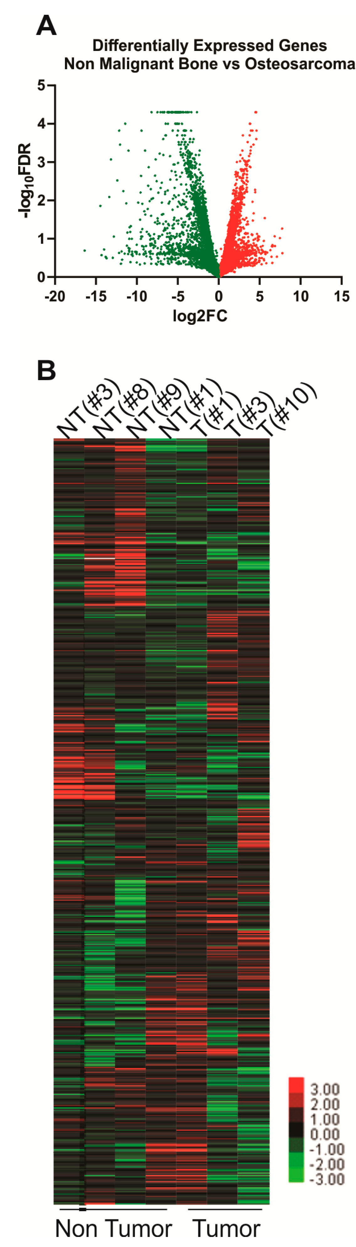
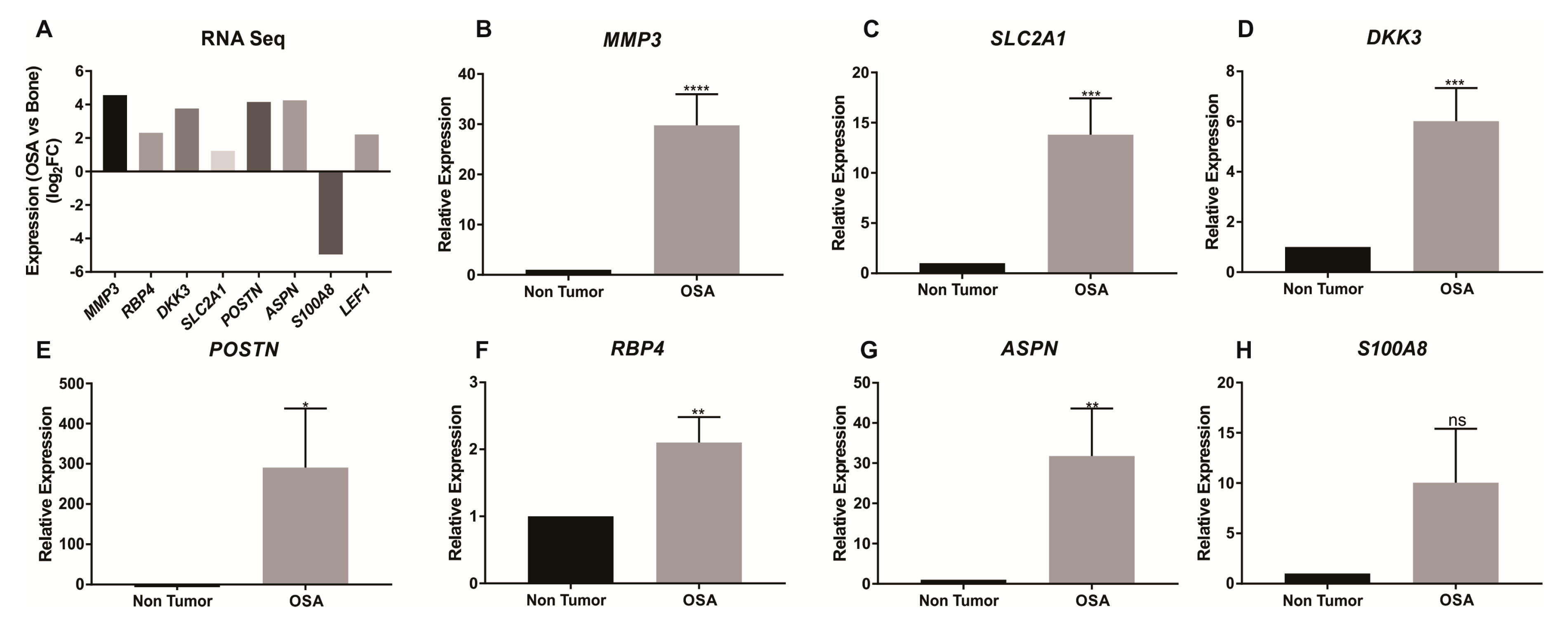
2.1. RNAseq Gene Expression

The Illumina iGenomes CanFam3.1 Ensembl (Illumina, San Diego, CA, USA) annotated genome comprising 24,580 genes was included in the RNAseq analysis. Of these genes, 1281 were identified with a fold change of ≥2 between tumor and non-tumor tissue and a p < 0.05 (n = 4 non-tumor and n = 3 tumor samples). Higher expression of 442 genes and lower expression of 839 genes was observed in tumor tissue compared to the non-tumor tissue (Figure 1A,B, Table 1 and Table 2, and Table S1). The biological relevance of these significantly differentially expressed genes was analysed using over-representation of enriched pathways, gene ontologies and predicted transcription factor networks using the WebGestalt platform (http://www.webgestalt.org/, Zhang Lab, Baylor College of Medicine, Houston, TX, USA; Tables S2 and S3). Differentially expressed genes were significantly associated with pathways associated with heme biosynthesis, calcium homeostasis and signalling (Table 2 and Table 3). Significantly enriched gene ontologies and enriched predicted transcription factor networks were also identified (Tables S2 and S3).

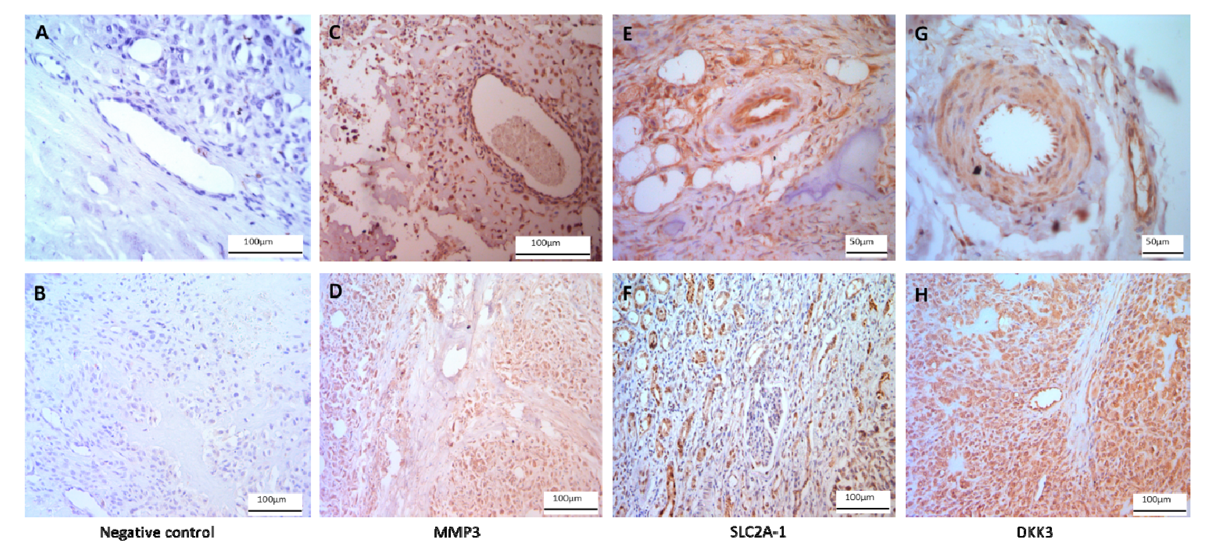
2.2. qRT-PCR and Immunohistochemistry Validation

Validation of seven differentially expressed genes identified by RNAseq was performed by qRT-PCR analysis of eight non-malignant bone specimens and seven OSA specimens. Expression for MMP3 (t = 4.884, p < 0.0001), SLC2A1 (t = 3.703, p = 0.0006), DDK3 (t = 3.981, p = 0.0003), POSTN (t = 2.061, p = 0.0455), RBP4 (t = 3.048, p = 0.004) and ASPN (t = 2.733, p = 0.0091) was confirmed to be higher in the tumor tissues (Figure 2A–G). Differential expression of S100A8 was not confirmed by qRT-PCR (t = 1.766 p = 0.0847; Figure 2H). Immunostaining confirmed positive expression of MMP3, SLC2A1 and DKK3 proteins in OSA tissue (Figure 3).

2.3. Cohort Epidemiology

From the buccal swab cohort, 12 IWHs (8 females and 4 males) and 31 Rottweilers (19 females and 12 males) were reported with OSA (diagnosed by veterinary surgeons and reported by owners), representing 0.53% of all IWHs (from n = 379) and 3.04% of Rottweilers (from n = 1019) within the entire Nottingham cohort inclusive of dogs of all ages. Significant differences were identified in the age at OSA diagnosis between males and females in both breeds. Mean age at OSA diagnosis for male IWHs was 5.34 years, whereas in females this was 7.65 years (t = 3.04, p = 0.0069). A similar difference in the age at diagnosis was observed in Rottweilers, with a mean age at diagnosis for males of 7.00 years while that for females was 8.50 years (t = 2.34, p = 0.027, summarised in Table 4). There were no differences between the sexes in the proportion of individuals affected by OSA in IWHs—χ2test result = 0.065 (p = 0.80), or Rottweilers—χ2test result = 0.17 (p = 0.68).
Of IWH males, 80% that developed OSA had done so before the age of 6.5 years, while 80% of IWH females that developed OSA had done so by 9 years. Rottweilers tend to live longer than IWHs, therefore a 90% threshold was used to determine the age restrictions in this breed. Of male Rottweilers, 90% of those that developed OSA were diagnosed before the age of 9 years, whereas 90% of OSA-affected females were diagnosed before the age of 10 years. Using these age restrictions, there were 23 male IWHs unaffected by OSA over 6.5 years and 6 female IWHs unaffected by OSA over 9 years from the cohort of 379. There were 51 male Rottweilers unaffected by OSA over the age of 9 years and 53 female Rottweilers unaffected by OSA over 10 years old from the cohort of 1019. These individuals were used as control samples for polyQ tract analysis (summarized in Table 5).

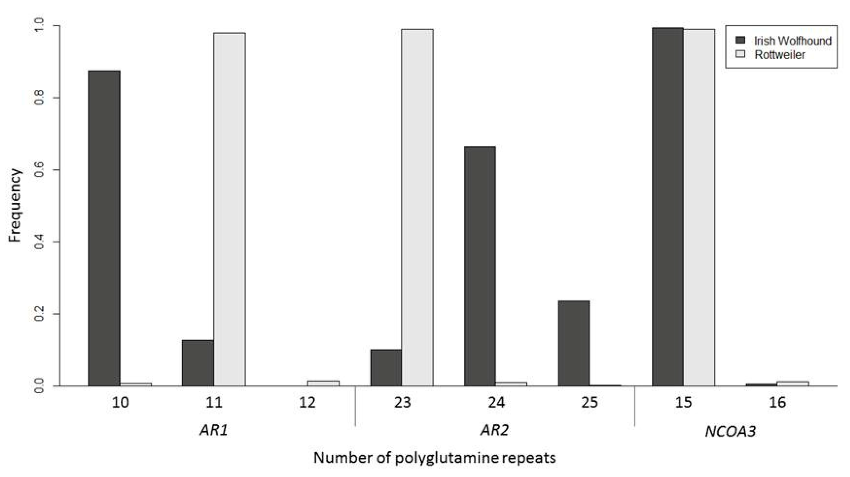
2.4. Androgen polyQ Analysis

Across all individuals genotyped at both AR1 and AR2, the total length of the AR polyQ tract ranged from 33 to 35 repeats. The length of the AR1 polyQ tract ranged from 10 to 12 repeats and the length of the AR2 polyQ tract ranged from 23 to 25 repeats (Figure 4). NCOA3 had a range of 15 to 16 repeats within the polyQ tract across all genotyped individuals (Figure 4). There were no significant differences in mean AR polyQ repeat length between IWHs (t = 2.04, p = 0.053) or Rottweilers (t = 1.00, p = 0.33) with and without a diagnosis of OSA. When individuals were split by sex there remained no significant differences between groups (IWH OSA: ANOVA—F = 1.28, p = 0.30; Rottweiler OSA: ANOVA—F = 1.88, p = 0.14, Table 5). Similar results were obtained when AR1 and AR2 were considered separately. There were also no significant differences in AR1 or AR2 length between individuals with and without OSA in IWHs (AR1 t = 1.02, p = 0.33; AR2 t = 1.83, p = 0.08) and Rottweilers (AR1 t = 1.00, p = 0.33; AR2 t = 1.00, p = 0.32). The length of AR1 or AR2 did not differ significantly when sex was compared in IWHs (AR1 F = 0.80, p = 0.50; AR2 F = 1.04, p = 0.39; Table 5) or Rottweilers (AR1 F = 1.88, p = 0.14; AR2 F = 0.55, p = 0.65; Table 5).
There were no significant differences in mean NCOA3 polyQ repeat length between IWHs with a diagnosis of OSA and IWHs included in the unaffected by OSA group, as all individuals had 15 repeats in their polyQ tract. There were two IWH individuals in the study that had 16 repeats; these individuals were unaffected by OSA but had not yet reached the age restrictions to be included in the unaffected group analysis. Similarly, there was little variation in the NCOA3 polyQ repeat length in Rottweilers with two individuals in the unaffected by OSA group having a mean repeat length of 15.5. The resulting t-test was not significant (t = 1.42, p = 0.16).
Analysis of the frequencies of the entire populations demonstrated significant differences in AR1 and AR2 polyQ repeat frequencies between the two breeds (AR1 χ2test result = 1707, p = < 0.00001; AR2 χ2test result = 1687; p = <0.00001). IWHs had fewer repeats at AR1, but more repeats at AR2, compared to Rottweilers (4). The mean repeat length overall of AR was significantly increased in IWHs at 34.25 (range 33–35.5) repeats in comparison to Rottweilers at 34.02 (range 33–35; t-test, t = 10.62, p = <0.0001). There was no significant difference between breeds in the NCOA3 polyQ repeat frequencies, χ2test result = 1.79, (p = 0.18).

3. Discussion

Differential gene expression in cancers has been associated with disease progression and prognosis [70,72,73,74]. This study identified 583 genes as differentially expressed between canine OSA (n = 3) and non-tumor (n = 5) bone tissue by RNAseq analysis (Figure 1). Many of these genes had previously been identified as differentially expressed in other cancers and could be potential drug targets in the treatment of OSA. qRT-PCR was used to validate RNAseq results for MMP3 SLC2A1, DKK3, POSTN, RBP4, and ASPN (Figure 2). Separately we used immunohistochemistry to confirm protein expression and localisation of MMP3, SLC2A1 and DKK3 in bone and OSA specimens (Figure 3).
The MMP3 gene encodes a member of the matrix metalloproteinase proteins, which degrades a range of extracellular matrix components and is therefore an important mediator of metastatic invasion [75,76]. High MMP3 protein expression has been shown in breast, lung, and pancreatic cancer, and has been associated with poor prognosis [77]. In addition, MMP3 expression in the murine model of mammary carcinoma has been demonstrated to be important for primary tumor and metastasis growth [78]. Formation of metastases in OSA is a critical stage in disease progression associated with poor prognosis [79,80]. A selective inhibitor of MMP3 is available (UK370106) but is yet to be tested for inhibition of primary tumor or metastatic tumor growth [81]. A generic MMP inhibitor is available (Marimastat, Sigma-Aldrich, St. Louis, MO, USA) [82], but it was not effective in clinical trials [82,83,84,85]. In pancreatic small-cell lung cancers trials, it was not established whether the tumours expressed high levels of MMPs, which could have had implications in establishing drug efficiency [82,85]. Marimastat has not been tested in OSA. In the present study, it was shown that expression of MMP3 is elevated in canine osteosarcoma in comparison to non-malignant bone tissue. Selective metalloprotease inhibitors such as UK370106 and/or marimastat and related candidate drugs may improve the prognosis of canine OSA in those cases with elevated MMP3 expression. Previously published studies found that MMP3 was more highly expressed in OSA tumors than in normal bone [86,87], which corroborated the results found in this study.
Glucose is an essential component in cellular metabolism, with transport into cells reliant on membrane glycoproteins [88]. SLC2A1 is a gene encoding one such cell membrane glycoprotein, which has been shown to be involved in glucose transport in a range of tumors including oral squamous cell carcinoma, non-small cell lung carcinoma, and non-gastrointestinal stromal tumor soft tissue sarcomas [89,90,91]. In patients with OSA, higher expression of SLC2A1 within tumors has been associated with a shorter disease-free interval, and poorer prognosis than in patients with lower expression of SLC2A1 [92]. In the current study, SLC2A1 expression was significantly higher in OSA tissue compared to non-tumor tissue. Survival time was not available for patients involved in the current study, but canine OSA survival time is typically short, up to 14.4 months on average [93]. Based on the current study results combined with those previously published [92], this could be due to the high expression of SLC2A1 [10,11,12]. There are several drugs available that have inhibitory effects on cellular glucose transport and SLC2A1, but few have been tested as a treatment for OSA [94,95,96,97].
A range of cancers have been shown to have aberrant Wnt activity [98,99,100,101]. Dickkopf proteins inhibit Wnt signalling and have been shown to be differentially expressed in colon cancer, prostate cancer, and breast cancer [102,103,104]. Reduced DKK3 expression has been associated with endometrial cancer, cervical cancer and breast cancer, and as such it has been implicated as a tumor suppressor [99,105,106,107]. Within OSA there are conflicting reports; in OSA cell lines and xenograft mice, DKK3 expression was shown to be downregulated, with subsequent restoration of DKK3 expression reducing tumor and metastatic growth [108]. In contrast, DKK3 has been shown to be more highly expressed in OSA cells that overexpress NKD2 [109]. The results from the current study showed that DKK3 was more highly expressed in OSA tissue than in non-tumor tissue. This is in contrast to expression in most other cancer types, but in agreement with other findings [109]. Knockdown of DKK3 in cells overexpressing NKD2 increased their proliferative potential indicating that DKK3 expression could be mediating NKD2-induced metastasis [109]. There are currently no drugs available that act on DKK3. MMP3 and DKK3 have been shown to have opposite effects on Wnt activation. MMP3 secretion has been shown to lead to increased Wnt activation [110,111], while increased DKK3 expression results in reduced Wnt activation [99,105,106,107]. The results presented in the current study thus appear to contradict these earlier findings, with both MMP3 and DKK3 expressed more highly in tumor compared to non-tumor tissue. It is possible that DKK3 is ameliorating the effect of MMP3 secretion in the tumors in this study in a similar way to the effect identified by Zhao and colleagues [109]. Further work is required to establish the relationship between MMP3 and DKK3 in OSA.
To assess the functional relevance of the differentially expressed genes identified here, we performed a comparative search of the KEGG, Panther and Reactome pathway databases using the WebGestalt platform (www.webgestalt.org; Table 3). By integrating the results obtained from these complementary databases, the most detailed insight into the mechanistic relevance of the differentially expressed genes can be obtained. Interestingly, this approach identified aberrant expression of genes associated with heme biosynthesis. While the exact role of heme in cancer is controversial [112], aberrant heme metabolism may influence tumor energy metabolism, the tumor microenvironment, angiogenesis and metastases [112]. Expression of the nine genes (ALAD, ALAS1, COX10, CPOX, FECH, HMBS, NFE2L1, UROD, UROS) of the heme biosynthesis ontology were lower in OSA as compared with non-malignant bone. This could be attributed to the loss of normal bone function as bone marrow is a major source of heme synthesis and hematopoiesis [113]. Bone marrow space may have been replaced with tumor tissue with a resulting loss in functional marrow. An additional possibility is that the loss of heme synthesis within the tumor contributes to its progression. There is some evidence that tetrapyrroles, such as heme, possess anti-cancer properties by inducing apoptosis [114]. Biliverdin and bilirubin, the catalytic products of heme generated by heme oxygenases, have been shown to have anti-oxidant properties [115]. Due to the reduced expression of genes involved in tetrapyrrole/heme/pigment metabolic and biosynthetic processes in tumor compared to non-tumor tissue in this study, it would seem likely that the apoptosis and anti-oxidant properties of tetrapyrroles is correspondingly reduced.
Pathways associated with calcium signalling and cardiomyopathy (Table 1) involved intracellular signalling enzymes including integrins, adenylate cyclases 4 and 9, ATPases, and calcium channels, and indeed calcium homeostasis may reflect aberrant osteoblastic differentiation during osteosarcoma carcinogenesis [116]. Consistent with this, multiple gene ontologies related to cellular differentiation, morphogenesis, development, cellular proliferation, and metabolism were associated with the differentially expressed genes identified here (Table S2).
We used the WebGestalt network module (www.webgestalt.org) to search the human MSigDB transcription factor target database. This analysis identified 51 transcription factor regulators (Table S3) predicted to act as regulators of OSA-associated differentially expressed genes (Table S1). Notable predicted regulators included LEF1, a key regulator of the Wnt/β-catenin pathway, which has previously been implicated in OSA [117,118]. There were 122 putative LEF1-target genes identified in the differentially expressed genes. LEF1 expression was significantly different between tumor and non-tumor tissue (p = 0.0405) with higher expression seen in tumor compared with non-tumor tissue. Among the genes regulated by LEF1, some demonstrated increased expression in tumor compared with non-tumor tissue, whereas others showed decreased expression. Genes do not act in isolation, therefore, those genes with lower expression in this dataset may have also required the expression of additional transcription factors or coactivators for activation [119]. LEF1 is associated with increased Wnt signalling, however, in the current study it appears that genes may be antagonising one another, with genes that both increase and decrease Wnt signalling expressed more highly in tumor compared with non-tumor tissue [109,110,111,120]. Wnt signalling has both canonical and non-canonical pathways, with non-canonical signalling split into planar cell polarity and Wnt/Ca2+ [121].
Expression of DKK3 and MMP3 are increased in OSA (Figure 2) and both are known to act on the binding of Wnt activator proteins so alterations in the amount of these proteins will affect both canonical and non-canonical Wnt signalling [110,121,122]. Combined with the increase in DKK3 and MMP3 expression seen in tumor tissue, the demonstration of aberrant activation of LEF1 in the current study adds OSA to the range of cancers that exhibit aberrant Wnt activity [98,99,100,101]. LEF1 is part of the canonical Wnt signalling pathway, requiring β-catenin as a coactivator for downstream gene transcription [123,124]. It may be that aberrant Wnt processes upstream of LEF1, such as DKK3 and MMP3 expression, are affecting LEF1 activity. This may explain the difference in the transcription of genes regulated by LEF1, but not LEF1 itself in OSA tissue compared to non-tumor tissue. LEF1 could be an additional drug target to influence Wnt signalling within OSA. In addition to association with CTGF, it has been shown that expression of MMP3 is inversely related to expression of IRF8 [78]. Due to this, IRF8 could also be an alternative drug target.
In total, 0.53% of all IWHs (n = 379) and 3.04% of Rottweilers (n = 1019) had OSA and although there were no overall differences in the rates of males and females affected, the males were younger at diagnosis than females, with a reduction of 2 years and 1.5 years seen for IWH and Rottweiler males, respectively. It is possible that other factors play a role in this. For example, IWH males are more likely to die younger from dilated cardiomyopathy and atrial fibrillation than females [125,126]. As heart disease is the most common specific cause of death in this breed, males may not always live long enough to develop OSA symptoms/get a diagnosis. The 379 IWHs used in this study were also used in previous publications to look at population outcomes in cardiomyopathy and atrial fibrillation [126]. The results showed that 65% of the male dogs with dilated cardiomyopathy and/or atrial fibrillation had been diagnosed before the age of 5.5 years—the average age at which OSA was diagnosed, whereas only 40% of females had been diagnosed as suffering from these heart diseases at that age [126].
This is the first study to examine the length of the canine NCOA3 polyQ tract. This is also the first evaluation of the AR polyQ tract in IWHs and Rottweilers. Most individuals had 15 polyQ repeats in both alleles of the NCOA3 gene, with only one additional repeat in 1% of Rottweiler and 0.5% of IWH NCOA3 polyQ tracts genotyped. The lack of variation in the length of the NCOA3 polyQ tract in these two breeds strongly suggests that variation in the length of the NCOA3 polyQ tract is not a contributing factor in the development of OSA in IWHs, or Rottweilers. The length of the canine AR polyQ tract has previously been associated with aggression in male Japanese Akita Inus and prostate cancer in a variety of breeds [127,128]. In the current study, there was no association between the length of the canine AR and OSA in either IWHs or Rottweilers. Additionally, RNAseq did not identify differential expression of the AR or NCOA3 in canine OSA relative to non-tumor bone tissue. It was interesting that IWHs and Rottweilers had significantly different allele frequencies at both AR1 and AR2, which supports previous findings across other breeds [129]. This difference in allele frequencies may be affected by different population histories, with founder effects, inbreeding, and genetic hitch-hiking [130,131,132].

4. Materials and Methods

4.1. Samples and Ethics

This study was approved by the University of Nottingham ethics committee (numbers 1823 160714 and 959 130925, 20 July 2016) in compliance with Home Office regulations and the Veterinary Surgeons Act. The call for owners to participate in research was made to the public and veterinary practices via breed health group collaborations, emails, social media, attending breed shows, and by holding breed health information days in-house. Informed consent was obtained from all dog owners.
Canine OSA tumor (n = 7) and non-tumor (n = 8) tissue was obtained from veterinary surgeons following amputation treatment or immediately following euthanasia. Confirmation of OSA was confirmed by pathologists via histopathological examination. A 1 cm3 piece of tumor was extracted along with a 1 cm3 piece of adjacent non-tumor affected bone tissue. These samples were placed directly into RNAlater® (Sigma-Aldrich, St. Louis, MO, USA), shipped at room temperature and stored at −20 °C.
DNA samples from 1019 Rottweilers and 379 IWHs were collected using Isohelix DNA Buccal Swabs (Cell Projects Ltd., Harrietsham, UK), according to the manufacturer’s instructions. Clinical histories of each animal were also obtained from owners. Swabs and clinical information were collected over a 3-year period with regular updates checked for a further 2 years. A Chi-Square (χ2test) was performed to establish any significant difference in the number of males and females diagnosed with OSA. In addition, t-tests were performed using R statistical software [133] to compare the mean age of diagnosis between males and females. From this, appropriate age restrictions for each sex were established for the inclusion of unaffected individuals in the control group, based on the age by which most affected individuals had been diagnosed. Individuals in the unaffected group were used in genetic association testing.

4.2. RNA Extraction and Next Generation Sequencing

OSA tumor (n = 3) and non-tumor (n = 4) bone tissue samples were homogenized (GentleMACS, Miltenyi Biotec, Bergisch Gladbach, Germany) and RNA extracted using Qiagen RNeasy Mini Kit (Qiagen, Hilden, Germany) according to manufacturer’s instructions. The extracted RNA was stored at −80 °C. RNA was quantified using both a NanoDrop 8000 (Thermo Fisher Scientific, Waltham, MA, USA) and an Agilent 2100 Bioanalyzer (Agilent, Santa Clara, CA, USA). RNAseq analysis of seven samples comprised of four non-malignant bone and three OSA specimens and was completed at Edinburgh Genomics using an Illumina HiSeq platform (Illumina, San Diego, CA, USA) with ~20 million reads obtained from all samples analysed. The resultant raw fastq reads were processed for quality (phred score >30) and adapter sequences removed using Trim Galore (https://www.bioinformatics.babraham.ac.uk/projects/trim_galore/). The processed raw reads were aligned to the canine genome (CanFam3.1, https://www.ncbi.nlm.nih.gov/assembly/GCF_000002285.3/) using the Ensembl annotated iGenomes build using Tophat2 [134]. Differential gene expression analysis was completed using Cufflinks/Cuffdiff [135] with significantly differentially expressed genes between the tumor and non-tumor tissue showing a fold change ≥2, and p < 0.05. WebGestalt [136] was also used to perform comparative gene ontology, KEGG, Panther and Reactome pathway analysis, and enriched transcription factor target networks to establish insights into the molecular mechanisms of canine OSA [137,138]. To do this, gene symbols were used to search the human pathway and network databases accessed via WebGestalt. A subset of these genes was selected for qPCR validation based on implication in cancer and biological function. RNA sequences in fastq format and gene quantification values as determined by Cufflinks/Cuffdiff expressed in FRPM have been deposited in the NCBI GEO database (accession = GSE155646, https://www.ncbi.nlm.nih.gov/geo/).

4.3. Quantitative Reverse Transcriptase PCR

cDNA was generated from the RNA extracted from OSA tumors and associated non-tumor tissue by reverse transcriptase PCR. The reverse transcriptase PCR reaction mixture consisted of 2 µL of 5× qScript™ cDNA SuperMix (Quanta Biosciences, Beverly, MA, USA), 0.5–1 µg RNA, adjusted to 10 µL using PCR grade H2O. Reverse transcriptase PCR was carried out at 25 °C for 5 min followed by 1 h at 42 °C. Gene expression of MMP3, MMP2, SLC2A1, DKK3, POSTN, RBP4, ASPN, and S100A8 were quantified using qRT-PCR using TaqMan assays Cf02625966_g1, Cf02649247_g1, Cf02633301_m1, Cf02633587_m1, Cf02634869_m1, Cf02659289_m1, Cf02655953_m1 respectively, ThermoFisher Scientific, Waltham, MA, USA) as described previously [139]. Expression for all samples was normalised to the internal control Actin (Taqman assays Cf03023880_g1, ThermoFisher Scientific, Waltham, MA, USA) and relative expression of tumor compared to non-tumor tissue was calculated utilising the methods described by Pfaffl [140]. t-tests were performed using GraphPad Prism version 7.01 for Windows (GraphPad Software, La Jolla, CA, USA).

4.4. Immunohistochemistry

Immunohistochemistry was performed to show positive protein expression of a subset of the genes analysed, SLC2A1, MMP3 and DKK3. Rottweiler post-mortem OSA tissue (n = 16) was obtained from Bridge Pathology, UK in the form of OSA tissue with neighbouring non-OSA bone. Tissue was fixed in 4% paraformaldehyde for 2 h, dehydrated through an ethanol series, embedded into paraffin blocks, and sectioned at 7 μm. Immunohistochemistry was carried out using a Leica Novolink Polymer Detection Kit (Leica, Wetzlar, Germany) according to manufacturer’s protocols with primary antibodies diluted in fetal calf serum 1:100; anti-SLC2A1 polyclonal unconjugated rabbit antibody (100732-TOB-SIB; Stratech, Ely, UK), anti-MMP3 polyclonal unconjugated rabbit antibody (GTX74514; GeneTex, Irvine, CA, USA), anti-DKK3 (N1C3) polyclonal unconjugated rabbit antibody (GTX100571; GeneTex, Irvine, CA, USA) were used to stain proteins of interest. Microscopy was carried out to confirm positive staining cytoplasmic and/or nuclear staining (Leica, Wetzlar, Germany). Negative controls received no primary antibody and were incubated in fetal calf serum only.

4.5. PolyQ Analysis

DNA extraction was performed as previously described [126]. PCR and fragment length analysis: The canine AR harbours two polyQ repeat regions within its locus, hereby denoted AR1 and AR2 [129]. AR1 and AR2 were PCR amplified using primers published by Maejima et al. [129]. Primers were designed to flank the polyQ region of the canine NCOA3 gene. One of each pair of primers was fluorescently labelled with HEX or 6-FAM dyes (Sigma-Aldrich, St. Louis, MO, USA) to allow fragment analysis to be carried out (Primers: AR1 forward 6-FAM-CCGTGAGCGCAGCACCTCCCGGTG, reverse AGGCTGACCGCTGTTGGGAAGGCTGC; AR2 forward 6-FAM-GCCAGCACCACCGGACGAGAATGA, reverse TAACTGTCCTTGGAGGAGGTGGAAGCA; NCOA3 forward HEX-CCCAGCAGGGTTTTCTGAATGCCC, reverse CACAGGCCCTGCCAAAACGCCATCC). Pre-PCR multiplexing, whereby two PCRs are carried out on a sample as a single reaction, was performed for AR1 and NCOA3 with no discernible effect on PCR efficiency. The reaction mixtures consisted of 1× LightCycler® 480 Probes Master Mix (Roche, Basel, Switzerland), 0.5 µM of primers, and 1.5 µL of template DNA, with the volume adjusted to 15 µL using PCR grade H2O. The calculation for a 15 µL reaction containing AR1 and NCOA3 primers combined was 7.5 µL 2× Roche Probe Master Mix, 1.5 µL of each primer from a stock with concentration of 5 µM, and 1.5 µL template DNA. AR2 primers were added to the multiplex post PCR; the calculation for a 15 µL reaction for the AR2 primer pair was 7.5 µL 2× Roche Probe Master Mix and 1.5 µL of each primer from a stock with concentration of 5 µM, 3 µL H2O, and 1.5 µL template DNA. The PCR reaction was run for 40 cycles at 94 °C for 30 s, annealing temperature for 30 s, and 72 °C for 30 s. The optimal annealing temperature was determined through a temperature gradient PCR, with temperature ranging between 52 °C and 64 °C. An annealing temperature of 61 °C was used for AR1 and NCOA3, and 57 °C was used for AR2.
All multiplex samples were diluted by a factor of 100. Analysis of the fluorescently-labelled PCR products was undertaken using the Applied Biosystems 3730 DNA Analyzer with GeneScan ROX-500 size standard (DBS Genomics, Durham, UK) for each sample. The size discrepancy between AR1 and AR2 allowed for discrimination between their FAM-labelled products. Genotypes were scored using Genemapper software version 3.7 (Applied Biosystems, Foster City, CA, USA). A homozygote of each allele was Sanger sequenced by Source Bioscience (Source Bioscience, Nottingham, UK) to confirm correct amplification and the number of polyQ repeats.
PolyQ statistical analysis: The lengths of AR1 and AR2 were combined to give an overall AR polyQ repeat tract length. As the AR locus is positioned on the X chromosome, males only have one copy of the gene while females have two. Therefore, the lengths of AR1 and AR2 were simply summed for males, while the mean of the summed lengths for both alleles was used for females. For NCOA3, the mean length of the polyQ tract for both alleles was determined for all individuals. Student’s t-tests were performed using R statistical software [133] to establish any differences in the length of the polyQ repeat in AR and NCOA3 between affected and unaffected control groups. The individuals included in the unaffected groups were established by the age restrictions as determined previously and as determined in the results. The groups were further split by sex and analysed by ANOVA using R statistical software [133].

5. Conclusions

Variations in the AR and NCOA3 genes polyQ repeat tracts are not associated with the development of canine OSA in IWHs and Rottweilers. However, this study has identified several potential therapeutic targets for improving the outcomes of dogs with OSA. Drugs targeting some of the genes identified already exist and thus could be rapidly investigated for an effect in OSA. Additional work is required to determine the functional/mechanistic importance of the difference in expression between tumor and non-tumor tissue and the potential for utilising spontaneous OSA in dogs to improve understanding of the disease in people.

Supplementary Materials

The following are available online at https://www.mdpi.com/2072-6694/12/9/2405/s1, Table S1: Differentially expressed genes in canine OSA relative to non-malignant bone, Table S2: Significantly enriched gene ontologies associated with differentially expressed genes in canine OSA, Table S3: Enrich transcription factor networks in differentially expressed genes in canine OSA.

Author Contributions

Conceptualization, M.D., N.P.M., R.D.E., C.A. and C.S.R.; Data curation, S.S., M.D., N.P.M. and C.S.R.; Formal analysis, S.S., S.d.B., M.M., J.L.-R., J.N.J., N.P.M. and C.S.R.; Funding acquisition, M.D., N.P.M., R.D.E., C.A., and C.S.R.; Investigation, S.S., M.D., S.d.B., A.A., C.B., C.L.W., J.L.-R., A.A.R., J.N.J., N.P.M. and C.S.R.; Methodology, M.D., A.A., C.B., M.M., S.A., J.L.-R., N.P.M. and C.S.R.; Project administration, M.D., N.P.M. and C.S.R.; Resources, S.S., M.D., S.A., N.P.M. and C.S.R.; Supervision, M.D., N.P.M. and C.S.R.; Validation, N.P.M., C.S.R.; Visualization, S.S., S.d.B., A.A., C.B., N.P.M. and C.S.R.; Writing—original draft, S.S., N.P.M. and C.S.R.; Writing—review and editing, S.S., M.D., S.d.B., A.A., C.B., C.L.W., M.M., S.A., J.N.J., J.L.-R., A.A.R., N.P.M. and C.S.R. All authors have read and agreed to the published version of the manuscript.

Funding

The authors gratefully acknowledge generous funding from the University of Nottingham BBSRC Doctoral Training Programmes BB/J014508/1 and BB/I024291/1 the School of Veterinary Medicine and Science, University of Nottingham, awarded to Catrin S. Rutland, Nigel P. Mongan, and Malcolm Cobb. We also acknowledge undergraduate student scholarships funded by The Wellcome Trust, The Academy of Medical Sciences awarded to Catrin S. Rutland, Nigel P. Mongan, V. James, J. Daly, K. Braithwaite, P. Voigt, K. Cobb and G. England to promote research and public engagement. A.A.R. was supported by Kazan Federal University Program of Competitive Growth.

Acknowledgments

The authors would like to thank the owners of the dogs involved for their participation and consent, the breed health committees for their support and the veterinary surgeons who collected tumor samples.

Conflicts of Interest

The authors declare no conflict of interest. The funders had no role in the design of the study, in the collection, analyses, or interpretation of data, in the writing of the manuscript, or in the decision to publish the results.

References

  1. Mirabello, L.; Troisi, R.J.; Savage, S.A. International osteosarcoma incidence patterns in children and adolescents, middle ages and elderly persons. Int. J. Cancer 2009, 125, 229–234. [Google Scholar] [CrossRef] [PubMed] [Green Version]
  2. Siegel, R.; Naishadham, D. jemal A: Cancer statistics, 2013. CA Cancer J. Clin. 2013, 63, 11–30. [Google Scholar] [CrossRef] [PubMed] [Green Version]
  3. Mirabello, L.; Troisi, R.J.; Savage, S.A. Osteosarcoma incidence and survival rates from 1973 to 2004: Data from the Surveillance, Epidemiology, and End Results Program. Cancer 2009, 115, 1531–1543. [Google Scholar] [CrossRef] [PubMed] [Green Version]
  4. Fenger, J.M.; London, C.A.; Kisseberth, W.C. Canine Osteosarcoma: A Naturally Occurring Disease to Inform Pediatric Oncology. ILAR J. 2014, 55, 69–85. [Google Scholar] [CrossRef] [Green Version]
  5. Tinkey, P.T.; Lembo, T.M.; Evans, G.R.D.; Cundiff, J.H.; Gray, K.N.; Price, R.E. Postirradiation sarcomas in Sprague-Dawley rats. Radiat. Res. 1998, 149, 401–404. [Google Scholar] [CrossRef]
  6. Jacks, T.; Remington, L.; Williams, B.O.; Schmitt, E.M.; Halachmi, S.; Bronson, R.T.; Weinberg, R.A. Tumor spectrum analysis in p53-mutant mice. Curr. Biol. 1994, 4, 1–7. [Google Scholar] [CrossRef]
  7. Berlin, O.; Samid, D.; Donthineni-Rao, R.; Akeson, W.; Amiel, D.; Woods, V.L. Development of a novel spontaneous metastasis model of human osteosarcoma transplanted orthotopically into bone of athymic mice. Cancer Res. 1993, 53, 4890–4895. [Google Scholar]
  8. Parkin, D.M.; Ferlay, J.; Bray, F.; Edwards, B.; Forman, D.; Curado, M.P.; Shin, H.-R. Fifty years of cancer incidence: CI5 I-IX. Int. J. Cancer 2010, 127, 2918–2927. [Google Scholar] [CrossRef]
  9. Egenvall, A.; Nødtvedt, A.; Von Euler, H. Bone tumors in a population of 400 000 insured Swedish dogs up to 10 y of age: Incidence and survival. Can. J. Vet. Res. 2007, 71, 292–299. [Google Scholar]
  10. Frimberger, A.E.; Chan, C.M.; Moore, A.S. Canine Osteosarcoma Treated by Post-Amputation Sequential Accelerated Doxorubicin and Carboplatin Chemotherapy: 38 Cases. J. Am. Anim. Hosp. Assoc. 2016, 52, 149–156. [Google Scholar] [CrossRef]
  11. Moore, A.S.; Dernell, W.S.; Ogilvie, G.K.; Kristal, O.; Elmslie, R.; Kitchell, B.; Susaneck, S.; Rosenthal, R.; Klein, M.K.; Obradovoich, J.; et al. Doxorubicin and BAY 12–9566 for the Treatment of Osteosarcoma in Dogs: A Randomized, Double-Blind, Placebo-Controlled Study. J. Vet. Intern. Med. 2007, 21, 783–790. [Google Scholar] [CrossRef] [PubMed]
  12. Straw, R.C.; Withrow, S.J.; Richter, S.L.; Powers, B.E.; Klein, M.K.; Postorino, N.C.; LaRue, S.M.; Ogilvie, G.K.; Vail, D.M.; Morrison, W.B.; et al. Amputation and Cisplatin for Treatment of Canine Osteosarcoma. J. Vet. Intern. Med. 1991, 5, 205–210. [Google Scholar] [CrossRef] [PubMed]
  13. Savage, S.A.; Mirabello, L.; Wang, Z.; Gastier-Foster, J.M.; Gorlick, R.; Khanna, C.; Flanagan, A.M.; Tirabosco, R.; Andrulis, I.L.; Wunder, J.S.; et al. Genome-wide association study identifies two susceptibility loci for osteosarcoma. Nat. Genet. 2013, 45, 799–803. [Google Scholar] [CrossRef] [PubMed] [Green Version]
  14. Mirabello, L.; Pfeiffer, R.; Murphy, G.; Daw, N.C.; Patiño-García, A.; Troisi, R.J.; Hoover, R.N.; Douglass, C.; Schüz, J.; Craft, A.W.; et al. Height at diagnosis and birth-weight as risk factors for osteosarcoma. Cancer Causes Control. 2011, 22, 899–908. [Google Scholar] [CrossRef] [Green Version]
  15. McNeill, C.J.; Overley, B.; Shofer, F.S.; Kent, M.S.; Clifford, C.A.; Samluk, M.; Haney, S.; Van Winkle, T.J.; Sorenmo, K.U. Characterization of the biological behaviour of appendicular osteosarcoma in Rottweilers and a comparison with other breeds: A review of 258 dogs. Vet. Comp. Oncol. 2007, 5, 90–98. [Google Scholar] [CrossRef]
  16. Ru, G.; Terracini, B.; Glickman, L. Host related risk factors for canine osteosarcoma. Vet. J. 1998, 156, 31–39. [Google Scholar] [CrossRef]
  17. Yang, Y.C.; Banuelos, C.A.; Mawji, N.R.; Wang, J.; Kato, M.; Haile, S.; McEwan, I.J.; Plymate, S.; Sadar, M.D. Targeting Androgen Receptor Activation Function-1 with EPI to Overcome Resistance Mechanisms in Castration-Resistant Prostate Cancer. Clin. Cancer Res. 2016, 22, 4466–4477. [Google Scholar] [CrossRef] [Green Version]
  18. Mongan, N.P.; Tadokoro-Cuccaro, R.; Bunch, T.; Hughes, I.A. Androgen insensitivity syndrome. Best Pract. Res. Clin. Endocrinol. Metab. 2015, 29, 569–580. [Google Scholar] [CrossRef]
  19. Lu, N.Z.; Wardell, S.E.; Burnstein, K.L.; DeFranco, D.; Fuller, P.J.; Giguère, V.; Hochberg, R.B.; McKay, L.; Renoir, J.-M.; Weigel, N.L.; et al. International Union of Pharmacology. LXV. The Pharmacology and Classification of the Nuclear Receptor Superfamily: Glucocorticoid, Mineralocorticoid, Progesterone, and Androgen Receptors. Pharmacol. Rev. 2006, 58, 782–797. [Google Scholar] [CrossRef]
  20. Fang, H.; Tong, W.; Branham, W.S.; Moland, C.L.; Dial, S.L.; Hong, H.; Xie, Q.; Perkins, R.; Owens, W.; Sheehan, D.M. Study of 202 Natural, Synthetic, and Environmental Chemicals for Binding to the Androgen Receptor. Chem. Res. Toxicol. 2003, 16, 1338–1358. [Google Scholar] [CrossRef]
  21. McKenna, N.J.; O’Malley, B.W. Combinatorial Control of Gene Expression by Nuclear Receptors and Coregulators. Cell 2002, 108, 465–474. [Google Scholar] [CrossRef] [Green Version]
  22. Dart, D.A.; Waxman, J.; Aboagye, E.O.; Bevan, C.L. Visualising Androgen Receptor Activity in Male and Female Mice. PLoS ONE 2013, 8, e71694. [Google Scholar] [CrossRef] [PubMed] [Green Version]
  23. Infante, J.B.; Alvelos, M.I.; Bastos, M.; Carrilho, F.; Lemos, M.C. Complete androgen insensitivity syndrome caused by a novel splice donor site mutation and activation of a cryptic splice donor site in the androgen receptor gene. J. Steroid Biochem. Mol. Biol. 2016, 155, 63–66. [Google Scholar] [CrossRef] [PubMed]
  24. Gottlieb, B.; Beitel, L.K.; Nadarajah, A.; Paliouras, M.; Trifiro, M. The androgen receptor gene mutations database: 2012 update. Hum. Mutat. 2012, 33, 887–894. [Google Scholar] [CrossRef]
  25. Batch, J.A.; Williams, D.M.; Davies, H.R.; Brown, B.D.; Evans, B.A.J.; Hughes, I.A.; Patterson, M.N. Androgen receptor gene mutations identified by SSCP in fourteen subjects with androgen insensitivity syndrome. Hum. Mol. Genet. 1992, 1, 497–503. [Google Scholar] [CrossRef]
  26. Brown, T.R.; Lubahn, D.B.; Wilson, E.M.; Joseph, D.R.; French, F.S.; Migeon, C.J. Deletion of the steroid-binding domain of the human androgen receptor gene in one family with complete androgen insensitivity syndrome: Evidence for further genetic heterogeneity in this syndrome. Proc. Natl. Acad. Sci. USA 1988, 85, 8151–8155. [Google Scholar] [CrossRef] [Green Version]
  27. Kazemi-Esfarjani, P.; Trifiro, M.A.; Pinsky, L. Evidence for a repressive function of the long polyglutamine tract in the human androgen receptor: Possible pathogenetic relevance for the (CAG)n-expanded neuronopathies. Hum. Mol. Genet. 1995, 4, 523–527. [Google Scholar] [CrossRef]
  28. Chamberlain, N.L.; Driver, E.D.; Miesfeld, R.L. The length and location of CAG trinucleotide repeats in the androgen receptor N-terminal domain affect transactivation function. Nucleic Acids Res. 1994, 22, 3181–3186. [Google Scholar] [CrossRef] [Green Version]
  29. Ibáñez, L.; Ong, K.K.; Mongan, N.P.; Jääskeläinen, J.; Marcos, M.V.; Hughes, I.A.; De Zegher, F.; Dunger, D.B. Androgen Receptor Gene CAG Repeat Polymorphism in the Development of Ovarian Hyperandrogenism. J. Clin. Endocrinol. Metab. 2003, 88, 3333–3338. [Google Scholar] [CrossRef] [Green Version]
  30. Hsing, A.W.; Gao, Y.T.; Wu, G.; Wang, X.; Deng, J.; Chen, Y.L.; Sesterhenn, I.A.; Mostofi, F.K.; Benichou, J.; Chang, C. Polymorphic CAG and GGN repeat lengths in the androgen receptor gene and prostate cancer risk: A population-based case-control study in China. Cancer Res. 2000, 60, 5111–5116. [Google Scholar]
  31. Rebbeck, T.R.; Kantoff, P.W.; Krithivas, K.; Neuhausen, S.; Blackwood, M.A.; Godwin, A.K.; Daly, M.B.; Narod, S.A.; Garber, J.E.; Lynch, H.T.; et al. Modification of BRCA1-associated breast cancer risk by the polymorphic androgen-receptor CAG repeat. Am. J. Hum. Genet. 1999, 64, 1371–1377. [Google Scholar] [CrossRef] [PubMed] [Green Version]
  32. Giovannucci, E.; Stampfer, M.J.; Krithivas, K.; Brown, M.; Brufsky, A.; Talcott, J.; Hennekens, C.H.; Kantoff, P.W. The CAG repeat within the androgen receptor gene and its relationship to prostate cancer. Proc. Natl. Acad. Sci. USA 1997, 94, 3320–3323. [Google Scholar] [CrossRef] [Green Version]
  33. La Spada, A.R.; Wilson, E.M.; Lubahn, D.B.; Harding, A.E.; Fischbeck, K.H. Androgen receptor gene mutations in X-linked spinal and bulbar muscular atrophy. Nature 1991, 352, 77–79. [Google Scholar] [CrossRef] [PubMed]
  34. Yepuru, M.; Wu, Z.; Kulkarni, A.; Yin, F.; Barrett, C.M.; Kim, J.; Steiner, M.S.; Miller, D.D.; Dalton, J.T.; Narayanan, R. Steroidogenic Enzyme AKR1C3 Is a Novel Androgen Receptor-Selective Coactivator that Promotes Prostate Cancer Growth. Clin. Cancer Res. 2013, 19, 5613–5625. [Google Scholar] [CrossRef] [PubMed] [Green Version]
  35. Boorjian, S.A.; Heemers, H.V.; Frank, I.; Farmer, S.A.; Schmidt, L.J.; Sebo, T.J.; Tindall, D.J. Expression and significance of androgen receptor coactivators in urothelial carcinoma of the bladder. Endocr. Relat. Cancer 2009, 16, 123–137. [Google Scholar] [CrossRef] [PubMed] [Green Version]
  36. Ngan, E.S.-W.; Hashimoto, Y.; Ma, Z.-Q.; Tsai, M.-J.; Tsai, S.Y. Overexpression of Cdc25B, an androgen receptor coactivator, in prostate cancer. Oncogene 2003, 22, 734–739. [Google Scholar] [CrossRef]
  37. Gnanapragasam, V.J.; Leung, H.Y.; Pulimood, A.S.; Neal, D.E.; Robson, C.N. Expression of RAC 3, a steroid hormone receptor co-activator in prostate cancer. Br. J. Cancer 2001, 85, 1928–1936. [Google Scholar] [CrossRef]
  38. Mongan, N.; Jääskeläinen, J.; Bhattacharyya, S.; Leu, R.; Hughes, I.; Mongan, N.P. Steroid receptor coactivator-3 glutamine repeat polymorphism and the androgen insensitivity syndrome. Eur. J. Endocrinol. 2003, 148, 277–279. [Google Scholar] [CrossRef] [Green Version]
  39. Tan, J.-A.; Hall, S.H.; Petrusz, P.; French, F.S. Thyroid Receptor Activator Molecule, TRAM-1, Is an Androgen Receptor Coactivator. Endocrinology 2000, 141, 3440–3450. [Google Scholar] [CrossRef]
  40. Han, G.; Xie, S.; Fang, H.; Li, G.; Han, Y.; Qin, Z. The AIB1 gene polyglutamine repeat length polymorphism contributes to risk of epithelial ovarian cancer risk: A case–control study. Tumor Biol. 2015, 36, 371–374. [Google Scholar] [CrossRef]
  41. Burandt, E.; Jens, G.; Holst, F.; Jänicke, F.; Müller, V.; Quaas, A.; Choschzick, M.; Wilczak, W.; Terracciano, L.; Simon, R.; et al. Prognostic relevance of AIB1 (NCoA3) amplification and overexpression in breast cancer. Breast Cancer Res. Treat. 2013, 137, 745–753. [Google Scholar] [CrossRef] [PubMed]
  42. Luo, F.; Li, W.; Zhang, J.; Huang, K.; Fu, J.; Xie, Z. Overexpression of steroid receptor coactivator-3 in bone cancers: An in vivo immunohistochemical study with tissue microarray. Pathol. Res. Pract. 2013, 209, 790–796. [Google Scholar] [CrossRef] [PubMed]
  43. Li, Z.; Fang, Z.-Y.; Ding, Y.; Yao, W.-T.; Yang, Y.; Zhu, Z.-Q.; Wang, W.; Zhang, Q.-X. Amplifications of NCOA3 gene in colorectal cancers in a Chinese population. World J. Gastroenterol. 2012, 18, 855–860. [Google Scholar] [CrossRef] [PubMed]
  44. Yang, J.; Benyamin, B.; McEvoy, B.P.; Gordon, S.; Henders, A.K.; Nyholt, D.R.; Madden, P.A.; Heath, A.C.; Martin, N.G.; Montgomery, G.; et al. Common SNPs explain a large proportion of the heritability for human height. Nat. Genet. 2010, 42, 565–569. [Google Scholar] [CrossRef] [PubMed] [Green Version]
  45. Villani, A.; Tabori, U.; Schiffman, J.; Shlien, A.; Beyene, J.; Druker, H.; Novokmet, A.; Finlay, J.; Malkin, D. Biochemical and imaging surveillance in germline TP53 mutation carriers with Li-Fraumeni syndrome: A prospective observational study. Lancet Oncol. 2011, 12, 559–567. [Google Scholar] [CrossRef]
  46. Limacher, J.-M.; Frebourg, T.; Natarajan-Ame, S.; Bergerat, J.-P. Two metachronous tumors in the radiotherapy fields of a patient with Li-Fraumeni syndrome. Int. J. Cancer 2001, 96, 238–242. [Google Scholar] [CrossRef]
  47. Stinco, G.; Governatori, G.; Mattighello, P.; Patrone, P. Multiple cutaneous neoplasms in a patient with Rothmund–Thomson syndrome: Case report and published work review. J. Dermatol. 2008, 35, 154–161. [Google Scholar] [CrossRef]
  48. Ognjanovic, S.; Olivier, M.; Bergemann, T.L.; Hainaut, P. Sarcomas in TP53 germline mutation carriers. Cancer 2012, 118, 1387–1396. [Google Scholar] [CrossRef]
  49. Thomas, D.M.; Ballinger, M.L. Etiologic, environmental and inherited risk factors in sarcomas. J. Surg. Oncol. 2015, 111, 490–495. [Google Scholar] [CrossRef]
  50. Poos, K.; Smida, J.; Nathrath, M.; Maugg, D.; Baumhoer, D.; Neumann, A.; Korsching, E. Structuring osteosarcoma knowledge: An osteosarcoma-gene association database based on literature mining and manual annotation. Database 2014, 2014, bau042. [Google Scholar] [CrossRef] [Green Version]
  51. De Bruin, E.C.; McGranahan, N.; Mitter, R.; Salm, M.; Wedge, D.C.; Yates, L.; Jamal-Hanjani, M.; Shafi, S.; Murugaesu, N.; Rowan, A.J.; et al. Spatial and temporal diversity in genomic instability processes defines lung cancer evolution. Science 2014, 346, 251–256. [Google Scholar] [CrossRef] [PubMed] [Green Version]
  52. Galanos, P.S.; Vougas, K.; Walter, D.; Polyzos, A.; Maya-Mendoza, A.; Haagensen, E.J.; Kokkalis, A.; Roumelioti, F.-M.; Gagos, S.; Tzetis, M.; et al. Chronic p53-independent p21 expression causes genomic instability by deregulating replication licensing. Nat. Cell Biol. 2016, 18, 777–789. [Google Scholar] [CrossRef] [PubMed]
  53. Al-Romaih, K.; Bayani, J.; Vorobyova, J.; Karaskova, J.; Park, P.; Zielenska, M.; Squire, J.A. Chromosomal instability in osteosarcoma and its association with centrosome abnormalities. Cancer Genet. Cytogenet. 2003, 144, 91–99. [Google Scholar] [CrossRef]
  54. Carter, S.L.; Eklund, A.C.; Kohane, I.S.; Harris, L.N.; Szállási, Z. A signature of chromosomal instability inferred from gene expression profiles predicts clinical outcome in multiple human cancers. Nat. Genet. 2006, 38, 1043–1048. [Google Scholar] [CrossRef]
  55. Phillips, J.C.; Stephenson, B.; Hauck, M.; Dillberger, J. Heritability and segregation analysis of osteosarcoma in the Scottish deerhound. Genomics 2007, 90, 354–363. [Google Scholar] [CrossRef] [Green Version]
  56. Vonholdt, B.M.; Pollinger, J.P.; Lohmueller, K.E.; Han, E.; Parker, H.G.; Quignon, P.; Degenhardt, J.D.; Boyko, A.R.; Earl, D.A.; Auton, A.; et al. Genome-wide SNP and haplotype analyses reveal a rich history underlying dog domestication. Nature 2010, 464, 898–902. [Google Scholar] [CrossRef] [Green Version]
  57. Phillips, J.C.; Lembcke, L.; Chamberlin, T. A novel locus for canine osteosarcoma (OSA1) maps to CFA34, the canine orthologue of human 3q26. Genomics 2010, 96, 220–227. [Google Scholar] [CrossRef] [Green Version]
  58. Karlsson, E.K.; Sigurdsson, S.; Ivansson, E.; Thomas, R.; Elvers, I.; Curtis, J.L.; Howald, C.; Tonomura, N.; Perloski, M.; Swofford, R.; et al. Genome-wide analyses implicate 33 loci in heritable dog osteosarcoma, including regulatory variants near CDKN2A/B. Genome Biol. 2013, 14, R132. [Google Scholar] [CrossRef]
  59. Millanta, F.; Asproni, P.; Cancedda, S.; Vignoli, M.; Poli, A. Immunohistochemical Expression of COX-2, mPGES and EP2 Receptor in Normal and Reactive Canine Bone and in Canine Osteosarcoma. J. Comp. Pathol. 2012, 147, 153–160. [Google Scholar] [CrossRef] [PubMed]
  60. McCleese, J.K.; Bear, M.D.; Kulp, S.K.; Mazcko, C.; Khanna, C.; London, C. Met interacts with EGFR and Ron in canine osteosarcoma. Vet. Comp. Oncol. 2013, 11, 124–139. [Google Scholar] [CrossRef] [PubMed] [Green Version]
  61. Pang, L.; Argyle, S.A.; Kamida, A.; Morrison, K.O.; Argyle, D.J. The long-acting COX-2 inhibitor mavacoxib (Trocoxil™) has anti-proliferative and pro-apoptotic effects on canine cancer cell lines and cancer stem cells in vitro. BMC Vet. Res. 2014, 10, 184. [Google Scholar] [CrossRef] [Green Version]
  62. Shahi, M.H.; York, D.; Gandour-Edwards, R.; Withers, S.S.; Holt, R.; Rebhun, R.B. BMI1 Is Expressed in Canine Osteosarcoma and Contributes to Cell Growth and Chemotherapy Resistance. PLoS ONE 2015, 10, e0131006. [Google Scholar] [CrossRef] [PubMed]
  63. Selvarajah, G.T.; Kirpensteijn, J.; Van Wolferen, M.E.; Rao, N.A.; Fieten, H.; Mol, J.A. Gene expression profiling of canine osteosarcoma reveals genes associated with short and long survival times. Mol. Cancer 2009, 8, 72. [Google Scholar] [CrossRef] [PubMed] [Green Version]
  64. Selvarajah, G.T.; Verheije, M.H.; Kik, M.; Slob, A.; Rottier, P.J.; Mol, J.A.; Kirpensteijn, J. Expression of epidermal growth factor receptor in canine osteosarcoma: Association with clinicopathological parameters and prognosis. Vet. J. 2012, 193, 412–419. [Google Scholar] [CrossRef] [PubMed]
  65. Selvarajah, G.T.; Bonestroo, F.A.S.; Kirpensteijn, J.; Kik, M.J.L.; Van Der Zee, R.; Van Eden, W.; Timmermans-Sprang, E.P.M.; Slob, A.; Mol, J.A. Heat shock protein expression analysis in canine osteosarcoma reveals HSP60 as a potentially relevant therapeutic target. Cell Stress Chaperon. 2013, 18, 607–622. [Google Scholar] [CrossRef] [Green Version]
  66. Maniscalco, L.; Iussich, S.; Morello, E.; Martano, M.; Gattino, F.; Miretti, S.; Biolatti, B.; Accornero, P.; Martignani, E.; Sánchez-Céspedes, R.; et al. Increased expression of insulin-like growth factor-1 receptor is correlated with worse survival in canine appendicular osteosarcoma. Vet. J. 2015, 205, 272–280. [Google Scholar] [CrossRef]
  67. Urfer, S. How the past affects the present: A genetic history of the Irish wolfhound. In Dogs: Biology, Behavior and Health Disorders; DeGiovine, V.M., Ed.; Nova Science Publishers, Inc.: Hauppauge, NY, USA, 2011; pp. 69–91. [Google Scholar]
  68. Dillberger, J.E.; McAtee, S.A. Osteosarcoma inheritance in two families of Scottish deerhounds. Canine Genet. Epidemiol. 2017, 4, 1–12. [Google Scholar] [CrossRef] [Green Version]
  69. The Cancer Genome Atlas Research Network. Comprehensive molecular characterization of urothelial bladder carcinoma. Nature 2014, 507, 315–322. [Google Scholar] [CrossRef] [Green Version]
  70. Calon, A.; Lonardo, E.; Berenguer, A.; Espinet, E.; Hernando-Momblona, X.; Iglesias, M.; Sevillano, M.; Palomo-Ponce, S.; Tauriello, D.V.F.; Byrom, D.; et al. Stromal gene expression defines poor-prognosis subtypes in colorectal cancer. Nat. Genet. 2015, 47, 320–329. [Google Scholar] [CrossRef] [Green Version]
  71. Pfefferle, A.D.; Agrawal, Y.N.; Koboldt, D.C.; Kanchi, K.L.; Herschkowitz, J.I.; Mardis, E.R.; Rosen, J.M.; Perou, C.M. Genomic profiling of murine mammary tumors identifies potential personalized drug targets for p53-deficient mammary cancers. Dis. Model. Mech. 2016, 9, 749–757. [Google Scholar] [CrossRef] [Green Version]
  72. Van ’t Veer, L.J.; Dai, H.; van de Vijver, M.J.; He, Y.D.; Hart, A.A.M.; Mao, M.; Peterse, H.L.; van der Kooy, K.; Marton, M.J.; Witteveen, A.T.; et al. Gene expression profiling predicts clinical outcome of breast cancer. Nature 2002, 415, 530–536. [Google Scholar] [CrossRef] [Green Version]
  73. Fehrmann, R.S.N.; Karjalainen, J.M.; Krajewska, M.; Westra, H.-J.; Maloney, D.; Simeonov, A.; Pers, T.H.; Hirschhorn, J.N.; Jansen, R.C.; Schultes, E.; et al. Gene expression analysis identifies global gene dosage sensitivity in cancer. Nat. Genet. 2015, 47, 115–125. [Google Scholar] [CrossRef] [PubMed]
  74. Gillet, J.-P.; Andersen, J.B.; Madigan, J.P.; Varma, S.; Bagni, R.K.; Powell, K.; Burgan, W.E.; Wu, C.-P.; Calcagno, A.M.; Ambudkar, S.V.; et al. A Gene Expression Signature Associated with Overall Survival in Patients with Hepatocellular Carcinoma Suggests a New Treatment Strategy. Mol. Pharmacol. 2016, 89, 263–272. [Google Scholar] [CrossRef] [PubMed] [Green Version]
  75. Okada, Y.; Nagase, H.; Harris, E.D. A metalloproteinase from human rheumatoid synovial fibroblasts that digests connective tissue matrix components. Purification and characterization. J. Biol. Chem. 1986, 261, 14245–14255. [Google Scholar]
  76. Kurahara, S.-I.; Shinohara, M.; Ikebe, T.; Nakamura, S.; Beppu, M.; Hiraki, A.; Takeuchi, H.; Shirasuna, K. Expression of MMPS, MT-MMP, and TIMPs in squamous cell carcinoma of the oral cavity: Correlations with tumor invasion and metastasis. Head Neck 1999, 21, 627–638. [Google Scholar] [CrossRef]
  77. Mehner, C.; Miller, E.; Nassar, A.; Bamlet, W.R.; Radisky, E.S.; Radisky, D.C. Tumor cell expression of MMP3 as a prognostic factor for poor survival in pancreatic, pulmonary, and mammary carcinoma. Genes Cancer 2015, 6, 480–489. [Google Scholar] [CrossRef] [PubMed] [Green Version]
  78. Banik, D.; Netherby, C.S.; Bogner, P.N.; Abrams, S.I. MMP3-Mediated tumor progression is controlled transcriptionally by a novel IRF8-MMP3 interaction. Oncotarget 2015, 6, 15164–15179. [Google Scholar] [CrossRef] [PubMed] [Green Version]
  79. Bielack, S.S.; Kempf-Bielack, B.; Delling, G.; Exner, G.U.; Flege, S.; Helmke, K.; Kotz, R.; Salzer-Kuntschik, M.; Werner, M.; Winkelmann, W.; et al. Prognostic Factors in High-Grade Osteosarcoma of the Extremities or Trunk: An Analysis of 1,702 Patients Treated on Neoadjuvant Cooperative Osteosarcoma Study Group Protocols. J. Clin. Oncol. 2002, 20, 776–790. [Google Scholar] [CrossRef] [PubMed]
  80. Reddy, K.I.A.; Wafa, H.; Gaston, C.L.; Grimer, R.J.; Abudu, A.T.; Jeys, L.M.; Carter, S.; Tillman, R.M.; Zuiderbaan, H.A.; Khamaisy, S.; et al. Does amputation offer any survival benefit over limb salvage in osteosarcoma patients with poor chemonecrosis and close margins? Bone Jt. J. 2015, 97, 115–120. [Google Scholar] [CrossRef]
  81. Fray, M.J.; Dickinson, R.P.; Huggins, J.P.; Occleston, N.L. A Potent, Selective Inhibitor of Matrix Metalloproteinase-3 for the Topical Treatment of Chronic Dermal Ulcers. J. Med. Chem. 2003, 46, 3514–3525. [Google Scholar] [CrossRef]
  82. Bramhall, S.; Schulz, J.; Nemunaitis, J.; Brown, P.D.; Baillet, M.; Buckels, J.A.C. A double-blind placebo-controlled, randomised study comparing gemcitabine and marimastat with gemcitabine and placebo as first line therapy in patients with advanced pancreatic cancer. Br. J. Cancer 2002, 87, 161–167. [Google Scholar] [CrossRef] [PubMed]
  83. Davies, B.; Brown, P.D.; East, N.; Crimmin, M.J.; Balkwill, F.R. A synthetic matrix metalloproteinase inhibitor decreases tumor burden and prolongs survival of mice bearing human ovarian carcinoma xenografts. Cancer Res. 1993, 53, 2087–2091. [Google Scholar] [PubMed]
  84. Watson, S.A.; Morris, T.M.; Collins, H.M.; Bawden, L.J.; Hawkins, K.; Bone, E.A. Inhibition of tumour growth by marimastat in a human xenograft model of gastric cancer: Relationship with levels of circulating CEA. Br. J. Cancer 1999, 81, 19–23. [Google Scholar] [CrossRef] [Green Version]
  85. Shepherd, F.A.; Giaccone, G.; Seymour, L.; Debruyne, C.; Bezjak, A.; Hirsh, V.; Smylie, M.; Rubin, S.; Martins, H.; Lamont, A.; et al. Prospective, Randomized, Double-Blind, Placebo-Controlled Trial of Marimastat After Response to First-Line Chemotherapy in Patients With Small-Cell Lung Cancer: A Trial of the National Cancer Institute of Canada-Clinical Trials Group and the European Organization for Research and Treatment of Cancer. J. Clin. Oncol. 2002, 20, 4434–4439. [Google Scholar] [CrossRef]
  86. Tsai, H.-C.; Su, H.-L.; Huang, C.-Y.; Fong, Y.-C.; Hsu, C.-J.; Tang, C.-H. CTGF increases matrix metalloproteinases expression and subsequently promotes tumor metastasis in human osteosarcoma through down-regulating miR-519d. Oncotarget 2014, 5, 3800–3812. [Google Scholar] [CrossRef] [PubMed] [Green Version]
  87. Huang, J.-F.; Du, W.-X.; Chen, J.-J. Elevated expression of matrix metalloproteinase-3 in human osteosarcoma and its association with tumor metastasis. J. BUON 2016, 21, 235–243. [Google Scholar]
  88. Shows, T.B.; Eddy, R.L.; Byers, M.G.; Fukushima, Y.; Dehaven, C.R.; Murray, J.C.; Bell, G.I. Polymorphic Human Glucose Transporter Gene (GLUT) is on Chromosome 1p31.3→p35. Diabetes 1987, 36, 546–549. [Google Scholar] [CrossRef] [Green Version]
  89. Younes, M.; Brown, R.W.; Stephenson, M.; Gondo, M.; Cagle, P.T. Overexpression of Glut1 and Glut3 in stage I nonsmall cell lung carcinoma is associated with poor survival. Cancer 1997, 80, 1046–1051. [Google Scholar] [CrossRef]
  90. Kunkel, M.; Reichert, T.E.; Benz, P.; Lehr, H.-A.; Jeong, J.-H.; Wieand, S.; Bartenstein, P.; Wagner, W.; Whiteside, T.L. Overexpression of Glut-1 and increased glucose metabolism in tumors are associated with a poor prognosis in patients with oral squamous cell carcinoma. Cancer 2003, 97, 1015–1024. [Google Scholar] [CrossRef]
  91. Smeland, E.; Kilvaer, T.; Sorbye, S.; Valkov, A.; Andersen, S.; Bremnes, R.M.; Busund, L.-T.; Donnem, T. Prognostic Impacts of Hypoxic Markers in Soft Tissue Sarcoma. Sarcoma 2012, 2012, 541650. [Google Scholar] [CrossRef]
  92. Kubo, T.; Shimose, S.; Fujimori, J.; Furuta, T.; Arihiro, K.; Ochi, M. Does Expression of Glucose Transporter Protein-1 Relate to Prognosis and Angiogenesis in Osteosarcoma? Clin. Orthop. Relat. Res. 2015, 473, 305–310. [Google Scholar] [CrossRef] [PubMed] [Green Version]
  93. MacEwen, E.G.; Kurzman, I.D. Canine osteosarcoma: Amputation and chemoimmunotherapy. Vet. Clin. N. Am. Small Anim. Pract. 1996, 26, 123–133. [Google Scholar] [CrossRef]
  94. Klepper, J.; Flörcken, A.; Fischbarg, J.; Voit, T. Effects of anticonvulsants on GLUT1-mediated glucose transport in GLUT1 deficiency syndrome in vitro. Eur. J. Nucl. Med. Mol. Imaging 2003, 162, 84–89. [Google Scholar] [CrossRef] [PubMed]
  95. Shuralyova, I.; Tajmir, P.; Bilan, P.J.; Sweeney, G.; Coe, I.R. Inhibition of glucose uptake in murine cardiomyocyte cell line HL-1 by cardioprotective drugs dilazep and dipyridamole. Am. J. Physiol. Circ. Physiol. 2004, 286, H627–H632. [Google Scholar] [CrossRef]
  96. Louters, L.L.; Stehouwer, N.; Rekman, J.; Tidball, A.M.; Cok, A.; Holstege, C.P. Verapamil Inhibits the Glucose Transport Activity of GLUTJ. Med. Toxicol. 2010, 6, 100–105. [Google Scholar] [CrossRef] [PubMed] [Green Version]
  97. Rufino, A.; Rosa, S.C.; Judas, F.; Mobasheri, A.; Lopes, M.; Mendes, A.F. Expression and function of K(ATP) channels in normal and osteoarthritic human chondrocytes: Possible role in glucose sensing. J. Cell. Biochem. 2013, 114, 1879–1889. [Google Scholar] [CrossRef] [PubMed] [Green Version]
  98. Woo, D.K.; Kim, H.S.; Lee, H.S.; Kang, Y.H.; Yang, H.-K.; Kim, W.H. Altered expression and mutation of β-catenin gene in gastric carcinomas and cell lines. Int. J. Cancer 2001, 95, 108–113. [Google Scholar] [CrossRef]
  99. Dellinger, T.; Planutis, K.; Jandial, D.D.; Eskander, R.N.; Martinez, M.E.; Zi, X.; Monk, B.J.; Holcombe, R.F. Expression of the Wnt antagonist Dickkopf-3 is associated with prognostic clinicopathologic characteristics and impairs proliferation and invasion in endometrial cancer. Gynecol. Oncol. 2012, 126, 259–267. [Google Scholar] [CrossRef] [Green Version]
  100. Dey, N.; Barwick, B.G.; Moreno, C.S.; Ordanic-Kodani, M.; Chen, Z.; Oprea-Ilies, G.; Tang, W.; Catzavelos, C.; Kerstann, K.F.; Sledge, J.G.W.; et al. Wnt signaling in triple negative breast cancer is associated with metastasis. BMC Cancer 2013, 13, 1–15. [Google Scholar] [CrossRef] [Green Version]
  101. Voloshanenko, O.; Erdmann, G.; Dubash, T.D.; Augustin, I.; Metzig, M.; Moffa, G.; Hundsrucker, C.; Kerr, G.; Sandmann, T.; Anchang, B.; et al. Wnt secretion is required to maintain high levels of Wnt activity in colon cancer cells. Nat. Commun. 2013, 4, 2610. [Google Scholar] [CrossRef] [Green Version]
  102. Baehs, S.; Herbst, A.; Thieme, S.E.; Perschl, C.; Behrens, A.; Scheel, S.; Jung, A.; Brabletz, T.; Göke, B.; Blum, H.; et al. Dickkopf-4 is frequently down-regulated and inhibits growth of colorectal cancer cells. Cancer Lett. 2009, 276, 152–159. [Google Scholar] [CrossRef] [PubMed]
  103. Hall, C.L.; Zhang, H.; Baile, S.; Ljungman, M.; Kuhstoss, S.; Keller, E.T. p21(CIP-1/WAF-1) Induction Is Required to Inhibit Prostate Cancer Growth Elicited by Deficient Expression of the Wnt Inhibitor Dickkopf-1 (DKK-1). Cancer Res. 2010, 70, 9916–9926. [Google Scholar] [CrossRef] [PubMed] [Green Version]
  104. Zhou, X.-L.; Qin, X.-R.; Zhang, S.; Ye, L.-H. Downregulation of Dickkopf-1 is responsible for high proliferation of breast cancer cells via losing control of Wnt/β-catenin signaling. Acta Pharmacol. Sin. 2010, 31, 202–210. [Google Scholar] [CrossRef] [PubMed]
  105. Hsieh, S.Y.; Hsieh, P.S.; Chiu, C.T.; Chen, W.Y. Dickkopf-3/REIC functions as a suppressor gene of tumor growth. Oncogene 2004, 23, 9183–9189. [Google Scholar] [CrossRef] [Green Version]
  106. Lee, E.-J.; Jo, M.; Rho, S.B.; Park, K.; Yoo, Y.-N.; Park, J.; Chae, M.; Zhang, W.; Lee, J.-H. Dkk3, downregulated in cervical cancer, functions as a negative regulator of β-catenin. Int. J. Cancer 2009, 124, 287–297. [Google Scholar] [CrossRef] [PubMed]
  107. Xiang, T.; Li, L.; Yin, X.; Zhong, L.; Peng, W.; Qiu, Z.; Ren, G.; Tao, Q. Epigenetic silencing of the WNT antagonist Dickkopf 3 disrupts normal Wnt/β-catenin signalling and apoptosis regulation in breast cancer cells. J. Cell. Mol. Med. 2013, 17, 1236–1246. [Google Scholar] [CrossRef]
  108. Lin, C.H.; Guo, Y.; Ghaffar, S.; McQueen, P.; Pourmorady, J.; Christ, A.B.; Rooney, K.; Ji, T.; Eskander, R.; Zi, X.; et al. Dkk-3, a Secreted Wnt Antagonist, Suppresses Tumorigenic Potential and Pulmonary Metastasis in Osteosarcoma. Sarcoma 2013, 2013, 1–11. [Google Scholar] [CrossRef] [Green Version]
  109. Zhao, S.; Kurenbekova, L.; Gao, Y.; Roos, A.; Creighton, C.J.; Rao, P.; Hicks, J.; Man, T.-K.; Lau, C.; Brown, A.M.C.; et al. NKD2, a negative regulator of Wnt signaling, suppresses tumor growth and metastasis in osteosarcoma. Oncogene 2015, 34, 5069–5079. [Google Scholar] [CrossRef] [Green Version]
  110. Kessenbrock, K.; Dijkgraaf, G.J.P.; Lawson, D.A.; Littlepage, L.E.; Shahi, P.; Pieper, U.; Werb, Z. A role for matrix metalloproteinases in regulating mammary stem cell function via the Wnt signaling pathway. Cell Stem Cell 2013, 13, 300–313. [Google Scholar] [CrossRef] [Green Version]
  111. Kim, H.J.; Kang, G.J.; Kim, E.J.; Park, M.K.; Byun, H.J.; Nam, S.; Lee, H.; Lee, C.H. Novel effects of sphingosylphosphorylcholine on invasion of breast cancer: Involvement of matrix metalloproteinase-3 secretion leading to WNT activation. Biochim. Biophys. Acta (BBA)—Mol. Basis Dis. 2016, 1862, 1533–1543. [Google Scholar] [CrossRef]
  112. Fiorito, V.; Chiabrando, D.; Petrillo, S.; Bertino, F.; Tolosano, E. The Multifaceted Role of Heme in Cancer. Front. Oncol. 2019, 9, 1540. [Google Scholar] [CrossRef] [PubMed] [Green Version]
  113. Morell, H.; Savoie, J.C.; London, I.M. The biosynthesis of heme and the incorporation of glycine into globin in rabbit bone marrow in vitro. J. Biol. Chem. 1958, 233, 923–929. [Google Scholar] [PubMed]
  114. Mölzer, C.; Pfleger, B.; Putz, E.; Roßmann, A.; Schwarz, U.; Wallner, M.; Bulmer, A.C.; Wagner, K.-H. In vitro DNA-damaging effects of intestinal and related tetrapyrroles in human cancer cells. Exp. Cell Res. 2013, 319, 536–545. [Google Scholar] [CrossRef]
  115. Jansen, T.; Hortmann, M.; Oelze, M.; Opitz, B.; Steven, S.; Schell, R.; Knorr, M.; Karbach, S.; Schuhmacher, S.; Wenzel, P.; et al. Conversion of biliverdin to bilirubin by biliverdin reductase contributes to endothelial cell protection by heme oxygenase-1—Evidence for direct and indirect antioxidant actions of bilirubin. J. Mol. Cell. Cardiol. 2010, 49, 186–195. [Google Scholar] [CrossRef]
  116. Adamopoulos, C.; Gargalionis, A.N.; Basdra, E.K.; Papavassiliou, A.G. Deciphering signaling networks in osteosarcoma pathobiology. Exp. Biol. Med. 2016, 241, 1296–1305. [Google Scholar] [CrossRef] [PubMed] [Green Version]
  117. Martins-Neves, S.R.; Corver, W.E.; Paiva-Oliveira, D.I.; van den Akker, B.E.; Briaire-de-Bruijn, I.H.; Bovee, J.V.; Gomes, C.M.; Cleton-Jansen, A.M. Osteosarcoma Stem Cells Have Active Wnt/beta-catenin and Overexpress SOX2 and KLFJ4. J. Cell Physiol. 2016, 231, 876–886. [Google Scholar] [CrossRef]
  118. Martins-Neves, S.R.; Paiva-Oliveira, D.I.; Fontes-Ribeiro, C.; Bovee, J.; Cleton-Jansen, A.M.; Gomes, C.M.F. IWR-1, a tankyrase inhibitor, attenuates Wnt/beta-catenin signaling in cancer stem-like cells and inhibits in vivo the growth of a subcutaneous human osteosarcoma xenograft. Cancer Lett. 2018, 414, 1–15. [Google Scholar] [CrossRef] [PubMed]
  119. Shakya, A.; Goren, A.; Shalek, A.K.; German, C.N.; Snook, J.; Kuchroo, V.K.; Yosef, N.; Chan, R.C.; Regev, A.; Williams, M.A.; et al. Oct1 and OCA-B are selectively required for CD4 memory T cell function. J. Exp. Med. 2015, 212, 2115–2131. [Google Scholar] [CrossRef]
  120. Reya, T.; O’Riordan, M.X.D.; Okamura, R.; Devaney, E.; Willert, K.; Nusse, R.; Grosschedl, R. Wnt Signaling Regulates B Lymphocyte Proliferation through a LEF-1 Dependent Mechanism. Immunity 2000, 13, 15–24. [Google Scholar] [CrossRef] [Green Version]
  121. Huelsken, J.; Behrens, J. The Wnt signalling pathway. J. Cell Sci. 2002, 115, 3977–3978. [Google Scholar] [CrossRef] [Green Version]
  122. Nakamura, R.E.I.; Hackam, A.S. Analysis of Dickkopf3 interactions with Wnt signaling receptors. Growth Factors 2010, 28, 232–242. [Google Scholar] [CrossRef] [PubMed]
  123. Behrens, J.; Von Kries, J.P.; Kühl, M.; Bruhn, L.; Wedlich, D.; Grosschedl, R.; Birchmeier, W. Functional interaction of β-catenin with the transcription factor LEF-1. Nature 1996, 382, 638–642. [Google Scholar] [CrossRef] [PubMed]
  124. Hsu, S.-C.; Galceran, J.; Grosschedl, R. Modulation of Transcriptional Regulation by LEF-1 in Response to Wnt-1 Signaling and Association with β-Catenin. Mol. Cell. Biol. 1998, 18, 4807–4818. [Google Scholar] [CrossRef] [PubMed] [Green Version]
  125. Simpson, S.; Edwards, J.; Ferguson-Mignan, T.F.N.; Cobb, M.; Mongan, N.P.; Rutland, C.S. Genetics of Human and Canine Dilated Cardiomyopathy. Int. J. Genom. 2015, 2015, 1–13. [Google Scholar] [CrossRef] [Green Version]
  126. Simpson, S.; Dunning, M.D.; Brownlie, S.; Patel, J.; Godden, M.; Cobb, M.; Mongan, N.P.; Rutland, C.S. Multiple Genetic Associations with Irish Wolfhound Dilated Cardiomyopathy. BioMed Res. Int. 2016, 2016, 1–14. [Google Scholar] [CrossRef]
  127. Lai, C.-L.; L’Eplattenier, H.; Ham, R.V.D.; Verseijden, F.; Jagtenberg, A.; Mol, J.; Teske, E. Androgen Receptor CAG Repeat Polymorphisms in Canine Prostate Cancer. J. Vet. Intern. Med. 2008, 22, 1380–1384. [Google Scholar] [CrossRef]
  128. Konno, A.; Inoue-Murayama, M.; Hasegawa, T. Androgen receptor gene polymorphisms are associated with aggression in Japanese Akita Inu. Biol. Lett. 2011, 7, 658–660. [Google Scholar] [CrossRef] [Green Version]
  129. Maejima, M.; Inoue-Murayama, M.; Koshimura, A.; Kato, S.; Nara, H.; Randi, E.; Kitagawa, H.; Iwasaki, T.; Murayama, Y. Allelic variation of two poly-glutamine repeats in the canine androgen receptor gene. J. Anim. Genet. 2005, 32, 3–11. [Google Scholar] [CrossRef] [Green Version]
  130. Wright, S. The Genetical Structure of Populations. Ann. Eugen. 1949, 15, 323–354. [Google Scholar] [CrossRef]
  131. Slatkin, M. A Measure of Population Subdivision Based on Microsatellite Allele Frequencies. Genetics 1995, 139, 457–462. [Google Scholar]
  132. Fu, Y.X. Statistical Tests of Neutrality of Mutations against Population Growth, Hitchhiking and Background Selection. Genetics 1997, 147, 915–925. [Google Scholar] [PubMed]
  133. R CoreTeam. R: A Language and Environment for Statistical Computing; R Foundation: Vienna, Austria, 2013. [Google Scholar]
  134. Kim, D.; Pertea, G.; Trapnell, C.; Pimentel, H.; Kelley, R.; Salzberg, S.L. TopHat2: Accurate alignment of transcriptomes in the presence of insertions, deletions and gene fusions. Genome Biol. 2013, 14, R13–R36. [Google Scholar] [CrossRef] [PubMed] [Green Version]
  135. Trapnell, C.; Hendrickson, D.G.; Sauvageau, M.; Goff, L.; Rinn, J.L.; Pachter, L. Differential analysis of gene regulation at transcript resolution with RNA-seq. Nat. Biotechnol. 2013, 31, 46–53. [Google Scholar] [CrossRef] [PubMed]
  136. Liao, Y.; Wang, J.; Jaehnig, E.J.; Shi, Z.; Zhang, B. WebGestalt 2019: Gene set analysis toolkit with revamped UIs and APIs. Nucleic Acids Res. 2019, 47, W199–W205. [Google Scholar] [CrossRef] [Green Version]
  137. Zhang, B.; Kirov, S.; Snoddy, J. WebGestalt: An integrated system for exploring gene sets in various biological contexts. Nucleic Acids Res. 2005, 33, W741–W748. [Google Scholar] [CrossRef]
  138. Wang, J.; Duncan, D.; Shi, Z.; Zhang, B. WEB-based GEne SeT AnaLysis Toolkit (WebGestalt): Update 2013. Nucleic Acids Res. 2013, 41, W77–W83. [Google Scholar] [CrossRef] [Green Version]
  139. Nilsson, E.M.; Laursen, K.B.; Whitchurch, J.; McWilliam, A.; Ødum, N.; Persson, J.L.; Heery, D.; Gudas, L.J.; Mongan, N.P. MiR137 is an androgen regulated repressor of an extended network of transcriptional coregulators. Oncotarget 2015, 6, 35710–35725. [Google Scholar] [CrossRef] [Green Version]
  140. Pfaffl, M.W. A new mathematical model for relative quantification in real-time RT-PCR. Nucleic Acids Res. 2001, 29, e45. [Google Scholar] [CrossRef]
Figure 1. Differentially expressed genes were identified in OSA relative to non-malignant bone. RNA sequencing was used to identify differentially expressed genes expressed lower (green) and higher (red) in OSA tumor specimens relative to non-malignant bone specimens. A volcano plot (A) and unsupervised hierarchical clustering was used to display the identified differentially expressed genes (B).
Cancers 12 02405 g001
Figure 2. qRT-PCR validation of RNA expression comparing osteosarcoma tumor tissue expression with non-tumor bone tissue expression. Differential expression of key genes was identified by RNAseq (A). Results from MMP3, SLC2A1, DKK3, POSTN, RBP4, and ASPN (BH) were consistent with the RNAseq results, however, no significant differences were observed in S100A8 (H). Significance levels * p < 0.05, ** p < 0.01, *** p < 0.001, **** p < 0.0001, ns = not significant.
Cancers 12 02405 g002
Figure 3. Protein expression of MMP3, SLC2A1, and DKK3 in osteosarcoma tumor. Negative control immunohistochemistry tissues received no primary antibody and were incubated in fetal calf serum instead (A,B). Immunohistochemistry confirmed protein expression of MMP3 (C,D), SLC2A1 (E,F), and DKK3 (G,H) in osteosarcoma tissue (n = 16). Scale bar represents 50 µm (E,G) and 100 µm (AD,F,H).
Cancers 12 02405 g003
Figure 4. PolyQ repeat allele frequencies for AR1, AR2, and NCOA3. Irish Wolfhounds (n = 379) and Rottweilers (n = 1019).
Cancers 12 02405 g004
Table 1. Top 25 genes expressed lower in osteosarcoma relative to non-malignant bone.
Gene_IDGeneLog2 Fold Changep Value
ENSCAFG00000007134MYBPC1−14.44410.01395
ENSCAFG00000001705MB−13.36270.0081
ENSCAFG00000013875MYL1−13.16890.00085
ENSCAFG00000008253ACTA1−12.38440.003
ENSCAFG00000011103NRAP−12.15370.00015
ENSCAFG00000005717ENSCAFG00000005717−12.06160.00035
ENSCAFG00000014281PYGM−11.64110.0057
ENSCAFG00000011619MYH7−11.40640.0001
ENSCAFG00000002620TNNT1−11.30520.0119
ENSCAFG00000001016TRDN−11.0340.0005
ENSCAFG00000009333CESDD1−10.72690.04655
ENSCAFG00000005896RYR1−9.834870.01135
ENSCAFG00000009404TNNC1−9.615990.0317
ENSCAFG00000015475DES−9.540080.0062
ENSCAFG00000005358HHATL−9.497790.0023
ENSCAFG00000008707CKMT2−9.432020.00015
ENSCAFG00000017254ATP2A1−9.343490.01225
ENSCAFG00000001140MYOT−9.083890.0191
ENSCAFG00000012927ALPK3−9.011170.0005
ENSCAFG00000005343KLHL40−8.960120.0234
ENSCAFG00000002982MYBPC2−8.668550.01675
ENSCAFG00000012432MYOZ2−8.620060.00415
ENSCAFG00000011638SPTA1−8.560140.00135
ENSCAFG00000001161OBSCN−8.443330.01045
ENSCAFG00000011418HFE2−8.311850.0051
RNA sequencing was used to identified differentially expressed genes in OSA (osteosarcoma) specimens relative to non-malignant bone. The entire list of 1281 differentially expressed genes is reported in Table S1.
Table 2. Top 25 genes expressed higher in osteosarcoma relative to non-malignant bone.
Gene_IDGeneLog2 Fold Changep Value
ENSCAFG00000010484PKP23.541830.00095
ENSCAFG00000005510EOMES3.561650.04155
ENSCAFG00000012983GPR643.669110.0009
ENSCAFG00000013448COLGALT23.694480.0007
ENSCAFG00000007845DKK33.768150.0013
ENSCAFG00000009722TOX33.779350.0002
ENSCAFG00000023615MMP-123.785470.0001
ENSCAFG00000002589COL9A13.843510.0001
ENSCAFG00000003503ARHGEF53.849450.0006
ENSCAFG00000005101RPSA3.868210.00135
ENSCAFG00000019918FOXF13.89360.0005
ENSCAFG00000017309RPL43.926780.00015
ENSCAFG00000018123ARSI3.927520.0004
ENSCAFG00000007280RNF1804.123350.00015
ENSCAFG00000010205ID44.125170.0108
ENSCAFG00000006073POSTN4.151740.03365
ENSCAFG00000009756ELOVL24.236190.00035
ENSCAFG00000002307ASPN4.256070.02175
ENSCAFG00000009135SAA14.425490.0053
ENSCAFG00000019036LIPG4.446540.0061
ENSCAFG00000016731MSX24.463570.0337
ENSCAFG00000015063MMP34.562360.0337
ENSCAFG00000018597LAMA14.587760.00025
ENSCAFG00000029131HAPLN14.610720.00015
ENSCAFG00000031443EREG5.613470.02875
RNA sequencing was used to identified differentially expressed genes in OSA specimens relative to non-malignant bone. The entire list of 1281 differentially expressed genes is reported in Table S1. Expressions of MMP3, ASPN, DKK3, and POSTN were validated using qRT-PCR.
Table 3. KEGG, Panther and Reactome pathway analysis identified as significantly (q < 0.05) enriched in the dataset. Differentially expressed genes associated with canine OSA were used in a comparative search of the human KEGG, Panther and Reactome pathway databases to identify over-represented pathways.
Gene SetDatabaseDescriptionSizeExpectRatioFalse Discovery Rate (FDR)
hsa05414KEGGDilated cardiomyopathy 905.98882.67170.04748
hsa05410KEGGHypertrophic cardiomyopathy835.5232.71590.04748
hsa04020KEGGCalcium signaling pathway18312.1772.0530.04748
P02746PantherHeme biosynthesis120.969117.22310.001603
R-HSA-397014ReactomeMuscle contraction20613.6052.79320.0075
R-HSA-189451ReactomeHeme biosynthesis110.7264511.0120.00
R-HSA-390522ReactomeStriated Muscle Contraction362.37755.4680.000151
R-HSA-189445ReactomeMetabolism of porphyrins171.12278.01640.00017
R-HSA-109582ReactomeHemostasis62040.9461.63630.014273
R-HSA-5578775ReactomeIon homeostasis563.69833.51510.018011
R-HSA-5576891ReactomeCardiac conduction1419.31182.36260.037904
Table 4. Breed and sex ages at diagnosis.
OSA Cases and DiagnosisIWH (n = 379)Rottweiler (n = 1019)
Number of OSA cases (%)0.533.04
Male age of diagnosis (mean, years)5.347.65
Female age of diagnosis (mean, years)7.00 **8.50 *
* p > 0.05 ** p > 0.01. No significant difference between number of affected males and females in each breed.
Table 5. Number of AR polyglutamine (polyQ) repeats in IWHs and Rottweilers in OSA/non OSA males and females.
BreedGroupDiagnosis and AgeAR1 Q-RepeatAR2 Q-RepeatAR Q-Repeat Number (Mean ± SEM)
IWHMaleOSA10.25 ± 0.25 (4)24.50 ± 0.29 (4)34.75 ± 0.25 (4)
Non OSA over 6.5 years10.05 ± 0.05 (19)24.31 ± 0.12 (16)34.31 ± 0.12 (16)
FemaleOSA10.13 ± 0.13 (8)24.57 ± 0.13 (7)34.57 ± 0.13 (7)
Non OSA over 9 years10.04 ± 0.04 (13)24.19 ± 0.16 (13)34.23 ± 0.15 (13)
RottweilerMaleOSA11.00 ± 0.00 (12)23.00 ± 0.00 (12)34.00 ± 0.00 (12)
Non OSA over 9 years11.00 ± 0.00 (49)23.00 ± 0.00 (50)34.00 ± 0.00 (49)
FemaleOSA11.03 ± 0.03 (19)23.00 ± 0.00 (18)34.03 ± 0.03 (18)
Non OSA over 10 years11.00 ± 0.00 (43)22.98 ± 0.02 (45)34.00 ± 0.00 (43)
Numbers represent mean ± SEM (number of samples), OSA = osteosarcoma.

