
Current Knowledge of Endolysosomal and Autophagy Defects in Hereditary Spastic Paraplegia


1
Institut du Cerveau—Paris Brain Institute—ICM, INSERM, CNRS, APHP, Sorbonne Université, Pitié-Salpêtrière Hospital, 75013 Paris, France
2
Neurogenetics Team, EPHE, Paris Sciences Lettres Research University, 75000 Paris, France
*
Author to whom correspondence should be addressed.
†
These authors contributed equally to this work.
Cells 2021, 10(7), 1678; https://doi.org/10.3390/cells10071678
Submission received: 7 May 2021
/
Revised: 26 June 2021
/
Accepted: 29 June 2021
/
Published: 2 July 2021
(This article belongs to the Special Issue Neurodegenerative Diseases: Updates and Challenges)
Abstract
:Hereditary spastic paraplegia (HSP) refers to a group of neurological disorders involving the degeneration of motor neurons. Due to their clinical and genetic heterogeneity, finding common effective therapeutics is difficult. Therefore, a better understanding of the common pathological mechanisms is necessary. The role of several HSP genes/proteins is linked to the endolysosomal and autophagic pathways, suggesting a functional convergence. Furthermore, impairment of these pathways is particularly interesting since it has been linked to other neurodegenerative diseases, which would suggest that the nervous system is particularly sensitive to the disruption of the endolysosomal and autophagic systems. In this review, we will summarize the involvement of HSP proteins in the endolysosomal and autophagic pathways in order to clarify their functioning and decipher some of the pathological mechanisms leading to HSP.
1. Introduction
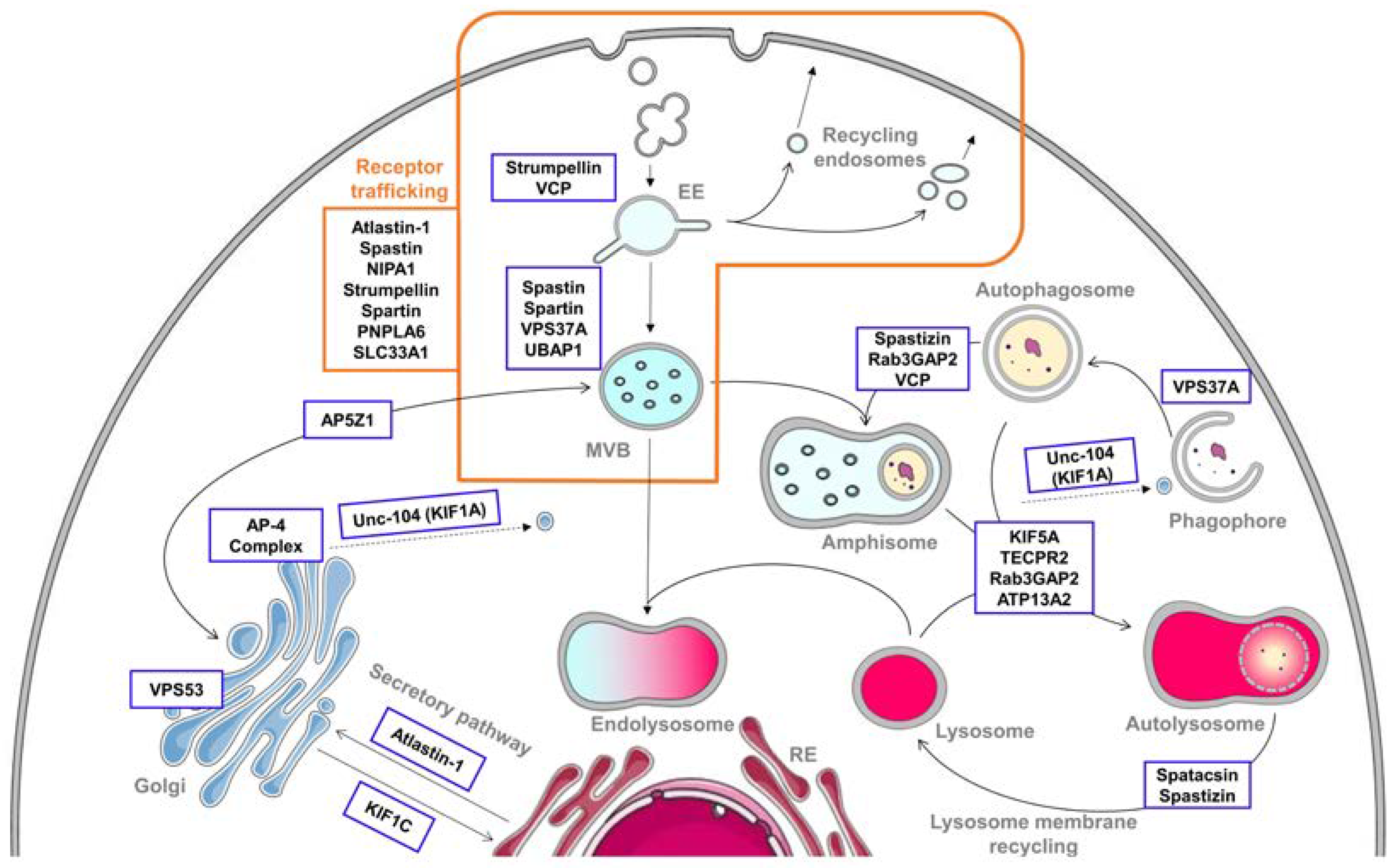
The endolysosomal pathway is an important cellular process that has been highly conserved during evolution. It allows the internalization of cargoes and their delivery to their intended destination, including all intracellular compartments, the plasma membrane and the extracellular compartment [1]. The first step of the endolysosomal pathway is endocytosis, which allows for the internalization of macromolecules, receptors and surface proteins [2]. Once internalized from the plasma membrane, the newly formed vesicles fuse with the early endosomes (EEs). Within the EEs, various cargoes’ fates can be decided. The cargoes can be directly recycled back to the plasma membrane, sent to the trans-Golgi network (TGN) or delivered to lysosomes for degradation [1,3,4]. Therefore, the endolysosomal pathway controls the localization and levels of hundreds of proteins in the cell. Consequently, this pathway is essential for regulating major cellular functions, such as cell signaling, establishment and maintenance of cell polarity, and cell migration.
It has been shown that the endolysosomal system is interconnected with another important cellular process, namely autophagy [5]. Autophagy is a key cellular process providing vital nutrients to the cell in response to various forms of stress, such as starvation, amino acid depletion and metabolic stress. This process is also involved in the degradation of defective cellular components, such as misfolded proteins and defective organelles, representing a potential danger for the cell. To answer the variety of inducing signals, different types of autophagy can take place, including macroautophagy, microautophagy and chaperone-mediated autophagy [6,7]. Macroautophagy can be described as the sequestration of cytoplasmic contents into a double-membrane vesicle called autophagosome and the degradation of the cytoplasmic content by fusion of the autophagosome with lysosomes to form an autolysosome [8]. In the context of this review, we decided to focus mainly on macroautophagy, which we will hereafter refer to as autophagy.
Due to their importance in many cellular functions, alterations of both endolysosomal and autophagic pathways have been associated with diseases, especially neurodegenerative conditions [7,9]. In particular, we will focus on hereditary spastic paraplegia (HSP), a family of motor neuron diseases in which numerous causative genes have been linked to these pathways. We believe these diseases are good illustrations of the difficulty to establish phenotype–genotype correlations: how altering these pathways at different steps can lead to similar consequences, while sometimes the alteration of a single protein can lead to different neurodegenerative diseases.
HSPs constitute a large family of clinically and genetically heterogeneous neurological disorders [10,11,12]. Patients present with the core clinical features of lower-limb spasticity and weakness due to degeneration in the corticospinal tracts. Clinically, there are “pure” and “complex” forms of HSPs, depending on the absence or presence of other neurological signs. Genetically, more than 70 causative genes have been described up to now, most of which are related to a relatively small number of pathological processes [11], among common genetic networks [13]. The endolysosomal and autophagic pathways are overrepresented with 24 genes whose mutations are directly involved in HSP (Table 1 and Figure 1). Multiple steps of those pathways are impaired in these disorders, not only directly by HSP protein loss of function but also as secondary consequences of the alteration of other systems. In this review, we will summarize current knowledge of the direct involvement of HSP proteins in the endolysosomal and autophagic pathways. A better understanding of the impaired cellular functions leading to HSP could help to decipher their functional organization and regulation and could also open opportunities for common therapeutic options for the various HSPs.
2. Alteration of the Endolysosomal Pathway in HSP
2.1. Endocytosis and Endosomes’ Dynamic
Endocytosis is crucial for uptake of nutrients and cell surface receptors, and for several other cellular processes [3]. Endocytosis is initiated with the invagination of the plasma membrane, leading to the formation of vesicles containing cargoes. There is diversity in the type of endocytic machinery, which classically differentiates the so-called “clathrin-dependent” or “clathrin-independent” pathways according to the requirement for the coat protein, clathrin [2].
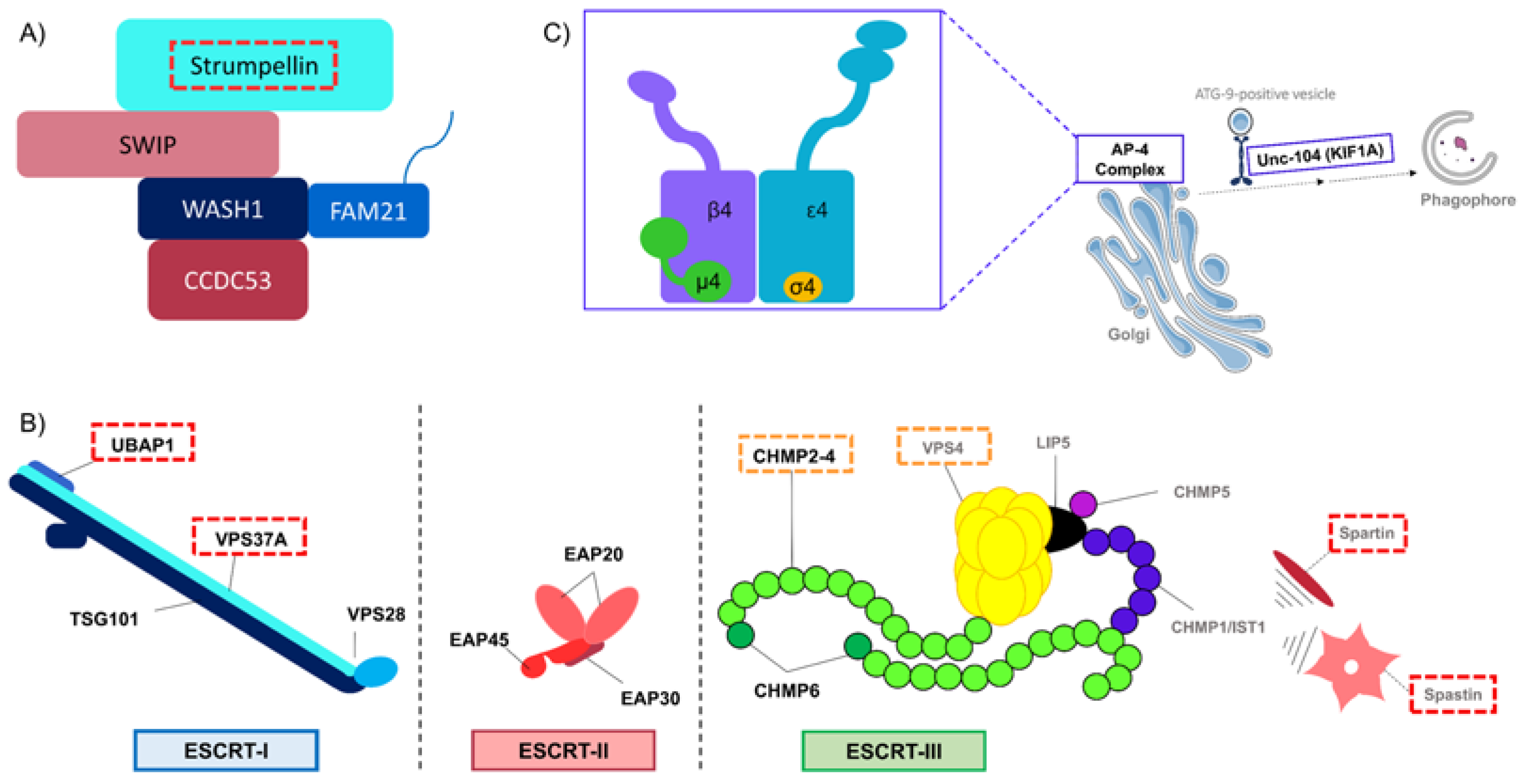
One of the clathrin-independent pathways is the caveolae and caveolin1 (CAV1) dependent pathway. CAV1 is enriched in caveolae, small invaginations of the plasma membrane that have various cellular functions, particularly lipid regulation via the ganglioside GM1 [2,14]. Of note, ganglioside dynamics are affected indirectly in SPG11 (for Spastic Paraplegia Gene 11), a frequent autosomal recessive subtype of HSP [15], through accumulation of GM2 species in lysosomes [16], and directly in SPG26, another HSP form, because of the loss of function of one key enzyme of ganglioside metabolism, B4GALNT1 [17]. More interestingly, the CAV1-dependent pathway is affected by mutations in the SPG8 gene, which cause a pure form of spastic paraplegia [18]. SPG8 encodes for the strumpellin protein, also called WASHC5, a member of the hetero-pentameric Wiskott–Aldrich syndrome protein and SCAR homolog (WASH) complex. WASH is composed of five subunits: WASH1, strumpellin, FAM21, KIAA1033(SWIP) and CCDC53 (Figure 2A) [19,20]. WASH activates the Arp2/3 complex on the surface of endosomes. Arp2/3 organizes the actin network and is required for endosome tubulation and fission [21]. WASH is involved in different steps of the endosomal tubulation and sorting dynamics. Thus, WASH depletion can lead to a wide variety of phenotypes, ranging from exaggerated tubulation to abolishment of tubulation associated with endosome enlargement [22]. The SPG8 mutations do not affect strumpellin interaction with the WASH complex or the WASH complex localization in the cell. However, in cells depleted in strumpellin or with mutated strumpellin, endosomal tubulation is impaired and an abnormal lysosomal morphology can be observed [23,24,25]. Moreover, strumpellin interacts with CAV1 and is necessary for maintenance of CAV1 abundance. Strumpellin-depleted cells have reduced levels of CAV1 due to an increase in its degradation. Indeed, CAV1 is ubiquitinated and targeted to lysosomes for degradation, but when protein degradation at lysosomes is inhibited, CAV1 abundance is restored in strumpellin-depleted cells. Thus, maintenance of the CAV1 protein level depends on strumpellin activity. The perturbation of caveola endocytosis in the case of strumpellin deficiency is certainly a primary event of SPG8 pathogenesis [25].

Table 1.
HSP proteins involved in the endolysosomal system and autophagy. The cellular consequences mentioned here are only in the context of this review, and therefore only include the function of the protein in the endolysosomal and autophagic pathways. All the references to the SPG numbers and their clinical involvement can be found in Hedera et al. or Boutry et al. [11,12].
| SPG | Gene | Protein | Relevant Section in the Text | Contribution in the Pathway | Cellular Consequences of Mutation/Depletion | References |
|---|---|---|---|---|---|---|
| SPG3A | ATL1 | Atlastin-1 | 2.2 Receptor trafficking 2.3 Secretory pathway | Regulation of BMP signaling ER-Golgi trafficking | Overactivation of BMP signaling Impairment of ER-Golgi trafficking and Golgi morphogenesis | [26,27,28,29] |
| SPG4 | SPAST | Spastin | 2.1 Endocytosis and endosomes’ dynamic 2.2 Receptor trafficking | Regulation of ESCRT-III | Perturbation of endosomal tubulation Overactivation of BMP signaling | [30,31,32,33,34] |
| SPG6 | NIPA1 | NIPA1 | 2.2 Receptor trafficking | Regulation of BMP signaling | Overactivation of BMP signaling | [28,35,36] |
| SPG8 | KIAA0196/ WASHC5 | Strumpellin/ WASHC5 | 2.1 Endocytosis and endosomes’ dynamic 2.2 Receptor trafficking | Member of WASH complex | Perturbation of clathrin-independent pathway Impairment of endosomal tubulation | [23,24,25] |
| SPG10 | KIF5 | KIF5A | 3.2 Autophagosome—lysosome fusion | Kinesin, motor protein | Impairment of the axonal transport and autophagic flux | [37] |
| SPG11 | SPG11 | Spatacsin | 3.3 Lysosome membrane recycling | Recruitment of Dynamin Interacts with spastizin and AP-5 | Autophagy defects due to reduction of autolysosome tubulation Accumulation of autophagic compartments Defective lysosomal clearance of gangliosides | [16,38,39,40,41] |
| SPG15 | ZFYVE26 | Spastizin | 3.3 Lysosome membrane recycling 3.4 Crossroads between endocytic and autophagic pathways | Interacts with spatacsin and AP-5 Interaction with Rab5A and Rab11 | Autophagy defects due to reduction of autolysosome tubulation Accumulation of autophagic compartments Altered maturation of autophagosomes | [38,39,40,42,43] |
| SPG20 | SPART | Spartin | 2.1 Endocytosis and endosomes’ dynamic 2.2 Receptor trafficking | Regulation of ESCRT-III | Perturbation of endosomal trafficking Overactivation of BMP signaling | [44,45,46,47,48,49] |
| SPG30 | KIF1A | KIF1A/Unc-104 | 3.1 Autophagosome biogenesis | Kinesin, motor protein | Impaired transport of ATG-9-positive vesicles leading to defects in autophagosome biogenesis | [50] |
| SPG39 | PNPLA6 | PNPLA6 | 2.2 Receptor trafficking | Regulation of BMP signaling | Overactivation of BMP signaling | [51] |
| SPG42 | SLC33A1 | SLC33A1 | 2.2 Receptor trafficking | Regulation of BMP signaling | Overactivation of BMP signaling | [52] |
| SPG47 | AP4B1 | AP4B1 | 2.3 Secretory pathway 3.1 Autophagosome biogenesis 3.3 Lysosome membrane recycling | Subunit of AP-4 complex | Impairment of ATG9A’s sorting and thus autophagosome biogenesis | [53,54,55] |
| SPG48 | AP5Z1 | AP5Z1 | 3.3 Lysosome membrane recycling 3.4 Crossroads between endocytic and autophagic pathways | AP-5 subunit spatacsin and spastizin interactor | Reduction of autolysosome tubulation Impaired endolysosomal system due to accumulation of endolysosomes Impairment of CIMPR trafficking towards TGN | [56,57,58,59] |
| SPG49 | TECPR2 | TECPR2 | 3.2 Autophagosome—lysosome fusion | Interactor of HOPS and ATG8 family members | Accumulation of autophagosomes due to impaired autophagosome—lysosome fusion | [60,61] |
| SPG50 | AP4M1 | AP4M1 | 2.3 Secretory pathway 3.1 Autophagosome biogenesis 3.3 Lysosome membrane recycling | Subunit of AP-4 complex | Impairment of ATG9A’s sorting and thus autophagosome biogenesis | [53,54,55] |
| SPG51 | AP4E1 | AP4E1 | 2.3 Secretory pathway 3.1 Autophagosome biogenesis 3.3 Lysosome membrane recycling | Subunit of AP-4 complex | Impairment of ATG9A’s sorting and thus autophagosome biogenesis | [53,54,55] |
| SPG52 | AP4S1 | AP4S1 | 2.3 Secretory pathway 3.1 Autophagosome biogenesis 3.3 Lysosome membrane recycling | Subunit of AP-4 complex | Impairment of ATG9A’s sorting and thus autophagosome biogenesis | [53,54,55] |
| SPG53 | VPS37A | VPS37A | 2.1 Endocytosis and endosomes’ dynamic 3.1 Autophagosome biogenesis | Subunit of ESCRT-I | Perturbation of endosomal sorting Altered capacity to recruit ESCRT-I subunits at the PAS leading to impaired autophagosome closure | [62,63] |
| SPG58 | KIF1C | KIF1C | 2.3 Secretory pathway | Kinesin, motor protein | Impairment of Golgi-ER transport | [64,65,66] |
| SPG69 | RAB3GAP2 | Rab3GAP2 | 3.4 Crossroads between endocytic and autophagic pathways | Subunit of Rab3GAP complex | Autophagy defects | [67,68] |
| SPG78 | ATP13A2/ PARK9 | ATP13A2 | 3.2 Autophagosome—lysosome fusion | Still unclear | Autophagy defects due to accumulation of autophagic compartments | [69,70] |
| SPG80 | UBAP1 | UBAP1 | 2.1 Endocytosis and endosomes’ dynamic | Subunit of ESCRT-I | Perturbation of endosomal sorting | [71,72] |
| \ | VCP | VCP | 2.1 Endocytosis and endosomes’ dynamic 3.4 Crossroads between endocytic and autophagic pathways | Interaction with strumpellin Autophagosome maturation | Perturbation of strumpellin localization and function Autophagy defects | [73,74,75,76] |
| \ | VPS53 | VPS53 | 2.3 Secretory pathway | Subunit of GARP complex | Still unclear | [77] |
Interestingly, the gene encoding for the protein valosin-containing protein (VCP), an interactor of strumpellin, can cause several neurodegenerative diseases such as amyotrophic lateral sclerosis (ALS), Charcot–Marie–Tooth (CMT) disease and notably HSP, when mutated [73,74,75,78,79]. VCP is a ubiquitously expressed AAA–ATPase protein with multiple cellular functions that include vesicular trafficking and protein degradation. Although VCP function is still unclear, its interaction with strumpellin could be necessary for normal functioning of strumpellin. Mutant VCP may then alter strumpellin function and even sequester it, since aggregates of VCP and strumpellin have been found in muscles biopsies from patients with VCP mutations [75]. Moreover, VCP-positive inclusion bodies have been found in several neurodegenerative diseases, such as Parkinson’s disease, amyotrophic lateral sclerosis (ALS) and spinocerebellar ataxia type 3. Additionally, strumpellin has been identified in pathological protein aggregates in inclusion body myopathy with Paget disease and frontotemporal dementia (IBMPFD), various myofibrillar myopathies and in cortical neurons of a Huntington’s disease mouse model [75,80].
The degradation pathway starts with the formation of late endosomes from the maturation of early endosomes. Cargoes that will be degraded are contained in intraluminal vesicles (ILV). Aggregation of several ILVs leads to the formation of a late endosome also called multivesicular body (MVB). The ubiquitination of ILVs is the key signal for their degradation. Indeed, ubiquitinated cargoes are trafficked and delivered to MVBs and lysosomes for degradation, whereas non-ubiquitinated ones are sorted and transported to the TGN or recycled back to the plasma membrane. The detection of the ubiquitination, the packaging of the cargoes into ILVs and the formation of the vesicles are mediated by the multiprotein complexes belonging to the endosomal sorting complexes required for transport (ESCRT) family [1,81]: ESCRT-0, -I, -II and –III (Figure 2B) [82,83,84]. The ESCRT system has been shown to play a central role in intracellular trafficking.
We know that defects in the ESCRT system can cause various neurodegenerative diseases and HSPs are no exception. For instance, mutations in two subunits of ESCRT-I lead to HSP. ESCRT-I composition can change depending on the cellular context and can be composed of VPS37A, UBAP1, TSG101 and VPS28 [85]. Indeed, mutation in SPG53, encoding VPS37A, causes a complex HSP [62] and mutation in SPG80, encoding UBAP1, causes a pure HSP [71,72]. Variation in clinical symptoms might suggest an additional role for these proteins besides involvement in ESCRT-I, notably a function in autophagy for VPS37A (see autophagy section), or redundancy. UBAP1 interacts directly with ubiquitin on endosomes and thus participates in driving cargoes into the degradation pathway. Studies on mouse hippocampal neurons have shown that mutation in Ubap1 alters UBAP1 recruitment to endosomes and then its function in endosomal sorting [71]. Similar results were obtained in cortical neurons derived from transgenic Ubap1 flox mice in which disruption of UBAP1 leads to dysregulation of both early-endosome processing and ubiquitinated protein sorting [72]. Moreover, in zebrafish, lack of UBAP1 leads to abnormal morphology, impaired motor neuron outgrowth and therefore decreased mobility [72]. Therefore, perturbation of the detection of cargoes fated to degradation seems deleterious for cells, and specifically for neurons, in HSP.
Unlike the other ESCRT complexes, ESCRT-III is not localized to endosomes but exists in an autoinhibited state in the cytoplasm. This complex is made up of at least four subunits, all members of the same protein family: “charged multivesicular body proteins” (CHMP). Activation of ESCRT-III occurs when the ESCRT-II subunit EAP20 (VPS25) binds to CHMP6 (VPS20), initiating ESCRT-III recruitment to the endosomes and complex formation [85]. ESCRT-III is responsible for the final sorting of cargoes and plays a direct role in the generation of luminal vesicles. ESCRT-III filaments surround the cargo, and its assembly induces the dissociation of the other ESCRT complexes. While ESCRT-III assembly stabilizes the membrane neck of a growing ILV, VPS4 binding to ESCRT-III subunits constricts the neck and facilitates its release from the membrane [86,87]. Alterations in ESCRT-III functions are known to induce neurological disorders. Indeed, mutations in CHMP2B, a gene encoding one of the four major proteins of ESCRT-III complex, can cause frontotemporal dementia (FTD), ALS or FTD-ALS [88,89,90,91]. In addition, mutations in the gene encoding VPS4A are found in patients with a multisystem disease with abnormal neurodevelopment [92].
Interestingly, the most common HSP genetic entity is due to mutations in SPG4, encoding spastin, which interacts with CHMP1B, a member of ESCRT-III [30]. The interaction occurs in a region containing an N-terminal microtubule interacting and trafficking (MIT) domain. This domain is shared with several proteins that have defined roles in membrane trafficking, including VPS4. Spastin’s MIT domain seems to be especially important since mutations in this domain lead to impaired endosomal tubule fission, altered endosome-to-Golgi Mannose-6-Phosphate Receptor (M6PR) traffic and abnormal lysosomal morphology [31,93]. In addition, spastin interacts with IST1, an ESCRT-III-like protein, and the lack of IST1 in cells also leads to an increase in endosomal tubulation. Moreover, abnormal endosomal tubulation has also been observed in spinal motor axons of zebrafish depleted of spastin or IST1. Furthermore, altering endosomal tubulation causes disturbance in receptor sorting and trafficking, such as BMP or transferrin receptors [32,33]. These results support the idea that spastin plays a role in intracellular transport and membrane trafficking.
Another HSP, caused by mutations in SPG20, could be associated with the ESCRT system. The encoded protein, spartin, localizes to endosomes [44,45] and also has an MIT domain, predicting a role in endosomal trafficking [46]. In addition, spartin interacts with the spastin interactor, IST1. Spartin seems to be an inhibitor of IST1 activity, possibly by preventing its interaction with spastin. Overexpression of IST1 in primary rat cortical neurons induces an increase in the number of axon branches, whereas overexpression of spartin in Spg20−/− primary neurons significantly decreases the number of axon branches [47]. However, the mechanisms underlying ESCRT-mediated effects on axon branching still need to be deciphered. Spartin also interacts with alsin, which has an important role in motor neuron integrity and accounts, in the case of loss of function mutations, for another motor neuron disease, ALS, and more rarely for HSP phenotypes [94].
Once the molecules destined to be degraded have been sorted, the MVBs fuse with lysosomes to form endolysosomes. In this compartment, the degrading enzymes formerly present in lysosomes will therefore be able to degrade the molecules contained in MVBs, marking this step as the end of the endolysosomal degradation pathway [95].
2.2. Receptor Trafficking Impairment
As we have seen, alteration of endosome tubulation is a recurrent pattern in HSP and has a direct impact on the fate of cargoes. Perturbation of sorting and recycling of cargoes, notably receptors, could be a key pathological mechanism of several HSPs. Indeed, failure of endosomal tubule fission can cause defective sorting of various receptors. For example, in strumpellin-depleted cells, the subcellular distribution of β-2-adrenergic receptors is altered [24]. Furthermore, spartin interacts with endocytic trafficking protein EPS15 and plays a role in intracellular trafficking of epidermal growth factor receptors (EGFR) [48].
More interestingly, the bone morphogenic protein (BMP) signaling pathway has progressively emerged as a common pathogenic mechanism in at least six HSP subtypes: SPG6 (NIPA1), SPG3A (atlastin-1), SPG4 (spastin), SPG20 (spartin), SPG39 (PNPLA6), SPG42 (SLC33A1) [26,27,34,35,36,49,51,52,96]. BMP constitutes a group of phylogenetically conserved growth factors and belongs to the transforming growth factor beta (TGFβ) superfamily [97]. First detected in extracts of bones, the BMP signaling pathway has a critical role in the nervous system. Initially, BMPs inhibit proliferation of neural precursors and promote the first steps of neuronal differentiation. Furthermore, in post-mitotic cells, they modulate neuronal subtype specification, promote dendritic and axonal growth and induce synapse formation and stabilization [98,99].
The first link between HSP and BMP signaling was made in 2007. Indeed, loss of spichthyin (SPICT), the Drosophila melanogaster orthologue of NIPA1, leads to upregulation of BMP signaling. SPICT localizes with early endosomes, interacts with BMP receptors, promotes their internalization from the plasma membrane and, finally, leads to BMP signaling inhibition [35]. In mammalian cells, NIPA1 physically interacts with the type II BMP receptor (BMPRII) and promotes its endocytosis and lysosomal degradation [36]. Mutations in ATL1, encoding for atlastin-1,have been found as causative in a subtype of HSP, SPG3A, and in hereditary sensory and autonomic neuropathy (HSAN) [12,100]. This HSP related protein strongly interacts with NIPA1 and BMPRII [28]. Even if atlastin-1 expression is not necessary for trafficking of BMPRII, the presence of mutant forms of atlastin-1 results in a complete absence of BMPRII from the plasma membrane and its clustering in the cytoplasm, suggesting a dominant negative effect [27]. Moreover, knockdown of Atl1 gene (coding for atlastin-1) in zebrafish induces a loss of motility of the zebrafish larvae, associated with impairment of branching axons. Overexpression and knockdown studies have shown that atlastin-1 inhibits BMP signaling. Atlastin-1 co-localizes with type I BMP receptors (BMPRI) in endosomal structures along neurites implying a role in BMP receptor trafficking regulation [26].
The HSP proteins spastin and spartin are inhibitors of BMP signaling. How they participate in the modulation of BMP signaling is not absolutely known, but both are involved in endosomal tubulation, which is associated with trafficking and recycling of receptors. A study in zebrafish depleted in spastin has shown motor circuit formation alterations and have linked a part of these defects to BMP signaling over-activation [34]. In Drosophila, spartin is required for downregulation of the BMP receptor Wit, which initiates a retrograde signal controlling microtubule stability and synaptic growth. In this model, both spartin depletion and elevated BMP signaling cause progressive neurodegeneration [49]. Finally, an upregulation of BMP signaling has also been noted in a mouse model of SPG42 (SLC33A1) [52] and in Pnpla6 (SPG39) knockdown in zebrafish [51]. The links between these two proteins and BMP signaling regulation need further investigation.
In conclusion, all these studies show a strong link between receptor trafficking, particularly involved in BMP signaling regulation, and HSP. BMP signaling has a critical role in motor circuit architecture and stability and small alterations in this pathway can cause major effects on motor axons and finally lead to neurodegeneration more or less associated with abnormal neurodevelopment as shown in some animal models.
2.3. Secretory Pathway
The Golgi apparatus in eukaryotic cells is composed of flattened and fenestrated membrane disks, called cisternae. Several cisternae, between 3 and 20, are aligned in parallel to form a stack. The Golgi stack is subdivided in three compartments: cis, medial and trans. Two networks responsible for the main tasks of this apparatus can be identified. The cis-Golgi network (CGN) is situated closest to the endoplasmic reticulum (ER) and is responsible for receiving ER-derived transport vesicles and shipping ER resident proteins back to the ER. On the contrary, the cisterna in the most trans position is continuous with a tubular, branching and reticulating compartment termed the TGN. The TGN is involved in the final stage of sorting, packing and delivering of most secretory proteins to their destinations. Moreover, cargoes from the early and late endosomes are targeted to the TGN via the retrograde route. Therefore, the TGN has conventionally been viewed as the main cargo sorting station where proteins and lipids are sorted and targeted to various downstream destinations [4,101].
Once the endosome-derived transport carrier reaches the proximity of the TGN, the carrier-Golgi interaction is mediated by tethers, one of them being the Golgi-associated retrograde proteins (GARP) complex. GARP is a heterotetrameric-tethering complex and consists of four subunits: VPS51, VPS52, VPS53 and VPS54 [102]. Dysfunction of the GARP complex leads to defects in both retrograde trafficking and anterograde trafficking. Mutations in the gene encoding the VPS54 subunit, which lead to a drastic reduction in levels of VPS54 and disturb the GARP complex assembly, result in spinal muscular atrophy in the “wobbler” mouse that is considered as a model for ALS [103]. Recently, mutations in two others GARP complex subunits, VPS53 and VPS51, have been associated with a complex form of HSP and a neurodevelopmental disorder in patients, respectively, demonstrating the importance of this complex in the pathway [77,104].
Cytosolic cargo adaptors play a fundamental role in protein sorting at the TGN. Five adaptor proteins (APs) have been identified in eukaryotic cells, and three of them (AP-1, AP-3 and AP-4) are involved in protein sorting at the TGN [105]. Each AP complex is formed of four subunits. The AP-4 complex is composed of four subunits encoded by four different genes: AP4E1, AP4B1, AP4M1 and AP4S1 (Figure 2C) [50,53,54,55,106]. Mutations in the genes encoding all subunits of AP-4 lead to severe neurodevelopmental forms of HSP (SPG47, SPG50, SPG51 and SPG52, respectively) [107,108,109,110,111]. In this context, mutations of AP-4 subunits mainly lead to autophagy defects that will be detailed in the autophagy section of this review.
Proteins destined for secretion, plasma membrane or intracellular organelles are synthesized in the ER and transported along the secretory pathway through the Golgi complex by coated vesicular carriers. These vesicles fuse with the CGN. In the opposite way, this flow of membranes needs to be counterbalanced by a reverse membrane transport pathway from Golgi to ER. This involves vesicles traveling along microtubules. Interestingly, mutations in proteins involved in Golgi-ER (or ER-Golgi) traffic can lead to HSP. Indeed, mutations in the gene encoding atlastin-1 (SPG3A), another HSP protein localized with cis-Golgi and ER markers, affect trafficking at the ER/Golgi interface and lead to impaired Golgi morphogenesis [29]. Similarly, mutations in a gene of the kinesin family, KIF1C, cause a complex HSP (SPG58) [64,65]. Little is known about the precise functions of the KIF1C protein but it is associated with the Golgi membranes and required for the retrograde transport of Golgi vesicles to the ER [66].
Finally, the ER is a major organelle involved in numerous cellular functions and is widespread in cells through a network of interconnected sheets and tubules. This network allows the ER to communicate with the other organelles and contribute to their functions, notably to obtain effective endocytic and endolysosomal pathways [112]. Among HSP proteins, many have been directly implicated in ER dysfunctions. In particular, the most represented ones are: atlastin-1 (SPG3A), reticulon-2 (SPG12), REEP1 (SPG31), REEP2 (SPG72) and erlin-2 (SPG18/37). Their precise involvement in those processes will not be dealt with here since it has already been extensively described recently [11,113,114,115].
3. Autophagy Defects in HSP
3.1. Autophagosome Biogenesis
Upon autophagy induction, Autophagy-related (ATG) proteins are recruited to the location of the defective material, called the phagophore assembly site (PAS), to initiate the formation of the phagophore. The phagophore can be described as a double-membrane saccule composed of isolation membranes budding from the ER and other sources of membranes [7,116]. The initiation step of phagophore formation is followed by a nucleation step and an elongation step, leading to the formation of a fully closed autophagosome sequestering the intracellular material [117]. Many ATG proteins are implicated in the initiation, nucleation and elongation of the phagophore. Notably, the arrival of ATG9-containing vesicles at any of these steps promotes the input of additional membrane material (lipids, proteins) to form the autophagosome [116].
ATG9, ATG9A in mammals, is a transmembrane protein belonging to the core machinery of autophagosome formation. When autophagy is not induced, this protein mainly localizes to the TGN and partly to the endosomes. However, upon autophagy induction, ATG9A is delivered to the PAS, by the intermediary of vesicles, to regulate the elongation of the phagophore [118]. As shown above, each of the four subunits of AP-4 complex has been involved in a complex form of HSP. The AP-4 complex is specific to the TGN, where it would allow the sorting of transmembrane proteins into cargoes and the recruitment of their transport machineries for their delivery to their destined localization [53,54]. In various cellular types, the depletion of each of the AP-4 subunits leads to the mislocalization of ATG9A with its retention at the TGN and its depletion in the periphery [53,54]. AP-4-dependent sorting and localization of ATG9A would be especially important in neuronal cells since an impairment in basal and stress-induced autophagy has been observed in iPSC-derived cortical neurons from SPG47 patients but not in fibroblasts derived from patients mutated in AP-4 subunits [53]. Consistently, a reduction of the axonal autophagosome formation with ER accumulation in axonal swellings has been observed in neurons of the Ap4e1−/− mouse model [55]. Of note, AP-4 is not only involved in ATG9A sorting but takes charge of other cargoes, such as the AMPA-type glutamate receptor, which has been reported with impaired trafficking in the Ap4b1−/− mouse model. However, the fact that the phenotype of the Ap4e1−/− mouse model is similar to the Atg9−/− mouse model, even if less severe, suggests an important role of ATG9A trafficking in AP-4-linked HSP [54]. This is also supported by the potential involvement of another HSP protein, KIF1A, in ATG9A trafficking. ATG9A-positive vesicles must be transported to their sites of action and especially to the axon terminals where ATG9A is highly localized [119]. It has been shown that in C. elegans, Unc-104, the orthologue of the mammalian KIF1A member of the Kinesin-3 family, is involved in the transport of ATG-9 to neurites [50]. Pathological variants in the gene encoding for KIF1A are involved in the development of autosomal recessive and dominant spastic paraplegia, namely SGP30 [120,121]. Of note, mutations in this gene can also give rise to a subtype of HSAN [100]. In vertebrates, ATG9A has not yet been identified as a cargo of KIF1A but could be an interesting candidate [122].
After its formation and elongation, the phagophore has to be closed to form the autophagosome but this process is still relatively unknown. However, it was recently shown that components of the ESCRT-I and ESCRT-III machineries, implicated in endosome tubulation as seen before, are involved in autophagosome closure [63]. Among the ESCRT proteins required, VPS37A (SPG53) would be involved in the promotion of autophagosome closure by recruiting the ESCRT-III subunit CHMP2A and the AAA ATPase VPS4 [63].
3.2. Autophagosome–Lysosome Fusion
Once the autophagosome has formed, a maturation step takes place. To be able to fuse, the autophagosome and lysosomes have to encounter one another in the cell despite their generally different cellular positions [123]. Bi-directional transport of both organelles then has to take place. Autophagosomes and lysosomes are transported by a kinesin-dependent plus-end transport, notably the kinesin-1/KIF5 [123,124]. Three isoforms of KIF5 exist: KIF5A, KIF5B and KIF5C [125]. The ubiquitously expressed subtype KIF5B has been shown to be largely involved in autophagy and autophagic lysosome reformation (ALR) [126,127]. KIF5A is a neuronal subtype of KIF5 that is involved in various allelic diseases when a mutation occurs in its corresponding gene: a form of HSP, SPG10, as well as ALS and CMT disease [125,128,129,130,131]. KIF5A has been mostly implicated in axonal transport of neurofilaments, mitochondria and amyloid precursor protein [132,133,134,135]. In addition to those roles, a recent study demonstrated that KIF5A is reduced in Neuro-2a cells treated with a neurotoxicity-inducing drug. That deficit would lead to an impairment of the autophagic flux due to a KIF5A-dependent axonal transport deficiency, suggesting that KIF5A could be important in autophagy [37].
The proteins of the ATG8 family, notably composed of the LC3 (light chain 3) proteins, are necessary to recruit interactors to initiate the transport, tethering and fusion of the autophagosome with lysosomes [123,136]. Among the necessary actors can be found the homotypic fusion and vacuole protein sorting (HOPS) tethering complex which is composed of six subunits: VPS39, VPS11, VPS18, VPS16, VPS33 and VPS41. This complex allows the Soluble N-ethylmaleimide-sensitive factor attachment protein receptors (SNARE)-mediated fusion between the outer membrane of the autophagosome and the lysosomal membrane. Notably, the syntaxin 17 (STX17) and synaptosomal-associated protein 29 (SNAP29) autophagosomal SNAREs and the lysosomal vesicle-associated membrane protein 8 (VAMP8) are involved in this process [60,116,137]. Mutations in three subunits of the HOPS complex, VPS11, VPS16 and VPS41, have been linked to neurological disorders. For example, patients mutated in VPS11 present with signs of hypomyelination that can be associated with leukoencephalopathy [138,139,140,141]. Mutations in VPS11 could also give rise to dystonia as observed in patients mutated in VPS16 and VPS41, associated with ataxia for the latter [141,142,143,144,145,146]. Of note, mutations in SNAP29 are also involved in neurological disorders since they can lead to the development of the cerebral dysgenesis, neuropathy, ichthyosis, and keratoderma (CEDNIK) syndrome [147,148,149].
The tectonin beta-propeller repeat containing 2 (TECPR2) protein has been identified as an interactor of the ATG8 family and of the HOPS complex [150]. Mutations in the gene coding for TECPR2 have been identified in a form of HSP, SPG49, and have been proposed as involved in a subtype of HSAN [12,151]. A link between SPG49 mutations and autophagy has been made by the observation of a defect in autophagy in SPG49 patient-derived fibroblasts [152]. Recently, it has been proposed that TECPR2 regulates autophagosome–lysosome tethering and fusion by binding the autophagosome with its LC3-interacting region (LIR) and its interaction with HOPS [60]. The precise molecular function of TECPR2 in this model still has to be explored but it is consistent with the observation of autophagosome accumulation in the brain and spinal cord of the Tecpr2−/− mouse model, suggesting an altered delivery of autophagosomes to lysosomes [61]. Interestingly, neurons of the Tecpr2−/− mouse model display age-dependent axonal spheroids or swellings with accumulation of autophagic material [61].
Another HSP protein could be involved in this process. ATP13A2 is a lysosomal transmembrane P5B-type ATPase localized at acidic compartments, which is notably involved in SPG78 and Parkinson’s disease [69,153,154,155]. Fibroblasts from patients carrying mutations in ATP13A2 present signs of autophagic pathway defects with an accumulation of lysosomal structures and abnormal lysosomal storage material [69]. The precise molecular involvement of ATP13A2 in autophagy is still unclear but a recent study in mouse embryonic fibroblasts (MEFs) of Atp13a2−/− mouse and ATP13A2-depleted HeLa cells suggests that it is required for autophagosome–lysosome fusion. In these cells, ATP13A2 depletion leads to both autophagosome and lysosome accumulation, indicating an autophagy defect that would notably lead to a defect in clearance of damaged mitochondria [70].
3.3. Lysosome Membrane Recycling
After formation of the autolysosome, the hydrolases and acidic environment provided by the lysosome will allow the degradation of the internal membrane and of the autophagosomal cargoes [116]. Following this fusion, the final step of the autophagic process is the recycling of some lysosomal components to allow the formation of a new pool of lysosomes, either by vesiculation in conditions of basal autophagy [38,156] or by tubulation in ALR in conditions of stress-induced autophagy [157,158]. Several HSP proteins have been implicated at different levels in the lysosomal reformation, notably spatacsin (SPG11), spastizin (SPG15), AP5Z1 (SPG48) and the AP-4 complex [38,56,57,159].
Spatacsin and spastizin are frequently mutated proteins in autosomal recessive forms of HSP. Spatacsin is encoded by SPG11 and mutated in three allelic motor neuron diseases: SPG11, ALS and CMT disease [15,79,160,161,162]. Spastizin is encoded by ZFYVE26 and mutated in SPG15 [163]. At the protein level, SPG11 and SPG15 interact with each other, might have an interdependent expression and stability [39] and both have also been shown to stably interact with the adaptor protein complex 5 (AP-5) [164]. The molecular functions and localizations of spatacsin and spastizin are still debated [165,166,167,168]. Among the propositions, the localization of those proteins at the late endosome/lysosomal compartments is especially interesting and is supported by the fact that spastizin possesses a FYVE domain able to target proteins to intracellular membranes enriched for phosphatidylinositol 3-phosphate, such as endosomal membranes [169]. Both spatacsin and spastizin have notably been involved in ALR since their mutations lead to a phenotype associated with ALR defects [38]. In conditions of stress-induced autophagy, ALR starts by the formation of tubules containing lysosomal membrane components, which are extruded from the autolysosome. From those tubules will bud vesicles called proto-lysosomes after their scission, and these proto-lysosomes will then become functional after a maturation step [157,158]. The depletion of both spatacsin and spastizin leads to a reduced tubulation in HeLa cells, which prompted the proposition that these proteins are necessary for the initiation of the tubulation [38]. This involvement is consistent with the global observation of an accumulation of autophagosomes and/or lysosomal structures that can be accompanied by a depletion of free lysosomes in various models, including fibroblasts from SPG11 and SPG15 patients [38,39,42], brain sections of Spg11−/− and Spg15−/− mouse models [40] and MEFs of both Spg11−/− and Spg15−/− mouse models [40,41], even if some discrepancies can be noted between the different reports or models, including animal models. Moreover, spatacsin could have another role in a later step in ALR since the recruitment of dynamin at the lysosomal structures, involved in the proto-lysosome scission from the tubule [38], is decreased in Spg11−/− mouse neurons [16].
At least two Spg11−/− mouse models have been established, based on two different approaches [41,170]. An accumulation of autolysosomes in neurons of the Spg11−/− mouse model has been reported in the former [41]. In the latter, neurons of Spg11−/− mouse present enlarged lysosomes with an accumulation of gangliosides linked to an impaired lysosome membrane recycling, as seen in iPSC-derived human neurons from a cortical organoid model [16,170]. Interestingly, gangliosides accumulation has been linked to neurodegeneration, given that increased glutamate-induced neuronal death has been observed and that inhibition of gangliosides accumulation in lysosomes by treatment with miglustat, an inhibitor of glucosylceramide synthase, increases the neuronal survival and improves the motor phenotype induced by Spg11 knock-down in zebrafish [16].
In addition to their roles in starvation-induced ALR, spatacsin and spastizin could also have a role in lysosome reformation in conditions of basal autophagy since an accumulation of autolysosomes has also been observed under feeding conditions in spatacsin- or spastizin-depleted cells [38]. This process seems to be especially important for neuronal cells, which have a reduced reaction to starvation and rely more on basal autophagy [171]. Therefore, loss-of-function mutations in SPG11 and ZFYVE26 would lead to global lysosomal renewal defects.
3.4. Crossroads between Endocytic and Autophagic Pathways
As previously mentioned, autophagy and endocytosis are two key, deeply interconnected, cellular processes. Crossroads between them can be seen at different levels of the pathways, notably the fusion between late endosomes and autophagosomes to form hybrid structures called amphisomes. Once formed, the amphisome can fuse with a lysosome to lead to the formation of an autolysosome to allow the degradation of the contained material [172] (Figure 1). It has been proposed that fusion between endosomes and autophagosomes is involved in the maturation of the autophagosome and that functional MVBs are needed for efficient autophagy [173]. A defect in this process can lead to an autophagic defect that can be linked to neurodegenerative diseases [5]. Despite spatacsin and spastizin interaction, an additional role for spastizin has been proposed. Indeed, spastizin is involved in the maturation of the autophagosomes since its depletion leads to an accumulation of immature autophagosomes in SPG15 patients’ cells [42]. This defect would be due to the incapacity of the mutated spastizin to interact with Rab5A and Rab11, leading to a more pronounced autophagy defect [42,43]. Rab5A localizes to early endosomes and Rab11 localizes to recycling endosomes and MVBs, and both proteins are necessary for endosome–autophagosome fusion at different levels [43,174,175].
Crossroads between endocytic and autophagic pathways involving HSP proteins have also been reported recently. Indeed, mutations in the AP5Z1 gene, encoding for a subunit of the AP-5 complex, have also been identified as causative in SPG48 [164]. AP-5 stably interacts with spatacsin and spastizin and its expression is dependent on SPG11 and SPG15 gene expression but not reciprocally, therefore a participation of AP-5 in spatacsin and spastizin cellular functions is very likely [58,166]. Although a decrease in tubule formation from autolysosomes in MEFs of the Ap5z1−/− mouse has been observed, as in SPG11 and SPG15 models, in conditions of starvation-induced autophagy [56,57], it has been suggested that the main role of AP-5 is in the endolysosomal system. Indeed, an accumulation of enlarged lysosomal structures identified as enlarged endolysosomes with abnormal storage material has been reported in SPG48 patient-derived fibroblasts [58]. In addition, recent investigations showed that AP-5 knockdown impairs retrograde trafficking of the cation-independent mannose 6-phosphate receptor (CIMPR) from endosomal compartments towards the TGN, which may indicate that AP-5 also acts on an alternative retrograde trafficking route [59]. Interestingly, axonal swellings with clustering of organelles and LAMP1-positive structures have been observed in neurons of the Ap5z1−/− mouse model, suggesting its importance in neuronal cells [56].
Additionally, the protein VCP, involved in various neurodegenerative diseases, including HSP [73], is necessary for autophagosome maturation in basal autophagy and proteasome-inhibition conditions [76], in addition to its role in conjunction with strumpellin (SPG8) in endocytosis, as discussed above.
Finally, it has been suggested that Rab3GAP2, whose loss of function causes SPG69 [13,176], is involved in autophagy at different levels: autophagosome formation and autophagosome–autolysosome maturation [67,68]. Rab3GAP2 is the noncatalytic subunit of the heterodimeric Rab3GAP complex, which has been proposed to regulate Rab18 activity and autolysosome maturation. Indeed, autophagy defects are observed in a Rab3GAP2-deficient Drosophila model [68]. In contrast, another study has also suggested a role of Rab3GAP in autophagy but earlier in the pathway since it was shown that, in human primary fibroblasts, depletion in Rab3GAP2 led to an impaired lipidation of ATG8 family members necessary for autophagosome formation and the autophagy pathway [67]. Rab3GAP2 cellular functions are therefore still unclear but the available data in different models of diseases suggest that this protein could be involved in autophagy. Of note, mutations in Rab3GAP2 have also been involved in the Martsolf syndrome and mutations in Rab3GAP2, Rab3GAP1 and Rab18 can give rise to the Warburg micro syndrome (clinically overlapping with the Martsolf syndrome and characterized by brain, eyes and endocrine abnormalities), further showing a link between these three proteins [68,177,178].
4. Lessons and Perspectives
Defects in the endolysosomal system and autophagy are a common pathological mechanism in several diseases and especially in neurodegenerative diseases, such as Alzheimer’s disease, Parkinson’s disease, Huntington’s disease and ALS, as strongly highlighted by numerous reviews [179,180,181,182,183,184,185]. For example, in the case of Parkinson’s disease, the endolysosomal pathway has been pinpointed as a key cellular pathway in the pathological mechanism of the disease and mutations in the gene encoding the glucocerebrosidase, a lysosomal enzyme, represent one of the most common genetic risk in Parkinson’s disease [183,186].
HSPs belong to motor neuron diseases which are a group of heterogeneous diseases in which overlaps at the clinical and genetic levels can be found. These pathways are particularly involved in motor neuron diseases, since they are altered not only in HSP, but also frequently in ALS and CMT disease, which are both motor neuron diseases [185,187,188]. For example, mutations of the genes encoding KIF5A, VCP and spatacsin are involved in subtypes of HSP but also in ALS and CMT disease [78,79,130,131,162]. In addition, other proteins of the pathways, not mutated in HSPs, have been founded mutated in motor neuron diseases like CHMPB2 mutations in ALS [90,91].
The recurrence of the implication of these pathways in neurodegenerative diseases indicates that the nervous system is particularly sensitive to the disruption of the endolysosomal and autophagic systems. This can be explained by three main reasons. First, neurons, as post-mitotic cells, are not able to eject undesired material during cell division, which means that autophagy defects in these cells have particularly high impacts. Second, these pathways have additional specific functions in neuronal cells such as the maintenance of the synaptic process and membrane trafficking along the axon. Finally, the unique morphology of neurons exerts high demands on accurate and coordinated delivery of proteins and lipids to their final destination [117]. This point is critical in the pathophysiology of HSP where motor neurons are the most affected and their degeneration is responsible for the major clinical signs.
Because of the need for accurate regulation of those processes, any perturbation can give rise to a plethora of events. As we pointed out before, many HSP proteins impact the global membrane and protein trafficking in the cells. However, the nature of the alterations varies considerably. For example, they can be caused by a defect in receptor trafficking (e.g., BMP signaling), failure to deliver cargo protein to the correct destination (AP-4), the inability to degrade substrates by autophagy (TECPR2/SPG49), or impairment of lysosome formation (spatacsin/SPG11 and spastizin/SPG15). In neurons, these defects lead to similar pathological phenotypes of particular interest, such as the axonal swellings that have been observed in SPG4, SPG48 and SPG51 [55,56,189,190] or defects in neurite architecture that have been seen in SPG3A, SPG4, SPG6, SPG8, SPG11, SPG20 and SPG39 for instance [26,35,49,51,191,192,193]. A better comprehension of the involvement of the endocytic and autophagic pathways in the altered neuronal phenotype could help us understand the complexity of this continuum and therefore lead to the development of common therapeutics.
As of now, no curative treatment for any form of HSP is available on the market. A few therapeutics are used to relieve patients, mainly targeting a reduction of the spasticity. The mutations are often affecting not only one but multiple pathways and the interplay between the mitochondrion compartment, the microtubule cytoskeleton, the endolysosomal/autophagy pathway and the tubular endoplasmic reticulum seems critical in HSP pathogenesis [11]. Regarding the endolysosomal and autophagy systems, attempts to increase their efficiency have been tested in various models of neurodegeneration with variable success, particularly acting on mTOR and TFEB levels, but these did not include HSP [194,195]. Some drugs are currently in development for HSP forms related to these pathways and have been tested in vitro. For example, tideglusib, an inhibitor of GSK3 activity, has been tested on brain organoids derived from cells of SPG11 patients and show a rescue of the premature neurogenesis induced by SPG11 mutation [196]. In addition, drugs have also been tested on animal models of HSP, notably zebrafish. Miglustat, an inhibitor of the glucosylceramide synthase known to decrease the levels of GM2 gangliosides, has been tested on zebrafish with a knock-down of Spg11 and allows the partial rescue of the motor phenotype [16]. Additionally, modulation of BMP signaling is an interesting target. Dorsomorphin, a small molecule that blocks the kinase activity of BMP receptors, has been tested in zebrafish depleted in atlastin-1 and is able to rescue the motor phenotype [26]. However, these drugs have not yet reached clinical trials.
Thus, there is still an unmet need for therapeutics, and a clearer understanding of endolysosomal and autophagy pathways alterations may pinpoint new therapeutic targets. It is striking to note that in many HSPs at least one step of the endolysosomal and autophagic pathways is impaired. The consequences of these alterations have been observed in patients and models, indicating that the regulation of all the machinery involved is very sensitive to any modification. Finding therapeutics may then be a real challenge. The induction or inhibition of the whole or a part of the endolysosomal or autophagy systems may not be efficacious and may even be deleterious, and therefore targeting a selective marker would probably be more beneficial than non-specific modulation.
Finally, it still remains puzzling why various disease presentations can occur due to mutations in the same gene, and the autophagy/endolysosomal pathways are no exception. This is the case, for example, for mutations in SPG11 accounting for diseases affecting the first (SPG11) or second (CMT) or both upper and lower motor neurons (ALS5). In the case of mutations in KIF1A, some phenotype–genotype correlations emerged recently. The diversity of KIF1A mutations, their inheritance models and associated phenotypes, while still puzzling for most mutations, seems due to dominant negative effects, haploinsufficiency or loss of function mechanisms according to the mutation and its location [197,198,199]. Understanding these mechanisms is critical for future personalized therapeutics and therefore multi-center reports of cohorts of patients mutated in these rare genes is still important [200].
Author Contributions
M.L., L.T.M. and G.S. did the literature search and drafted, reviewed and edited the manuscript; G.S. supervised the study. All authors have read and agreed to the published version of the manuscript.
Funding
The authors’ work has been financed by grants from the European Union (H2020 project SOLVE-RD, to G.S.), the Spastic Paraplegia Foundation (to G.S.) and the ALS-HSP-France association (to G.S.). M.L. and L.T. were the recipients of PhD fellowships from the PSL-Biogen program and the French Research Ministry through the EPHE-472 doctoral school, respectively.
Institutional Review Board Statement
Not applicable.
Informed Consent Statement
Not applicable.
Data Availability Statement
Not applicable.
Acknowledgments
We thank Alexandre Pierga and Maxime Boutry for careful reading of the manuscript.
Conflicts of Interest
G.S. and M.L. received support from Biogen. L.T.M. declares no conflict of interest.
Abbreviations
| ALR | Autophagic lysosome reformation |
| ALS | Amyotrophic lateral sclerosis |
| AP | Adaptor protein |
| ATG | Autophagy-related |
| BMP | Bone morphogenic protein |
| BMPRI | Type I BMP receptors |
| BMPRII | Type II BMP receptors |
| CAV1 | Caveolin1 |
| CEDNIK | Cerebral dysgenesis, neuropathy, ichthyosis, and keratoderma |
| CGN | Cis-Golgi network |
| CHMP | Charged multivesicular body proteins |
| CIMPR | Cation-independent mannose 6-phosphate receptor |
| CMT | Charcot–Marie–Tooth |
| EE | Early endosomes |
| EGFR | Epidermal growth factor receptors |
| ER | Endoplasmic reticulum |
| ESCRT | Endosomal sorting complexes required for transport |
| FTD | Frontotemporal dementia |
| GARP | Golgi-associated retrograde proteins |
| HOPS | Homotypic fusion and vacuole protein sorting |
| HSAN | Hereditary sensory and autonomic neuropathy |
| HSP | Hereditary spastic paraplegia |
| IBMPFD | Inclusion body myopathy with Paget disease and frontotemporal dementia |
| ILV | Intraluminal vesicles |
| LC3 | Light chain 3 |
| LIR | LC3-interacting region |
| M6PR | Mannose-6-phosphate receptor |
| MEF | Mouse embryonic fibroblast |
| MIT | Microtubule interacting and trafficking |
| MVB | Multivesicular body |
| PAS | Phagophore assembly site |
| SNAP29 | Synaptosomal-associated protein 29 |
| SNARE | Soluble N-ethylmaleimide-sensitive factor attachment protein receptors |
| SPG | Spastic Paraplegia Gene |
| SPICT | Spichthyin |
| STX17 | Syntaxin 17 |
| TECPR2 | Tectonin beta-propeller repeat containing 2 |
| TGFβ | Transforming growth factor beta |
| TGN | Trans-Golgi network |
| VAMP8 | Vesicle-associated membrane protein 8 |
| VCP | Valosin-containing protein |
| WASH | Wiskott–Aldrich syndrome protein and SCAR homolog |
References
- Cullen, P.J.; Steinberg, F. To degrade or not to degrade: Mechanisms and significance of endocytic recycling. Nat. Rev. Mol. Cell Biol. 2018, 19, 679–696. [Google Scholar] [CrossRef] [PubMed]
- Doherty, G.J.; McMahon, H.T. Mechanisms of endocytosis. Annu. Rev. Biochem. 2009, 78, 857–902. [Google Scholar] [CrossRef] [Green Version]
- Scott, C.C.; Vacca, F.; Gruenberg, J. Endosome maturation, transport and functions. Semin. Cell Dev. Biol. 2014, 31, 2–10. [Google Scholar] [CrossRef] [PubMed]
- Saimani, U.; Kim, K. Traffic from the endosome towards trans-golgi network. Eur. J. Cell Biol. 2017, 96, 198–205. [Google Scholar] [CrossRef] [PubMed]
- Filimonenko, M.; Stuffers, S.; Raiborg, C.; Yamamoto, A.; Malerød, L.; Fisher, E.M.C.; Isaacs, A.; Brech, A.; Stenmark, H.; Simonsen, A. Functional multivesicular bodies are required for autophagic clearance of protein aggregates associated with neurodegenerative disease. J. Cell Biol. 2007, 179, 485–500. [Google Scholar] [CrossRef]
- Xie, Z.; Klionsky, D.J. Autophagosome formation: Core machinery and adaptations. Nat. Cell Biol. 2007, 9, 1102–1109. [Google Scholar] [CrossRef] [PubMed]
- Sarkar, S. Regulation of autophagy by MTOR-dependent and MTOR-independent pathways: Autophagy dysfunction in neurodegenerative diseases and therapeutic application of autophagy enhancers. Biochem. Soc. Trans. 2013, 41, 1103–1130. [Google Scholar] [CrossRef] [Green Version]
- Wartosch, L.; Günesdogan, U.; Graham, S.C.; Luzio, J.P. Recruitment of VPS33A to HOPS by VPS16 is required for lysosome fusion with endosomes and autophagosomes. Traffic 2015, 16, 727–742. [Google Scholar] [CrossRef] [PubMed] [Green Version]
- Schreij, A.M.A.; Fon, E.A.; McPherson, P.S. Endocytic membrane trafficking and neurodegenerative disease. Cell. Mol. Life Sci. 2016, 73, 1529–1545. [Google Scholar] [CrossRef] [PubMed]
- Walusinski, O. A historical approach to hereditary spastic paraplegia. Rev. Neurol. 2020, 176, 225–234. [Google Scholar] [CrossRef] [PubMed]
- Boutry, M.; Morais, S.; Stevanin, G. Update on the genetics of spastic paraplegias. Curr. Neurol. Neurosci. Rep. 2019, 19, 18. [Google Scholar] [CrossRef]
- Hedera, P. Hereditary Spastic Paraplegia Overview. In GeneReviews®; Adam, M.P., Ardinger, H.H., Pagon, R.A., Wallace, S.E., Bean, L.J., Mirzaa, G., Amemiya, A., Eds.; University of Washington, Seattle: Seattle, WA, USA, 1993. [Google Scholar]
- Novarino, G.; Fenstermaker, A.G.; Zaki, M.S.; Hofree, M.; Silhavy, J.L.; Heiberg, A.D.; Abdellateef, M.; Rosti, B.; Scott, E.; Mansour, L.; et al. Exome sequencing links corticospinal motor neuron disease to common neurodegenerative disorders. Science 2014, 343, 506–511. [Google Scholar] [CrossRef] [Green Version]
- Fra, A.M.; Masserini, M.; Palestini, P.; Sonnino, S.; Simons, K. A photo-reactive derivative of ganglioside GM1 specifically cross-links VIP21-caveolin on the cell surface. FEBS Lett. 1995, 375, 11–14. [Google Scholar] [CrossRef] [Green Version]
- Stevanin, G.; Santorelli, F.M.; Azzedine, H.; Coutinho, P.; Chomilier, J.; Denora, P.S.; Martin, E.; Ouvrard-Hernandez, A.-M.; Tessa, A.; Bouslam, N.; et al. Mutations in SPG11, encoding spatacsin, are a major cause of spastic paraplegia with thin corpus callosum. Nat. Genet. 2007, 39, 366–372. [Google Scholar] [CrossRef] [PubMed]
- Boutry, M.; Branchu, J.; Lustremant, C.; Pujol, C.; Pernelle, J.; Matusiak, R.; Seyer, A.; Poirel, M.; Chu-Van, E.; Pierga, A.; et al. Inhibition of lysosome membrane recycling causes accumulation of gangliosides that contribute to neurodegeneration. Cell Rep. 2018, 23, 3813–3826. [Google Scholar] [CrossRef] [PubMed]
- Boukhris, A.; Schule, R.; Loureiro, J.L.; Lourenço, C.M.; Mundwiller, E.; Gonzalez, M.A.; Charles, P.; Gauthier, J.; Rekik, I.; Acosta Lebrigio, R.F.; et al. Alteration of ganglioside biosynthesis responsible for complex hereditary spastic paraplegia. Am. J. Hum. Genet. 2013, 93, 118–123. [Google Scholar] [CrossRef] [Green Version]
- Valdmanis, P.N.; Meijer, I.A.; Reynolds, A.; Lei, A.; MacLeod, P.; Schlesinger, D.; Zatz, M.; Reid, E.; Dion, P.A.; Drapeau, P.; et al. Mutations in the KIAA0196 gene at the SPG8 locus cause hereditary spastic paraplegia. Am. J. Hum. Genet. 2007, 80, 152–161. [Google Scholar] [CrossRef] [Green Version]
- Jia, D.; Gomez, T.S.; Metlagel, Z.; Umetani, J.; Otwinowski, Z.; Rosen, M.K.; Billadeau, D.D. WASH and WAVE actin regulators of the Wiskott–Aldrich Syndrome Protein (WASP) family are controlled by analogous structurally related complexes. Proc. Natl. Acad. Sci. USA 2010, 107, 10442–10447. [Google Scholar] [CrossRef] [Green Version]
- McNally, K.E.; Cullen, P.J. Endosomal retrieval of cargo: Retromer is not alone. Trends Cell Biol. 2018, 28, 807–822. [Google Scholar] [CrossRef] [Green Version]
- Suetsugu, S.; Gautreau, A. Synergistic BAR-NPF interactions in actin-driven membrane remodeling. Trends Cell Biol. 2012, 22, 141–150. [Google Scholar] [CrossRef]
- Gautreau, A.; Oguievetskaia, K.; Ungermann, C. Function and regulation of the endosomal fusion and fission machineries. Cold Spring Harb. Perspect. Biol. 2014, 6, a016832. [Google Scholar] [CrossRef] [Green Version]
- Song, L.; Rijal, R.; Karow, M.; Stumpf, M.; Hahn, O.; Park, L.; Insall, R.; Schröder, R.; Hofmann, A.; Clemen, C.S.; et al. Expression of N471D strumpellin leads to defects in the endolysosomal system. Dis. Model. Mech. 2018, 11, dmm033449. [Google Scholar] [CrossRef] [Green Version]
- Freeman, C.; Seaman, M.N.J.; Reid, E. The hereditary spastic paraplegia protein strumpellin: Characterisation in neurons and of the effect of disease mutations on WASH complex assembly and function. Biochim. Biophys. Acta 2013, 1832, 160–173. [Google Scholar] [CrossRef] [Green Version]
- Lee, S.; Park, H.; Zhu, P.-P.; Jung, S.-Y.; Blackstone, C.; Chang, J. Hereditary spastic paraplegia SPG8 mutations impair CAV1-dependent, integrin-mediated cell adhesion. Sci. Signal. 2020, 13, eaau7500. [Google Scholar] [CrossRef] [PubMed]
- Fassier, C.; Hutt, J.A.; Scholpp, S.; Lumsden, A.; Giros, B.; Nothias, F.; Schneider-Maunoury, S.; Houart, C.; Hazan, J. Zebrafish atlastin controls motility and spinal motor axon architecture via inhibition of the BMP pathway. Nat. Neurosci. 2010, 13, 1380–1387. [Google Scholar] [CrossRef] [PubMed]
- Zhao, J.; Hedera, P. Hereditary spastic paraplegia-causing mutations in atlastin-1 interfere with BMPRII trafficking. Mol. Cell. Neurosci. 2013, 52, 87–96. [Google Scholar] [CrossRef] [Green Version]
- Botzolakis, E.J.; Zhao, J.; Gurba, K.N.; Macdonald, R.L.; Hedera, P. The effect of HSP-causing mutations in SPG3A and NIPA1 on the assembly, trafficking, and interaction between atlastin-1 and NIPA1. Mol. Cell. Neurosci. 2011, 46, 122–135. [Google Scholar] [CrossRef] [Green Version]
- Namekawa, M.; Muriel, M.-P.; Janer, A.; Latouche, M.; Dauphin, A.; Debeir, T.; Martin, E.; Duyckaerts, C.; Prigent, A.; Depienne, C.; et al. Mutations in the SPG3A gene encoding the GTPase atlastin interfere with vesicle trafficking in the ER/Golgi interface and golgi morphogenesis. Mol. Cell. Neurosci. 2007, 35, 1–13. [Google Scholar] [CrossRef] [PubMed]
- Reid, E.; Connell, J.; Edwards, T.L.; Duley, S.; Brown, S.E.; Sanderson, C.M. The hereditary spastic paraplegia protein spastin interacts with the ESCRT-III complex-associated endosomal protein CHMP1B. Hum. Mol. Genet. 2005, 14, 19–38. [Google Scholar] [CrossRef] [PubMed]
- Allison, R.; Edgar, J.R.; Reid, E. Spastin MIT domain disease-associated mutations disrupt lysosomal function. Front. Neurosci. 2019, 13, 1179. [Google Scholar] [CrossRef] [Green Version]
- Allison, R.; Lumb, J.H.; Fassier, C.; Connell, J.W.; Ten Martin, D.; Seaman, M.N.J.; Hazan, J.; Reid, E. An ESCRT–spastin interaction promotes fission of recycling tubules from the endosome. J. Cell Biol. 2013, 202, 527–543. [Google Scholar] [CrossRef] [Green Version]
- Connell, J.W.; Allison, R.J.; Rodger, C.E.; Pearson, G.; Zlamalova, E.; Reid, E. ESCRT-III-associated proteins and spastin inhibit protrudin-dependent polarised membrane traffic. Cell. Mol. Life Sci. 2020, 77, 2641–2658. [Google Scholar] [CrossRef] [Green Version]
- Jardin, N.; Giudicelli, F.; Ten Martín, D.; Vitrac, A.; De Gois, S.; Allison, R.; Houart, C.; Reid, E.; Hazan, J.; Fassier, C. BMP- and neuropilin 1-mediated motor axon navigation relies on spastin alternative translation. Dev. Camb. Engl. 2018, 145. [Google Scholar] [CrossRef] [Green Version]
- Wang, X.; Shaw, W.R.; Tsang, H.T.H.; Reid, E.; O’Kane, C.J. Drosophila Spichthyin Inhibits BMP Signaling and Regulates Synaptic Growth and Axonal Microtubules. Nat. Neurosci. 2007, 10, 177–185. [Google Scholar] [CrossRef] [Green Version]
- Tsang, H.T.H.; Edwards, T.L.; Wang, X.; Connell, J.W.; Davies, R.J.; Durrington, H.J.; O’Kane, C.J.; Luzio, J.P.; Reid, E. The hereditary spastic paraplegia proteins NIPA1, spastin and spartin are inhibitors of mammalian BMP signalling. Hum. Mol. Genet. 2009, 18, 3805–3821. [Google Scholar] [CrossRef] [PubMed] [Green Version]
- Liu, M.; Pi, H.; Xi, Y.; Wang, L.; Tian, L.; Chen, M.; Xie, J.; Deng, P.; Zhang, T.; Zhou, C.; et al. KIF5A-dependent axonal transport deficiency disrupts autophagic flux in trimethyltin chloride-induced neurotoxicity. Autophagy 2020, 903–924. [Google Scholar] [CrossRef] [PubMed]
- Chang, J.; Lee, S.; Blackstone, C. Spastic Paraplegia proteins spastizin and spatacsin mediate autophagic lysosome reformation. J. Clin. Invest. 2014, 124, 5249–5262. [Google Scholar] [CrossRef] [Green Version]
- Renvoisé, B.; Chang, J.; Singh, R.; Yonekawa, S.; FitzGibbon, E.J.; Mankodi, A.; Vanderver, A.; Schindler, A.B.; Toro, C.; Gahl, W.A.; et al. Lysosomal abnormalities in hereditary spastic paraplegia types SPG 15 and SPG 11. Ann. Clin. Transl. Neurol. 2014, 1, 379–389. [Google Scholar] [CrossRef] [PubMed]
- Khundadze, M.; Ribaudo, F.; Hussain, A.; Stahlberg, H.; Brocke-Ahmadinejad, N.; Franzka, P.; Varga, R.-E.; Zarkovic, M.; Pungsrinont, T.; Kokal, M.; et al. Mouse models for hereditary spastic paraplegia uncover a role of PI4K2A in autophagic lysosome reformation. Autophagy 2021. [Google Scholar] [CrossRef] [PubMed]
- Varga, R.-E.; Khundadze, M.; Damme, M.; Nietzsche, S.; Hoffmann, B.; Stauber, T.; Koch, N.; Hennings, J.C.; Franzka, P.; Huebner, A.K.; et al. In vivo evidence for lysosome depletion and impaired autophagic clearance in hereditary spastic paraplegia type spg11. PLOS Genet. 2015, 11, e1005454. [Google Scholar] [CrossRef] [PubMed]
- Vantaggiato, C.; Crimella, C.; Airoldi, G.; Polishchuk, R.; Bonato, S.; Brighina, E.; Scarlato, M.; Musumeci, O.; Toscano, A.; Martinuzzi, A.; et al. Defective autophagy in spastizin mutated patients with hereditary spastic paraparesis type 15. Brain 2013, 136, 3119–3139. [Google Scholar] [CrossRef] [PubMed] [Green Version]
- Vantaggiato, C.; Panzeri, E.; Castelli, M.; Citterio, A.; Arnoldi, A.; Santorelli, F.M.; Liguori, R.; Scarlato, M.; Musumeci, O.; Toscano, A.; et al. ZFYVE26/SPASTIZIN and SPG11/SPATACSIN mutations in hereditary spastic paraplegia types AR-SPG15 and AR-SPG11 have different effects on autophagy and endocytosis. Autophagy 2019, 15, 34–57. [Google Scholar] [CrossRef]
- Patel, H.; Cross, H.; Proukakis, C.; Hershberger, R.; Bork, P.; Ciccarelli, F.D.; Patton, M.A.; McKusick, V.A.; Crosby, A.H. SPG20 is mutated in troyer syndrome, an hereditary spastic paraplegia. Nat. Genet. 2002, 31, 347–348. [Google Scholar] [CrossRef] [PubMed]
- Robay, D.; Patel, H.; Simpson, M.A.; Brown, N.A.; Crosby, A.H. Endogenous spartin, mutated in hereditary spastic paraplegia, has a complex subcellular localization suggesting diverse roles in neurons. Exp. Cell Res. 2006, 312, 2764–2777. [Google Scholar] [CrossRef]
- Ciccarelli, F.D.; Proukakis, C.; Patel, H.; Cross, H.; Azam, S.; Patton, M.A.; Bork, P.; Crosby, A.H. The identification of a conserved domain in both spartin and spastin, mutated in hereditary spastic paraplegia. Genomics 2003, 81, 437–441. [Google Scholar] [CrossRef]
- Renvoisé, B.; Stadler, J.; Singh, R.; Bakowska, J.C.; Blackstone, C. Spg20−/−mice reveal multimodal functions for troyer syndrome protein spartin in lipid droplet maintenance, cytokinesis and BMP signaling. Hum. Mol. Genet. 2012, 21, 3604–3618. [Google Scholar] [CrossRef] [PubMed] [Green Version]
- Bakowska, J.C.; Jupille, H.; Fatheddin, P.; Puertollano, R.; Blackstone, C. Troyer syndrome protein spartin is mono-ubiquitinated and functions in EGF RECEPTOR TRAFfiCKing D. Mol. Biol. Cell 2007, 18, 10. [Google Scholar] [CrossRef] [PubMed] [Green Version]
- Nahm, M.; Lee, M.-J.; Parkinson, W.; Lee, M.; Kim, H.; Kim, Y.-J.; Kim, S.; Cho, Y.S.; Min, B.-M.; Bae, Y.C.; et al. Spartin regulates synaptic growth and neuronal survival by inhibiting BMP-mediated microtubule stabilization. Neuron 2013, 77, 680–695. [Google Scholar] [CrossRef] [PubMed] [Green Version]
- Stavoe, A.K.H.; Hill, S.E.; Hall, D.H.; Colón-Ramos, D.A. KIF1A/UNC-104 transports ATG-9 to regulate neurodevelopment and autophagy at synapses. Dev. Cell 2016, 38, 171–185. [Google Scholar] [CrossRef] [Green Version]
- Song, Y.; Wang, M.; Mao, F.; Shao, M.; Zhao, B.; Song, Z.; Shao, C.; Gong, Y. Knockdown of Pnpla6 protein results in motor neuron defects in zebrafish. Dis. Model. Mech. 2013, 6, 404–413. [Google Scholar] [CrossRef] [Green Version]
- Liu, P.; Jiang, B.; Ma, J.; Lin, P.; Zhang, Y.; Shao, C.; Sun, W.; Gong, Y. S113R Mutation in SLC33A1 leads to neurodegeneration and augmented BMP signaling in a mouse model. Dis. Model. Mech. 2017, 10, 53–62. [Google Scholar] [CrossRef] [Green Version]
- Behne, R.; Teinert, J.; Wimmer, M.; D’Amore, A.; Davies, A.K.; Scarrott, J.M.; Eberhardt, K.; Brechmann, B.; Chen, I.P.-F.; Buttermore, E.D.; et al. Adaptor protein complex 4 deficiency: A paradigm of childhood-onset hereditary spastic paraplegia caused by defective protein trafficking. Hum. Mol. Genet. 2020, 29, 320–334. [Google Scholar] [CrossRef]
- Mattera, R.; Park, S.Y.; De Pace, R.; Guardia, C.M.; Bonifacino, J.S. AP-4 mediates export of ATG9A from the Trans -golgi network to promote autophagosome formation. Proc. Natl. Acad. Sci. USA 2017, 114, E10697–E10706. [Google Scholar] [CrossRef] [Green Version]
- Ivankovic, D.; Drew, J.; Lesept, F.; White, I.J.; López Doménech, G.; Tooze, S.A.; Kittler, J.T. Axonal autophagosome maturation defect through failure of ATG9A sorting underpins pathology in AP-4 deficiency syndrome. Autophagy 2020, 16, 391–407. [Google Scholar] [CrossRef] [PubMed] [Green Version]
- Khundadze, M.; Ribaudo, F.; Hussain, A.; Rosentreter, J.; Nietzsche, S.; Thelen, M.; Winter, D.; Hoffmann, B.; Afzal, M.A.; Hermann, T.; et al. A mouse model for SPG48 reveals a block of autophagic flux upon disruption of adaptor protein complex five. Neurobiol. Dis. 2019, 127, 419–431. [Google Scholar] [CrossRef] [PubMed]
- Hirst, J.; Hesketh, G.G.; Gingras, A.-C.; Robinson, M.S. Rag GTPases and phosphatidylinositol 3-phosphate mediate recruitment of the AP-5/SPG11/SPG15 complex. J. Cell Biol. 2021, 220, e202002075. [Google Scholar] [CrossRef] [PubMed]
- Hirst, J.; Edgar, J.R.; Esteves, T.; Darios, F.; Madeo, M.; Chang, J.; Roda, R.H.; Dürr, A.; Anheim, M.; Gellera, C.; et al. Loss of AP-5 results in accumulation of aberrant endolysosomes: Defining a new type of lysosomal storage disease. Hum. Mol. Genet. 2015, 24, 4984–4996. [Google Scholar] [CrossRef] [Green Version]
- Hirst, J.; Itzhak, D.N.; Antrobus, R.; Borner, G.H.H.; Robinson, M.S. Role of the AP-5 adaptor protein complex in late endosome-to-golgi retrieval. PLOS Biol. 2018, 16, e2004411. [Google Scholar] [CrossRef] [Green Version]
- Fraiberg, M.; Tamim-Yecheskel, B.-C.; Kokabi, K.; Subic, N.; Heimer, G.; Eck, F.; Nalbach, K.; Behrends, C.; Ben-Zeev, B.; Shatz, O.; et al. Lysosomal targeting of autophagosomes by the TECPR domain of TECPR2. Autophagy 2020. [Google Scholar] [CrossRef]
- Tamim-Yecheskel, B.-C.; Fraiberg, M.; Kokabi, K.; Freud, S.; Shatz, O.; Marvaldi, L.; Subic, N.; Brenner, O.; Tsoory, M.; Eilam-Altstadter, R.; et al. A Tecpr2 knockout mouse exhibits age-dependent neuroaxonal dystrophy associated with autophagosome accumulation. Autophagy 2020. [Google Scholar] [CrossRef]
- Zivony-Elboum, Y.; Westbroek, W.; Kfir, N.; Savitzki, D.; Shoval, Y.; Bloom, A.; Rod, R.; Khayat, M.; Gross, B.; Samri, W.; et al. A Founder mutation in Vps37A causes autosomal recessive complex hereditary spastic paraparesis. J. Med. Genet. 2012, 49, 462–472. [Google Scholar] [CrossRef]
- Takahashi, Y.; Liang, X.; Hattori, T.; Tang, Z.; He, H.; Chen, H.; Liu, X.; Abraham, T.; Imamura-Kawasawa, Y.; Buchkovich, N.J.; et al. VPS37A directs ESCRT recruitment for phagophore closure. J. Cell Biol. 2019, 218, 3336–3354. [Google Scholar] [CrossRef] [Green Version]
- Marchionni, E.; Méneret, A.; Keren, B.; Melki, J.; Denier, C.; Durr, A.; Apartis, E.; Boespflug-Tanguy, O.; Mochel, F. KIF1C variants are associated with hypomyelination, ataxia, tremor, and dystonia in fraternal twins. Tremor Hyperkinetic Mov. 2019, 9. [Google Scholar] [CrossRef]
- Duchesne, A.; Vaiman, A.; Frah, M.; Floriot, S.; Legoueix-Rodriguez, S.; Desmazières, A.; Fritz, S.; Beauvallet, C.; Albaric, O.; Venot, E.; et al. Progressive ataxia of charolais cattle highlights a role of KIF1C in sustainable myelination. PLOS Genet. 2018, 14, e1007550. [Google Scholar] [CrossRef] [PubMed]
- Dorner, C.; Ciossek, T.; Müller, S.; Møller, N.P.H.; Ullrich, A.; Lammers, R. Characterization of KIF1C, a new kinesin-like protein involved in vesicle transport from the golgi apparatus to the endoplasmic reticulum *. J. Biol. Chem. 1998, 273, 20267–20275. [Google Scholar] [CrossRef] [PubMed] [Green Version]
- Spang, N.; Feldmann, A.; Huesmann, H.; Bekbulat, F.; Schmitt, V.; Hiebel, C.; Koziollek-Drechsler, I.; Clement, A.M.; Moosmann, B.; Jung, J.; et al. RAB3GAP1 and RAB3GAP2 modulate basal and rapamycin-induced autophagy. Autophagy 2014, 10, 2297–2309. [Google Scholar] [CrossRef] [PubMed] [Green Version]
- Takáts, S.; Lévay, L.; Boda, A.; Tóth, S.; Simon-Vecsei, Z.; Rubics, A.; Varga, Á.; Lippai, M.; Lőrincz, P.; Glatz, G.; et al. The warburg micro syndrome-associated Rab3GAP-Rab18 module promotes autolysosome maturation through the Vps34 complex, I. FEBS J. 2021, 288, 190–211. [Google Scholar] [CrossRef]
- Estrada-Cuzcano, A.; Martin, S.; Chamova, T.; Synofzik, M.; Timmann, D.; Holemans, T.; Andreeva, A.; Reichbauer, J.; De Rycke, R.; Chang, D.-I.; et al. Loss-of-function mutations in the ATP13A2/ PARK9 gene cause complicated hereditary spastic paraplegia (SPG78). Brain 2017, 140, 287–305. [Google Scholar] [CrossRef] [Green Version]
- Wang, R.; Tan, J.; Chen, T.; Han, H.; Tian, R.; Tan, Y.; Wu, Y.; Cui, J.; Chen, F.; Li, J.; et al. ATP13A2 facilitates HDAC6 recruitment to lysosome to promote autophagosome–lysosome fusion. J. Cell Biol. 2019, 218, 267–284. [Google Scholar] [CrossRef] [PubMed]
- Nan, H.; Ichinose, Y.; Tanaka, M.; Koh, K.; Ishiura, H.; Mitsui, J.; Mizukami, H.; Morimoto, M.; Hamada, S.; Ohtsuka, T.; et al. UBAP1 mutations cause juvenile-onset hereditary spastic paraplegias (SPG80) and impair UBAP1 targeting to endosomes. J. Hum. Genet. 2019, 64, 1055–1065. [Google Scholar] [CrossRef]
- Lin, X.; Su, H.-Z.; Dong, E.-L.; Lin, X.-H.; Zhao, M.; Yang, C.; Wang, C.; Wang, J.; Chen, Y.-J.; Yu, H.; et al. Stop-gain mutations in UBAP1 cause pure autosomal-dominant spastic paraplegia. Brain J. Neurol. 2019, 142, 2238–2252. [Google Scholar] [CrossRef] [Green Version]
- de Bot, S.T.; Schelhaas, H.J.; Kamsteeg, E.-J.; van de Warrenburg, B.P.C. Hereditary spastic paraplegia caused by a mutation in the VCP gene. Brain 2012, 135, e223. [Google Scholar] [CrossRef]
- Nakamura, T.; Kawarabayashi, T.; Koh, K.; Takiyama, Y.; Ikeda, Y.; Shoji, M. Spastic paraplegia with paget’s disease of bone due to a VCP gene mutation. Intern. Med. Tokyo Jpn. 2021, 60, 141–144. [Google Scholar] [CrossRef]
- Clemen, C.S.; Tangavelou, K.; Strucksberg, K.-H.; Just, S.; Gaertner, L.; Regus-Leidig, H.; Stumpf, M.; Reimann, J.; Coras, R.; Morgan, R.O.; et al. Strumpellin is a novel valosin-containing protein binding partner linking hereditary spastic paraplegia to protein aggregation diseases. Brain J. Neurol. 2010, 133, 2920–2941. [Google Scholar] [CrossRef] [PubMed] [Green Version]
- Tresse, E.; Salomons, F.A.; Vesa, J.; Bott, L.C.; Kimonis, V.; Yao, T.-P.; Dantuma, N.P.; Taylor, J.P. VCP/P97 is essential for maturation of ubiquitin-containing autophagosomes and this function is impaired by mutations that cause IBMPFD. Autophagy 2010, 6, 217–227. [Google Scholar] [CrossRef] [PubMed] [Green Version]
- Hausman-Kedem, M.; Ben-Shachar, S.; Menascu, S.; Geva, K.; Sagie, L.; Fattal-Valevski, A. VPS53 gene is associated with a new phenotype of complicated hereditary spastic paraparesis. Neurogenetics 2019, 20, 187–195. [Google Scholar] [CrossRef] [PubMed]
- Gonzalez, M.A.; Feely, S.M.; Speziani, F.; Strickland, A.V.; Danzi, M.; Bacon, C.; Lee, Y.; Chou, T.-F.; Blanton, S.H.; Weihl, C.C.; et al. A novel mutation in VCP causes charcot–marie–tooth type 2 disease. Brain 2014, 137, 2897–2902. [Google Scholar] [CrossRef] [PubMed]
- Siddique, N.; Siddique, T. Amyotrophic Lateral Sclerosis Overview. In GeneReviews®; Adam, M.P., Ardinger, H.H., Pagon, R.A., Wallace, S.E., Bean, L.J., Mirzaa, G., Amemiya, A., Eds.; University of Washington: Seattle, WA, USA, 2001. [Google Scholar]
- Kakizuka, A. Roles of VCP in human neurodegenerative disorders. Biochem. Soc. Trans. 2008, 36, 105–108. [Google Scholar] [CrossRef] [PubMed] [Green Version]
- Raiborg, C.; Stenmark, H. The ESCRT machinery in endosomal sorting of ubiquitylated membrane proteins. Nature 2009, 458, 445–452. [Google Scholar] [CrossRef] [PubMed]
- Christ, L.; Raiborg, C.; Wenzel, E.M.; Campsteijn, C.; Stenmark, H. Cellular functions and molecular mechanisms of the ESCRT membrane-scission machinery. Trends Biochem. Sci. 2017, 42, 42–56. [Google Scholar] [CrossRef]
- Remec Pavlin, M.; Hurley, J.H. The ESCRTs–converging on mechanism. J. Cell Sci. 2020, 133. [Google Scholar] [CrossRef]
- Guo, E.Z.; Xu, Z. Distinct Mechanisms of recognizing endosomal sorting complex required for transport III (ESCRT-III) protein IST1 by different Microtubule Interacting and Trafficking (MIT) domains. J. Biol. Chem. 2015, 290, 8396–8408. [Google Scholar] [CrossRef] [PubMed] [Green Version]
- Henne, W.M.; Buchkovich, N.J.; Emr, S.D. The ESCRT pathway. Dev. Cell 2011, 21, 77–91. [Google Scholar] [CrossRef] [Green Version]
- Hanson, P.I.; Shim, S.; Merrill, S.A. Cell biology of the ESCRT machinery. Curr. Opin. Cell Biol. 2009, 21, 568–574. [Google Scholar] [CrossRef]
- Adell, M.A.Y.; Vogel, G.F.; Pakdel, M.; Müller, M.; Lindner, H.; Hess, M.W.; Teis, D. Coordinated binding of Vps4 to ESCRT-III drives membrane neck constriction during MVB vesicle formation. J. Cell Biol. 2014, 205, 33–49. [Google Scholar] [CrossRef]
- Skibinski, G.; Parkinson, N.J.; Brown, J.M.; Chakrabarti, L.; Lloyd, S.L.; Hummerich, H.; Nielsen, J.E.; Hodges, J.R.; Spillantini, M.G.; Thusgaard, T.; et al. Mutations in the endosomal ESCRTIII-complex subunit CHMP2B in frontotemporal dementia. Nat. Genet. 2005, 37, 806–808. [Google Scholar] [CrossRef]
- Parkinson, N.; Ince, P.G.; Smith, M.O.; Highley, R.; Skibinski, G.; Andersen, P.M.; Morrison, K.E.; Pall, H.S.; Hardiman, O.; Collinge, J.; et al. ALS phenotypes with mutations in CHMP2B (charged multivesicular body protein 2B). Neurology 2006, 67, 1074–1077. [Google Scholar] [CrossRef]
- Cox, L.E.; Ferraiuolo, L.; Goodall, E.F.; Heath, P.R.; Higginbottom, A.; Mortiboys, H.; Hollinger, H.C.; Hartley, J.A.; Brockington, A.; Burness, C.E.; et al. Mutations in CHMP2B in lower motor neuron predominant Amyotrophic Lateral Sclerosis (ALS). PLoS ONE 2010, 5, e9872. [Google Scholar] [CrossRef] [PubMed]
- Ugbode, C.; West, R.J.H. Lessons learned from CHMP2B, implications for frontotemporal dementia and amyotrophic lateral sclerosis. Neurobiol. Dis. 2021, 147, 105144. [Google Scholar] [CrossRef]
- Rodger, C.; Flex, E.; Allison, R.J.; Sanchis-Juan, A.; Hasenahuer, M.A.; Cecchetti, S.; French, C.E.; Edgar, J.R.; Carpentieri, G.; Ciolfi, A.; et al. De Novo VPS4A mutations cause multisystem disease with abnormal neurodevelopment. Am. J. Hum. Genet. 2020, 107, 1129–1148. [Google Scholar] [CrossRef] [PubMed]
- Allison, R.; Edgar, J.R.; Pearson, G.; Rizo, T.; Newton, T.; Günther, S.; Berner, F.; Hague, J.; Connell, J.W.; Winkler, J.; et al. Defects in ER–endosome contacts impact lysosome function in hereditary spastic paraplegia. J. Cell Biol. 2017, 216, 1337–1355. [Google Scholar] [CrossRef]
- Çobanoğlu, G.; Ozansoy, M.; Başak, A.N. Are alsin and spartin novel interaction partners? Biochem. Biophys. Res. Commun. 2012, 427, 1–4. [Google Scholar] [CrossRef] [PubMed]
- Huotari, J.; Helenius, A. Endosome maturation: Endosome maturation. EMBO J. 2011, 30, 3481–3500. [Google Scholar] [CrossRef] [PubMed]
- Mao, F.; Li, Z.; Zhao, B.; Lin, P.; Liu, P.; Zhai, M.; Liu, Q.; Shao, C.; Sun, W.; Gong, Y. Identification and functional analysis of a SLC33A1: C.339T>G (p.Ser113Arg) variant in the original SPG42 family. Hum. Mutat. 2015, 36, 240–249. [Google Scholar] [CrossRef] [PubMed]
- Lowery, J.W.; Rosen, V. The BMP pathway and its inhibitors in the skeleton. Physiol. Rev. 2018, 98, 2431–2452. [Google Scholar] [CrossRef] [Green Version]
- Gámez, B.; Rodriguez-Carballo, E.; Ventura, F. BMP signaling in telencephalic neural cell specification and maturation. Front. Cell. Neurosci. 2013, 7, 87. [Google Scholar] [CrossRef] [PubMed] [Green Version]
- Zhong, J.; Zou, H. BMP signaling in axon regeneration. Curr. Opin. Neurobiol. 2014, 0, 127–134. [Google Scholar] [CrossRef] [Green Version]
- Kurth, I. Hereditary Sensory and Autonomic Neuropathy Type II. In GeneReviews®; Adam, M.P., Ardinger, H.H., Pagon, R.A., Wallace, S.E., Bean, L.J., Mirzaa, G., Amemiya, A., Eds.; University of Washington: Seattle, WA, USA, 2010. [Google Scholar]
- Makaraci, P.; Kim, K. Trans-golgi network-bound cargo traffic. Eur. J. Cell Biol. 2018, 97, 137–149. [Google Scholar] [CrossRef]
- Siniossoglou, S.; Pelham, H.R.B. Vps51p Links the VFT complex to the SNARE Tlg1p. J. Biol. Chem. 2002, 277, 48318–48324. [Google Scholar] [CrossRef] [Green Version]
- Schmitt-John, T. VPS54 and the wobbler mouse. Front. Neurosci. 2015, 9, 381. [Google Scholar] [CrossRef] [Green Version]
- Gershlick, D.C.; Ishida, M.; Jones, J.R.; Bellomo, A.; Bonifacino, J.S.; Everman, D.B. A neurodevelopmental disorder caused by mutations in the VPS51 subunit of the GARP and EARP complexes. Hum. Mol. Genet. 2019, 28, 1548–1560. [Google Scholar] [CrossRef] [Green Version]
- Guo, Y.; Sirkis, D.W.; Schekman, R. Protein sorting at the trans-golgi network. Annu. Rev. Cell Dev. Biol. 2014, 30, 169–206. [Google Scholar] [CrossRef]
- Gadbery, J.E.; Abraham, A.; Needle, C.D.; Moth, C.; Sheehan, J.; Capra, J.A.; Jackson, L.P. Integrating structural and evolutionary data to interpret variation and pathogenicity in adapter protein complex 4. Protein Sci. Publ. Protein Soc. 2020, 29, 1535–1549. [Google Scholar] [CrossRef]
- Ebrahimi-Fakhari, D.; Cheng, C.; Dies, K.; Diplock, A.; Pier, D.B.; Ryan, C.S.; Lanpher, B.C.; Hirst, J.; Chung, W.K.; Sahin, M.; et al. Clinical and genetic characterization of AP4B1 -associated SPG47. Am. J. Med. Genet. A 2018, 176, 311–318. [Google Scholar] [CrossRef] [PubMed] [Green Version]
- Abou Jamra, R.; Philippe, O.; Raas-Rothschild, A.; Eck, S.H.; Graf, E.; Buchert, R.; Borck, G.; Ekici, A.; Brockschmidt, F.F.; Nöthen, M.M.; et al. Adaptor protein complex 4 deficiency causes severe autosomal-recessive intellectual disability, progressive spastic paraplegia, shy character, and short stature. Am. J. Hum. Genet. 2011, 88, 788–795. [Google Scholar] [CrossRef] [PubMed] [Green Version]
- Kong, X.-F.; Bousfiha, A.; Rouissi, A.; Itan, Y.; Abhyankar, A.; Bryant, V.; Okada, S.; Ailal, F.; Bustamante, J.; Casanova, J.-L.; et al. A Novel homozygous p.R1105X mutation of the AP4E1 gene in twins with hereditary spastic paraplegia and mycobacterial disease. PLoS ONE 2013, 8, e58286. [Google Scholar] [CrossRef] [PubMed] [Green Version]
- Tessa, A.; Battini, R.; Rubegni, A.; Storti, E.; Marini, C.; Galatolo, D.; Pasquariello, R.; Santorelli, F.M. Identification of mutations in AP4S1/ SPG52 through next generation sequencing in three families. Eur. J. Neurol. 2016, 23, 1580–1587. [Google Scholar] [CrossRef]
- Bettencourt, C.; Salpietro, V.; Efthymiou, S.; Chelban, V.; Hughes, D.; Pittman, A.M.; Federoff, M.; Bourinaris, T.; Spilioti, M.; Deretzi, G.; et al. Genotype-phenotype correlations and expansion of the molecular spectrum of AP4M1-related hereditary spastic paraplegia. Orphanet J. Rare Dis. 2017, 12. [Google Scholar] [CrossRef] [Green Version]
- Raiborg, C.; Wenzel, E.M.; Stenmark, H. ER–endosome contact sites: Molecular compositions and functions. EMBO J. 2015, 34, 1848–1858. [Google Scholar] [CrossRef] [Green Version]
- Blackstone, C. Cellular pathways of hereditary spastic paraplegia. Annu. Rev. Neurosci. 2012, 35, 25–47. [Google Scholar] [CrossRef] [Green Version]
- Lee, C.A.; Blackstone, C. ER Morphology and endo-lysosomal crosstalk: Functions and disease implications. Biochim. Biophys. Acta Mol. Cell Biol. Lipids 2020, 1865, 158544. [Google Scholar] [CrossRef]
- Blackstone, C. Converging cellular themes for the hereditary spastic paraplegias. Curr. Opin. Neurobiol. 2018, 51, 139–146. [Google Scholar] [CrossRef]
- Dikic, I.; Elazar, Z. Mechanism and medical implications of mammalian autophagy. Nat. Rev. Mol. Cell Biol. 2018, 19, 349–364. [Google Scholar] [CrossRef]
- Overhoff, M.; De Bruyckere, E.; Kononenko, N.L. Mechanisms of neuronal survival safeguarded by endocytosis and autophagy. J. Neurochem. 2020, jnc.15194. [Google Scholar] [CrossRef]
- Ravussin, A.; Brech, A.; Tooze, S.A.; Stenmark, H. The phosphatidylinositol 3-phosphate-binding protein SNX4 controls ATG9A recycling and autophagy. J. Cell Sci. 2021, 134, jcs250670. [Google Scholar] [CrossRef]
- Tamura, H.; Shibata, M.; Koike, M.; Sasaki, M.; Uchiyama, Y. Atg9A protein, an autophagy-related membrane protein, is localized in the neurons of mouse brains. J. Histochem. Cytochem. 2010, 58, 443–453. [Google Scholar] [CrossRef] [Green Version]
- Pennings, M.; Schouten, M.I.; van Gaalen, J.; Meijer, R.P.P.; de Bot, S.T.; Kriek, M.; Saris, C.G.J.; van den Berg, L.H.; van Es, M.A.; Zuidgeest, D.M.H.; et al. KIF1A variants are a frequent cause of autosomal dominant hereditary spastic paraplegia. Eur. J. Hum. Genet. 2020, 28, 40–49. [Google Scholar] [CrossRef]
- Klebe, S.; Lossos, A.; Azzedine, H.; Mundwiller, E.; Sheffer, R.; Gaussen, M.; Marelli, C.; Nawara, M.; Carpentier, W.; Meyer, V.; et al. KIF1A missense mutations in SPG30, an autosomal recessive spastic paraplegia: Distinct phenotypes according to the nature of the mutations. Eur. J. Hum. Genet. EJHG 2012, 20, 645–649. [Google Scholar] [CrossRef] [PubMed] [Green Version]
- Gabrych, D.R.; Lau, V.Z.; Niwa, S.; Silverman, M.A. Going too far is the same as falling short†: Kinesin-3 family members in hereditary spastic paraplegia. Front. Cell. Neurosci. 2019, 13, 419. [Google Scholar] [CrossRef] [PubMed]
- Lőrincz, P.; Juhász, G. Autophagosome-Lysosome Fusion. J. Mol. Biol. 2020, 432, 2462–2482. [Google Scholar] [CrossRef] [PubMed]
- Guardia, C.M.; Farías, G.G.; Jia, R.; Pu, J.; Bonifacino, J.S. BORC functions upstream of kinesins 1 and 3 to coordinate regional movement of lysosomes along different microtubule tracks. Cell Rep. 2016, 17, 1950–1961. [Google Scholar] [CrossRef] [Green Version]
- Kanai, Y.; Okada, Y.; Tanaka, Y.; Harada, A.; Terada, S.; Hirokawa, N. KIF5C, a novel neuronal kinesin enriched in motor neurons. J. Neurosci. 2000, 20, 6374–6384. [Google Scholar] [CrossRef] [Green Version]
- Cardoso, C.M.P.; Groth-Pedersen, L.; Høyer-Hansen, M.; Kirkegaard, T.; Andersen, J.S. Depletion of kinesin 5B affects lysosomal distribution and stability and induces peri-nuclear accumulation of autophagosomes in cancer cells. PLoS ONE 2009, 4, e4424. [Google Scholar] [CrossRef] [Green Version]
- Du, W.; Su, Q.P.; Chen, Y.; Zhu, Y.; Jiang, D.; Rong, Y.; Zhang, S.; Zhang, Y.; Ren, H.; Zhang, C.; et al. Kinesin 1 drives autolysosome tubulation. Dev. Cell 2016, 37, 326–336. [Google Scholar] [CrossRef]
- Reid, E.; Kloos, M.; Ashley-Koch, A.; Hughes, L.; Bevan, S.; Svenson, I.K.; Graham, F.L.; Gaskell, P.C.; Dearlove, A.; Pericak-Vance, M.A.; et al. A Kinesin Heavy Chain (KIF5A) mutation in hereditary spastic paraplegia (SPG10). Am. J. Hum. Genet. 2002, 71, 1189–1194. [Google Scholar] [CrossRef] [PubMed] [Green Version]
- Crimella, C.; Baschirotto, C.; Arnoldi, A.; Tonelli, A.; Tenderini, E.; Airoldi, G.; Martinuzzi, A.; Trabacca, A.; Losito, L.; Scarlato, M.; et al. Mutations in the motor and stalk domains of KIF5A in spastic paraplegia type 10 and in axonal charcot-marie-tooth type 2. Clin. Genet. 2012, 82, 157–164. [Google Scholar] [CrossRef] [PubMed]
- Nicolas, A.; Kenna, K.P.; Renton, A.E.; Ticozzi, N.; Faghri, F.; Chia, R.; Dominov, J.A.; Kenna, B.J.; Nalls, M.A.; Keagle, P.; et al. Genome-wide analyses identify KIF5A as a novel ALS gene. Neuron 2018, 97, 1268–1283.e6. [Google Scholar] [CrossRef] [PubMed] [Green Version]
- He, J.; Liu, X.; Tang, L.; Zhao, C.; He, J.; Fan, D. Whole-exome sequencing identified novel KIF5A mutations in Chinese patients with amyotrophic lateral sclerosis and charcot-marie-tooth type 2. J. Neurol. Neurosurg. Psychiatry 2020, 91, 326–328. [Google Scholar] [CrossRef]
- Wang, L.; Brown, A. A hereditary spastic paraplegia mutation in kinesin-1A/KIF5A disrupts neurofilament transport. Mol. Neurodegener. 2010, 5, 52. [Google Scholar] [CrossRef] [PubMed] [Green Version]
- Karle, K.N.; Möckel, D.; Reid, E.; Schöls, L. Axonal transport deficit in a KIF5A–/–mouse model. Neurogenetics 2012, 13, 169–179. [Google Scholar] [CrossRef] [Green Version]
- Campbell, P.D.; Shen, K.; Sapio, M.R.; Glenn, T.D.; Talbot, W.S.; Marlow, F.L. Unique function of kinesin Kif5A in localization of mitochondria in axons. J. Neurosci. 2014, 34, 14717–14732. [Google Scholar] [CrossRef] [PubMed] [Green Version]
- Kamal, A.; Stokin, G.B.; Yang, Z.; Xia, C.-H.; Goldstein, L.S.B. Axonal transport of amyloid precursor protein is mediated by direct binding to the kinesin light chain subunit of kinesin-I. Neuron 2000, 28, 449–459. [Google Scholar] [CrossRef] [Green Version]
- Jia, R.; Guardia, C.M.; Pu, J.; Chen, Y.; Bonifacino, J.S. BORC coordinates encounter and fusion of lysosomes with autophagosomes. Autophagy 2017, 13, 1648–1663. [Google Scholar] [CrossRef] [Green Version]
- Itakura, E.; Kishi-Itakura, C.; Mizushima, N. The hairpin-type tail-anchored SNARE syntaxin 17 targets to autophagosomes for fusion with endosomes/lysosomes. Cell 2012, 151, 1256–1269. [Google Scholar] [CrossRef] [Green Version]
- Edvardson, S.; Gerhard, F.; Jalas, C.; Lachmann, J.; Golan, D.; Saada, A.; Shaag, A.; Ungermann, C.; Elpeleg, O. Hypomyelination and developmental delay associated with VPS11 mutation in ashkenazi-jewish patients. J. Med. Genet. 2015, 52, 749–753. [Google Scholar] [CrossRef]
- Hörtnagel, K.; Krägeloh-Mann, I.; Bornemann, A.; Döcker, M.; Biskup, S.; Mayrhofer, H.; Battke, F.; du Bois, G.; Harzer, K. The second report of a new hypomyelinating disease due to a defect in the VPS11 gene discloses a massive lysosomal involvement. J. Inherit. Metab. Dis. 2016, 39, 849–857. [Google Scholar] [CrossRef] [Green Version]
- Zhang, J.; Lachance, V.; Schaffner, A.; Li, X.; Fedick, A.; Kaye, L.E.; Liao, J.; Rosenfeld, J.; Yachelevich, N.; Chu, M.-L.; et al. A founder mutation in VPS11 causes an autosomal recessive leukoencephalopathy linked to autophagic defects. PLOS Genet. 2016, 12, e1005848. [Google Scholar] [CrossRef] [Green Version]
- Van der Beek, J.; Jonker, C.; van der Welle, R.; Liv, N.; Klumperman, J. CORVET, CHEVI and HOPS–multisubunit tethers of the endo-lysosomal system in health and disease. J. Cell Sci. 2019, 132. [Google Scholar] [CrossRef] [PubMed] [Green Version]
- Monfrini, E.; Zech, M.; Steel, D.; Kurian, M.A.; Winkelmann, J.; Di Fonzo, A. HOPS-associated neurological disorders (HOPSANDs): Linking endolysosomal dysfunction to the pathogenesis of dystonia. Brain 2021. [Google Scholar] [CrossRef]
- Cai, X.; Chen, X.; Wu, S.; Liu, W.; Zhang, X.; Zhang, D.; He, S.; Wang, B.; Zhang, M.; Zhang, Y.; et al. Homozygous mutation of VPS16 gene is responsible for an autosomal recessive adolescent-onset primary dystonia. Sci. Rep. 2016, 6, 25834. [Google Scholar] [CrossRef] [Green Version]
- Sanderson, L.E.; Lanko, K.; Alsagob, M.; Almass, R.; Al-Ahmadi, N.; Najafi, M.; Al-Muhaizea, M.A.; Alzaidan, H.; AlDhalaan, H.; Perenthaler, E.; et al. Bi-allelic variants in HOPS complex subunit VPS41 cause cerebellar ataxia and abnormal membrane trafficking. Brain 2021, 144, 769–780. [Google Scholar] [CrossRef]
- Steel, D.; Zech, M.; Zhao, C.; Barwick, K.E.S.; Burke, D.; Demailly, D.; Kumar, K.R.; Zorzi, G.; Nardocci, N.; Kaiyrzhanov, R.; et al. Loss-of-function variants in HOPS complex genes VPS16 and VPS41 cause early onset dystonia associated with lysosomal abnormalities. Ann. Neurol. 2020, 88, 867–877. [Google Scholar] [CrossRef]
- Van der Welle, R.E.N.; Jobling, R.; Burns, C.; Sanza, P.; van der Beek, J.A.; Fasano, A.; Chen, L.; Zwartkruis, F.J.; Zwakenberg, S.; Griffin, E.F.; et al. Neurodegenerative VPS41 variants inhibit HOPS function and MTORC1-dependent TFEB/TFE3 regulation. EMBO Mol. Med. 2021, 13. [Google Scholar] [CrossRef] [PubMed]
- Fuchs-Telem, D.; Stewart, H.; Rapaport, D.; Nousbeck, J.; Gat, A.; Gini, M.; Lugassy, Y.; Emmert, S.; Eckl, K.; Hennies, H.C.; et al. CEDNIK syndrome results from loss-of-function mutations in SNAP29. Br. J. Dermatol. 2011, 164, 610–616. [Google Scholar] [CrossRef] [PubMed]
- Sprecher, E.; Ishida-Yamamoto, A.; Mizrahi-Koren, M.; Rapaport, D.; Goldsher, D.; Indelman, M.; Topaz, O.; Chefetz, I.; Keren, H.; O’Brien, T.J.; et al. A mutation in SNAP29, coding for a SNARE protein involved in intracellular trafficking, causes a novel neurocutaneous syndrome characterized by cerebral dysgenesis, neuropathy, ichthyosis, and palmoplantar keratoderma. Am. J. Hum. Genet. 2005, 77, 242–251. [Google Scholar] [CrossRef] [PubMed] [Green Version]
- Poojary, S.; Shah, K.S.; Bhalala, K.B.; Hegde, A.U. CEDNIK syndrome in an indian patient with a novel mutation of the SNAP29 gene. Pediatr. Dermatol. 2019, 36, 372–376. [Google Scholar] [CrossRef] [PubMed]
- Stadel, D.; Millarte, V.; Tillmann, K.D.; Huber, J.; Tamin-Yecheskel, B.-C.; Akutsu, M.; Demishtein, A.; Ben-Zeev, B.; Anikster, Y.; Perez, F.; et al. TECPR2 cooperates with LC3C to regulate COPII-dependent ER export. Mol. Cell 2015, 60, 89–104. [Google Scholar] [CrossRef] [Green Version]
- Heimer, G.; Oz-Levi, D.; Eyal, E.; Edvardson, S.; Nissenkorn, A.; Ruzzo, E.K.; Szeinberg, A.; Maayan, C.; Mai-Zahav, M.; Efrati, O.; et al. TECPR2 mutations cause a new subtype of familial dysautonomia like hereditary sensory autonomic neuropathy with intellectual disability. Eur. J. Paediatr. Neurol. 2016, 20, 69–79. [Google Scholar] [CrossRef]
- Oz-Levi, D.; Ben-Zeev, B.; Ruzzo, E.K.; Hitomi, Y.; Gelman, A.; Pelak, K.; Anikster, Y.; Reznik-Wolf, H.; Bar-Joseph, I.; Olender, T.; et al. Mutation in TECPR2 reveals a role for autophagy in hereditary spastic paraparesis. Am. J. Hum. Genet. 2012, 91, 1065–1072. [Google Scholar] [CrossRef] [Green Version]
- Van Veen, S.; Sørensen, D.M.; Holemans, T.; Holen, H.W.; Palmgren, M.G.; Vangheluwe, P. Cellular function and pathological role of ATP13A2 and related P-type transport ATPases in Parkinson’s disease and other neurological disorders. Front. Mol. Neurosci. 2014, 7. [Google Scholar] [CrossRef] [Green Version]
- Odake, Y.; Koh, K.; Takiyama, Y.; Ishiura, H.; Tsuji, S.; Yamada, M.; Yoshita, M. Identification of a novel mutation in ATP13A2 associated with a complicated form of hereditary spastic paraplegia. Neurol. Genet. 2020, 6. [Google Scholar] [CrossRef] [PubMed]
- Park, J.-S.; Blair, N.F.; Sue, C.M. The role of ATP13A2 in Parkinson’s disease: Clinical phenotypes and molecular mechanisms: ATP13A2 in Parkinson’s disease. Mov. Disord. 2015, 30, 770–779. [Google Scholar] [CrossRef] [PubMed]
- Sridhar, S.; Patel, B.; Aphkhazava, D.; Macian, F.; Santambrogio, L.; Shields, D.; Cuervo, A.M. The Lipid Kinase PI4KIIIβ preserves lysosomal identity. EMBO J. 2013, 32, 324–339. [Google Scholar] [CrossRef]
- Chen, Y.; Yu, L. Autophagic lysosome reformation. Exp. Cell Res. 2013, 319, 142–146. [Google Scholar] [CrossRef] [PubMed]
- Chen, Y.; Yu, L. Recent progress in autophagic lysosome reformation. Traffic 2017, 18, 358–361. [Google Scholar] [CrossRef] [PubMed]
- Rong, Y.; Liu, M.; Ma, L.; Du, W.; Zhang, H.; Tian, Y.; Cao, Z.; Li, Y.; Ren, H.; Zhang, C.; et al. Clathrin and phosphatidylinositol-4,5-bisphosphate regulate autophagic lysosome reformation. Nat. Cell Biol. 2012, 14, 924–934. [Google Scholar] [CrossRef]
- Denora, P.S.; Smets, K.; Zolfanelli, F.; Ceuterick-de Groote, C.; Casali, C.; Deconinck, T.; Sieben, A.; Gonzales, M.; Zuchner, S.; Darios, F.; et al. Motor Neuron degeneration in spastic paraplegia 11 mimics amyotrophic lateral sclerosis lesions. Brain 2016. [Google Scholar] [CrossRef]
- Orlacchio, A.; Babalini, C.; Borreca, A.; Patrono, C.; Massa, R.; Basaran, S.; Munhoz, R.P.; Rogaeva, E.A.; St George-Hyslop, P.H.; Bernardi, G.; et al. SPATACSIN mutations cause autosomal recessive juvenile amyotrophic lateral sclerosis. Brain 2010, 133, 591–598. [Google Scholar] [CrossRef] [Green Version]
- Montecchiani, C.; Pedace, L.; Lo Giudice, T.; Casella, A.; Mearini, M.; Gaudiello, F.; Pedroso, J.L.; Terracciano, C.; Caltagirone, C.; Massa, R.; et al. ALS5/SPG11/ KIAA1840 mutations cause autosomal recessive axonal charcot–marie–tooth disease. Brain 2016, 139, 73–85. [Google Scholar] [CrossRef] [Green Version]
- Hanein, S.; Martin, E.; Boukhris, A.; Byrne, P.; Goizet, C.; Hamri, A.; Benomar, A.; Lossos, A.; Denora, P.; Fernandez, J.; et al. Identification of the SPG15 gene, encoding spastizin, as a frequent cause of complicated autosomal-recessive spastic paraplegia, including kjellin syndrome. Am. J. Hum. Genet. 2008, 82, 992–1002. [Google Scholar] [CrossRef] [Green Version]
- Słabicki, M.; Theis, M.; Krastev, D.B.; Samsonov, S.; Mundwiller, E.; Junqueira, M.; Paszkowski-Rogacz, M.; Teyra, J.; Heninger, A.-K.; Poser, I.; et al. A genome-scale DNA repair RNAi screen identifies SPG48 as a novel gene associated with hereditary spastic paraplegia. PLoS Biol. 2010, 8, e1000408. [Google Scholar] [CrossRef] [PubMed]
- Murmu, R.P.; Martin, E.; Rastetter, A.; Esteves, T.; Muriel, M.-P.; El Hachimi, K.H.; Denora, P.S.; Dauphin, A.; Fernandez, J.C.; Duyckaerts, C.; et al. Cellular distribution and subcellular localization of spatacsin and spastizin, two proteins involved in hereditary spastic paraplegia. Mol. Cell. Neurosci. 2011, 47, 191–202. [Google Scholar] [CrossRef] [PubMed]
- Hirst, J.; Borner, G.H.H.; Edgar, J.; Hein, M.Y.; Mann, M.; Buchholz, F.; Antrobus, R.; Robinson, M.S. Interaction between AP-5 and the hereditary spastic paraplegia proteins SPG11 and SPG15. Mol. Biol. Cell 2013, 24, 2558–2569. [Google Scholar] [CrossRef] [PubMed]
- Pérez-Brangulí, F.; Mishra, H.K.; Prots, I.; Havlicek, S.; Kohl, Z.; Saul, D.; Rummel, C.; Dorca-Arevalo, J.; Regensburger, M.; Graef, D.; et al. Dysfunction of spatacsin leads to axonal pathology in SPG11-linked hereditary spastic paraplegia. Hum. Mol. Genet. 2014, 23, 4859–4874. [Google Scholar] [CrossRef]
- Pozner, T.; Regensburger, M.; Engelhorn, T.; Winkler, J.; Winner, B. Janus-faced spatacsin (SPG11): Involvement in neurodevelopment and multisystem neurodegeneration. Brain 2020, 143, 2369–2379. [Google Scholar] [CrossRef] [PubMed]
- Khundadze, M.; Kollmann, K.; Koch, N.; Biskup, C.; Nietzsche, S.; Zimmer, G.; Hennings, J.C.; Huebner, A.K.; Symmank, J.; Jahic, A.; et al. A hereditary spastic paraplegia mouse model supports a role of ZFYVE26/SPASTIZIN for the endolysosomal system. PLoS Genet. 2013, 9, e1003988. [Google Scholar] [CrossRef] [PubMed] [Green Version]
- Branchu, J.; Boutry, M.; Sourd, L.; Depp, M.; Leone, C.; Corriger, A.; Vallucci, M.; Esteves, T.; Matusiak, R.; Dumont, M.; et al. Loss of spatacsin function alters lysosomal lipid clearance leading to upper and lower motor neuron degeneration. Neurobiol. Dis. 2017, 102, 21–37. [Google Scholar] [CrossRef]
- Stavoe, A.K.H.; Holzbaur, E.L.F. Autophagy in neurons. Annu. Rev. Cell Dev. Biol. 2019, 35, 477–500. [Google Scholar] [CrossRef]
- Tooze, S.A.; Abada, A.; Elazar, Z. Endocytosis and autophagy: Exploitation or cooperation? Cold Spring Harb. Perspect. Biol. 2014, 6. [Google Scholar] [CrossRef]
- Fader, C.M.; Colombo, M.I. Autophagy and multivesicular bodies: Two closely related partners. Cell Death Differ. 2009, 16, 70–78. [Google Scholar] [CrossRef] [Green Version]
- Ravikumar, B.; Imarisio, S.; Sarkar, S.; O’Kane, C.J.; Rubinsztein, D.C. Rab5 modulates mutant huntingtin aggregation/toxicity in cell and fly models via macroautophagy. J. Cell Sci. 2008, 121, 1649–1660. [Google Scholar] [CrossRef] [Green Version]
- Szatmári, Z.; Kis, V.; Lippai, M.; Hegedűs, K.; Faragó, T.; Lőrincz, P.; Tanaka, T.; Juhász, G.; Sass, M. Rab11 facilitates cross-talk between autophagy and endosomal pathway through regulation of hook localization. Mol. Biol. Cell 2014, 25, 522–531. [Google Scholar] [CrossRef]
- De Souza, P.V.S.; de Rezende Pinto, W.B.V.; de Rezende Batistella, G.N.; Bortholin, T.; Oliveira, A.S.B. Hereditary spastic paraplegia: Clinical and genetic hallmarks. Cerebellum 2017, 16, 525–551. [Google Scholar] [CrossRef] [PubMed]
- Handley, M.T.; Morris-Rosendahl, D.J.; Brown, S.; Macdonald, F.; Hardy, C.; Bem, D.; Carpanini, S.M.; Borck, G.; Martorell, L.; Izzi, C.; et al. Mutation spectrum in RAB3GAP1, RAB3GAP2, and RAB18 and genotype–phenotype correlations in warburg micro syndrome and martsolf syndrome. Hum. Mutat. 2013, 34, 686–696. [Google Scholar] [CrossRef] [PubMed]
- Handley, M.T.; Aligianis, I.A. RAB3GAP1, RAB3GAP2 and RAB18: Disease genes in micro and martsolf syndromes. Biochem. Soc. Trans. 2012, 40, 1394–1397. [Google Scholar] [CrossRef] [PubMed] [Green Version]
- Malik, B.R.; Maddison, D.C.; Smith, G.A.; Peters, O.M. Autophagic and endo-lysosomal dysfunction in neurodegenerative disease. Mol. Brain 2019, 12, 100. [Google Scholar] [CrossRef] [PubMed]
- Whyte, L.S.; Lau, A.A.; Hemsley, K.M.; Hopwood, J.J.; Sargeant, T.J. Endo-lysosomal and autophagic dysfunction: A driving factor in Alzheimer’s disease? J. Neurochem. 2017, 140, 703–717. [Google Scholar] [CrossRef] [Green Version]
- Sjödin, S.; Brinkmalm, G.; Öhrfelt, A.; Parnetti, L.; Paciotti, S.; Hansson, O.; Hardy, J.; Blennow, K.; Zetterberg, H.; Brinkmalm, A. Endo-lysosomal proteins and ubiquitin CSF concentrations in Alzheimer’s and Parkinson’s disease. Alzheimers Res. Ther. 2019, 11, 1–16. [Google Scholar] [CrossRef]
- Miranda, A.M.; Di Paolo, G. Endolysosomal dysfunction and exosome secretion: Implications for neurodegenerative disorders. Cell Stress 2018, 2, 115–118. [Google Scholar] [CrossRef]
- Vidyadhara, D.J.; Lee, J.E.; Chandra, S.S. Role of the endolysosomal system in Parkinson’s disease. J. Neurochem. 2019, 150, 487–506. [Google Scholar] [CrossRef] [Green Version]
- Giovedì, S.; Ravanelli, M.M.; Parisi, B.; Bettegazzi, B.; Guarnieri, F.C. Dysfunctional autophagy and endolysosomal system in neurodegenerative diseases: Relevance and therapeutic options. Front. Cell. Neurosci. 2020, 14. [Google Scholar] [CrossRef] [PubMed]
- Yarwood, R.; Hellicar, J.; Woodman, P.G.; Lowe, M. Membrane trafficking in health and disease. Dis. Model. Mech. 2020, 13. [Google Scholar] [CrossRef] [PubMed]
- Park, H.; Kang, J.-H.; Lee, S. Autophagy in neurodegenerative diseases: A hunter for aggregates. Int. J. Mol. Sci. 2020, 21, 3369. [Google Scholar] [CrossRef] [PubMed]
- Otomo, A.; Pan, L.; Hadano, S. Dysregulation of the autophagy-endolysosomal system in amyotrophic lateral sclerosis and related motor neuron diseases. Neurol. Res. Int. 2012, 2012. [Google Scholar] [CrossRef]
- Rudolf, R.; Khan, M.M.; Wild, F.; Hashemolhosseini, S. The impact of autophagy on peripheral synapses in health and disease. Front. Biosci. Landmark Ed. 2016, 21, 1474–1487. [Google Scholar] [CrossRef] [Green Version]
- Tarrade, A.; Fassier, C.; Courageot, S.; Charvin, D.; Vitte, J.; Peris, L.; Thorel, A.; Mouisel, E.; Fonknechten, N.; Roblot, N.; et al. A mutation of spastin is responsible for swellings and impairment of transport in a region of axon characterized by changes in microtubule composition. Hum. Mol. Genet. 2006, 15, 3544–3558. [Google Scholar] [CrossRef] [Green Version]
- De Pace, R.; Skirzewski, M.; Damme, M.; Mattera, R.; Mercurio, J.; Foster, A.M.; Cuitino, L.; Jarnik, M.; Hoffmann, V.; Morris, H.D.; et al. Altered distribution of ATG9A and accumulation of axonal aggregates in neurons from a mouse model of AP-4 deficiency syndrome. PLOS Genet. 2018, 14, e1007363. [Google Scholar] [CrossRef] [Green Version]
- Denton, K.R.; Xu, C.; Shah, H.; Li, X.-J. Modeling axonal defects in hereditary spastic paraplegia with human pluripotent stem cells. Front. Biol. 2016, 11, 339–354. [Google Scholar] [CrossRef] [Green Version]
- Butler, R.; Wood, J.D.; Landers, J.A.; Cunliffe, V.T. Genetic and chemical modulation of spastin-dependent axon outgrowth in zebrafish embryos indicates a role for impaired microtubule dynamics in hereditary spastic paraplegia. Dis. Model. Mech. 2010, 3, 743–751. [Google Scholar] [CrossRef] [Green Version]
- Zhao, J.; Hedera, P. Strumpellin and spartin, hereditary spastic paraplegia proteins, are binding partners. J. Exp. Neurosci. 2015, 9, 15–25. [Google Scholar] [CrossRef] [Green Version]
- Sharma, J.; di Ronza, A.; Lotfi, P.; Sardiello, M. Lysosomes and brain health. Annu. Rev. Neurosci. 2018, 41, 255–276. [Google Scholar] [CrossRef]
- Darios, F.; Stevanin, G. Impairment of lysosome function and autophagy in rare neurodegenerative diseases. J. Mol. Biol. 2020, 432, 2714–2734. [Google Scholar] [CrossRef]
- Pérez-Brangulí, F.; Buchsbaum, I.Y.; Pozner, T.; Regensburger, M.; Fan, W.; Schray, A.; Börstler, T.; Mishra, H.; Gräf, D.; Kohl, Z.; et al. Human SPG11 cerebral organoids reveal cortical neurogenesis impairment. Hum. Mol. Genet. 2019, 28, 961–971. [Google Scholar] [CrossRef] [Green Version]
- Chiba, K.; Takahashi, H.; Chen, M.; Obinata, H.; Arai, S.; Hashimoto, K.; Oda, T.; McKenney, R.J.; Niwa, S. Disease-associated mutations hyperactivate KIF1A motility and anterograde axonal transport of synaptic vesicle precursors. Proc. Natl. Acad. Sci. USA 2019, 116, 18429–18434. [Google Scholar] [CrossRef] [Green Version]
- Cheon, C.K.; Lim, S.-H.; Kim, Y.-M.; Kim, D.; Lee, N.-Y.; Yoon, T.-S.; Kim, N.-S.; Kim, E.; Lee, J.-R. Autosomal dominant transmission of complicated hereditary spastic paraplegia due to a dominant negative mutation of KIF1A, SPG30 Gene. Sci. Rep. 2017, 7, 12527. [Google Scholar] [CrossRef] [PubMed] [Green Version]
- Guo, Y.; Chen, Y.; Yang, M.; Xu, X.; Lin, Z.; Ma, J.; Chen, H.; Hu, Y.; Ma, Y.; Wang, X.; et al. A Rare KIF1A missense mutation enhances synaptic function and increases seizure activity. Front. Genet. 2020, 11, 61. [Google Scholar] [CrossRef] [PubMed] [Green Version]
- Breza, M.; Hirst, J.; Chelban, V.; Banneau, G.; Tissier, L.; Kol, B.; Bourinaris, T.; Ait-Said, S.; Péréon, Y.; Heinzmann, A.; et al. Expanding the spectrum of AP5Z1–related Hereditary Spastic Paraplegia (HSP-SPG48): A multi-center study on a rare disease. Mov. Dis. 2021, 36, 1034–1038. [Google Scholar] [CrossRef] [PubMed]
Figure 1.
Implication of hereditary spastic paraplegia (HSP) proteins in endolysosomal and autophagic pathways. The endolysosomal pathway begins with endocytosis, consisting in the uptake of extracellular contents into vesicles. They fuse with early endosomes (EEs), which mature into multivesicular bodies (MVBs). During this fusion and maturation, the cargoes are sorted and can have three different fates. They can be directly recycled back to the plasma membrane via recycling endosomes, sent to the trans-Golgi network (TGN) or delivered to lysosomes for degradation. This process is notably highly involved in receptor trafficking, where numerous HSP proteins are implicated. For the degradation, the MVB has to fuse with the lysosome to form an endolysosome. However, MVBs can also fuse with autophagic compartments for their maturation, forming an amphisome. This hybrid structure will merge with a lysosome to give an autolysosome, where the degradation will take place, highlighting the importance of interconnections between endocytosis and autophagy. Autophagy involves the sequestration of cytoplasmic contents into a double-membrane vesicle called autophagosome and their degradation by fusion of the autophagosome with lysosomes to form an autolysosome. Once the degradation is effective, new lysosomes have to be formed using the lysosomal membrane components present in the autolysosome by the lysosome membrane recycling. All these compartments are highly interconnected, especially the Golgi and endoplasmic reticulum (ER), which also communicate with each other along the secretory pathway. Numerous HSP proteins, framed on the figure, are involved in various steps of the endocytic and autophagic pathways. Their precise involvement is summarized in Table 1. Of note, some elements of the figure are adapted from Servier Medical Art by Servier licensed under a Creative Commons Attribution 3.0 Unported License.

Figure 2.
Schematic representations of the WASH, ESCRT-I to –III and AP-4 complexes. (A) Schematic representation of the WASH complex. The WASH complex is composed of five subunits: strumpellin (or WASHC5), SWIP, WASH1, CCDC53 and FAM21. The WASH complex plays a critical role in endosome tubulation and fission. Mutations in the WASHC5/strumpellin subunit (highlighted by a dashed red frame) led to a pure form of HSP, SPG8. (B) Schematic representation of the ESCRT system from ESCRT-I to ESCRT-III. The ESCRT system plays a central role in intracellular trafficking. Notably, the ESCRT machinery is involved in the formation of MVBs since they are implicated in the sorting of cargoes and the formation of ILVs. ESCRT-I is a tetrameric complex composed of four subunits with two core subunits: TSG101 and VPS28 and of two additional subunits: MVB12A/B, UBAP1, UBA1L or UMAD1 and one isoform of VPS37 (A/B/C/D). ESCRT-II is also a tetramer composed of EAP45, EAP30 and two EAP20. Finally, ESCRT-III is composed of four main proteins, CHMP2A/B, CHMP3, CHMP4A/B/C, and CHMP6. To those proteins, can be associated at least three ESCRT-III related proteins: CHMP1A/B, CHMP5 and IST1. On the right panel are also represented VPS4 and its cofactor LIP5 as well as spastin and spartin which are interactors of ESCRT-III and ESCRT-III related proteins. Importantly, VPS37A, UBAP1, spastin and spartin are mutated in HSP, corresponding to SPG53, SPG80, SPG4 and SPG20 respectively. Of note, mutations in the gene encoding for CHMP2B can give rise to other neurodegenerative conditions: amyotrophic lateral sclerosis (ALS), frontotemporal dementia (FTD) and ALS-FTD. In addition, mutations in one isoform of VPS4, VPS4A, has been found in a multisystemic disease with abnormal neurodevelopment. The proteins mutated in HSP or other neurological disorders are highlighted by dashed red or orange frames, respectively. (C) Schematic representation of the AP-4 complex and ATG-9-positive vesicles transport. The AP-4 complex specifically localizes to the TGN where it would allow the sorting of transmembrane proteins into cargoes and the recruitment of their transport machineries for their delivery to their destined localization. Among those proteins can be found ATG9A in mammals that will be delivered to the phagophore in ATG9A-positive vesicles. In the zoom box, there is a representative schematic model of AP-4 with the four subunits composing the complex: ε4, β4, μ4 and σ4, respectively encoded by AP4E1, AP4B1, AP4M1 and AP4S1. Mutations in these four genes lead to severe neurodevelopmental forms of HSP (SPG47, SPG50, SPG51 and SPG52, respectively). In C. elegans, the motor protein Unc-104, the homologue of KIF1A, mutated in SPG30, would participate in the transport of ATG-9-positive vesicles to the phagophore for the autophagosome biogenesis. Of note, some elements of the figures are adapted from Servier Medical Art by Servier licensed under a Creative Commons Attribution 3.0 Unported License.

Publisher’s Note: MDPI stays neutral with regard to jurisdictional claims in published maps and institutional affiliations. |
© 2021 by the authors. Licensee MDPI, Basel, Switzerland. This article is an open access article distributed under the terms and conditions of the Creative Commons Attribution (CC BY) license (https://creativecommons.org/licenses/by/4.0/).
Share and Cite
MDPI and ACS Style
Toupenet Marchesi, L.; Leblanc, M.; Stevanin, G. Current Knowledge of Endolysosomal and Autophagy Defects in Hereditary Spastic Paraplegia. Cells 2021, 10, 1678. https://doi.org/10.3390/cells10071678
AMA Style
Toupenet Marchesi L, Leblanc M, Stevanin G. Current Knowledge of Endolysosomal and Autophagy Defects in Hereditary Spastic Paraplegia. Cells. 2021; 10(7):1678. https://doi.org/10.3390/cells10071678
Chicago/Turabian StyleToupenet Marchesi, Liriopé, Marion Leblanc, and Giovanni Stevanin. 2021. "Current Knowledge of Endolysosomal and Autophagy Defects in Hereditary Spastic Paraplegia" Cells 10, no. 7: 1678. https://doi.org/10.3390/cells10071678
Note that from the first issue of 2016, this journal uses article numbers instead of page numbers. See further details here.