Review

Heterogeneity in Colorectal Cancer: A Challenge for Personalized Medicine?

1
Biosciences Laboratory, Istituto Scientifico Romagnolo per lo Studio e la Cura dei Tumori (IRST) IRCCS, 47014 Meldola, Italy
2
Department of Medical Oncology, Istituto Scientifico Romagnolo per lo Studio e la Cura dei Tumori (IRST) IRCCS, 47014 Meldola, Italy
*
Author to whom correspondence should be addressed.
Int. J. Mol. Sci. 2018, 19(12), 3733; https://doi.org/10.3390/ijms19123733
Submission received: 31 October 2018 / Revised: 19 November 2018 / Accepted: 20 November 2018 / Published: 23 November 2018
(This article belongs to the Special Issue Basic and Translational Research in Colorectal Cancer)

Abstract

:
High inter-patient variability and high spatial heterogeneity are features of colorectal cancer (CRC). This may influence the molecular characterization of tumor tissue, now mandatory for patients with metastatic CRC who are candidates for treatment with an anti-EGFR mAb, as false-negative results can occur, leading to non optimal therapy. Moreover, temporal molecular heterogeneity during treatment is known to influence the response to therapy and prognosis. We present a literature overview of advances made in characterizing molecular heterogeneity in CRC, underlining that the analysis of liquid biopsy could represent an efficient non-invasive tool to overcome the problem. We believe that understanding CRC heterogeneity is fundamental for a more accurate diagnosis, for selecting the best targets to ensure prolonged antitumor response, and for monitoring minimal residual disease and the onset of resistance to therapy, all essential components of successful personalized treatment.

1. Introduction

Colorectal cancer (CRC) is the third most commonly diagnosed cancer in males and the second in females, with 1.7 million new cases and almost 832,000 deaths worldwide in 2016 [1]. It has been estimated that more than 20% of patients present with metastatic CRC (mCRC), and around 50% of patients with localized CRC will develop metastases [2]. In Europe the overall survival (OS) rate for mCRC is estimated at 12%, with a median OS of 30 months [3]. A mortality rate of 15.8/100,000 for men and 9.2/100,000 for women has been predicted for 2018 [4].
mCRC may be resectable or non resectable. Up to 30% of patients can be cured if metastases to the liver are completely removed. Chemotherapy (CT) may be recommended before surgery if metastatic disease is confined to the liver or if there is a limited metastatic disease in the lung [5]. If surgical removal of the liver metastases is successful, additional chemotherapy is often recommended. However, in the majority of cases, surgery is not possible and CT is recommended to reduce symptoms and prolong survival. Fluoropyrimidine-based chemotherapy regimens in combination with antiangiogenic agents or monoclonal antibodies directed against epidermal growth factor receptor (anti-EGFR mAb) are approved worldwide for first-line treatment of the disease. The determination of KRAS, NRAS, and BRAF mutational status is essential when metastatic disease is diagnosed to optimize the choice and sequencing of therapy.
In this era of precision medicine, it must be taken into account that CRC is characterized by high inter-patient heterogeneity and high intra-tumor heterogeneity (ITH), given the remarkable genomic instability (microsatellite and genomic instability) and field cancerization of the disease [6,7]. Moreover, there is evidence that exogenous factors such as lifestyle, diet, nutrition, environment, and microbiome contribute to pathogenesis, also influencing non neoplastic cells, including immune cells, and leading to further heterogeneity [8,9]. In this context, the importance of investigating the interactive effects of tumor molecular changes and the totality of exposures (called exposome) that differ from individual to individual has now been acknowledged. This has become a new research field known as molecular pathological epidemiology (MPE), whose aim is to provide novel insights into interactions among the environment, tumor, and host to better understand how endogenous and exogenous factors modify tumor phenotypes [10,11].
Many of the genome-scale studies on heterogeneity are limited to the analysis of primary cancers and normal paired tissues, and further research into metastatic disease is needed [12,13]. Indeed, data on the concordance between the genes analyzed in the primary tumor and in paired metastases are increasing thanks to -omics technology, but are sometimes conflicting [14]. The need to better characterize the molecular features of CRC heterogeneity has also been acknowledged given the varied response of patients with similar phenotypes to therapy [15].
In the present review we focus on tumor heterogeneity in CRC (both primary and metastatic) and describe its impact on response to treatment and prognosis. We also discuss its evolution during treatment, paying particular attention to the role of liquid biopsy in tumor heterogeneity characterization and monitoring.

2. Inter-Patient Heterogeneity

The majority of sporadic CRCs (almost 85%) arise from adenomas according to the model proposed by Fearon and Vogelstein in 1990 [16]. They are characterized by chromosomal instability (CIN), a negative CpG island methylator phenotype (CIMP), microsatellite stability (MSS), and a KRAS-mutated and BRAF wild type (wt) gene. Conversely, about 15% of CRCs originate from serrated adenomas following a pathway characterized by CIMP-high, microsatellite instability (MSI) and BRAF mutation [17]. A subset of these tumors possess markedly elevated mutation rates, being characterized by dysfunction of mismatch repair (dMMR) genes leading to high MSI (MSI-H). MSI-H tumors make up a minority of CRCs, with a favorable prognosis in early-stage disease and a lower frequency in more advanced stages [18]. Interestingly, some authors have hypothesized that even mCRC patients with KRAS mutations may be a heterogeneous population, reporting that only patients with KRAS G13D mutation rather than KRAS G12R mutation show a survival benefit from treatment with targeted therapy combined with chemotherapy [19]. Molecular and phenotypic differences also exist in relation to tumor localization. The different origins of left- and right-sided tumors lead to different gene expression and mutation profiles, ultimately influencing prognosis [20]. In particular, right-sided tumors show a higher frequency of BRAF mutations and MSI, and tend to develop in patients with a genetic predisposition to CRC (e.g., Lynch syndrome). Conversely, left-sided tumors are characterized by CIN and a gene expression profile involving the activation of the EGFR pathway [20,21,22]. In addition, a significantly higher number of mutations are present in right- compared to left-sided tumors (102 vs. 66, p = 0.004) [23]. Ulivi et al. also reported a different distribution of angiogenic and inflammatory markers in relation to tumor localization. In particular, higher expression levels of cyclooxygenase-2 (COX2), ephrin type-B receptor 4 (EPHB4), and endothelial nitric oxide synthase (eNOS) were observed in right-sided with respect to left-sided tumors, whereas baseline inflammatory indexes such as neutrophil-to-lymphocyte ratio, platelet-lymphocyte rate, and systemic immune-inflammation index were higher in the latter [24]. Finally, the microbial richness and composition that is believed to play an important part in the formation of CRC vary on the basis of the primary tumor location [25].
All of these differences lead to a heterogeneous sensitivity to treatment and to a varied prognosis [20,26,27,28].
CRC is known to be characterized by a high number of mutations, but only 20% are drivers of the disease, and even fewer are common to more than one tumor. Moreover, as with other tumors, only a weak correlation exists between somatic mutations and phenotype expression [12]. The concept of consensus molecular subtypes (CMS) was proposed by Guinney et al. to classify CRCs into four subtypes [12,22,29]. CMS1 is enriched in MSI tumors with an immune activation and is known as the “MSI immune” subtype. CMS2 tumors display epithelial characteristics and upregulation of WNT and MYC downstream targets, while CMS3, the metabolic subtype, includes KRAS-mutated tumors with the activation of metabolic pathways. Finally, CMS4, known as the mesenchymal subtype, includes tumors with mesenchymal features showing a high stromal content and activation of tumor growth factor beta (TGF-β) and vascular endothelial growth factor receptor (VEGFR) pathways. In addition to all of the obvious biological differences, there are also clear clinical distinctions between the CRC subtypes, CMS4 tumors having a poor prognosis and CMS1 a relatively good one [15].
It appears that increased genomic instability as a result of an abnormal tumor microenvironment could further contribute to tumor cell diversification. In fact, a higher spatial and temporal variability of tumor microenvironment components, such as stromal, inflammatory, and microbial cells, may translate into a higher phenotypic heterogeneity of tumor cells [30]. This is in line with findings recently reported by Bramsen et al. who demonstrated that microbiome deeply influences tumor archetypes and is associated in particular with a new metabiome-dependent CIN subtype [8].

3. Spatial Heterogeneity in Colorectal Cancer

In addition to inter-patient heterogeneity, three types of spatial heterogeneity are important in tumorigenesis. First, ITH may refer to differences in cells within the same primary tumor. Secondly, ITH may be at the basis of the inter-metastatic heterogeneity that consists of differences in various metastatic lesions in the same patient. Finally, distinct mutations can also co-exist within the cells of a single metastatic lesion [7]. This spatial heterogeneity is related to the presence of clones that are genetically distinct as a consequence of evolutionary processes, and also to the co-existence of fully-differentiated cancer cells and immature cancer stem cells within the same cancer [31]. As demonstrated by Kreso et al. in CRC, there may also be functional heterogeneity between tumor cells with a uniform genetic lineage, with cells differently capable of growing and responding to chemotherapy [32]. In addition, different immune cell distribution and action, influenced by local tumor cells, stroma, and microenvironmental factors, such as microbiota, contributes to spatial heterogeneity in CRC [9].
Five different models of CRC molecular evolution are described by Amaro et al. each model highlighting a different scenario about the possibility of predicting distant metastases by analyzing the primary tumor [31]. Among them, the “big bang” model proposed by Sottoriva et al. appears to fit in best with the mutation patterns observed in CRC. In Sottoriva’s study, more than 300 individual glands from 15 CRCs were profiled by massive genomic characterization, revealing that ITH derives from early passenger alterations rather than from clonal selection. The different clones generated by genomic instability are indicative of a highly heterogeneous primary cancer and are capable of stabilizing and moving through a linear evolution towards metastatization [33]. Thus, according to this model, the characterization of the bulk of the primary tumor may be sufficient for prognosis.
ITH leads to an underestimation of the mutational landscape portrayed by single needle biopsy, and consequently affects treatment accuracy. Thus, spatial heterogeneity is at the basis of resistance to treatment, especially when selective pressure has been exerted, for example, by drugs in late-stage cancers [34]. This is not only due to the wide-ranging mutagenic effect of chemotherapy, but also to the so-called “competitive release” induced by chemotherapy that allows treatment-resistant subclones to repopulate and drive the relapsed tumor [35,36].
Furthermore, some studies have suggested that heterogeneity, measured by the existence of multiple subclonal alterations within the same tumor, may be associated with poor outcomes in many cancers [37]. With regard to CRC patients, Sveen at al. demonstrated that intra-patient and inter-metastatic heterogeneity were strong prognostic determinants. Patients with a low level of heterogeneity had a three-year progression-free (PFS) and OS rate of 23% and 66%, respectively, compared to 5% and 18% for those with high heterogeneity (hazard ratio (HR) = 0.4, 95% confidence interval (95% CI) 0.2–0.8, p = 0.01; and HR = 0.3, 95% CI 0.1–0.7, p = 0.007; respectively) [38].
The lack of accurate standard methods to assess ITH is currently limiting our ability to explore its clinical implications [39]. As mentioned by Wang et al. in a recent review, single-cell sequencing and computational deconvolution of bulk-tumor gene expression profiles could represent important strategies for analyzing the link between inter- and intra-tumor heterogeneity, even though the accurate characterization of the tumor cell subpopulations remains a challenge. It has also been shown that multi-omic data integration at genetic, epigenetic, transcriptomic, and proteomic levels is needed to further understand the heterogeneity of primary and metastatic lesions [40].
Whole-exome sequencing or high-coverage multigene panels carried out on multiple biopsy sites are currently the standard procedure for assessing spatial heterogeneity. However, ultra-deep sequencing may be required for an accurate assessment of ITH. Digital PCR also has a high sensitivity, but is costly and technically difficult to use in these kinds of studies [41]. Apart from -omic studies aimed at further characterizing CRC, more targeted approaches could be used to improve patient management. Recently, NGS targeted panels were proposed for use in clinical practice as they are less expensive and time-consuming [42,43]. However, their usefulness is still open to discussion, and the genetic alterations limited to the spectrum of RAS and BRAF mutations are still the most widely analyzed markers.

3.1. Molecular Differences within the Primary Tumor

Considering the ITH found in CRC, contradictory results published on the prognostic and predictive significance of the most common genetic alterations in these tumors may be a result of the analysis of only one small tumor area. In their study in the late 1990s, Baisse et al. demonstrated that 67% of the advanced CRCs analyzed had ITH when gene alterations and loss of heterozygosity (LOH) were examined in 15–20 areas within the tumor, indicating the importance of tumor sampling [44]. Later, numerous other studies revealed that, despite the frequently high degree of concordance between alterations in different tumor areas [45], analysis of a single tumor block led to the incorrect assignment of KRAS and BRAF status in 10–30% of cases [46,47,48]. Interestingly, as highlighted by Buttner et al. morphological heterogeneity detected by conventional histology is important in the molecular assessment of CRC, as it may be an indicator of genetic ITH. The authors suggested that all morphologically different components identified within a tumor must be included in the analysis in order to detect alterations that might be involved in primary drug resistance [49]. This is in line with results reported by Reggiani-Bonetti et al. who showed that biomolecular ITH in CRC may depend on the grade of differentiation of the tumor. These authors found that poorly differentiated clusters (PDC), which are the most likely to give rise to metastatic disease, have a different mutational status that overlaps more with nodal metastases than with the tumor, thus demonstrating that peculiar histologic features may indicate ITH [50].
In the majority of studies, a specific tissue map was not considered when tissue was formalin-fixed and paraffin-embedded, and so the heterogeneity between specific locations within the tumor could not be analyzed. There are still no guidelines available to assess the adequacy of tissue sampling in representing the molecular features of the entire tumor. This represents a particular challenge when preoperative treatment is considered, since the choice is often based on the status of small biopsies taken from the surface of the tumor and sometimes leads to inappropriate decisions. For this reason, it is recommended that at least two biopsies be taken from different parts of the tumor to take into account ITH and avoid mis-sampling [48].
In addition to tissue sampling, the sensitivity of the technique used to evaluate heterogeneity undoubtedly influences results. Laurent-Puig et al. using highly sensitive picodroplet digital PCR, suggested that in patients with mCRC showing KRAS-mutated subclones, ≤1% benefitted from anti-EGFR therapies, while others did not [51].
Suzuki et al. recently reported that a variable pattern of ITH was observed using deep-targeted NGS followed by ultra-deep amplicon sequencing in different CRC patients [41]. In their analysis of 24 specimens from 4 CRC patients, the authors found that the different tumor sectors not only shared mutations in driver genes including APC, KRAS, and TP53, but also many other mutations. However, numerous mutations were present at the subclonal level around the primary tumor and were only revealed by an ultra-high-depth sequencing approach. Among patients, more extensive ITH was observed in MSI tumors, probably depending on the higher mutational rate [41]. Li et al. analyzed 747 CRC samples by NGS to look for mutations in 22 cancer-related genes and found that intratumor mutational heterogeneity, estimated by mutant allele frequency and tumor cellularity, most often occurred in PIK3CA mutant tumors [52].
ITH also affects mitochondrial DNA (mtDNA), known to play a critical role in the initiation, metastatization, and metabolic changes of cancer cells. In fact, different mtDNA mutations were detected in multiple regions of 13 CRCs, in particular in tissue samples from the peripheral part of the tumor [53].
A different degree of heterogeneity has been observed in tumors at various stages of the disease. Losi et al. reported higher ITH in early CRC with respect to advanced stages in terms of TP53 and KRAS point mutations (70% vs. 20% and 60% vs. 20%, respectively), with an almost complete absence of heterogeneity in distant metastases. However, this was not true for 5q and 18q allelic losses, suggesting that a high level of CIN is not compensated by clonal selection [54]. The reduction in ITH for point mutations and the relative stability of ITH in terms of copy number alterations (CNAs) observed during CRC progression observed by Losi et al. seem to be in line with the previously mentioned “big bang” model for CRC evolution proposed later by Sottoriva [33].
Yaeger et al. recently described very few genomic differences in various stages of CRC, with only FBXW7 and TP53 alterations significantly enriched in early stage tumors and metastatic disease, respectively. Furthermore, a high concordance between genomic alterations was demonstrated when multiple samples from the same patient were sequenced [55].

3.2. Molecular Differences between the Primary Tumor and Metastases

The liver is the most frequent site of distant spread of CRC because much of the intestinal mesenteric drainage enters the hepatic portal venous system. As a result, liver metastases are a major cause of death from CRC. Around 20% of patients present with synchronous liver metastases [56], while another 40% develop metachronous liver metastases after resection of the primary tumor [57]. The second most common metastatic site is the lung [58,59], followed by the peritoneum (around 10% of patients) [60,61]. Ovarian metastases occur in 5%–10% of all women with mCRC [62], while brain metastases are relatively rare, with a reported incidence of 1%–2% [63]. CRC originating in the left colon and rectum has a higher incidence of liver and lung metastases than that of right-sided cancer, which shows a higher rate of peritoneal metastases and other-site metastases [55]. Moreover, patients with liver metastases from right-sided CRC have a significantly worse OS than those with liver-metastasized left-sided colon cancer [64].
Several studies have explored the mutational concordance between the major pathway genes, including RAS, BRAF, and PIK3CA, of CRC tissue lesions through paired analysis of surgical specimens of primary and metastatic lesions, generating contradictory results. Concordance of mutational status was defined as either the absence or presence of the mutation in both the primary tumor and the matched metastases. The majority of studies mainly examined liver metastases, revealing that mutation profiles were highly concordant (>90%) between primary and metastatic lesions [13,23,38,65,66,67,68,69], regardless of the temporal nature of the metastases (synchronous or metachronous) [70,71,72]. However, some authors have suggested that synchronous primary CRC lesions have a high frequency of heterogeneity in KRAS mutations [45]. In their study of spatio-temporal changes in CRC by targeted NGS, Kovaleva et al. confirmed the conservation of RAS mutation status in primary CRC and paired metastases, albeit with a different allele frequency. However, multiple metastatic lesions of the lung resected at the same time may differ in their mutation profile, whereas this is not true for liver metastases. Interestingly, the authors revealed that a number of de novo mutations occurred in late disease stages of metachronous CRC lung metastases, several of which may be targetable by specific therapies [73]. Conversely, Lee et al. reported that about half of the liver lesions analyzed from mCRC showed genetic heterogeneity with respect to their corresponding primary CRC, indicating the need to evaluate metastatic lesions separately to optimize therapy rather than using primary tumor data [74]. Similarly, Vermaat et al. observed a substantial loss or gain of gene variants from primary tumors with respect to their metastases. This indicates that genetic analysis of metastases may have greater predictive power when patients are selected for specific treatment modalities, thus allowing for further refinement of treatment algorithms [75]. Other studies demonstrated that CNAs differed in primary tumor and metastases, but were rarely observed in CRC-specific genes [23].
With regard to peritoneal [72], brain [76], and ovarian metastases [77], several authors observed a high concordance rate (>95%) for driver genes between primary tumors and these metastases, whereas Tortola et al. reported a discordance between KRAS mutations in bone-marrow micro metastases and the primary CRC [78]. Interestingly, Watanabe et al. found that all the metastatic lesions from 25 patients with multiple liver metastases and 11 patients with multiple lung metastases showed identical KRAS mutations, highlighting the importance of KRAS status in the primary tumor [79]. However, despite the overall concordance rate of 93.5% between paired tumors and metastases regardless of their localization, Kim et al. observed that nearly all samples sequenced by NGS had at least one discordant mutation, with APC and TP53 among the seven genes with two or more discordant alterations [13]. Beije et al. analyzed a panel of 21 target genes by NGS, reporting a concordance for all variants between the primary tumor and metastases of around 72% [80]. Mogensen et al. found that the number of shared mutations between primary CRC and synchronous liver metastases varied from 50% to 96% when whole-exome sequencing was performed, depending on the gene considered. No discordance was observed for RAS or BRAF mutations, while some private mutations in well-known driver genes, such as PIK3CA and SMAD4, were observed [23].
It would appear that the localization of metastases may, in itself, be an indicator of the risk of discordance. In their subanalysis of the concordance of KRAS between primary tumors and hepatic versus extrahepatic metastases, Baas et al. observed a concordance rate of 95% for hepatic metastases and of 86% for extrahepatic lesions (p = 0.01). In particular, discordance with the primary tumor occurred more frequently in lymph node metastases (95% vs 84%; p = 0.01), and concordance was lower in patients with primary KRAS-mutated tumors (95% vs. 86%; p =0.01) [66]. In agreement with these findings, Mao et al. reported that lymph node metastases showed a lower concordance rate of KRAS mutation status [14]. Similar results were obtained by Fujiyoshi et al. who also underlined the importance of considering the cellularity and degree of necrosis of samples, especially when analyzing lymph nodes [72].
Finally, in an analysis of mitochondrial DNA alterations, a higher discordance was observed between lymph node metastases and primary tumor with respect to distant metastases and primary tumor [81]. Overall, the higher discordance observed for lymph nodes lesions is in line with data recently published by Naxerova et al. who showed that 2 distinct patterns of metastatic dissemination exist in CRC. The authors found that in about 65% of mCRC patients, lymphatic and distant metastases were generated from independent subclones in the primary tumor, whereas only about one third of cases shared a common subclonal origin [82]. They thus hypothesized that, in the case of the former, cancers contain multiple genetically distinct metastasis ancestors. In particular, lymph node colonization may occur more frequently and earlier when, according to the big bang model, ITH may be higher [33,82]. Otherwise, liver metastases may arise in later stages of disease from more stable clones and therefore appear to have more mutations, but also to be more homogeneous [82]. Given that lung metastases are more frequently seeded through lymph nodes, the discordance of KRAS status observed for lung by Kim et al. [83] may be supported by this model. Mutated KRAS tumors developed lung metastases more frequently as the initial metastatic site, whereas the liver and distant lymph nodes tended to be the first sites of metastasis in wt KRAS tumors. However, other metastatic sites (e.g., peritoneum) would seem to be affected by PIK3CA mutations rather than by KRAS status [83,84].

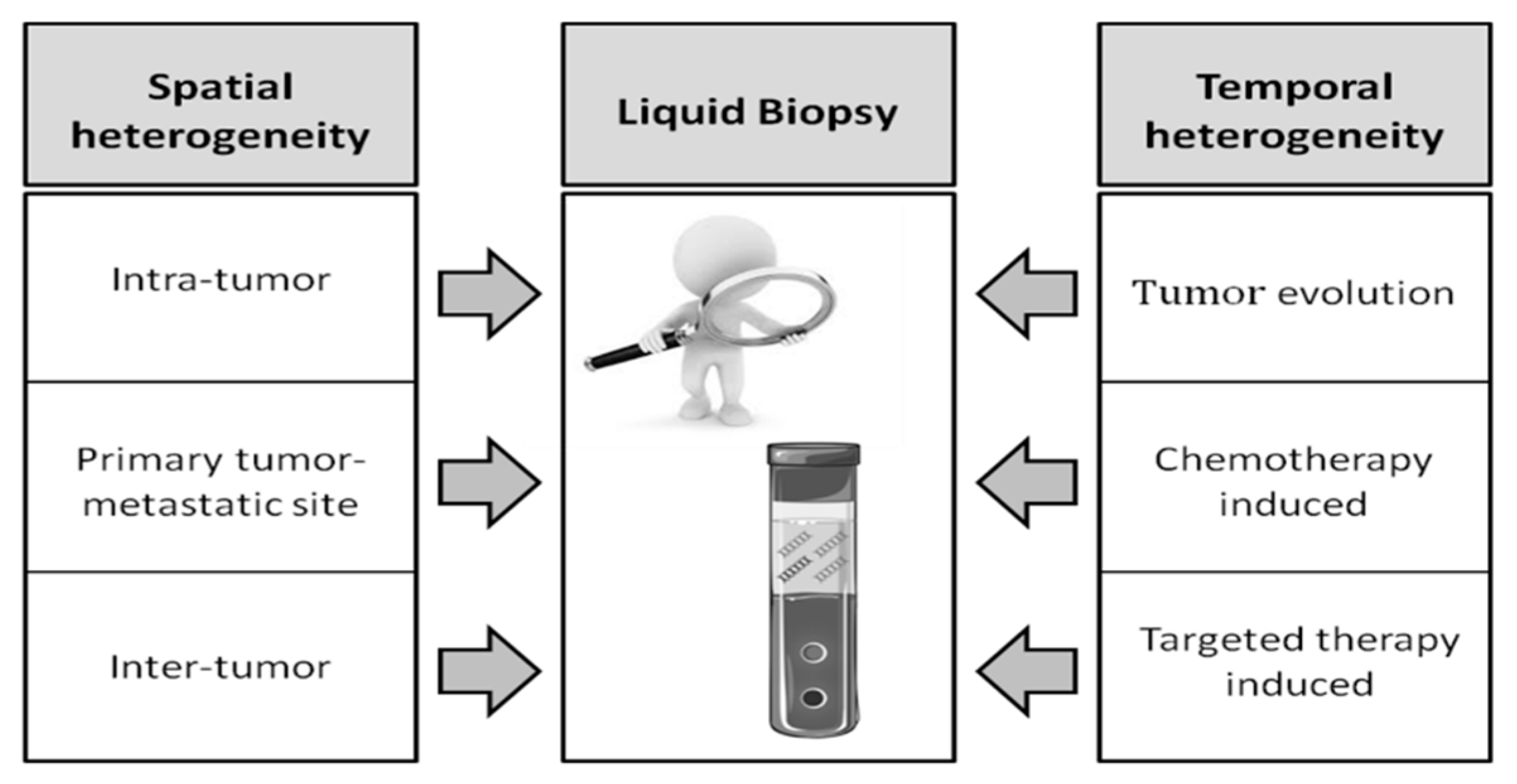
4. Liquid Biopsy to Overcome Tumor Tissue Heterogeneity

Different tumor components (circulating tumor cells (CTCs), circulating tumor DNA (ctDNA), exosomes, microRNAs) isolated from body fluids through “liquid biopsy” can provide additional useful information for diagnostic, prognostic, and predictive purposes. The study of liquid biopsy has acquired a prominent place in cancer research, and one of the features that renders it especially promising is its capacity to overcome the problem of tumor heterogeneity (Figure 1). Assuming that tumor tissue is heterogeneous, a tissue biopsy performed in a particular part of the lesion may not be representative of the genetic landscape of the entire tumor. Liquid biopsy on the other hand is more widely representative of the entire lesion and enables tumor evolution to be followed in real time. In a recent review, Hench et al. discussed the advantages and limitations of CTC and ctDNA evaluation in breast, lung, and CRC, envisioning a potential role of liquid biopsy for second-line diagnosis and tumor surveillance, even though its analytical and clinical validity has yet to be definitively demonstrated [85].
ITH, multiple metastatic clones that disseminate and remain dormant for years, and methodological pitfalls may be responsible for the discordance often observed in KRAS status between CTCs and CRC tissue. Fabbri et al. reported a concordance of only 50% in their case series of 40 CRC patients [86]. Kondo et al. analyzed primary tumor tissue and CTCs in 11 patients, observing a concordance of around 64% in KRAS mutational status [87], while other studies obtained a concordance of 71–77% [88,89]. Of note, even KRAS status in CTCs from the same patient proved to be heterogeneous [90].
Given that cell-free (cfDNA) and ctDNA appear to have a strong prognostic value in mCRC and are directly related to disease burden, they might be useful to monitor disease from early stages through disease evolution under treatment. Interestingly, Bettegowda et al. observed that ctDNA was always detected even when no CTCs were found, whereas there were no instances when CTCs were present but ctDNA was absent [91].
Finally, the analysis of exosomal DNA or RNA to detect tumor-specific genetic mutations in CRC is a potentially promising area of research. Given its concentration and abundant content, exosomes could help to increase the sensitivity of liquid biopsy, despite the presence of some methodological limitations [92]. A high consistency rate (73.7% sensitivity and 100% specificity) has been seen for KRAS mutations detected using exosomes from serum [93]. Moreover, as it has been found that mutant KRAS rather than wt KRAS DNA is loaded preferably into exosomes, the analysis of these vesicles could likely facilitate patient stratification and thus improve clinical management [94].

4.1. Comparison between cfDNA and Tumor Tissue

Several studies have been performed to compare gene mutations in cfDNA and tumor tissue (Table 1). The majority of these focused on the analysis of RAS genes as their alterations are useful to predict the response to anti-EGFR mAb treatment [91,95,96,97,98,99,100,101,102,103]. Spindler et al. were among the first to analyze the concordance of KRAS mutation between tissue and cfDNA, obtaining a sensitivity of 78% and absolute specificity, as none of the patients without mutations in primary tumor tissue showed positive plasma samples [95]. Another study reported a concordance of 93% between tissue and plasma analyses in 115 mCRC patients, with the RAS mutation status discrepancy explained by spatial and temporal tumor heterogeneity [99]. The authors found that peritoneal and lung metastases, and mucinous histology, were associated with low RAS cfDNA detection, whereas the number of metastases was not, suggesting that intrinsic biological characteristics of the tumor rather than tumor burden may impact cfDNA release [99]. Similarly, a concordance of about 90% between tissue and plasma analyses was observed by Grasselli et al. in a case series of 146 mCRC patients. The 10% discordance prevalently referred to cases of RAS mutation positivity in cfDNA but negativity in tumor tissue, once more suggesting that such a result could be due to tumor tissue heterogeneity [100]. In another study performed on two independent case series, the positive and negative concordance between plasma and tumor RAS mutation was 90.4% and 93.5%, respectively, with an overall concordance of 91.8% [102].
Recently, a very high concordance (97%) was reported in RAS mutation between plasma and tumor tissue in mCRC patients with liver metastases. These results confirm the high concordance between primary tissue and liver metastases, suggesting that liquid biopsy could be contemplated for patients in this subgroup for whom no tumor tissue is available [104].
Similarly, a recent study by Normanno et al. on a case series of 92 KRAS exon 2 wt patients treated with first-line cetuximab plus FOLFIRI showed a mutation concordance between tissue and cfDNA of 78.3%, with 10 cases of RAS mutated in tissue and RAS wt in plasma, and a further 10 cases of RAS mutated in plasma and RAS wt in tissue [103]. Of note, the use of highly sensitive methodologies, such as droplet digital PCR, in discordant cases revealed the presence of mutations in all tissues that were negative by NGS but had matched positive plasma, and in 20% of plasma that was negative by OncoBeam but had matched positive tissue, suggesting that the main reason for the discordance between tissue and cfDNA was the sensitivity of the methodologies, which may not always identify underrepresented alterations. In Normanno’s study, RAS mutation in tumor tissue but not in plasma was associated with a better prognosis, implying that low amounts of cfDNA could be indicative of less aggressive disease [103].

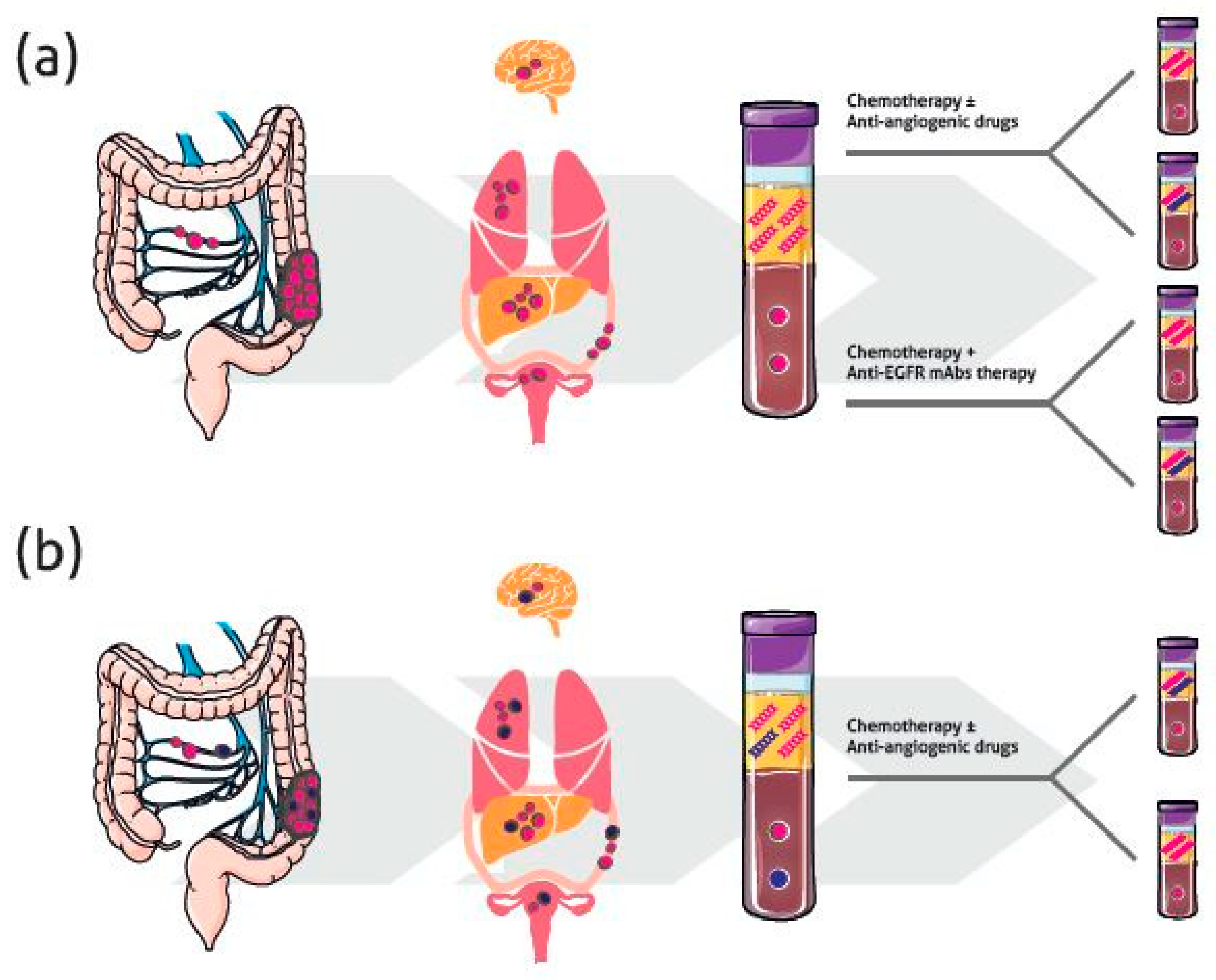
4.2. Temporal Heterogeneity and Monitoring Response to Therapy

The problem of tumor heterogeneity becomes even more prominent during pharmacological treatment, when a clonal selection develops and a series of secondary resistance mechanisms is established. As tumor re-biopsy is an invasive procedure that may not be feasible, in particular for metastases deep within the body; the only opportunity to investigate and monitor these alterations occurring during treatment is the study of liquid biopsy.

4.2.1. Anti-EGFR Therapy

In patients treated with targeted therapy, tumor subclones with resistance-conferring aberrations in the target that cannot compete in the absence of therapy will eventually be selected to grow under the treatment pressure. Moreover, de novo mutations may drive resistance to anti-EGFR mAb treatment. Figure 2a summarizes the possible scenario in this patient setting, where KRAS mutations represent the principal mechanism of acquired resistance and can be detected non-invasively in blood months before the appearance of radiographic progression [105]. Remarkably, not only KRAS alterations, but also NRAS and BRAF mutations, as well as amplification of ERBB2 and MET, are detected in patients who respond to EGFR blockade and then relapse, highlighting that although the mechanisms of resistance are genetically heterogeneous, they converge on key signaling pathways [106].
In a study by Morelli et al. analysis of plasma collected at progression in 60 patients refractory to anti-EGFR mAbs and KRAS wt revealed the presence of KRAS mutation in 44% of cases [107]. In 30% of these, mutations were found at a low allele frequency in pre-treatment tumor tissue [107], suggesting that newly detected RAS mutation in plasma from patients refractory to anti-EGFR treatment may derive from rare, pre-existing clones in primary tumors. The presence of cfDNA RAS mutations was associated with a shorter PFS [107].
In a case series of 20 RAS-BRAF wt patients showing progression after anti-EGFR mAbs treatment, NGS analysis revealed that RAS mutations and HER2/MET amplifications were the most frequently detected resistance mechanisms in both tissue and plasma samples, the authors demonstrating the involvement of both intralesion and interlesion tumor heterogeneity in the emergence of the complex resistance mechanisms [108].
In a similar patient setting in which cfDNA was evaluated, Strickler et al. observed multiple tumor subclones characterized by resistance-related alterations emerging during therapy that originate from different metastases throughout the body [109].
In another recent study performed on a case series of mCRC patients treated in a phase II trial with panitumumab plus irinotecan, the incidence of RAS mutation in tumor tissue was 9.5% by NGS and 6.3% by BEAMing, whereas cfDNA evaluated by BEAMing revealed an incidence of 37%. This result demonstrated that cfDNA was more indicative of resistance mechanisms than tumor tissue, and this is more probably attributable to tumor heterogeneity, which increased dramatically during treatment. Interestingly, RAS mutations in cfDNA preceded clinical progression by a median of 3.6 months, indicating that the serial monitoring of RAS mutations on cfDNA during treatment could be useful for the timely identification of patients with a high risk of progression [110]. Finally, the highest temporal heterogeneity (94%) was detected by Yamada et al. who reported that 17/18 chemo-naive KRAS wt mCRC patients submitted to first-line systemic chemotherapy including EGFR blockade were characterized by the emergence of KRAS mutation [111].
Overall, tumor heterogeneity at the time of acquired resistance to anti-EGFR treatment represents a significant obstacle to the development of precision medicine strategies, and suggests the potential usefulness of large-scale cfDNA profiling to define the genomic landscape of tumor heterogeneity and therapeutic resistance.

4.2.2. Chemotherapy and Other Targeted Therapies

Interesting data based on tissue analyses has demonstrated that mutation shifts after chemotherapy may be related to ITH due to tumor clone selection or cancer evolution after chemotherapy. Like targeted therapies, systemic chemotherapies may impose an evolutionary pressure on cancer, thus increasing heterogeneity in terms of new driver and passenger mutations, creating neoantigens that strongly influence immune response [36] (Figure 2a). Conversely, Brannon et al. hypothesized that prior chemotherapy treatment results in a decrease in apparent tumor heterogeneity [69]. A reduction in RAS/BRAF/PIK3CA mutations after chemotherapy was observed by Li et al. even when samples with low cellularity, which may give rise to false-negative results, were excluded [52].
Few studies have been published to date on the use of liquid biopsy to evaluate the effect of chemotherapy on heterogeneity. Interestingly, in their preliminary results, Gazzaniga et al. showed that 30% of mutated KRAS mCRCs treated with anti-angiogenic drugs switched to prevalently wt KRAS ctDNA in peripheral blood during therapy (Figure 2b), probably due to the induction of hypoxia [112]. This is in line with the observation in tissue of Li et al. [52], and if confirmed in larger populations, should be taken into account when a second-line therapeutic option is required. Notably, and in agreement with the above, Yamada et al. recently reported that mutations that emerged in some patients after anti-EGFR mAb treatment disappeared after starting second-line chemotherapy [111].

5. Impact of Heterogeneity in Clinical Molecular Diagnostics

Considering the importance of molecular targeted therapy in CRC, it is clear that the accurate detection of gene mutations to bypass tumor heterogeneity is essential for correct patient selection. In fact, although it is difficult to predict whether patients with a low frequency of RAS mutations will respond or not to anti-EGFR therapy, it is important to identify and characterize all mutant subpopulations during the routine diagnostic process, because their impact on the response to target therapy is still unclear [49]. International guidelines indicate that RAS mutation analysis should be performed at diagnosis by analyzing the most recent and representative tumor tissue available [3]. The choice of the specimen and the selection of the appropriate tissue block represent the first challenge, because poor tumor cellularity, ITH, and adjuvant therapy may be confounding factors [52]. Nelson et al. recommended pooling three tumor sites with adequate cellularity for genetic testing to minimize the risk of false negative results [113]. Moreover, since for a subset of mCRC patients the diagnosis of metastases is done only by radiological studies, often only a primary tumor sample is available to perform molecular determinations, even if the principal goal of anti-EGFR therapy is to treat metastases. Despite the fact that the high concordance between primary tissue and metastasis has led to the approval of guidelines indicating that either type of material can be used indifferently, a proportion of patients for whom there is no concordance between primary and metastatic lesions are destined not to receive optimal treatment [14,114]. Moreover, this may be a pitfall for patients with locally advanced CRC who relapse after surgery and adjuvant treatment, as the mutational status of the remaining tumor cells may be altered due to chemotherapy [52]. Similarly, in non-chemo-naive patients, new biopsies of primary or metastatic lesions are probably needed following chemotherapy resistance or insensitivity to fully understand the complexity of the disease and to choose the most appropriate therapy [13]. Overall, when looking at advanced stages of CRC, patients need to be individually oriented due to case-specific prior surgical and clinical treatment and molecular progression.

6. Conclusions

In summary, spatial heterogeneity and drug-selected clonal evolution in CRC influences tumor aggressiveness, resistance to therapy, and patient prognosis. Although a high concordance of the mutational status exists between primary tumor and liver metastases, it appears that the localization of secondary lesions predict the risk of molecular discordance from the primary tumor. Thus, although the use of liquid biopsy in CRC clinical practice is not as urgent as in other malignancies, such as lung cancer, it could represent a tool for further improving the molecular diagnostics of this disease. Moreover, the increasing development of high-throughput sequencing methodologies in both tumor tissue and liquid biopsy, and the gradual decrease in their costs, will provide the possibility of performing genome wide tumor characterization in a non-invasive manner and at any time during treatment, bypassing the problem of tumor heterogeneity and substantially improving mCRC patient management.

Author Contributions

C.M., G.M., A.P., L.M., G.D.M., and P.U. drafted the manuscript. C.M. and G.M. assisted in the figure design. C.M., G.M., and P.U. outlined and revised the manuscript. Reviewing and editing was done by all authors. C.M. and P.U. conceived the idea for the review. C.M., G.M., L.M., G.D.M., and P.U. performed the literature search. C.M. and P.U. drafted the manuscript. A.P. critically reviewed the manuscript for important intellectual content. All authors read and approved the final version for submission.

Funding

This research received no external funding.

Acknowledgments

The authors would like to thank Francesco Mazza for his help with figure editing and Gráinne Tierney for editorial assistance.

Conflicts of Interest

The authors declare no conflict of interest.

References

  1. Moraga-Serrano, P.E. Global, Regional, and National Cancer Incidence, Mortality, Years of Life Lost, Years Lived with Disability, and Disability-Adjusted Life-Years for 29 Cancer Groups, 1990 to 2016: A Systematic Analysis for the Global Burden of Disease Study. JAMA Oncol. 2018, 2018, 126049. [Google Scholar]
  2. Siegel, R.; Naishadham, D.; Jemal, A. Cancer Statistics, 2013. CA Cancer J. Clin. 2013, 63, 11–30. [Google Scholar] [CrossRef] [PubMed]
  3. Van Cutsem, E.; Cervantes, A.; Adam, R.; Sobrero, A.; Van Krieken, J.H.; Aderka, D.; Aranda Aguilar, E.; Bardelli, A.; Benson, A.; Bodoky, G.; et al. ESMO Consensus Guidelines for the Management of Patients with Metastatic Colorectal Cancer. Ann. Oncol. 2016, 27, 1386–1422. [Google Scholar] [CrossRef] [PubMed]
  4. Malvezzi, M.; Carioli, G.; Bertuccio, P.; Boffetta, P.; Levi, F.; La Vecchia, C.; Negri, E. European Cancer Mortality Predictions for the Year 2018 with Focus on Colorectal Cancer. Ann. Oncol. 2018, 29, 1016–1022. [Google Scholar] [CrossRef] [PubMed]
  5. Adam, R.; Pascal, G.; Castaing, D.; Azoulay, D.; Delvart, V.; Paule, B.; Levi, F.; Bismuth, H. Tumor Progression while on Chemotherapy: A Contraindication to Liver Resection for Multiple Colorectal Metastases? Ann. Surg. 2004, 240, 1052–1061. [Google Scholar] [CrossRef] [PubMed]
  6. Grady, W.M.; Carethers, J.M. Genomic and Epigenetic Instability in Colorectal Cancer Pathogenesis. Gastroenterology 2008, 135, 1079–1099. [Google Scholar] [CrossRef] [PubMed] [Green Version]
  7. Vogelstein, B.; Papadopoulos, N.; Velculescu, V.E.; Zhou, S.; Diaz, L.A., Jr.; Kinzler, K.W. Cancer Genome Landscapes. Science 2013, 339, 1546–1558. [Google Scholar] [CrossRef] [PubMed] [Green Version]
  8. Bramsen, J.B.; Rasmussen, M.H.; Ongen, H.; Mattesen, T.B.; Orntoft, M.W.; Arnadottir, S.S.; Sandoval, J.; Laguna, T.; Vang, S.; Oster, B.; et al. Molecular-Subtype-Specific Biomarkers Improve Prediction of Prognosis in Colorectal Cancer. Cell. Rep. 2017, 19, 1268–1280. [Google Scholar] [CrossRef] [PubMed]
  9. Ogino, S.; Nowak, J.A.; Hamada, T.; Phipps, A.I.; Peters, U.; Milner, D.A., Jr.; Giovannucci, E.L.; Nishihara, R.; Giannakis, M.; Garrett, W.S.; et al. Integrative Analysis of Exogenous, Endogenous, Tumour and Immune Factors for Precision Medicine. Gut 2018, 67, 1168–1180. [Google Scholar] [CrossRef] [PubMed]
  10. Ogino, S.; Chan, A.T.; Fuchs, C.S.; Giovannucci, E. Molecular Pathological Epidemiology of Colorectal Neoplasia: An Emerging Transdisciplinary and Interdisciplinary Field. Gut 2011, 60, 397–411. [Google Scholar] [CrossRef] [PubMed]
  11. Ogino, S.; Nowak, J.A.; Hamada, T.; Milner DA, J.; Nishihara, R. Insights into Pathogenic Interactions among Environment, Host, and Tumor at the Crossroads of Molecular Pathology and Epidemiology. Annu. Rev. Pathol. 2018. [Google Scholar] [CrossRef] [PubMed]
  12. Cancer Genome Atlas Network. Comprehensive Molecular Characterization of Human Colon and Rectal Cancer. Nature 2012, 487, 330–337. [Google Scholar] [CrossRef] [PubMed]
  13. Kim, R.; Schell, M.J.; Teer, J.K.; Greenawalt, D.M.; Yang, M.; Yeatman, T.J. Co-Evolution of Somatic Variation in Primary and Metastatic Colorectal Cancer may Expand Biopsy Indications in the Molecular Era. PLoS ONE 2015, 10, e0126670. [Google Scholar] [CrossRef] [PubMed]
  14. Mao, C.; Wu, X.Y.; Yang, Z.Y.; Threapleton, D.E.; Yuan, J.Q.; Yu, Y.Y.; Tang, J.L. Concordant Analysis of KRAS, BRAF, PIK3CA Mutations, and PTEN Expression between Primary Colorectal Cancer and Matched Metastases. Sci. Rep. 2015, 5, 8065. [Google Scholar] [CrossRef] [PubMed]
  15. Linnekamp, J.F.; Hooff, S.R.V.; Prasetyanti, P.R.; Kandimalla, R.; Buikhuisen, J.Y.; Fessler, E.; Ramesh, P.; Lee, K.A.S.T.; Bochove, G.G.W.; de Jong, J.H.; et al. Consensus Molecular Subtypes of Colorectal Cancer are Recapitulated in in Vitro and in Vivo Models. Cell Death Differ. 2018, 25, 616–633. [Google Scholar] [CrossRef] [PubMed]
  16. Fearon, E.R.; Vogelstein, B. A Genetic Model for Colorectal Tumorigenesis. Cell 1990, 61, 759–767. [Google Scholar] [CrossRef]
  17. Jass, J.R.; Smith, M. Sialic Acid and Epithelial Differentiation in Colorectal Polyps and Cancer--a Morphological, Mucin and Lectin Histochemical Study. Pathology 1992, 24, 233–242. [Google Scholar] [CrossRef] [PubMed]
  18. Singh, P.P.; Sharma, P.K.; Krishnan, G.; Lockhart, A.C. Immune Checkpoints and Immunotherapy for Colorectal Cancer. Gastroenterol. Rep. 2015, 3, 289–297. [Google Scholar] [CrossRef] [PubMed]
  19. Yokota, T. Are KRAS/BRAF Mutations Potent Prognostic and/or Predictive Biomarkers in Colorectal Cancers? Anticancer Agents Med. Chem. 2012, 12, 163–171. [Google Scholar] [CrossRef] [PubMed]
  20. Tejpar, S.; Stintzing, S.; Ciardiello, F.; Tabernero, J.; Van Cutsem, E.; Beier, F.; Esser, R.; Lenz, H.J.; Heinemann, V. Prognostic and Predictive Relevance of Primary Tumor Location in Patients with RAS Wild-Type Metastatic Colorectal Cancer: Retrospective Analyses of the CRYSTAL and FIRE-3 Trials. JAMA Oncol. 2016. [Google Scholar] [CrossRef] [PubMed]
  21. Missiaglia, E.; Jacobs, B.; D’Ario, G.; Di Narzo, A.F.; Soneson, C.; Budinska, E.; Popovici, V.; Vecchione, L.; Gerster, S.; Yan, P.; et al. Distal and Proximal Colon Cancers Differ in Terms of Molecular, Pathological, and Clinical Features. Ann. Oncol. 2014, 25, 1995–2001. [Google Scholar] [CrossRef] [PubMed]
  22. Guinney, J.; Dienstmann, R.; Wang, X.; de Reynies, A.; Schlicker, A.; Soneson, C.; Marisa, L.; Roepman, P.; Nyamundanda, G.; Angelino, P.; et al. The Consensus Molecular Subtypes of Colorectal Cancer. Nat. Med. 2015, 21, 1350–1356. [Google Scholar] [CrossRef] [PubMed]
  23. Mogensen, M.B.; Rossing, M.; Ostrup, O.; Larsen, P.N.; Heiberg Engel, P.J.; Jorgensen, L.N.; Hogdall, E.V.; Eriksen, J.; Ibsen, P.; Jess, P.; et al. Genomic Alterations Accompanying Tumour Evolution in Colorectal Cancer: Tracking the Differences between Primary Tumours and Synchronous Liver Metastases by Whole-Exome Sequencing. BMC Cancer 2018, 18, 752. [Google Scholar] [CrossRef] [PubMed]
  24. Ulivi, P.; Scarpi, E.; Chiadini, E.; Marisi, G.; Valgiusti, M.; Capelli, L.; Casadei Gardini, A.; Monti, M.; Ruscelli, S.; Frassineti, G.L.; et al. Right- Vs. Left-Sided Metastatic Colorectal Cancer: Differences in Tumor Biology and Bevacizumab Efficacy. Int. J. Mol. Sci. 2017, 18, 1240. [Google Scholar] [CrossRef] [PubMed]
  25. Stintzing, S.; Tejpar, S.; Gibbs, P.; Thiebach, L.; Lenz, H.J. Understanding the Role of Primary Tumour Localisation in Colorectal Cancer Treatment and Outcomes. Eur. J. Cancer 2017, 84, 69–80. [Google Scholar] [CrossRef] [PubMed]
  26. Boisen, M.K.; Johansen, J.S.; Dehlendorff, C.; Larsen, J.S.; Osterlind, K.; Hansen, J.; Nielsen, S.E.; Pfeiffer, P.; Tarpgaard, L.S.; Hollander, N.H.; et al. Primary Tumor Location and Bevacizumab Effectiveness in Patients with Metastatic Colorectal Cancer. Ann. Oncol. 2013, 24, 2554–2559. [Google Scholar] [CrossRef] [PubMed]
  27. Moretto, R.; Cremolini, C.; Rossini, D.; Pietrantonio, F.; Battaglin, F.; Mennitto, A.; Bergamo, F.; Loupakis, F.; Marmorino, F.; Berenato, R.; et al. Location of Primary Tumor and Benefit from Anti-Epidermal Growth Factor Receptor Monoclonal Antibodies in Patients with RAS and BRAF Wild-Type Metastatic Colorectal Cancer. Oncologist 2016, 21, 988–994. [Google Scholar] [CrossRef] [PubMed]
  28. Cremolini, C.; Antoniotti, C.; Lonardi, S.; Bergamo, F.; Cortesi, E.; Tomasello, G.; Moretto, R.; Ronzoni, M.; Racca, P.; Loupakis, F.; et al. Primary Tumor Sidedness and Benefit from FOLFOXIRI Plus Bevacizumab as Initial Therapy for Metastatic Colorectal Cancer. Retrospective Analysis of the TRIBE Trial by GONO. Ann. Oncol. 2018, 29, 1528–1534. [Google Scholar] [CrossRef] [PubMed]
  29. Komor, M.A.; Bosch, L.J.; Bounova, G.; Bolijn, A.S.; Delis-van Diemen, P.M.; Rausch, C.; Hoogstrate, Y.; Stubbs, A.P.; de Jong, M.; Jenster, G.; et al. Consensus Molecular Subtype Classification of Colorectal Adenomas. J. Pathol. 2018. [Google Scholar] [CrossRef] [PubMed]
  30. Marusyk, A.; Almendro, V.; Polyak, K. Intra-Tumour Heterogeneity: A Looking Glass for Cancer? Nat. Rev. Cancer 2012, 12, 323–334. [Google Scholar] [CrossRef] [PubMed]
  31. Amaro, A.; Chiara, S.; Pfeffer, U. Molecular Evolution of Colorectal Cancer: From Multistep Carcinogenesis to the Big Bang. Cancer Metastasis Rev. 2016, 35, 63–74. [Google Scholar] [CrossRef] [PubMed]
  32. Kreso, A.; O’Brien, C.A.; van Galen, P.; Gan, O.I.; Notta, F.; Brown, A.M.; Ng, K.; Ma, J.; Wienholds, E.; Dunant, C.; et al. Variable Clonal Repopulation Dynamics Influence Chemotherapy Response in Colorectal Cancer. Science 2013, 339, 543–548. [Google Scholar] [CrossRef] [PubMed]
  33. Sottoriva, A.; Kang, H.; Ma, Z.; Graham, T.A.; Salomon, M.P.; Zhao, J.; Marjoram, P.; Siegmund, K.; Press, M.F.; Shibata, D.; et al. A Big Bang Model of Human Colorectal Tumor Growth. Nat. Genet. 2015, 47, 209–216. [Google Scholar] [CrossRef] [PubMed]
  34. Fisher, R.; Pusztai, L.; Swanton, C. Cancer Heterogeneity: Implications for Targeted Therapeutics. Br. J. Cancer 2013, 108, 479–485. [Google Scholar] [CrossRef] [PubMed]
  35. Enriquez-Navas, P.M.; Kam, Y.; Das, T.; Hassan, S.; Silva, A.; Foroutan, P.; Ruiz, E.; Martinez, G.; Minton, S.; Gillies, R.J.; et al. Exploiting Evolutionary Principles to Prolong Tumor Control in Preclinical Models of Breast Cancer. Sci. Transl. Med. 2016, 8, 327ra24. [Google Scholar] [CrossRef] [PubMed]
  36. Venkatesan, S.; Swanton, C.; Taylor, B.S.; Costello, J.F. Treatment-Induced Mutagenesis and Selective Pressures Sculpt Cancer Evolution. Cold Spring Harb. Perspect. Med. 2017, 7. [Google Scholar] [CrossRef] [PubMed]
  37. Andor, N.; Graham, T.A.; Jansen, M.; Xia, L.C.; Aktipis, C.A.; Petritsch, C.; Ji, H.P.; Maley, C.C. Pan-Cancer Analysis of the Extent and Consequences of Intratumor Heterogeneity. Nat. Med. 2016, 22, 105–113. [Google Scholar] [CrossRef] [PubMed]
  38. Sveen, A.; Loes, I.M.; Alagaratnam, S.; Nilsen, G.; Holand, M.; Lingjaerde, O.C.; Sorbye, H.; Berg, K.C.; Horn, A.; Angelsen, J.H.; et al. Intra-Patient Inter-Metastatic Genetic Heterogeneity in Colorectal Cancer as a Key Determinant of Survival After Curative Liver Resection. PLoS Genet. 2016, 12, e1006225. [Google Scholar] [CrossRef] [PubMed]
  39. McGranahan, N.; Swanton, C. Clonal Heterogeneity and Tumor Evolution: Past, Present, and the Future. Cell 2017, 168, 613–628. [Google Scholar] [CrossRef] [PubMed] [Green Version]
  40. Wang, W.; Kandimalla, R.; Huang, H.; Zhu, L.; Li, Y.; Gao, F.; Goel, A.; Wang, X. Molecular Subtyping of Colorectal Cancer: Recent Progress, New Challenges and Emerging Opportunities. In Seminars in Cancer Biology; Academic Press: Cambridge, MA, USA, 2018. [Google Scholar]
  41. Suzuki, Y.; Ng, S.B.; Chua, C.; Leow, W.Q.; Chng, J.; Liu, S.Y.; Ramnarayanan, K.; Gan, A.; Ho, D.L.; Ten, R.; et al. Multiregion Ultra-Deep Sequencing Reveals Early Intermixing and Variable Levels of Intratumoral Heterogeneity in Colorectal Cancer. Mol. Oncol. 2017, 11, 124–139. [Google Scholar] [CrossRef] [PubMed]
  42. Giardina, T.; Robinson, C.; Grieu-Iacopetta, F.; Millward, M.; Iacopetta, B.; Spagnolo, D.; Amanuel, B. Implementation of Next Generation Sequencing Technology for Somatic Mutation Detection in Routine Laboratory Practice. Pathology 2018, 50, 389–401. [Google Scholar] [CrossRef] [PubMed]
  43. D’Haene, N.; Fontanges, Q.; De Neve, N.; Blanchard, O.; Melendez, B.; Delos, M.; Dehou, M.F.; Maris, C.; Nagy, N.; Rousseau, E.; et al. Clinical Application of Targeted Next-Generation Sequencing for Colorectal Cancer Patients: A Multicentric Belgian Experience. Oncotarget 2018, 9, 20761–20768. [Google Scholar] [CrossRef] [PubMed]
  44. Baisse, B.; Bouzourene, H.; Saraga, E.P.; Bosman, F.T.; Benhattar, J. Intratumor Genetic Heterogeneity in Advanced Human Colorectal Adenocarcinoma. Int. J. Cancer 2001, 93, 346–352. [Google Scholar] [CrossRef] [PubMed]
  45. Petaccia de Macedo, M.; Melo, F.M.; Ribeiro, H.S.C.; Marques, M.C.; Kagohara, L.T.; Begnami, M.D.; Neto, J.C.; Ribeiro, J.S.; Soares, F.A.; Carraro, D.M.; et al. KRAS Mutation Status is Highly Homogeneous between Areas of the Primary Tumor and the Corresponding Metastasis of Colorectal Adenocarcinomas: One Less Problem in Patient Care. Am. J. Cancer Res. 2017, 7, 1978–1989. [Google Scholar] [PubMed]
  46. Farber, L.; Efrati, E.; Elkin, H.; Peerless, Y.; Sabo, E.; Ben-Izhak, O.; Hershkovitz, D. Molecular Morphometric Analysis shows Relative Intra-Tumoural Homogeneity for KRAS Mutations in Colorectal Cancer. Virchows Arch. 2011, 459, 487–493. [Google Scholar] [CrossRef] [PubMed]
  47. Jeantet, M.; Tougeron, D.; Tachon, G.; Cortes, U.; Archambaut, C.; Fromont, G.; Karayan-Tapon, L. High Intra- and Inter-Tumoral Heterogeneity of RAS Mutations in Colorectal Cancer. Int. J. Mol. Sci. 2016, 17, 2015. [Google Scholar] [CrossRef] [PubMed]
  48. Jones, H.G.; Jenkins, G.; Williams, N.; Griffiths, P.; Chambers, P.; Beynon, J.; Harris, D. Genetic and Epigenetic Intra-Tumour Heterogeneity in Colorectal Cancer. World J. Surg. 2017, 41, 1375–1383. [Google Scholar] [CrossRef] [PubMed]
  49. Buttner, J.; Johrens, K.; Klauschen, F.; Hummel, M.; Lenze, D.; Saeger, W.; Lehmann, A. Intratumoral Morphological Heterogeneity can be an Indicator of Genetic Heterogeneity in Colorectal Cancer. Exp. Mol. Pathol. 2018, 104, 76–81. [Google Scholar] [CrossRef] [PubMed]
  50. Reggiani Bonetti, L.; Barresi, V.; Bettelli, S.; Caprera, C.; Manfredini, S.; Maiorana, A. Analysis of KRAS, NRAS, PIK3CA, and BRAF Mutational Profile in Poorly Differentiated Clusters of KRAS-Mutated Colon Cancer. Hum. Pathol. 2017, 62, 91–98. [Google Scholar] [CrossRef] [PubMed]
  51. Laurent-Puig, P.; Cayre, A.; Manceau, G.; Buc, E.; Bachet, J.B.; Lecomte, T.; Rougier, P.; Lievre, A.; Landi, B.; Boige, V.; et al. Analysis of PTEN, BRAF, and EGFR Status in Determining Benefit from Cetuximab Therapy in Wild-Type KRAS Metastatic Colon Cancer. J. Clin. Oncol. 2009, 27, 5924–5930. [Google Scholar] [CrossRef] [PubMed]
  52. Li, W.; Qiu, T.; Guo, L.; Ying, J. Major Challenges Related to Tumor Biological Characteristics in Accurate Mutation Detection of Colorectal Cancer by Next-Generation Sequencing. Cancer Lett. 2017, 410, 92–99. [Google Scholar] [CrossRef] [PubMed]
  53. Li, X.; Guo, X.; Li, D.; Du, X.; Yin, C.; Chen, C.; Fang, W.; Bian, Z.; Zhang, J.; Li, B.; et al. Multi-Regional Sequencing Reveals Intratumor Heterogeneity and Positive Selection of Somatic mtDNA Mutations in Hepatocellular Carcinoma and Colorectal Cancer. Int. J. Cancer 2018, 143, 1143–1152. [Google Scholar] [CrossRef] [PubMed]
  54. Losi, L.; Baisse, B.; Bouzourene, H.; Benhattar, J. Evolution of Intratumoral Genetic Heterogeneity during Colorectal Cancer Progression. Carcinogenesis 2005, 26, 916–922. [Google Scholar] [CrossRef] [PubMed]
  55. Yaeger, R.; Chatila, W.K.; Lipsyc, M.D.; Hechtman, J.F.; Cercek, A.; Sanchez-Vega, F.; Jayakumaran, G.; Middha, S.; Zehir, A.; Donoghue, M.T.A.; et al. Clinical Sequencing Defines the Genomic Landscape of Metastatic Colorectal Cancer. Cancer. Cell 2018, 33, 125–136. [Google Scholar] [CrossRef] [PubMed]
  56. Manfredi, S.; Lepage, C.; Hatem, C.; Coatmeur, O.; Faivre, J.; Bouvier, A.M. Epidemiology and Management of Liver Metastases from Colorectal Cancer. Ann. Surg. 2006, 244, 254–259. [Google Scholar] [CrossRef] [PubMed]
  57. Garden, O.J.; Rees, M.; Poston, G.J.; Mirza, D.; Saunders, M.; Ledermann, J.; Primrose, J.N.; Parks, R.W. Guidelines for Resection of Colorectal Cancer Liver Metastases. Gut 2006, 55 (Suppl. 3), iii1–iii8. [Google Scholar] [CrossRef] [PubMed]
  58. Mitry, E.; Guiu, B.; Cosconea, S.; Jooste, V.; Faivre, J.; Bouvier, A.M. Epidemiology, Management and Prognosis of Colorectal Cancer with Lung Metastases: A 30-Year Population-Based Study. Gut 2010, 59, 1383–1388. [Google Scholar] [CrossRef] [PubMed]
  59. Riihimaki, M.; Hemminki, A.; Sundquist, J.; Hemminki, K. Patterns of Metastasis in Colon and Rectal Cancer. Sci. Rep. 2016, 6, 29765. [Google Scholar] [CrossRef] [PubMed]
  60. Lemmens, V.E.; Klaver, Y.L.; Verwaal, V.J.; Rutten, H.J.; Coebergh, J.W.; de Hingh, I.H. Predictors and Survival of Synchronous Peritoneal Carcinomatosis of Colorectal Origin: A Population-Based Study. Int. J. Cancer 2011, 128, 2717–2725. [Google Scholar] [CrossRef] [PubMed]
  61. Goere, D.; Allard, M.A.; Honore, C.; Dumont, F.; Elias, D. Incidence and Prognosis of Synchronous Colorectal Carcinomatosis. Future Oncol. 2013, 9, 541–549. [Google Scholar] [CrossRef] [PubMed]
  62. Hanna, N.N.; Cohen, A.M. Ovarian Neoplasms in Patients with Colorectal Cancer: Understanding the Role of Prophylactic Oophorectomy. Clin. Colorectal Cancer 2004, 3, 215–222. [Google Scholar] [CrossRef] [PubMed]
  63. Go, P.H.; Klaassen, Z.; Meadows, M.C.; Chamberlain, R.S. Gastrointestinal Cancer and Brain Metastasis: A Rare and Ominous Sign. Cancer 2011, 117, 3630–3640. [Google Scholar] [CrossRef] [PubMed]
  64. Engstrand, J.; Nilsson, H.; Stromberg, C.; Jonas, E.; Freedman, J. Colorectal Cancer Liver Metastases—A Population-Based Study on Incidence, Management and Survival. BMC Cancer 2018, 18, 78. [Google Scholar] [CrossRef] [PubMed]
  65. Artale, S.; Sartore-Bianchi, A.; Veronese, S.M.; Gambi, V.; Sarnataro, C.S.; Gambacorta, M.; Lauricella, C.; Siena, S. Mutations of KRAS and BRAF in Primary and Matched Metastatic Sites of Colorectal Cancer. J. Clin. Oncol. 2008, 26, 4217–4219. [Google Scholar] [CrossRef] [PubMed]
  66. Baas, J.M.; Krens, L.L.; Guchelaar, H.J.; Morreau, H.; Gelderblom, H. Concordance of Predictive Markers for EGFR Inhibitors in Primary Tumors and Metastases in Colorectal Cancer: A Review. Oncologist 2011, 16, 1239–1249. [Google Scholar] [CrossRef] [PubMed] [Green Version]
  67. Knijn, N.; Mekenkamp, L.J.; Klomp, M.; Vink-Borger, M.E.; Tol, J.; Teerenstra, S.; Meijer, J.W.; Tebar, M.; Riemersma, S.; van Krieken, J.H.; et al. KRAS Mutation Analysis: A Comparison between Primary Tumours and Matched Liver Metastases in 305 Colorectal Cancer Patients. Br. J. Cancer 2011, 104, 1020–1026. [Google Scholar] [CrossRef] [PubMed]
  68. Vakiani, E.; Janakiraman, M.; Shen, R.; Sinha, R.; Zeng, Z.; Shia, J.; Cercek, A.; Kemeny, N.; D’Angelica, M.; Viale, A.; et al. Comparative Genomic Analysis of Primary Versus Metastatic Colorectal Carcinomas. J. Clin. Oncol. 2012, 30, 2956–2962. [Google Scholar] [CrossRef] [PubMed] [Green Version]
  69. Brannon, A.R.; Vakiani, E.; Sylvester, B.E.; Scott, S.N.; McDermott, G.; Shah, R.H.; Kania, K.; Viale, A.; Oschwald, D.M.; Vacic, V.; et al. Comparative Sequencing Analysis Reveals High Genomic Concordance between Matched Primary and Metastatic Colorectal Cancer Lesions. Genome Biol. 2014, 15, 454. [Google Scholar] [CrossRef] [PubMed]
  70. Tan, I.B.; Malik, S.; Ramnarayanan, K.; McPherson, J.R.; Ho, D.L.; Suzuki, Y.; Ng, S.B.; Yan, S.; Lim, K.H.; Koh, D.; et al. High-Depth Sequencing of Over 750 Genes Supports Linear Progression of Primary Tumors and Metastases in most Patients with Liver-Limited Metastatic Colorectal Cancer. Genome Biol. 2015, 16, 32. [Google Scholar] [CrossRef] [PubMed]
  71. Kim, K.P.; Kim, J.E.; Hong, Y.S.; Ahn, S.M.; Chun, S.M.; Hong, S.M.; Jang, S.J.; Yu, C.S.; Kim, J.C.; Kim, T.W. Paired Primary and Metastatic Tumor Analysis of Somatic Mutations in Synchronous and Metachronous Colorectal Cancer. Cancer Res. Treat. 2017, 49, 161–167. [Google Scholar] [CrossRef] [PubMed] [Green Version]
  72. Fujiyoshi, K.; Yamamoto, G.; Takahashi, A.; Arai, Y.; Yamada, M.; Kakuta, M.; Yamaguchi, K.; Akagi, Y.; Nishimura, Y.; Sakamoto, H.; et al. High Concordance Rate of KRAS/BRAF Mutations and MSI-H between Primary Colorectal Cancer and Corresponding Metastases. Oncol. Rep. 2017, 37, 785–792. [Google Scholar] [CrossRef] [PubMed]
  73. Kovaleva, V.; Geissler, A.L.; Lutz, L.; Fritsch, R.; Makowiec, F.; Wiesemann, S.; Hopt, U.T.; Passlick, B.; Werner, M.; Lassmann, S. Spatio-Temporal Mutation Profiles of Case-Matched Colorectal Carcinomas and their Metastases Reveal Unique De Novo Mutations in Metachronous Lung Metastases by Targeted Next Generation Sequencing. Mol. Cancer 2016, 15, 63. [Google Scholar] [CrossRef] [PubMed]
  74. Lee, S.Y.; Haq, F.; Kim, D.; Jun, C.; Jo, H.J.; Ahn, S.M.; Lee, W.S. Comparative Genomic Analysis of Primary and Synchronous Metastatic Colorectal Cancers. PLoS ONE 2014, 9, e90459. [Google Scholar] [CrossRef] [PubMed]
  75. Vermaat, J.S.; Nijman, I.J.; Koudijs, M.J.; Gerritse, F.L.; Scherer, S.J.; Mokry, M.; Roessingh, W.M.; Lansu, N.; de Bruijn, E.; van Hillegersberg, R.; et al. Primary Colorectal Cancers and their Subsequent Hepatic Metastases are Genetically Different: Implications for Selection of Patients for Targeted Treatment. Clin. Cancer Res. 2012, 18, 688–699. [Google Scholar] [CrossRef] [PubMed]
  76. Aprile, G.; Casagrande, M.; De Maglio, G.; Fontanella, C.; Rihawi, K.; Bonotto, M.; Pisa, F.E.; Tuniz, F.; Pizzolitto, S.; Fasola, G. Comparison of the Molecular Profile of Brain Metastases from Colorectal Cancer and Corresponding Primary Tumors. Future Oncol. 2017, 13, 135–144. [Google Scholar] [CrossRef] [PubMed]
  77. Crobach, S.; Ruano, D.; van Eijk, R.; Schrumpf, M.; Fleuren, G.; van Wezel, T.; Morreau, H.; PALGA Group. Somatic Mutation Profiles in Primary Colorectal Cancers and Matching Ovarian Metastases: Identification of Driver and Passenger Mutations. J. Pathol. Clin. Res. 2016, 2, 166–174. [Google Scholar] [CrossRef] [PubMed]
  78. Tortola, S.; Steinert, R.; Hantschick, M.; Peinado, M.A.; Gastinger, I.; Stosiek, P.; Lippert, H.; Schlegel, W.; Reymond, M.A. Discordance between K-Ras Mutations in Bone Marrow Micrometastases and the Primary Tumor in Colorectal Cancer. J. Clin. Oncol. 2001, 19, 2837–2843. [Google Scholar] [CrossRef] [PubMed]
  79. Watanabe, T.; Kobunai, T.; Yamamoto, Y.; Matsuda, K.; Ishihara, S.; Nozawa, K.; Iinuma, H.; Shibuya, H.; Eshima, K. Heterogeneity of KRAS Status may Explain the Subset of Discordant KRAS Status between Primary and Metastatic Colorectal Cancer. Dis. Colon Rectum 2011, 54, 1170–1178. [Google Scholar] [CrossRef] [PubMed]
  80. Beije, N.; Helmijr, J.C.; Weerts, M.J.A.; Beaufort, C.M.; Wiggin, M.; Marziali, A.; Verhoef, C.; Sleijfer, S.; Jansen, M.P.H.M.; Martens, J.W.M. Somatic Mutation Detection using various Targeted Detection Assays in Paired Samples of Circulating Tumor DNA, Primary Tumor and Metastases from Patients Undergoing Resection of Colorectal Liver Metastases. Mol. Oncol. 2016, 10, 1575–1584. [Google Scholar] [CrossRef] [PubMed]
  81. Kleist, B.; Meurer, T.; Poetsch, M. Mitochondrial DNA Alteration in Primary and Metastatic Colorectal Cancer: Different Frequency and Association with Selected Clinicopathological and Molecular Markers. Tumour Biol. 2017, 39. [Google Scholar] [CrossRef] [PubMed]
  82. Naxerova, K.; Reiter, J.G.; Brachtel, E.; Lennerz, J.K.; van de Wetering, M.; Rowan, A.; Cai, T.; Clevers, H.; Swanton, C.; Nowak, M.A.; et al. Origins of Lymphatic and Distant Metastases in Human Colorectal Cancer. Science 2017, 357, 55–60. [Google Scholar] [CrossRef] [PubMed]
  83. Kim, M.J.; Lee, H.S.; Kim, J.H.; Kim, Y.J.; Kwon, J.H.; Lee, J.O.; Bang, S.M.; Park, K.U.; Kim, D.W.; Kang, S.B.; et al. Different Metastatic Pattern According to the KRAS Mutational Status and Site-Specific Discordance of KRAS Status in Patients with Colorectal Cancer. BMC Cancer 2012, 12, 347. [Google Scholar] [CrossRef] [PubMed]
  84. Christensen, T.D.; Palshof, J.A.; Larsen, F.O.; Poulsen, T.S.; Hogdall, E.; Pfeiffer, P.; Jensen, B.V.; Yilmaz, M.K.; Nielsen, D. Associations between Primary Tumor RAS, BRAF and PIK3CA Mutation Status and Metastatic Site in Patients with Chemo-Resistant Metastatic Colorectal Cancer. Acta Oncol. 2018, 57, 1057–1062. [Google Scholar] [CrossRef] [PubMed]
  85. Hench, I.B.; Hench, J.; Tolnay, M. Liquid Biopsy in Clinical Management of Breast, Lung, and Colorectal Cancer. Front. Med. 2018, 5, 9. [Google Scholar] [CrossRef] [PubMed]
  86. Fabbri, F.; Carloni, S.; Zoli, W.; Ulivi, P.; Gallerani, G.; Fici, P.; Chiadini, E.; Passardi, A.; Frassineti, G.L.; Ragazzini, A.; et al. Detection and Recovery of Circulating Colon Cancer Cells using a Dielectrophoresis-Based Device: KRAS Mutation Status in Pure CTCs. Cancer Lett. 2013, 335, 225–231. [Google Scholar] [CrossRef] [PubMed]
  87. Kondo, Y.; Hayashi, K.; Kawakami, K.; Miwa, Y.; Hayashi, H.; Yamamoto, M. KRAS Mutation Analysis of Single Circulating Tumor Cells from Patients with Metastatic Colorectal Cancer. BMC Cancer 2017, 17, 311. [Google Scholar] [CrossRef] [PubMed]
  88. Buim, M.E.; Fanelli, M.F.; Souza, V.S.; Romero, J.; Abdallah, E.A.; Mello, C.A.; Alves, V.; Ocea, L.M.; Mingues, N.B.; Barbosa, P.N.; et al. Detection of KRAS Mutations in Circulating Tumor Cells from Patients with Metastatic Colorectal Cancer. Cancer Biol. Ther. 2015, 16, 1289–1295. [Google Scholar] [CrossRef] [PubMed]
  89. Denis, J.A.; Patroni, A.; Guillerm, E.; Pepin, D.; Benali-Furet, N.; Wechsler, J.; Manceau, G.; Bernard, M.; Coulet, F.; Larsen, A.K.; et al. Droplet Digital PCR of Circulating Tumor Cells from Colorectal Cancer Patients can Predict KRAS Mutations before Surgery. Mol. Oncol. 2016, 10, 1221–1231. [Google Scholar] [CrossRef] [PubMed]
  90. Gasch, C.; Bauernhofer, T.; Pichler, M.; Langer-Freitag, S.; Reeh, M.; Seifert, A.M.; Mauermann, O.; Izbicki, J.R.; Pantel, K.; Riethdorf, S. Heterogeneity of Epidermal Growth Factor Receptor Status and Mutations of KRAS/PIK3CA in Circulating Tumor Cells of Patients with Colorectal Cancer. Clin. Chem. 2013, 59, 252–260. [Google Scholar] [CrossRef] [PubMed]
  91. Bettegowda, C.; Sausen, M.; Leary, R.J.; Kinde, I.; Wang, Y.; Agrawal, N.; Bartlett, B.R.; Wang, H.; Luber, B.; Alani, R.M.; et al. Detection of Circulating Tumor DNA in Early- and Late-Stage Human Malignancies. Sci. Transl. Med. 2014, 6, 224ra24. [Google Scholar] [CrossRef] [PubMed]
  92. Jia, S.; Zhang, R.; Li, Z.; Li, J. Clinical and Biological Significance of Circulating Tumor Cells, Circulating Tumor DNA, and Exosomes as Biomarkers in Colorectal Cancer. Oncotarget 2017, 8, 55632–55645. [Google Scholar] [CrossRef] [PubMed]
  93. Hao, Y.X.; Li, Y.M.; Ye, M.; Guo, Y.Y.; Li, Q.W.; Peng, X.M.; Wang, Q.; Zhang, S.F.; Zhao, H.X.; Zhang, H.; et al. KRAS and BRAF Mutations in Serum Exosomes from Patients with Colorectal Cancer in a Chinese Population. Oncol. Lett. 2017, 13, 3608–3616. [Google Scholar] [CrossRef] [PubMed]
  94. Ruiz-Lopez, L.; Blancas, I.; Garrido, J.M.; Mut-Salud, N.; Moya-Jodar, M.; Osuna, A.; Rodriguez-Serrano, F. The Role of Exosomes on Colorectal Cancer: A Review. J. Gastroenterol. Hepatol. 2018, 33, 792–799. [Google Scholar] [CrossRef] [PubMed]
  95. Spindler, K.L.; Pallisgaard, N.; Vogelius, I.; Jakobsen, A. Quantitative Cell-Free DNA, KRAS, and BRAF Mutations in Plasma from Patients with Metastatic Colorectal Cancer during Treatment with Cetuximab and Irinotecan. Clin. Cancer Res. 2012, 18, 1177–1185. [Google Scholar] [CrossRef] [PubMed] [Green Version]
  96. Spindler, K.L.; Pallisgaard, N.; Appelt, A.L.; Andersen, R.F.; Schou, J.V.; Nielsen, D.; Pfeiffer, P.; Yilmaz, M.; Johansen, J.S.; Hoegdall, E.V.; et al. Clinical Utility of KRAS Status in Circulating Plasma DNA Compared to Archival Tumour Tissue from Patients with Metastatic Colorectal Cancer Treated with Anti-Epidermal Growth Factor Receptor Therapy. Eur. J. Cancer 2015, 51, 2678–2685. [Google Scholar] [CrossRef] [PubMed]
  97. Thierry, A.R.; El Messaoudi, S.; Mollevi, C.; Raoul, J.L.; Guimbaud, R.; Pezet, D.; Artru, P.; Assenat, E.; Borg, C.; Mathonnet, M.; et al. Clinical Utility of Circulating DNA Analysis for Rapid Detection of Actionable Mutations to Select Metastatic Colorectal Patients for Anti-EGFR Treatment. Ann. Oncol. 2017, 28, 2149–2159. [Google Scholar] [CrossRef] [PubMed]
  98. Taly, V.; Pekin, D.; Benhaim, L.; Kotsopoulos, S.K.; Le Corre, D.; Li, X.; Atochin, I.; Link, D.R.; Griffiths, A.D.; Pallier, K.; et al. Multiplex Picodroplet Digital PCR to Detect KRAS Mutations in Circulating DNA from the Plasma of Colorectal Cancer Patients. Clin. Chem. 2013, 59, 1722–1731. [Google Scholar] [CrossRef] [PubMed]
  99. Vidal, J.; Muinelo, L.; Dalmases, A.; Jones, F.; Edelstein, D.; Iglesias, M.; Orrillo, M.; Abalo, A.; Rodriguez, C.; Brozos, E.; et al. Plasma ctDNA RAS Mutation Analysis for the Diagnosis and Treatment Monitoring of Metastatic Colorectal Cancer Patients. Ann. Oncol. 2017, 28, 1325–1332. [Google Scholar] [CrossRef] [PubMed]
  100. Grasselli, J.; Elez, E.; Caratu, G.; Matito, J.; Santos, C.; Macarulla, T.; Vidal, J.; Garcia, M.; Vieitez, J.M.; Paez, D.; et al. Concordance of Blood- and Tumor-Based Detection of RAS Mutations to Guide Anti-EGFR Therapy in Metastatic Colorectal Cancer. Ann. Oncol. 2017, 28, 1294–1301. [Google Scholar] [CrossRef] [PubMed]
  101. Thierry, A.R.; Mouliere, F.; El Messaoudi, S.; Mollevi, C.; Lopez-Crapez, E.; Rolet, F.; Gillet, B.; Gongora, C.; Dechelotte, P.; Robert, B.; et al. Clinical Validation of the Detection of KRAS and BRAF Mutations from Circulating Tumor DNA. Nat. Med. 2014, 20, 430–435. [Google Scholar] [CrossRef] [PubMed]
  102. Schmiegel, W.; Scott, R.J.; Dooley, S.; Lewis, W.; Meldrum, C.J.; Pockney, P.; Draganic, B.; Smith, S.; Hewitt, C.; Philimore, H.; et al. Blood-Based Detection of RAS Mutations to Guide Anti-EGFR Therapy in Colorectal Cancer Patients: Concordance of Results from Circulating Tumor DNA and Tissue-Based RAS Testing. Mol. Oncol. 2017, 11, 208–219. [Google Scholar] [CrossRef] [PubMed]
  103. Normanno, N.; Esposito Abate, R.; Lambiase, M.; Forgione, L.; Cardone, C.; Iannaccone, A.; Sacco, A.; Rachiglio, A.M.; Martinelli, E.; Rizzi, D.; et al. RAS Testing of Liquid Biopsy Correlates with the Outcome of Metastatic Colorectal Cancer Patients Treated with First-Line FOLFIRI Plus Cetuximab in the CAPRI-GOIM Trial. Ann. Oncol. 2018, 29, 112–118. [Google Scholar] [CrossRef] [PubMed]
  104. Bachet, J.B.; Bouche, O.; Taieb, J.; Dubreuil, O.; Garcia, M.L.; Meurisse, A.; Normand, C.; Gornet, J.M.; Artru, P.; Louafi, S.; et al. RAS Mutation Analysis in Circulating Tumor DNA from Patients with Metastatic Colorectal Cancer: The AGEO RASANC Prospective Multicenter Study. Ann. Oncol. 2018, 29, 1211–1219. [Google Scholar] [CrossRef] [PubMed]
  105. Misale, S.; Yaeger, R.; Hobor, S.; Scala, E.; Janakiraman, M.; Liska, D.; Valtorta, E.; Schiavo, R.; Buscarino, M.; Siravegna, G.; et al. Emergence of KRAS Mutations and Acquired Resistance to Anti-EGFR Therapy in Colorectal Cancer. Nature 2012, 486, 532–536. [Google Scholar] [CrossRef] [PubMed]
  106. Misale, S.; Arena, S.; Lamba, S.; Siravegna, G.; Lallo, A.; Hobor, S.; Russo, M.; Buscarino, M.; Lazzari, L.; Sartore-Bianchi, A.; et al. Blockade of EGFR and MEK Intercepts Heterogeneous Mechanisms of Acquired Resistance to Anti-EGFR Therapies in Colorectal Cancer. Sci. Transl. Med. 2014, 6, 224ra26. [Google Scholar] [CrossRef] [PubMed]
  107. Morelli, M.P.; Overman, M.J.; Dasari, A.; Kazmi, S.M.; Mazard, T.; Vilar, E.; Morris, V.K.; Lee, M.S.; Herron, D.; Eng, C.; et al. Characterizing the Patterns of Clonal Selection in Circulating Tumor DNA from Patients with Colorectal Cancer Refractory to Anti-EGFR Treatment. Ann. Oncol. 2015, 26, 731–736. [Google Scholar] [CrossRef] [PubMed]
  108. Pietrantonio, F.; Vernieri, C.; Siravegna, G.; Mennitto, A.; Berenato, R.; Perrone, F.; Gloghini, A.; Tamborini, E.; Lonardi, S.; Morano, F.; et al. Heterogeneity of Acquired Resistance to Anti-EGFR Monoclonal Antibodies in Patients with Metastatic Colorectal Cancer. Clin. Cancer Res. 2017, 23, 2414–2422. [Google Scholar] [CrossRef] [PubMed]
  109. Strickler, J.H.; Loree, J.M.; Ahronian, L.G.; Parikh, A.R.; Niedzwiecki, D.; Pereira, A.A.L.; McKinney, M.; Korn, W.M.; Atreya, C.E.; Banks, K.C.; et al. Genomic Landscape of Cell-Free DNA in Patients with Colorectal Cancer. Cancer. Discov. 2018, 8, 164–173. [Google Scholar] [CrossRef] [PubMed]
  110. Siena, S.; Sartore-Bianchi, A.; Garcia-Carbonero, R.; Karthaus, M.; Smith, D.; Tabernero, J.; Van Cutsem, E.; Guan, X.; Boedigheimer, M.; Ang, A.; et al. Dynamic Molecular Analysis and Clinical Correlates of Tumor Evolution within a Phase II Trial of Panitumumab-Based Therapy in Metastatic Colorectal Cancer. Ann. Oncol. 2018, 29, 119–126. [Google Scholar] [CrossRef] [PubMed]
  111. Yamada, T.; Takahashi, G.; Iwai, T.; Takeda, K.; Furuki, H.; Koizumi, M.; Shinji, S.; Matsuda, A.; Yokoyama, Y.; Hotta, M.; et al. PD-009Emergence of KRAS Mutation may Play a Major Role in the Secondary Resistance to EGFR Blockade. Ann. Oncol. 2018, 29, mdy150.008. [Google Scholar] [CrossRef]
  112. Gazzaniga, P.; Raimondi, C.; Nicolazzo, C.; Gradilone, A.; Cortesi, E. CtDNA might Expand Therapeutic Options for Second Line Treatment of KRAS Mutant mCRC. Ann. Oncol. 2017, 28 (Suppl. 5), v573–v594. [Google Scholar] [CrossRef]
  113. Nelson, A.C.; Boone, J.; Cartwright, D.; Thyagarajan, B.; Kincaid, R.; Lambert, A.P.; Karnuth, K.; Henzler, C.; Yohe, S. Optimal Detection of Clinically Relevant Mutations in Colorectal Carcinoma: Sample Pooling Overcomes Intra-Tumoral Heterogeneity. Mod. Pathol. 2018, 31, 343–349. [Google Scholar] [CrossRef] [PubMed]
  114. Furuki, H.; Yamada, T.; Takahashi, G.; Iwai, T.; Koizumi, M.; Shinji, S.; Yokoyama, Y.; Takeda, K.; Taniai, N.; Uchida, E. Evaluation of Liquid Biopsies for Detection of Emerging Mutated Genes in Metastatic Colorectal Cancer. Eur. J. Surg. Oncol. 2018, 44, 975–982. [Google Scholar] [CrossRef] [PubMed]
Figure 1. Overview of the main types of heterogeneity in colorectal cancer (CRC), and the central role of liquid biopsy.
Ijms 19 03733 g001
Figure 2. Spatio-temporal heterogeneity and the role of liquid biopsy in the management of mCRC. Example based on RAS mutational status analysis. (a) RAS wild type (wt) tumors. Even in an apparently homogeneous genetic context, the selective pressure exerted by drugs can induce de novo mutations or the selection of mutations present at subclonal level in the primary tumor. (b) RAS mutated tumors. Heterogeneity can be present within the primary tumor, within metastases, and between primary tumor and metastases. Treatment based on chemotherapy + anti-angiogenic drugs can determine a switch to prevalently wt RAS circulating tumor DNA (ctDNA).
Ijms 19 03733 g002
Table 1. Concordance between tumor tissue and cell-free DNA (cfDNA) for RAS, and BRAF mutations in metastatic CRC (mCRC).
ReferenceNo. PatientsMethodologyGeneMutations in cfDNA/Tumor TissueSensitivity/SpecificityConcordance
Spindler et al. [95]98qPCRKRAS34%/43%78%/100%NA
Taly et al. [98]50ddPCRKRAS28%/38%74%/89%NA
Thierry et al. [101]95Intplex qPCRKRAS
BRAF
39%/38%
6%/6%
92%/98%
100%/100%
96%
100%
Spindler et al. [96]140qPCRKRAS23%/34%NANA
Vidal et al. [99]115BeamingRAS47.8%/51.3%96.4%/90%93%
Bachet et al. [104]406NGSRAS42%/55%92%/94%93%
Thierry et al. [97]34
97
Intplex qPCRRAS
BRAF
12%/9%
14%/7%
67%/94%
57%/89%
92%
87%
Grasselli et al. [100]146BeamingRAS39%/46%85%/91%90%
Schmiegel et al. [102]98BeamingRAS51%/53%90%/94%91.8%
Normanno et al. [103]92NGSRAS36%/36%70%/83%78%
NA: Not available.

