Article

Inflammatory and Hypercoagulable Biomarkers and Clinical Outcomes in COVID-19 Patients

1
Department of Cardiology, Keio University School of Medicine, Tokyo 160-8582, Japan
2
Department of Cardiovascular Medicine, Cleveland Clinic, Cleveland, OH 44195, USA
3
Department of Cardiovascular Medicine, Kanazawa University Graduate School of Medical Sciences, Kanazawa 920-8641, Japan
4
Department of Cardiovascular Medicine, Kobe City Medical Center General Hospital, Kobe 650-0047, Japan
5
Department of Interventional Cardiology, Tokyo Medical and Dental University, Tokyo 113-8513, Japan
6
Department of Cardiology, Tokai University School of Medicine, Isehara 259-1193, Japan
7
Department of Cardiovascular Biology and Medicine, Juntendo University Graduate School of Medicine, Tokyo 113-8421, Japan
8
Cardiovascular Respiratory Sleep Medicine, Juntendo University Graduate School of Medicine, Tokyo 113-8421, Japan
9
Department of Cardiovascular Medicine, Department of Internal Medicine, Toho University Faculty of Medicine, Tokyo 143-8540, Japan
*
Author to whom correspondence should be addressed.
J. Clin. Med. 2021, 10(14), 3086; https://doi.org/10.3390/jcm10143086
Submission received: 16 June 2021 / Revised: 9 July 2021 / Accepted: 10 July 2021 / Published: 13 July 2021

Abstract

:
Systemic inflammation and hypercoagulopathy are known pathophysiological processes of coronavirus disease 2019 (COVID-19), particularly in patients with known cardiovascular disease or its risk factors (CVD). However, whether a cumulative assessment of these biomarkers at admission could contribute to the prediction of in-hospital outcomes remains unknown. The CLAVIS-COVID registry was a Japanese nationwide retrospective multicenter observational study, supported by the Japanese Circulation Society. Consecutive hospitalized patients with pre-existing CVD and COVID-19 were enrolled. Patients were stratified by the tertiles of CRP and D-dimer values at the time of admission. Multivariable Cox proportional hazard models were constructed. In 461 patients (65.5% male; median age, 70.0), the median baseline CRP and D-dimer was 58.3 (interquartile range, 18.2–116.0) mg/L and 1.5 (interquartile range, 0.8–3.0) mg/L, respectively. Overall, the in-hospital mortality rate was 16.5%, and the rates steadily increased in concordance with both CRP (5.0%, 15.0%, and 28.2%, respectively p < 0.001) and D-dimer values (6.8%, 19.6%, and 22.5%, respectively p = 0.001). Patients with the lowest tertiles of both biomarkers (CRP, 29.0 mg/L; D-dimer, 1.00 mg/L) were at extremely low risk of in-hospital mortality (0% until day 50, and 1.4% overall). Conversely, the elevation of both CRP and D-dimer levels was a significant predictor of in-hospital mortality (Hazard ratio, 2.97; 95% confidence interval, 1.57–5.60). A similar trend was observed when the biomarker threshold was set at a clinically relevant threshold. In conclusion, the combination of these abnormalities may provide a framework for rapid risk estimation for in-hospital COVID-19 patients with CVD.

1. Introduction

Coronavirus disease 2019 (COVID-19) is a respiratory disease caused by severe acute respiratory syndrome coronavirus 2 (SARS-CoV-2) [1]. As of 14 February 2021, the total number of confirmed cases reached over 100 million, and the number of global deaths was estimated to be over 2 million people [2]. While pre-existing cardiovascular disease or its risk factors (CVD) have been reportedly associated with poor outcomes in patients with COVID-19 [3,4,5,6,7], further identification of risk factors for poor outcomes among these subjects is not well known.
Recent investigations have demonstrated that moderate or severe COVID-19 is characterized by an intense inflammatory syndrome associated with hypercoagulopathy [8,9]. Both of these pathophysiological processes have known representative biomarkers: C-reactive protein (CRP) and D-dimer. Several studies have described the potential role of biomarkers in evaluating the severity of COVID-19 [10]. Liu et al. showed that serum levels of CRP and IL-6 could effectively predict clinical severity in patients with COVID-19 [11]. Another study also reported that the initial CRP level on admission was an independent predictor of severe or critical illness in COVID-19 patients [12]. More recently, Zhang et al. reported that D-dimer level on admission could effectively predict in-hospital mortality in patients with COVID-19 [13]. However, most of the studies were conducted in a single hospital and were relatively small, which precludes the generalizability of their results.
The clinical outcomes of COVID-19 infection in hospitalized patients with cardiovascular disease and/or risk factors (CLAVIS-COVID) was a Japanese nationwide retrospective multicenter observational study supported by the Japanese Circulation Society. This study was designed to elucidate the outcomes of COVID-19 infection among patients with pre-existing CVD. In Japan, biomarkers are routinely measured as the associated cost is universally covered, providing us the unique opportunity for their assessment.
Here, we evaluated whether a cumulative assessment of biomarkers such as CRP and D-dimer levels at admission and their dynamics would provide prognostic information for risk stratification in COVID-19 patients with pre-existing CVD.

2. Methods

Data that support the findings of this study are available from the corresponding author upon reasonable request.

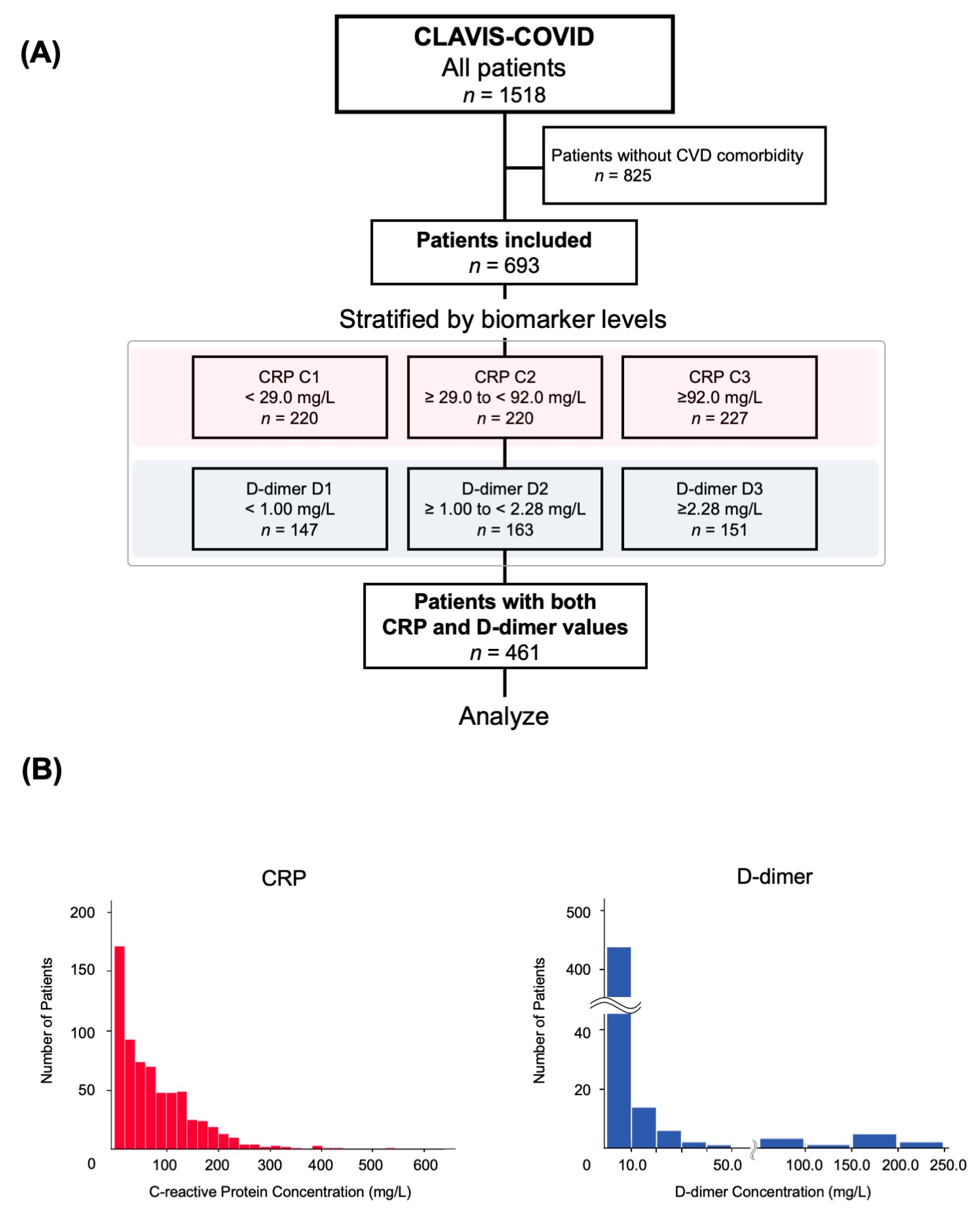
2.1. Study Design and Patient Population

CLAVIS-COVID is a case-control study that included 693 consecutive patients with pre-existing CVD and COVID-19 who were admitted to 49 nationwide Japanese acute care hospitals between 1 January 2020 and 31 May 2020. The infection of COVID-19 was diagnosed on the basis of a positive polymerase chain reaction (PCR) result from nasal or pharyngeal swabs in all patients.
In the present study, the main population consisted of patients with pre-existing CVD or their risk factors (hypertension, diabetes mellitus, or dyslipidemia). Pre-existing CVD was defined as history and/or manifestations of heart failure, coronary artery disease, myocardial infarction, peripheral artery disease, valvular heart disease, cardiac arrhythmia, pericarditis, myocarditis, congenital heart disease, pulmonary hypertension, deep vein thrombosis, pulmonary embolism, aortic dissection, aortic aneurysm, cerebral infarction/transient ischemic attack, the use of cardiac devices (pacemaker, implantable cardioverter defibrillation, cardiac resynchronization therapy, and left ventricular assist device), heart transplantation, and cardiac arrest. Detailed definitions of each comorbidity have been described in the literature [14]. Patients who were under 20 years of age at admission were excluded from this study.
The study protocol conformed to the ethical guidelines of the 1975 Declaration of Helsinki. The study protocol, including the use of an opt-out consent method, was approved by the ethics committee of Toho University Omori Medical Center (no. M20253), and each of the ethics committees of all participating institutions. This clinical study was registered with the University Hospital Medical Information Network Clinical Trial Registry, in accordance with the International Committee of Medical Journal Editors (UMIN-ID: UMIN000040598).

2.2. Data Collection

Our dataset referred to the case report form proposed by the International Severe Acute Respiratory and Emerging Infection Consortium [15]. The collected baseline clinical information, which included symptoms, demographics, medical history, home medications, baseline comorbidities, physical findings, laboratory tests, X-ray and chest CT findings, electrocardiogram and cardiac echocardiography results, treatment information, and outcomes, were obtained from electronic medical records using data collection forms. Additionally, all laboratory and imaging data were collected at the time of admission, which was considered the date of COVID-19 onset if the patient presented with symptoms of COVID-19. In the case that the patient had no symptoms on admission, the date of onset was defined as the day of the first positive PCR test.

2.3. Decision of Hospitalization and Discharge in Patients with COVID-19

During the period of patient enrollment, between 1 January 2020 and 31 May 2020, the Japanese government mandated the hospitalization of all patients diagnosed with COVID-19 through PCR testing, regardless of their severity. The hospitals participating in the current study fundamentally followed this government recommendation. The triaging and discharge thresholds of all COVID-19 patients were pre-defined according to the Japanese government guidelines for COVID-19 management [16]. To prevent a shortage of hospital beds, the Japanese health ministry notified municipalities to have COVID-19 patients with mild or no symptoms stay at accommodation facilities. The ministry guidelines encourage the patient to stay in-hospital until (1) systemic conditions and respiratory symptoms are stable, (2) body temperature is consistently under 37.5 °C for at least 24 h, and (3) negative PCR results are confirmed twice, at least 12 h apart.

2.4. Biomarker Measurement

The assays for CRP and D-dimer were performed in the laboratory of each study site. The assays included latex agglutination and latex turbidimetry (Supplemental Table S1A,B). Data on laboratory values for these biomarkers were reviewed, and the maximum and final levels prior to discharge for each biomarker were recorded. Patients were categorized by the tertiles of CRP (C1, <29.0 mg/L; C2, ≥29.0 to <92.0 mg/L; C3, ≥92.0 mg/L) and D-dimer (D1, <1.00 mg/L; D2, ≥1.00 to <2.28 mg/L; D3, ≥2.28 mg/L) concentrations at the timing of admission. The threshold was set on the values from the lowest tertile (CRP, 29.0 mg/L; D-dimer, 1.00 mg/L), and a cumulative incidence of the primary outcome was analyzed (“both values above threshold” vs. “either of the values above threshold” vs. “neither value above threshold”). For sensitivity analyses, we set three additional cut-off values, and the incidence of in-hospital death was evaluated: (1) Thresholds from the highest tertiles (CRP, 92.0 mg/L; D-dimer, 2.28 mg/L); (2) thresholds from the clinically relevant cut-off values (CRP, 50.0 mg/L; D-dimer, 1.00 mg/L) [17,18,19,20], and (3) thresholds calculated by ROC analysis for each biomarker.

2.5. Study Outcome

All patients enrolled in this study had in-hospital information available until 8 November 2020, the deadline of data transfer. The primary outcome of this study was all-cause in-hospital mortality. The secondary outcomes were the major cardiovascular events during admission. Major cardiovascular events were defined as the composite of stroke, coronary artery disease including myocardial infarction, acute decompensated heart failure, the incidence of moderate or severe valvular heart disease, myocarditis, embolism in large vessels, aortic dissection, aortic aneurysm, and peripheral artery disease. Designated data entry operators and data managers in each institution were able to access the REDCap [21] website to register and edit the case data. The credibility of the data was maintained systematically through a checking system by cardiologists in each institution. Moreover, the quality of the reporting was verified by the core investigator (Shunsuke Kuroda), and queries were conducted to ensure quality.

2.6. Statistical Analysis
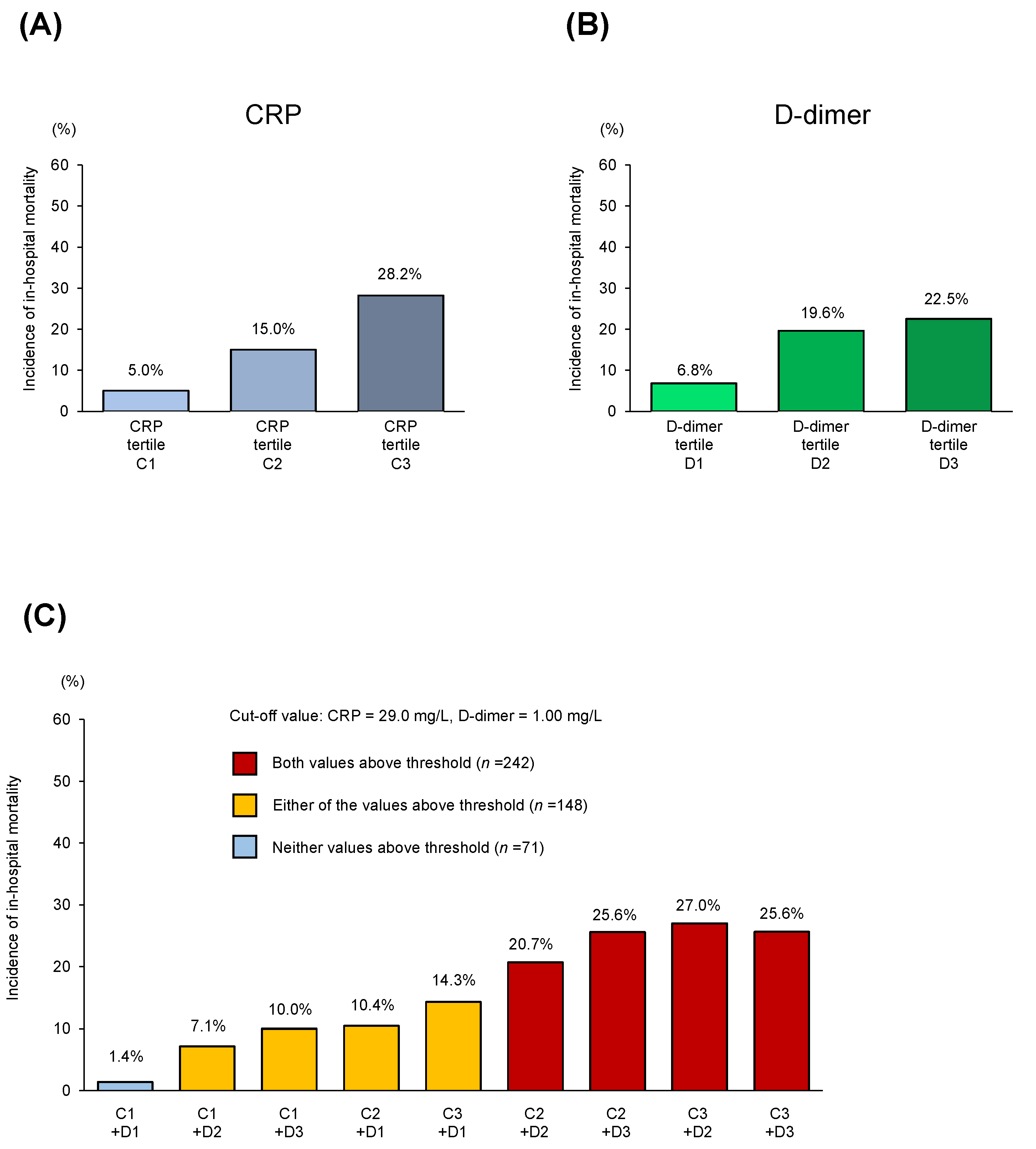
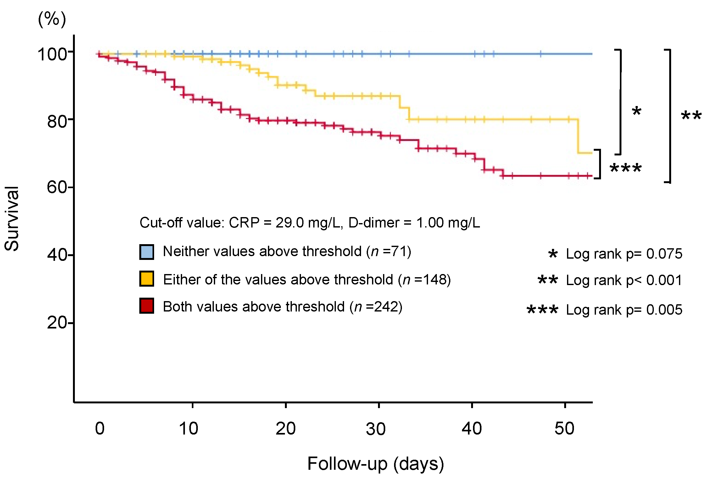
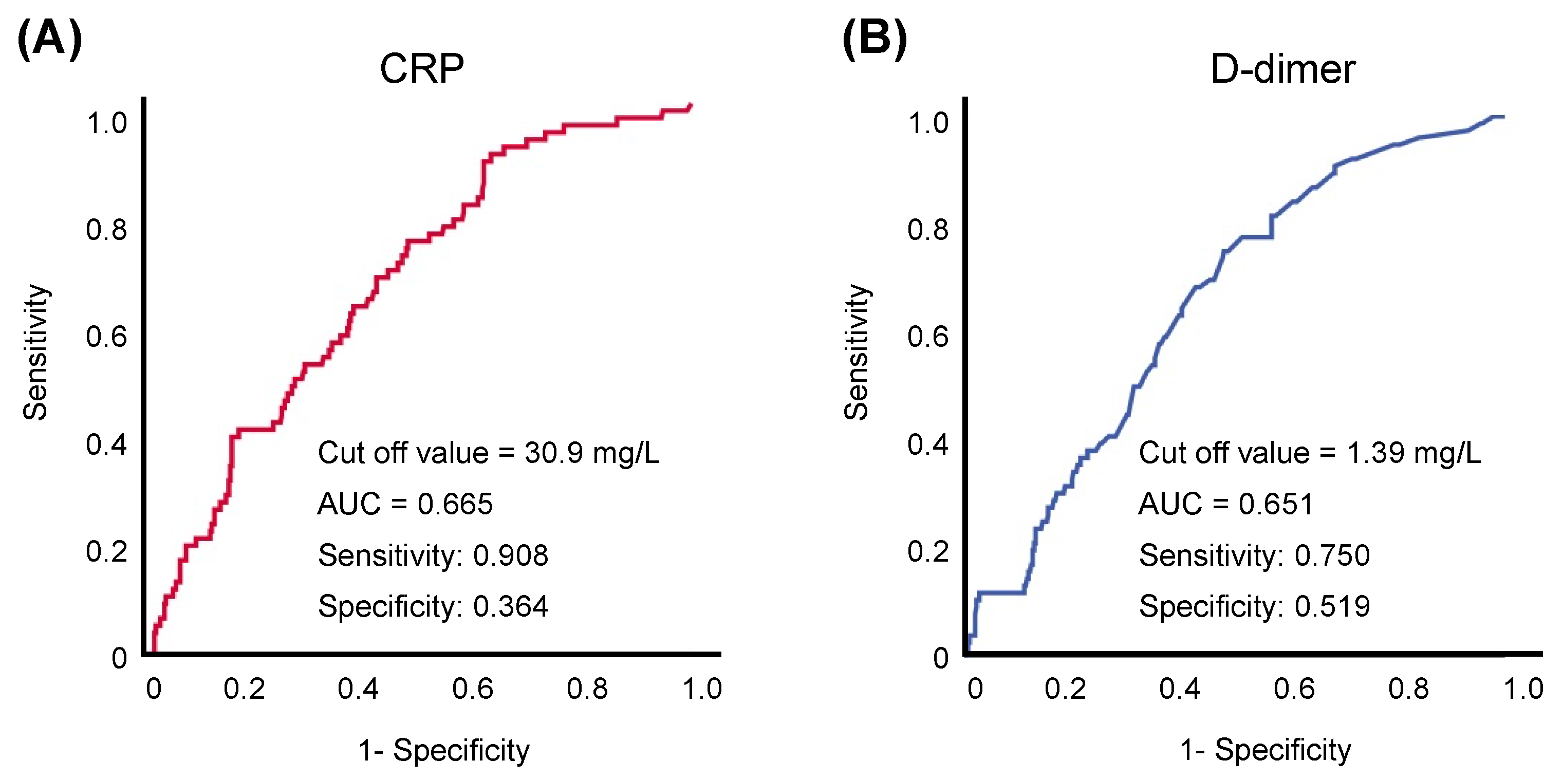
Continuous variables were expressed as mean ± standard deviation for normally distributed variables and median with interquartile range (IQR) for non-normally distributed variables. Categorical variables were expressed as percentages. Student’s t-test or Mann–Whitney’s U test was used to compare normally or non-normally distributed variables and Pearson’s chi-squared test was used to compare categorical variables. Differences in CRP and D-dimer values by group were compared using repeated-measures analysis of variance (ANOVA). The Cox proportional hazard model was used to estimate the odds of in-hospital mortality, adjusted for demographics and clinical comorbidities. Covariates included in the multivariable models were age, sex, body mass index, hypertension, diabetes mellitus, hyperlipidemia, coronary artery disease, previous history of cancer, chronic obstructive pulmonary disease, chronic kidney disease, and baseline laboratory values. Furthermore, for another model, we analyzed all clinical and laboratory parameters for their prognostic value in univariate analysis and include only variables with p < 0.1 in multivariate analysis. Cumulative incidence and adjusted odds of the primary outcome were analyzed to evaluate the combination of CRP and D-dimer levels (“both above threshold” vs. “either of the values above threshold” vs. “neither above threshold”). The Kaplan–Meier method was used to evaluate the impact of the initial CRP and D-dimer levels on subsequent in-hospital death and was compared using the log-rank statistic. We also performed sensitivity analysis based on the clinical threshold provided in the Biomarker Measurement section. For all statistical analyses, significance was defined at a p value of <0.05. Data were analyzed using SPSS version 26 (IBM Corp., Armonk, NY, USA).

3. Results

First, a total of 693 patients with a history of CVD (64.8% male; median age, 70.0 years old) were analyzed (Figure 1A). Among them, the proportion of Japanese patients was 96.1%, and the median length of hospital stay was 18 (IQR, 11–29) days. CRP and D-dimer levels were measured at the time of admission in 667 (96.2%) and 461 (66.5%) patients, respectively. The median baselines were 58.3 (IQR, 18.2–116.0) mg/L and 1.5 (IQR, 0.8–3.0) mg/L, respectively.
The distribution of CRP and D-dimer levels is shown in Figure 1B. Among the 461 patients with initial values of both CRP and D-dimer, in-hospital mortality was 16.5% (n = 76). Figure 2 demonstrates the in-hospital mortality among the patients in this study stratified by the initial CRP (Figure 2A) and D-dimer levels (Figure 2B). In-hospital death was concordantly associated with baseline CRP values (C1, 5.0%; C2, 15.0%; C3, 28.2% [p < 0.001]) and D-dimer values (D1, 6.8%; D2, 19.6%; D3, 22.5% [p = 0.001]).
Patients with initial values of both CRP and D-dimer (n = 461) were divided into three groups (“both values above threshold” vs. “either of the values above threshold” vs. “neither value above threshold”; Table 1) with a cut-off value (CRP, 29.0 mg/L; D-dimer, 1.00 mg/L) for a cumulative analysis. The cumulative evaluation demonstrated that patients in the “neither value above threshold” group were at the lowest risk for in-hospital mortality (1.4%; Figure 2C). Kaplan–Meier estimates demonstrated that the “Both” (both values above threshold) group showed significantly higher crude rates of in-hospital mortality compared with the patients in the “Either” (either of the values above threshold) or the “Neither” (neither value above threshold) groups (Both vs. Either, p = 0.005; Both vs. Neither, p < 0.001; Figure 3). Notably, patients in the “Neither” group showed no in-hospital deaths within 50 days of hospitalization. Univariate cox hazard proportional analysis demonstrated that the elevation of both biomarkers was an independent risk factor for in-hospital mortality (hazard ratio (HR), 3.46; 95% confidence interval (CI, 1.72–6.96)), while the single elevation of the D-dimer was not (HR, 1.00; 95% CI, 1.00–1.01; Supplemental Table S2). After adjustment, the elevation of both biomarkers remained an independent risk factor for in-hospital mortality (HR, 2.97; 95% CI, 1.57–5.60; Table 2). Another model also demonstrated that the elevation of both biomarkers remained an independent risk factor for in-hospital mortality (HR, 3.88; 95% CI, 1.19–12.7; Supplemental Table S3).
Supplemental Figure S1A demonstrates the outcomes when a threshold is set on the second cut-off value on each tertile (CRP, 92.0 mg/L; D-dimer, 2.28 mg/L). Kaplan–Meier analysis demonstrated that the “Both” group showed significantly higher crude rates of in-hospital mortality compared with the patients in the “Neither” group (Both vs. Neither; p = 0.024; Supplemental Figure S1A). Furthermore, even when we set the cut-off value along with previous literature (CRP, 50.0 mg/L; D-dimer, 1.00 mg/L), the “Both” group still had higher crude rates of in-hospital mortality compared with the patients in the “Neither” group (Both vs. Either, p = 0.119; Both vs. Neither; p < 0.001; Supplemental Figure S1B). In addition, the ROC analysis demonstrated that the threshold for CRP and D-dimer were 30.9 mg/L and 1.39 mg/L, respectively (Figure 4A,B). When we set the cut-off value based on this analysis, the “Both” group still had higher crude rates of in-hospital mortality compared with the patients in the “Neither” group (Both vs. Either, p = 0.002; Both vs. Neither; p < 0.001; Figure 5).
Supplemental Table S4 summarizes the initial, maximum, and final (prior to discharge) values of each biomarker stratified by the in-hospital death group and survival group. During their hospital stay, most of the patients experienced a steady increase in biomarker levels, regardless of in-hospital outcomes. However, the increment between baseline and peak values were higher in deceased patients compared to survivors (CRP, 117 mg/L vs. 0.15 mg/L (p < 0.001); D-dimer, 6.05 mg/L vs. 0.00 mg/L (p < 0.001); Supplemental Table S5). Furthermore, the timing of peak values of both biomarkers in the in-hospital group was significantly proximal to the discharge timing (i.e., delayed from admission; Supplemental Table S6). Supplemental Figure S2 demonstrates the proportion of patients whose peak biomarker values were recorded at the indicated time points (days from admission). In particular, while almost half of the patients in the survival group showed the highest CRP values on day 0 (i.e., at the time of admission), approximately 80% of patients in the in-hospital death group were detected after day 1. The measurement of other biomarkers is also shown as a comparison of CRP and D-dimer dynamics (Supplemental Figure S3A–E).

4. Discussion

In our study, the following key points were demonstrated. First, baseline CRP and D-dimer values were both associated with in-hospital mortality, even in patients with pre-existing CVD. Second, the combination of baseline CRP and D-dimer levels can predict in-hospital mortality. Finally, our study highlighted that patients with initial values of CRP (<29.0 mg/L) and D-dimer (<1.00 mg/L) showed the lowest in-hospital death during the 50 days of hospitalization.
CVD and its risk factors have been repeatedly demonstrated as major predictors of adverse outcomes in COVID-19 patients [3,4,5,22]. However, as CVD risk factors include a wide range of conditions (hypertension, diabetes, and hyperlipidemia), a considerable number of patients with COVID-19 meet this definition [5]. Therefore, further identification of risk factors for poor outcomes among patients with CVD crucial in improving disease management and patient outcomes. A number of recent studies have demonstrated that CRP and D-dimer levels are associated with the severity of COVID-19 symptoms and prolonged hospital stay [8,9,12,19,23,24,25,26,27]. In a multicenter prospective observational cohort study from Europe, Knight et al. demonstrated that CRP was an essential biomarker for risk stratification in COVID-19 [28]. Moreover, Smilowitz et al. demonstrated that CRP was strongly associated with critical illness and mortality [29]. In their study, they analyzed the initial CRP value among approximately 2500 patients who were hospitalized for COVID-19 infection during a pandemic period in New York. Our study adds that the clinical impact of CRP levels could be further strengthened by combining the results of D-dimer level measurements. In particular, the negative predictive value of the combination approach was substantial in our study, demonstrating no mortality within 50 days when neither biomarker was elevated. In line with our results, Spanish investigators recently provided a protocol for outpatient management for patients with COVID-19, stating that CRP and D-dimer levels could extract groups with very low rates of admission and no mortality [30].
We set our threshold based on tertiles considering the applicability of the study results (cut-off values of these biomarkers need to be set according to the distribution of the population). Although we were able to provide directionally similar results with absolute values set from previous clinical studies, regional disparities and differences in cut-off values are important aspects for interpreting the outcome of the present study. Significant differences in fatalities between Europe and Asia due to COVID-19 have been documented [31]. In our study, the highest tertile of CRP value (92.0 mg/L) was similar to the cut-off value defined by Smilowitz et al. (108 mg/L) [29]. The proportion of in-hospital mortality among patients in their cohort was approximately equivalent to that in our study (32.2% vs. 28.8%). Another study also demonstrated that the in-hospital mortality rate was 29.2% (33/113 patients) among COVID-19 patients with a CRP level of >100 mg/L [32]. Finally, a large-scale study using mathematical models also concluded that, when appropriately adjusted, the prevalence of mortality is quite similar across eight countries, including Europe, the United States, and East Asia [33]. Our findings underscore the multiple-step approach for biomarker interpretation in predicting the prognosis of COVID-19 [29,34,35].
Another strength of this study is that we elucidated not only the association between initial biomarker levels and clinical outcomes, but also unveiled transient changes along with the outcomes. Our results demonstrated that the peak value of biomarker timing was significantly proximal to the discharge timing in the in-hospital death group compared to in the survivor group, which was consistent with previous literature [36]. The findings of our study have significant clinical implications in that elevated CRP values during admission could be a sign of subsequent worse prognosis in patients with COVID-19 and pre-existing CVD. Indeed, while almost half of the patients in the survival group showed that their initial CRP values were the highest, the CRP values were further increased among approximately 80% of patients in the in-hospital death group. Furthermore, the increments between baseline and peak values of both CRP and D-dimer were higher in the in-hospital death group than in the survivor group. These tendencies were also detected in other biomarkers, such as ferritin and procalcitonin. Given that CRP and D-dimer require a minimal number of resources and are commonly tested in medical hospitals [29,37,38], it is reasonable to routinely follow these biomarkers to predict the clinical trajectory of COVID-19.
There are some other limitations to the present study that should be considered when interpreting the results. First, this study was a retrospective multicenter observational study; missing values of baseline serum biomarkers, especially IL-6, troponin, and BNP/NT-proBNP, which limited our further analysis. We did not unify the way of testing cardiac troponin (high-sense troponin or troponin) or BNP (BNP/NT-proBNP), which was also a limitation of our registry. In addition, as mentioned above, patients with a relatively stable clinical course were not transiently checked for biomarkers; therefore, there might have been a potential selection bias in interpreting the data. Furthermore, some values of biomarkers, especially for D-dimer, may not be comparable among the assays and manufacturers, although previous literature from Japan demonstrated that the current interassay imprecision is low [39,40]. Secondly, since this study targeted patients with COVID-19 and pre-existing CVD, the number of patients was relatively small. Further, it is unclear whether our results can be extrapolated to COVID-19 patients in other countries. In particular, the Japanese government mandated the hospitalization of all COVID-19 patients during patient enrollment; thus, the severity of hospitalized patients would be lower than that of other areas. A larger study with international collaboration is warranted for a robust evaluation of the relationship between biomarkers and COVID-19 outcomes. Finally, we did not evaluate the impact of treatment, especially for anticoagulation and steroids, which could confound the systemic immune response and transient biomarker levels [8]. However, despite these limitations, our study demonstrated that both CRP and D-dimer levels may be useful predictors for in-hospital mortality rates in COVID-19 patients with pre-existing CVD. In conclusion, our CLAVIS-COVID study revealed that the majority of patients had biomarker elevation representative of marked inflammation or hypercoagulopathy at hospital presentation in patients with COVID-19 and pre-existing CVD. The combination of these biomarkers may predict the incremental prognostic information for risk stratification and provide a framework for rapid risk estimation.

Supplementary Materials

The following are available online at https://www.mdpi.com/article/10.3390/jcm10143086/s1, Figure S1: Sensitivity analysis with additional threshold values to evaluate the incidence of in-hospital death, Figure S2: Proportion of patients whose peak CRP and D-dimer values were recorded at the indicated time points (days from admission), Figure S3: Trajectories of other biomarkers over time among survival and in-hospital deaths, Table S1: Assays and Manufactures for C-Reactive Protein and D-dimer Measurements, Table S2: Univariate cox proportional hazard analysis for predictor of in-hospital mortality, Table S3: Multivariate cox proportional hazard analysis for predictor of in-hospital mortality, Table S4: Biomarker levels at each timing of admission, peak, and final examination, Table S5: The increment of biomarker values between baseline and peak, Table S6: Timing of peak value of each biomarker (Days from discharge).

Author Contributions

S.K. (Shun Kohsaka) takes responsibility for the content of the manuscript, including the data and analysis. H.K., S.K. (Shun Kuroda), T.K., Y.M. and S.M. conceived the idea, designed and supervised the study, drafted the manuscript, and had full access to all of the data and take responsibility for the integrity of the data. H.K., A.N., T.K., T.Y., S.T., Y.M. and S.M. collected data. H.K. and S.K. (Shun Kohsaka), analyzed data and performed statistical analysis. All authors have read and agreed to the published version of the manuscript.

Funding

This research received no external funding.

Institutional Review Board Statement

The study was conducted according to the guidelines of the Declaration of Helsinki, and approved by the ethics committee of Toho University Omori Medical Center (1 June 2020; Protocol no. M20253), and each of the ethics committees of all participating institutions. This clinical study was registered with the University Hospital Medical Information Network Clinical Trial Registry, in accordance with the International Committee of Medical Journal Editors (UMIN-ID: UMIN000040598).

Informed Consent Statement

We used an opt-out consent method, which was approved by the ethics committee of Toho University Omori Medical Center and each of the ethics committees of all participating institutions.

Data Availability Statement

The data presented in this study are available on request from the corresponding author.

Acknowledgments

We would like to thank all study coordinators, investigators, and the patients who participated in the CLAVIS-COVID registry.

Conflicts of Interest

Yonetsu belongs to the endowed departments of Abbott Vascular Japan, Boston Scientific Japan, Japan Lifeline, WIN International, and Takeyama KK. Kohsaka received unrestricted research grants from the Department of Cardiology, Keio University School of Medicine provided by Daiichi Sankyo Co., Ltd. and Bristol-Meyers Squibb, and lecture fees from AstraZeneca and Bristol-Meyers Squibb. Torii received unrestricted research grants from Japan Medical Device Technology Co., Ltd., Boston Scientific Japan and Asahi Intecc Co., Ltd., received an honorarium from Boston Scientific Japan, Abbott Vascular Japan, and Medtronic. Matsue is affiliated with a department endowed by Philips Respironics, ResMed, Teijin Home Healthcare, and Fukuda Denshi, received an honorarium from Otsuka Pharmaceutical Co. and Novartis Japan, received consultant fee from Otsuka Pharmaceutical Co., and joint research funds from Otsuka Pharmaceutical Co. and Pfizer Inc.

References

  1. Wiersinga, W.J.; Rhodes, A.; Cheng, A.C.; Peacock, S.J.; Prescott, H.C. Pathophysiology, Transmission, Diagnosis, and Treatment of Coronavirus Disease 2019 (COVID-19): A Review. JAMA 2020, 324, 782–793. [Google Scholar] [CrossRef]
  2. Coronavirus COVID-19 Global Cases by the Center for Systems Science and Engineering (CSSE) at Johns Hopkins University (JHU). Available online: https://coronavirus.jhu.edu/map.html (accessed on 14 February 2021).
  3. Wu, Z.; McGoogan, J.M. Characteristics of and Important Lessons From the Coronavirus Disease 2019 (COVID-19) Outbreak in China: Summary of a Report of 72,314 Cases From the Chinese Center for Disease Control and Prevention. JAMA 2020, 323, 1239–1242. [Google Scholar] [CrossRef]
  4. Li, X.; Guan, B.; Su, T.; Liu, W.; Chen, M.; Waleed, K.B.; Guan, X.; Gary, T.; Zhu, Z. Impact of cardiovascular disease and cardiac injury on in-hospital mortality in patients with COVID-19: A systematic review and meta-analysis. Heart 2020, 106, 1142–1147. [Google Scholar] [CrossRef] [PubMed]
  5. Bae, S.; Kim, S.R.; Kim, M.N.; Shim, W.J.; Park, S.M. Impact of cardiovascular disease and risk factors on fatal outcomes in patients with COVID-19 according to age: A systematic review and meta-analysis. Heart 2021, 107, 373–380. [Google Scholar] [CrossRef]
  6. Guo, T.; Fan, Y.; Chen, M.; Wu, X.; Zhang, L.; He, T.; Wang, H.; Wan, J.; Wang, X.; Lu, Z. Cardiovascular Implications of Fatal Outcomes of Patients With Coronavirus Disease 2019 (COVID-19). JAMA Cardiol. 2020, 5, 811–818. [Google Scholar] [CrossRef] [PubMed] [Green Version]
  7. Shi, S.; Qin, M.; Shen, B.; Cai, Y.; Liu, T.; Yang, F.; Gong, W.; Liu, X.; Liang, J.; Zhao, Q.; et al. Association of Cardiac Injury With Mortality in Hospitalized Patients With COVID-19 in Wuhan, China. JAMA Cardiol. 2020, 5, 802–810. [Google Scholar] [CrossRef] [PubMed] [Green Version]
  8. Wu, C.; Chen, X.; Cai, Y.; Xia, J.; Zhou, X.; Xu, S.; Huang, H.; Zhang, L.; Zhou, X.; Du, C.; et al. Risk Factors Associated With Acute Respiratory Distress Syndrome and Death in Patients With Coronavirus Disease 2019 Pneumonia in Wuhan, China. JAMA Intern Med. 2020, 180, 934–943. [Google Scholar] [CrossRef] [Green Version]
  9. Levi, M.; Thachil, J.; Iba, T.; Levy, J.H. Coagulation abnormalities and thrombosis in patients with COVID-19. Lancet Haematol. 2020, 7, e438–e440. [Google Scholar] [CrossRef]
  10. Malik, P.; Patel, U.; Mehta, D.; Patel, N.; Kelkar, R.; Akrmah, M.; Gabrilove, J.L.; Sacks, H. Biomarkers and outcomes of COVID-19 hospitalisations: Systematic review and meta-analysis. BMJ Evid. Based Med. 2020, 3, 107–108. [Google Scholar] [CrossRef]
  11. Liu, F.; Li, L.; Xu, M.; Wu, J.; Luo, D.; Zhu, Y.; Li, B.; Song, X.; Zhou, X. Prognostic value of interleukin-6, C-reactive protein, and procalcitonin in patients with COVID-19. J. Clin. Virol. 2020, 127, 104370. [Google Scholar] [CrossRef]
  12. Luo, X.; Zhou, W.; Yan, X.; Guo, T.; Wang, B.; Xia, H.; Ye, L.; Xiong, J.; Jiang, Z.; Liu, Y.; et al. Prognostic Value of C-Reactive Protein in Patients With Coronavirus 2019. Clin. Infect. Dis. 2020, 71, 2174–2179. [Google Scholar] [CrossRef]
  13. Zhang, L.; Yan, X.; Fan, Q.; Liu, H.; Liu, X.; Liu, Z.; Zhang, Z. D-dimer levels on admission to predict in-hospital mortality in patients with Covid-19. J. Thromb. Haemost. 2020, 18, 1324–1329. [Google Scholar] [CrossRef]
  14. Matsumoto, S.; Kuroda, S.; Sano, T.; Kitai, T.; Yonetsu, T.; Kohsaka, S.; Torii, S.; Kishi, T.; Komuro, I.; Hirata, K.I.; et al. Clinical and Biomarker Profiles and Prognosis of Elderly Patients With Coronavirus Disease 2019 (COVID-19) With Cardiovascular Diseases and/or Risk Factors. Circ. J. 2021, 85, 921–928. [Google Scholar] [CrossRef]
  15. International Severe Acute Respiratory and emerging Infection Consortium. Clinical data collection: The COVID-19 case report forms (CRF). Available online: https://isaric.org/ (accessed on 1 December 2020).
  16. Ministry of Health, Labour and Welfare. Clinical management of patients with COVID-19 [in Japanese]. Available online: https://www.mhlw.go.jp/stf/seisakunitsuite/bunya/0000121431_00111.html (accessed on 1 June 2020).
  17. Daniels, J.M.; Schoorl, M.; Snijders, D.; Knol, D.L.; Lutter, R.; Jansen, H.M.; Boersma, W.G. Procalcitonin vs C-reactive protein as predictive markers of response to antibiotic therapy in acute exacerbations of COPD. Chest 2010, 138, 1108–1115. [Google Scholar] [CrossRef] [PubMed]
  18. Komiya, K.; Ishii, H.; Teramoto, S.; Takahashi, O.; Eshima, N.; Yamaguchi, O.; Ebi, N.; Murakami, J.; Yamamoto, H.; Kadota, J. Diagnostic utility of C-reactive protein combined with brain natriuretic peptide in acute pulmonary edema: A cross sectional study. Respir. Res. 2011, 12, 83. [Google Scholar] [CrossRef] [PubMed] [Green Version]
  19. Zhou, F.; Yu, T.; Du, R.; Fan, G.; Liu, Y.; Liu, Z.; Xiang, J.; Wang, Y.; Song, B.; Gu, X.; et al. Clinical course and risk factors for mortality of adult inpatients with COVID-19 in Wuhan, China: A retrospective cohort study. Lancet 2020, 395, 1054–1062. [Google Scholar] [CrossRef]
  20. Zhang, G.; Zhang, J.; Wang, B.; Zhu, X.; Wang, Q.; Qiu, S. Analysis of clinical characteristics and laboratory findings of 95 cases of 2019 novel coronavirus pneumonia in Wuhan, China: A retrospective analysis. Respir. Res. 2020, 21, 74. [Google Scholar] [CrossRef] [PubMed]
  21. Harris, P.A.; Taylor, R.; Thielke, R.; Payne, J.; Gonzalez, N.; Conde, J.G. Research electronic data capture (REDCap)--a metadata-driven methodology and workflow process for providing translational research informatics support. J. Biomed. Inform. 2009, 42, 377–381. [Google Scholar] [CrossRef] [PubMed] [Green Version]
  22. Gonzalez-Jaramillo, N.; Low, N.; Franco, O.H. The double burden of disease of COVID-19 in cardiovascular patients: Overlapping conditions could lead to overlapping treatments. Eur. J. Epidemiol. 2020, 35, 335–337. [Google Scholar] [CrossRef] [Green Version]
  23. Gao, Y.; Li, T.; Han, M.; Li, X.; Wu, D.; Xu, Y.; Zhu, Y.; Liu, Y.; Wang, X.; Wang, L. Diagnostic utility of clinical laboratory data determinations for patients with the severe COVID-19. J. Med. Virol. 2020, 92, 791–796. [Google Scholar] [CrossRef] [PubMed]
  24. Liang, W.; Liang, H.; Ou, L.; Chen, B.; Chen, A.; Li, C.; Li, Y.; Guan, W.; Sang, L.; Lu, J.; et al. Development and Validation of a Clinical Risk Score to Predict the Occurrence of Critical Illness in Hospitalized Patients With COVID-19. JAMA Intern Med. 2020, 180, 1081–1089. [Google Scholar] [CrossRef] [PubMed]
  25. Iba, T.; Levy, J.H.; Levi, M.; Connors, J.M.; Thachil, J. Coagulopathy of Coronavirus Disease 2019. Crit. Care Med. 2020, 48, 1358–1364. [Google Scholar] [CrossRef]
  26. Chen, N.; Zhou, M.; Dong, X.; Qu, J.; Gong, F.; Han, Y.; Qiu, Y.; Wang, J.; Liu, Y.; Wei, Y.; et al. Epidemiological and clinical characteristics of 99 cases of 2019 novel coronavirus pneumonia in Wuhan, China: A descriptive study. Lancet 2020, 395, 507–513. [Google Scholar] [CrossRef] [Green Version]
  27. Bansal, A.; Singh, A.D.; Jain, V.; Aggarwal, M.; Gupta, S.; Padappayil, R.P.; Nadeem, M.; Joshi, S.; Mian, A.; Greathouse, T.; et al. The association of D-dimers with mortality, intensive care unit admission or acute respiratory distress syndrome in patients hospitalized with coronavirus disease 2019 (COVID-19): A systematic review and meta-analysis. Heart Lung. 2021, 50, 9–12. [Google Scholar] [CrossRef] [PubMed]
  28. Knight, S.R.; Ho, A.; Pius, R.; Buchan, I.; Carson, G.; Drake, T.M.; Dunning, J.; Fairfield, C.J.; Gamble, C.; Green, C.A.; et al. Risk stratification of patients admitted to hospital with covid-19 using the ISARIC WHO Clinical Characterisation Protocol: Development and validation of the 4C Mortality Score. BMJ 2020, 370, m3339. [Google Scholar] [CrossRef]
  29. Smilowitz, N.R.; Kunichoff, D.; Garshick, M.; Shah, B.; Pillinger, M.; Hochman, J.S.; Berger, J.S. C-reactive protein and clinical outcomes in patients with COVID-19. Eur. Heart J. 2021, 23, 2270–2279. [Google Scholar] [CrossRef] [PubMed]
  30. Teigell Muñoz, F.J.; García-Guijarro, E.; García-Domingo, P.; Pérez-Nieto, G.; Roque Rojas, F.; García-Peña, M.; Nieto Gallo, M.A.; Melero Bermejo, J.A.; de Guzman García-Monge, M.T.; Granizo, J.J. A safe protocol to identify low-risk patients with COVID-19 pneumonia for outpatient management. Intern Emerg. Med. 2021, 1–9. [Google Scholar]
  31. Yamamoto, N.; Bauer, G. Apparent difference in fatalities between Central Europe and East Asia due to SARS-COV-2 and COVID-19: Four hypotheses for possible explanation. Med. Hypotheses. 2020, 144, 110160. [Google Scholar] [CrossRef]
  32. Li, L.; Zhang, S.; He, B.; Chen, X.; Wang, S.; Zhao, Q. Risk factors and electrocardiogram characteristics for mortality in critical inpatients with COVID-19. Clin. Cardiol. 2020, 43, 1624–1630. [Google Scholar] [CrossRef]
  33. Goldstein, J.R.; Lee, R.D. Demographic perspectives on the mortality of COVID-19 and other epidemics. Proc. Natl. Acad. Sci. USA 2020, 117, 22035–22041. [Google Scholar] [CrossRef]
  34. Biamonte, F.; Botta, C.; Mazzitelli, M.; Rotundo, S.; Trecarichi, E.M.; Foti, D.; Torti, C.; Viglietto, G.; Torella, D.; Costanzo, F. Combined lymphocyte/monocyte count, D-dimer and iron status predict COVID-19 course and outcome in a long-term care facility. J. Transl. Med. 2021, 19, 79. [Google Scholar] [CrossRef] [PubMed]
  35. Manocha, K.K.; Kirzner, J.; Ying, X.; Yeo, I.; Peltzer, B.; Ang, B.; Li, H.A.; Lerman, B.B.; Safford, M.M.; Goyal, P.; et al. Troponin and Other Biomarker Levels and Outcomes Among Patients Hospitalized with COVID-19: Derivation and Validation of the HA(2)T(2) COVID-19 Mortality Risk Score. J. Am. Heart. Assoc. 2020, 6, e018477. [Google Scholar]
  36. Sharifpour, M.; Rangaraju, S.; Liu, M.; Alabyad, D.; Nahab, F.B.; Creel-Bulos, C.M.; Jabaley, C.S. C-Reactive protein as a prognostic indicator in hospitalized patients with COVID-19. PLoS ONE 2020, 15, e0242400. [Google Scholar] [CrossRef]
  37. Escadafal, C.; Incardona, S.; Fernandez-Carballo, B.L.; Dittrich, S. The good and the bad: Using C reactive protein to distinguish bacterial from non-bacterial infection among febrile patients in low-resource settings. BMJ Glob. Health 2020, 5, e002396. [Google Scholar] [CrossRef] [PubMed]
  38. Crawford, F.; Andras, A.; Welch, K.; Sheares, K.; Keeling, D.; Chappell, F.M. D-dimer test for excluding the diagnosis of pulmonary embolism. Cochrane Database Syst. Rev. 2016, 2016, Cd010864. [Google Scholar] [CrossRef] [Green Version]
  39. Yamamoto, Y.; Hosogaya, S.; Osawa, S.; Ichihara, K.; Onuma, T.; Saito, A.; Banba, K.; Araki, H.; Nagamine, Y.; Shinohara, K.; et al. Nationwide multicenter study aimed at the establishment of common reference intervals for standardized clinical laboratory tests in Japan. Clin. Chem. Lab. Med. 2013, 51, 1663–1672. [Google Scholar] [CrossRef] [PubMed]
  40. Oi, M.; Yamashita, Y.; Toyofuku, M.; Morimoto, T.; Motohashi, Y.; Tamura, T.; Kaitani, K.; Amano, H.; Takase, T.; Hiramori, S.; et al. D-dimer levels at diagnosis and long-term clinical outcomes in venous thromboembolism: From the COMMAND VTE Registry. J. Thromb. Thrombolysis. 2020, 49, 551–561. [Google Scholar] [CrossRef] [PubMed]
Figure 1. Flowchart describing the study design from the CLAVIS-COVID registry (A), and patient distribution of initial CRP and D-dimer concentrations (B). CVD: Cardiovascular medical condition, CRP: C-reactive protein.
Jcm 10 03086 g001
Figure 2. Mortality rate (%) among patients with pre-existing CVD and COVID-19 infection, stratified by initial CRP (A) or D-dimer (B) concentrations. Patients were categorized by the tertiles of CRP (C1, <29.0 mg/L; C2, ≥29.0 to <92.0 mg/L; C3, ≥92.0 mg/L) or D-dimer (D1, <1.00 mg/L; D2, ≥1.00 to <2.28 mg/L; D3, ≥2.28 mg/L) concentration at the timing of admission. (C) The cumulative incidence of in-hospital mortality, stratified by “both values above threshold”, “either of the values above threshold”, and “neither value above threshold”. CVD: Cardiovascular medical condition, COVID-19: Coronavirus disease 2019, CRP: C-reactive protein.
Jcm 10 03086 g002
Figure 3. Kaplan–Meier analysis demonstrating the impact of the initial CRP and D-dimer level on subsequent in-hospital death. Threshold values were CRP, 29.0 mg/L; D-dimer, 1.00 mg/L. CRP: C-reactive protein.
Jcm 10 03086 g003
Figure 4. ROC analysis demonstrating the predictive threshold values of (A) CRP and (B) D-dimer for predicting in-hospital death. Threshold values were CRP, 30.9 mg/L; D-dimer, 1.39 mg/L. CRP: C-reactive protein. AUC: Area under the curve.
Jcm 10 03086 g004
Figure 5. Kaplan–Meier analysis demonstrating the impact of the initial CRP and D-dimer level on subsequent in-hospital death. Threshold values were based on ROC analysis: CRP, 30.9 mg/L; D-dimer, 1.39 mg/L. CRP: C-reactive protein.
Jcm 10 03086 g005
Table 1. Baseline characteristics stratified by a combination of biomarkers.
VariablesNeither above Threshold (n = 71)Either above Threshold (n = 148)Both above Threshold (n = 242)p Value
Demographics
  Age, years62.0 ± 14.369.5 ± 16.370.6 ± 13.0<0.001
  Male, %53.564.969.40.045
  Japanese, %94.498.898.80.079
  BMI, kg/m225.2 ± 4.723.9 ± 4.923.9 ± 5.10.006
Comorbidities and medical history
  Hypertension, %74.675.068.20.282
  Diabetes mellitus, %22.535.848.8<0.001
  Dyslipidemia, %49.335.141.70.124
  Heart failure, %7.011.59.90.589
  Coronary artery disease,%7.011.59.50.573
  Myocardial infarction, %0.05.45.80.120
  CI/TIA, %4.26.810.30.188
  COPD, %4.23.47.40.206
  CKD, %5.67.49.50.528
  Cancer, %4.28.110.70.218
Symptoms
  Fever (>38.0 °C), %55.161.969.50.062
  Cough, %47.945.353.70.248
  Pharyngitis, %16.914.28.70.087
  Rhinorrhea, %7.04.13.70.472
  Dyspnea, %15.533.847.5<0.001
  Arthritis, %7.04.12.50.192
  Headache, %9.98.85.80.374
  Olfactory dysfunction, %9.910.15.40.167
  Asymptom, %9.99.53.70.038
4C Mortality Score8 (6–11)12 (9–14)14 (11–15)< 0.001
Physical findings
  Max body temperature38.0 (37.5–38.3)38.0 (37.6–38.5)38.0 (37.7–38.6)0.005
  Herat rate (bpm)85.0 (76.0–95.0)82.0 (73.0–95.0)88.0 (75.0–101.0)0.028
  Systolic BP (mmHg)140.0 (119.0–154.0)130.0 (117.0–150.0)130.0 (115.0–142.5)0.042
  Respiratory rate (/min)18.0 (16.0–22.0)20.0 (17.0–24.0)20.0 (18.0–25.3)0.001
  SpO2, %97.0 (96.0–98.0)96.0 (95.0–98.0)95.0 (92.0–97.0)<0.001
Laboratory data at admission
  White blood cell, /μL4800 (3760–5900)5400 (4400–7400)6600 (5000–8800)<0.001
  Lymphocyte, %24.0 (17.9–28.5)18.3 (12.9–25.4)12.3 (8.4–17.5)<0.001
  Neutrocyte, %65.2 (60.7–73.4)72.3 (63.8–80.7)80.3 (73.7–86.2)<0.001
  Eosinocyte, %0.50 (0.00–1.20)0.30 (0.00–1.15)0.00 (0.00–0.500)<0.001
  Hemoglobin, g/dl14.0 (12.8–15.1)13.4 (11.6–14.9)13.0 (11.5–14.5)0.003
  Platelet, 103/μL184.0 (149.0–238.0)178.0 (140.0–235.0)196.0 (140.0–251.0)0.657
  Creatinin, mg/dL0.77 (0.62–0.88)0.85 (0.65–1.10)0.87 (0.65–1.14)0.012
  eGFR, mL/min/1.73 m294.6 (77.4–105.5)86.1 (64.1–106.6)82.6 (60.3–109.2)0.058
  LDH, IU/L219.0 (183.5–256.0)276.5 (216.0–344.5)380.0 (265.0–501.0)<0.001
  HbA1c, %6.1 (5.9–6.8)6.2 (5.7–6.8)6.5 (6.1–7.4)0.001
  CK, U/L73.5 (49.0–108.3)84.5 (43.5–142.5)88.0 (55.0–189.0)0.089
  Serum Alb, gL4.0 (3.7–4.2)3.4 (3.0–3.8)3.0 (2.6–3.3)<0.001
Specific biomarker at admission
  CRP, mg/L7.8 (2.5–14.7)30.5 (11.9–74.9)106.0 (66.0–159.0)<0.001
  D-dimer, mg/L0.60 (0.50–0.70)0.90 (0.60–1.70)2.24 (1.49–5.31)<0.001
  FDP, μg/mL2.50 (1.60–2.95)5.25 (3.42–6.40)6.6 (5.0–12.9)<0.001
  Ferritin, ng/mL234.0 (105.5–460.0)476.0 (175.8–1000.8)752.0 (312.0–1395.0)<0.001
  Procalcitonin, ng/mL0.050 (0.020–0.750)0.100 (0.058–0.193)0.160 (0.080–0.570)<0.001
  KL-6, U/mL277.0 (187.5–398.8)276.0 (204.0–490.5)332.0 (234.3–471.5)0.071
  BNP, pg/mL11.0 (5.8–34.9)29.1 (11.7–134.0)58.9 (15.6–169.3)0.001
Length of Stay, days15.0 (11.0–25.0)19.0 (13.0–27.8)19.5 (10.0–32.0)0.097
Data are shown as mean ± standard deviation or median with interquartile range or percentage. BMI: Body mass index, CI: Cerebral infarction, TIA: Transient ischemic attack, COPD: Chronic obstructive pulmonary disease, CKD: Chronic kidney disease, BP: Blood pressure, eGFR: Estimated glomerular filtration rate, LDH: Lactic acid dehydrogenase, HbA1c: Hemoglobin A1c, CK: Creatine kinase, Alb: Albumin, CRP: C-reactive protein, FDP: Fibrin degradation products, KL-6: Krebs von den Lungen-6 antigen, BNP: Brain natriuretic peptide.
Table 2. Independent predictor of in-hospital mortality.
VariablesHRCIp Value
Age1.081.04–1.11<0.000
Male1.600.85–3.010.147
BMI1.111.04–1.180.001
Hypertension0.970.51–1.860.930
Diabetes mellitus1.060.60–1.890.833
Dyslipidemia0.940.52–1.680.831
Coronary artery disease2.091.05–4.150.035
Cancer1.260.60–2.620.541
COPD2.561.23–5.340.012
CKD1.280.60–2.750.524
Both values above threshold
(CRP = 29.0 mg/L, D-dimer = 1.00 mg/L)
2.971.57–5.600.001
The Cox proportional hazard model predicting the odds of in-hospital mortality, adjusted for demographics and clinical comorbidities. Covariates included in the multivariable models were age, sex, body mass index (BMI), hypertension, diabetes mellitus, dyslipidemia, coronary artery disease, previous history of cancer, chronic obstructive pulmonary disease (COPD), chronic kidney disease (CKD), and baseline laboratory values. HR: Hazard ratio, CI: Confidence interval, BMI: Body mass index, COPD: Chronic obstructive pulmonary disease, CKD: Chronic kidney disease, CRP: C-reactive protein.
Publisher’s Note: MDPI stays neutral with regard to jurisdictional claims in published maps and institutional affiliations.

