Combined Effect of Caspase-Dependent and Caspase-Independent Apoptosis in the Anticancer Activity of Gold Complexes with Phosphine and Benzimidazole Derivatives

 , , , , ,
Abstract
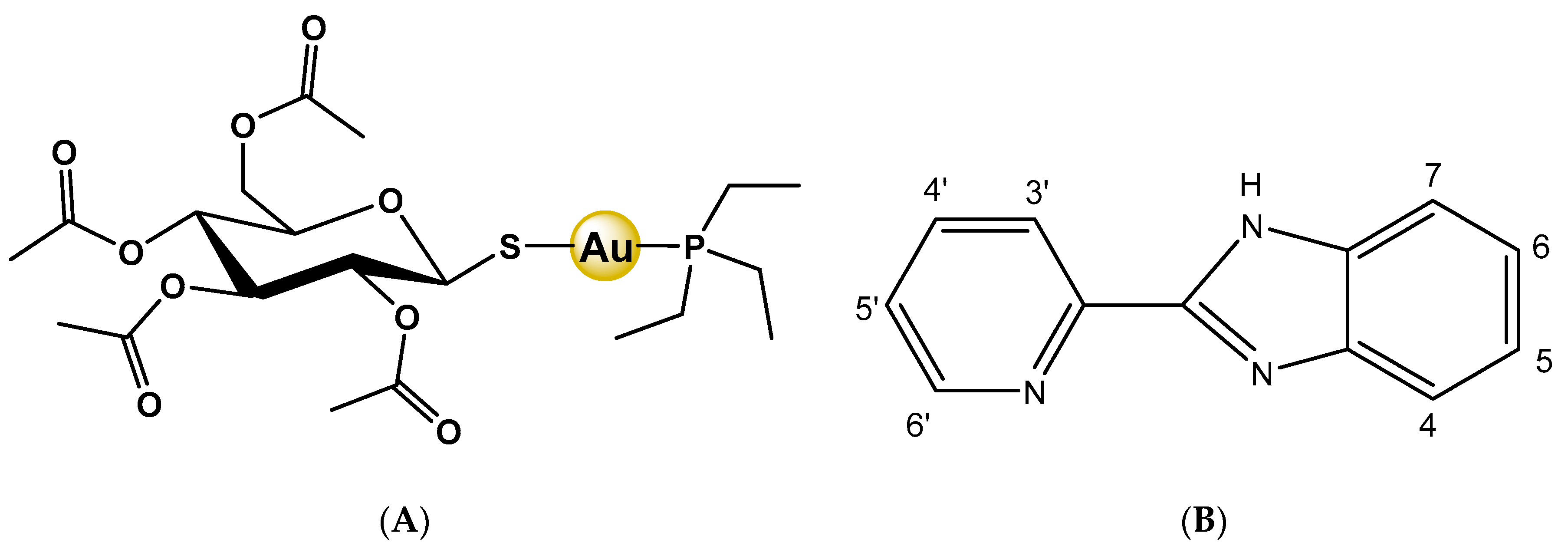
:1. Introduction
2. Results
2.1. Analytical and Spectroscopic Characterization of 1 and 2
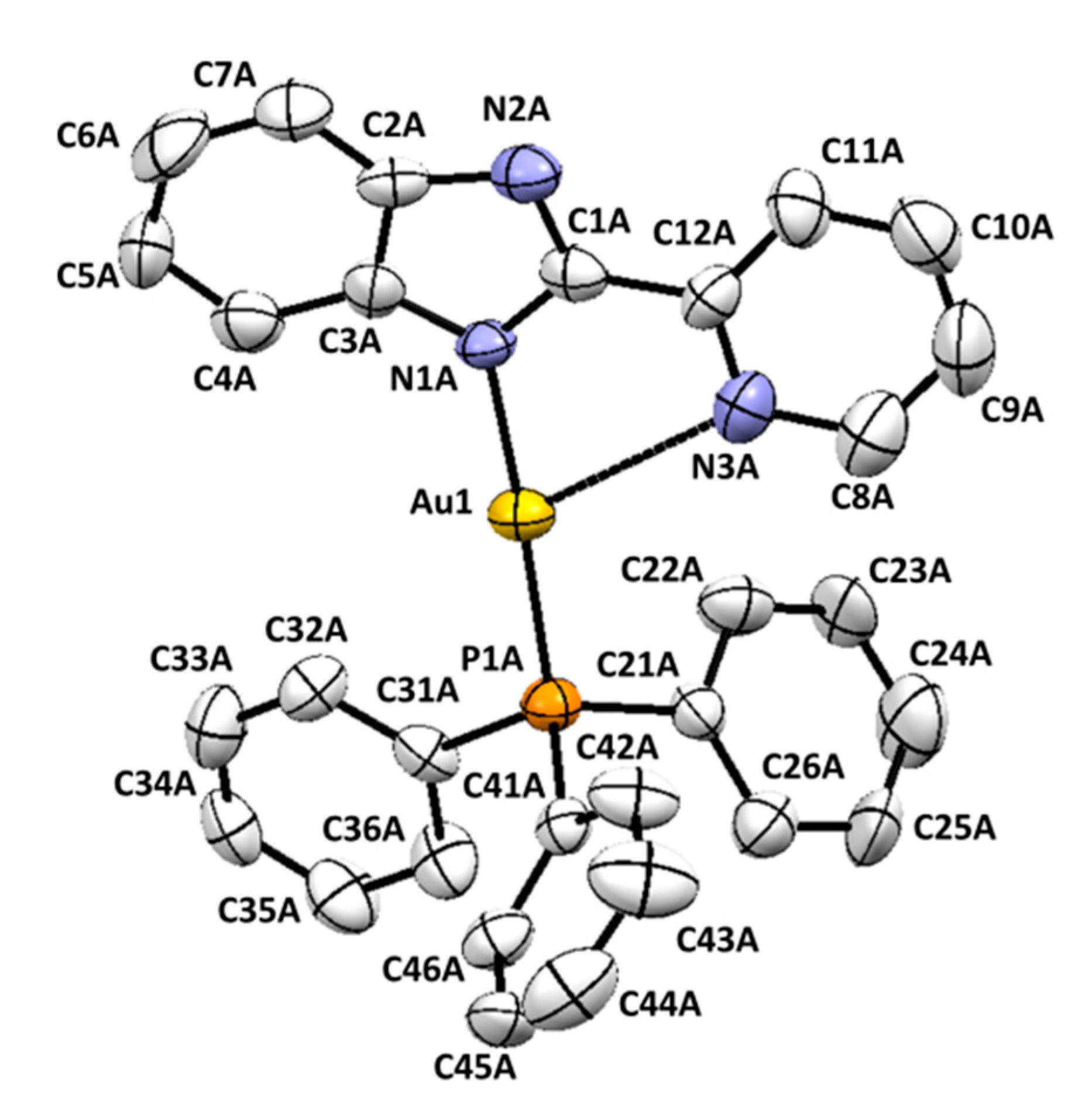
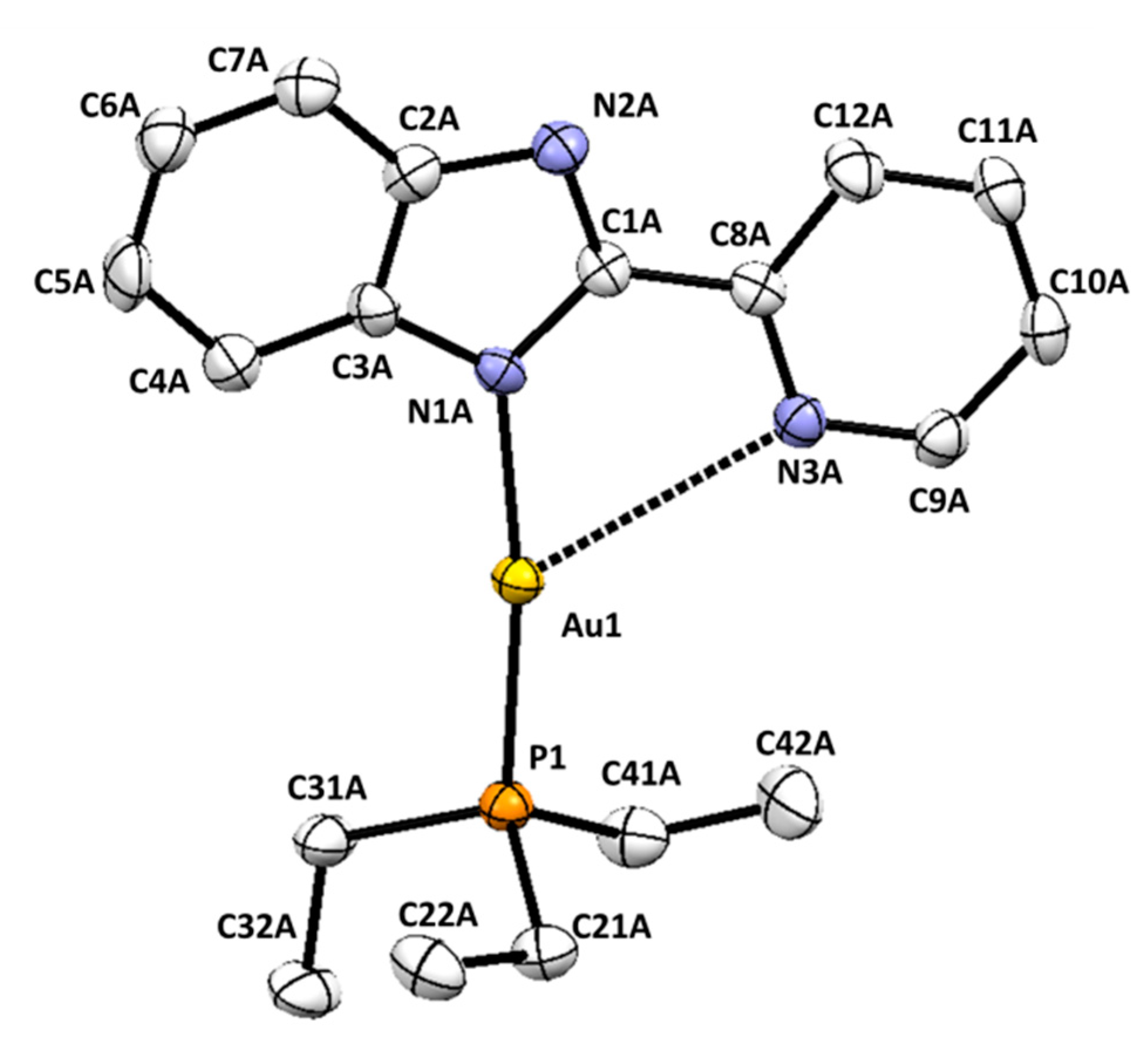
2.2. Crystal Structures of Complexes 1 and 2
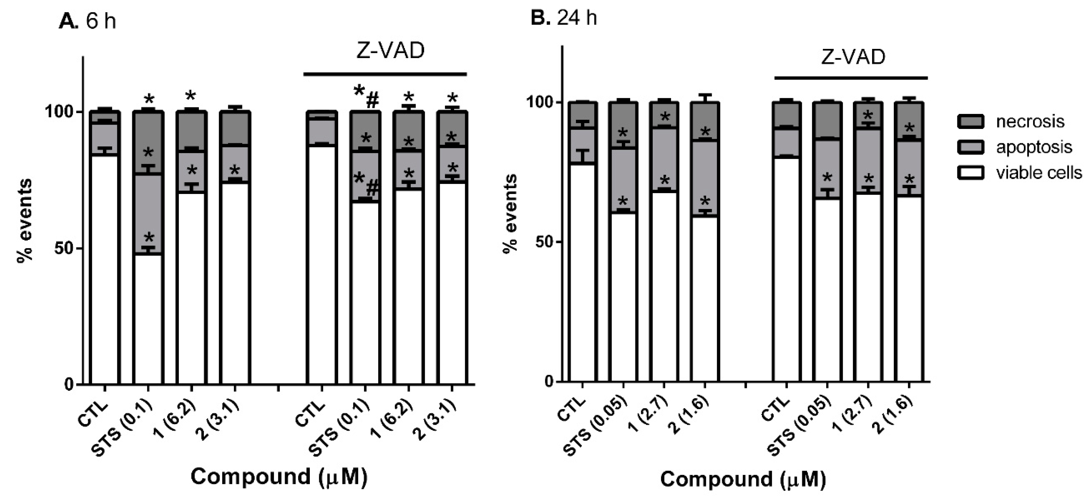
2.3. Cytotoxicity Studies and Determination of Cell Death Type
3. Discussion
4. Materials and Methods
4.1. Materials
4.2. Physical Measurements
4.3. Synthesis of the Complexes
4.3.1. (Au(pben)(PPh3)) (1)
4.3.2. (Au(pben)(PEt3)) (2)
4.4. Cell Culture
4.5. Cytotoxic Effects
4.6. Flow Cytometry Analysis of Cell Death Type
4.7. Caspase-3 Assay
4.8. Determination of ROS Production
4.9. Statistical Analysis
4.10. Crystallographic Studies
5. Conclusions
Supplementary Materials
Author Contributions
Funding
Institutional Review Board Statement
Informed Consent Statement
Data Availability Statement
Conflicts of Interest
References
- Casini, A.; Sun, R.W.; Ott, I. Medicinal chemistry of gold anticancer metallodrugs. Metallo-Drugs: Development and Action of Anticancer Agents. Met. Ions Life Sci. 2018, 18, 199–218. [Google Scholar]
- Dabrowiak, J.C. Metal Complexes for Treating Arthritis and Diabetes. In Metals in Medicine, 2nd ed.; John Wiley & Sons Ltd.: Hoboken, NJ, USA, 2017; pp. 245–263. [Google Scholar]
- Sutton, B.M. Gold compounds for rheumatoid arthritis. Gold Bull. 1986, 19, 15–16. [Google Scholar] [CrossRef] [Green Version]
- Messori, L.; Marcon, G. Gold Complexes in the Treatment of. In Metal Ions in Biological Systems: Volume 41: Metal Ions and Their Complexes in Medication; CRC Press: Boca Raton, FL, USA, 2004; pp. 279–304. [Google Scholar]
- Berners-Price, S.J.; Filipovska, A. Gold compounds as therapeutic agents for human diseases. Metallomics 2011, 3, 863–873. [Google Scholar] [CrossRef]
- Madeira, J.M.; Renschler, C.J.; Mueller, B.; Hashioka, S.; Gibson, D.L.; Klegeris, A. Novel protective properties of auranofin: Inhibition of human astrocyte cytotoxic secretions and direct neuroprotection. Life Sci. 2013, 92, 1072–1080. [Google Scholar] [CrossRef]
- Roder, C.; Thomson, M.J. Auranofin: Repurposing an old drug for a golden new age. Drugs R&D 2015, 15, 13–20. [Google Scholar] [CrossRef] [Green Version]
- Rothan, H.A.; Stone, S.; Natekar, J.; Kumari, P.; Arora, K.; Kumar, M. The FDA-approved gold drug auranofin inhibits novel coronavirus (SARS-COV-2) replication and attenuates inflammation in human cells. Virology 2020, 547, 7–11. [Google Scholar] [CrossRef]
- Mirzadeh, N.; Reddy, T.S.; Bhargava, S.K. Advances in diphosphine ligand-containing gold complexes as anticancer agents. Coord. Chem. Rev. 2019, 388, 343–359. [Google Scholar] [CrossRef]
- Mora, M.; Gimeno, M.C.; Visbal, R. Recent advances in gold–NHC complexes with biological properties. Chem. Soc. Rev. 2019, 48, 447–462. [Google Scholar] [CrossRef]
- Iacopetta, D.; Rosano, C.; Sirignano, M.; Mariconda, A.; Ceramella, J.; Ponassi, M.; Saturnino, C.; Sinicropi, M.S.; Longo, P. Is the way to fight cancer paved with gold? Metal-based carbene complexes with multiple and fascinating biological features. Pharmaceuticals 2020, 13, 91. [Google Scholar] [CrossRef]
- Marzo, T.; Massai, L.; Pratesi, A.; Stefanini, M.; Cirri, D.; Magherini, F. Replacement of the Thiosugar of Auranofin with Iodide Enhances the Anticancer Potency in a Mouse Model of Ovarian Cancer. ACS Med. Chem. Lett. 2019, 10, 656–660. [Google Scholar] [CrossRef]
- Marzo, T.; Cirri, D.; Gabbiani, C.; Gamberi, T.; Magherini, F.; Pratesi, A. Auranofin, Et3PAuCl, and Et3PAuI Are Highly Cytotoxic on Colorectal Cancer Cells: A Chemical and Biological Study. ACS Med. Chem. Lett. 2017, 8, 997–1001. [Google Scholar] [CrossRef] [PubMed]
- Gonzalez-Barcia, L.M.; Fernandez-Fariña, S.; Rodríguez-Silva, L.; Bermejo, M.R.; González-Noya, A.M.; Pedrido, R. Comparative study of the antitumoral activity of phosphine-thiosemicarbazone gold(I) complexes obtained by different methodologies. J. Inorg. Biochem. 2020, 203, 110931. [Google Scholar] [CrossRef] [PubMed]
- Yoo, M.-H.; Xu, X.-M.; Carlson, B.A.; Gladyshev, V.N.; Hatfield, D.L. Thioredoxin reductase 1 deficiency reverses tumor phenotype and tumorigenicity of lung carcinoma cells. J. Biol. Chem. 2006, 281, 13005–13008. [Google Scholar] [CrossRef] [PubMed] [Green Version]
- Casini, A.; Hartinger, C.; Gabbiani, C.; Mini, E.; Dyson, P.J.; Keppler, B.K.; Messori, L. Gold (III) compounds as anticancer agents: Relevance of gold–protein interactions for their mechanism of action. J. Inorg. Biochem. 2008, 102, 564–575. [Google Scholar] [CrossRef] [PubMed]
- Nobili, S.; Mini, E.; Landini, I.; Gabbiani, C.; Casini, A.; Messori, L. Gold compounds as anticancer agents: Chemistry, cellular pharmacology, and preclinical studies. Med. Res. Rev. 2010, 30, 550–580. [Google Scholar] [CrossRef]
- Ott, I. On the medicinal chemistry of gold complexes as anticancer drugs. J. Coord. Chem. Rev. 2009, 253, 1670–1681. [Google Scholar] [CrossRef]
- Hassan, F.; Al-Aridhi, D.T. Antitumor effect of 4-(N, N-dimethyl)-3-(3-Mercapto-5-Phenyl [1,2,4] triazol-4yl)-thiazolidin-4-one in liver carcinoma cell lines Hep G2 by (HCS) technique. Int. J. Pharm. Sci. 2015, 5, 1317–1322. [Google Scholar]
- Zhong, L.; Arnér, E.S.J.; Holmgren, A. Targeting thioredoxin reductase is a basis for cancer therapy by arsenic trioxide. Proc. Natl. Acad. Sci. USA 2007, 104, 12288–12293. [Google Scholar]
- Zhang, X.; Frezza, M.; Milacic, V.; Ronconi, L.; Fan, Y.; Bi, C.; Fregona, D.; Dou, Q.P. Inhibition of tumor proteasome activity by gold-dithiocarbamato complexes via both redox-dependent and -independent processes. J. Cell. Biochem. 2010, 109, 162–172. [Google Scholar] [CrossRef] [Green Version]
- Barnard, P.; Berners-Price, S.J. Targeting the mitochondrial cell death pathway with gold compounds. Coord. Chem. Rev. 2007, 251, 1889–1902. [Google Scholar] [CrossRef]
- González-Barcia, L.M.; Romero, M.J.; González-Noya, A.M.; Bermejo, M.R.; Maneiro, M.; Zaragoza, G.; Pedrido, R. “The Golden Method”: Electrochemical synthesis is an efficient route to gold complexes. Inorg. Chem. 2016, 55, 7823–7825. [Google Scholar] [CrossRef] [PubMed]
- Rodríguez-Fanjul, V.; López-Torres, E.; Mendiola, M.A.; Pizarro, A.M. Gold (III) bis (thiosemicarbazonate) compounds in breast cancer cells: Cytotoxicity and thioredoxin reductase targeting. Eur. J. Med. Chem. 2018, 148, 372–383. [Google Scholar] [CrossRef]
- Ortego, L.; Cardoso, F.; Manrins, S.; Fillat, M.F.; Laguna, A.; Meireles, M.; Villacampa, M.D.; Gimeno, M.C. Strong inhibition of thioredoxin reductase by highly cytotoxic gold (I) complexes. DNA binding studies. J. Inorg. Biochem. 2014, 130, 32–37. [Google Scholar] [CrossRef]
- Serebryanskaya, T.V.; Lyakhov, A.S.; Ivashkevich, L.S.; Schur, J.; Frias, C.; Prokop, L. Gold (I) thiotetrazolates as thioredoxin reductase inhibitors and antiproliferative agents. Dalton Trans. 2015, 44, 1161–1169. [Google Scholar] [CrossRef] [Green Version]
- Saggioro, D.; Rigobello, M.P.; Paloschi, L.; Folda, A.; Moggach, S.A.; Parsons, S.; Ronconi, L.; Fregona, D.; Bindoli, A. Gold (III)-dithiocarbamato complexes induce cancer cell death triggered by thioredoxin redox system inhibition and activation of ERK pathway. Chem. Biol. 2007, 14, 1128–1139. [Google Scholar] [CrossRef]
- Barreiro, E.; Casas, J.S.; Couce, M.D.; Sánchez, A.; Sánchez-González, A.; Sordo, J.; Vázquez-López, E. Mono and dinuclear phosphinegold(I) sulfanylcarboxylates: Influence of nuclearity and substitution of PPh3 for Pet3 on cytotoxicity. J. Inorg. Biochem. 2014, 138, 89–98. [Google Scholar] [CrossRef]
- Bian, M.; Fan, R.; Jiang, G.; Wang, Y.; Lu, Y.; Liu, W. Halo and pseudohalo gold(I)-NHC complexes derived from 4,5-diarylimidazoles with excellent in vitro and in vivo anticancer activities against HCC. J. Med. Chem. 2020, 63, 9197–9211. [Google Scholar] [CrossRef]
- Meyer, A.; Oehninger, L.; Geldmacher, Y.; Alborzinia, H.; Wölfl, S.; Sheldrick, W.S.; Ott, I. Gold(I) N-heterocyclic carbene complexes with naphthalimide ligands as combined thioredoxin reductase inhibitors and DNA intercalators. ChemMedChem 2014, 9, 1794–1800. [Google Scholar] [CrossRef] [PubMed]
- Terrón, A.; Buils, J.; Mooibroek, T.J.; Barceló-Oliver, M.; García-Raso, A.; Fiol, J.J.; Frontera, A. Synthesis, X-ray characterization and regium bonding interactions of a trichlorido(1-hexylcytosine)gold(III) complex. Chem. Commun. 2020, 56, 3524–3527. [Google Scholar] [CrossRef]
- Casini, A.; Cinellu, M.A.; Minghetti, G.; Gabbiani, C.; Coronnello, M.; Mini, E.; Messori, L. Structural and solution chemistry, antiproliferative effects, and DNA and protein binding properties of a series of dinuclear gold(III) compounds with bypyridyl ligands. J. Med. Chem. 2006, 49, 5524–5531. [Google Scholar] [CrossRef] [PubMed]
- Olsen, P.M.; Ruiz, C.; Lussier, D.; Le, B.K.; Angel, N.; Smith, M.; Hwang, C.; Khatib, R.; Jenkins, J.; Adams, K.; et al. Synthesis, characterization, and antitumor activity of unusual pseudo five coordinate gold(III) complexes: Distinct cytotoxic mechanism or expensive ligand delivery systems? J. Inorg. Biochem. 2014, 141, 121–131. [Google Scholar] [CrossRef] [PubMed]
- Serratrice, M.; Cinellu, M.A.; Maiore, L.; Pilo, M.; Zucca, A.; Gabbiani, C.; Guerri, A.; Landini, I.; Nobili, S.; Mini, E.; et al. Synthesis, structural characterization, solution behavior, and in vitro antiproliferative properties of a series of gold complexes with 2-(2′-pyridyl)benzimidazole as ligand: Comparisons of gold(III) versus gold(I) and mononuclear versus binuclear derivatives. Inorg. Chem. 2012, 51, 3161–3171. [Google Scholar] [CrossRef] [PubMed]
- Maiore, L.; Aragoni, M.C.; Deiana, C.; Cinellu, M.A.; Isaia, F.; Lippolis, V.; Pintus, A.; Serratrice, M.; Arca, M. Structure-activity relationships in cytotoxic AuI/AuIII complexes derived from 2-(2′-pyridyl)benzimidazole. Inorg. Chem. 2014, 53, 4068–4080. [Google Scholar] [CrossRef] [PubMed]
- Serratrice, M.; Maiore, L.; Zucca, A.; Stoccoro, S.; Landini, I.; Mini, E.; Massai, L.; Ferraro, G.; Merlino, A.; Messori, L.; et al. Cytotoxic properties of a new organometallic platinum(II) complex and its gold(I) heterobimetallic derivatives. Dalton Trans. 2016, 45, 579–590. [Google Scholar] [CrossRef] [PubMed]
- Jin, I.S.; Jo, M.J.; Park, C.-W.; Chung, Y.B.; Kim, J.-S.; Shin, D.H. Physicochemical, pharmacokinetic, and toxicity evaluation of Soluplus polymeric micelles encapsulating fenbendazole. Pharmaceutics 2020, 12, 1000. [Google Scholar] [CrossRef] [PubMed]
- Vasava, M.S.; Bhoi, M.N.; Rathwa, S.K.; Jethava, D.J.; Acharya, P.T.; Patel, D.B.; Patel, H.D. Benzimidazole: A milestone in the field of medicinal chemistry. Mini-Rev. Med. Chem. 2020, 20, 532–565. [Google Scholar] [CrossRef] [PubMed]
- Spek, A.L. PLATON, an Integrated Tool for the Analysis of the Results of a Single Crystal Structure Determination. Acta Crystallogr. 1990, 46, C34. [Google Scholar]
- Munakata, M.; Yan, S.G.; Maekawa, M.; Akiyama, M.; Kitagawa, S. Solid and solution structures of ternary gold(I) complexes with triphenylphosphine and nitrogen-containing ligands. J. Chem. Soc. Dalton Trans. 1997, 4257–4262. [Google Scholar] [CrossRef]
- Nomiya, K.; Noguchi, R.; Ohsawa, K.; Tsuela, K. Synthesis and crystal structure of gold(I) complexes with triazole and triphenylphosphine ligands: Monomeric complex [Au(1,2,3-L)(PPh3)] and dimeric complex [Au(1,2,4-L)(PPh3)]2 (HL = triazole) through an Au–Au bond in the solid state. J. Chem. Soc. Dalton trans. 1998, 4101–4108. [Google Scholar] [CrossRef]
- Barreiro, E.; Casas, J.S.; Couce, M.D.; Sánchez, A.; Sánchez-González, A.; Sordo, J. Dinuclear triphenylphosphinegold(I) sulfanylcarboxylates: Synthesis, structure and cytotoxic activity against cancer cell lines. J. Inorg. Biochem. 2010, 104, 551–559. [Google Scholar] [CrossRef]
- Van Tonder, A.; Joubert, A.M.; Cromarty, A.D. Limitations of the 3-(4, 5-dimethylthiazol-2-yl)-2, 5-diphenyl-2H-tetrazolium bromide (MTT) assay when compared to three commonly used cell enumeration assays. BMC Res. Notes 2015, 8, 47. [Google Scholar] [CrossRef] [Green Version]
- Scudiero, D.A.; Shoemaker, R.H.; Paull, K.D.; Monks, A.; Tierney, S.; Nofziger, T.H.; Boyd, M.R. Evaluation of a soluble tetrazolium/formazan assay for cell growth and drug sensitivity in culture using human and other tumor cell lines. Cancer Res. 1988, 48, 4827–4833. [Google Scholar]
- Stepanenko, A.A.; Dmitrenko, V.V. Pitfalls of the MTT assay: Direct and off-target effects of inhibitors can result in over/underestimation of cell viability. Gene 2015, 574, 193–203. [Google Scholar] [CrossRef]
- Belmokhtar, C.A.; Hillion, J.; Ségal-Bendirdjian, E. Staurosporine induces apoptosis through both caspase-dependent and caspase-independent mechanisms. Oncogene 2001, 20, 3354–3362. [Google Scholar] [CrossRef] [PubMed] [Green Version]
- Déas, O.; Dumont, C.; MacFarlane, M.; Rouleau, M.; Hebib, C.; Harper, F.; Senik, A. Caspase-independent cell death induced by anti-CD2 or staurosporine in activated human peripheral T lymphocytes. J. Immunol. 1998, 161, 3375–3383. [Google Scholar]
- Van Le, H.; Babak, M.V.; Ali Ehsan, M.; Altaf, M.; Reichert, L.; Gushchin, A.L.; Han Ang, W.; Isab, A.A. Highly cytotoxic gold(I)-phosphane dithiocarbamate complexes trigger an ER stress-dependent immune response in ovarian cancer cells. Dalton Trans. 2020, 49, 7355–7363. [Google Scholar] [CrossRef]
- Wang, Y.; He, Q.Y.; Sun, R.W.Y.; Che, C.M.; Chiu, J.F. Gold (III) porphyrin 1a induced apoptosis by mitochondrial death pathways related to reactive oxygen species. Cancer Res. 2005, 65, 11553–11564. [Google Scholar] [CrossRef] [PubMed]
- Donzelli, E.; Carfi, M.; Miloso, M.; Strada, A.; Galbiati, S.; Bayssas, M.; Griffon-Etienne, G.; Cavaletti, G.; Petruccioli, M.G.; Tredici, G. Neurotoxicity of platinum compounds: Comparison of the effects of cisplatin and oxaliplatin on the human neuroblastoma cell line SH-SY5Y. J. Neurooncol. 2004, 67, 65–73. [Google Scholar] [CrossRef] [PubMed]
- Bindoli, A.; Rigobello, M.P.; Scutari, G.; Gabbiani, C.; Casini, A.; Messori, L. Thioredoxin reductase: A target for gold compounds acting as potential anticancer drugs. Coord. Chem. Rev. 2009, 253, 1692–1707. [Google Scholar] [CrossRef]
- Scalcon, V.; Bindoli, A.; Rigobello, M.P. Significance of the mitochondrial thioredoxin reductase in cancer cells: An update on role, targets and inhibitors. Free Rad. Biol. Med. 2018, 127, 62–79. [Google Scholar] [CrossRef]
- You, B.R.; Park, W.H. Auranofin induces mesothelioma cell death through oxidative stress and GSH depletion. Oncol. Rep. 2016, 35, 546–551. [Google Scholar] [CrossRef] [PubMed]
- McCord, J.M.; Fridovich, I. Superoxide dismutase an enzymic function for erythrocuprein (hemocuprein). J. Biol. Chem. 1969, 244, 6049–6055. [Google Scholar] [PubMed]
- Vega-Avila, E.; Pugsley, M.K. An overview of colorimetric assay methods used to assess survival or proliferation of mammalian cells. Proc. West. Pharmacol. Soc. 2011, 54, 10–14. [Google Scholar]
- Alvariño, R.; Alonso, E.; Bornancin, L.; Bonnard, I.; Inguimbert, N.; Banaigs, B.; Botana, L.M. Biological Activities of Cyclic and Acyclic B-Type Laxaphycins in SH-SY5Y Human Neuroblastoma Cells. Mar. Drugs 2020, 18, 364. [Google Scholar] [CrossRef]
- Tetz, L.M.; Kamau, P.W.; Cheng, A.A.; Meeker, J.D.; Loch-Caruso, R. Troubleshooting the dichlorofluorescein assay to avoid artifacts in measurement of toxicant-stimulated cellular production of reactive oxidant species. J. Pharmacol. Toxicol. Methods 2013, 67, 56–60. [Google Scholar] [CrossRef] [PubMed] [Green Version]
- Sheldrick, G.M. SADABS. Program for Absorption Correction; University of Göttingen: Göttingen, Germany, 1996. [Google Scholar]
- Sheldrick, G.M. SHELX97. An Integrated System for Solving and Refining Crystal Structures from Diffraction Data; University of Göttingen: Göttingen, Germany, 1997. [Google Scholar]
- Ibers, J.A.; Hamilton, W.C. (Eds.) International Tables for X-ray Crystallography; Kynoch Press: Birmingham, UK; Kluwer Academic Publishers: Dordrecht, The Netherlands, 1974; Volume IV. [Google Scholar]
- Burnett, M.N.; Johnson, C.K. ORTEPIII, ORNL-6895; Oak Ridge National Laboratory: Oak Ridge, TN, USA, 1996.
- Macrae, C.F.; Edgington, P.R.; McCabe, P.; Shields, G.P.; Taylor, R.; Towler, M.; van de Streek, J. Mercury: Visualization and analysis of crystal structures. J. Appl. Crystallogr. 2006, 39, 453–457. [Google Scholar] [CrossRef] [Green Version]


| Bond | Lengths (Å) | Bond | Angles (°) |
|---|---|---|---|
| Au(1)-N(1) | 2.058(6) | Au(2)-N(4) | 2.060(6) |
| Au(1)-P(1) | 2.230(2) | Au(2)-P(2) | 2.235(2) |
| Au(1)···N(3) | 2.792(7) | Au(2)···N(6) | 2.782(7) |
| N(1)-Au(1)-P(1) | 176.40(18) | N(4)-Au(2)-P(2) | 174.75(18) |
| N(1)-Au(1)-N(3) | 68.5(2) | N(4)-Au(2)-N(6) | 68.8(2) |
| P(1)-Au(1)-N(3) | 113.52(17) | P(2)-Au(2)-N(6) | 114.48(16) |
| C(13)-P(1)-Au(1) | 110.5(3) | C(43)-P(2)-Au(2) | 113.9(3) |
| C(25)-P(1)-Au(1) | 115.1(3) | C(49)-P(2)-Au(2) | 108.0(3) |
| C(19)-P(1)-Au(1) | 112.9(3) | C(55)-P(2)-Au(2) | 114.9(3) |
| C(3)-N(1)-Au(1) | 129.6(6) | C(31)-N(4)-Au(2) | 126.3(5) |
| C(1)-N(1)-Au(1) | 125.5(5) | C(33)-N(4)-Au(2) | 128.7(5) |
| C(8)-N(3)-Au(1) | 106.2(5) | C(38)-N(6)-Au(2) | 104.7(5) |
| C(9)-N(3)-Au(1) | 134.2(7) | C(39)-N(6)-Au(2) | 135.3(6) |
| Bond | Lengths (Å) | Bond | Angles (°) |
|---|---|---|---|
| Au(1)-N(1) | 2.050(6) | Au(2)-N(4) | 2.058(5) |
| Au(1)-P(1) | 2.2288(18) | Au(2)-P(2) | 2.224(2) |
| Au(1)···N(3) | 2.975(6) | Au(2)···N(6) | 2.849(6) |
| N(1)-Au(1)-P(1) | 172.59(16) | N(4)-Au(2)-P(2) | 174.37(17) |
| N(1)-Au(1)-N(3) | 66.34(19) | N(4)-Au(2)-N(6) | 67.38(19) |
| P(1)-Au(1)-N(3) | 120.75(12) | P(2)-Au(2)-N(6) | 116.88(13) |
| Compound | Incubation Time (h) | IC50 (μM) | R2 | 95% Confidence Interval |
|---|---|---|---|---|
| 1 | 6 | 6.2 | 0.97 | 3.851 to 8.30 |
| 24 | 2.7 | 0.94 | 1.534 to 4.684 | |
| 2 | 6 | 3.1 | 0.93 | 1.340 to 5.015 |
| 24 | 1.6 | 0.91 | 0.8094 to 3.018 |
| Compound | 1 | 2 |
|---|---|---|
| Empirical formula | C30H23AuN3P | C18H23AuN3P |
| Formula weight | 653.45 | 509.33 |
| Temperature (K) | 293(2) | 100(2) |
| Wavelength (Å) | 0.71073 | 0.71073 |
| Crystal system | Monoclinic | Monoclinic |
| Space group | P21/c | P21/n |
| a (Å) | 9.4636(5) | 14.6206(10) |
| b (Å) | 18.3327(10) | 13.0799(9) |
| c (Å) | 32.0984(17) | 20.0179(14) |
| α (°) | 90 | 90 |
| β (°) | 98.2780(10) | 102.335(2)° |
| γ (°) | 90 | 90 |
| Volume (Å3) | 5510.8(5) | 3739.8(4) |
| Z | 8 | 8 |
| Density (calculated) (g cm−3) | 1.575 | 1.809 |
| Absorption coefficient (mm−1) | 5.419 | 7.956 |
| F(000) | 2544 | 1968 |
| Crystal size (mm3) | 0.35 × 0.15 × 0.09 | 0.311 × 0.281 × 0.252 |
| Theta range for data collection (°) | 1.70 to 28.01 | 2.485 to 28.423 |
| Index ranges | −12 ≤ h ≤ 12, −17 ≤ k ≤ 24, −39 ≤ l ≤ 42 | −19 ≤ h ≤ 19, −17 ≤ k ≤ 17, −25 ≤ l ≤ 26 |
| Reflections collected | 30301 | 89059 |
| Independent reflections | 12296 | 9338 |
| Data/restraints/parameters | 12296/0/631 | 9338/10/444 |
| Goodness-of-fit on F2 | 0.805 | 1.140 |
| Largest diff. peak and hole (e Å−3) | 1.345 and −1.339 | 4.100 and −5.096 |
| Final R indices (I>2sigma(I)) | R1 = 0.0440, wR2 = 0.0875 | R1 = 0.0463, wR2 = 0.0881 |
| R indices (all data) | R1 = 0.1218, wR2 = 0.0979 | R1 = 0.0640, wR2 = 0.0997 |
Publisher’s Note: MDPI stays neutral with regard to jurisdictional claims in published maps and institutional affiliations. |
© 2020 by the authors. Licensee MDPI, Basel, Switzerland. This article is an open access article distributed under the terms and conditions of the Creative Commons Attribution (CC BY) license (http://creativecommons.org/licenses/by/4.0/).
Share and Cite
Rouco, L.; Sánchez-González, Á.; Alvariño, R.; Alfonso, A.; Vázquez-López, E.M.; García-Martínez, E.; Maneiro, M. Combined Effect of Caspase-Dependent and Caspase-Independent Apoptosis in the Anticancer Activity of Gold Complexes with Phosphine and Benzimidazole Derivatives. Pharmaceuticals 2021, 14, 10. https://doi.org/10.3390/ph14010010
Rouco L, Sánchez-González Á, Alvariño R, Alfonso A, Vázquez-López EM, García-Martínez E, Maneiro M. Combined Effect of Caspase-Dependent and Caspase-Independent Apoptosis in the Anticancer Activity of Gold Complexes with Phosphine and Benzimidazole Derivatives. Pharmaceuticals. 2021; 14(1):10. https://doi.org/10.3390/ph14010010
Chicago/Turabian StyleRouco, Lara, Ángeles Sánchez-González, Rebeca Alvariño, Amparo Alfonso, Ezequiel M. Vázquez-López, Emilia García-Martínez, and Marcelino Maneiro. 2021. "Combined Effect of Caspase-Dependent and Caspase-Independent Apoptosis in the Anticancer Activity of Gold Complexes with Phosphine and Benzimidazole Derivatives" Pharmaceuticals 14, no. 1: 10. https://doi.org/10.3390/ph14010010