Article

Preventive and Regenerative Effect of Glutamine and Probiotics on Gastric Mucosa in an Experimental Model of Alcohol-Induced Injury in Male Holtzman Rats

by
Michelle Lozada-Urbano
1,2,*,
Christian Pitot
3,
Paulo Recoba-Obregón
1,4,
Diego Paredes-Inofuente
5,
Cristina Cáceres
6,
Oriana Rivera-Lozada
2,
Fiorella Inga-Berrospi
7 and
Cesar Bonilla-Asalde
8
1
Faculty of Health Sciences, School of Human Nutrition, Norbert Wiener Private University, Lima 15108, Peru
2
South American Center for Education and Research in Public Health, Universidad Norbert Wiener, Lima 15108, Peru
3
Automated Biotherium of the Office of the Vice Chancellor for Research, Andean University of Cusco, Cusco 08006, Peru
4
Specialized Group of Nutrition in Peru (GENUP), Lima 07006, Peru
5
Faculty of Sciences and Philosophy “Alberto Cazorla Talleri”, Universidad Peruana Cayetano Heredia, Lima 15102, Peru
6
San Fernando Faculty of Medicine, School of Nutrition, Universidad Nacional Mayor de San Marcos, Lima 15081, Peru
7
Research Center, Universidad Norbert Wiener, Lima 15108, Peru
8
Escuela Profesional de Medicina Humana, Universidad Privada San Juan Bautista, Lima 15067, Peru
*
Author to whom correspondence should be addressed.
Processes 2022, 10(3), 504; https://doi.org/10.3390/pr10030504
Submission received: 1 January 2022 / Revised: 11 February 2022 / Accepted: 23 February 2022 / Published: 2 March 2022

Abstract

:
Background: The purpose of this study was to measure the preventive and regenerative effect of glutamine and probiotics induced by alcohol injury in Holtzman rats. Methods: Analytical, experimental and prospective study. The population consisted of 56 male rats between 300 and 350 g, distributed in three experimental phases: Pre-pilot phase PPP (6 rats), Pilot phase PP (10 rats), and Experimental phase EP (40 rats). In the pilot phase, 10 rats were subjected to damage with 8.5% ethanol, which was given intragastrically. The dosage was calculated for 10 rats in two groups: the first with 7.5 mL/kg in 5 rats and the second with 8.5 mL/kg in 5 rats. The experimental phase was performed in 40 rats divided into 6 groups, the negative control group (healthy), positive control group (injured), preventive experimental group (glutamine and glutamine with probiotic) and regenerative experimental group (glutamine and glutamine with probiotic). At the end of each phase, the rats were sacrificed with sodium pentobarbital (Halathal) and a portion of their stomachs was stored in formol. Results: The evaluation of stomach tissue samples (desquamation, erythema, hyperemia) showed that in the preventive phase, glutamine shows effectiveness in comparison to glutamine with probiotic. In the regenerative phase, glutamine and glutamine with probiotic did not show significant differences. Conclusions: Glutamine and probiotics can potentially serve as a therapy for the treatment for gastritis.

1. Introduction

Gastric ulcers are mucosal lesions that can be disabling, painful or can even lead to stomach perforation and internal bleeding [1]. Gastric mucosa in gastritis shows special characteristics and can be classified by time course; also, if it is untreated, it can progress from acute to chronic. The cause of gastritis may be related to smoking, alcohol consumption and/or the use of non-steroidal anti-inflammatory drugs (NSAIDs) or steroids [2]. Long-term alcohol abuse may cause acute erosive hemorrhagic gastritis, upset stomach, and chronic atrophic gastritis [3,4]. Alcohol is able to activate cellular responses and increases reactive oxygen species (ROS), which is a cytotoxic agent of the stomach mucosa [5]. Ethanol can cause edema, erosion, hemorrhage and mucosal necrosis, and when the gastric mucosa is affected it interferes with its ability to mediate gastric acids and juices [6].
Gastric mucosa in aging humans is more susceptible to injury and it heals [4]. Probiotics and supplementation with Lactobacillus reuteri DSM 17938 have been used, significantly preventing ethanol-induced gastric injury [7]. Prophylactic properties are attributed through positive regulation of prostaglandin E2 [8], improving mucus secretion [9]; the regulation of the inflammatory response [10]; and the promotion of angiogenesis [11].
Glutamine has been used as a supplement to avoid gastrointestinal tract complications. It has multiple effects on gastrointestinal function and has produced variable results [12]. L-glutamine supplementation prevented intestinal inflammation, improving anti-inflammatory cytokines and reducing oxidative stress without altering gastric motility in rats with ulcerative colitis (UC) [13].
Therefore, supplementation of Lactobacillus reuteri and glutamine would be recommendable in order to reduce inflammation, which is necessary for immune-related chronic diseases and for suppressing potential pathogenic invasions. Therefore, exploring the mechanisms of gastritis and finding new therapeutic agents is critical. This scenario led to the following research question: What will be the preventive and gastric regenerative effect of a dietary supplementation with glutamine and glutamine with probiotic on the gastric mucosa in rats?

1.1. Theory

1.1.1. Ethanol in Gastric Inflammation

Ethanol induces gastric inflammation and increases the levels of molecular oxygen [14]. The development of that condition is complex and results from an imbalance between aggressive and protective factors, free radical formation and altered nitric oxide (NO) [15]. In addition, other aggressive factors contribute to the accumulation of ROS in the stomach, such as reduced levels of antioxidant enzymes (such as SOD) [16]. Excessive levels of ROS damage cellular proteins [17] (Singh, 2019) and stimulate PMNs (polymorphonuclear leukocytes), causing further tissue damage, which can lead to malignant cell expansion [18]. Other authors mention a process known as “adaptive cytoprotection” mediated by prostaglandin (PG) and histamine release. These factors increase vascular permeability (VP) and create a perivascular edema that has the capacity to slow the arrival of toxic agents to the subepithelial capillaries. This complex process, together with the secretion of mucus and/or bicarbonate, dilutes gastrotoxic chemicals [19].

1.1.2. The Effect of Probiotics

Chang et al. (2013) worked with a mixture of probiotics such as: Bifidobacterium breve, Bifidobacterium longum, Bifidobacterium infantis, Lactobacillus acidophilus, Lactobacillus plantarum, Lactobacillus paracasei, Lactobacillus bulgaricus and freeze-dried live Stmobacillus thermophilus. They investigated the protective effect on alcoholic intestinal injury using an animal model, and found that this treatment reduces colonic paracellular permeability and the expression of tight junction proteins (ZO-1 and occludin) [19]. In addition, the intestinal barrier prevents endotoxins and other bacterial products from passing from the intestinal lumen into the portal circulation and thus protects against hepatic inflammation [20]. The results suggest that probiotics may regulate the intestinal microflora by protecting the epithelial barrier.

1.1.3. The Effect of Probiotics and Glutamine

Glutamine is an immunomodulator, prevents intestinal lesions, regulates the growth of epithelial cells and promotes the release of growth hormone. In conjunction with probiotics, glutamine has been shown to synergistically maintain gastrointestinal homeostasis [21].

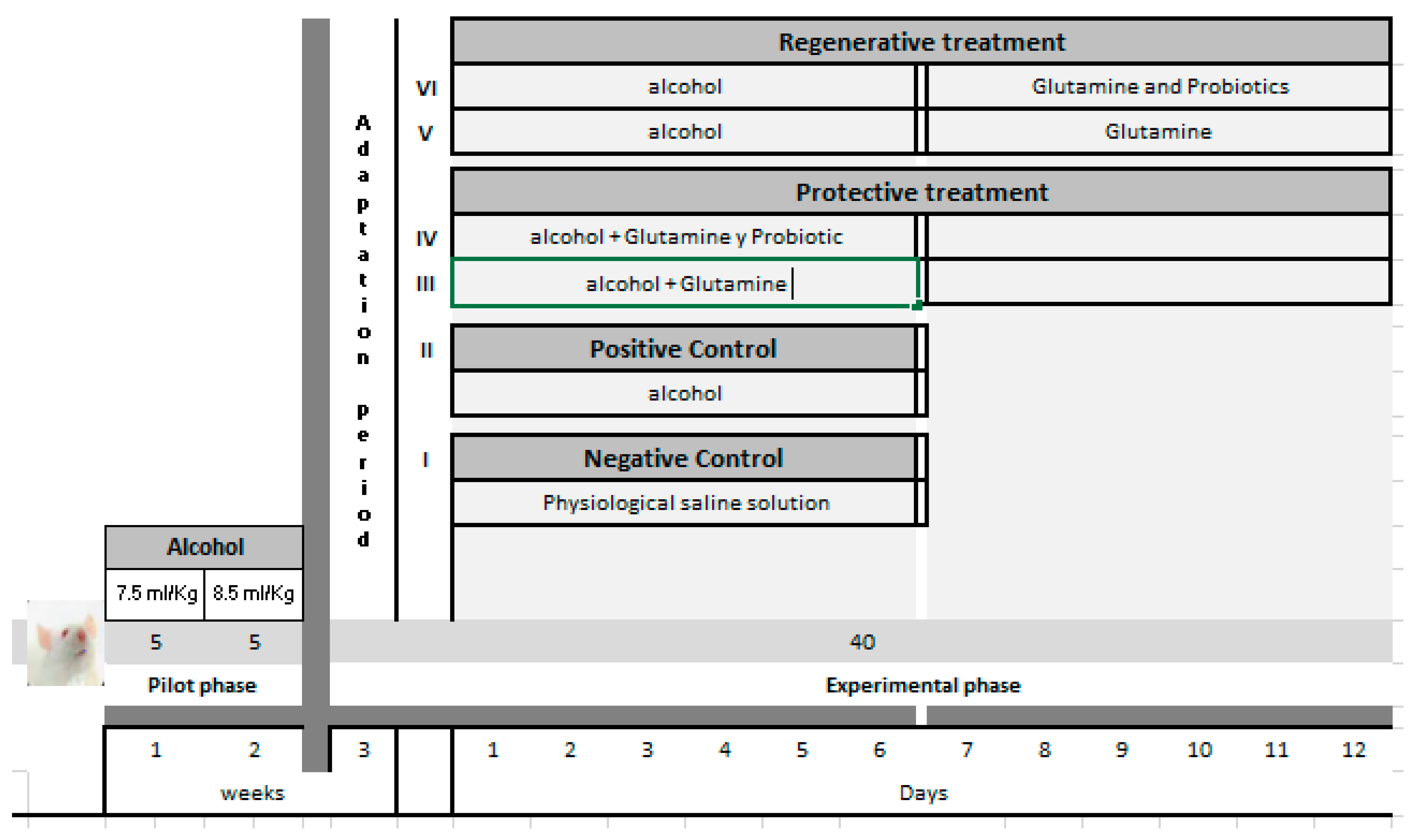
2. Materials and Methods

2.1. Animals for Experimentation

A total of 56 male Holtzman rats were obtained from a supplier. The rats weighed between 300 and 350 g and were placed in a biotherium with a temperature of 21 °C ± 2, with 12 h of light and 12 h of darkness. They had access to water and food at all times. Before the start of the pilot phase, all animals had one week of adaptation in the biotherium at Norbert Wiener University.

2.2. Reagents and Drugs

Alcohol 96° (805–812 mg/Kg), glutamine (trade name Glutapak 10) and glutamine with probiotic (trade name Glutapack R), were purchased at a local pharmacy. The preparations and dilutions were made before being offered to the rats. Glutamine in humans is recommended, according to ASPEN and ESPEN guidelines [22], at doses of 0.3 to 0.5 g per kg of body weight per person orally.
In rats weighing 300 g, each rat was given 0.15 g/kg of glutamine and 0.23 g/kg of Glutapak (glutamine + probiotic). For every 10 rats, we used approximately 1.5 g of glutamine per dose, equivalent to 2.3 of Glutapak. It should be remarked that one sachet of Glutapak 10 contains 15 g (10 g of glutamine + 5 g of maltodextrin) and the sachet of Glutapak R contains 15 g (10 g of glutamine + 5 g of maltodextrin + Lactobacillus reuteri − 1 × 10⁸ Colony Forming Units). For enteral use it is dissolved in 100 mL of water.

2.3. Treatments

The rats received the alcohol without fasting. We did not select a time of food restriction, as we found it difficult to decide the fasting time. However, studies have shown that a three-hour restriction in the animal feeding period corresponds to one meal per day for humans [23]. They received food in the morning and evening. The dose of alcohol per kilogram of weight was as follows: R1: 0 mL/kg, R2: 2.5 mL/kg, R3: 5 mL/kg, R4: 7.5 mL/kg, R5: 8.5 mL/kg, R6: 10 mL/kg. It was determined that the doses 7.5 mL/kg and 8.5 mL/kg generated greater gastric damage. Figure 1 shows the entire design and scheme of work.

2.4. Pilot Phase

The pilot phase was carried out with 10 rats and they were separated into two equal groups with different concentrations. Group A: 7.5 mL/kg of rat weight and Group B: 8.5 mL/kg of rat weight, to determine the dose (7.5 or 8.5 mL/kg) and the number of doses (2, 5, 8 or 12) that generate the gastric damage caused by alcohol. The time it lasted was two weeks. Histological evidence of gastric dysplasia was observed at a histological level after the different doses of alcohol were ingested through a cannula by each one of the rats. The pilot phase lasted two weeks, with each animal receiving its established dose daily. It was determined that the dose of 8.5 mL/kg generated the greatest gastric damage in the pilot phase. Thus, we were able to establish the dose that would induce ethanol injury to be used in the experimental phase, which was offered without fasting and given to the experimental animals in their diet to simulate normal feeding.

2.5. Experimental Phase

Glutamine and glutamine plus probiotics were calculated according to the initial weight of the Holtzman rats, and each dose of glutamine or glutamine plus probiotics was 0.5 g/kg per rat. The experimental phase lasted 12 days. Forty rats were selected in 6 groups and were distributed as follows: Group I, negative control (Healthy); Group II, positive control (Damage); Group III, protective treatment with glutamine, which received a diet plus glutamine supplementation (0.5 g/Kg); Group IV, preventive treatment with glutamine and probiotics (0.5 g/Kg + 5 million CFU Lactobacillus reuteri); Group V, regenerative treatment with glutamine received diets plus glutamine supplementation (0.5 g/Kg); Group VI, regenerative treatment with glutamine plus probiotics (0.5 g/Kg + 5 million CFU Lactobacillus reuteri).
The amount of food consumed and weight gained were recorded daily for each of the rats in the six experimental groups. Halathal was applied intraperitoneally (0.8 mL/400 g rat). Portions of the gastric wall corresponding to the fundic region were removed from each of the animals and immediately preserved in 10% formalin solution for shipping, processing and histopathological analysis (see Figure 2).

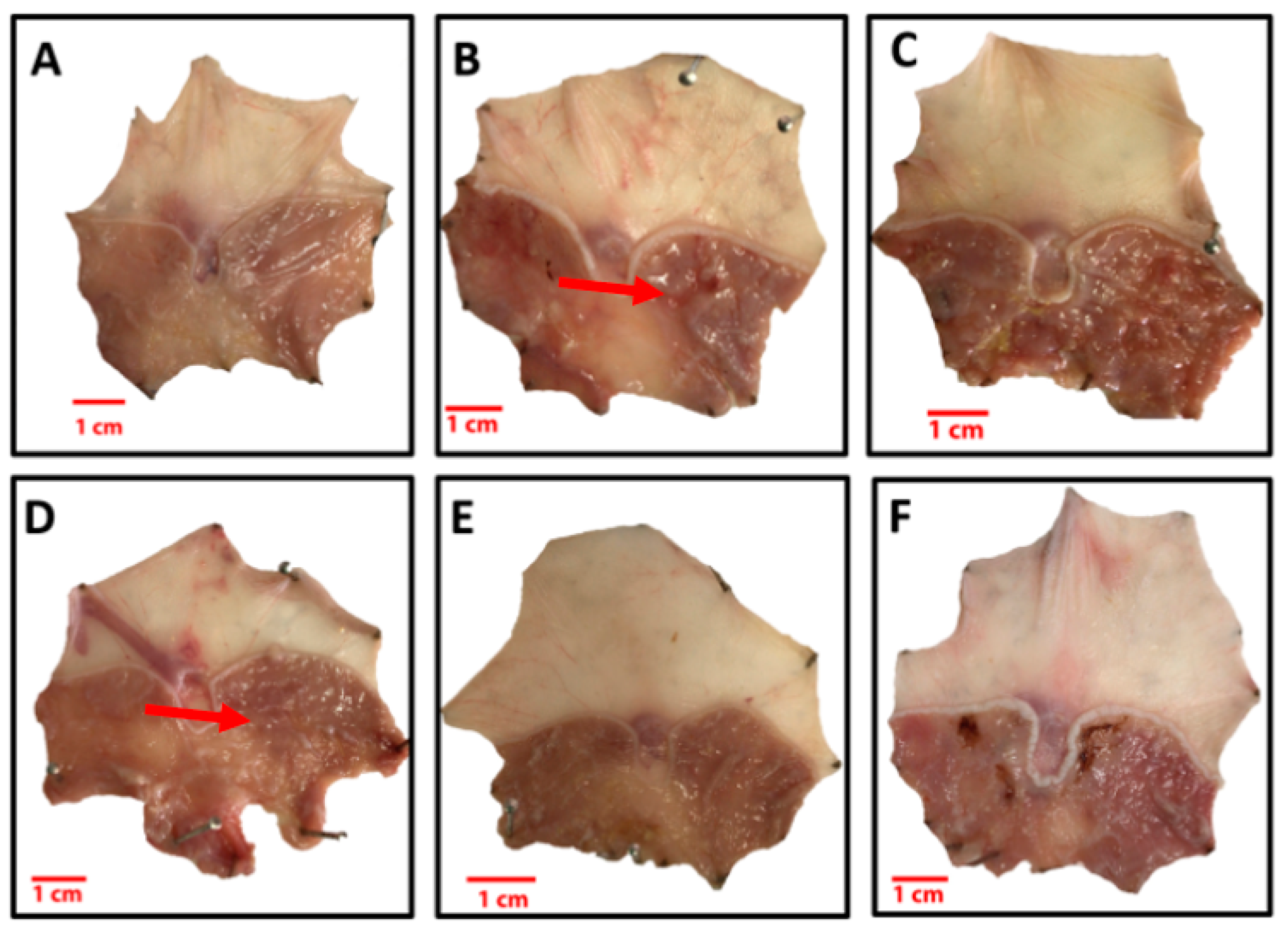
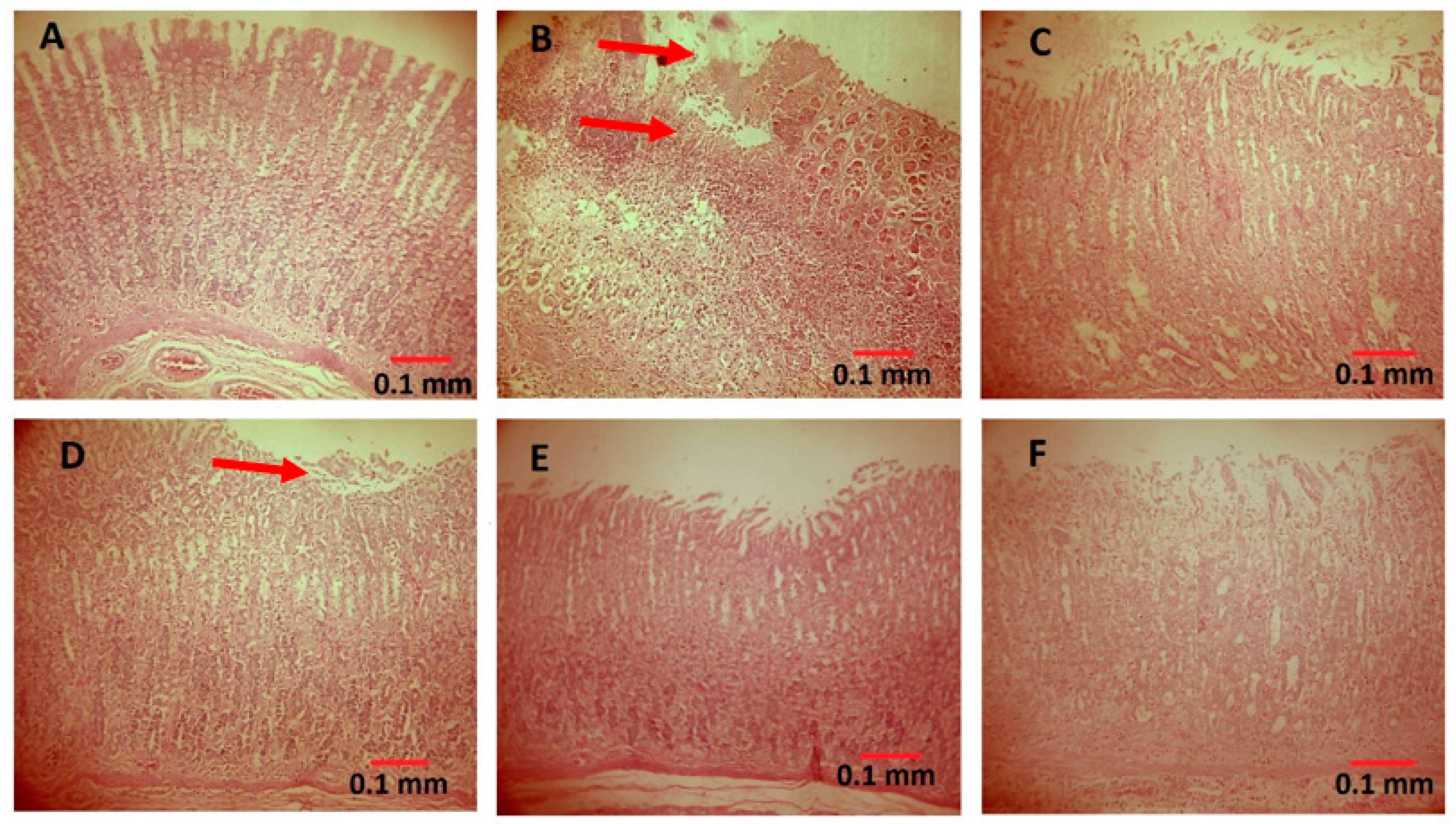
2.6. Histopathological Analysis

The histopathological analysis of the Holtzman rats’ stomachs was developed based on the technique published by Montalvo in 2010, which includes sample collection, fixation, embedding, diaphanization, microtomy, staining and mounting [24]. The results found in the histopathological analysis of the stomachs corresponding to the experimental groups were based on the presence of desquamation, edema and hyperemia [25].

2.7. Data Collection Instruments

We created sheets for these groups: negative control, positive control, preventive treatment with glutamine, preventive treatment with glutamine plus probiotics, regenerative treatment with glutamine and regenerative treatment with glutamine plus probiotics.

2.8. Statistical Analysis Plan

In vivo study data such as weight were shown as the mean ± SD in all experimental groups. The results of the histopathological analysis of the gastric mucosa showed epithelial desquamation in the apical region of the villi; also, mucus secretion was observed on the surface (which would be favorable), or an irritant substance in mild concentration. These were representative results for the four different experiments. Differences in the studies were measured and compared by using the ANOVA test. Tukey’s test for post hoc analysis was used for comparison between two groups, with a p value < 0.05 to be significant.

3. Results

3.1. Histopathological Analysis

The results found in the histopathological analysis of the stomach are described according to the experimental groups, see Figure 2. Desquamation, edema and hyperemia were controlled. In Group I (negative control), a gastric mucosa without structural alteration was observed in all samples. In Group II (positive control), a notorious erosion of the gastric mucosa was observed in all the samples, from the apical region to the intermediate region of the gastric mucosa. In the case of preventive or protective treatment with glutamine, (Group III), a desquamation of the apical region of the gastric mucosa was observed in most of the samples, which varied from mild to moderate.
In addition, preventive or protective treatment of glutamine plus probiotics (Group IV) showed a moderate-to-severe desquamation of the apical region of the gastric mucosa, which was observed in most of the samples. In regard to regenerative glutamine treatment (Group V), severe desquamation of the apical region of the gastric mucosa was observed in most of the samples. In one of the samples, an erosive zone could be seen at the level of the apical region.
Finally, regenerative treatment with glutamine plus probiotic (Group VI) showed a mild-to-moderate desquamation of the apical region of the gastric mucosa in most of the samples. They are shown in Figure 3.

3.2. Preventive Effect

There were three rats whose stomach cells showed desquamation and received glutamine, and eight rats that received glutamine with probiotic; the test showed that there exists an association between the intervention and outcome at p < 0.05. For rats whose stomach cells showed edema, three received glutamine and six received glutamine with probiotic. The results show no association. Hyperemia was not shown in any case in the group that received glutamine, and two received glutamine and probiotics. The results showed no association.

3.3. Regenerative Effect

Eight rats showed desquamation in their stomach cells and received glutamine, while four rats received glutamine with probiotic. The test showed no association between the intervention and the outcome (p > 0.05) (Table 1). Five rats that received glutamine showed edema in the stomach, while four received glutamine with probiotic. The results showed no association. Since none of the rats had hyperemia, the contrast and its consequent calculation did not apply.
Preventive glutamine seemed to have an impact in comparison to not doing anything in the face of injury due to alcohol (positive control) (Table 2).
The results of the initial and final weight of the rats that received glutamine and probiotic in a preventive and regenerative way are described (Table 3). According to the results, an increase in weight was only found in the first experimental group, see Figure 4. Intraesophageal cannulas were used in this study, ensuring that all animals received equal doses of alcohol 96°, glutamine and glutamine with probiotic. It was also ensured that all animals reached the same degree of injury with alcohol.
According to the graphic, there exists an increase in weight in the first experimental group.

4. Discussion

4.1. Our Findings

Our results showed that glutamine had better preventive efficacy than glutamine plus probiotic. Glutamine plus probiotic showed better regenerative effectiveness than glutamine, despite no significant statistical difference.
This would be due to the fact that since the mucosal epithelium was previously damaged, infectious agents would find a favorable environment, and without the use of probiotics, the action of glutamine in cell growth would be hindered as part of the expected regeneration process.
Glutamine would have a better regenerative effect associated with the stimulation of epithelial cells, specifically those producing mucopolysaccharides or mucus (mucous cells), as has been demonstrated in studies at the colon level. Since there is no previous lesion at the beginning of the treatment and also since glutamine is not associated with another product such as probiotics, the cells would have a better response for the expected effect.

4.2. Preventive and Regenerative Effect of Glutamine

It was observed that the preventive and regenerative group, based on a supplementation with glutamine, had a positive effect on the process of mild-to-moderate desquamation, but the glutamine group had a greater loss of body weight in comparison to the glutamine-with-probiotics group according to the microscopic evaluation of the gastric mucosa in the animals subjected to alcohol injury. Enteral protein supply altered the proteome of the duodenal mucosa, stimulated the biosynthesis of proteins and those related to the cytoskeleton [26].
In mice with dietary supplementation with glutamine, spontaneous cytokine-induced apoptosis was prevented through the formation of glutathione [27]. Glutamine also repaired intestinal mucosal injury by blocking the expression of HMGB1 and inflammatory cytokines [28].
The administration of glutamine as a preventive therapy avoids desquamation because the mucosa had greater protection (p < 0.05). However, the glutamine given as regenerative treatment did not provide protection.
When comparing the preventive therapy (glutamine) with the positive control (alcohol 96° injury), we obtained significant differences (p < 0.05). We hypothesize a positive impact of glutamine as a coadjuvant.

4.3. Preventive Effect of Probiotics

It was observed that it had less effect in comparison to the glutamine plus probiotics supplementation with Lactobacillus reuteri as regenerative effect. It was evidenced that although the two groups presented with moderate level damage of desquamation in the gastric mucosa, this group presented with greater loss of weight.
Different strains of lactic acid bacteria, and particularly, Lactobacillus spp., are some of the most commonly used probiotics, defined as living microorganisms which, when administered in adequate amounts, provide a health benefit to the host [29]. Similarly to our results, other authors reported the improvement of alcohol-induced gastric ulcers through the administration of Lactobacillus plantarum isolated from green tea. Propionate levels increased, and this favors and impacts on the microbial population and its metabolic activity [30]. Bifidobacterium bifidum BF-1 alleviates gastric injury, improving gastric mucin production [31]. A mixture of probiotics, with 13 different bacteria, decreased lesions, reduced lipid peroxidation, improved sIgA production in the mucosa and stabilized the degranulation of gastric mucosa during pretreatment. It also inhibited lipid peroxidation in gastric mucosa [9]. Lactobacillus reuteri DSM 17938 (DSM) showed gastroprotective activity and reduced the ulcer area with three days of pretreatment, decreasing the expression of transient receptor potential vanilloid type 1 (TRPV1), with the reduction in oxidative stress [7].

4.4. Regenerative Effect of the Probiotic
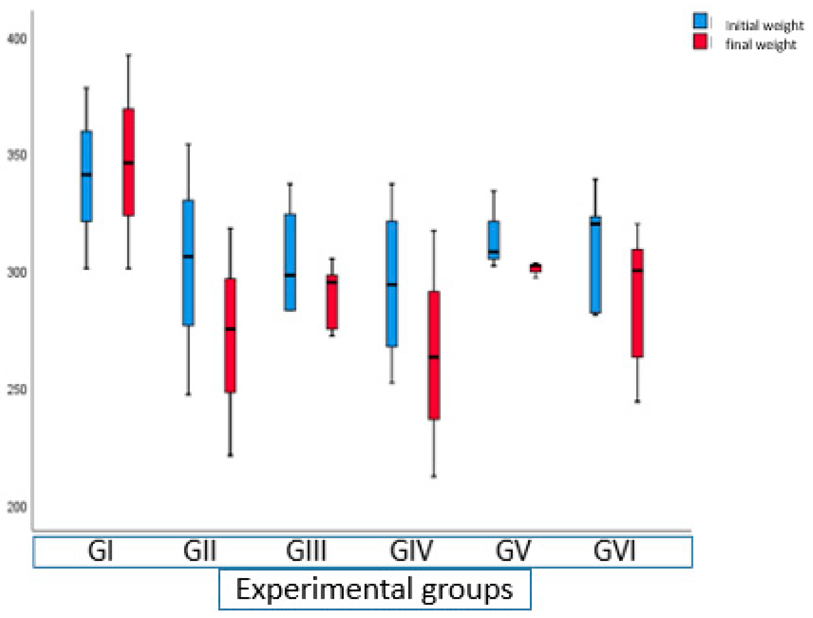
It was observed that its preventive effect was greater in comparison to glutamine plus probiotics supplementation with Lactobacillus reuteri. In addition, it was evidenced that although the two groups presented with a moderate level of desquamation damage in the gastric mucosa, this group had less weight loss than the other.
The effect of the efficacy of several strains of Lactobacillus on gastric ulcers is already known [32,33]. It has been demonstrated that strains such as Lactobacillus gasseri LG21 inhibit erosive lesions through the positive regulation of prostaglandin E2 of the gastric mucosa [11]. Lactobacillus acidophilus was proven to be a healing agent in the damage of stomachs ulcers [33], and in angiogenesis [11,33]. The preventive anti-ulcer effect of multiprobiotics (Bifidobacterium animalis VKL and Bifidobacterium animalis VKB) managed to reduce erosive and ulcerative lesions through the restoration of antioxidant balance [32], decreasing proinflammatory cytokines and regulating the increase in anti-inflammatory cytokines [34,35].

4.5. Weight of Rats

The alcohol-treated groups lost weight, except for the control group (p < 0.05), see Figure 4. Weight loss was less in the groups that received only glutamine in comparison to those that received glutamine plus probiotics (see Table 1). This may have happened due to the fact that if there is greater gastric damage induced by ethanol in the experimental animals, they will not consume adequate food, and therefore, will progressively lose body weight. Authors such as Malherbe found that alcohol intake in adolescent rats decreased body weight [35], even in moderate doses.
This may be based on products generated as a result of metabolizing alcohol (free radicals and acetaldehyde), formed inside the cell, which can produce tissue damage and lead to death [36]. The process of lipid pre-oxidation can alter membranes, which leads to steatosis and cell death.
Alcohol interacts with the absorption of amino acids and interferes with the uptake of these essential amino acids; thus, it has been demonstrated that intestinal absorption of amino acids is significantly reduced in experimental animals after receiving a dose of alcohol 96° [37].
Cai et al., 2020, administered ethanol to rats intragastrically for seven consecutive days to evaluate the protective effect of citrulline on ulcerative colitis. The weight change of the rats was measured every four hours; the authors concluded that the loss of weight by 25% in relation to initial weight showed that the rats had difficulty ingesting food and drinking water by themselves [38].
In order to avoid that effect in our study, the rats were given enough food and water and their weight was evaluated daily. Morampudi et al., 2014, describe that such weight loss is due to dehydration, and when it approaches maximum values, Ringer’s lactate should be administered [39].

5. Conclusions

Our study shows the positive preventive effect of glutamine and the regenerative effect of glutamine plus probiotic, demonstrated through histological analysis. Although there are few in vivo studies that have explored the impact of probiotics on gastric ulcers, moderate protective and therapeutic effects have been suggested. Therefore, the use of probiotics in the treatment of gastric ulcers appears promising, and further studies are required considering probiotic strains, dosage and commercial preparations. Future explorations could include the study of only glutamine or glutamine in a combination in a preventive form in desquamation of the gastric mucosa.

Author Contributions

Conceptualization, M.L.-U. and P.R.-O.; methodology, C.P.; software, P.R.-O.; formal analysis, C.P.; investigation, D.P.-I. and C.C.; resources, O.R.-L.; writing—original draft preparation, M.L.-U.; writing—review and editing, F.I.-B.; visualization, C.B.-A.; supervision, M.L.-U.; project administration, F.I.-B.; funding acquisition, O.R.-L. All authors have read and agreed to the published version of the manuscript.

Funding

This Project was financed by the Competitive Fund of the Universidad Privada Norbert Wiener of the year 2019. RESOLUCIÓN N° 112-2019-R-UPNW.

Institutional Review Board Statement

This project was approved by the Ethics Committee of the Norbert Wiener Private University (Exp. No. 199-2020).

Informed Consent Statement

Not applicable.

Data Availability Statement

Underlying data Figshare: GLUTAMIN AND PROBIOTICS DOI: https://doi.org/10.6084/m9.figshare.14751561 (accessed on 22 February 2022). The project contains the following underlying data: Database: The data, including the experimental and prospective studies, are the result of working with 56 male rats between 300 and 350 g; three phases were worked: the first the pre-pilot phase (6 rats), the second the pilot phase (10 rats) and the third experimental phase (40 rats).

Acknowledgments

We would like to thank Juana Chávez Flores, who supported us at all times during the development of the study.

Conflicts of Interest

The authors declare no conflict of interest.

References

  1. Stewart, D.J.; Ackroyd, R. Peptic ulcers and their complications. Surgery 2011, 29, 568–574. [Google Scholar]
  2. Azer, S.; Akhondi, H. Gastritis-StatPearls-NCBI Bookshelf [Internet]. NCBI Bookshelf. 2020; pp. 1–14. Available online: https://www.ncbi.nlm.nih.gov/books/NBK544250/ (accessed on 15 May 2021).
  3. Park, J.-H.; Jang, K.-J.; Kim, C.-H.; Kim, J.-H.; Kim, Y.-K.; Yoon, H.-M. Ganoderma Lucidum Pharmacopuncture for Teating Ethanol-induced Chronic Gastric Ulcers in Rats. J. Pharmacopunct. 2015, 18, 72–78. [Google Scholar] [CrossRef] [PubMed]
  4. Li, G.; Zhu, L.; Cao, Z.; Wang, J.; Zhou, F.; Wang, X.; Li, X.; Nie, G. A New Participant in the Pathogenesis of Alcoholic Gastritis: Pyroptosis. Cell Physiol. Biochem. 2018, 49, 406–418. [Google Scholar] [CrossRef] [PubMed]
  5. Gazzieri, D.; Trevisani, M.; Springer, J.; Harrison, S.; Cottrell, G.S.; Andre, E.; Nicoletti, P.; Massi, D.; Zecchi, S.; Nosi, D. Substance P released by TRPV1-expressing neurons produces reactive oxygen species that mediate ethanol-induced gastric injury. Free Radic. Biol. Med. 2007, 43, 581–589. [Google Scholar] [CrossRef]
  6. Roberts, D.M. Chronic gastritis, alcohol, and non-ulcer dyspepsia. Gut 1972, 13, 768–774. [Google Scholar] [CrossRef]
  7. Oliveira, A.P.; Souza, L.K.M.; Araújo, T.S.L.; de Araújo, S.; Nogueira, K.M.; Sousa, F.B.M.; Silva, R.O.; Pacífico, D.M.; Martins, C.S.; Brito, G.A.; et al. Lactobacillus reuteri DSM 17938 protects against gastric damage induced by ethanol administration in mice: Role of TRPV1/substance P axis. Nutrients 2019, 11, 208. [Google Scholar] [CrossRef] [PubMed] [Green Version]
  8. Uchida, M.; Kurakazu, K. Yogurt Containing Lactobacillus gasseri OLL2716 Exerts Gastroprotective Action Agaisnt Acute Gastric Lesion and Antral Ulcer in Rats. J. Pharmacol. Sci. 2004, 96, 84–90. [Google Scholar] [CrossRef] [Green Version]
  9. Gomi, A.; Harima-Mizusawa, N.; Shibahara-Sone, H.; Kano, M.; Miyazaki, K.; Ishikawa, F. Effect of Bifidobacterium bifidum BF-1 on gastric protection and mucin production in an acute gastric injury rat model. J. Dairy Sci. 2013, 96, 832–837. [Google Scholar] [CrossRef] [PubMed] [Green Version]
  10. Wang, R.; Zhou, K.; Xiong, R.; Yang, Y.; Yi, R.; Hu, J.; Liao, W.; Zhao, X. Pretreatment with lactobacillus fermentum xy18 relieves gastric injury induced by HCL/ethanol in mice via antioxidant and anti-inflammatory mechanisms. Drug Des. Dev. Ther. 2020, 14, 5721–5734. [Google Scholar] [CrossRef] [PubMed]
  11. Dharmani, P.; De Simone, C.; Chadee, K. The Probiotic Mixture VSL#3 Accelerates Gastric Ulcer Healing by Stimulating Vascular Endothelial Growth Factor. PLoS ONE 2013, 8, e58671. [Google Scholar]
  12. Souba, W.W.; Smith, R.J.; Wilmore, D.W. Glutamine metabolism by the intestinal tract. J. Parenter. Enter. Nutr. 1985, 9, 608–617. [Google Scholar] [CrossRef] [PubMed]
  13. de Oliveira Santos, R.; da Silva Cardoso, G.; da Costa Lima, L.; de Sousa Cavalcante, M.L.; Silva, M.S.; Cavalcante, A.K.M.; Tolentino, M. l-Glutamine and Physical Exercise Prevent Intestinal Inflammation and Oxidative Stress Without Improving Gastric Dysmotility in Rats with Ulcerative Colitis. Inflammation 2020, 44, 617–632. [Google Scholar] [CrossRef] [PubMed]
  14. Hernández-Muñoz, R.; Montiel-Ruíz, C.; Vázquez-Martínez, O. Gastric Mucosal Cell Proliferation in Ethanol-Induced Chronic Mucosal Injury Is Related to Oxidative Stress and Lipid Peroxidation in Rats. Lab. Investig. 2000, 80, 1161–1169. [Google Scholar] [CrossRef]
  15. Szabo, S. “Gastric cytoprotection” is still relevant. J. Gastroenterol. Hepatol. 2014, 29, 124–132. [Google Scholar] [CrossRef] [PubMed] [Green Version]
  16. Naito, Y.; Yoshikawa, T.; Ando, T.; Kishi, A.; Ueda, S.; Oyamada, H.; Kondo, M. Changes in superoxide dismutase activity in the gastric mucosa of peptic ulcer patients. J. Clin. Gastroenterol. 1992, 14, S131–S134. [Google Scholar] [CrossRef] [PubMed]
  17. Singh, A.; Kukreti, R.; Saso, L.; Kukreti, S. Oxidative stress: A key modulator in neurodegenerative diseases. Molecules 2019, 24, 1583. [Google Scholar] [CrossRef] [Green Version]
  18. Carneiro, J.G.; De Brito, T.; Holanda, L.; Gomes Quinderé, A.L.; Fernandes Frota, A.; Virgínia, V.; Benevides, N.M.B. Gastroprotective Effects of Sulphated Polysaccharides from the Alga Caulerpa mexicana Reducing Ethanol-Induced Gastric Damage. Pharmaceuticals 2018, 11, 6. [Google Scholar] [CrossRef] [PubMed] [Green Version]
  19. Chang, B.; Sang, L.; Wang, Y.; Tong, J.; Zhang, D.; Wang, B. The protective effect of VSL#3 on intestinal permeability in a rat model of alcoholic intestinal injury. BMC Gastroenterol. 2013, 13, 151. [Google Scholar]
  20. Ziegler Evans, M.; Fernández-Estívariz, C.; Jones, D. Trophic and cytoprotective nutrition for intestinal adaptation, mucosal repair, and barrier function. Annu. Rev. Nutr. 2003, 23, 229–261. [Google Scholar] [CrossRef] [PubMed]
  21. Chen, Y.; Tsai, Y.H.; Tseng, B.J.; Tseng, S.H. Influence of growth hormone and glutamine on intestinal stem cells: A narrative review. Nutrients 2019, 11, 1941. [Google Scholar] [CrossRef] [PubMed] [Green Version]
  22. Bejarano Rosales, M.; Álvarez Altamirano, K.; Fuchs-Tarlovsky, V. Análisis comparativo de las guías de la ESPEN y la Academia de Nutrición y Dietética Americana sobre cuidado nutricional del paciente con cáncer publicadas en 2017. Rev. Nutr. Clín. Metab. 2019, 2, 29–41. [Google Scholar] [CrossRef]
  23. Park, S.; Yoo, K.; Hyun, J.; Kang, S. Intermittent fasting reduces body fat but exacerbates hepatic insulin resistance in young rats regardless of high protein and fat diets. J. Nutr. Biochem. 2017, 40, 14–22. [Google Scholar] [CrossRef] [PubMed]
  24. Cui, D. Histologia con Correlaciones Funcionales y Clinicas, 1st ed.; Wolters Kluwer: Alphen aan den Rijn, The Netherlands, 2011; 482p. [Google Scholar]
  25. Ramos, D.; Angulo, P.; Chavera, A.; MAyón, M. Propuesta de un modelo experimental de enteritis aguda inducida por indometacina en ratas albinas. Rev. Investig. Vet. Peru 2004, 15, 37–43. [Google Scholar] [CrossRef] [Green Version]
  26. Goichon, A.; Bertrand, J.; Chan, P.; Lecleire, S.; Coquard, A.; Cailleux, A.F.; Vaudry, D.; Déchelotte, P.; Coeffier, M. Enteral delivery of proteins enhances the expression of proteins involved in the cytoskeleton and protein biosynthesis in human duodenal mucosa. Am. J. Clin. Nutr. 2015, 102, 359–367. [Google Scholar] [CrossRef] [PubMed] [Green Version]
  27. Deniel, N.; Marion-Letellier, R.; Charlionet, R.; Tron, F.; Leprince, J.; Vaudry, H.; Ducrotté, P.; Déchelotte, P.; Thébault, S. Glutamine regulates the human epithelial intestinal HCT-8 cell proteome under apoptotic conditions. Mol. Cell Proteom. 2007, 6, 1671–1679. [Google Scholar] [CrossRef] [PubMed] [Green Version]
  28. Shu, X.; Zhang, J.; Wang, Q.; Xu, Z.; Yu, T. Glutamine decreases intestinal mucosal injury in a rat modelof intestinal ischemia-reperfusion by downregulating HMGB1 and inflammatory cytokine expression. Exp. Ther. Med. 2016, 12, 1367–1372. [Google Scholar] [CrossRef] [PubMed] [Green Version]
  29. Berkes, E.; Liao, Y.H.; Neef, D.; Grandalski, M.; Monsul, N. Potentiated In Vitro Probiotic Activities of Lactobacillus fermentum LfQi6 Biofilm Biomass Versus Planktonic Culture. Probiotics Antimicrob. Proteins 2020, 12, 1097–1114. [Google Scholar] [CrossRef]
  30. Park, H.; Cho, D.; Huang, E.; Seo, J.Y.; Kim, W.G.; Todorov, S.D.; Ji, Y.; Holzapfel, W.H. Amelioration of Alcohol Induced Gastric Ulcers Through the Administration of Lactobacillus plantarum APSulloc 331261 Isolated From Green Tea. Front. Microbiol. 2020, 11, 420. [Google Scholar] [CrossRef] [PubMed] [Green Version]
  31. Senol, A.; Isler, M.; Karahan, A.G.; Kilic, G.B.; Kuleasan, H.; Kaya, S.; Keskin, M.; Goren, I.; Saritas, U.; Aridogan, B.C.; et al. Preventive effect of probiotics and α-tocopherol on ethanol-induced gastric mucosal injury in rats. J. Med. Food 2011, 14, 173–179. [Google Scholar] [CrossRef] [PubMed]
  32. Uchida, M.; Shimizu, K.; Kurakazu, K. Yogurt containing lactobacillus gasseri OLL 2716 (LG21 yogurt) accelerated the healing of acetic acid-induced gastric ulcer in rats. Biosci. Biotechnol. Biochem. 2010, 74, 1891–1894. [Google Scholar] [CrossRef] [PubMed] [Green Version]
  33. Singh, P.K.; Kaur, I.P. Synbiotic (probiotic and ginger extract) loaded floating beads: A novel therapeutic option in an experimental paradigm of gastric ulcer. J. Pharm. Pharmacol. 2012, 64, 207–217. [Google Scholar] [CrossRef] [PubMed]
  34. Falalyeyeva, T.; Leschenko, I.; Beregova, T.; Lazarenko, L.; Savchuk, O.; Sichel, L.; Tsyryuk, O.; Vovk, T.; Spivak, M.; Diaprof, K.L. Probiotic strains of lactobacilli and bifidobacteria alter pro- and anti-inflammatory cytokines production in rats with monosodium glutamate-induced obesity. Fiziolohichnyĭ Zhurnal 2017, 63, 17–25. [Google Scholar] [CrossRef] [PubMed] [Green Version]
  35. García Barceló, M.; Malherbe Pérez, T.; Batista Castro, Z.; Escalona Rabaza, M.; Aguilar González, V. Variaciones del peso corporal en ratas macho adolescentes tratadas con dosis moderadas de alcohol. Med. UTA 2017, 1, 1. [Google Scholar] [CrossRef] [Green Version]
  36. Cheeseman, K.H.; Slater, T.F. An introduction to free radical biochemistry. Br. Med. Bull. 1993, 49, 481–493. [Google Scholar] [CrossRef] [PubMed]
  37. Baroana, E.; Lieber, C.S. Effects of ethanol on hepatic protein metabolism. Subst. Alcohol Actions Misuse 1981, 2, 188–189. [Google Scholar]
  38. Cai, B.; Zhou, M.; Huang, H.; Zhou, A.; Chu, Z.; Huang, X.; Li, C.-W. Protective effects of citrulline supplementation in ulcerative colitis rats. PLoS ONE 2020, 15, e0240883. [Google Scholar] [CrossRef] [PubMed]
  39. Morampudi, V.; Bhinder, G.; Wu, X.; Dai, C.; Sham, H.P.; Vallance, B.A.; Jacobson, K. DNBS/TNBS Colitis Models: Providing Insights Into Inflammatory Bowel Disease and Effects of Dietary Fat. J. Vis. Exp. 2014, 84, 51297. [Google Scholar] [CrossRef] [PubMed] [Green Version]
Figure 1. Study design and work scheme. The schematic representation of the periods included in each phase is shown. The pre-pilot phase is considered in weeks and the experimental phase in days. In the pilot phase, the dosages with alcohol to generate the injury were carried out with 10 rats between 7.5 mL/kg (5 rats) and 8.5 mL/kg (5 rats). The experimental phase was carried out with 40 rats separated into 6 groups: negative control, positive control, preventive treatment with glutamine (0.5 g of glutamine per kilogram of weight), preventive treatment with glutamine plus probiotics (0.5 g of glutamine per kilogram of weight and 5 million CFU Lactobacillus reuteri), regenerative treatment with glutamine (0.5 g of glutamine per kilogram of weight) and regenerative treatment with glutamine plus probiotics (0.5 g of glutamine per kilogram of weight and 5 million CFU Lactobacillus reuteri). The administration was intragastric.
Processes 10 00504 g001
Figure 2. Gastric mucosa photographs of animals in the experimental groups (A) Negative control group, no observable alteration (B) Positive control group, slight-to-moderate degree of edema (C) Preventive treatment group with 0.5 g of glutamine per kilogram of weight, slight degree of edema (D) Preventive treatment group with 0.5 g of glutamine per kilogram of weight and 5 million CFU Lactobacillus reuteri No observable macroscopic alteration, slight degree of edema (E) Regenerative treatment group with 0.5 g of glutamine per kilogram of weight, slight degree of edema (F) Regenerative treatment group with 0.5 g of glutamine per kilogram of weight and 5 million CFU Lactobacillus reuteri, slight degree of edema.
Processes 10 00504 g002
Figure 3. Histopathological analysis: Microscopic photographs of the gastric mucosa of the animals in the experimental groups, taken with a 10× objective. (A) Negative control group, with evident preservation of its histological architecture. (B) Positive control group, with erosion mainly involving the apical and intermediate region of the mucosa. (C) Preventive treatment group with 0.5 g of glutamine per kilogram of weight, in which a desquamation and erosion process is observed at the level of the apical region of the mucosa. (D) Preventive treatment group with 0.5 g of glutamine per kilogram of weight and 5 million CFU Lactobacillus reuteri, with a moderate desquamation process at the level of the apical region of the mucosa. (E) Regenerative treatment group with 0.5 g of glutamine per kilogram of weight, in which a moderate desquamation process is observed at the level of the apical region of the mucosa. (F) Regenerative treatment group with 0.5 g of glutamine per kilogram of weight and 5 million CFU Lactobacillus reuteri, with a mild desquamation process at the level of the apical region of the mucosa.
Processes 10 00504 g003
Figure 4. Results regarding initial weight and final weight in the experimental groups.
Processes 10 00504 g004
Table 1. Comparison between the preventive and regenerative effect according to the presence of desquamation, edema and hyperemia.
Preventive EffectRegenerative Effect
Desquamation
yesnopyesnop
Glutamine350.026800.077
Glutamine and Probiotic 8044
Edema
Glutamine350.315531.00
Glutamine and Probiotic 6244
Hyperemia
Glutamine080.46708NA
Glutamine and Probiotic 2608
Own elaboration.
Table 2. Comparison between the preventive and regenerative in desquamation.
Desquamation
YesNo p
Positive control vs.800.026 (*)
preventive glutamine350.007 (**)
Preventive glutamine vs. 350.026 (*)
glutamine with preventive probiotic800.007 (**)
Preventive glutamine vs.350.026 (*)
regenerative glutamine 800.007 (**)
*: Fisher’s exact p = 0.026; **: Pearson chi2 = 7.2727 p = 0.007/Degrees of freedom: 1.
Table 3. Weight of the rats in each phase of the experiment.
Experimental GroupInitial Weight (g)Final Weight (g)Change in Weight (g)
Mean (SD)Min–MaxMean (DS)Min–Max
G I: Negative control340.00 (38.51)(301–378)346.33 (45.50)(301–392)6.33 ± 7.09 a
G II: Positive control302.33 (53.59)(247–354)271.33 (48.60)(221–318)−31.00 ± 5.00
G III: Preventive treatment 1305.00 (24.50)(283–337)289.00 (14.64)(272–305)−16.00 ± 12.39
G IV: Preventive treatment 2294.25 (35.84)(252–337)263.75 (42.90)(212–317)−30.50 ± 13.40
G V: Regenerative treatment 1314.67 (17.01)(281–339)300.67 (3.21)(297–303)−14.00 ± 14.73
G VI: Regenerative treatment 2309.00 (26.12)(302–334)287.20 (32.27)(244–320)−21.80 ± 8.81
Own elaboration at p < 0.05.
Publisher’s Note: MDPI stays neutral with regard to jurisdictional claims in published maps and institutional affiliations.

