
Abstract
There is currently no accepted classification of autosomal recessive cerebellar ataxias, a group of disorders characterized by important genetic heterogeneity and complex phenotypes. The objective of this task force was to build a consensus on the classification of autosomal recessive ataxias in order to develop a general approach to a patient presenting with ataxia, organize disorders according to clinical presentation, and define this field of research by identifying common pathogenic molecular mechanisms in these disorders. The work of this task force was based on a previously published systematic scoping review of the literature that identified autosomal recessive disorders characterized primarily by cerebellar motor dysfunction and cerebellar degeneration. The task force regrouped 12 international ataxia experts who decided on general orientation and specific issues. We identified 59 disorders that are classified as primary autosomal recessive cerebellar ataxias. For each of these disorders, we present geographical and ethnical specificities along with distinctive clinical and imagery features. These primary recessive ataxias were organized in a clinical and a pathophysiological classification, and we present a general clinical approach to the patient presenting with ataxia. We also identified a list of 48 complex multisystem disorders that are associated with ataxia and should be included in the differential diagnosis of autosomal recessive ataxias. This classification is the result of a consensus among a panel of international experts, and it promotes a unified understanding of autosomal recessive cerebellar disorders for clinicians and researchers.
Introduction
The classification of hereditary ataxias represents a significant challenge due to the large number of neurological and metabolic diseases that present with cerebellar dysfunction and the phenotypic heterogeneity in known genetically defined disorders. Indeed, ataxia is a presenting feature in degenerative disorders that target mainly the cerebellum, but it may be present in hereditary spastic paraplegias, inborn errors of metabolism, and various encephalopathies. Proper classification and phenotypic understanding is of primary importance in this field where the high prevalence of repeat expansion disorders, which are not adequately covered by the next-generation sequencing (NGS) techniques [1, 2], precludes NGS as a first diagnostic step and requires phenotypic evaluation to perform custom gene testing when applicable. Nevertheless, autosomal recessive cerebellar ataxias have remained an ill-defined and disorganized group of disorders for two main reasons. First, unlike the dominant ataxias that have been organized with a numerical naming system, recessive disorders presenting with ataxia have been named in a highly heterogeneous manner according to clinical features, physicians’ surname, or regions of high prevalence. Second, several recessive multisystemic or complex metabolic disorders present with ataxia, such that it is difficult to properly circumscribe this group of disorders and classify it in a meaningful way for both clinicians and researchers. Hence, the Society for Research on the Cerebellum and Ataxias (SRCA) Task Force on the Classification of Recessive Cerebellar Ataxias was created in 2016 to regroup a panel of international ataxia experts in order to propose a classification relevant to clinical practice and researchers. As a first step, we undertook a systematic scoping review of the literature to identify all recessive disorders presenting with ataxia, select those in which cerebellar degeneration was a core feature, and propose a first classification. This systematic scoping review has been previously published [3] and served as the basis for the current work.
Recently, the Movement Disorder Society Task Force on Classification and Nomenclature of Genetic Movement Disorders proposed a revised naming system based on the gene name associated with a phenotypical prefix. They presented a list of 92 gene-defined recessive disorders associated with ataxia for which this naming system would be applied and an exhaustive list of disorders that may occasionally present with ataxia [4]. This represents a useful reference for interpretation of NGS results. However, in a significant number of listed disorders, the cerebellum is only one of the many affected organs in multisystemic and metabolic disorders. For example, maple syrup urine disease, caused by BCKDHB mutations, and congenital disorders of glycosylation 1a, 1c, and 1q have been included. These disorders are inborn errors of metabolism characterized by developmental delay, hypotonia, and metabolic defects, and ataxia is only mild, found in a minority of patients, or present solely during episodes of metabolic decompensation. Hence, there remains a need for a classification system that focuses on disorders affecting primarily the cerebellum and organizes clinical and paraclinical information to promote an understanding of cerebellar disorders useful not only to ataxia experts but also to general neurologists, learners, patients, and researchers.
The objective of this task force was to build a consensus on the classification of autosomal recessive ataxias in order to develop a general approach to a patient presenting with ataxia, organize disorders according to clinical presentation, and define this field of research by identifying common pathophysiological mechanisms in recessive disorders presenting with ataxia. This aims at bringing together clinicians and researchers to promote a common understanding of recessive cerebellar disorders in order to advance research and improve patient care.
Materials and Methods
The first step was to identify all recessive disorders presenting with ataxia. Recessive cerebellar ataxias were defined as disorders with autosomal recessive inheritance characterized by a cerebellar motor syndrome of gait ataxia, dysmetria, adiadochokinesia, nystagmus, and dysarthria associated with cerebellar degeneration as demonstrated by imagery or pathology. A pathogenic mutation had to be identified in at least two independent families for a specific gene to be included. Purely malformative disorders were excluded, and disorders with complex phenotypes where ataxia is a secondary or late feature were also excluded. We conducted a systematic scoping review of the literature to identify relevant reports. The methodology and results of this systematic review have been published previously [3]. In the first publication, this review process had allowed the identification of 2354 records and was current as of September 2016. The literature search was updated and is current as of October 2018.
The second step was to regroup a panel of 12 international ataxia experts to create a logical classification system and build a consensus. Ataxia experts were identified from various geographical regions and areas of expertise within the field of ataxias, ensuring proper representation of regional differences in prevalence and clinical approach to ataxias. Discussions spanned over 2 years, included meetings at two SRCA international conferences, and concerned general orientation, clinical approach, specific disorders, classification issues, and regional specificities. The first author (MB) reviewed identified records for inclusion, extracted clinical, epidemiological, and molecular data to build the classifications and wrote the text integrating all authors’ input and comments. All authors approved the final manuscript and list of included disorders.
Results
The final list of included autosomal recessive cerebellar ataxias is presented in Table 1 and includes 59 primary recessive ataxias, which regroup 15 disorders that are more prevalent and widely distributed and 44 disorders that are less frequent and reported only in certain populations or few families. Because ethnic and regional specificities are an essential element to consider in the appraisal of a patient with a recessive ataxia, areas where the disorder has been reported to date are listed. Metabolic or mitochondrial disorders where ataxia is only a secondary nonspecific finding in a multisystemic phenotype were excluded, as cerebellar pathology is not central in these disorders. However, clinicians must bear in mind that some of these disorders may present with a milder juvenile or adult onset phenotype where cerebellar ataxia may predominate, for example, in Niemann-Pick disease type C, Tay-Sachs disease, sialic acid storage disorders, congenital disorders of glycosylation, and Zellweger spectrum disorders. As some of these metabolic disorders may benefit from early treatment, clinicians must keep a high index of suspicion to test for these disorders, and they should be included in large NGS gene panels for ataxia. These and other complex disorders that may occasionally present with ataxia are presented in Table 2. This second list is not exhaustive and presents only the main or most frequent disorders occasionally associated with ataxia. Disorders in which the cerebellar phenotype is not clearly established have been excluded.
Clinical Approach to a Patient Presenting with Ataxia
-
1.
The first step in evaluating a patient with ataxia is to perform a detailed clinical evaluation that includes a clinical history, a family history, a targeted neurological and systemic physical evaluation, and relevant paraclinical tests. The temporal course is a central element in determining the underlying etiology. Indeed, a chronic progressive evolution over months to years, without trauma or toxin exposure, is suggestive of a hereditary disorder, whereas acute or subacute onset points towards an acquired etiology. A clinical history and physical examination are essential to assess the severity of the cerebellar syndrome and the presence of associated neurological features or systemic involvement. Headache, fever, or an associated autoimmune disorder should prompt the consideration of acquired etiologies. A detailed family history should be obtained to search for relatives with similar symptomatology. Laboratory tests may be useful to rule out acquired causes or as biomarkers for certain disorders. Neuroimaging, preferably with magnetic resonance imaging, is an essential tool to evaluate the presence of cerebellar atrophy or signal anomalies, to search for associated pontine atrophy, and to rule out space-occupying lesions. Electromyography and nerve conduction studies can prove the presence of clinically suspected or subclinical neuropathy and provide evidence of associated myopathy.
-
2.
Following the clinical assessment, one should verify that acquired and treatable causes for ataxia have been excluded. These include vascular disease, trauma, infection, primary or metastatic tumor, excess alcohol consumption, vitamin deficiency, Creutzfeldt-Jakob disease, and immune-mediated cerebellar ataxias such as multiple sclerosis, gluten ataxia, anti-GAD (glutamic acid decarboxylase) ataxia, and paraneoplastic cerebellar degenerations. Clinical evaluation should reveal previous exposure to toxins or traumatic injuries, along with specific signs and symptoms suggestive of infectious, vascular, or metastatic disease. Laboratory tests are useful to identify vitamin deficiencies or autoimmune conditions. Specifically, testing for antibodies involved in paraneoplastic or autoimmune cerebellar degeneration may be particularly useful for patients with a subacute progression, older age at onset, and absence of family history. The paraneoplastic antibodies most associated with cerebellar degeneration are anti-Yo, anti-Hu, anti-Tr, and anti-mGluR1 antibodies; the tumors most often involved are breast and gynecological tumors, Hodgkin lymphoma, and small-cell lung carcinoma [218]. Large paraneoplastic autoantibody panels are now available and may reduce the delay associated with serial testing.
-
3.
Once acquired causes have been ruled out, a genetic etiology may be considered, especially in the presence of a positive family history, early onset, chronic progressive course, or with a set of clinical signs and symptoms that is reminiscent of a well-described genetic disorder. One should bear in mind that a negative family history does not rule out a genetic cause, and sporadic cases may be due to recessive or mitochondrial inheritance, de novo mutations, genetic anticipation, incomplete penetrance, variability in disease expression, paternity error, gonadic mosaicism, or incomplete phenotyping of family members. Indeed, recessive disorders may appear as sporadic in small kindred or with incomplete family history. In other cases, a complete family history should allow identification of the mode of transmission.
-
4.
If autosomal recessive inheritance is suspected, the next step in clinical evaluation is to consider age at onset and clinical signs and symptoms to evaluate if the clinical picture is reminiscent of a well-described disorder. Presentation in infancy suggests ataxia telangiectasia or autosomal recessive ataxia of Charlevoix-Saguenay. Childhood or teenage onset should raise the suspicion for Friedreich ataxia, ataxia with oculomotor apraxia 1 and 2, and POLG-related disorders. Finally, recessive ataxia with onset in adulthood is evocative of autosomal recessive cerebellar ataxia 1 and 3 and spastic paraplegia 7. However, there are large variations in the age at onset of most of the presented disorders, and Friedreich ataxia is one of the best examples with some patients presenting with late-onset (> 25 years of age) or very-late-onset Friedreich ataxia (> 40 years of age). Clinical signs and symptoms may provide clues to identify the mutated gene. Indeed, certain discriminating clinical features or combinations of neurological symptoms may be helpful to guide the clinician towards specific genes (Fig. 1 and Table 1). As one may observe in Fig. 1, none of the autosomal recessive ataxias reported up to now presents with a pure cerebellar phenotype. Even SYNE1-related autosomal recessive cerebellar ataxia 1, which used to be the prototype of a pure cerebellar phenotype [21], has recently been reported to be associated with upper and/or lower motor neuron involvement in 58% of cases, with some rare patients presenting with a very severe early-onset neuromuscular phenotype [22]. The presence of motor neuron involvement, polyneuropathy, extrapyramidal movement disorders, eye movement abnormalities such as oculomotor apraxia, intellectual impairment, and associated multisystemic involvement may guide the clinician towards a particular diagnosis. Some clinical syndromes are particularly evocative of specific disorders. Multisystemic involvement with sensory loss, muscle weakness, cardiomyopathy, diabetes, optic atrophy, and sensorineuronal hearing loss is characteristic of Friedreich ataxia, which is the prototype of a disorder associated with mitochondrial dysfunction. Other associated disorders present with similar features and occasionally epilepsy, retinal involvement, or ophthalmoplegia, such as POLG-related disorders, autosomal recessive cerebellar ataxia 2, and Marinesco-Sjogren syndrome. Extrapyramidal involvement with oculomotor apraxia, elevated α-fetoprotein, and occasional polyneuropathy are typical findings of ataxia telangiectasia, ataxia telangiectasia-like disorder, spinocerebellar ataxia recessive 26, and ataxia with oculomotor apraxia types 1, 2, and 4. Nevertheless, autosomal recessive ataxias are characterized by important phenotypic variability and significant clinical overlap between different pathologies, such that predicting the mutated gene according to the clinical phenotype is prone to errors even for ataxia experts [219]. Some laboratory tests may serve as useful biomarkers for recessive ataxias. Altered levels of vitamin E, α-fetoprotein, albumin, coenzyme Q10, cholesterol, cholestanol, lactate, sex hormones, and gonadotropins have been associated with specific disorders (see Table 1). Dosing of immunoglobulins, very long chain fatty acids, and hexosaminidase may be relevant according to clinical suspicion.
Fig. 1 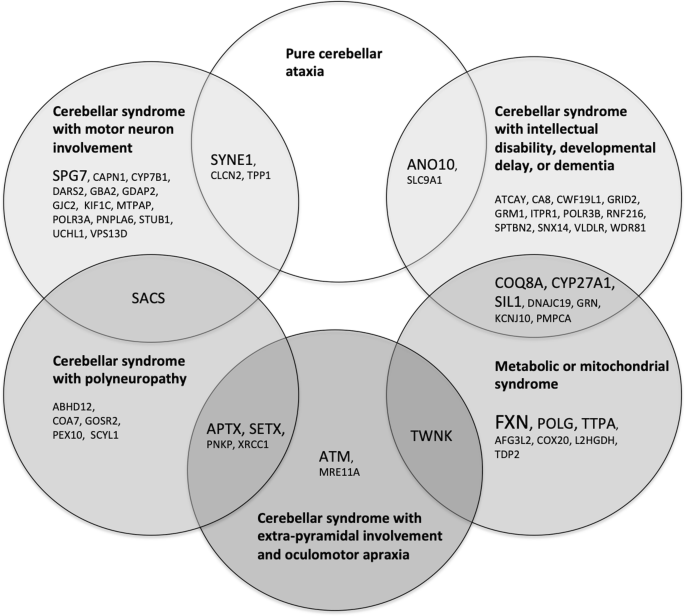
Clinical classification of autosomal recessive ataxias. The gene associated with each primary recessive ataxia is classified according to the most frequent clinical syndrome described for this disorder. Note that some disorders have more complex or variable phenotypes and are placed in the overlapping areas between two categories. Genes presented in larger font represent the most prevalent ataxias

-
5.
Once the clinical assessment is complete, genetic testing is indicated to confirm the mutated gene or allow a more specific diagnosis if the clinical picture is nonspecific. Initial testing should include searching for the Friedreich ataxia-associated trinucleotide repeat expansion in the FXN gene considering the high prevalence of this mutation, its incomplete coverage through the next-generation sequencing methods [1], and the heterogeneous clinical phenotype. Searching for a FXN repeat expansion can be done with frataxin protein analysis or gene analysis with Southern blot or PCR. Moreover, clinicians may consider testing for another specific gene through Sanger sequencing or multiplex ligation-dependent probe amplification (MLPA) if the clinical and paraclinical data are highly evocative of a particular disorder, if there is a confirmed mutation in a relative or in isolated populations where selected disorders are highly prevalent. Finally, a panel for the dominantly inherited CAG-repeat expansion spinocerebellar ataxias may also be considered as part of the initial assessment if family history is inconclusive regarding the mode of inheritance and considering the high prevalence of these mutations and their incomplete coverage through the next-generation sequencing methods [1].
-
6.
If single gene testing does not provide a molecular diagnosis, one should consider the high-throughput NGS methods either with a multigene panel, whole exome sequencing, or whole genome sequencing. Several studies have demonstrated the efficacy and cost efficiency of multigene panels [220], targeted exome sequencing [219, 221], or whole exome sequencing [222, 223], with a diagnostic yield varying between 18 and 80%. The highest yield is obtained for patients with early-onset ataxia and positive family history and consanguinity among parents. NGS panels allow for better coverage of included genes and reduce the volume of genetic variants that are unrelated to the clinical phenotype, while exome sequencing may reveal mutations in genes that were not previously known to be associated with ataxia [1]. Whole genome sequencing may be considered in selected cases with appropriate genetic counseling, but its diagnostic yield is uncertain [224]. Once genetic testing is completed and a pathogenic mutation has been identified, it is of primary importance to provide specialized genetic counseling for the patient and his or her relatives along with symptom management and disease treatment when available. Figure 2 presents a graphical summary of the proposed clinical approach.
Fig. 2 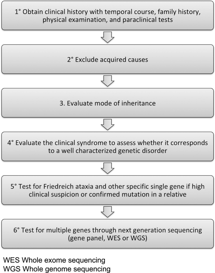
Graphical summary of the clinical approach to a patient presenting with ataxia

Pathophysiological Mechanisms Underlying Autosomal Recessive Cerebellar Ataxias
The importance of a proper recessive ataxia classification goes beyond the clinical diagnosis perspective. Autosomal recessive ataxias can be regrouped according to the deficient cellular and metabolic pathways involved, which provide a better understanding of cerebellar physiology and of its selective vulnerability to certain metabolic defects. This is also essential from a therapeutic perspective, as disorders that belong to the same metabolic pathway may respond to the same treatment options, indicating potential for drug repurposing. Figure 3 presents a pathophysiological classification of autosomal recessive ataxias. Certain genes are presented more than once since some proteins are involved in several metabolic pathways or may interfere with other cellular processes as they accumulate in neurons or glial cells. Table 3 presents a more detailed listing of the pathogenic pathways involved along with relevant references.

Pathophysiological classification of autosomal recessive ataxias. A Purkinje cell is depicted along with a granule cell and parallel fibers. Subcellular organelles and structures are represented graphically. Each gene is classified at one or more subcellular localizations according to the different metabolic pathways involved
Certain pathways are predominantly involved, notably mitochondrial dysfunction, which may result from abnormal mitochondrial DNA maintenance with progressive mutagenesis, defective mitochondrial protein synthesis and quality control, increased levels of reactive oxygen species and oxidative stress, deficient coenzyme Q10 metabolism, altered mitochondrial dynamics, defective mitochondrial chain assembly, or abnormal mitochondrial RNA maturation and processing (Table 3). Interestingly, many of the disorders caused by mitochondrial dysfunction also present with a mitochondrial clinical syndrome as shown in Fig. 1. Disorders of DNA repair mechanisms are also common, with double-strand break repair pathway or single-strand break repair complexes predominantly involved. Pathogenic mutations in these genes are also associated with a susceptibility to ionizing radiations and predisposition for cancers, but the neurological syndrome is characterized by cerebellar involvement and extrapyramidal movement disorders. It remains debated whether defective DNA repair is the main pathogenic mechanism causing the neurological phenotype [230], but the fact that several interacting genes in this pathway are involved in degenerative cerebellar ataxias suggests that the cerebellum has a peculiar susceptibility to DNA damage for which the underlying mechanism is not understood. Finally, altered synaptic morphology or synaptic dysfunction of Purkinje cells (PC) is frequently involved in recessive ataxias and is associated with aberrant morphology at the PC/parallel fiber synapse, impaired dendritic architecture, or dysregulation of glutamate transmission. Other disorders have been implicated in synaptic dysfunction through indirect evidence, for example, SLC9A1, which localizes in presynaptic terminals and is involved in the modulation of synaptic activity [254, 275]. Of interest, many of these disorders are characterized by significant cognitive impairment that goes beyond what is expected in the cerebellar cognitive-affective syndrome and cause intellectual disability, developmental delay, or dementia, highlighting the importance of synaptogenesis in cognitive development.
Discussion
We present a new clinical classification of autosomal recessive ataxias in parallel with a pathophysiological classification. The objective of this classification is to provide a tool for clinicians and researchers that facilitates the understanding of this complex group of disorders and defines this field of research. This work is based on the results of our systematic scoping review of the literature [3]. We updated this literature review and regrouped a panel of 12 international ataxia experts to build a consensus on the definition and classification of cerebellar ataxias. The task force vision is that a classification goes beyond the listing of disorders and must organize diseases in a way that allows better understanding and clinical mastery of this group of disorders. Hence, we proposed a clinical classification along with a pathophysiological classification, which enabled us to observe that there is significant overlap between these two classifications, highlighting how clinical presentation is in some cases a good projection of the underlying biochemical defect. This has potential applications from bench to bedside since treatments that address a specific pathogenic pathway may have therapeutic potential in all disorders in which this pathway is affected. The clinical classification is presented along with a structured clinical approach to a patient presenting with ataxia, which is intended as a clinical tool for expert and nonexpert clinicians. Despite the increasing accessibility of the NGS techniques, there remains a critical place for clinical judgment in the prescription of genetic tests and interpretation of results, taking into account the technical limitations and risk of finding variants of unknown significance. Recently, Renaud and colleagues published the results of a diagnostic algorithm for recessive ataxias that integrates 124 clinical features to propose three potential diagnoses among a list of 67 recessive disorders that may present with ataxia [285]. This is a very promising tool, but its pragmatic impact on molecular testing strategy, final diagnostic rate, patient management, or time efficiency remains to be validated. In the meantime, it is essential for clinicians to be at ease with a general approach to recessive ataxias with the NGS techniques often permitting molecular diagnosis when the clinical picture is nonspecific.
One of the major strengths of this classification proposal is that it is based on a consensus from a panel of international ataxia experts, thereby ensuring a proper representation of regional differences in the prevalence and clinical approach to ataxias. Moreover, the literature search was based on a systematic scoping review of the literature whose methodology has been published before and which permitted an unbiased appraisal of all potentially relevant articles. Nevertheless, there are some limitations to this classification proposal that are inherent to classifying a group of diseases that evolves very rapidly and that is highly heterogeneous. First, as new evidence emerges regarding the identification of novel ataxia-associated genes and as new phenotypes are described for previously described disorders, this classification will need to be updated. This was highlighted by the significant additions to the list of primary recessive ataxias since the original systematic review was conducted in 2016. Indeed, many new genes and new phenotypes of previously described genes have been reported in only 2 years, which suggests that there is a need for periodic updates to the present classification or an online resource. Moreover, several decisions were made in the elaboration of this classification regarding general orientation, purpose of a classification, inclusion of specific disorders, and classification categories. The lists presented here offer in our opinion the best compromise between synthesis and exhaustiveness for the expert and nonexpert clinician.
Compared with a previously published report by the Movement Disorders Society Task Force [4], we decided to exclude disorders in which cerebellar involvement is a minor or late finding in a complex multisystem phenotype or disorders that are already classified on their own, such as genes associated with Joubert syndrome. The objective was to identify the core disorders that are involved in autosomal recessive ataxias in order to define this field of research and build a classification that would be accessible for all clinicians. Indeed, with the progressive advent of affordable NGS diagnostic testing, we believe that it is most important for clinicians to be at ease with one classification and familiar with the most frequent disorders in their unique ethnical and clinical context. Disorders in which ataxia has been reported as a rare or late finding should be included in large NGS testing strategies, but in our opinion should not be categorized as primary ataxias per se. From this perspective, our classification complements the proposal by the Movement Disorders Society Task Force.
There remain some important challenges to be addressed in the field of autosomal recessive ataxias. First, the issue of a proper nomenclature system has been much debated. Recently, the Movement Disorders Society Task Force proposed a revised naming system based on an ataxia prefix associated with the gene name [4]; this was part of a larger effort to revise the nomenclature of all genetic movement disorders. This system overcomes the limitations of the numbered nomenclature, notably unconfirmed genes, and erroneously attributed phenotypes, but its ease of use by nonexperts and patients remains uncertain. Moreover, some disorders were assigned as many as three phenotypic prefixes while some other disorders that are among the most prevalent causes of recessive ataxia, such as POLG, were not assigned an ataxia prefix. Hence, there remains a debate concerning the attribution of prefixes and the integration of this naming system with other fields in neurology and other specialties as many genes involved in ataxia have very complex multisystem phenotypes. Finally, one of the most important challenges in this field of orphan diseases is to develop targeted treatment strategies that address the pathogenic mechanism underlying symptom progression. To this end, we believe that identifying common pathophysiological pathways may provide an opportunity for drug repurposing or enlarge the number of patients that are admissible for drug trials in order to find treatments for these rare but debilitating diseases.
Conclusion or Summary
We present a clinical and a pathophysiological classification of autosomal recessive cerebellar ataxias along with a clinical approach to a patient presenting with ataxia. This classification is the result of a consensus among a panel of international experts, and it promotes a unified understanding of autosomal recessive cerebellar disorders for clinicians and researchers.
References
Klein CJ, Foroud TM. Neurology individualized medicine: when to use next-generation sequencing panels. Mayo Clin Proc. 2017;92(2):292–305. https://doi.org/10.1016/j.mayocp.2016.09.008.
Bahlo M, Bennett MF, Degorski P, Tankard RM, Delatycki MB, Lockhart PJ. Recent advances in the detection of repeat expansions with short-read next-generation sequencing. F1000Res. 2018;7. https://doi.org/10.12688/f1000research.13980.1.
Beaudin M, Klein CJ, Rouleau GA, Dupre N. Systematic review of autosomal recessive ataxias and proposal for a classification. Cerebellum Ataxias. 2017;4:3. https://doi.org/10.1186/s40673-017-0061-y.
Rossi M, Anheim M, Durr A, Klein C, Koenig M, Synofzik M, et al. The genetic nomenclature of recessive cerebellar ataxias. Mov Disord. 2018;33(7):1056–76. https://doi.org/10.1002/mds.27415.
Campuzano V, Montermini L, Molto MD, Pianese L, Cossee M, Cavalcanti F, et al. Friedreich’s ataxia: autosomal recessive disease caused by an intronic GAA triplet repeat expansion. Science. 1996;271(5254):1423–7.
Durr A, Cossee M, Agid Y, Campuzano V, Mignard C, Penet C, et al. Clinical and genetic abnormalities in patients with Friedreich’s ataxia. N Engl J Med. 1996;335(16):1169–75. https://doi.org/10.1056/nejm199610173351601.
Savitsky K, Bar-Shira A, Gilad S, Rotman G, Ziv Y, Vanagaite L, et al. A single ataxia telangiectasia gene with a product similar to PI-3 kinase. Science. 1995;268(5218):1749–53.
Wright J, Teraoka S, Onengut S, Tolun A, Gatti RA, Ochs HD, et al. A high frequency of distinct ATM gene mutations in ataxia-telangiectasia. Am J Hum Genet. 1996;59(4):839–46.
Levy A, Lang AE. Ataxia-telangiectasia: a review of movement disorders, clinical features, and genotype correlations. Mov Disord. 2018. https://doi.org/10.1002/mds.27319.
Date H, Onodera O, Tanaka H, Iwabuchi K, Uekawa K, Igarashi S, et al. Early-onset ataxia with ocular motor apraxia and hypoalbuminemia is caused by mutations in a new HIT superfamily gene. Nat Genet. 2001;29(2):184–8. https://doi.org/10.1038/ng1001-184.
Moreira MC, Barbot C, Tachi N, Kozuka N, Uchida E, Gibson T, et al. The gene mutated in ataxia-ocular apraxia 1 encodes the new HIT/Zn-finger protein aprataxin. Nat Genet. 2001;29(2):189–93. https://doi.org/10.1038/ng1001-189.
Renaud M, Moreira MC, Ben Monga B, Rodriguez D, Debs R, Charles P, et al. Clinical, biomarker, and molecular delineations and genotype-phenotype correlations of ataxia with oculomotor apraxia type 1. JAMA Neurol. 2018;75(4):495–502. https://doi.org/10.1001/jamaneurol.2017.4373.
Moreira MC, Klur S, Watanabe M, Nemeth AH, Le Ber I, Moniz JC, et al. Senataxin, the ortholog of a yeast RNA helicase, is mutant in ataxia-ocular apraxia 2. Nat Genet. 2004;36(3):225–7. https://doi.org/10.1038/ng1303.
Le Ber I, Bouslam N, Rivaud-Pechoux S, Guimaraes J, Benomar A, Chamayou C, et al. Frequency and phenotypic spectrum of ataxia with oculomotor apraxia 2: a clinical and genetic study in 18 patients. Brain. 2004;127(Pt 4):759–67. https://doi.org/10.1093/brain/awh080.
Anheim M, Fleury M, Monga B, Laugel V, Chaigne D, Rodier G, et al. Epidemiological, clinical, paraclinical and molecular study of a cohort of 102 patients affected with autosomal recessive progressive cerebellar ataxia from Alsace, Eastern France: implications for clinical management. Neurogenetics. 2010;11(1):1–12. https://doi.org/10.1007/s10048-009-0196-y.
Engert JC, Berube P, Mercier J, Dore C, Lepage P, Ge B, et al. ARSACS, a spastic ataxia common in northeastern Quebec, is caused by mutations in a new gene encoding an 11.5-kb ORF. Nat Genet. 2000;24(2):120–5. https://doi.org/10.1038/72769.
Criscuolo C, Banfi S, Orio M, Gasparini P, Monticelli A, Scarano V, et al. A novel mutation in SACS gene in a family from southern Italy. Neurology. 2004;62(1):100–2.
Van Goethem G, Martin JJ, Dermaut B, Lofgren A, Wibail A, Ververken D, et al. Recessive POLG mutations presenting with sensory and ataxic neuropathy in compound heterozygote patients with progressive external ophthalmoplegia. Neuromuscul Disord. 2003;13(2):133–42.
Winterthun S, Ferrari G, He L, Taylor RW, Zeviani M, Turnbull DM, et al. Autosomal recessive mitochondrial ataxic syndrome due to mitochondrial polymerase gamma mutations. Neurology. 2005;64(7):1204–8. https://doi.org/10.1212/01.wnl.0000156516.77696.5a.
Cohen BH, Chinnery PF, Copeland WC. POLG-related disorders. In: Adam MP, Ardinger HH, Pagon RA, Wallace SE, Bean LJH, Stephens K et al., editors. GeneReviews((R)). Seattle (WA) 1993.
Gros-Louis F, Dupre N, Dion P, Fox MA, Laurent S, Verreault S, et al. Mutations in SYNE1 lead to a newly discovered form of autosomal recessive cerebellar ataxia. Nat Genet. 2007;39(1):80–5. https://doi.org/10.1038/ng1927.
Synofzik M, Smets K, Mallaret M, Di Bella D, Gallenmuller C, Baets J, et al. SYNE1 ataxia is a common recessive ataxia with major non-cerebellar features: a large multi-centre study. Brain. 2016;139(Pt 5):1378–93. https://doi.org/10.1093/brain/aww079.
Izumi Y, Miyamoto R, Morino H, Yoshizawa A, Nishinaka K, Udaka F et al. Cerebellar ataxia with SYNE1 mutation accompanying motor neuron disease. Neurology. 2013;80(1).
Casari G, De Fusco M, Ciarmatori S, Zeviani M, Mora M, Fernandez P, et al. Spastic paraplegia and OXPHOS impairment caused by mutations in paraplegin, a nuclear-encoded mitochondrial metalloprotease. Cell. 1998;93(6):973–83.
Pfeffer G, Pyle A, Griffin H, Miller J, Wilson V, Turnbull L, et al. SPG7 mutations are a common cause of undiagnosed ataxia. Neurology. 2015;84(11):1174–6. https://doi.org/10.1212/WNL.0000000000001369.
Lagier-Tourenne C, Tazir M, Lopez LC, Quinzii CM, Assoum M, Drouot N, et al. ADCK3, an ancestral kinase, is mutated in a form of recessive ataxia associated with coenzyme Q10 deficiency. Am J Hum Genet. 2008;82(3):661–72. https://doi.org/10.1016/j.ajhg.2007.12.024.
Mollet J, Delahodde A, Serre V, Chretien D, Schlemmer D, Lombes A, et al. CABC1 gene mutations cause ubiquinone deficiency with cerebellar ataxia and seizures. Am J Hum Genet. 2008;82(3):623–30. https://doi.org/10.1016/j.ajhg.2007.12.022.
Vermeer S, Hoischen A, Meijer RP, Gilissen C, Neveling K, Wieskamp N, et al. Targeted next-generation sequencing of a 12.5 Mb homozygous region reveals ANO10 mutations in patients with autosomal-recessive cerebellar ataxia. Am J Hum Genet. 2010;87(6):813–9. https://doi.org/10.1016/j.ajhg.2010.10.015.
Chamova T, Florez L, Guergueltcheva V, Raycheva M, Kaneva R, Lochmuller H, et al. ANO10 c.1150_1151del is a founder mutation causing autosomal recessive cerebellar ataxia in Roma/Gypsies. J Neurol. 2012;259(5):906–11. https://doi.org/10.1007/s00415-011-6276-6.
Renaud M, Anheim M, Kamsteeg EJ, Mallaret M, Mochel F, Vermeer S, et al. Autosomal recessive cerebellar ataxia type 3 due to ANO10 mutations: delineation and genotype-phenotype correlation study. JAMA Neurol. 2014;71(10):1305–10. https://doi.org/10.1001/jamaneurol.2014.193.
Ouahchi K, Arita M, Kayden H, Hentati F, Ben Hamida M, Sokol R, et al. Ataxia with isolated vitamin E deficiency is caused by mutations in the alpha-tocopherol transfer protein. Nat Genet. 1995;9(2):141–5. https://doi.org/10.1038/ng0295-141.
Yokota T, Shiojiri T, Gotoda T, Arita M, Arai H, Ohga T, et al. Friedreich-like ataxia with retinitis pigmentosa caused by the His101Gln mutation of the alpha-tocopherol transfer protein gene. Ann Neurol. 1997;41(6):826–32. https://doi.org/10.1002/ana.410410621.
El Euch-Fayache G, Bouhlal Y, Amouri R, Feki M, Hentati F. Molecular, clinical and peripheral neuropathy study of Tunisian patients with ataxia with vitamin E deficiency. Brain. 2014;137(Pt 2):402–10. https://doi.org/10.1093/brain/awt339.
Cali JJ, Hsieh CL, Francke U, Russell DW. Mutations in the bile acid biosynthetic enzyme sterol 27-hydroxylase underlie cerebrotendinous xanthomatosis. J Biol Chem. 1991;266(12):7779–83.
Leitersdorf E, Reshef A, Meiner V, Levitzki R, Schwartz SP, Dann EJ, et al. Frameshift and splice-junction mutations in the sterol 27-hydroxylase gene cause cerebrotendinous xanthomatosis in Jews or Moroccan origin. J Clin Invest. 1993;91(6):2488–96. https://doi.org/10.1172/JCI116484.
Wong JC, Walsh K, Hayden D, Eichler FS. Natural history of neurological abnormalities in cerebrotendinous xanthomatosis. J Inherit Metab Dis. 2018;41(4):647–56. https://doi.org/10.1007/s10545-018-0152-9.
Anttonen AK, Mahjneh I, Hamalainen RH, Lagier-Tourenne C, Kopra O, Waris L, et al. The gene disrupted in Marinesco-Sjogren syndrome encodes SIL1, an HSPA5 cochaperone. Nat Genet. 2005;37(12):1309–11. https://doi.org/10.1038/ng1677.
Senderek J, Krieger M, Stendel C, Bergmann C, Moser M, Breitbach-Faller N, et al. Mutations in SIL1 cause Marinesco-Sjogren syndrome, a cerebellar ataxia with cataract and myopathy. Nat Genet. 2005;37(12):1312–4. https://doi.org/10.1038/ng1678.
Nikali K, Suomalainen A, Saharinen J, Kuokkanen M, Spelbrink JN, Lonnqvist T, et al. Infantile onset spinocerebellar ataxia is caused by recessive mutations in mitochondrial proteins Twinkle and Twinky. Hum Mol Genet. 2005;14(20):2981–90. https://doi.org/10.1093/hmg/ddi328.
Park MH, Woo HM, Hong YB, Park JH, Yoon BR, Park JM, et al. Recessive C10orf2 mutations in a family with infantile-onset spinocerebellar ataxia, sensorimotor polyneuropathy, and myopathy. Neurogenetics. 2014;15(3):171–82. https://doi.org/10.1007/s10048-014-0405-1.
Fiskerstrand T, H’Mida-Ben Brahim D, Johansson S, M’Zahem A, Haukanes BI, Drouot N, et al. Mutations in ABHD12 cause the neurodegenerative disease PHARC: an inborn error of endocannabinoid metabolism. Am J Hum Genet. 2010;87(3):410–7. https://doi.org/10.1016/j.ajhg.2010.08.002.
Eisenberger T, Slim R, Mansour A, Nauck M, Nurnberg G, Nurnberg P, et al. Targeted next-generation sequencing identifies a homozygous nonsense mutation in ABHD12, the gene underlying PHARC, in a family clinically diagnosed with Usher syndrome type 3. Orphanet J Rare Dis. 2012;7:59. https://doi.org/10.1186/1750-1172-7-59.
Pierson TM, Adams D, Bonn F, Cherikuri PF, Teer JK, Hanson NF, et al. Whole exome sequencing identifies AFG3L2 mutation in a novel recessive progressive myoclonic epilepsy-ataxia-neuropathy syndrome. Ann Neurol. 2010;68:S68–S9.
Eskandrani A, AlHashem A, Ali ES, AlShahwan S, Tlili K, Hundallah K, et al. Recessive AFG3L2 mutation causes progressive microcephaly, early onset seizures, spasticity, and basal ganglia involvement. Pediatr Neurol. 2017;71:24–8. https://doi.org/10.1016/j.pediatrneurol.2017.03.019.
Bomar JM, Benke PJ, Slattery EL, Puttagunta R, Taylor LP, Seong E, et al. Mutations in a novel gene encoding a CRAL-TRIO domain cause human Cayman ataxia and ataxia/dystonia in the jittery mouse. Nat Genet. 2003;35(3):264–9. https://doi.org/10.1038/ng1255.
Manzoor H, Bruggemann N, Hussain HMJ, Baumer T, Hinrichs F, Wajid M, et al. Novel homozygous variants in ATCAY, MCOLN1, and SACS in complex neurological disorders. Parkinsonism Relat Disord. 2018. https://doi.org/10.1016/j.parkreldis.2018.02.005.
Paternoster L, Soblet J, Aeby A, Vilain C, Smits G, Deconinck N. A new mutation of carbonic anhydrase 8 gene expanding the cerebellar ataxia, mental retardation and disequilibrium syndrome (CAMRQ) subtype 3. J Neurol Sci. 2017;381:1136–7. https://doi.org/10.1016/j.jns.2017.08.3200.
Kaya N, Aldhalaan H, Al-Younes B, Colak D, Shuaib T, Al-Mohaileb F, et al. Phenotypical spectrum of cerebellar ataxia associated with a novel mutation in the CA8 gene, encoding carbonic anhydrase (CA) VIII. Am J Med Genet B Neuropsychiatr Genet. 2011;156b(7):826–34. https://doi.org/10.1002/ajmg.b.31227.
Gan-Or Z, Bouslam N, Birouk N, Lissouba A, Chambers DB, Veriepe J, et al. Mutations in CAPN1 cause autosomal-recessive hereditary spastic paraplegia. Am J Hum Genet. 2016;98(6):1271. https://doi.org/10.1016/j.ajhg.2016.05.009.
Wang Y, Hersheson J, Lopez D, Hammer M, Liu Y, Lee KH, et al. Defects in the CAPN1 gene result in alterations in cerebellar development and cerebellar ataxia in mice and humans. Cell Rep. 2016;16(1):79–91. https://doi.org/10.1016/j.celrep.2016.05.044.
Depienne C, Bugiani M, Dupuits C, Galanaud D, Touitou V, Postma N, et al. Brain white matter oedema due to ClC-2 chloride channel deficiency: an observational analytical study. Lancet Neurol. 2013;12(7):659–68. https://doi.org/10.1016/s1474-4422(13)70053-x.
Zeydan B, Uygunoglu U, Altintas A, Saip S, Siva A, Abbink TEM, et al. Identification of 3 novel patients with CLCN2-related leukoencephalopathy due to CLCN2 mutations. Eur Neurol. 2017;78(3–4):125–7. https://doi.org/10.1159/000478089.
Martinez Lyons A, Ardissone A, Reyes A, Robinson AJ, Moroni I, Ghezzi D, et al. COA7 (C1orf163/RESA1) mutations associated with mitochondrial leukoencephalopathy and cytochrome c oxidase deficiency. J Med Genet. 2016;53(12):846–9. https://doi.org/10.1136/jmedgenet-2016-104194.
Higuchi Y, Okunushi R, Hara T, Hashiguchi A, Yuan J, Yoshimura A, et al. Mutations in COA7 cause spinocerebellar ataxia with axonal neuropathy. Brain. 2018. https://doi.org/10.1093/brain/awy104.
Szklarczyk R, Wanschers BF, Nijtmans LG, Rodenburg RJ, Zschocke J, Dikow N, et al. A mutation in the FAM36A gene, the human ortholog of COX20, impairs cytochrome c oxidase assembly and is associated with ataxia and muscle hypotonia. Hum Mol Genet. 2013;22(4):656–67. https://doi.org/10.1093/hmg/dds473.
Doss S, Lohmann K, Seibler P, Arns B, Klopstock T, Zuhlke C, et al. Recessive dystonia-ataxia syndrome in a Turkish family caused by a COX20 (FAM36A) mutation. J Neurol. 2014;261(1):207–12. https://doi.org/10.1007/s00415-013-7177-7.
Burns R, Majczenko K, Xu J, Peng W, Yapici Z, Dowling JJ, et al. Homozygous splice mutation in CWF19L1 in a Turkish family with recessive ataxia syndrome. Neurology. 2014;83(23):2175–82. https://doi.org/10.1212/wnl.0000000000001053.
Nguyen M, Boesten I, Hellebrekers DM, Vanoevelen J, Kamps R, de Koning B, et al. Pathogenic CWF19L1 variants as a novel cause of autosomal recessive cerebellar ataxia and atrophy. Eur J Hum Genet. 2016;24(4):619–22. https://doi.org/10.1038/ejhg.2015.158.
Tsaousidou MK, Ouahchi K, Warner TT, Yang Y, Simpson MA, Laing NG, et al. Sequence alterations within CYP7B1 implicate defective cholesterol homeostasis in motor-neuron degeneration. Am J Hum Genet. 2008;82(2):510–5. https://doi.org/10.1016/j.ajhg.2007.10.001.
Schols L, Rattay TW, Martus P, Meisner C, Baets J, Fischer I, et al. Hereditary spastic paraplegia type 5: natural history, biomarkers and a randomized controlled trial. Brain. 2017;140(12):3112–27. https://doi.org/10.1093/brain/awx273.
Scheper GC, van der Klok T, van Andel RJ, van Berkel CG, Sissler M, Smet J, et al. Mitochondrial aspartyl-tRNA synthetase deficiency causes leukoencephalopathy with brain stem and spinal cord involvement and lactate elevation. Nat Genet. 2007;39(4):534–9. https://doi.org/10.1038/ng2013.
van Berge L, Hamilton EM, Linnankivi T, Uziel G, Steenweg ME, Isohanni P, et al. Leukoencephalopathy with brainstem and spinal cord involvement and lactate elevation: clinical and genetic characterization and target for therapy. Brain. 2014;137(Pt 4):1019–29. https://doi.org/10.1093/brain/awu026.
Al Teneiji A, Siriwardena K, George K, Mital S, Mercimek-Mahmutoglu S. Progressive cerebellar atrophy and a novel homozygous pathogenic DNAJC19 variant as a cause of dilated cardiomyopathy ataxia syndrome. Pediatr Neurol. 2016;62:58–61. https://doi.org/10.1016/j.pediatrneurol.2016.03.020.
Davey KM, Parboosingh JS, McLeod DR, Chan A, Casey R, Ferreira P, et al. Mutation of DNAJC19, a human homologue of yeast inner mitochondrial membrane co-chaperones, causes DCMA syndrome, a novel autosomal recessive Barth syndrome-like condition. J Med Genet. 2006;43(5):385–93. https://doi.org/10.1136/jmg.2005.036657.
Ucar SK, Mayr JA, Feichtinger RG, Canda E, Coker M, Wortmann SB. Previously unreported biallelic mutation in DNAJC19: are sensorineural hearing loss and basal ganglia lesions additional features of dilated cardiomyopathy and ataxia (DCMA) syndrome? JIMD Rep. 2017;35:39–45. https://doi.org/10.1007/8904_2016_23.
Hammer MB, Eleuch-Fayache G, Schottlaender LV, Nehdi H, Gibbs JR, Arepalli SK, et al. Mutations in GBA2 cause autosomal-recessive cerebellar ataxia with spasticity. Am J Hum Genet. 2013;92(2):245–51. https://doi.org/10.1016/j.ajhg.2012.12.012.
Votsi C, Zamba-Papanicolaou E, Middleton LT, Pantzaris M, Christodoulou K. A novel GBA2 gene missense mutation in spastic ataxia. Ann Hum Genet. 2014;78(1):13–22. https://doi.org/10.1111/ahg.12045.
Eidhof I, Baets J, Kamsteeg EJ, Deconinck T, van Ninhuijs L, Martin JJ, et al. GDAP2 mutations implicate susceptibility to cellular stress in a new form of cerebellar ataxia. Brain. 2018;141(9):2592–604. https://doi.org/10.1093/brain/awy198.
Uhlenberg B, Schuelke M, Ruschendorf F, Ruf N, Kaindl AM, Henneke M, et al. Mutations in the gene encoding gap junction protein alpha 12 (connexin 46.6) cause Pelizaeus-Merzbacher-like disease. Am J Hum Genet. 2004;75(2):251–60. https://doi.org/10.1086/422763.
Henneke M, Combes P, Diekmann S, Bertini E, Brockmann K, Burlina AP, et al. GJA12 mutations are a rare cause of Pelizaeus-Merzbacher-like disease. Neurology. 2008;70(10):748–54. https://doi.org/10.1212/01.wnl.0000284828.84464.35.
Corbett MA, Schwake M, Bahlo M, Dibbens LM, Lin M, Gandolfo LC, et al. A mutation in the Golgi Qb-SNARE gene GOSR2 causes progressive myoclonus epilepsy with early ataxia. Am J Hum Genet. 2011;88(5):657–63. https://doi.org/10.1016/j.ajhg.2011.04.011.
van Egmond ME, Verschuuren-Bemelmans CC, Nibbeling EA, Elting JW, Sival DA, Brouwer OF, et al. Ramsay Hunt syndrome: clinical characterization of progressive myoclonus ataxia caused by GOSR2 mutation. Mov Disord. 2014;29(1):139–43. https://doi.org/10.1002/mds.25704.
Utine GE, Haliloglu G, Salanci B, Cetinkaya A, Kiper PO, Alanay Y, et al. A homozygous deletion in GRID2 causes a human phenotype with cerebellar ataxia and atrophy. J Child Neurol. 2013;28(7):926–32. https://doi.org/10.1177/0883073813484967.
Hills LB, Masri A, Konno K, Kakegawa W, Lam AT, Lim-Melia E, et al. Deletions in GRID2 lead to a recessive syndrome of cerebellar ataxia and tonic upgaze in humans. Neurology. 2013;81(16):1378–86. https://doi.org/10.1212/WNL.0b013e3182a841a3.
Guergueltcheva V, Azmanov DN, Angelicheva D, Smith KR, Chamova T, Florez L, et al. Autosomal-recessive congenital cerebellar ataxia is caused by mutations in metabotropic glutamate receptor 1. Am J Hum Genet. 2012;91(3):553–64. https://doi.org/10.1016/j.ajhg.2012.07.019.
Rossi PI, Musante I, Summa M, Pittaluga A, Emionite L, Ikehata M, et al. Compensatory molecular and functional mechanisms in nervous system of the Grm1(crv4) mouse lacking the mGlu1 receptor: a model for motor coordination deficits. Cereb Cortex. 2013;23(9):2179–89. https://doi.org/10.1093/cercor/bhs200.
Smith KR, Damiano J, Franceschetti S, Carpenter S, Canafoglia L, Morbin M, et al. Strikingly different clinicopathological phenotypes determined by progranulin-mutation dosage. Am J Hum Genet. 2012;90(6):1102–7. https://doi.org/10.1016/j.ajhg.2012.04.021.
Almeida MR, Macario MC, Ramos L, Baldeiras I, Ribeiro MH, Santana I. Portuguese family with the co-occurrence of frontotemporal lobar degeneration and neuronal ceroid lipofuscinosis phenotypes due to progranulin gene mutation. Neurobiol Aging. 2016;41:200.e1–5. https://doi.org/10.1016/j.neurobiolaging.2016.02.019.
Gerber S, Alzayady KJ, Burglen L, Bremond-Gignac D, Marchesin V, Roche O, et al. Recessive and dominant de novo ITPR1 mutations cause Gillespie syndrome. Am J Hum Genet. 2016;98(5):971–80. https://doi.org/10.1016/j.ajhg.2016.03.004.
Paganini L, Pesenti C, Milani D, Fontana L, Motta S, Sirchia SM, et al. A novel splice site variant in ITPR1 gene underlying recessive Gillespie syndrome. Am J Med Genet A. 2018. https://doi.org/10.1002/ajmg.a.38704.
Dor T, Cinnamon Y, Raymond L, Shaag A, Bouslam N, Bouhouche A, et al. KIF1C mutations in two families with hereditary spastic paraparesis and cerebellar dysfunction. J Med Genet. 2014;51(2):137–42. https://doi.org/10.1136/jmedgenet-2013-102012.
Yucel-Yilmaz D, Yucesan E, Yalnizoglu D, Oguz KK, Sagiroglu MS, Ozbek U, et al. Clinical phenotype of hereditary spastic paraplegia due to KIF1C gene mutations across life span. Brain Dev. 2018;40(6):458–64. https://doi.org/10.1016/j.braindev.2018.02.013.
Bockenhauer D, Feather S, Stanescu HC, Bandulik S, Zdebik AA, Reichold M, et al. Epilepsy, ataxia, sensorineural deafness, tubulopathy, and KCNJ10 mutations. N Engl J Med. 2009;360(19):1960–70. https://doi.org/10.1056/NEJMoa0810276.
Scholl UI, Choi M, Liu T, Ramaekers VT, Hausler MG, Grimmer J, et al. Seizures, sensorineural deafness, ataxia, mental retardation, and electrolyte imbalance (SeSAME syndrome) caused by mutations in KCNJ10. Proc Natl Acad Sci U S A. 2009;106(14):5842–7. https://doi.org/10.1073/pnas.0901749106.
Celmina M, Micule I, Inashkina I, Audere M, Kuske S, Pereca J, et al. EAST/SeSAME syndrome: review of the literature and introduction of four new Latvian patients. Clin Genet. 2018. https://doi.org/10.1111/cge.13374.
Topcu M, Jobard F, Halliez S, Coskun T, Yalcinkayal C, Gerceker FO, et al. L-2-Hydroxyglutaric aciduria: identification of a mutant gene C14orf160, localized on chromosome 14q22.1. Hum Mol Genet. 2004;13(22):2803–11. https://doi.org/10.1093/hmg/ddh300.
Steenweg ME, Jakobs C, Errami A, van Dooren SJ, Adeva Bartolome MT, Aerssens P, et al. An overview of L-2-hydroxyglutarate dehydrogenase gene (L2HGDH) variants: a genotype-phenotype study. Hum Mutat. 2010;31(4):380–90. https://doi.org/10.1002/humu.21197.
Stewart GS, Maser RS, Stankovic T, Bressan DA, Kaplan MI, Jaspers NG, et al. The DNA double-strand break repair gene hMRE11 is mutated in individuals with an ataxia-telangiectasia-like disorder. Cell. 1999;99(6):577–87.
Pitts SA, Kullar HS, Stankovic T, Stewart GS, Last JI, Bedenham T, et al. hMRE11: genomic structure and a null mutation identified in a transcript protected from nonsense-mediated mRNA decay. Hum Mol Genet. 2001;10(11):1155–62.
Crosby AH, Patel H, Chioza BA, Proukakis C, Gurtz K, Patton MA, et al. Defective mitochondrial mRNA maturation is associated with spastic ataxia. Am J Hum Genet. 2010;87(5):655–60. https://doi.org/10.1016/j.ajhg.2010.09.013.
Martin NT, Nakamura K, Paila U, Woo J, Brown C, Wright JA, et al. Homozygous mutation of MTPAP causes cellular radiosensitivity and persistent DNA double-strand breaks. Cell Death Dis. 2014;5:e1130. https://doi.org/10.1038/cddis.2014.99.
Regal L, Ebberink MS, Goemans N, Wanders RJ, De Meirleir L, Jaeken J, et al. Mutations in PEX10 are a cause of autosomal recessive ataxia. Ann Neurol. 2010;68(2):259–63. https://doi.org/10.1002/ana.22035.
Yamashita T, Mitsui J, Shimozawa N, Takashima S, Umemura H, Sato K, et al. Ataxic form of autosomal recessive PEX10-related peroxisome biogenesis disorders with a novel compound heterozygous gene mutation and characteristic clinical phenotype. J Neurol Sci. 2017;375:424–9. https://doi.org/10.1016/j.jns.2017.02.058.
Jobling RK, Assoum M, Gakh O, Blaser S, Raiman JA, Mignot C, et al. PMPCA mutations cause abnormal mitochondrial protein processing in patients with non-progressive cerebellar ataxia. Brain. 2015;138(Pt 6):1505–17. https://doi.org/10.1093/brain/awv057.
Choquet K, Zurita-Rendon O, La Piana R, Yang S, Dicaire MJ, Boycott KM, et al. Autosomal recessive cerebellar ataxia caused by a homozygous mutation in PMPCA. Brain. 2016;139(Pt 3):e19. https://doi.org/10.1093/brain/awv362.
Joshi M, Anselm I, Shi J, Bale TA, Towne M, Schmitz-Abe K, et al. Mutations in the substrate binding glycine-rich loop of the mitochondrial processing peptidase-alpha protein (PMPCA) cause a severe mitochondrial disease. Cold Spring Harb Mol Case Stud. 2016;2(3):a000786. https://doi.org/10.1101/mcs.a000786.
Bras J, Alonso I, Barbot C, Costa MM, Darwent L, Orme T, et al. Mutations in PNKP cause recessive ataxia with oculomotor apraxia type 4. Am J Hum Genet. 2015;96(3):474–9. https://doi.org/10.1016/j.ajhg.2015.01.005.
Paucar M, Malmgren H, Taylor M, Reynolds JJ, Svenningsson P, Press R, et al. Expanding the ataxia with oculomotor apraxia type 4 phenotype. Neurol Genet. 2016;2(1):e49. https://doi.org/10.1212/nxg.0000000000000049.
Schiess N, Zee DS, Siddiqui KA, Szolics M, El-Hattab AW. Novel PNKP mutation in siblings with ataxia-oculomotor apraxia type 4. J Neurogenet. 2017;31(1–2):23–5. https://doi.org/10.1080/01677063.2017.1322079.
Synofzik M, Gonzalez MA, Lourenco CM, Coutelier M, Haack TB, Rebelo A, et al. PNPLA6 mutations cause Boucher-Neuhauser and Gordon Holmes syndromes as part of a broad neurodegenerative spectrum. Brain. 2014;137(Pt 1):69–77. https://doi.org/10.1093/brain/awt326.
Wiethoff S, Bettencourt C, Paudel R, Madon P, Liu YT, Hersheson J, et al. Pure cerebellar ataxia with homozygous mutations in the PNPLA6 gene. Cerebellum. 2016. https://doi.org/10.1007/s12311-016-0769-x.
Bernard G, Chouery E, Putorti ML, Tetreault M, Takanohashi A, Carosso G, et al. Mutations of POLR3A encoding a catalytic subunit of RNA polymerase Pol III cause a recessive hypomyelinating leukodystrophy. Am J Hum Genet. 2011;89(3):415–23. https://doi.org/10.1016/j.ajhg.2011.07.014.
Wolf NI, Vanderver A, van Spaendonk RM, Schiffmann R, Brais B, Bugiani M, et al. Clinical spectrum of 4H leukodystrophy caused by POLR3A and POLR3B mutations. Neurology. 2014;83(21):1898–905. https://doi.org/10.1212/WNL.0000000000001002.
Saitsu H, Osaka H, Sasaki M, Takanashi J, Hamada K, Yamashita A, et al. Mutations in POLR3A and POLR3B encoding RNA polymerase III subunits cause an autosomal-recessive hypomyelinating leukoencephalopathy. Am J Hum Genet. 2011;89(5):644–51. https://doi.org/10.1016/j.ajhg.2011.10.003.
Tetreault M, Choquet K, Orcesi S, Tonduti D, Balottin U, Teichmann M, et al. Recessive mutations in POLR3B, encoding the second largest subunit of Pol III, cause a rare hypomyelinating leukodystrophy. Am J Hum Genet. 2011;89(5):652–5. https://doi.org/10.1016/j.ajhg.2011.10.006.
Margolin DH, Kousi M, Chan YM, Lim ET, Schmahmann JD, Hadjivassiliou M, et al. Ataxia, dementia, and hypogonadotropism caused by disordered ubiquitination. N Engl J Med. 2013;368(21):1992–2003. https://doi.org/10.1056/NEJMoa1215993.
Alqwaifly M, Bohlega S. Ataxia and hypogonadotropic hypogonadism with intrafamilial variability caused by RNF216 mutation. Neurol Int. 2016;8(2):6444. https://doi.org/10.4081/ni.2016.6444.
Schmidt WM, Rutledge SL, Schule R, Mayerhofer B, Zuchner S, Boltshauser E, et al. Disruptive SCYL1 mutations underlie a syndrome characterized by recurrent episodes of liver failure, peripheral neuropathy, cerebellar atrophy, and ataxia. Am J Hum Genet. 2015;97(6):855–61. https://doi.org/10.1016/j.ajhg.2015.10.011.
Shohet A, Cohen L, Haguel D, Mozer Y, Shomron N, Tzur S, et al. Variant in SCYL1 gene causes aberrant splicing in a family with cerebellar ataxia, recurrent episodes of liver failure, and growth retardation. Eur J Hum Genet. 2018. https://doi.org/10.1038/s41431-018-0268-2.
Thomas AC, Williams H, Seto-Salvia N, Bacchelli C, Jenkins D, O’Sullivan M, et al. Mutations in SNX14 cause a distinctive autosomal-recessive cerebellar ataxia and intellectual disability syndrome. Am J Hum Genet. 2014;95(5):611–21. https://doi.org/10.1016/j.ajhg.2014.10.007.
Akizu N, Cantagrel V, Zaki MS, Al-Gazali L, Wang X, Rosti RO, et al. Biallelic mutations in SNX14 cause a syndromic form of cerebellar atrophy and lysosome-autophagosome dysfunction. Nat Genet. 2015;47(5):528–34. https://doi.org/10.1038/ng.3256.
Guissart C, Li X, Leheup B, Drouot N, Montaut-Verient B, Raffo E, et al. Mutation of SLC9A1, encoding the major Na(+)/H(+) exchanger, causes ataxia-deafness Lichtenstein-Knorr syndrome. Hum Mol Genet. 2015;24(2):463–70. https://doi.org/10.1093/hmg/ddu461.
Iwama K, Osaka H, Ikeda T, Mitsuhashi S, Miyatake S, Takata A, et al. A novel SLC9A1 mutation causes cerebellar ataxia. J Hum Genet. 2018;63(10):1049–54. https://doi.org/10.1038/s10038-018-0488-x.
Lise S, Clarkson Y, Perkins E, Kwasniewska A, Sadighi Akha E, Schnekenberg RP, et al. Recessive mutations in SPTBN2 implicate beta-III spectrin in both cognitive and motor development. PLoS Genet. 2012;8(12):e1003074. https://doi.org/10.1371/journal.pgen.1003074.
Yildiz Bolukbasi E, Afzal M, Mumtaz S, Ahmad N, Malik S, Tolun A. Progressive SCAR14 with unclear speech, developmental delay, tremor, and behavioral problems caused by a homozygous deletion of the SPTBN2 pleckstrin homology domain. Am J Med Genet A. 2017;173(9):2494–9. https://doi.org/10.1002/ajmg.a.38332.
Shi Y, Wang J, Li JD, Ren H, Guan W, He M, et al. Identification of CHIP as a novel causative gene for autosomal recessive cerebellar ataxia. PLoS One. 2013;8(12):e81884. https://doi.org/10.1371/journal.pone.0081884.
Synofzik M, Schule R, Schulze M, Gburek-Augustat J, Schweizer R, Schirmacher A, et al. Phenotype and frequency of STUB1 mutations: next-generation screenings in Caucasian ataxia and spastic paraplegia cohorts. Orphanet J Rare Dis. 2014;9:57. https://doi.org/10.1186/1750-1172-9-57.
Gomez-Herreros F, Schuurs-Hoeijmakers JH, McCormack M, Greally MT, Rulten S, Romero-Granados R, et al. TDP2 protects transcription from abortive topoisomerase activity and is required for normal neural function. Nat Genet. 2014;46(5):516–21. https://doi.org/10.1038/ng.2929.
Zagnoli-Vieira G, Bruni F, Thompson K, He L, Walker S, de Brouwer APM, et al. Confirming TDP2 mutation in spinocerebellar ataxia autosomal recessive 23 (SCAR23). Neurol Genet. 2018;4(4):e262. https://doi.org/10.1212/nxg.0000000000000262.
Sun Y, Almomani R, Breedveld GJ, Santen GW, Aten E, Lefeber DJ, et al. Autosomal recessive spinocerebellar ataxia 7 (SCAR7) is caused by variants in TPP1, the gene involved in classic late-infantile neuronal ceroid lipofuscinosis 2 disease (CLN2 disease). Hum Mutat. 2013;34(5):706–13. https://doi.org/10.1002/humu.22292.
Dy ME, Sims KB, Friedman J. TPP1 deficiency: rare cause of isolated childhood-onset progressive ataxia. Neurology. 2015;85(14):1259–61. https://doi.org/10.1212/wnl.0000000000001876.
Bilguvar K, Tyagi NK, Ozkara C, Tuysuz B, Bakircioglu M, Choi M, et al. Recessive loss of function of the neuronal ubiquitin hydrolase UCHL1 leads to early-onset progressive neurodegeneration. Proc Natl Acad Sci U S A. 2013;110(9):3489–94. https://doi.org/10.1073/pnas.1222732110.
Rydning SL, Backe PH, Sousa MML, Iqbal Z, Oye AM, Sheng Y, et al. Novel UCHL1 mutations reveal new insights into ubiquitin processing. Hum Mol Genet. 2017;26(6):1217–8. https://doi.org/10.1093/hmg/ddx072.
Boycott KM, Flavelle S, Bureau A, Glass HC, Fujiwara TM, Wirrell E, et al. Homozygous deletion of the very low density lipoprotein receptor gene causes autosomal recessive cerebellar hypoplasia with cerebral gyral simplification. Am J Hum Genet. 2005;77(3):477–83. https://doi.org/10.1086/444400.
Ali BR, Silhavy JL, Gleeson MJ, Gleeson JG, Al-Gazali L. A missense founder mutation in VLDLR is associated with dysequilibrium syndrome without quadrupedal locomotion. BMC Med Genet. 2012;13:80. https://doi.org/10.1186/1471-2350-13-80.
Gauthier J, Meijer IA, Lessel D, Mencacci NE, Krainc D, Hempel M, et al. Recessive mutations in >VPS13D cause childhood onset movement disorders. Ann Neurol. 2018;83(6):1089–95. https://doi.org/10.1002/ana.25204.
Seong E, Insolera R, Dulovic M, Kamsteeg EJ, Trinh J, Bruggemann N, et al. Mutations in VPS13D lead to a new recessive ataxia with spasticity and mitochondrial defects. Ann Neurol. 2018;83(6):1075–88. https://doi.org/10.1002/ana.25220.
Gulsuner S, Tekinay AB, Doerschner K, Boyaci H, Bilguvar K, Unal H, et al. Homozygosity mapping and targeted genomic sequencing reveal the gene responsible for cerebellar hypoplasia and quadrupedal locomotion in a consanguineous kindred. Genome Res. 2011;21(12):1995–2003. https://doi.org/10.1101/gr.126110.111.
Komara M, John A, Suleiman J, Ali BR, Al-Gazali L. Clinical and molecular delineation of dysequilibrium syndrome type 2 and profound sensorineural hearing loss in an inbred Arab family. Am J Med Genet A. 2016;170a(2):540–3. https://doi.org/10.1002/ajmg.a.37421.
Hoch NC, Hanzlikova H, Rulten SL, Tetreault M, Komulainen E, Ju L, et al. XRCC1 mutation is associated with PARP1 hyperactivation and cerebellar ataxia. Nature. 2017;541(7635):87–91. https://doi.org/10.1038/nature20790.
O’Connor E, Vandrovcova J, Bugiardini E, Chelban V, Manole A, Davagnanam I, et al. Mutations in XRCC1 cause cerebellar ataxia and peripheral neuropathy. J Neurol Neurosurg Psychiatry. 2018. https://doi.org/10.1136/jnnp-2017-317581.
Ferland RJ, Eyaid W, Collura RV, Tully LD, Hill RS, Al-Nouri D, et al. Abnormal cerebellar development and axonal decussation due to mutations in AHI1 in Joubert syndrome. Nat Genet. 2004;36(9):1008–13. https://doi.org/10.1038/ng1419.
Parisi M, Glass I. Joubert syndrome and related disorders. In: Pagon RA, Adam MP, Ardinger HH, Wallace SE, Amemiya A, Bean LJH, et al., editors. GeneReviews(R). Seattle: University of Washington, Seattle University of Washington, Seattle. All rights reserved; 1993.
Pearl PL, Gibson KM, Acosta MT, Vezina LG, Theodore WH, Rogawski MA, et al. Clinical spectrum of succinic semialdehyde dehydrogenase deficiency. Neurology. 2003;60(9):1413–7.
Trettel F, Malaspina P, Jodice C, Novelletto A, Slaughter CA, Caudle DL, et al. Human succinic semialdehyde dehydrogenase. Molecular cloning and chromosomal localization. Adv Exp Med Biol. 1997;414:253–60.
Imbach T, Burda P, Kuhnert P, Wevers RA, Aebi M, Berger EG, et al. A mutation in the human ortholog of the Saccharomyces cerevisiae ALG6 gene causes carbohydrate-deficient glycoprotein syndrome type-Ic. Proc Natl Acad Sci U S A. 1999;96(12):6982–7.
Morava E, Tiemes V, Thiel C, Seta N, de Lonlay P, de Klerk H, et al. ALG6-CDG: a recognizable phenotype with epilepsy, proximal muscle weakness, ataxia and behavioral and limb anomalies. J Inherit Metab Dis. 2016;39(5):713–23. https://doi.org/10.1007/s10545-016-9945-x.
Bull PC, Thomas GR, Rommens JM, Forbes JR, Cox DW. The Wilson disease gene is a putative copper transporting P-type ATPase similar to the Menkes gene. Nat Genet. 1993;5(4):327–37. https://doi.org/10.1038/ng1293-327.
Onat OE, Gulsuner S, Bilguvar K, Nazli Basak A, Topaloglu H, Tan M, et al. Missense mutation in the ATPase, aminophospholipid transporter protein ATP8A2 is associated with cerebellar atrophy and quadrupedal locomotion. Eur J Hum Genet. 2013;21(3):281–5. https://doi.org/10.1038/ejhg.2012.170.
Alsahli S, Alrifai MT, Al Tala S, Mutairi FA, Alfadhel M. Further delineation of the clinical phenotype of cerebellar ataxia, mental retardation, and disequilibrium syndrome type 4. J Cent Nerv Syst Dis. 2018;10:1179573518759682. https://doi.org/10.1177/1179573518759682.
Boukhris A, Schule R, Loureiro JL, Lourenco CM, Mundwiller E, Gonzalez MA, et al. Alteration of ganglioside biosynthesis responsible for complex hereditary spastic paraplegia. Am J Hum Genet. 2013;93(1):118–23. https://doi.org/10.1016/j.ajhg.2013.05.006.
Pomponio RJ, Reynolds TR, Cole H, Buck GA, Wolf B. Mutational hotspot in the human biotinidase gene causes profound biotinidase deficiency. Nat Genet. 1995;11(1):96–8. https://doi.org/10.1038/ng0995-96.
Wolf B. Biotinidase deficiency. In: Adam MP, Ardinger HH, Pagon RA, Wallace SE, Bean, LJH, Stephens K et al., editors. GeneReviews((R)). Seattle (WA)1993.
Klockars T, Savukoski M, Isosomppi J, Peltonen L. Positional cloning of the CLN5 gene defective in the Finnish variant of the LINCL. Mol Genet Metab. 1999;66(4):324–8. https://doi.org/10.1006/mgme.1999.2832.
Yoshida K, Furihata K, Takeda S, Nakamura A, Yamamoto K, Morita H, et al. A mutation in the ceruloplasmin gene is associated with systemic hemosiderosis in humans. Nat Genet. 1995;9(3):267–72. https://doi.org/10.1038/ng0395-267.
Pennacchio LA, Lehesjoki AE, Stone NE, Willour VL, Virtaneva K, Miao J, et al. Mutations in the gene encoding cystatin B in progressive myoclonus epilepsy (EPM1). Science. 1996;271(5256):1731–4.
Leegwater PA, Vermeulen G, Konst AA, Naidu S, Mulders J, Visser A, et al. Subunits of the translation initiation factor eIF2B are mutant in leukoencephalopathy with vanishing white matter. Nat Genet. 2001;29(4):383–8. https://doi.org/10.1038/ng764.
Fogli A, Schiffmann R, Bertini E, Ughetto S, Combes P, Eymard-Pierre E, et al. The effect of genotype on the natural history of eIF2B-related leukodystrophies. Neurology. 2004;62(9):1509–17.
Minassian BA, Lee JR, Herbrick JA, Huizenga J, Soder S, Mungall AJ, et al. Mutations in a gene encoding a novel protein tyrosine phosphatase cause progressive myoclonus epilepsy. Nat Genet. 1998;20(2):171–4. https://doi.org/10.1038/2470.
Chan EM, Young EJ, Ianzano L, Munteanu I, Zhao X, Christopoulos CC, et al. Mutations in NHLRC1 cause progressive myoclonus epilepsy. Nat Genet. 2003;35(2):125–7. https://doi.org/10.1038/ng1238.
Sijbers AM, de Laat WL, Ariza RR, Biggerstaff M, Wei YF, Moggs JG, et al. Xeroderma pigmentosum group F caused by a defect in a structure-specific DNA repair endonuclease. Cell. 1996;86(5):811–22.
Doi H, Koyano S, Miyatake S, Nakajima S, Nakazawa Y, Kunii M, et al. Cerebellar ataxia-dominant phenotype in patients with ERCC4 mutations. J Hum Genet. 2018;63(4):417–23. https://doi.org/10.1038/s10038-017-0408-5.
Edvardson S, Hama H, Shaag A, Gomori JM, Berger I, Soffer D, et al. Mutations in the fatty acid 2-hydroxylase gene are associated with leukodystrophy with spastic paraparesis and dystonia. Am J Hum Genet. 2008;83(5):643–8. https://doi.org/10.1016/j.ajhg.2008.10.010.
Steinfeld R, Grapp M, Kraetzner R, Dreha-Kulaczewski S, Helms G, Dechent P, et al. Folate receptor alpha defect causes cerebral folate transport deficiency: a treatable neurodegenerative disorder associated with disturbed myelin metabolism. Am J Hum Genet. 2009;85(3):354–63. https://doi.org/10.1016/j.ajhg.2009.08.005.
Grapp M, Just IA, Linnankivi T, Wolf P, Lucke T, Hausler M, et al. Molecular characterization of folate receptor 1 mutations delineates cerebral folate transport deficiency. Brain. 2012;135(Pt 7):2022–31. https://doi.org/10.1093/brain/aws122.
Bomont P, Cavalier L, Blondeau F, Ben Hamida C, Belal S, Tazir M, et al. The gene encoding gigaxonin, a new member of the cytoskeletal BTB/kelch repeat family, is mutated in giant axonal neuropathy. Nat Genet. 2000;26(3):370–4. https://doi.org/10.1038/81701.
Kuhlenbaumer G, Timmerman V, Bomont P. Giant axonal neuropathy. In: Adam MP, Ardinger HH, Pagon RA, Wallace SE, Bean LJH, Stephens K et al., editors. GeneReviews((R)). Seattle (WA) 1993.
Nishimoto J, Nanba E, Inui K, Okada S, Suzuki K. GM1-gangliosidosis (genetic beta-galactosidase deficiency): identification of four mutations in different clinical phenotypes among Japanese patients. Am J Hum Genet. 1991;49(3):566–74.
Karimzadeh P, Naderi S, Modarresi F, Dastsooz H, Nemati H, Farokhashtiani T, et al. Case reports of juvenile GM1 gangliosidosisis type II caused by mutation in GLB1 gene. BMC Med Genet. 2017;18(1):73. https://doi.org/10.1186/s12881-017-0417-4.
Myerowitz R, Costigan FC. The major defect in Ashkenazi Jews with Tay-Sachs disease is an insertion in the gene for the alpha-chain of beta-hexosaminidase. J Biol Chem. 1988;263(35):18587–9.
Gort L, de Olano N, Macias-Vidal J, Coll MA, Spanish GMWG. GM2 gangliosidoses in Spain: analysis of the HEXA and HEXB genes in 34 Tay-Sachs and 14 Sandhoff patients. Gene. 2012;506(1):25–30. https://doi.org/10.1016/j.gene.2012.06.080.
O’Dowd BF, Klavins MH, Willard HF, Gravel R, Lowden JA, Mahuran DJ. Molecular heterogeneity in the infantile and juvenile forms of Sandhoff disease (O-variant GM2 gangliosidosis). J Biol Chem. 1986;261(27):12680–5.
Pierce SB, Walsh T, Chisholm KM, Lee MK, Thornton AM, Fiumara A, et al. Mutations in the DBP-deficiency protein HSD17B4 cause ovarian dysgenesis, hearing loss, and ataxia of Perrault syndrome. Am J Hum Genet. 2010;87(2):282–8. https://doi.org/10.1016/j.ajhg.2010.07.007.
Matsukawa T, Koshi KM, Mitsui J, Bannai T, Kawabe M, Ishiura H, et al. Slowly progressive d-bifunctional protein deficiency with survival to adulthood diagnosed by whole-exome sequencing. J Neurol Sci. 2017;372:6–10. https://doi.org/10.1016/j.jns.2016.11.009.
Stevanin G, Santorelli FM, Azzedine H, Coutinho P, Chomilier J, Denora PS, et al. Mutations in SPG11, encoding spatacsin, are a major cause of spastic paraplegia with thin corpus callosum. Nat Genet. 2007;39(3):366–72. https://doi.org/10.1038/ng1980.
Schule R, Schlipf N, Synofzik M, Klebe S, Klimpe S, Hehr U, et al. Frequency and phenotype of SPG11 and SPG15 in complicated hereditary spastic paraplegia. J Neurol Neurosurg Psychiatry. 2009;80(12):1402–4. https://doi.org/10.1136/jnnp.2008.167528.
Van Bogaert P, Azizieh R, Desir J, Aeby A, De Meirleir L, Laes JF, et al. Mutation of a potassium channel-related gene in progressive myoclonic epilepsy. Ann Neurol. 2007;61(6):579–86. https://doi.org/10.1002/ana.21121.
Kousi M, Anttila V, Schulz A, Calafato S, Jakkula E, Riesch E, et al. Novel mutations consolidate KCTD7 as a progressive myoclonus epilepsy gene. J Med Genet. 2012;49(6):391–9. https://doi.org/10.1136/jmedgenet-2012-100859.
Malm D, Nilssen O. Alpha-mannosidosis. Orphanet J Rare Dis. 2008;3:21. https://doi.org/10.1186/1750-1172-3-21.
Leegwater PA, Yuan BQ, van der Steen J, Mulders J, Konst AA, Boor PK, et al. Mutations of MLC1 (KIAA0027), encoding a putative membrane protein, cause megalencephalic leukoencephalopathy with subcortical cysts. Am J Hum Genet. 2001;68(4):831–8. https://doi.org/10.1086/319519.
Nasca A, Scotton C, Zaharieva I, Neri M, Selvatici R, Magnusson OT, et al. Recessive mutations in MSTO1 cause mitochondrial dynamics impairment, leading to myopathy and ataxia. Hum Mutat. 2017;38(8):970–7. https://doi.org/10.1002/humu.23262.
Gal A, Balicza P, Weaver D, Naghdi S, Joseph SK, Varnai P, et al. MSTO1 is a cytoplasmic pro-mitochondrial fusion protein, whose mutation induces myopathy and ataxia in humans. EMBO Mol Med. 2017;9(7):967–84. https://doi.org/10.15252/emmm.201607058.
Sharp D, Blinderman L, Combs KA, Kienzle B, Ricci B, Wager-Smith K, et al. Cloning and gene defects in microsomal triglyceride transfer protein associated with abetalipoproteinaemia. Nature. 1993;365(6441):65–9. https://doi.org/10.1038/365065a0.
Bonten E, van der Spoel A, Fornerod M, Grosveld G, d’Azzo A. Characterization of human lysosomal neuraminidase defines the molecular basis of the metabolic storage disorder sialidosis. Genes Dev. 1996;10(24):3156–69.
Lai SC, Chen RS, Wu Chou YH, Chang HC, Kao LY, Huang YZ, et al. A longitudinal study of Taiwanese sialidosis type 1: an insight into the concept of cherry-red spot myoclonus syndrome. Eur J Neurol. 2009;16(8):912–9. https://doi.org/10.1111/j.1468-1331.2009.02622.x.
Dorboz I, Aiello C, Simons C, Stone RT, Niceta M, Elmaleh M, et al. Biallelic mutations in the homeodomain of NKX6-2 underlie a severe hypomyelinating leukodystrophy. Brain. 2017;140(10):2550–6. https://doi.org/10.1093/brain/awx207.
Chelban V, Patel N, Vandrovcova J, Zanetti MN, Lynch DS, Ryten M, et al. Mutations in NKX6-2 cause progressive spastic ataxia and hypomyelination. Am J Hum Genet. 2017;100(6):969–77. https://doi.org/10.1016/j.ajhg.2017.05.009.
Carstea ED, Morris JA, Coleman KG, Loftus SK, Zhang D, Cummings C, et al. Niemann-Pick C1 disease gene: homology to mediators of cholesterol homeostasis. Science. 1997;277(5323):228–31.
Nadjar Y, Hutter-Moncada AL, Latour P, Ayrignac X, Kaphan E, Tranchant C, et al. Adult Niemann-Pick disease type C in France: clinical phenotypes and long-term miglustat treatment effect. Orphanet J Rare Dis. 2018;13(1):175. https://doi.org/10.1186/s13023-018-0913-4.
Naureckiene S, Sleat DE, Lackland H, Fensom A, Vanier MT, Wattiaux R, et al. Identification of HE1 as the second gene of Niemann-Pick C disease. Science. 2000;290(5500):2298–301. https://doi.org/10.1126/science.290.5500.2298.
Schaaf CP, Blazo M, Lewis RA, Tonini RE, Takei H, Wang J, et al. Early-onset severe neuromuscular phenotype associated with compound heterozygosity for OPA1 mutations. Mol Genet Metab. 2011;103(4):383–7. https://doi.org/10.1016/j.ymgme.2011.04.018.
Bonneau D, Colin E, Oca F, Ferre M, Chevrollier A, Gueguen N, et al. Early-onset Behr syndrome due to compound heterozygous mutations in OPA1. Brain. 2014;137(Pt 10):e301. https://doi.org/10.1093/brain/awu184.
Shimozawa N, Suzuki Y, Orii T, Moser A, Moser HW, Wanders RJ. Standardization of complementation grouping of peroxisome-deficient disorders and the second Zellweger patient with peroxisomal assembly factor-1 (PAF-1) defect. Am J Hum Genet. 1993;52(4):843–4.
Sevin C, Ferdinandusse S, Waterham HR, Wanders RJ, Aubourg P. Autosomal recessive cerebellar ataxia caused by mutations in the PEX2 gene. Orphanet J Rare Dis. 2011;6:8. https://doi.org/10.1186/1750-1172-6-8.
van den Brink DM, Brites P, Haasjes J, Wierzbicki AS, Mitchell J, Lambert-Hamill M, et al. Identification of PEX7 as the second gene involved in Refsum disease. Am J Hum Genet. 2003;72(2):471–7.
Mihalik SJ, Morrell JC, Kim D, Sacksteder KA, Watkins PA, Gould SJ. Identification of PAHX, a Refsum disease gene. Nat Genet. 1997;17(2):185–9. https://doi.org/10.1038/ng1097-185.
Morgan NV, Westaway SK, Morton JE, Gregory A, Gissen P, Sonek S, et al. PLA2G6, encoding a phospholipase A2, is mutated in neurodegenerative disorders with high brain iron. Nat Genet. 2006;38(7):752–4. https://doi.org/10.1038/ng1826.
Salih M, Mundwiller E, Khan A, Al Drees A, Elmalik S, Hassan H, et al. PLA2G6 gene mutations cause evolving spinocerebellar ataxia influenced by the genotype. J Neurol. 2013;260:S10–S1.
Matthijs G, Schollen E, Pardon E, Veiga-Da-Cunha M, Jaeken J, Cassiman JJ, et al. Mutations in PMM2, a phosphomannomutase gene on chromosome 16p13, in carbohydrate-deficient glycoprotein type I syndrome (Jaeken syndrome). Nat Genet. 1997;16(1):88–92. https://doi.org/10.1038/ng0597-88.
Schiff M, Roda C, Monin ML, Arion A, Barth M, Bednarek N, et al. Clinical, laboratory and molecular findings and long-term follow-up data in 96 French patients with PMM2-CDG (phosphomannomutase 2-congenital disorder of glycosylation) and review of the literature. J Med Genet. 2017;54(12):843–51. https://doi.org/10.1136/jmedgenet-2017-104903.
Meneret A, Gaudebout C, Riant F, Vidailhet M, Depienne C, Roze E. PRRT2 mutations and paroxysmal disorders. Eur J Neurol. 2013;20(6):872–8. https://doi.org/10.1111/ene.12104.
Delcourt M, Riant F, Mancini J, Milh M, Navarro V, Roze E, et al. Severe phenotypic spectrum of biallelic mutations in PRRT2 gene. J Neurol Neurosurg Psychiatry. 2015;86(7):782–5. https://doi.org/10.1136/jnnp-2014-309025.
Hu H, Matter ML, Issa-Jahns L, Jijiwa M, Kraemer N, Musante L, et al. Mutations in PTRH2 cause novel infantile-onset multisystem disease with intellectual disability, microcephaly, progressive ataxia, and muscle weakness. Ann Clin Transl Neurol. 2014;1(12):1024–35. https://doi.org/10.1002/acn3.149.
Picker-Minh S, Mignot C, Doummar D, Hashem M, Faqeih E, Josset P, et al. Phenotype variability of infantile-onset multisystem neurologic, endocrine, and pancreatic disease IMNEPD. Orphanet J Rare Dis. 2016;11(1):52. https://doi.org/10.1186/s13023-016-0433-z.
Agamy O, Ben Zeev B, Lev D, Marcus B, Fine D, Su D, et al. Mutations disrupting selenocysteine formation cause progressive cerebello-cerebral atrophy. Am J Hum Genet. 2010;87(4):538–44. https://doi.org/10.1016/j.ajhg.2010.09.007.
van Dijk T, Vermeij JD, van Koningsbruggen S, Lakeman P, Baas F, Poll-The BT. A SEPSECS mutation in a 23-year-old woman with microcephaly and progressive cerebellar ataxia. J Inherit Metab Dis. 2018;41(5):897–8. https://doi.org/10.1007/s10545-018-0151-x.
Verheijen FW, Verbeek E, Aula N, Beerens CE, Havelaar AC, Joosse M, et al. A new gene, encoding an anion transporter, is mutated in sialic acid storage diseases. Nat Genet. 1999;23(4):462–5. https://doi.org/10.1038/70585.
Aula N, Salomaki P, Timonen R, Verheijen F, Mancini G, Mansson JE, et al. The spectrum of SLC17A5-gene mutations resulting in free sialic acid-storage diseases indicates some genotype-phenotype correlation. Am J Hum Genet. 2000;67(4):832–40. https://doi.org/10.1086/303077.
Barmherzig R, Bullivant G, Cordeiro D, Sinasac DS, Blaser S, Mercimek-Mahmutoglu S. A new patient with intermediate severe Salla disease with hypomyelination: a literature review for Salla disease. Pediatr Neurol. 2017;74:87–91 e2. https://doi.org/10.1016/j.pediatrneurol.2017.05.022.
Seidner G, Alvarez MG, Yeh JI, O’Driscoll KR, Klepper J, Stump TS, et al. GLUT-1 deficiency syndrome caused by haploinsufficiency of the blood-brain barrier hexose carrier. Nat Genet. 1998;18(2):188–91. https://doi.org/10.1038/ng0298-188.
Hully M, Vuillaumier-Barrot S, Le Bizec C, Boddaert N, Kaminska A, Lascelles K, et al. From splitting GLUT1 deficiency syndromes to overlapping phenotypes. Eur J Med Genet. 2015;58(9):443–54. https://doi.org/10.1016/j.ejmg.2015.06.007.
Johnson JO, Gibbs JR, Megarbane A, Urtizberea JA, Hernandez DG, Foley AR, et al. Exome sequencing reveals riboflavin transporter mutations as a cause of motor neuron disease. Brain. 2012;135(Pt 9):2875–82. https://doi.org/10.1093/brain/aws161.
Foley AR, Menezes MP, Pandraud A, Gonzalez MA, Al-Odaib A, Abrams AJ, et al. Treatable childhood neuronopathy caused by mutations in riboflavin transporter RFVT2. Brain. 2014;137(Pt 1):44–56. https://doi.org/10.1093/brain/awt315.
Guissart C, Drouot N, Oncel I, Leheup B, Gershoni-Barush R, Muller J, et al. Genes for spinocerebellar ataxia with blindness and deafness (SCABD/SCAR3, MIM# 271250 and SCABD2). Eur J Hum Genet. 2016;24(8):1154–9. https://doi.org/10.1038/ejhg.2015.259.
Kleta R, Romeo E, Ristic Z, Ohura T, Stuart C, Arcos-Burgos M, et al. Mutations in SLC6A19, encoding B0AT1, cause Hartnup disorder. Nat Genet. 2004;36(9):999–1002. https://doi.org/10.1038/ng1405.
Abrams AJ, Hufnagel RB, Rebelo A, Zanna C, Patel N, Gonzalez MA, et al. Mutations in SLC25A46, encoding a UGO1-like protein, cause an optic atrophy spectrum disorder. Nat Genet. 2015;47(8):926–32. https://doi.org/10.1038/ng.3354.
Hammer MB, Ding J, Mochel F, Eleuch-Fayache G, Charles P, Coutelier M, et al. SLC25A46 mutations associated with autosomal recessive cerebellar ataxia in North African families. Neurodegener Dis. 2017;17(4–5):208–12. https://doi.org/10.1159/000464445.
Cantagrel V, Lefeber DJ, Ng BG, Guan Z, Silhavy JL, Bielas SL, et al. SRD5A3 is required for converting polyprenol to dolichol and is mutated in a congenital glycosylation disorder. Cell. 2010;142(2):203–17. https://doi.org/10.1016/j.cell.2010.06.001.
Wheeler PG, Ng BG, Sanford L, Sutton VR, Bartholomew DW, Pastore MT, et al. SRD5A3-CDG: expanding the phenotype of a congenital disorder of glycosylation with emphasis on adult onset features. Am J Med Genet A. 2016;170(12):3165–71. https://doi.org/10.1002/ajmg.a.37875.
Ghezzi D, Arzuffi P, Zordan M, Da Re C, Lamperti C, Benna C, et al. Mutations in TTC19 cause mitochondrial complex III deficiency and neurological impairment in humans and flies. Nat Genet. 2011;43(3):259–63. https://doi.org/10.1038/ng.761.
Morino H, Miyamoto R, Ohnishi S, Maruyama H, Kawakami H. Exome sequencing reveals a novel TTC19 mutation in an autosomal recessive spinocerebellar ataxia patient. BMC Neurol. 2014;14:5. https://doi.org/10.1186/1471-2377-14-5.
Ardissone A, Granata T, Legati A, Diodato D, Melchionda L, Lamantea E, et al. Mitochondrial complex III deficiency caused by TTC19 defects: report of a novel mutation and review of literature. JIMD Rep. 2015;22:115–20. https://doi.org/10.1007/8904_2015_419.
Colin E, Huynh Cong E, Mollet G, Guichet A, Gribouval O, Arrondel C, et al. Loss-of-function mutations in WDR73 are responsible for microcephaly and steroid-resistant nephrotic syndrome: Galloway-Mowat syndrome. Am J Hum Genet. 2014;95(6):637–48. https://doi.org/10.1016/j.ajhg.2014.10.011.
Vodopiutz J, Seidl R, Prayer D, Khan MI, Mayr JA, Streubel B, et al. WDR73 mutations cause infantile neurodegeneration and variable glomerular kidney disease. Hum Mutat. 2015;36(11):1021–8. https://doi.org/10.1002/humu.22828.
Strom TM, Hortnagel K, Hofmann S, Gekeler F, Scharfe C, Rabl W, et al. Diabetes insipidus, diabetes mellitus, optic atrophy and deafness (DIDMOAD) caused by mutations in a novel gene (wolframin) coding for a predicted transmembrane protein. Hum Mol Genet. 1998;7(13):2021–8.
Mallaret M, Lagha Boukbiza O, Drouot N, Renaud M, Klein FAC, Anheim M, et al. The tumor suppressor gene WWOX is mutated in autosomal recessive cerebellar ataxia with epilepsy and mental retardation. Mov Disord. 2013;28:S398.
Mignot C, Lambert L, Pasquier L, Bienvenu T, Delahaye-Duriez A, Keren B, et al. WWOX-related encephalopathies: delineation of the phenotypical spectrum and emerging genotype-phenotype correlation. J Med Genet. 2015;52(1):61–70. https://doi.org/10.1136/jmedgenet-2014-102748.
Dalmau J, Rosenfeld MR. Paraneoplastic syndromes of the CNS. Lancet Neurol. 2008;7(4):327–40. https://doi.org/10.1016/S1474-4422(08)70060-7.
Coutelier M, Hammer MB, Stevanin G, Monin ML, Davoine CS, Mochel F, et al. Efficacy of exome-targeted capture sequencing to detect mutations in known cerebellar ataxia genes. JAMA Neurol. 2018. https://doi.org/10.1001/jamaneurol.2017.5121.
Nemeth AH, Kwasniewska AC, Lise S, Parolin Schnekenberg R, Becker EB, Bera KD, et al. Next generation sequencing for molecular diagnosis of neurological disorders using ataxias as a model. Brain. 2013;136(Pt 10):3106–18. https://doi.org/10.1093/brain/awt236.
Sun M, Johnson AK, Nelakuditi V, Guidugli L, Fischer D, Arndt K, et al. Targeted exome analysis identifies the genetic basis of disease in over 50% of patients with a wide range of ataxia-related phenotypes. Genet Med. 2018. https://doi.org/10.1038/s41436-018-0007-7.
Valence S, Cochet E, Rougeot C, Garel C, Chantot-Bastaraud S, Lainey E, et al. Exome sequencing in congenital ataxia identifies two new candidate genes and highlights a pathophysiological link between some congenital ataxias and early infantile epileptic encephalopathies. Genet Med. 2018. https://doi.org/10.1038/s41436-018-0089-2.
Fogel BL, Lee H, Deignan JL, Strom SP, Kantarci S, Wang X, et al. Exome sequencing in the clinical diagnosis of sporadic or familial cerebellar ataxia. JAMA Neurol. 2014;71(10):1237–46. https://doi.org/10.1001/jamaneurol.2014.1944.
Kang C, Liang C, Ahmad KE, Gu Y, Siow SF, Colebatch JG, et al. High degree of genetic heterogeneity for hereditary cerebellar ataxias in Australia. Cerebellum. 2018. https://doi.org/10.1007/s12311-018-0969-7.
Jiang B, Glover JN, Weinfeld M. Neurological disorders associated with DNA strand-break processing enzymes. Mech Ageing Dev. 2017;161(Pt A):130–40. https://doi.org/10.1016/j.mad.2016.07.009.
Tahbaz N, Subedi S, Weinfeld M. Role of polynucleotide kinase/phosphatase in mitochondrial DNA repair. Nucleic Acids Res. 2012;40(8):3484–95. https://doi.org/10.1093/nar/gkr1245.
El-Hattab AW, Craigen WJ, Scaglia F. Mitochondrial DNA maintenance defects. Biochim Biophys Acta Mol basis Dis. 2017;1863(6):1539–55. https://doi.org/10.1016/j.bbadis.2017.02.017.
Almajan ER, Richter R, Paeger L, Martinelli P, Barth E, Decker T, et al. AFG3L2 supports mitochondrial protein synthesis and Purkinje cell survival. J Clin Invest. 2012;122(11):4048–58. https://doi.org/10.1172/JCI64604.
Levytskyy RM, Germany EM, Khalimonchuk O. Mitochondrial quality control proteases in neuronal welfare. J NeuroImmune Pharmacol. 2016;11(4):629–44. https://doi.org/10.1007/s11481-016-9683-8.
Choy KR, Watters DJ. Neurodegeneration in ataxia-telangiectasia: multiple roles of ATM kinase in cellular homeostasis. Dev Dyn. 2018;247(1):33–46. https://doi.org/10.1002/dvdy.24522.
Wong A, Yang J, Cavadini P, Gellera C, Lonnerdal B, Taroni F, et al. The Friedreich’s ataxia mutation confers cellular sensitivity to oxidant stress which is rescued by chelators of iron and calcium and inhibitors of apoptosis. Hum Mol Genet. 1999;8(3):425–30.
Abeti R, Parkinson MH, Hargreaves IP, Angelova PR, Sandi C, Pook MA, et al. Mitochondrial energy imbalance and lipid peroxidation cause cell death in Friedreich’s ataxia’. Cell Death Dis. 2016;7:e2237. https://doi.org/10.1038/cddis.2016.111.
Cuddihy SL, Ali SS, Musiek ES, Lucero J, Kopp SJ, Morrow JD, et al. Prolonged alpha-tocopherol deficiency decreases oxidative stress and unmasks alpha-tocopherol-dependent regulation of mitochondrial function in the brain. J Biol Chem. 2008;283(11):6915–24. https://doi.org/10.1074/jbc.M702572200.
Garcia-Diaz B, Barca E, Balreira A, Lopez LC, Tadesse S, Krishna S, et al. Lack of aprataxin impairs mitochondrial functions via downregulation of the APE1/NRF1/NRF2 pathway. Hum Mol Genet. 2015;24(16):4516–29. https://doi.org/10.1093/hmg/ddv183.
Stefely JA, Licitra F, Laredj L, Reidenbach AG, Kemmerer ZA, Grangeray A, et al. Cerebellar ataxia and coenzyme Q deficiency through loss of unorthodox kinase activity. Mol Cell. 2016;63(4):608–20. https://doi.org/10.1016/j.molcel.2016.06.030.
Lariviere R, Gaudet R, Gentil BJ, Girard M, Conte TC, Minotti S, et al. Sacs knockout mice present pathophysiological defects underlying autosomal recessive spastic ataxia of Charlevoix-Saguenay. Hum Mol Genet. 2015;24(3):727–39. https://doi.org/10.1093/hmg/ddu491.
Bradshaw TY, Romano LE, Duncan EJ, Nethisinghe S, Abeti R, Michael GJ, et al. A reduction in Drp1-mediated fission compromises mitochondrial health in autosomal recessive spastic ataxia of Charlevoix Saguenay. Hum Mol Genet. 2016;25(15):3232–44. https://doi.org/10.1093/hmg/ddw173.
Anding AL, Wang C, Chang TK, Sliter DA, Powers CM, Hofmann K, et al. Vps13D encodes a ubiquitin-binding protein that is required for the regulation of mitochondrial size and clearance. Curr Biol. 2018;28(2):287–95 e6. https://doi.org/10.1016/j.cub.2017.11.064.
Aradjanski M, Dogan SA, Lotter S, Wang S, Hermans S, Wibom R, et al. DARS2 protects against neuroinflammation and apoptotic neuronal loss, but is dispensable for myelin producing cells. Hum Mol Genet. 2017;26(21):4181–9. https://doi.org/10.1093/hmg/ddx307.
Bratic A, Clemente P, Calvo-Garrido J, Maffezzini C, Felser A, Wibom R, et al. Mitochondrial polyadenylation is a one-step process required for mRNA integrity and tRNA maturation. PLoS Genet. 2016;12(5):e1006028. https://doi.org/10.1371/journal.pgen.1006028.
Lu YW, Claypool SM. Disorders of phospholipid metabolism: an emerging class of mitochondrial disease due to defects in nuclear genes. Front Genet. 2015;6:3. https://doi.org/10.3389/fgene.2015.00003.
Rzem R, Achouri Y, Marbaix E, Schakman O, Wiame E, Marie S, et al. A mouse model of L-2-hydroxyglutaric aciduria, a disorder of metabolite repair. PLoS One. 2015;10(3):e0119540. https://doi.org/10.1371/journal.pone.0119540.
Rao L, Sha Y, Eissa NT. The E3 ubiquitin ligase STUB1 regulates autophagy and mitochondrial biogenesis by modulating TFEB activity. Mol Cell Oncol. 2017;4(6):e1372867. https://doi.org/10.1080/23723556.2017.1372867.
Regal JA, Festerling TA, Buis JM, Ferguson DO. Disease-associated MRE11 mutants impact ATM/ATR DNA damage signaling by distinct mechanisms. Hum Mol Genet. 2013;22(25):5146–59. https://doi.org/10.1093/hmg/ddt368.
Cohen S, Puget N, Lin YL, Clouaire T, Aguirrebengoa M, Rocher V, et al. Senataxin resolves RNA:DNA hybrids forming at DNA double-strand breaks to prevent translocations. Nat Commun. 2018;9(1):533. https://doi.org/10.1038/s41467-018-02894-w.
Fogel BL, Cho E, Wahnich A, Gao F, Becherel OJ, Wang X, et al. Mutation of senataxin alters disease-specific transcriptional networks in patients with ataxia with oculomotor apraxia type 2. Hum Mol Genet. 2014;23(18):4758–69. https://doi.org/10.1093/hmg/ddu190.
Hirasawa M, Xu X, Trask RB, Maddatu TP, Johnson BA, Naggert JK, et al. Carbonic anhydrase related protein 8 mutation results in aberrant synaptic morphology and excitatory synaptic function in the cerebellum. Mol Cell Neurosci. 2007;35(1):161–70. https://doi.org/10.1016/j.mcn.2007.02.013.
Kakegawa W, Miyoshi Y, Hamase K, Matsuda S, Matsuda K, Kohda K, et al. D-serine regulates cerebellar LTD and motor coordination through the delta2 glutamate receptor. Nat Neurosci. 2011;14(5):603–11. https://doi.org/10.1038/nn.2791.
Sugawara T, Hisatsune C, Le TD, Hashikawa T, Hirono M, Hattori M, et al. Type 1 inositol trisphosphate receptor regulates cerebellar circuits by maintaining the spine morphology of Purkinje cells in adult mice. J Neurosci. 2013;33(30):12186–96. https://doi.org/10.1523/JNEUROSCI.0545-13.2013.
Perkins E, Suminaite D, Jackson M. Cerebellar ataxias: beta-III spectrin’s interactions suggest common pathogenic pathways. J Physiol. 2016;594(16):4661–76. https://doi.org/10.1113/JP271195.
Razafsky D, Hodzic D. A variant of Nesprin1 giant devoid of KASH domain underlies the molecular etiology of autosomal recessive cerebellar ataxia type I. Neurobiol Dis. 2015;78:57–67. https://doi.org/10.1016/j.nbd.2015.03.027.
Itoh M, Li S, Ohta K, Yamada A, Hayakawa-Yano Y, Ueda M, et al. Cayman ataxia-related protein is a presynapse-specific caspase-3 substrate. Neurochem Res. 2011;36(7):1304–13. https://doi.org/10.1007/s11064-011-0430-5.
Bossi S, Musante I, Bonfiglio T, Bonifacino T, Emionite L, Cerminara M, et al. Genetic inactivation of mGlu5 receptor improves motor coordination in the Grm1(crv4) mouse model of SCAR13 ataxia. Neurobiol Dis. 2018;109(Pt A):44–53. https://doi.org/10.1016/j.nbd.2017.10.001.
Dietrich CJ, Morad M. Synaptic acidification enhances GABAA signaling. J Neurosci. 2010;30(47):16044–52. https://doi.org/10.1523/JNEUROSCI.6364-09.2010.
Praschberger R, Lowe SA, Malintan NT, Giachello CNG, Patel N, Houlden H, et al. Mutations in Membrin/GOSR2 reveal stringent secretory pathway demands of dendritic growth and synaptic integrity. Cell Rep. 2017;21(1):97–109. https://doi.org/10.1016/j.celrep.2017.09.004.
Husain N, Yuan Q, Yen YC, Pletnikova O, Sally DQ, Worley P, et al. TRIAD3/RNF216 mutations associated with Gordon Holmes syndrome lead to synaptic and cognitive impairments via arc misregulation. Aging Cell. 2017;16(2):281–92. https://doi.org/10.1111/acel.12551.
Sakurai M, Sekiguchi M, Zushida K, Yamada K, Nagamine S, Kabuta T, et al. Reduction in memory in passive avoidance learning, exploratory behaviour and synaptic plasticity in mice with a spontaneous deletion in the ubiquitin C-terminal hydrolase L1 gene. Eur J Neurosci. 2008;27(3):691–701. https://doi.org/10.1111/j.1460-9568.2008.06047.x.
Duncan EJ, Lariviere R, Bradshaw TY, Longo F, Sgarioto N, Hayes MJ, et al. Altered organization of the intermediate filament cytoskeleton and relocalization of proteostasis modulators in cells lacking the ataxia protein sacsin. Hum Mol Genet. 2017;26(16):3130–43. https://doi.org/10.1093/hmg/ddx197.
Menade M, Kozlov G, Trempe JF, Pande H, Shenker S, Wickremasinghe S, et al. Structures of ubiquitin-like (Ubl) and Hsp90-like domains of sacsin provide insight into pathological mutations. J Biol Chem. 2018;293(33):12832–42. https://doi.org/10.1074/jbc.RA118.003939.
Ichhaporia VP, Kim J, Kavdia K, Vogel P, Horner L, Frase S, et al. SIL1, the endoplasmic-reticulum-localized BiP co-chaperone, plays a crucial role in maintaining skeletal muscle proteostasis and physiology. Dis Model Mech. 2018;11(5). https://doi.org/10.1242/dmm.033043.
Shi CH, Schisler JC, Rubel CE, Tan S, Song B, McDonough H, et al. Ataxia and hypogonadism caused by the loss of ubiquitin ligase activity of the U box protein CHIP. Hum Mol Genet. 2014;23(4):1013–24. https://doi.org/10.1093/hmg/ddt497.
Lenz D, McClean P, Kansu A, Bonnen PE, Ranucci G, Thiel C, et al. SCYL1 variants cause a syndrome with low gamma-glutamyl-transferase cholestasis, acute liver failure, and neurodegeneration (CALFAN). Genet Med. 2018;20(10):1255–65. https://doi.org/10.1038/gim.2017.260.
Patron M, Sprenger HG, Langer T. m-AAA proteases, mitochondrial calcium homeostasis and neurodegeneration. Cell Res. 2018;28(3):296–306. https://doi.org/10.1038/cr.2018.17.
Wanitchakool P, Ousingsawat J, Sirianant L, Cabrita I, Faria D, Schreiber R, et al. Cellular defects by deletion of ANO10 are due to deregulated local calcium signaling. Cell Signal. 2017;30:41–9. https://doi.org/10.1016/j.cellsig.2016.11.006.
Synofzik M, Helbig KL, Harmuth F, Deconinck T, Tanpaiboon P, Sun B, et al. De novo ITPR1 variants are a recurrent cause of early-onset ataxia, acting via loss of channel function. Eur J Hum Genet. 2018;26(11):1623–34. https://doi.org/10.1038/s41431-018-0206-3.
Shanmughapriya S, Rajan S, Hoffman NE, Higgins AM, Tomar D, Nemani N, et al. SPG7 is an essential and conserved component of the mitochondrial permeability transition pore. Mol Cell. 2015;60(1):47–62. https://doi.org/10.1016/j.molcel.2015.08.009.
Gotzl JK, Colombo AV, Fellerer K, Reifschneider A, Werner G, Tahirovic S, et al. Early lysosomal maturation deficits in microglia triggers enhanced lysosomal activity in other brain cells of progranulin knockout mice. Mol Neurodegener. 2018;13(1):48. https://doi.org/10.1186/s13024-018-0281-5.
Sleat DE, Wiseman JA, El-Banna M, Kim KH, Mao Q, Price S, et al. A mouse model of classical late-infantile neuronal ceroid lipofuscinosis based on targeted disruption of the CLN2 gene results in a loss of tripeptidyl-peptidase I activity and progressive neurodegeneration. J Neurosci. 2004;24(41):9117–26. https://doi.org/10.1523/JNEUROSCI.2729-04.2004.
Bryant D, Liu Y, Datta S, Hariri H, Seda M, Anderson G, et al. SNX14 mutations affect endoplasmic reticulum-associated neutral lipid metabolism in autosomal recessive spinocerebellar ataxia 20. Hum Mol Genet. 2018;27(11):1927–40. https://doi.org/10.1093/hmg/ddy101.
Ulatowski L, Parker R, Warrier G, Sultana R, Butterfield DA, Manor D. Vitamin E is essential for Purkinje neuron integrity. Neuroscience. 2014;260:120–9. https://doi.org/10.1016/j.neuroscience.2013.12.001.
Joshi A, Shaikh M, Singh S, Rajendran A, Mhetre A, Kamat SS. Biochemical characterization of the PHARC-associated serine hydrolase ABHD12 reveals its preference for very-long-chain lipids. J Biol Chem. 2018;293(44):16953–63. https://doi.org/10.1074/jbc.RA118.005640.
Nie S, Chen G, Cao X, Zhang Y. Cerebrotendinous xanthomatosis: a comprehensive review of pathogenesis, clinical manifestations, diagnosis, and management. Orphanet J Rare Dis. 2014;9:179. https://doi.org/10.1186/s13023-014-0179-4.
Woeste MA, Wachten D. The enigmatic role of GBA2 in controlling locomotor function. Front Mol Neurosci. 2017;10:386. https://doi.org/10.3389/fnmol.2017.00386.
Dutta S, Rieche F, Eckl N, Duch C, Kretzschmar D. Glial expression of Swiss cheese (SWS), the Drosophila orthologue of neuropathy target esterase (NTE), is required for neuronal ensheathment and function. Dis Model Mech. 2016;9(3):283–94. https://doi.org/10.1242/dmm.022236.
Liu Y, Zaun HC, Orlowski J, Ackerman SL. CHP1-mediated NHE1 biosynthetic maturation is required for Purkinje cell axon homeostasis. J Neurosci. 2013;33(31):12656–69. https://doi.org/10.1523/JNEUROSCI.0406-13.2013.
Abrams CK. Diseases of connexins expressed in myelinating glia. Neurosci Lett. 2019;695:91–9. https://doi.org/10.1016/j.neulet.2017.05.037.
Schirmer L, Mobius W, Zhao C, Cruz-Herranz A, Ben Haim L, Cordano C, et al. Oligodendrocyte-encoded Kir4.1 function is required for axonal integrity. Elife. 2018;7. https://doi.org/10.7554/eLife.36428.
Tingaud-Sequeira A, Raldua D, Lavie J, Mathieu G, Bordier M, Knoll-Gellida A, et al. Functional validation of ABHD12 mutations in the neurodegenerative disease PHARC. Neurobiol Dis. 2017;98:36–51. https://doi.org/10.1016/j.nbd.2016.11.008.
Blanz J, Schweizer M, Auberson M, Maier H, Muenscher A, Hubner CA, et al. Leukoencephalopathy upon disruption of the chloride channel ClC-2. J Neurosci. 2007;27(24):6581–9. https://doi.org/10.1523/JNEUROSCI.0338-07.2007.
Duchesne A, Vaiman A, Frah M, Floriot S, Legoueix-Rodriguez S, Desmazieres A, et al. Progressive ataxia of Charolais cattle highlights a role of KIF1C in sustainable myelination. PLoS Genet. 2018;14(8):e1007550. https://doi.org/10.1371/journal.pgen.1007550.
Takanashi J, Osaka H, Saitsu H, Sasaki M, Mori H, Shibayama H, et al. Different patterns of cerebellar abnormality and hypomyelination between POLR3A and POLR3B mutations. Brain and Development. 2014;36(3):259–63. https://doi.org/10.1016/j.braindev.2013.03.006.
Ady V, Toscano-Marquez B, Nath M, Chang PK, Hui J, Cook A, et al. Altered synaptic and firing properties of cerebellar Purkinje cells in a mouse model of ARSACS. J Physiol. 2018;596(17):4253–67. https://doi.org/10.1113/JP275902.
Cavallin M, Rujano MA, Bednarek N, Medina-Cano D, Bernabe Gelot A, Drunat S, et al. WDR81 mutations cause extreme microcephaly and impair mitotic progression in human fibroblasts and Drosophila neural stem cells. Brain. 2017;140(10):2597–609. https://doi.org/10.1093/brain/awx218.
Kizhakkedath P, Loregger A, John A, Bleijlevens B, Al-Blooshi AS, Al-Hosani AH, et al. Impaired trafficking of the very low density lipoprotein receptor caused by missense mutations associated with dysequilibrium syndrome. Biochim Biophys Acta. 2014;1843(12):2871–7. https://doi.org/10.1016/j.bbamcr.2014.08.013.
Renaud M, Tranchant C, Martin JVT, Mochel F, Synofzik M, van de Warrenburg B, et al. A recessive ataxia diagnosis algorithm for the next generation sequencing era. Ann Neurol. 2017;82(6):892–9. https://doi.org/10.1002/ana.25084.
Acknowledgments
We thank Miruna Anohim for her contribution to the data collection on geographical specificities. Marie Beaudin is supported by the Canadian Institutes of Health Research.
Author information
Authors and Affiliations
Corresponding author
Ethics declarations
Conflict of Interest
The authors declare that they have no conflict of interest.
Additional information
Publisher’s Note
Springer Nature remains neutral with regard to jurisdictional claims in published maps and institutional affiliations.
Rights and permissions
Open Access This article is distributed under the terms of the Creative Commons Attribution 4.0 International License (http://creativecommons.org/licenses/by/4.0/), which permits unrestricted use, distribution, and reproduction in any medium, provided you give appropriate credit to the original author(s) and the source, provide a link to the Creative Commons license, and indicate if changes were made.
About this article

Cite this article
Beaudin, M., Matilla-Dueñas, A., Soong, BW. et al. The Classification of Autosomal Recessive Cerebellar Ataxias: a Consensus Statement from the Society for Research on the Cerebellum and Ataxias Task Force. Cerebellum 18, 1098–1125 (2019). https://doi.org/10.1007/s12311-019-01052-2
Published:
Issue Date:
DOI: https://doi.org/10.1007/s12311-019-01052-2