
Abstract
Magnetorelaxometry imaging (MRXI) is a non-invasive, quantitative imaging technique for magnetic nanoparticles (MNPs). The image resolution of this technique significantly depends on the relaxation amplitude (ΔB). For this work, we measured the room temperature (299 K) relaxation signals of eight commercial MNP sample systems with different magnetic properties, in both fluid and immobilized states, in order to select the most suitable sample for a particular MRXI setting. Additionally, the effect of elevated temperatures (up to hyperthermia temperature, 335 K) on the relaxation signals of four different MNP systems (Synomag, Perimag, BNF and Nanomag) in both states were investigated. The ΔB values of fluid samples significantly decreased with increasing temperature, and the behaviour for immobilized samples depended on their blocking temperature (TB). For samples with TB < 299 K, ΔB also decreased with increasing temperature. Whereas for samples with TB > 299 K, the opposite behaviour was observed. These results are beneficial for improving the image resolution in MRXI and show, among the investigated systems, and for our setup, Synomag is the best candidate for future in vitro and in vivo studies. This is due to its consistently high ΔB between 299 and 335 K in both states. Our findings demonstrate the feasibility of temperature imaging by MRXI.
Export citation and abstract BibTeX RIS

Original content from this work may be used under the terms of the Creative Commons Attribution 4.0 licence. Any further distribution of this work must maintain attribution to the author(s) and the title of the work, journal citation and DOI.
1. Introduction
Magnetic nanoparticles (MNPs) have been extensively used for biomedical applications such as cancer therapies. During recent years, cancer types such as glioblastoma multiform, which is the most aggressive type of brain tumour, were treated in patients by magnetic hyperthermia therapy (Mahmoudi et al
2018). This technique is based on the subsequent heating of MNPs by an alternating magnetic field after injecting them into the target region (Liu et al
2020). Any information about the quantity and the location of the MNP distribution in the target region as well as in other body regions for the heat treatment procedure is required, leading to subsequent progress in cancer treatment. Magnetorelaxometry imaging (MRXI) is a method which can provide this information (Baumgarten et al
2008, Liebl et al
2015). MRXI is a non-invasive imaging technique that is performed by the combination of distinct multi-channel magnetorelaxometry (MRX) measurements and solving the inverse problem of the forward model (Liebl et al
2014, Schier et al
2022) to reconstruct a MNP source distribution from the measured relaxation field maps. MRX is based on measuring the magnetic (stray) field of the decaying net magnetization of an MNP ensemble after switching off a magnetizing field applied before to align their magnetic moments. The magnetic moments of the particles can be reoriented via two mechanisms, (1) the rotation of the whole particle or (2) the switching of the magnetic moment inside the particle core. These mechanisms are parameterized by characteristic relaxation times called Brownian relaxation time (Brown 1963),  and Néel relaxation time (Néel 1949),
  respectively:


where η(T) is the temperature dependent viscosity of the dispersion medium. Vh is the hydrodynamic volume of the particle, kB is the Boltzmann constant, T is the temperature, τ0 is a time-constant (attempt time) with values between 10−8 and 10−12 s (Coene et al 2017), K is the anisotropy constant, and V is the magnetic core volume of the particle.
Where both Brownian and Néel relaxation mechanisms are present, the faster mechanism will dominate the relaxation, leading to an effective relaxation time  (Shliomis 1972):

According to equations (1)–(3), the relaxation time is affected by the hydrodynamic diameter, viscosity, anisotropy energy and temperature of the MNPs. However, the Brownian and Néel relaxation times also depend on the ac and dc magnetic field as shown by Dieckhoff and Eberbeck (Dieckhoff et al 2016).
Since MRXI can be used for quantitative imaging of MNP distributions inside a specific body region (Arsalani et al 2022), and the relaxation signals of MNPs depend on temperature, one of the most essential factors influencing biological processes of the body, it is important to investigate the effect of temperature on the relaxation signal of MNPs. In this work, we measured the relaxation signals of various MNP samples in fluid and immobilized states from room temperature (299 K), important for in vitro applications, to nearly body temperature (308 K) and up to hyperthermia temperature (335 K), essential for in vivo treatment applications with our 6-channel MRX system. From the samples, the MNP system with the highest relaxation amplitude ΔB at different temperatures and for a given set of MRXI parameters can be selected to improve image resolution. Additionally, the results suggest that MRXI can potentially be used as a temperature imaging.
2. Materials and methods
2.1. Materials
In this work, eight commercially available MNP systems, consisting of aqueous suspensions of iron oxide nanoparticles, were used. Hydrodynamic diameter (dh) and polydispersity index (PDI) of the samples were determined by dynamic light scattering using a Zetasizer system (Malvern Instruments, UK) (Arsalani et al 2021). Relevant size distribution parameters are given in table 1. Note, due to the cluster-type or multi-core character of most samples the single core diameter is not quoted.
Table 1. Summary of the different MNP systems used in this work. Given are the sample and supplier names, surface coating of the MNP, and characteristics of the MNP samples measured by DLS (hydrodynamic diameter dh and polydispersity index PDI).
| MNP systems | Supplier | Surface coating | dh (nm) | PDI |
|---|---|---|---|---|
| Iron oxide particles (IONP_1) | Micromod | COOH | 66 | 0.16 |
| Synomag®-D | Micromod | Dextran-OH | 71 | 0.16 |
| Bionisiertes NanoFerrit (BNF) | Micromod | Starch-OH | 110 | 0.05 |
| Perimag® | Micromod | Dextran-OH | 112 | 0.22 |
| FluidMAG-D (FLU100) | Chemicell | Starch-OH | 132 | 0.33 |
| Nanomag®-D | Micromod | Dextran-OH | 150 | 0.08 |
| FluidMAG-D (FLU200) | Chemicell | Starch-OH | 162 | 0.10 |
| Iron oxide particles (IONP_2) | Micromod | OH | 283 | 0.23 |
2.2. Methods
2.2.1. Magnetorelaxometry
We first recorded the room temperature (299 K) relaxation signal of the MNP sample systems in fluid and immobilized states (freeze dried) with a 6-channel low- superconducting quantum interference device (SQUID) MRX system (Ackermann et al
2007), see figure 1. To enable a consistent comparison, we always used the same channel for subsequent analysis. The samples with 100 μl volume and an iron concentration of c(Fe) = 6 mmol l−1 were exposed to a homogenous magnetic field
superconducting quantum interference device (SQUID) MRX system (Ackermann et al
2007), see figure 1. To enable a consistent comparison, we always used the same channel for subsequent analysis. The samples with 100 μl volume and an iron concentration of c(Fe) = 6 mmol l−1 were exposed to a homogenous magnetic field  = 3.2 kA m−1 (4 mT), generated by a cylindrical coil. After a magnetizing time of
  = 1 s, a net magnetic moment
  of the sample is achieved by (partial) alignment of the individual MNP moments with the external magnetic field direction according to:

where  is the magnetic susceptibility of the MNPs and
  is the amount of MNPs in the sample.

Figure 1. 6-channel low- SQUID MRX system. The Dewar has a horizontal warm bore (indicated by the yellow circle) which is shielded by a superconducting niobium tube. The SQUIDs are located midway of the warm bore, orientated in x, y, and z direction (not shown here), and operated with a flux-locked-loop (FLL) electronics (adopted from Everaert et al (2023) with permission from the Royal Society of Chemistry). The sample is inserted into the center of warm bore close to the SQUIDs.
Download figure:
Standard image High-resolution imageAfter switching off  and a delay time of 0.25 ms, necessary to prevent saturation of the highly sensitive SQUID electronics by the strong transient magnetic field of the magnetization coil, the decaying net magnetic moment of the particles
  where
where  is the relaxation shape function (with values between zero and one, as described in details in Eberbeck et al (2006)), was recorded for a measurement time of
  = 400 ms at a sampling rate of 100 kHz. The decaying net magnetic moment, modelled as a point-like source at sample location
  gives rise to a flux density B (t, r) that is detected by a sensor at location r with its sensitive direction n = [n
x
, n
y
, n
z
] (Liebl et al
2014)
gives rise to a flux density B (t, r) that is detected by a sensor at location r with its sensitive direction n = [n
x
, n
y
, n
z
] (Liebl et al
2014)

The relaxation signal  is characterized by two parameters: (1) the relaxation amplitude
  which denotes the field difference between two time points
  ms and
  ms (
ms ( ) which strongly depends on the quantity of MNPs, magnetic susceptibility and relaxation properties of the MNPs. (2) The relaxation time (
 ) indicating the time at which the relaxation signal
  has dropped to
  or 36.8% of its value
After MRX measurements at room temperature, four selected sample systems in both fluid and immobilized state were further analyzed by MRX measurements at elevated temperatures. The temperature dependence was determined by first placing the sample container in a water bath until it reached the steady state at the nominal temperature of 300, 310, 320, 330, 340 and 350 K next to the MRX system and then quickly (within about 12 s) transferring it into the warm bore of MRX system close to the SQUID sensors and start a measurement. For each measurement, we used 3 repetitions from which the average relaxation signal was calculated. To estimate the temperature drop during the measurement more precisely, we independently monitored the temperature inside a separate fluid sample with a fiberoptic thermometer when transferring it into the warm bore. The average temperature and the uncertainty during the 3 repetitions for the 6 used temperatures were determined as 299 ± 0.4, 308 ± 0.7, 316 ± 1.5, 325 ± 2, 330 ± 3 and 335 ± 2.5 K. The uncertainty was obtained from the temperature differences relative to the start and the end of the measurement.
2.2.2. Magnetic properties measurement system
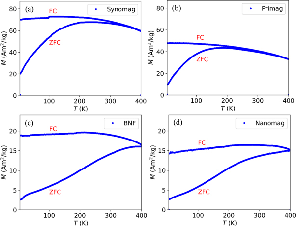
The relaxation behavior of MNP samples can be related to their blocking temperature (TB). Therefore, we examined the temperature dependent magnetization M(T) of MNPs by a SQUID-based magnetic property measurement system (MPMS-XL5, Quantum Design, USA) to investigate TB. The measurements were performed on the freeze dried MNP samples with iron concentration of c(Fe) = 6 mmol l−1 and a volume of 60 μl. First, the sample was cooled in the absence of a magnetic field down to 5 K. Then the magnetization M(T) was recorded while slowly heating from 5 to 400 K in an applied magnetic field of 9.5 kA m−1 (12 mT) (zero-field-cooled (ZFC)) with a measurement time of 100 s. Subsequently, M(t) was acquired while the sample was slowly cooled down again to 5 K in the same magnetic field (field-cooled (FC)). Note, a larger magnetic field, relative to the MRX measurements, was applied to increase the signal to noise ratio and the corresponding effect on the TB is expected to be insignificant for the purpose of this work (Kachkachi et al 2000).
3. Results and discussions
The measured relaxation signals of MNP samples with c(Fe) = 6 mmol l−1 in fluid and immobilized states at 299 K are shown for one channel in figures 2(a), (b) and the determined ΔB and  are listed in table 2. It should be noted, that all MNP samples used in this work are heterogenous systems and consist of a broad size distribution of particles, see the PDI in table 1. Therefore, the measured relaxation signal of a fluid MNP ensemble is the superposition of various exponential decays caused by Néel and Brownian processes (Liebl et al
2015), whereas for an immobilized sample the signal consists of the superposition of merely Néel relaxations since Brownian rotation is suppressed.
are listed in table 2. It should be noted, that all MNP samples used in this work are heterogenous systems and consist of a broad size distribution of particles, see the PDI in table 1. Therefore, the measured relaxation signal of a fluid MNP ensemble is the superposition of various exponential decays caused by Néel and Brownian processes (Liebl et al
2015), whereas for an immobilized sample the signal consists of the superposition of merely Néel relaxations since Brownian rotation is suppressed.

Figure 2. Relaxation signals of different MNP systems in (a) fluid, and (b) immobilized states at a temperature of 299 K recorded by a 6-channel MRX system (only one channel shown). Each curve was offset corrected by subtracting its  -value.
Download figure:
Standard image High-resolution imageTable 2. MRX parameters ΔB and  of all MNP systems at 299 K in fluid and immobilized states.
| Fluid | Immobilized | |||
|---|---|---|---|---|
| MNP systems |

|

|

|

|
| BNF | 1384 | 0.74 | 155 | 29.13 |
| FLU100 | 365 | 3.23 | 221 | 9.75 |
| FLU200 | 781 | 1.90 | 359 | 12.93 |
| IONP_1 | 2 | 0.86 | 36 | 3.62 |
| IONP_2 | 794 | 12.93 | 184 | 16.49 |
| Nanomag | 837 | 1.29 | 101 | 21.91 |
| Perimag | 737 | 1.12 | 484 | 7.67 |
| Synomag | 1625 | 1.24 | 1452 | 9.37 |
Among fluid samples, Synomag and BNF have the highest ΔB with 1.6 nT and 1.4 nT, about a factor of two larger than Nanomag with 837 pT, see figure 2(a). This can be interpreted within the concept of measurement time window (Kötitz et al
1999), i.e. only the particles of the size fraction with the relaxation time that is within the measurement time window of the apparatus ( ) significantly contribute to the MRX signal. Specifically, if the Néel relaxation of MNP-moments is blocked, the relaxation is purely determined by the Brownian relaxation time (equation (1)). This is obviously the case for IONP_2, where the relaxation curve is closest to a single exponential decay among the investigated sample systems (figure 2(a)) although the width of the distribution of dh is very large (PDI in table 1). This could be explained by the fact, that
  of an ensemble of size distributed MNP is usually much wider distributed than
  because of the exponential dependency of
  on the core volume, equation (2). We infer that the shape of the IONP_2's relaxation curve is to a large extend determined by the Brownian relaxation deducing that the
  of a large part of these MNP is much larger than the upper limit of the measurement time window ('blocked particles'). This corresponds well to the relatively small relaxation amplitude of the immobilized MNP of IONP_2. On the other hand, Brownian and Néel relaxation of IONP_1 is much smaller than the lower limit of the relaxation time window, thus the relaxation is too fast to be detected by the given measurement system so that no significant relaxation is observed. The strongly non-exponential shape of relaxation of the other systems is evidence for a wide distribution of
  The relatively large relaxation amplitudes of Synomag and BNF imply a distribution of core diameters d and hydrodynamic diameters dh where
  lies within the measurement time window for a relatively large fraction of MNP.
Among the immobilized systems (figure 2(b)), Synomag shows again the highest ΔB with 1.5 nT, followed by Perimag which has a substantially smaller ΔB of 484 pT. A large signal results from (i) a large core volume V (high magnetic moment) having a low anisotropy constant K so that the relaxing magnetic moment is large. Thereby, K has a value which shifts the relaxation time (equation (2)) into the measurement time window. Note, the optimal combination of V and K values is constrained by the requirement of prevention of aggregation, the probability of which increases with the magnitude of the magnetic moment, more details can be found in Eberbeck et al (2006). (ii) The width of the size distribution should be very small so that  of a large fraction of MNP fits the measurement time window, i.e. contributes strongly to the signal. Obviously, for Synomag the core volumes and the anisotropy constant are closer to the optimal values than the values of the other systems.
Thus, Synomag exposed to a magnetic field 3.2 kA m−1 has the highest ΔB of all samples at 299 K in both fluid and immobilized states, demonstrating its suitability for MRXI with the employed time window. However, to fully assess its suitability and to confirm that it has the highest ΔB in both fluid and immobilized states also at elevated temperatures, we investigated the relaxation signals at body and hyperthermia temperatures of Synomag and the three further MNP systems (BNF, Nanomag and Perimag), which exhibited the highest relaxation amplitudes at 299 K.
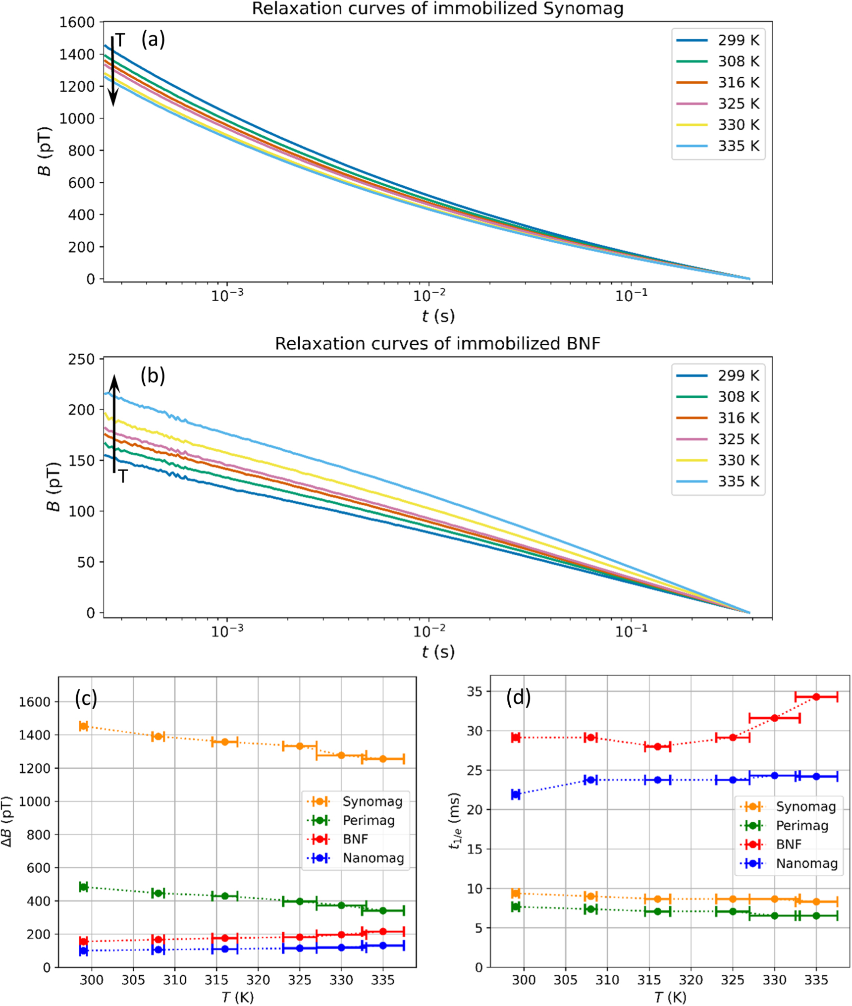
The relaxation signals of fluid Synomag and BNF at different temperatures 299, 308, 316, 325, 330 and 335 K are shown in figures 3(a), (b). We do not show the relaxation signals for the other two samples as they are qualitatively similar. In figures 3(c), (d), it can be seen, that with increasing temperature ΔB reduces, and  gets shorter for all samples. As outlined above, in MRX only particles of size fraction which exhibit a
  within the measurement time window contribute strongly to the relaxation signal. Following equations (1) and (2), by increasing the temperature, which also lead to the reduction of η(T) of the aqueous MNP suspensions, the relaxation time becomes shorter. Therefore, an increasing fraction of particles relaxes faster than the lower limit of the measurement time window reducing its contribution to the signal, resulting in a decrease of the relaxation amplitude.

Figure 3. Measured relaxation signals of (a) Synomag, and (b) BNF in fluid state at different temperatures from 299 to 335 K. Each curve was offset corrected by subtracting its  -value. (c), (d) MRX parameters ΔB and
  of Synomag, BNF, Nanomag and Perimag at different temperatures in the range of 299–335 K.
Download figure:
Standard image High-resolution imageWhen increasing the temperature from 299 to 335 K the amplitude drops by about 36%, 50%, 40% and 59% for Synomag, BNF, Nanomag and Perimag, respectively. As can be seen, the effect is least pronounced for Synomag. This is mainly related to the smaller size fraction of MNP for which  moves out of the measurement time window.
The relaxation signals of immobilized Synomag and BNF at different temperatures from 299 to 335 K are shown in figures 4(a), (b). As can be seen, the signals of the samples behave differently by increasing temperature. For Synomag and Perimag, the ΔB decrease by about 13% and 29%, respectively, and their  shorten with increasing temperature, see figures 4(c), (d). However, BNF and Nanomag show opposite behaviour; their ΔB values increase by about 28% and 23%, respectively, and their
  values become longer with rising temperature. Again, Synomag shows the least change. Repeating the measurements under nominal identical conditions revealed that changes in ΔB for the highest temperatures of fluid and immobilized samples are below 8%, indicating that the effect of temperature on ΔB is substantial.

Figure 4. Measured relaxation signals of (a) Synomag, and (b) BNF in immobilized state at different temperatures from 299 to 335 K. Each curve was offset corrected by subtracting its  -value. (c), (d) MRX parameters ΔB and
  of Synomag, BNF, Nanomag and Perimag at different temperatures in the range of 299–335 K.
Download figure:
Standard image High-resolution imageTo explain the above results, we measured the magnetization of these samples in dependence of the temperature. The M(T) curves (ZFC-FC) of the immobilized samples at temperature from 5 to 400 K are shown in figures 5(a)–(d). We estimated the average blocking temperatures (TB) from the peak of ZFC curves (Shim et al
2008) to be about 230 K and 190 K for Synomag and Perimag, respectively. Above TB, the magnetic moments of these particles are roughly free to reorient by thermal agitation, so that particles with  within the MRX measurement time window strongly contribute to the MRX signal at 299 K. However, with elevating the temperature up to 335 K, the thermal fluctuations will significantly increase, so that a size fraction of the sample has relaxed within the delay time due to their shorter
  and cannot contribute to the MRX signal resulting in a reduction of ΔB (figure 4(c)). The ZFC-FC curves also show that the magnetization reduces for increasing the temperature from 299 to 335 K for both samples (figures 5(a), (b)).
Figure 5. Measured ZFC-FC magnetization curves of Synomag, Perimag, BNF and Nanomag in a temperature range of 5–400 K.
Download figure:
Standard image High-resolution image
From the ZFC-FC curves of Nanomag and BNF we infer that TB is above 400 K, see figures 5(c), (d). Hence, a high portion of particles, specifically the large particles, are still thermally blocked at 299 K; therefore, they cannot contribute to the MRX signal at this temperature. However, with increasing temperature, some of the particles acquire enough thermal energy to overcome the anisotropy energy and contribute to the MRX signal, leading to a larger ΔB (figures 4(c), (d)).
4. Conclusions
We measured the relaxation signals of different MNP sample systems in fluid and immobilized states at room temperature using an MRX system. This allows us to improve the MRX image resolution by selecting the MNP sample with the highest ΔB for a given set of MRX imaging parameters. The results showed that MNP systems with relatively similar hydrodynamic diameter and PDI, e.g. IONP_1 and Synomag, can have vastly different relaxation amplitudes both in fluid and immobilized states. This indicates the importance of the core volume which likely is greater for Synomag leading to the largest ΔB in both states.
Furthermore, we investigated the effect of temperature on the relaxation signal of some MNP systems. We focused on a range which is important for in vivo applications, i.e. body and hyperthermia temperature. The results demonstrated the impact of temperature and reveal that the relaxation amplitude of fluid systems is strongly reduced with increasing temperature whereas the immobilized samples showed the opposite behavior. This can be explained by the blocking temperature TB of the MNP systems which were independently determined. Synomag exposed to a magnetic field 3.2 kA m−1 showed the largest ΔB not only at room temperature but also at elevated temperatures. Therefore, it can be considered among the best candidate for our given MRXI setup, for future in vitro and in vivo studies. Furthermore, the results show the feasibility of MRXI to be used for temperature imaging which is a beneficial result of our work.
Acknowledgments
Financial support by the German Science Foundation (DFG), project 'quantMRX', Grant 428329263, WI4230/4-1 and Austrian Science Fund (FWF), Grant number I 4357-B, is gratefully acknowledged. Thanks to Katrijn Everaert for providing help with data analysis.
Data availability statement
The data cannot be made publicly available upon publication because no suitable repository exists for hosting data in this field of study.