

Abstract
Interleukins (ILs) are a major subclass of cytokines acting as molecular messengers playing role in immune system responses via a cascade of signaling pathways. Belonging to the cytokine family, the ILs play a crucial role in the theranostics of various diseases. Their abnormal expression leads to the development of various diseases such as cancer, neurodegenerative diseases, allergies, asthma, autoimmune diseases, and other physiological abnormalities. This paves the path of exploring the ILs for the development of sensitive and efficient biosensors and promoting them for clinical testing in a wide array of diseases. Further, detecting the level of ILs is very important for their early diagnosis and their progression within the body, and simultaneously their possible immunotherapeutic approaches. To achieve this goal, multidisciplinary scientific approaches involving immunology, electrochemistry, nanotechnology, photometry, etc. are already being put into action. The advancements in nanoscience and nanotechnology are aiding the development of highly sensitive biosensors for ILs detection. This review focuses on giving a detailed description of all the presently discovered ILs and their role in various diseases. Simultaneously, it also discusses the various electrochemical biosensors that can be employed for the detection of ILs in body fluids. Moreover, the role of nanomaterials in electrochemical biosensing is also discussed in this review.
Export citation and abstract BibTeX RIS

This is an open access article distributed under the terms of the Creative Commons Attribution 4.0 License (CC BY, http://creativecommons.org/licenses/by/4.0/), which permits unrestricted reuse of the work in any medium, provided the original work is properly cited.
Cytokines are soluble signaling proteins with molecular weights ranging from 8 to 40 kDa. They play important role in balancing the interaction of innate and adaptive immunity. Almost all the cells bearing the nucleus, especially leukocytes secrete cytokines in response to an inflammatory stimulus such as antigenic stimulation, ultra-violet (UV) light, heat-shock, and other stress inducers. 1 The sources of cytokine synthesis are B- cells, T- cells, macrophages, dendritic cells, and natural killer cells. The cytokines function in synchronization with each other, thereby, generating an immunoregulatory system as shown in Fig. 1.

Figure 1. Metabolic pathways of cytokines in humans [reproduced with permission from Ref. 2 ©, 2022 PubMed Central].
Download figure:
Standard image High-resolution imageThe cytokine family comprises of ILs, chemokines (CXCLs), transforming growth factors (TGFs) interferons (IFNs), mesenchymal growth factors, tumor necrosis factors (TNFs), hematopoietic growth factors (HGFs), and adipocytokines. 3 Among them ILs are a subclass of cytokines deriving their identity from the word leukocytes, named so because one leukocyte act on other leukocytes. 4 Basically, ILs secreted by white blood cells (WBCs), are either low molecular weight glycoproteins or proteins, having regulatory roles. 5,6 Owing to high affinity, ILs bring about biological effects at picomolar (pM) concentrations which confer a very coordinated and interactive cellular activity due to pleiotropy, redundancy, synergy, antagonism, and cascade induction. 7,8 The important functions performed by ILs are eliciting an immune response against pathogens, inflammatory responses, curing wounds, and proliferation and differentiation of the cells. 7 The ILs are produced as small signaling proteins that play crucial role in establishing intracellular communication, particularly leukocyte communication. The aberrant overexpression or underexpression of ILs signifies diseased conditions such as bacterial/viral infection, cancer, autoimmunity, or neurodegenerative. The profiling of ILs expressions can also distinguish between bacterial and viral infections. 9,10 Moreover, it has been reported that ILs are also involved in psychiatric diseases and suicide 6,11 as during chronic stress and depression the levels of IL-1β, IL-6, and TNF-α in the brain are upregulated. 12–14 The autoimmune diseases and tumorigenesis are also been evidenced to be caused by the dysregulation of ILs. 15–19
The concentration of ILs in blood/serum ranges from picogram per milliliter (pg mL−1) to nanogram per milliliter (ng mL−1), while in other body fluids, such as saliva, urine, and sweat, it is present in much lower concentrations ranging in femtogram per milliliter (fg mL−1), 20,21 hence involving highly sensitive detection techniques. The analytical methods presently being employed for the ILs detection and quantification are immunoassays such as ELISA, RIA, and fluorescence immunoassay; molecular biology techniques like flow cytometry. 8 These techniques are although very efficient but are quite time-consuming, very expensive, and demand trained persons and a complex sample handling system. 22 Therefore, the advanced technology which should be rapid, cost-effective, selective, reliable, simplified, and miniaturized to provide a "fit-for-use" approach for both the patient and the on-site medical staff is being looked after by the scientific community.
The low cost, easy miniaturization, requiring small sample volume, and ease of operating and handling make the electrochemical biosensing technique a highly potential method for ILs biosensing.
The incorporation of nanomaterials in electrochemical biosensors has enabled the detection of ILs with very high sensitivity and wide dynamic ranges as compared to other conventional techniques. These electrochemical biosensors are quite flexible and time-saving, contributing to the rapid evaluation of ILs. A biosensor is a device capable of detecting biological signals upon their chemical interactions. The acquired signal basically remains proportional to the level of analyte undergoing chemical reactions. 23 Nowadays biosensors are of utmost requirement in the field of monitoring diseases, drug discovery, pollutant detection, indicators of disease-causing microbes in our body fluids, etc. 22,23 The constitutive components of biosensors are analyte, bioreceptor, transducer, electronics, and display. 23 Bioreceptors can include enzymes, aptamers, DNA antibodies, etc. An electrochemical biosensor is a special class of biosensor where the biochemical reaction is transduced into electrical signals. For enhancing the sensitivity of electrochemical immunosensors, a large number of signaling molecules must be added to label the recognition molecules. For this purpose, the nanomaterials play a crucial role by offering unique catalytic and electronic properties, and a high surface-area-to-volume ratio, thereby allowing the incorporation of the large number of receptor molecules which amplify the target response synergistically and produce the high electrochemical response. Various nanomaterials, such as quantum dots (QDs), metal oxide nanoparticles (MNPs), metal NPs, etc. are being widely employed for enhanced stability and sensitivity of electrochemical biosensors. 24,2
Cytokines acting as alarmins (e.g., IL-1α, IL-33, HMGB1), are maintained intracellularly and perform many essential functions [Fig. 2]. This review focuses on the description of almost 40 ILs, and how their levels in our body fluids affect our health conditions. It will give a briefing about the diseases that are associated with the abnormal levels of these ILs and some other cytokines too. Here, various electrochemical biosensing techniques are also discussed that can be very well utilized for the detection of the level of the ILs, and hence play a crucial role in the early-stage detection of any disease progression in which ILs levels deviate from the normal healthy concentrations. Moreover, we also discussed the role of NMs in the fabrication of various types of electrochemical biosensors along with the challenges and future perspectives of biosensors.

Figure 2. Physiological cytokines behavior determines their levels at any corporal fluid, with a special emphasis on blood. After sampling, it may be processed by conventional tests (e.g., ELISA, flow cytometry-based methods, Microspot) or by the use of electrochemical techniques [reproduced with permission from Ref. 25 © 2022 Wiley].
Download figure:
Standard image High-resolution imageILs: Types and Expression
ILs play a vital role in the clinical sector for the diagnosis of many diseases, thereby contributing efficiently to medicine and drug discovery. Earlier studies suggested that only leukocytes express the ILs, but the recent advancement in immunology has revealed that many other body cells also secrete the ILs having their roles in differentiation, proliferation, maturation, migration, and adhesion of immune cells. They exhibit pro-inflammatory and anti-inflammatory activities corresponding to specific immune responses. Table I summarizes the various types of ILs expression levels in body fluids and their role in healthy and diseased conditions. Around 40 ILs are discovered to date as shown in Fig. 3, and it is proposed to expand this list further. The families of ILs include IL-1, γc, IL-10, CXCLs, Il-17, and IL-6, and are classified on the basis of immune responses as type- 1 (Th-1 like), and type 2 (Th-2 like) cytokines.

Figure 3. Classification of ILs.
Download figure:
Standard image High-resolution imageIL-1
IL-1 having a molecular weight is 17 kDa, was discovered by Gery and Waksman and is shown to elicit a T-cell response to antigens. It is an assembly of two different molecules, namely IL-1α and IL-1β, present on the same chromosome, i.e., chromosome 2, but are produced by two distinct genes. 7,26,27 IL-1β, are present in secrete form, while IL-1 is present in membrane-bound form and is biologically active. Cells manufacturing IL-1 are macrophages, keratinocytes, astrocytes, and mesangial cells. The binding of IL-1 to their receptors on T-cells causes them to stimulate protein kinase C activity, and elevate calcium concentration in cytosol, except in the case of neutrophils. 6 Mostly, it is responsible for bringing about inflammation upon infection with pathogens, or autoimmune processes. IL-1 has organ-specific effects on the central nervous system (CNS), and liver, and less organ-specific effects on differentiation of connective tissue cells, collagen formation, osteoclast activity, and production of prostaglandin. 4,7,13 IL-1 also aids in TNFs catabolism, and inflammation of the vascular system leading to vasodilation, and eventually causing clot formation. 5 The pathologic effect, for instance in the case of arthritis, is due to their secretion of prostaglandin and collagenase, thereby causing osteoclasts, leukocyte degranulation, and secretion of toxic substances such as proteolytic enzymes, etc. 7
IL-2
IL-2 gene, located on chromosome 4 long arm, expresses a 15.4 kDa glycosylated protein, the IL-2 in humans. The source of IL-2 production is T-cells and large granular lymphocytes. IL-2 protein leads to the proliferation of B-cells, T-cells, natural killer cells, and thymocytes, which can elicit cytotoxic properties. 28 The high affinity heterodimeric IL-2 receptor on the T-cell has a p55 moiety which binds to IL-2 with low affinity, and a p75 moiety binding IL-2 with intermediate affinity. The HTLV-1 viral infected cells have five-to ten-fold elevated expression of IL-2 as compared to T-cells activated by mitogen. 29 The dysregulation of IL-2 production is involved in various diseases such as acquired immune deficiency syndrome (AIDS), caused by human immunodeficiency virus (HIV), type 1 diabetes mellitus, and hypogammaglobulinemia. 30,31,32 As a therapy molecule, IL-2 also activates lymphokine-activated killer (LAK) cells which kills various tumor cells. 32
IL-3
IL-3 is a cytokine family which causes cell growth and differentiation of hematopoietic and lymphoid precursor cells. 33 In the bone marrow culture it has the capacity to initiate granulocyte and/or macrophage colony formation. 34 IL-3 is derived from activated T-cells, and its gene is located on chromosome 5. 35
IL-4
IL-4 is composed of 153 amino acids 36 which enhances the growth of activated B-cells, induces MHC-II, CD-23 (IgE receptor) expression, and elevation of IL-4 receptor itself. Some data from earlier studies suggest that IL-4 can lead to uplift the IgE synthesis through B-cells present in human peripheral blood. 37 It also causes proliferation and division of myelomonocytes, macrophages, and mast cells. 38 Being an anti-inflammatory cytokine, IL-4 has a very important role in tumor immunology, 39 hence can be used for tumor therapy. 39 Their level are generally increased in various cancers such as renal cancer, colorectal cancer, spontaneous adenocarcinoma, colon carcinoma, fibrosarcoma, and melanoma. 40 They are present in high concentrations in pancreatic cancer (PANC-1) cells (5.3 ± 1.7 s.d. (pg mL−1 10−5 cells). 40
IL-5
IL-5, having a total molecular weight of 50–60 kDa, is comprised of 18 kDa smaller subunits that are linked via disulfide bonds. 41 Receptor binding occurs via N-acetylgalactosamine moieties. They are synthesized by T-lymphocytes (Th2 cells), and the gene coding IL-5 is located on chromosome 5q13. 42,43 They are crucial for eosinophils activation, growth, and differentiation, hence there are involved in asthma and allergies. 44 Additionally they have also been reported in studies to be involved in lung, stomach, and colon carcinoma, and Hodgkin's disease. 45–47
IL-6
The pleiotropic functions of IL-6 have drawn the attention of immunologists and molecular pathologists for clinical diagnosis in the human body playing key roles in immunomodulation and inflammation processes. The dysregulation of IL-6 leads to disruption of the normal functioning of multiple organ systems in the human body, as illustrated in Fig. 4. IL-6 is a 21–26 kDa protein made up of 212 amino acids, and is located on chromosome 7p21. 48 It is synthesized from B and T lymphocytes, monocytes and fibroblasts. IL-6 is evident to be associated with inflammatory conditions such as inflammatory bowel diseases (IBD), RA, and various tumors like kidney, prostate, and bladder cancer. 49 IL-6 is the crucial cytokine that is highly overexpressed and dysregulated in cancer. 50 It is also reported that cancer-associated fibroblasts (CAFs) producing IL-6 assist in developing chemoresistance in gastric cancer (GC). 51 The IL-6 produced by CAF also inhibits the cancer cell death caused by doxorubicin by attenuating P53 through JAK/STAT signaling pathway. 52

Figure 4. Pleiotropic function of interleukin 6 (IL-6) in humans. Black colored pathway shows a normal physiological role, and the red-colored pathway indicates impaired functions [[Reproduced from Ref. 53], © 2020 MDPI]. CRF = corticotropin-releasing factor; LPL = lipoprotein lipase; GCC = glucocorticoid; ACTH = adrenocorticotrophic hormone.
Download figure:
Standard image High-resolution imageWithin the tumor microenvironment, IL-6 also plays an anchoring role in mediating the interaction between the cancerous and non-cancerous cells. It has been reported that M1- macrophages entering the tumor cells start secreting high concentrations of IL-6 to impart an anti-tumor effect. It has been reported that IL-6 enhanced the survivability of HCT-116 CRC cells, HT-29, and colo205. IL-6 level was also upregulated in carcinoma-associated mesenchymal stem cells (CA-MSC) as compared to control mesenchymal stem cells (MSC) in the case of primary tumors of ovarian cancer patients. 54 The adipocytes secreting IL-6 have been shown to stimulate epithelial-mesenchymal transition (EMT) in epithelial tumors. 55 IL-6 also stimulates NF- κB signaling whose overexpression can be fatal to bone promoting survival of bone metastatic cancers, such as prostate, neuroblastoma, breast, multiple myeloma (MM), and acute myeloid leukemia (AML). 37
IL-7
IL-7 in human is a 25 kDa glycoprotein with 152 amino acids whose gene is located on chromosome 8q12–13. 56 IL-7 are secreted from the adherent stromal cells of bone marrow, spleen, thymus, kidney, and epithelial cells have a crucial role in T- lymphocytes survival. 57 It also has an important role in the development of CD8 killer T-lymphocytes and lymphokine-activated killer (LAK) cells. 58 It is also reported to play role in lymph node metastasis, T-lymphoma, and breast cancer. 59 Inhibiting IL-7 receptors simultaneously inhibit effector and memory T-cells, thereby curing autoimmune diabetes. 60
IL-8
IL-8 is encoded by the CXCL8 gene and its precursor peptide is of 99 amino acids. 61 High level of Interleukin-8 in serum implies the worst prognosis in the case of the late-stage of cancer. 62 IL-8 has the potential to form new blood vessels (angiogenesis) in multiple cancers. 63 Regulation of neutrophilic chemotaxis is maintained by IL-8, thereby imparting tumor-promoting effects such as angiogenesis, tumor cell dedifferentiation, and metastasis. 64 The clinical cut-off level of IL-8 in serum is 23 pg mL−1. The concentrations exceeding this cut-off level are responsible for fatal outcomes in melanoma, squamous and non-squamous non-small-cell lung cancer (NSCLC), and renal cell carcinoma (RCC). 65 One of the prominent biomarkers overexpressed in colorectal cancer (CRC) screening is IL-8 leading to angiogenesis, proliferation, and tube-like formation of cancer cells. 37 The site of synthesis of IL-8 is monocytes, endothelial, and many epithelial cells. Pleiotropically IL-8 recruits neutrophils and stimulates angiogenesis and tumor-cell proliferation. 64 Its overexpression is also involved in a worse prognosis of head and neck squamous cell carcinoma (HNSCC) due to its ability to promote MMP2, MMP9, snail, and vimentin in HNSCC cells, and also simultaneously phosphorylate PTEN leading to its inactivation, thereby phosphorylate STAT3. 66 In patient-derived xenograft (PDX) GBM cells, the increased concentration of IL-8 improved their self-renewal capacity. 67 It is evident from earlier studies that there is constitutive overexpression of IL-8 in the case of solid tumors of the metastatic stages, such as in ovarian, prostate, breast cancer, and melanoma. 68
IL-9
IL-9 protein (32–39 kDa) is encoded by a gene located on chromosome 5q31 and mainly secreted by Th9 cells. 69 IL-9 is an anti-inflammatory cytokine that stimulates IL-6 production, thereby causing proliferation and differentiation of mast cells from bone marrow independently or in association with IL-3. 70 The elevation in IL-9 in systemic flow is associated with T-lineage tumors, asthma, and coronary antherosclerosis. 71–73
IL-10
IL-10 in humans is an 18 kDa non-glycosylated protein made up of 160 amino acids 37 located on chromosome 1q3–32. 74 Apart from immune cells, cancer cells are also known to produce IL-10. In the case of ovarian cancer, the production of cytokines, and proliferation of T-cells are lowered primarily due to the secretion of IL-10 by the peritoneal monocytes. 75 The high serum IL-10 is indicative of advanced CRC and poor prognosis of colon cancer patients. 76 IL-10 also plays role in dysfunctioning the T- cells which is the primary cause of the onset of Chronic Lymphocytic Leukemia (CLL) of the B-cells. It is well reported that suppressing IL-10 derived from CLL promotes T-cell immunity. 77 It is also evident that the regulatory B-cells (Bregs) producing IL-10 were elevated in the case of gastric cancer (GC). 78 IL-10 has also been shown to be highly expressed in the case of glioblastomas (GBM) via the activation of the STAT3 signaling pathway. 79 IL-10 in conjugation with IL-4 also elevates antitumor inflammatory markers expression in bone marrow-derived macrophages.
IL-11
IL-11 is composed of 19–21 kDa protein whose gene is located on chromosome 19q13. 80 Majorly it is produced from the stroma of bone marrow and endometrial tissues. It is an anti-inflammatory cytokine with hematopoietic and neurogenic activities. 81 Its high expression has been found to be involved in Hodgkin carcinoma, colorectal adenocarcinoma, and breast cancer. 82
IL-12
IL-12 is a heterodimeric protein with a smaller subunit of 35 kDa and a larger subunit of 40 kDa. 83 The gene encoding this cytokine is located on chromosome 5q31–33. 84 Various roles of IL-12 are to activate adaptive immunity, inhibit humoral immunity, and also display antibacterial, antiviral, and antitumor activities. 85 Overproduction of IL-12 has association with many diseases, such as organ-specific autoimmune diseases, RA, diabetes mellitus, multiple sclerosis, Crohn's diseases, leprosy, and many cancers as well such as breast, cervical, and nasopharyngeal cancers, and gliomas. 86
IL-13
IL-13 is a 14–40 kDa cytokine with its gene located on chromosome 5q31. 87 Mainly IL-13 is secreted by activated Th2 cells, but under stimulation of IL-5 and GM-CSF eosinophil granulocytes can also secrete IL-13. 88 It activates monocytes cell lines, and inhibits synthesis of inflammatory cytokines. 89 It also leads to class switching of B-lymphocytes to IgE, thus playing crucial roles in allergies. 90 Therefore, IL-13- antagonists could be used in the treatment of allergies and asthma.
IL-15
Two types of IL-15 include tarns-membrane, and secretory, with the latter being 14–15 kDa. 91,92 It is apoptosis inhibitors that are stimulated by IL-2. Membrane form IL-15 stimulates the synthesis of proinflammatory cytokines. 93 It is also reported to play roles in regulating muscle mass, as it was seen to be elevated in people undergoing workout sessions of about 10 weeks. 94 Dysregulation of IL-15 is associated with certain diseased conditions such as diabetes, polymyositis and dermatomyositis, ulcerative colitis, and RA. 95–97
IL-16
Discovered as the first chemoattractant in 1982, the IL-16 is now being researched extensively for its therapeutic models against inflammatory diseases, and HIV. 98 It is synthesized from a precursor molecule, i.e., pro-IL-16, which is 631 amino acids long. The bioactive IL-16 is basic in nature with an isoelectric point of 9.1. 99
IL-17
Interleukin-17 finds its important place in inflammation, defense, and autoimmunity against some bacteria. It is associated with the enhancement of autoimmune diseases such as RA, MS, inflammatory bowel disease (IBD), and psoriasis. Stimulating the secretion of IL-8 and CXCL1 (GROα), it synergistically targets epithelial, stromal, fibroblast, and endothelial cells. 100
IL-18
IL-18 is a 17.2 kDa protein made up of 193 amino acids present on chromosome 11q22. 101 Dendritic cells, and macrophages are the major source of IL-18 secretion. Possessing immunomodulatory functions, it stimulates Th1 and inhibits Th2 development. Overproduction of IL-18 has been reported to be associated with various diseases such as Crohn's disease, heart diseases, endometriosis, RA, atherosclerosis, diabetic nephropathy, pleural and pulmonary tuberculosis, T-lymphoma, and melanoma. 102 Belonging to the IL-1 family, IL-18 was first isolated in the mice serum as an inducer of interferon-γ (IFN-γ) back in 1989 when the mice were given endotoxin injection intraperitoneally. 103 It is like IL-1β promotes inflammation, and immune responses, but shares critical differences, like, IL-18 is present in monocytes in the blood, and epithelial cells of the gastrointestinal tract, but IL-1β is absent in these locations. 103 The secretion of FasL by IL-18 leads to hepatic damage in the case of macrophage activating syndrome (MAS). Because IL-18 does not affect cyclooxygenase secretion, there is no synthesis of prostaglandin E2, hence it has been reported to be used in cancer therapy due to its potential to increase cytotoxic T cells. 104
IL-19
IL-19 is a member of the IL-10 family located on chromosome 1q32. 105 Its receptor is a heterodimer, IL-20R1/IL-20R2, and is shared by IL-20 and IL-24 also. IL-19 is a proinflammatory cytokine associated with autoimmune diseases, especially psoriasis and RA. Its promoter sequence (TGTGGT) also serves as a binding site for acute myelogenous leukemia (AML)-1. 105 IL-19 is produced mainly by monocytes, and simultaneously stimulates the production of IL-6, TNF-α, IL-4, IL-5, IL-10, and IL-13 via Th2 cells, while inhibiting IFN-γ via Th1 cells.
IL-20
IL-20 is a 20 kDa cytokine made up of 176 amino acids and is produced via monocytes and T- cells. 105 It is associated with Th-2-based immune responses and is reported to be involved in psoriasis. Together with IL-19 and IL-24, it is also located on chromosome 1q32. Mostly, it is present in monocytes and skin keratinocytes. Recently, Wolk showed that even maturing dendritic cells are an important source of IL-20 secretion.
IL-21
IL-21 is known to have protective roles from parasites whose receptors are highly expressed in parasitized organs. 106 Its expression in serum increases when infected with the malarial parasite Plasmodium falciparum or Plasmodium vivax. Overall its expression in serum increases in Protozoa, Helminth, and Nematode parasites. 106 The molecular weight of IL-21 is 15.4 kDa, containing 132 amino acids, and located on chromosome 4q26–27. 107 It is primarily secreted by CD4+ T-cells when stimulated by antigens. It is strongly linked to inflammation and autoimmunity and has been reported to be involved in diseases such as type 1 diabetes, and inflammatory bowel diseases (IBL).
IL-22
IL-22 belongs to the IL-10 family of cytokines. It is a ∼20 kDa cytokine with 179 amino acids, and its gene is located on chromosome 12q15. 108 After a systematic investigation, it is now reported that IL-22 can only be expressed in activated T-cells and to a little bit extent by natural killer (NK) cells. 109 The dendritic cells, monocyte-derived macrophages, and nonhematopoietic cells never expressed IL-22. At the tissue level, it enhances innate immunity and contributes to tissue regeneration. It shows an antibacterial role against bowel and lung infections. In intestinal epithelial cells, RegIII proteins belonging to AMP members were secreted by IL-22, which showed a great antibacterial effect, and it was seen that the mice lacking IL-22 died from the intestinal infection of Citrobacter rodentium . Its level is reported to be elevated in blood in HIV-infected individuals giving a glimpse of its potential antiviral effect. Zenewicz et al, showed the defensive role of IL-22 in case of acute liver inflammation. Overexpression of IL-22 has been seen to be associated with diseases such as IBD, psoriasis, and RA.
IL-23
IL-23 is a member of the IL-12 family and IL-6 superfamily cytokines. It is a heterodimer containing units IL-23p19 and IL-23p40. 110 The genetic association of IL-23 has been reported with Chrohn's disease. 111 Its molecular weight is 60 kDa and both subunits are located on different chromosomes namely, 5q31–33 for IL-23p19, and 12q13 for IL-23p40. 112 The major source for IL-23 production in dendritic cells, and intestinal epithelial cells. Leading to rising in the number of neutrophils and eosinophils, IL-23 is also reported to cause acanthosis and dermal inflammation. 113
IL-24
IL-24 is an 18 kDa cytokine belonging to the IL-10 family and has its gene located on chromosome 1q32. 105 Activated monocytes and Th2 cells are the source of IL-24 synthesis. IL-24 is a highly potent candidate for cancer therapy, and it has already been reported to be utilized efficiently for multiple advanced cancers. 11 It inhibits angiogenesis and chemotherapeutic- sensitization. Its overexpression along with IL-19, and IL-20 is a hallmark for IBD, RA, and psoriasis. 105 It is reported to have the potential to induce resistance in the host against Salmonella family infection by stimulating IFN-γ, IL-12, and nitric oxide (NO) production via CD8+ T cells. Wang et al. showed that normally non-fibrotic liver has elevated expression of IL-24, but with the progression of disease severity, IL-24 expression level decreased. 114
IL-25
Human IL-25, known to be IL-17E as well, is a member of the IL-17 family but is distinct from other members by supporting pathogenic cellular responses of T helper cells. 115 Its gene is located on chromosome 14q11.2. 116 It is a 17 kDa cytokine consisting of 161 amino acid residues. 116 Mostly IL-25 is synthesized by subepithelial macrophages, and it has been reported that patients with IBD produce a low level of IL-25. IL-25 is known to possess anti-inflammatory activities and its low level of expression is associated with various diseases such as multiple sclerosis, IBD, colitides, and autoimmune encephalomyelitis. 22,117
IL-26
IL-26 is a part of the IL-10 family of cytokines and its overexpression is associated with human chronic inflammatory diseases. Its gene is located on chromosome 12q15 in the midst of IFN-γ and IL-22. 118 Present in the form of a homodimer, its individual subunits are approximate 19 kDa each. It owes unique physicochemical properties having around 30 positively charged amino acids impairing them at a high isoelectric point of 10.77. 118 The major sources of IL-26 generation are T-cells, and cells involved in eliciting the inflammation responses. 100 Consisting of 171 amino acids, IL-26 owns six α-helices, joined together by loops and four cysteine residues providing disulfide bridges. IL-26 has been reported to be highly expressed in the case of Crohn's disease, and it also facilitates the expression of pro-inflammatory cytokines such as IL-8 and TNFα. It also plays its role as an antibacterial IL, and has been reported to kill both gram-positive (Staphylococcus aureus) and gram-negative bacteria (E.coli, Klebsiella pneumonia). 119 IL-26 is a key inducer of inflammation. Having the ability to bind to extracellular DNA, it can be used for DNA sensors via the aid of shuttling molecules association, thereby allowing the IL-26 to complex with intracellular DNA sensor. 120 Its level is also increased in hepatitis C virus (HCV) infected patients. Serum and synovial fluids are reported to have a significant rise in IL-26 concentration in the case of RA. IL-26 and IL-26-DNA complex are both found to be highly expressed in patients with anti-neutrophil cytoplasmic antibodies-associated vasculitis (AAV). 120 The important cellular sources of IL-26 are alveolar macrophages, bronchial epithelial cells, and fibroblasts. Predictions are being made as evident in a few studies that even neutrophils and endothelial and smooth muscle cells also produce IL-26. 121 Its overexpression is also evident in the case of chronic bronchitis and asthma. 122 The plethora of potentials with a few of them mentioned above allows it to qualify as a highly potent candidate to be used as a biomarker for various diseases.
IL-27
IL-27 is a 70 kDa cytokine consisting of two subunits, namely, EBI3 (Epstein–Barr-like 3) and p28. 123 The gene encoding IL-27 is located on chromosome 16p11. 124 It is a member of the IL-12 interleukin family and is majorly produced by antigen-presenting cells (APC). Its prime role is to act as a suppressor of proinflammatory cytokines (IL-17) and elevate the level of anti-inflammatory cytokines (IL-10). 125 The IL-27 concentration of serum in patients with systemic sclerosis (SSc) was reported to be around 319.6 ng L−1 as median and 51–184.2 ng L−1 as range values, which is far more than the healthy individual (median-104.2 ng L−1; range- 51–184.2 ng L−1). 126 It induces Th1 differentiation, thereby producing cytotoxic T lymphocytes (CTL), simultaneously it also prohibits the CD4+ cells to differentiate into Th2, Th9, and Th17 cells. 127 The IL-27 mediated activation of IL-9, and IL-10 induces anti-angiogenesis against melanomas, thereby eliciting anti-tumor and anti-metastatic activities. 128 It can also serve as a potential diagnostic biomarker for sepsis. 129 ELISA study confirmed that the serum level of IL-27 was remarkably high in patients with breast cancer, as compared to healthy individuals. 130 The bacterial infection in critically ill children or systemic inflammatory response syndrome (SIRS) is a clinically easily diagnostically biomarker.
IL-28 and IL-29
IL-28 and IL-29 are together clubbed as IFNγ sharing the antiviral, antiproliferative, and antitumor properties of type 1 IFNs. 131 In humans they are comprised of 3 genes, and accordingly, they are classified as IFNγ1 (IL-29), IFNγ2 (IL-28A), and IFNγ3 (IL-28B). 131 They encode around 20 kDa monomeric proteins. According to individual studies conducted by Kontenko et al, and Sheppard et al, it was shown that IFNγ was produced when infected with the Sindbis virus in HeLa, HuH7, and HT29 cell lines. Later it was shown that IFNγ was induced in lymphoid, myeloid, or epithelial cell lines upon infection with viruses including HSV-1, HSV-2, influenza A, measles, mumps, and reoviruses. The RT-PCR and ELISA studies indicated that the IL-28 mRNA expression in tumor-bearing mice inhibited tumor growth and metastasis in the lungs. 132 It also elevated the IFNγ secretion and CTL responses specific to tumors. IL-28 and IL-29 are also reported to have a protective role against encephalomyocarditis virus causing hepatocellular carcinoma. 14
IL-30
Among Il-30–40, IL-31, IL-32, IL-34, and IL-40 were isolated with great difficulty as whole genomic data needed to be screened out employing bioinformatics methods. IL-30 belongs to the IL-12 cytokine family. 133 Predictions are being made that the signaling of IL-30 may occur through WSX-1 and glycoprotein 130. 133 Few studies predict that IL-30 may have an anti-inflammatory role like IL-27, and may also inhibit IL-17A production. Latest research has indicated that this IL has a crucial role in the regulation of breast and prostate cancer metastasis via inhibiting tumor and metastasis promoter cytokines such as CXCL1, CSF1, IL-6, etc. 133 It can also tell the stage of cancer in prostate cancer cases. IL-30 level was reported to be significantly elevated in the case of psoriasis as it leads to epidermal cell proliferation. ELISA report showed that the serum level of IL-30 in psoriasis patients was nearly 111.96 pg mL−1, while healthy individuals had 33.76 pg mL−1 IL-30 in serum.
IL-31
IL-31 was identified back in 2004, and it belongs to the IL-6 cytokine family. The source of IL-31 synthesis is activated Th2 cells, and it has been predicted that eosinophils also lead to IL-31 production. It is an 18 kDa interleukin having its gene located on chromosome 12q24. Atopic dermatitis occurring due to allergies is closely associated with IL-31. It has been reported in studies that IL-31 synthesis in leukocytes is induced by Staphylococcus enterotoxin B (SEB) infections. 133 During wound healing, at the peak point of itching, there is a high amount of IL-31 at the wound site indicating its role in cutaneous wound healing. The expression of IL-31 is seen in fibroblasts, macrophages, and keratinocytes. Overexpression of IL-31 and IL-31 receptors has been shown by Yaseen et al, in the case of SSc. 134
IL-32
IL-32 is a pro-inflammatory cytokine having a molecular weight of 22 kDa, and its signaling via a mitogen-activated protein kinase (MAPK), p38, and NF-κB are linked with many autoimmune diseases such as inflammatory disorders, RA, IBD, ulcerative colitis, Crohn's disease, etc. 133 It is predicted to have its role against intracellular pathogens by developing resistance against them. Overexpression of IL-32 stimulates IL-2 synthesis by the bone marrow, thereby promoting cancer progression (such as multiple myeloma), bone loss, and poor prognosis. Its gene is found to be located on chromosome 16p13.3. It is also reported to be overexpressed in the case of Non- alcoholic Fatty liver diseases (NAFLD), which leads to advanced liver cancer worldwide. 135 It also serves as an airway inflammatory biomarker. It also has the potential to act as therapeutics in the case of asthma. 136 Being an anti-inflammatory cytokine, it also upregulates IL-10 production.
IL-33
IL-33 is a member of the IL-1 superfamily having a molecular weight of 31 kDa. 133,137,138 It plays a pivotal role in the development of innate and adaptive immunity. 133 Suppression of tumerogenecity2 (ST2) receptor, which is expressed by T helper cell 2, but not by T helper cell-1, is a unique receptor of IL-33. 138 Early studies reported that IL-33 can also lead to activation of group 2 innate lymphoid cells, regulatory T cells (Treg), TH1 cells, CD8+ T cells, and natural killer cells, and all these activations have a crucial role in AD progression. In the mice model, its expression is constitutive in epithelial, endothelial, and fibroblast cells. As an inducible expression, nuclear IL-33 overexpression has been reported in the case of chronic obstructive pulmonary diseases (COPD) in the airway epithelium. 139 Increased IL-33 level is also observed in the intestinal epithelium in case of graft-vs-host disease (GVHD). 140 Atopic dermatitis patients also have high levels of IL-33 in skin keratinocytes, and blood vessels. 141 Cigarette smoke has also been reported to cause a rise in IL-33 in type 2 pneumocytes in alveoli, which is further aggravated by nematode infection. The reported infectious diseases in which IL-33 level is increased are Helminth infection where increased IL-33 is seen in the colon, protozoa infection as reported in leishmaniasis, fungal infection where IL-33 has a fungicidal role, bacterial infections caused by Staphylococcus aureus, Pseudomonas aeruginosa, and viral infections which is evident by the increase in the level of serum ST2 in case of dengue virus, and HIV infections. 142,143,137 Inflammatory diseases associated with IL-33 levels are respiratory and allergies, cardiovascular diseases, musculoskeletal diseases, IBD, obesity, type 2 diabetes, etc. increased IL-33 mRNA has been reported in the case of colorectal cancer. 138
IL-34
IL-34 is a highly conserved ligand of the CSF-1 receptor and is expressed in all amphibians, mammals, and birds. It is composed of 241 amino acids and has a molecular weight of 39 kDa, and its gene is located on chromosome 16q22.1 Its dysregulation is associated with certain cancers, autoimmunity, inflammation, metabolic disorders, and bone diseases. Its high expression has a significant role in neuronal tube growth during embryogenesis. 133
IL-35
IL-35 belongs to the IL-12 family which is a heterodimeric protein. IL-35 stimulates regulatory B cells to convert into a subtype that can produce both IL-35 and IL-10. In mice models injecting with IL-35 was reported to protect them from autoimmune uveitis, giving the scientific community a clue of its anti-inflammatory actions and its therapeutic role against autoimmune and inflammatory diseases. It has also been reported that it protect from various diseases in mice model, such as autoimmune encephalomyelitis, allergies, diabetes, colitis, etc. 133
IL-36
IL-36 is a pro-inflammatory cytokine belonging to the IL-1 family. In healthy individuals, the IL-36 is present in low concentration. For dermal health, IL-36 undergoes an inflammatory response to elicit the host defense. But the overexpression of IL-36 activates the keratinocyte and immune cells leading to upregulation of the Th17/Th23 axis and leading to the development of psoriatic-like skin disorder. Other organs such as the lungs, the intestine, the joints, and the brain also express IL-36 family members upon stimulation. 133
IL-37
IL 37 belongs to the IL-1 family and is a recently discovered ILs. It's mostly expressed in normal cells and tissues and is regulated by inflammatory response and pro-cytokines. 133 IL 37 gene cluster is found on chromosome 2 in humans; however, they are not found in mice. 144 IL 37 is 3.617 kb in size and is comprised of 6 exons that encode a protein of 17–26 kb in size. 145 It undergoes alternative splicing which results in 5 spliced exons out of which IL 37 b is the largest exon comprising 218 amino acids. 145 Apart from normal cells and tissues, it's also expressed in natural killer (NK) cells, stimulated B cells, monocytes, skin keratinocytes, epithelial cells, lymph nodes, thymus, lung, colon, uterus, and bone marrow. IL 37 is expressed in IBD (Inflammatory bowel disease) and Crohn's disease. 146
IL-38
IL-38 is likewise an individual from IL-1 cytokines and offers a few qualities of IL-1Ra, restricting a similar IL-1 receptor type I. 147 IL-38 is exceptionally homologous to IL-36Ra and IL-1Ra, proposing that it may go about as an IL-1 family antagonist. IL-38 articulation was reported in the skin, tonsil, thymus, spleen, fetal liver, and salivary organs. 26,148 IL-38 assumes a part in the pathogenesis of incendiary infections, applying a defensive impact in some immune system illnesses. The impacts of IL-38 may take after that of IL-36Ra as it ties to the IL-36 receptor and hinders its effects, especially the Th17-reaction.
IL-39
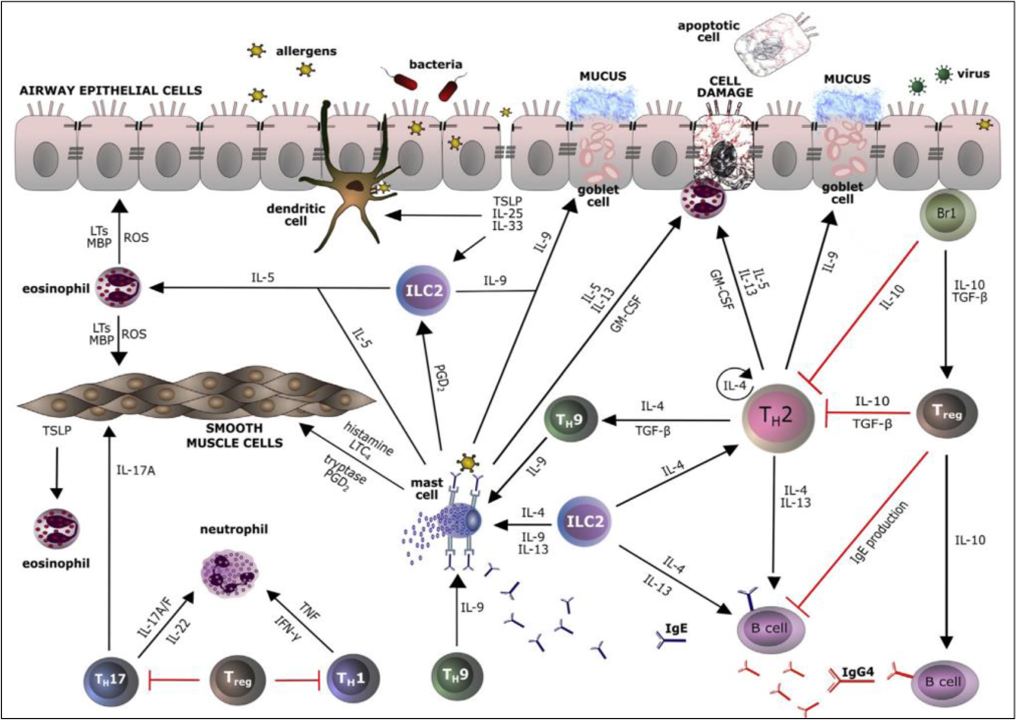
IL-39 is a heterodimer having 54 kDa that belongs to the IL-12 cytokine family upregulated in B cells. 149 IL 39 is comprised of subunits of p19 and Ebi3 (Epstein-Barr-virus-induced-gene) from the IL 12 family present on human chromosome 17. Lipopolysaccharide (LPS) activated B cells secrete natural IL 39. IL 39 elicits an inflammatory response by activating STAT1/STAT3. It is also found to be associated with SLE autoimmune diseases. Ebi3 levels are higher in lung cancer, resulting in Ebi3 being highly expressed in serum samples in patients. And again, reduction of Ebi3 by siRNA suppression results in inhibition of cancer proliferation. IL 39 may play a pro-inflammatory response in systematic lupus erythematosus. 149 Figure 5 illustrates ILs and other mediator networks in allergic inflammation.

Figure 5. ILs and other mediator networks in allergic inflammation. During allergic inflammation, activated epithelial cells release TSLP, IL-25, and IL-33, which also contribute to the TH2 response. [reproduced from Ref. 150 © 2016 Elsevier].
Download figure:
Standard image High-resolution imageIL-40
IL-40 is the presently most recent cytokine to be discovered so far. IL-40 is a 27 kDa secreted protein consisting of 265 amino acids, and a 20-amino-acid signal peptide. It is located on chromosome 17 and is expressed by the fetal liver, bone marrow, and activated B cells. 133 It has also been found that IL 40 takes part in the maturation of B-cells in the bone marrow and periphery of cells. Since it is involved in B cell activation it has been under study to find out more about how IL 40 is related to B-cell-related diseases and autoimmunity disorders.
Table I. Human Interleukin levels in healthy and diseased conditions in body fluids.
| ILs | Role in cancer and other diseases | Disease condition range | References |
|---|---|---|---|
| IL-1-(α and β) | Gastric cancer, Acute, and chronic myelogenous leukemia, hypochlorhydria, Atherosclerosis, Acute myocardial infarction, Heart failure, Hashimoto, Thyroiditis (HT), rheumatoid arthritis, sepsis syndrome, Inflammatory bowel disease, Insulin-dependent diabetes mellitus, Osteoarthritis, multiple sclerosis, Alzheimer's disease | 151,152 | |
| IL-2 | Chronic myeloma, HIV, sarcoidosis | Chronic myeloma serologic IL-2–190 pg mL−1 (normal range- 23 pg mL−1) | 31, 153,154 |
| sIL-2Rα- 1880 pg mL−1 (normal range- 1200 pg mL−1) HIV-seronegative soluble IL-2R receptor level- 368 ± 35 U mL−1 in serum (normal range- 158 ± 19 u mL−1) sarcoidosis | |||
| sIL-2R level-5418 pg mL−1 (normal range- 3000 pg mL−1) | |||
| IL-3 | Multiple myeloma (MM), autoimmune encephalitis (EAE), Sepsis, COVID-19, chronic stable coronary heart disease (CHD) | Multiple myeloma (MM) bone marrow plasma - 66.4 ± 12 pg mL−1 (normal range- 22.11 ± 8.2 pg mL−1) Sepsis 91.2 pg mL−1 (normal range- 36 pg mL−1) COVID-19 plasma IL-3 levels- ≥20 pg mL−1 (normal range- <20 pg mL−1) CHD plasma IL-3 concentrations- 173 ng mL−1 (normal range- 118 ng mL−1) | 153, 155,156 |
| IL-4 | COVID-19 | — | 157 |
| IL-6 | Multiple myeloma, Kaposi's sarcoma, rheumatoid arthritis, AIDS, mesangial proliferative glomerulonephritis, psoriasis, Sepsis, Osteoporosis | Septic shock Serum level- 189 ng mL−1 (normal range- 0.05 ng mL−1) | 158 |
| IL-8 | Pancreatic cancer, Breast Cancer, Urinary bladder cancer, Osteomyelitis, Cystic fibrosis (CF), Acute pyelonephritis, Urinary tract infection, Prostatitis | Breast cancer (normal range in serum- 5.4 pg mL−1) operable stage serum level- 8.3 pg mL−1; progressive metastatic breast cancer stage serum level- 34.54 pg mL−1 | 153 |
| IL-10 | CRC, CLL, GC, GBM COVID-19 | — | 157,159 |
| Il-11 | Hodgkin carcinoma, colorectal adenocarcinoma, breast cancer | — | 153 |
| Il-12 | Breast, cervical, nasopharyngeal cancers, gliomas, RA, diabetes mellitus, multiple sclerosis, Crohn's diseases, leprosy | — | 153 |
| IL-15 | Rheumatoid arthritis, inflammatory bowel disease, type C chronic liver disease, multiple sclerosis, HIV AIDS | Inflammatory bowel disease (normal serum level- 25 pg mL−1) Serum levels in: | 160–162 |
| Right colitis- 28 pg mL−1; Colon-50 pg mL−1; | |||
| Terminal lleum- 35 pg mL−1; Pancolitis- 30 pg mL−1 type C chronic liver disease (normal range- 6.7+/- 6.3 pg mL−1) serum level - 77.4+/- 78.0 pg mL−1 | |||
| HIV AIDS Serum level- 11.79 ± 1.70 (normal range- 23.35 ± 5.06) | |||
| IL-18 | T-lymphoma melanoma Crohn's disease, heart diseases, endometriosis, RA, Atherosclerosis, Diabetic nephropathy, pleural and pulmonary tuberculosis | — | 153 |
| IL-27 | SSc | SSc disease 319.6 ng l−1 (normal range- 104.2 ng L−1) | 163 |
| IL-30 | prostate cancer, psorasis | Psoriasis disease 33.76 pg mL−1 in serum (normal range- 111.96 pg mL−1) | 164 |
| IL-33 | Colorectal cancer, chronic obstructive pulmonary diseases (COPD), Atopic dermatitis, Leishmaniasis, Helminth infection, cardiovascular diseases, musculoskeletal, diseases, IBD, Obesity, type 2 diabetes | — | 153 |
Interleukins: Biomarkers of Clinical Uses
Early and quick diagnosis of diseases is very crucial for their optimum cure. Making the diagnosis process efficient is the prime focus of clinicians. Some diseases like cancers need to be detected at very early stages for their easy cure, and this process to date remains a very challenging task. The role of cytokines is to maintain the homeostasis of the immune system and its overexpression or expression play a crucial role in the diagnosis process as shown in Fig. 6. In HIV, IL-2 and IL-13 level is decreased, whereas IL-4, IL-10, and pro-inflammatory cytokines, such as IL-1, IL-6, and IL-8 are elevated. 165 The first immunological dysregulation that can be reported in the case of HIV is the underexpression of IL-2. In the supernatant of the culture (in vitro) in the HIV sample, there is overexpression and constitutive secretion of IL-1 and IL-6. Tb with pleural effusions has been reported to possess the IL-6 cut-off value of 4000 pg mL−1, while for the diseased state ventilator-associated pneumonia (VAP), the IL-6 cut-off level is 620 pg mL−1. 21 IL-6 is also employed for the diagnosis of neonatal sepsis. 166 The neonatal sepsis is well diagnosed by the cut-off value of IL-12 as 75 pg mL−1, IL-8 as 60 pg ml−1, and IL-10 as 14 pg mL−1. 21 The three-color flow cytometry assay clearly showed the upregulation of IL-4. Overexpression of IL-8 in bronchoalveolar fluid and serum has also been reported in HIV patients. 167 IL-16 production is also reported to be decreased sharply in HIV patients, and treating with IL-2 uplifts the IL-16 production, thereby giving a potential way to be used for HIV- therapy. IL-2 is as associated with the generation of anti-cancer immunity, whereas overexpression of IL-1 is reported to have a poor prognosis in tumor cases. IL-4 and IL-13 stimulate the Th2 response and inhibit the Th1 cell differentiation. This dual role of IL-4 and IL-13 jointly suppresses the antitumor immunity. IL-6 and IL-11 stimulate the transcription of the STAT3 signaling pathway which is involved in oncogenic responses. Siltuximab is FDA approved drug that has been proposed for multicentric Castleman disease. It works by acting as an IL-6 antagonist, so we can think in this direction about its application for treating other cancers also. Overexpression of IL-17 is linked to ductal carcinoma, but clinical trials are yet to be done against its inhibitors or antagonists to confirm their efficacies. IL-18 is reported to be overexpressed in the case of thyroid cancer, with mast cells playing a crucial role in developing the malignancy. 168 IL-17 dysregulation has been reported in many cancers with its ability to induce angiogenesis. The thyroid diseases could easily be detected by checking the levels of IL-13, and IL-8. The steroids-induced inhibition of IL-1 recruits the adhesive molecules and leukocytes, thereby preventing inflammation development. IL-13 has been reported to be 18-fold lower in AD as compared to healthy individuals, and serum measurements of these cytokines have to be efficiently developed, especially in developing nations for the AD diagnosis. 101

Figure 6. Illustration of role of various ILs as biomarkers in different diseases. (i) The role of IL-1 in systemic inflammatory diseases and stroke. Injury, infection, high-fat-containing diet, and other metabolic alterations [Reproduced from Ref. 169 © 2011 Cerebrovascular Diseases]; (ii) Role of IL-1 and IL-6 in the induction of fever via production of prostaglandin E2 (PGE2) and in the release of acute phase response proteins [Reproduced from Ref. 170 © 2021 Frontiers]; and (iii) The pleiotropic effects of IL-6 driving inflammation, fever, cytokinaemia, and tumorigenesis, along with regulation of metabolism, bone turnover, and hematopoiesis [Reproduced from Ref. 171 © 2021 Frontiers].
Download figure:
Standard image High-resolution imageBiosensors Technology: Towards Point of Care Biosensing of ILs
A variety of methods for cytokines detection with high sensitivity, low cost, and linear response in biological and physical samples has been proposed by the researchers. Therefore, special emphasis needs to be given to POC applications for designing portable, specific, sensitive, and rapid tools for ILs detection, resulting in the early diagnosis of diseases. An electrochemical biosensor can fulfill the characteristic ASSURED criteria i.e., Affordable, Specific, Sensitive, User-friendly, Rapid and robust, Equipment-free, and Delivered to the end-users and POC testing.
An electrochemical biosensor is an analytical device comprising biorecognition elements (nucleic acids, tissue, cell receptors, microorganisms, organelles, antibodies, enzymes, proteins), three-electrode systems, and an electronic transducer. 172,173 The system detects the chemical, physical, or biological properties of a specific substance. The transducer performs signal processing, amplification, recording, and displaying the result in a readable format as shown in Fig. 7. 174–176 Target analytes are recognized by specific adsorption or some other chemical/physical interaction. Thereafter, the transducer converts molecular interactions into detectable form.

Figure 7. Schematic flowchart of biosensor components.
Download figure:
Standard image High-resolution imageElectrochemical biosensors for ILs detection
Electrochemical biosensors are the sensing instrumentation based on the phenomena of electrochemistry and redox reactions giving out the biochemical response in form of electric signals. 177 In this method, the electron movement on account of biochemical reactions, and the immobilization of biological analytes, both occur on the electrodes. 178–180 Based on operating principles, electrochemical biosensors are divided into four types- potentiometric, amperometric, conductimetric, and impedimetric. 181,182 The first diagnostic biosensor was established in 1967 for blood glucose levels detection employing natural human enzyme (glucose oxidase) using an electrochemical detector. 183 Nowadays sophisticated, miniaturized, and advanced biosensors are being utilized for screening and controlling diabetes, and several other diseases. 184
A label-free electrochemical impedance spectroscopy (EIS) system was developed for the detection of IL-2 to screen for Multiple Sclerosis (MS). Sri et al., developed for the first time, a sensitive and low-cost electrochemical biosensor based on carbon dots (CDs) and Poly methyl methacrylate (PMMA) for the electrochemical detection of TNF-α as shown in Fig. 8i. The fabricated immunosensor has a wide dynamic range of 0.05–160 pg mL−1 with a lower detection limit (LOD) of 0.05 pg ml−1 and sensitivity of 5.56 pg mL-1cm−2. Furthermore, this CDs-based immunosensor retains high sensitivity, selectivity, and stability. 185 In another work, a nano biosensor based on MoS2 nanoflower synthesized by the hydrothermal method was designed for the detection of TNF-α in cancer patients' serum by Sri et al. 186 The linear detection range of this biosensor was broad (1–200 pg ml−1) with LOD as 0.202 pg ml−1 and limit of quantification (LOQ) was 0.01 pg ml−1. The electrodes showed high sensitivity of 23.156 Ω pgmL-1 cm−2 with higher selectivity. [Fig. 8ii]. Moreover, a viologen-functionalized Single-Walled Carbon Nanotube (V-Phe-SWCNT) was utilized for the design of electrochemical immunosensing of TGF-β1 in human saliva samples. The advantage of this biosensor is its reversible redox nature enabling electron transfer between electrode and proteins with subsequent reduction in ohmic overpotential. However, the viologen cation gets solubilized in an aqueous solution, and these are highly toxic as well. These issues are a major hindrance for them to serve as a clinical diagnosis., although clinically it is being employed for the detection of H2O2 via direct electron transfer of hemoglobin (Hb). Fig. 8iii. 187–189

Figure 8. (i) Schematic representation of stepwise assembly CDs and PMMA nanocomposite-based fabricated immunosensor [Reproduced from Ref. 185 © 2021 Elsevier]; (ii) Schematic representation of BSA/anti-TNF-a/MoS2 nf/ITO electrode fabrication [Reproduced from Ref. 186 © 2021 Elsevier]; and (iii) Schematics of different steps involved in the construction of an amperometric immunosensor for TGF-β1 using V-Phe-SWCNT hybrids [Reproduced from Ref. 187 © 2017 Elsevier].
Download figure:
Standard image High-resolution imageFurther, alkaline phosphatase (ALP) enzyme is also used for the electrochemical biosensing of TNF-β upon functionalization of AuNPs and anti-TNFα Ab2 coated poly(styrene-acrylic acid) (PSA) spheres with polyallylamine hydrochloride (PAH). 8 The label-free electrochemical immunosensor utilizing K3[Fe(CN)6] as a signal has been developed for sensitive identification of TNF-α offering high sensitivity, specificity, stability, and reproducibility. They were also reported to exhibit to medical protein diagnostics. 190
Moreover, Pachauri et al., 191 developed non-invasive electrochemical immunosensors utilizing silver molybdate nanoparticles (β-Ag2MoO4 NPs) for label-free detection of IL-8 biomarker as displayed in Fig. 9i. Electrochemical response was taken in the range of 1 fg mL−1− 40 ng mL−1. The sensitivity and LOD were 7.03 μA ng mL−1 cm−2 and 90 pg mL−1, respectively. The biosensor reported a fast response time of 10 min along with exceptional detection of IL-8 in saliva spiked samples, enabling it to set a promising platform for IL-8 detection in real samples. The fabricated biosensor showed decent stability for 4 weeks. Further, Dave et al. 192 fabricated a novel, non-invasive reduced graphene oxide (RGO) incorporated paper-based biosensing platform for IL-8 detection as shown in Fig. 9ii. They utilized the chronoamperometry technique for direct electron transfer without using any mediator for electronic and electrochemical applications including point-of-care (POC) devices. This technique enabled the direct transfer of electrons eliminating the need for any mediator. It also has the ability to replace the commercially available ITO and FTO coated electrodes for electrochemical applications including POCdevices. The challenge with this technique is to improve its stability. Upon analyzing the reproducibility of the Ab-CysAuNPs-RGO paper electrode, the RSD value was found to be ∼6.81% for the mean current of 5 electrodes.

Figure 9. (i) Schematic illustration showing the synthesis of β-Ag2MoO4 NPs and immunoelectrode fabrication for IL-8 detection [Reproduced from Ref. 191 © 2020 Elsevier]; and (ii) Schematic representation of the step-wise modification of paper and fabrication of immunosensor [Reproduced from Ref. 192 © 2018 Elsevier].
Download figure:
Standard image High-resolution imageOptical biosensors for ILS detection
A highly sensitive human IL-6 detection was achieved by an innovative magnetic colorimetric immunoassay approach employing ceria spheres as labels as shown in Fig. 10i. 193 Recently, for PDGF assessment, a fluorescent nanometer sensing device integrating the ferrocene-marked adapter (ssDNA-Fc) with β-cyclodextrin- modified Carbon Dot (β-CD-CD) fluorescent nano-probe, has been developed exhibiting a broad linear detection range and a low LOD. This can be applied for monitoring and screening cytokines spiked in human serum. 194 In a study, Electrochemiluminescence (ECL) immunoassay integrated with a pattern microfluidic tool was developed which ensured highly sensitive detection of Prostate-Specific Antigen (PSA) and IL-6 in the serum sample. 195 The system is capable of detecting a minimum of two biomarkers in serum and it mostly outperformed ELISA and commercial assay systems and kits [Fig. 10ii].

Figure 10. (i) Schematics of the construction of a magnetic sandwich colorimetric immunoassay for detection of IL-6 [Reproduced from Ref. 193 © 2020 Elsevier]; (ii) Schematics of the LSPR microarray chip patterning process entailing glass pre-treating, AuNR deposition, and antibody function [Reproduced from Ref. 196 © 2020 Elsevier]; and (iii) Schematics of microfluidic ECL array for detection of IL-6 [Reproduced from Ref. 195 © 2020 Elsevier].
Download figure:
Standard image High-resolution imagePlasmonic biosensing is an amazing fluorescence-based for rapid, real-time, and label-free biological species detection. 197 It involves negligible reaction with peripheral interferences and offers high stability in severe environments. 198 Nano-based plasmonic bio-sensing technologies have been developed which utilize the principle of Localized Surface Plasmon Resonance (LSPR) which efficiently detected serum cytokines. 196 Also, the raised cytokine levels were efficiently measured by this system, particularly for IL- 10 and IL-6, in 24 h after cardiopulmonary bypass surgery for hereditary heart disease as shown in Fig. 10iii.
Aptasensors for ILs detection
Aptamers are single-stranded oligonucleotides that fold themselves into unique shapes suitable to bind to the target molecules such as proteins very specifically. 199 Aptamers, because they have sizes comparable to the size of biological metabolites, and because they are highly specific for their analyte and can be modified chemically fairly easily, are proving to be a highly competitive candidate for molecular detection technologies. A lot of studies have been conducted to design the aptamer-based biosensing of ILs.
IL-5RA is a biomarker for eosinophilic inflammation and the concentration of sIL-5RA in blood in the case of eosinophilic inflammation is highly elevated. 200 For instance, Youn et al., Developed the aptasensor for IL-5RA utilizing rod-shaped gold electrodes with a circular gold surface. Its detection limit in PBS has been reported to be 1.69 pg mL−1 which is lower than ELISA kits available commercially. Chen et al., have designed an aptamer placed on the interdigitated electrode (IDE) for IL-6 level detection in case of RA with the limit of detection in the range of 1 fM to 100 pM (0.021 pg mL−1−2.1 ng mL−1). 201 Tertis et al., have developed a highly sensitive and specific aptasensor for IL-6 detection utilizing a glassy carbon electrode loaded with p-aminobenzoic acid, p-aminothiophenol, and gold nanoparticles. The DNA oligonucleotide acts as a probe capturing the target protein at the surface of the biosensor, thereby facilitating label-free detection via electrochemical impedance spectroscopy. The linear response range of the developed aptasensor varied from 5 pg mL−1 to 100 ng mL−1 with the limit of detection being 1.6 pg mL−1. Its novelty is targeting macromolecules like proteins, which was not reported before. The combined effect of covalent binding of AuNPs and fast and sensitive label-free IL-6 detection has amazing potential to serve as clinical screening of colorectal cancer. Its reproducibility was checked by experimenting with ten electrodes, and an RSD of 5.1% was reported. The stability data showed that after 7 days 93% of the initial signal was retained, while after 14, and 28 days 89% and 81 % of the initial signal were retained, respectively. 202 Ciui et al., developed a screen-printed carbon electrode for the development of an electrochemical aptasensor for IL-6 detection. For this purpose, the electrode was modified with a nanocomposite of polypyrrole and AuNP. The method used for detection was cyclic voltammetry and impedance spectroscopy. The linear range of detection was 1 pg mL−1 to 15 mg mL−1 with a detection limit of 0.33 pg mL−1. The advantage of this biosensor is its fast shift of the potential between positive and negative values, thereby generating a pulse with an amplitude showing zero charge value of the modified electrode. It imparts very high speed (tens of ms) enabling the diagnosis to be completed in approximately 30 min, therefore it can serve as a clinical POC device. 93% of the initial signal was retained after 10 days when these aptasensors were tested for stability which reflected that these aptasensors had moderate stability. 203 Jo et al., designed an impedimetric aptasensor with high selectivity and sensitivity for IL-17RA detection utilizing the electrodeposition of AuNP. The detection range was found to be 10 to 10000 pg mL−1 in buffer, while the detection limit was 2.13 pg mL−1 which is lower than commercially available ELISA kits. This biosensor has been approved by the Institute Review Board of Ajou University Hospital, Suwon, Korea for the Clinical diagnosis of neutrophil-related diseases. AuNPs deposition enhances the conductivity and biocompatibility of the fabricated impedimetric aptasensor. Additionally, AuNPs also increase the surface area of the reaction and pave the path for self-assembly. 204 Further, as shown in Fig. 11A, Kumar et al., designed a label-free impedimetric-based sensor modified with gold nanoparticle/aptamer for IL-6 detection in artificial sweat. 205 In another study, an ultra-sensitive and specific aptamer-based biosensor was developed by Tertis et al., for IL-6 detection in the blood samples of CRC patients as shown in Fig. 11B. 202 Moreover, Liu et al., designed DNA aptamer-based electrochemical biosensor for interferon (IFN)-γ detection as shown in Fig. 11C. The LOD for the optimized biosensor was 0.06 nM with linear response extending to 10 nM. The designed aptasensor showed specificity to IFN-γ in the presence of abundant serum proteins. Also, it can be regenerated by treating aptamer-IFN-γ complex in urea buffer enabling it to be reused. These aptasensors do not need any reagents or are washed several times as against standard sandwich immunoassays. It has promising future applications in immunology, cancer research, and infectious disease monitoring. One challenge it needs to address is the stability issue, as it lost approximately 20% of the initial signal in the 10th cycle of cytokine exposure during stability testing. 206

Figure 11. (A) Schematic representation of the fabrication of the aptamer-based sensor and the experimental procedure followed for obtaining the analytical signal of IL-6 detection [Reproduced from Ref. 205 © 2016 RSC]; (B) Schematic representation of the aptasensor elaboration protocol and testing [reproduced from ref., © 2019 Elsevier]; and (C) Schematic of aptamer-based electrochemical sensor for IFN-γ [Reproduced from Ref. 202 © 2010 ACS].
Download figure:
Standard image High-resolution imageSelf-assembled monolayer-based biosensors for ILs detection
Self-assembled monolayer (SAM) allows the electrode surface to be coated by organic molecules giving free anchor residues such as thiols, amines, silanes, etc. 202 Electrochemical introductions to SAM-based biosensors enhance the specificity and signal output even at a very low concentration of target analyte. 207 The immobilization of biomolecules occurs mostly by covalent, non-covalent, and affinity-based attachment. Dijksma utilized N-acetylcysteine (NAC) as SAM for the detection of IFN-γ utilizing SAM-based impedimetric immunosensor, where NAC was used to immobilize anti- IFN-γ on the Au electrode. 11-(triethoxysilyl)undecanal (TESUD) is utilized as SAM by Lee et al., to design an IL-10 biosensor, where the substrate was composed of silicon functionalized with hafnium oxide (HfO2). 8 It has the ability to detect even 0.1 pg mL−1 concentration of IL-10. For TNF-α detection via EIS a reduced dithiobis-succinimidyl propionate SAM was utilized by Pui et al., it was fixed on an Au electrode immobilized with anti-TNF-α Ab. Sharma et al., used monothiol-alkane-PEG-acid capacitative SAM to attach Ab to the Au electrode for IL-8 detection. This construct enabled the detection limit of IL-8 to as low as 90 fg mL−1.
Further, 3-glycidoxypropyltriethoxysilane (GPTES) monolayer coated indium tin oxide-polyethylene terephthalate (ITO-PET) electrode has been reported by Aydin et al., to be utilized for Tumour necrosis factor α (TNF α) cancer biomarker. 208,209 Results showed a wide linear range of detection from 0.01 to 1.5 pg mL−1 and a LOD of 3.1 fg mL−1. It also effectively detected TNF α cancer biomarkers in human serum samples with high sensitivity and recovery ranges as shown in Fig. 12.

Figure 12. Processes of fabricating TNF-α immunosensor based on GPTES-modified ITO-PET electrode [Reproduced from Ref. 208 © 2019 Taylor and Francis].
Download figure:
Standard image High-resolution imageMultiplexed biosensors for ILs detection
The biosensors mentioned above are mostly for single cytokine/IL detection. Researchers have also utilized more than single working electrodes immobilized with the different target analyte while keeping the reference electrode, and counter electrode common. This method allows multiple cytokine detection simultaneously. Li et al. utilized it for simultaneous detection of IL-6 and IL-17 employing polystyrene beads coated with PDDA which are tagged with different metal nanoparticles. 8 FET- based biosensor was constructed for detection of continuous secretion of IL-6, and TNF-α by macrophages in cell culture fed with lipopolysaccharide. Anti- TNF- α Ab was immobilized on the APTES-glutaraldehyde pair, and anti-IL-6 Ab was fixed over the silicon nanowire (SiNW) gates. Conductance change is detected to estimate antigen concentration. 8 Detection of multiplexed IL-1 β and IL-10 is reported by Baraket et al., utilizing two 4-carboxymethyl aryl diazonium-grafted Au working electrodes having functionalized with respective antibodies. Moreover, an electrochemical immunosensor employing dual screen-printed electrodes modified with double-walled carbon nanotubes was developed for real-time identification of IL-1β and TNFα in serum and saliva. The clinical detection of IL-1β/ TNFα in both saliva and serum is shown in Fig. 13. The range of linearity for IL-1β was shown to be 0.5 to 100 pg mL−1 and that for TNF-α it was reported to be 1 to 200 pg mL−1. These ranges are adequate for them to be employed on clinical samples. They have enhanced life-span and stability under chemical, mechanical, or thermal treatments as compared to MWCNTs. Also, their electron transfer and overpotential reduction are much better than SWCNTs these biosensors showed good stability for around 40 days. 27

Figure 13. Schematics of the dual electrochemical immunosensor for determining IL-1β and TNF-a cytokines [Reproduced from Ref. 27 © 2017 Elsevier].
Download figure:
Standard image High-resolution imageFurther, Chen et al., designed a multiarray LSPR optical biosensor possessing 480 nanoplasmonic sensing spots in microfluidic channel arrays which performed parallel multiplex immunoassays of six cytokines in a complex serum matrix on a single device chip [Fig. 14]. The device was fabricated using easy-to-implement, one-step microfluidic patterning and antibody conjugation of gold nanorods (AuNRs) allowing cytokine measurements at concentrations down to 5–20 pg mL−1 from a 1 μL serum sample. 196

Figure 14. Schematic and principle of the method. (a) Schematic of the LSPR microarray chip; (b) Histograms of the particle-to-particle distance of the AuNRs on the LSPR microarray chip characterized using SEM images, and (c) LSPR microarray method. Analyte molecules are introduced to an antibody-functionalized AuNR LSPR biosensor [Reproduced from Ref. 196 © 2015 ACS].
Download figure:
Standard image High-resolution imageSmart point of care biosensors for ILs detection
POCs devices are being applied in biosensors due to their rapid testing, quick results, ease of use, and their small size. Affordability and no need for special training for use have made POCs a common household, clinical and professional device for daily use. Microfluidics devices are a class of instruments that are automated and provide precise measurements of liquid samples. But the use of microfluid devices is difficult to integrate with POCs because microfluid systems require pumps. For IL-6 detection in saliva, a graphene-based portable/wearable nano-sensing device has been designed with decent sensitivity and a good linear range and offers an efficient clinical analysis of diseases at their early stage as shown in Fig. 15. 210 The results showed that the designed portable device responds to IL-6 concentration has a detection limit as low as 12 pM, which falls in the physiological level of IL-6. These features enable this biosensor to be practically employed for salivary cytokine biomarkers as well as for the diagnosis of diseases at an early stage. The observed response signal recorded a fluctuation of 7.47 % which might result from the unknown interference due to the non-uniformity of a different real saliva sample. This fluctuation of biosensor signals must be addressed by future work. Further, Lateral Flow Immunoassay (LFIA) with the fluorescent label has been developed as a remarkable technique to detect ILs and offers easy- to- use platform for POC diagnostics as shown in Fig. 16. 211

Figure 15. Aptameric GFET nano-sensing system for detection of cytokines: (a) photograph of a fully united portable graphene-based nano-sensing system with an Android smartphone; the white dashed box: Modified App for the wireless presentation of the cytokine level data; (b) System-level block diagram of the nano-sensing system presenting the power supply (Vds and Vg), signal (Ids) transduction, handling, exhibition, and wireless transmission pathways from portable nano-sensing platforms to smartphones and cloud servers, one to one; and (c) Snapshots of a GFET nano-sensing system [Reproduced from Ref. 210 © 2015 ACS].
Download figure:
Standard image High-resolution image
Figure 16. Fluorescent lateral flow assay: (a) components of IL-6 test strips, (b) positive sample added to the sample pad, and (c) negative sample added to the sample pad [Reproduced from Ref. 211 © 2015 ACS].
Download figure:
Standard image High-resolution imageBeads and multi-well plates are mostly utilized by multiplexed commercial tests for cytokine detection. Nanobiosensors can offer a highly sensitive, stable, and reliable alternative in POC testing. Similar to other biosensors, electrochemical nanobiosensors function via analyte recognition, signal transduction, and measurable signal readout [Fig. 17i]. In a study, for IL-3 POC testing, an identical system involving magnetic beads was utilized as an anti-biofouling strategy [Fig. 17ii]. The Bluetooth incorporated into the miniaturized potentiostat transfer the information from the SPE readout to a smartphone (or laptop), where an app performs the processing and transduction of signal from the chronoamperometric readout into concentration values (Fig. 3C). This platform offers goods at ∼$50 for the IBS reader; reagent at ∼$5 per test which reached a LOD of ∼5 pg mL−1 in whole blood after one hour of testing, as against ∼7 h needed for IL-3 ELISA commercial kits costing $11.

Figure 17. (i) Components and strategies for assembling electrochemical nanobiosensors based on screen-printed electrodes (SPEs) [reproduced from Ref. 25 © 2021 Wiley]; and (ii) Integrated biosensor for sepsis (IBS) relied on SPEs [Reproduced from Ref. 25 © 2021 Wiley].
Download figure:
Standard image High-resolution image
Role of Nanomaterials in Electrochemical Biosensors
Nanomaterials are objects of nano-domain whose at least one dimension should be 1–100 nm. Currently, many nanomaterial candidates such as nanoparticles, nanotubes, nanowires, etc. are being employed to construct highly sensitive biosensors. These constructs when analyzed electrochemically have the ability to sense trace amounts of the target analyte. Nanomaterials present enhanced electrical conductivity and biocompatibility enabling them to amplify the signals by great amounts. For instance, carbon nanomaterials can be conjugated with a wide array of biomolecules such as DNA, peptides, cells, etc., thereby enhancing the sensitivity and lowering the limit of detection to several orders of magnitude. 212 The high performance of nanobiosensors is basically imparted due to nanomaterial's high surface-to-volume ratio, high mechanical strength, efficient catalytic and diffusivity, and chemical reactivity. 212
The optical property of AuNP allows efficient electron transfer and also acts as a signal amplifier. Au NP-based sensors can be fabricated via the Nano imprinting process on polyethylene terephthalate (PET) filament and even these nanoparticles can be modified into silica optical fibers. Pieces of evidence showed that the device fabricated managed to detect minimal concentrations even as low as 1 pg mL−1 of IL-6 from (1 μL) volume sample. The AuNP aggregation has been reported to undergo a visible color change which is utilized for IL-6 detection. AgNP has been reported to be utilized for the detection of IL-1β, IL-10, IL-4, and IL-6 via colorimetry technique via linking of AgNPs to secondary antibodies. Bimetallic Pd–Pt NPs have been employed for IL-6 detection, and it has been shown to have a wide range of detection (0.1 to 2000 pg mL−1) and LOD of 0.032 pg mL−1 in serum samples. The high refractive index sensitivity and exceptional catalytic activity of Pd compared to AuNPs and AgNP makes Pd a most sought choice. Metal oxide NPs such as titanium dioxide (TiO2), iron oxide, nickel oxide (NiO), zinc oxide (ZnO), copper oxide (CuO), etc. are widely utilized nowadays for nano-based biosensor preparations. These biosensors offer high stability, small size, shape, optimum porosity, flexibility to incorporate into hydrophobic and hydrophilic systems, and easy functionalization with various molecules due to charged surface. The characteristic physicochemical properties offered to the nano-based biosensors due to their small size play a crucial role in strengthening the conductive sensing interface by establishing electrical contact with the transducer surface. The high IEP of ZnO NPs leads to efficient adsorption of biomolecules via electrostatic interactions. Also, their nontoxicity and compatibility with human skin throw light on establishing them as permanent biosensors in chronic diseases. The ELISA-based sensors showed a detection limit of 5.5 pg mL−1 for TNF-α and 7.5 pg mL−1 for IL-8, whereas the detection limits utilizing ZnO nanorod sensors were reported to be 4.2 fg mL−1 for TNF-α and 5.5 fg mL−1 for IL-8 in urine. IONPs have also gathered enormous attraction owing to their biocompatibility, stability, and special magnetic, optical, and electrical properties for the detection of IL-3 and IL-6. Titanium dioxide (TiO2) nanoparticles owing to their semi-conductance property offer great stability and biocompatibility. TiO2 nanotube offers brilliant surface modification suitable for IL-6 biosensing. Employing TiO2 nanotubes for IL-6 biosensing with impedimetric and amperometric electrochemical biosensors offers a very tight interaction of antibody and antigen and can detect IL-6 levels as low as 5 pg mL−1, which is even below the detection limit by the ELISA kits available in the current market. 212,213 An oxide of the Lanthenide family, the Cerium dioxide nanoparticles have the very efficient property of fixing enzymes or proteins on the surface of biosensing electrodes. The provision of switching the oxidation states between Ce(III) to Ce(II) generates oxygen vacancies offering tremendous electrocatalytic properties. The electrostatic force of interaction guides the interaction between the high IEP of CeO2 nanoparticles and the low IEP of biological analytes. Prussian blue (PB) functionalized CeO2 nanoparticles with the aid of employing positively charged chitosan (CS) coating have been developed for TNF-α detection. Conjugation of CeO2 nanoparticles with magnetic Fe3O4 nanoparticles has also shown promising results for IL-6 detection with a detection limit of 0.04 pg mL−1. The exceptional porosity, thermal resistance, surface activity, and adsorption efficiency of SiO2 nanoparticles has been employed for efficient detection of IL-10 with a limit of detection as low as 0.1–20 pg mL−1. Electrodes modified with graphene, nanodiamond (ND), and carbon nanotube (CNT) are some of the very important carbon-based nanomaterials which are highly efficient for interleukin biosensing. 0.1 pg mL−1 IL-6 level has been detected employing Graphene modified electrode. ND has been successfully tested for detecting the concentration of IL-1β, IL-6, and IL-12. AuNP deposited on SWCNT utilizing SiO2/Si as the substrate has enhanced the detection limit (0.01fg mL−1) as compared to detection with bare AuNP.
Conclusion and Future Perspective
Interleukins are an amazing class of biomolecules offering excellent prognosis and diagnosis of a wide array of diseases and syndromes such as cancer, microbial diseases, HIV-AIDS, neurodegenerative diseases, etc. Electrochemical biosensors are proving to be a high potential and highly efficient approach to detecting the concentrations of various interleukins and cytokines that are involved in many diseased conditions. IL-6, IL-8, TNF-α, IL-10, etc. are already been reported as biomarkers for many diseases such as rheumatoid arthritis, AIDS, psoriasis, sepsis, multiple myeloma, many cancers, CRC, CLL, Urinary tract infection, etc. at very low concentrations as compared to other methods such as ELISA. As far as current literature is concerned there is no commercially available electrochemical biosensor for the detection of interleukins which is mainly due to a lack of long-term stability as mentioned in the above-reported biosensors. Future works should be emphasized increasing the shelf life of electrochemical biosensors utilizing various stabilizing agents and nanomaterials. Moreover, the other limitations of these fabricated biosensors are their use in clinical trials or make their commercialization. Thus, efforts have been made to improve the other parameters of the biosensors, thus enabling their use in clinical trials. Various nanomaterials also have the potential to enhance biocompatibility and biodegradability.
Acknowledgments
PRS is thankful to the Biomedical Device and Technology Development (BDTD) sponsored project No.TDP/BDTD/49/2021 General, Department of Science and Technology (DST), New Delhi, India; and Indian Council of Medical Research (ICMR) sponsored project No. 34/13/2019-TF/Nano/BMS, New Delhi, India for funding. Amit K. Yadav recognized the Ministry of Education, Govt. of India for the Prime Minister Research Fellowship for the financial assistants.
Declaration of Competing Interest
The authors declare that they have no known competing financial interests or personal relationships that could have appeared to influence the work reported in this paper.