Adenomyoma of Ampulla: A Report of Three Cases of a Rare Entity
Article Information
Maryam Cheddadi1*,Rihane El Mohtarim1, Samia Sassi1, Sabrine Derqaoui, Fouad zouaidia1, Kaoutar Znati1, Zakia el bernoussi1, Ahmed Jahid1
1Department of Pathology, Ibn Sina Teaching Hospital, Rabat, Morocco
*Corresponding author: Maryam Cheddadi, Department of Pathology, Ibn Sina Teaching Hospital, Rabat, Morocco.
Received: February 07, 2024; Accepted: February 14, 2024; Published: February 23, 2024
Citation: Maryam Cheddadi, Rihane El Mohtarim, Samia Sassi, Sabrine Derqaoui, Fouad zouaidia, Kaoutar Znati, Zakia el bernoussi, Ahmed Jahid Adenomyoma of Ampulla: A Report of Three Cases of a Rare Entity. Journal of Cancer Science and Clinical Therapeutics 8 (2024): 80-82.
View / Download Pdf Share at FacebookAbstract
Adenomyoma is an uncommon, benign lesion with reactive and hamartomatous features resembling a tumor. While it can manifest anywhere along the gastrointestinal tract, including the gallbladder, stomach, duodenum, and jejunum, its occurrence in the extrahepatic bile duct and ampulla of Vater (AOV) is exceedingly rare. Adenomyomas affecting the Vaterian system (ampulla and common bile duct) carry significant clinical implications, as most of these lesions manifest with obstructive biliary tract symptoms and exhibit characteristics similar to malignancies. Histologically, it can be easily confused with well-differentiated adenocarcinoma. Hence, meticulous histological assessment is imperative to confirm the diagnosis definitively. Here, we present the pathological observations from three cases of ampullary adenomyoma, highlighting the rarity of this condition and the diagnostic challenges it presents.
Keywords
Adenomyoma, Ampulla of Vater, Case series, Histopathology.
Article Details
Introduction
Adenomyoma is a rare, non-neoplastic tumor-like lesion occurring commonly in the fundus of the gallbladder but can arise throughout the entire biliary tree including papilla of Vater [1, 2]. It’s also called adenomyomatous hyperplasia and defined as a well circumscribed, nodular proliferation of smooth muscle cells, ducts and glands, clearly disorganized compared to normal [3]. Despite being a benign lesion in most cases, ampullary adenomyoma present clinical signs of obstructive jaundice and mimic malignant neoplasm thereby leading to extensive surgical resection with a high risk of complications [4]. Here, we report the pathologic findings of three cases of ampullary adenomyoma because of the rarity of this disease and the diagnostic pitfalls involved.
Materials and Methods
All pancreatico-duodenectomy and ampullectomy cases received in the Department of Pathology for 8 consecutive years at our institute were reviewed and we found 3 cases of adenomyoma.
Case 1: A 73-year-old female patient presented with hepatic colic, obstructive jaundice, and loss of appetite. An endoscopic retrograde cholangiopancreatography (ERCP) was performed, revealing a nodular mass originating from the peripancreatic orifice of the ampulla. An ampullectomy was conducted as the mass was well-demarcated and showed no evidence of involvement of the distal common bile duct.
Case 2 and 3: Two male patients, aged 68 and 65 years, presented with epigastric pain and obstructive jaundice.
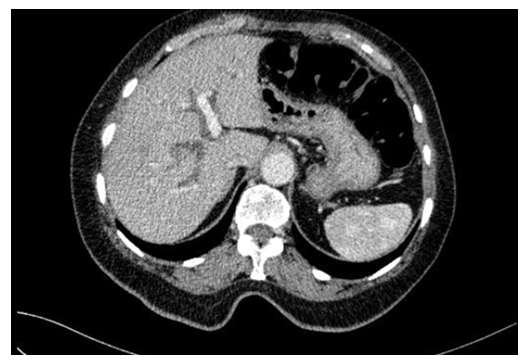
Abdominal ultrasonography showed common bile duct dilation in both cases, along with nodular masses measuring 2 cm and 0.8 cm at the ampulla, respectively. Additionally, abdominal computed tomography revealed mild extrahepatic bile duct dilatation with diffuse wall thickening and enhancement. (Figure 1)
Despite no evidence of malignancy found during the investigation, both patients underwent cephalic duodenopancreatectomy with lymph node dissection.
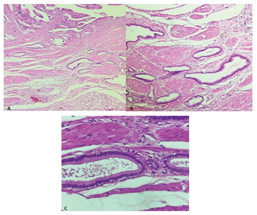
Figure 2: Microscopic examination of the specimen. H&E. A: Low magnification showing microscopic features of atypical glandular structure located in the muscular layer of the duodenal wall (25X). B: lobular arrangement of ducts ducts of variable sizes without desmoplastic reaction (100X). C: At high magnification, the glandular lesions are composed of columnar epithelial cells with basally located nuclei without atypia or mitosis (400X)
Results
Macroscopically, the ampulla appeared firm and exhibited a well-defined polypoid lesion measuring 1.5 cm, 2 cm, and 0.8 cm in our three cases, respectively. Histological examination revealed consistent morphology across all cases. The lesion consisted of proliferating benign glands of varying sizes, lined by gastric antral epithelium and separated by bands of fibromuscular tissue. The overlying mucosa appeared unremarkable. (Figure 2)
Discussion
Adenomyoma is a rare benign nodular lesions showing proliferation of both epithelial and smooth muscle components [5]. It can occur anywhere in the gastrointestinal tract including the gallbladder, stomach, duodenum, and jejunum. It is very rarely observed in the extrahepatic bile duct and ampulla of Vater (AOV) [1]. Histogenesis of adenomyoma in the Vaterian system remains ambiguous. One theory claims that papilla inflammation may cause dysfunction of the muscle fibers of the sphincter of Oddi, leading to muscular hyperplasia, as well as adenomyomatous hyperplasia. Another theory argues that these lesions may represent a form of an incomplete heterotopic pancreas that could be justified by the presence of pancreatic tissue containing scattered ducts and hyperplastic smooth cells, with no acinar or endocrine tissue [1, 6]. Despite its benign nature, patients with ampullary adenomyoma usually present with features of biliary obstruction and weight loss with rare reports of acute pancreatitis with clinical similarities to periampullary tumors. This resemblance malignancies makes the preoperative diagnosis challenging. Definite diagnosis is confirmed by histological examination [7]. Adenomyoma is histologically characterized by a well circumscribed, dense proliferation of ducts and ductules with smooth muscle bundles and collagen fibers. Multiple lobules of the glands are mainly located in the muscular layers of the Vaterian system, thereby resulting in the hypertrophy of the sphincter of Oddi. Ducts and glands are lined by columnar or cuboidal cells, and show no atypia or mitoses. Moreover, it has immunohistochemical characteristics of cytokeratin 7 expression without cytokeratin 20 expression [3, 6]. Differential diagnosis with adenocarcimoma may be challenging, but glands with basally located monotonous nuclei and a lobular arrangement of small ducts without desmoplastic reaction is in favor of an adenomyoma. Additionally, low proliferative activity estimated by immunohistochemical staining with a Ki67 antibody can help with the diagnosis [6, 8]. In conclusion, adenomyoma of the ampulla of Vater is a rare benign tumor of clinical importance. Morphologically, it can easily be mistaken for well differentiated adenocarcinoma therefore, careful histological evaluation is necessary for a definite diagnosis.
References
- Kwon HJ, Kim SG, Park Adenomyoma of the ampulla of Vater mimicking malignancy: A case report and literature review. Medicine (Baltimore) 102 (2023): e34080.
- Colovic R, Micev M, Markovic J, et al. Adenomyoma of the common hepatic duct. HPB (Oxford) 4 (2002): 187-
- Gulwani Adenomyoma. PathologyOutlines.com
- Catarina Gouveia, Catarina Fidalgo, Rui Loureiro, et Adenomyomatosis of the Common Bile Duct and Ampulla of Vater. GE Port J Gastroenterol 28 (2021): 121-133.
- Tae-Hee Kwon, Do Hyun Park, Kwang Yeon Shim, et Ampullary adenomyoma presenting as acute recurrent pancreatitis; World J Gastroenterol 13 (2007): 2892-2894.
- Gialamas E, Mormont M, Bagetakos I, et Combination of adenomyoma and adenomyomatous hyperplasia of the ampullary system: a first case report. Int J Surg Pathol 26 (2018): 644-648.
- Kumari N, Vij M. Adenomyoma of ampulla: a rare cause of obstructive jaundice. JSCR 8 (2011): 6.
- Kee Taek Jang, Jin Seok Heo, Seoung Ho Choi, et al. Adenomyoma of Ampulla of Vater or the Common Bile Duct - A Report of Three Cases - Journal of Pathology and Translational Medicine 39 (2005): 59-62.


 Impact Factor: * 4.1
 CiteScore: 2.9
  Acceptance Rate: 11.01%
  Time to first decision: 10.4 days
  Time from article received to acceptance: 2-3 weeks