Ferroptosis and its Role in Gastric Cancer
 Renjun Gu1,2†
  Yawen Xia3,4†
  Pengfei Li5†
  Defang Zou3,4
  Keqin Lu3,4 Lang Ren1,2 Hongru Zhang6* Zhiguang Sun1,2*
- 1Nanjing University of Chinese Medicine, Nanjing, China
- 2Jiangsu Provincial Second Chinese Medicine Hospital, The Second Affiliated Hospital of Nanjing University of Chinese Medicine, Nanjing, China
- 3Jiangsu Key Laboratory for Pharmacology and Safety Evaluation of Chinese Materia Medica, School of Pharmacy, Nanjing University of Chinese Medicine, Nanjing, China
- 4Jiangsu Collaborative Innovation Center of Traditional Chinese Medicine Prevention and Treatment of Tumor, Nanjing University of Chinese Medicine, Nanjing, China
- 5Department of Clinical Laboratory, Jiangsu Province Hospital of Chinese Medicine, Affiliated Hospital of Nanjing University of Chinese Medicine, Nanjing, China
- 6School of Basic Medical Sciences, Nanjing University of Chinese Medicine, Nanjing, China
Gastric cancer (GC) is the fifth most common cancer and the third leading cause of cancer-related deaths worldwide. Currently, surgery is the treatment of choice for GC. However, the associated expenses and post-surgical pain impose a huge burden on these patients. Furthermore, disease recurrence is also very common in GC patients, thus necessitating the discovery and development of other potential treatment options. A growing body of knowledge about ferroptosis in different cancer types provides a new perspective in cancer therapeutics. Ferroptosis is an iron-dependent form of cell death. It is characterized by intracellular lipid peroxide accumulation and redox imbalance. In this review, we summarized the current findings of ferroptosis regulation in GC. We also tackled on the action of different potential drugs and genes in inducing ferroptosis for treating GC and solving drug resistance. Furthermore, we also explored the relationship between ferroptosis and the tumor microenvironment in GC. Finally, we discussed areas for future studies on the role of ferroptosis in GC to accelerate the clinical utility of ferroptosis induction as a treatment strategy for GC.
Introduction
Gastric cancer (GC) is the fifth most common cancer and the third leading cause of cancer-related deaths worldwide (Smyth et al., 2020). Some of the risk factors for this disease include Helicobacter pylori infection, age, high salt intake, and unhealthy diet (Chen et al., 2021a). GC is commonly treated with surgery (Mihmanli et al., 2016). However, the associated expenses and post-surgical pain impose a huge burden on GC patients. Several studies which focused on identifying molecular signatures and genetic alterations in GC in order to improve treatment selection and aid drug development have already been conducted (Lordick et al., 2017). However, the underlying mechanisms in disease progression are still unclear. Thus, an in-depth understanding of the GC pathobiology will not only facilitate the identification of new drug targets but also provide help in the development of new clinical treatment strategies.
Ferroptosis is a relatively new form of programmed cell death, which was first described in 2012 (Dixon et al., 2012). Several studies have implicated the contribution of ferroptosis in the progression of multiple diseases (Galluzzi et al., 2018; Stockwell et al., 2020), including GC (Jiang et al., 2021a; Liu G. et al., 2021). In this review, we summarized the relationship between ferroptosis and gastric cancer. Furthermore, we suggested that effective regulation of iron metabolism may provide a novel strategy for treating gastric cancer.
Iron Metabolism in Gastric Cancer
Iron is an indispensable molecule in almost all living organisms. Iron-containing enzymes are involved in many physiological activities (Dixon and Stockwell, 2014), such as cellular metabolism, oxygen transport, DNA synthesis, energy production, and cellular respiration. Aside from its roles in various life processes, the catalytic form of iron can also catalyze the formation of reactive oxygen species (ROS) in oxygen-rich environments. Interestingly, iron and ROS can initiate and mediate cell death in several organisms and disease states (Fischbacher et al., 2017). In addition, ROS can also affect several processes, such as cell survival, proliferation, and differentiation through multiple signaling pathways (Lambeth and Neish, 2014). Although low cellular ROS levels are beneficial to some extent, it has also been found to result in base modification and DNA strand breaks (Inoue and Kawanishi, 1987; Dizdaroglu et al., 1991). These findings hint at the potential contribution of free radical-induced DNA damage in the etiology of numerous diseases, including cancer (Dizdaroglu and Jaruga, 2012). Consistent with this idea, extensive studies have shown that poor regulation of iron metabolism is associated with many diseases, including atherosclerosis, neurodegenerative disorders, and cancer (Lambeth and Neish, 2014; Zhou et al., 2018; Vinchi et al., 2019).
Iron levels and stomach health are closely interrelated. For example, several iron-related conditions, such as unexplained iron deficiency, idiopathic thrombocytopenic purpura, and anemia, were found to be associated with H. pylori infection (Hagymási and Tulassay, 2014; Durazzo et al., 2021). Iron homeostasis has also been implicated in cancer development. In one study, iron oxidation has been shown to contribute to tumor formation and subsequent cancer development (Torti and Torti, 2013). On the other hand, several studies reported that iron deficiency may enhance the risk of developing cancer (Janssen et al., 2020). Anemia, low serum ferritin levels, and autoimmune gastritis–related iron malabsorption were identified as risk factors associated with gastrointestinal tumors and GC (Nomura et al., 1992; Cover et al., 2013; Kamada et al., 2021). Consistent with these studies, in vivo data from rodent models show that iron deficiency may contribute to early progression of gastrointestinal tumors (Prá et al., 2009). Taken together, these studies highlight the significant role of iron in the development of various gastrointestinal malignancies, and the potential value of iron regulation as a treatment strategy (Palzer et al., 2021).
Mechanism of Ferroptosis
Ferroptosis is a unique form of cell death (Kerr et al., 1972; Jacobson and Raff, 1995; Christofferson and Yuan, 2010; Shimada et al., 2016). Some protein modulators, such as p53, can exert their physiologic functions either through apoptosis or ferroptosis (Jiang et al., 2015). Similarly, several small molecules can initiate cell death via specific molecular events related to either ferroptosis, apoptosis, or necrosis (Kerr et al., 1972; Schweichel and Merker, 1973; Yang and Stockwell, 2008; Dixon et al., 2012; Shimada et al., 2016). The activation of distinct pathways suggests that the molecular mechanism involved in ferroptosis differs from those of apoptosis and necrosis (Dixon et al., 2012; Dong et al., 2015; Shimada et al., 2016; Torii et al., 2016).
Iron accumulation is the first step in ferroptosis (Galluzzi et al., 2015). Free ferric iron (Fe3+) in the blood conjugates with transferrin proteins. The iron-bound transferrin molecules are then captured by the transferrin receptors present on the cell membrane and enter the cell through endocytosis (Gao et al., 2019). Reducing proteins, such as six transmembrane proteins of prostate 3 (STEAP3), reduces Fe3+ to its highly reactive ferrous ion (Fe2+) form. Upon conversion, Fe2+ is transported from the endosomes to the cytoplasm and is included to the labile iron pool. To protect the cells and tissues from iron-mediated damage, excess Fe2+ in the iron pool is stored in ferritin, while the remaining Fe2+ can be pumped out of the cell through ferroportin molecules on the cell membrane (Yang et al., 2016; Hassannia et al., 2019). Under normal conditions, intracellular iron concentrations remain stable (Yamaguchi et al., 2021). However, in cases of iron overload, excessive Fe2+ is produced within the cell. The accumulation of intracellular Fe2+ further leads to the production of Fe3+ and ROS through the Fenton chemical reaction (Talvenmäki et al., 2019). In addition, excess Fe3+ can also be reduced to Fe2+ through the Haber–Weiss reaction (Kehrer, 2000). Furthermore, under stress conditions, ferritin can self-degrade into Fe2+ through iron autophagy (Talvenmäki et al., 2019). Collectively, these processes can lead to ferroptosis, which in turn induces the formation of more ROS. Excessive ROS can damage biofilms, proteins, and nucleic acids eventually leading to cell death (Kehrer, 2000).
The high intracellular ROS and free radical levels are usually controlled by cells through the actions of antioxidants such as glutathione (GSH) and glutathione peroxidase 4 (GPX4) (Aldini et al., 2018). However, in some cases, glutathione and GPX4 are used up by the cells in other processes, such as in regulating amino acid metabolism. The intracellular levels of glutathione are also affected by amino acid availability. Increased glutamine decomposition may affect the synthesis of glutathione and cause a cell death event similar to a GSH consumption–induced ferroptosis (Linkermann et al., 2014a; Yang et al., 2014). Furthermore, since GPX4 converts the potentially toxic lipid hydroperoxides (L-OOH) to non-toxic lipid alcohols (L-OH) (Ursini et al., 1982), the inactivation of GPX4 can ultimately cause cell death (Linkermann et al., 2014b) (Figure 1).
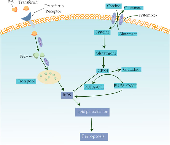
FIGURE 1. Mechanisms of ferroptosis. Excess iron is related to lipid peroxidation and abnormal iron metabolism of mercaptan, which induces the production of ROS. On the one hand, circulating iron in the form of Fe3+ binds to the transferrin receptor and enters the cell. Iron oxide reductase, STEAP3, reduces Fe3+ to Fe2 +, which is transported to the iron pool through DMT1 to induce the formation of ROS. Finally, it promotes lipid peroxidation and causes ferroptosis. On the other hand, the Xc system transports intracellular Glu to the extracellular space and extracellular cystine simultaneously into the cells, which is then transformed into cysteine for GSH synthesis. GPX4 converts -OOH to -OH in polyunsaturated fatty acid (PUFA) to reduce ROS accumulation.
Ferroptosis in Gastric Cancer
Proliferation, Invasion, and Metastasis of Gastric Cancer
The proliferation, invasion, and metastasis of tumor cells are crucial events in the occurrence and development of malignant tumors. These cell activities lead to varying degrees of clinical responses (Machlowska et al., 2018). Like in other cancer types, GC develops from preneoplastic and early neoplastic precursor lesions (Song et al., 2015). These lesions may develop into tumors when the rate of cell proliferation is faster than cell death (Ginsberg et al., 1996; Li et al., 2018). Early stages of GC are characterized by good prognosis with 5-year survival rates reaching >90%; however, most patients are already in the advanced stages of the disease upon initial diagnosis (Tan, 2019). To date, curative surgical resection procedure is the only available treatment for GC (Santoro et al., 2014). Unfortunately, the metastasis of malignant tumors often causes treatment failure (Coburn et al., 2018; Hatta et al., 2020). Tumor invasion and metastasis refer to cellular events when malignant tumor cells continue to grow from the primary site into other sites through lymphatic, vascular, or the body cavity routes. The origin of tumor cells, genetic variations, circulatory mode, and the physiological structure of the metastatic organ determine the specific sites for distant metastasis (Jin et al., 2014).
Several studies try to identify other potential candidates for GC treatment (Table 1). One of these substances, Tanshinone IIA, can inhibit tumor proliferation and metastasis by increasing the level of lipid peroxides and decreasing that of glutathione in the GC cells (Ni et al., 2021a). Another extract, Actinidia chinensis (Planch) exerts anti-proliferation and anti-migration effects on GC cells. Additionally, it can significantly downregulate the expression of GPX4 in a dose-dependent manner (Gao et al., 2020). On the other hand, physcion 8-O-β-glucopyranoside displays antitumor effects in several cancer types, and it induces ferroptosis by regulating the miR-103a-3p/GLS2 axis in GC (Niu et al., 2019).
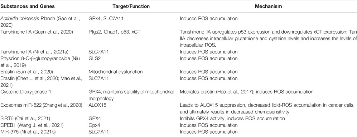
TABLE 1. Candidate substances and genes for inducing ferroptosis in gastric cancer.
Consistent with the studies on the relationship of iron and GC, ferroptosis has also been found to be closely related to the proliferation, invasion, and metastasis in GC (Chen et al., 2021a; Huang et al., 2021). However, the predictive role of ferroptosis in GC remains elusive (Shao et al., 2021). Thus, understanding the processes underlying ferroptosis is promising for the development of cancer treatment strategies.
Tumor Microenvironment in Gastric Cancer
A tumor is closely connected with where it arises and develops in the organism (Chen et al., 2015). Tumor cells in tumor microenvironment (TME) play an active role in the disease progression (Bubnovskaya and Osinsky, 2020; Jiang et al., 2020). The TME favors the growth and expansion of cancer cells (Oya et al., 2020; Rojas et al., 2020). Interestingly, tumor cells and their surrounding microenvironment can be shaped by varying degrees of ferroptosis activation (Zavros, 2017; Xiao et al., 2021). It has been found that ferroptosis serves as an important factor in the formation of the TME in GC (Jiang et al., 2021b; Wang F. et al., 2021; Chen et al., 2021b). In addition, numerous studies have demonstrated that dying cells, including ferroptotic cancer cells, communicate with the immune cells in the TME via a series of signals (Friedmann Angeli et al., 2019). These signals produced during cell death allow the recruitment and activation of immune cells, such as macrophages, regulatory T cell, and neutrophils (Matsushita et al., 2015; Klöditz and Fadeel, 2019; Li et al., 2019), which regulate the growth and expansion of other cancer cells.
Tumor-associated macrophages are emerging as key players in the development of GC (Gambardella et al., 2020). Aside from its role in phagocytosis of foreign antigens, another physiological function of macrophages is to maintain the iron balance in human tissues. Iron homeostasis should be tightly maintained since excess labile iron is toxic (Henle and Linn, 1997; Muckenthaler et al., 2017). Surprisingly, malignant cells can evade the deleterious effect of excessive iron and require high amounts of these reactive ions for their proliferation (Pfeifhofer-Obermair et al., 2018). Depending on the circumstances, increased iron traffic by tumor-associated macrophages either promotes tumor progression or tumor protraction (Soares and Hamza, 2016). Therefore, the detection of macrophages and iron levels in the TME may provide a basis for predicting tumor progression (Liu S. J. et al., 2021; Xiang et al., 2021) (Figure 2). To better understand the functional role of ferroptosis and immune cells in TME, a comprehensive investigation of ferroptosis-related signals and the immune responses they trigger is warranted.
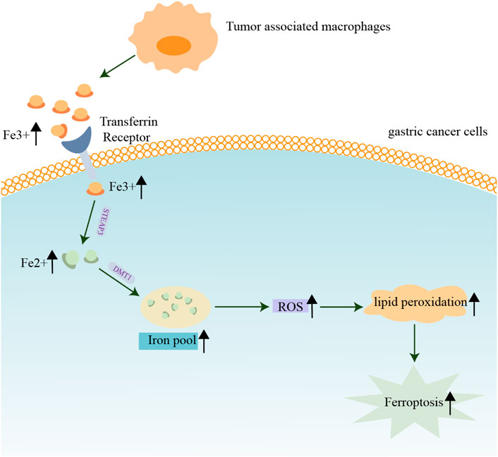
FIGURE 2. Contribution of iron and macrophages in the tumor microenvironment of gastric cancer. Macrophages maintain iron balance in human tissues. The proliferation of GC cells requires a large amount of iron, and the increased iron flow from tumor-associated macrophages promotes tumor progression or tumor protraction.
Despite of reduction in the incidence of GC and the development of novel therapeutic strategies, the prognosis of GC remains poor (Lazăr et al., 2018). Biomarkers for the characterization of the tumor immune microenvironment may add to the predictive value of the current staging system (Jiang et al., 2019). In recent decades, large-scale clinical trans-omics studies allowed the identification of some crucial ferroptosis-related genes as reliable biomarkers to describe the tumor immune microenvironment landscape and predict response to antitumor therapy (Liu S. J. et al., 2021; Shao et al., 2021).
Drug Resistance in Gastric Cancer
Resistance to cisplatin and paclitaxel has become increasingly severe in GC patients (Shao et al., 2019; Zhai et al., 2019). This proves to be a major hurdle in clinical oncology and leads to poor prognosis (Chen Z. et al., 2020; Wei et al., 2020). Resistance to chemotherapy is usually related to mutations in genes regulating cell apoptosis and increased levels of glutathione (Silva et al., 2019). Interestingly, ferroptosis inducers may help in overcoming drug resistance and warrants further investigation (Shin et al., 2018).
Owing to genetic alterations and abnormal growth, cancer cells have higher oxidative tolerance from ROS than non-malignant cells. This ability is attributed to the maintenance of high levels of the antioxidant GSH, which is essential for cell survival and proliferation (Cramer et al., 2017). Studies show that blocking CAF-exosomes–mediated lipid-ROS inhibition leads to increased levels of ferroptosis in cancer cells, which in turn enhances cell sensitivity towards chemotherapy (Zhang et al., 2020). Another potential target for GC therapy is through the blockage of the ROS-activated GCN2-eIF2α-ATF4-xCT pathway, a signaling cascade leading to mitochondrial dysfunction-enhanced cisplatin resistance (Wang et al., 2016). In addition, regulating ROS levels may serve as another novel therapeutic strategy, since ROS can disturb the cellular oxidative environment and induce cell death (Dharmaraja, 2017). In line with this, studies have shown that the antioxidant enzyme, peroxiredoxin 2, significantly sensitizes the AGS and SNU-1 cells towards cisplatin treatment by regulating the level of ROS (Wang et al., 2020). As chronic and exorbitant ROS levels instigate drug resistance (Liao et al., 2019; Xu et al., 2020; Zhu et al., 2020), regulating ferroptosis may be a useful strategy for targeting the drug-resistant tumor cells (Yang et al., 2017; Huang et al., 2019; Choi et al., 2020; Zhang et al., 2021). A possible relevant mechanism is presented in Figure 3.
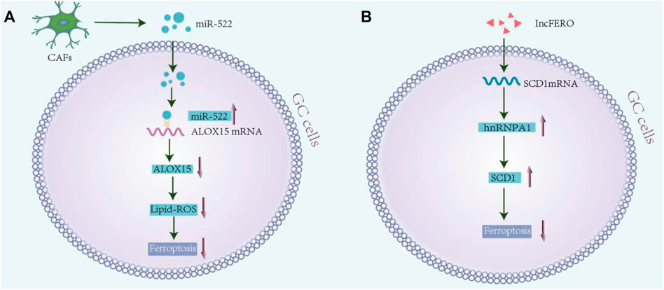
FIGURE 3. Drug resistance and ferroptosis in gastric cancer. (A) Exosomal mir-522 secreted from cancer-associated fibroblasts (CAFs) enter the GC cells and bind to ALOX15 mRNA, resulting in ALOX15 inhibition and reduction in lipid-ROS accumulation in cancer cells. It inhibits ferroptosis in GC cells, and finally reduces chemosensitivity (Namee and O'Driscoll, 2018). (B) Exosomal lnc-ENDOG-1:1 from GC cells can promote the expression of SCD1 by directly interacting with the SCD1 mRNA in GC cells and recruiting heterogenous ribonucleoprotein A1 (hnRNPA1), thereby leading to the inhibition of ferroptosis in GC cells.
Conclusion
Gastric adenocarcinoma is a common disease worldwide. Currently, surgery is the only considered effective treatment strategy. However, disease recurrence is very common even after complete resection (Johnston and Beckman, 2019). Interestingly, ferroptosis has been found to have a very vital role in several cancer types, especially in GC (Lee et al., 2020). As a relatively new discovered mode of cell death, the field of ferroptosis is a research hotspot. Although numerous studies have examined the biological mechanisms underlying ferroptosis, its relationship to tumor progression remains to be poorly understood.
In this review, we have highlighted the importance of iron metabolism and ferroptosis in GC. Iron is an important nutrient in humans (Tan et al., 1997; Goddard et al., 2011). However, iron oxidation also contributes to tumor formation and development of cancer (Doll et al., 2019). In addition, iron in macrophages of the tumor microenvironment is an important index for predicting and detecting GC as well as for evaluating the clinical utility of the related gene signature. Meanwhile, ferroptosis is an iron-dependent form of cell death, which is often characterized by the accumulation of lipid peroxidation products in a cellular-iron–dependent manner (Stockwell et al., 2017; Tang et al., 2021). It functions through two main pathways: iron metabolism and Xc system–induced ROS production (Zheng and Conrad, 2020). Different inducers can affect different steps in ferroptosis to regulate GC proliferation, invasion, and metastasis. Furthermore, the development of drug resistance in GC cells poses a major hurdle. As chronic and exorbitant ROS levels instigate drug resistance, ROS homeostasis may provide a useful treatment strategy for targeting the drug-resistant tumor cells.
Taken together, this review tries to elucidate the relationship between ferroptosis and GC, based on available research findings. We summarized the known ferroptosis processes mediated by gastric cancer-related biomolecules and discussed the actions of some drugs in the different pathways involved in ferroptosis. Lastly, this may serve as a reference for future studies on the mechanism of ferroptosis and the treatment of GC.
Author Contributions
All authors listed have made a substantial, direct, and intellectual contribution to the work, and approved it for publication.
Funding
This project was supported in part by the National Natural Science Foundation of China (82074312).
Conflict of Interest
The authors declare that the research was conducted in the absence of any commercial or financial relationships that could be construed as a potential conflict of interest.
Publisher’s Note
All claims expressed in this article are solely those of the authors and do not necessarily represent those of their affiliated organizations, or those of the publisher, the editors, and the reviewers. Any product that may be evaluated in this article, or claim that may be made by its manufacturer, is not guaranteed or endorsed by the publisher.
Abbreviations
CAFs, cancer-associated fibroblasts; DMT1, divalent metal transporter 1; GC, gastric cancer; Gpx4, glutathione peroxidase 4; ncRNAs, noncoding RNAs; PUFA, polyunsaturated fatty acid; ROS, reactive oxygen species; STEAP3, six transmembrane protein of prostate 3.
References
Aldini, G., Altomare, A., Baron, G., Vistoli, G., Carini, M., Borsani, L., et al. (2018). N-acetylcysteine as an Antioxidant and Disulphide Breaking Agent: the Reasons Why. Free Radic. Res. 52 (7), 751–762. doi:10.1080/10715762.2018.1468564
Bubnovskaya, L., and Osinsky, D. (2020). Tumor Microenvironment and Metabolic Factors: Contribution to Gastric Cancer. Exp. Oncol. 42 (1), 2–10. doi:10.32471/exp-oncology.2312-8852.vol-42-no-1.14056
Cai, S., Fu, S., Zhang, W., Yuan, X., Cheng, Y., and Fang, J. (2021). SIRT6 Silencing Overcomes Resistance to Sorafenib by Promoting Ferroptosis in Gastric Cancer. Biochem. Biophysical Res. Commun. 577, 158–164. doi:10.1016/j.bbrc.2021.08.080
Chen, D., Chen, H., Chi, L., Fu, M., Wang, G., Wu, Z., et al. (2021). Association of Tumor-Associated Collagen Signature with Prognosis and Adjuvant Chemotherapy Benefits in Patients with Gastric Cancer. JAMA Netw. Open 4 (11), e2136388. doi:10.1001/jamanetworkopen.2021.36388
Chen, F., Zhuang, X., Lin, L., Yu, P., Wang, Y., Shi, Y., et al. (2015). New Horizons in Tumor Microenvironment Biology: Challenges and Opportunities. BMC Med. 13, 45. doi:10.1186/s12916-015-0278-7
Chen, L., Qiao, L., Bian, Y., and Sun, X. (2020). GDF15 Knockdown Promotes Erastin-Induced Ferroptosis by Decreasing SLC7A11 Expression. Biochem. Biophysical Res. Commun. 526 (2), 293–299. doi:10.1016/j.bbrc.2020.03.079
Chen, X., Zeh, H. J., Kang, R., Kroemer, G., and Tang, D. (2021a). Cell Death in Pancreatic Cancer: from Pathogenesis to Therapy. Nat. Rev. Gastroenterol. Hepatol. 18 (11), 804–823. doi:10.1038/s41575-021-00486-6
Chen, X., Zhu, Z., Li, X., Yao, X., and Luo, L. (2021b). The Ferroptosis-Related Noncoding RNA Signature as a Novel Prognostic Biomarker in the Tumor Microenvironment, Immunotherapy, and Drug Screening of Gastric Adenocarcinoma. Front. Oncol. 11, 778557. doi:10.3389/fonc.2021.778557
Chen, Z., Li, Y., Tan, B., Zhao, Q., Fan, L., Li, F., et al. (2020). Progress and Current Status of Molecule-Targeted Therapy and Drug Resistance in Gastric Cancer. Drugs Today 56 (7), 469–482. doi:10.1358/dot.2020.56.7.3112071
Choi, H.-J., Jhe, Y.-L., Kim, J., Lim, J. Y., Lee, J. E., Shin, M.-K., et al. (2020). FoxM1-dependent and Fatty Acid Oxidation-Mediated ROS Modulation Is a Cell-Intrinsic Drug Resistance Mechanism in Cancer Stem-like Cells. Redox Biol. 36, 101589. doi:10.1016/j.redox.2020.101589
Christofferson, D. E., and Yuan, J. (2010). Necroptosis as an Alternative Form of Programmed Cell Death. Curr. Opin. Cel Biol. 22 (2), 263–268. doi:10.1016/j.ceb.2009.12.003
Coburn, N., Cosby, R., Klein, L., Knight, G., Malthaner, R., Mamazza, J., et al. (2018). Staging and Surgical Approaches in Gastric Cancer: A Systematic Review. Cancer Treat. Rev. 63, 104–115. doi:10.1016/j.ctrv.2017.12.006
Cover, T. L., and Peek, Jr, R. M. (2013). Diet, Microbial Virulence, andHelicobacter Pylori-Induced Gastric Cancer. Gut Microbes 4 (6), 482–493. doi:10.4161/gmic.26262
Cramer, S. L., Saha, A., Liu, J., Tadi, S., Tiziani, S., Yan, W., et al. (2017). Systemic depletion of L-cyst(e)ine with cyst(e)inase increases reactive oxygen species and suppresses tumor growth. Nat. Med. 23 (1), 120–127. doi:10.1038/nm.4232
Dharmaraja, A. T. (2017). Role of Reactive Oxygen Species (ROS) in Therapeutics and Drug Resistance in Cancer and Bacteria. J. Med. Chem. 60 (8), 3221–3240. doi:10.1021/acs.jmedchem.6b01243
Dixon, S. J., Lemberg, K. M., Lamprecht, M. R., Skouta, R., Zaitsev, E. M., Gleason, C. E., et al. (2012). Ferroptosis: an Iron-dependent Form of Nonapoptotic Cell Death. Cell 149 (5), 1060–1072. doi:10.1016/j.cell.2012.03.042
Dixon, S. J., and Stockwell, B. R. (2014). The Role of Iron and Reactive Oxygen Species in Cell Death. Nat. Chem. Biol. 10, 9–17. doi:10.1038/nchembio.1416
Dizdaroglu, M., and Jaruga, P. (2012). Mechanisms of Free Radical-Induced Damage to DNA. Free Radic. Res. 46 (4), 382–419. doi:10.3109/10715762.2011.653969
Dizdaroglu, M., Rao, G., Halliwell, B., and Gajewski, E. (1991). Damage to the DNA Bases in Mammalian Chromatin by Hydrogen Peroxide in the Presence of Ferric and Cupric Ions. Arch. Biochem. Biophys. 285 (2), 317–324. doi:10.1016/0003-9861(91)90366-q
Doll, S., Freitas, F. P., Shah, R., Aldrovandi, M., da Silva, M. C., Ingold, I., et al. (2019). FSP1 Is a Glutathione-independent Ferroptosis Suppressor. Nature 575 (7784), 693–698. doi:10.1038/s41586-019-1707-0
Dong, T., Liao, D., Liu, X., and Lei, X. (2015). Using Small Molecules to Dissect Non-apoptotic Programmed Cell Death: Necroptosis, Ferroptosis, and Pyroptosis. Chembiochem 16 (18), 2557–2561. doi:10.1002/cbic.201500422
Durazzo, M., Adriani, A., Fagoonee, S., Saracco, G. M., and Pellicano, R. (2021). Helicobacter pylori and Respiratory Diseases: 2021 Update. Microorganisms 9 (10), 2033. doi:10.3390/microorganisms9102033
Fischbacher, A., von Sonntag, C., and Schmidt, T. C. (2017). Hydroxyl Radical Yields in the Fenton Process under Various pH, Ligand Concentrations and Hydrogen peroxide/Fe(II) Ratios. Chemosphere 182, 738–744. doi:10.1016/j.chemosphere.2017.05.039
Friedmann Angeli, J. P., Krysko, D. V., and Conrad, M. (2019). Ferroptosis at the Crossroads of Cancer-Acquired Drug Resistance and Immune Evasion. Nat. Rev. Cancer 19 (7), 405–414. doi:10.1038/s41568-019-0149-1
Galluzzi, L., Bravo-San Pedro, J. M., Vitale, I., Aaronson, S. A., Abrams, J. M., Adam, D., et al. (2015). Essential versus Accessory Aspects of Cell Death: Recommendations of the NCCD 2015. Cell Death Differ 22 (1), 58–73. doi:10.1038/cdd.2014.137
Galluzzi, L., Vitale, I., Aaronson, S. A., Abrams, J. M., Adam, D., Agostinis, P., et al. (2018). Molecular Mechanisms of Cell Death: Recommendations of the Nomenclature Committee on Cell Death 2018. Cel Death Differ 25 (3), 486–541. doi:10.1038/s41418-017-0012-4
Gambardella, V., Castillo, J., Tarazona, N., Gimeno-Valiente, F., Martínez-Ciarpaglini, C., Cabeza-Segura, M., et al. (2020). The Role of Tumor-Associated Macrophages in Gastric Cancer Development and Their Potential as a Therapeutic Target. Cancer Treat. Rev. 86, 102015. doi:10.1016/j.ctrv.2020.102015
Gao, M., Yi, J., Zhu, J., Minikes, A. M., Monian, P., Thompson, C. B., et al. (2019). Role of Mitochondria in Ferroptosis. Mol. Cel 73 (2), 354–363. e3. doi:10.1016/j.molcel.2018.10.042
Gao, Z., Deng, G., Li, Y., Huang, H., Sun, X., Shi, H., et al. (2020). Actinidia Chinensis Planch Prevents Proliferation and Migration of Gastric Cancer Associated with Apoptosis, Ferroptosis Activation and Mesenchymal Phenotype Suppression. Biomed. Pharmacother. 126, 110092. doi:10.1016/j.biopha.2020.110092
Ginsberg, G. G., Al-Kawas, F. H., Fleischer, D. E., Reilly, H. F., and Benjamin, S. B. (1996). Gastric Polyps: Relationship of Size and Histology to Cancer Risk. Am. J. Gastroenterol. 91 (4), 714–717.
Goddard, A. F., James, M. W., McIntyre, A. S., and Scott, B. B.British Society of Gastroenterology (2011). Guidelines for the Management of Iron Deficiency Anaemia. Gut 60 (10), 1309–1316. doi:10.1136/gut.2010.228874
Guan, Z., Chen, J., Li, X., and Dong, N. (2020). Tanshinone IIA Induces Ferroptosis in Gastric Cancer Cells through P53-Mediated SLC7A11 Down-Regulation. Biosci. Rep. 40 (8), BSR20201807. doi:10.1042/BSR20201807
Hagymási, K., and Tulassay, Z. (2014). Helicobacter Pyloriinfection: New Pathogenetic and Clinical Aspects. Wjg 20 (21), 6386–6399. doi:10.3748/wjg.v20.i21.6386
Hao, S., Yu, J., He, W., Huang, Q., Zhao, Y., Liang, B., et al. (2017). Cysteine Dioxygenase 1 Mediates Erastin-Induced Ferroptosis in Human Gastric Cancer Cells. Neoplasia 19 (12), 1022–1032. doi:10.1016/j.neo.2017.10.005
Hassannia, B., Vandenabeele, P., and Vanden Berghe, T. (2019). Targeting Ferroptosis to Iron Out Cancer. Cancer Cell 35 (6), 830–849. doi:10.1016/j.ccell.2019.04.002
Hatta, W., Gotoda, T., Koike, T., and Masamune, A. (2020). History and Future Perspectives in Japanese Guidelines for Endoscopic Resection of Early Gastric Cancer. Dig. Endosc. 32 (2), 180–190. doi:10.1111/den.13531
Henle, E. S., and Linn, S. (1997). Formation, Prevention, and Repair of DNA Damage by Iron/hydrogen Peroxide. J. Biol. Chem. 272 (31), 19095–19098. doi:10.1074/jbc.272.31.19095
Huang, R., Chen, H., Liang, J., Li, Y., Yang, J., Luo, C., et al. (2021). Dual Role of Reactive Oxygen Species and Their Application in Cancer Therapy. J. Cancer 12 (18), 5543–5561. doi:10.7150/jca.54699
Huang, T., Song, C., Zheng, L., Xia, L., Li, Y., and Zhou, Y. (2019). The Roles of Extracellular Vesicles in Gastric Cancer Development, Microenvironment, Anti-cancer Drug Resistance, and Therapy. Mol. Cancer 18 (1), 62. doi:10.1186/s12943-019-0967-5
Inoue, S., and Kawanishi, S. (1987). Hydroxyl Radical Production and Human DNA Damage Induced by Ferric Nitrilotriacetate and Hydrogen Peroxide. Cancer Res. 47 (24 Pt 1), 6522–6527.
Jacobson, M. D., and Raff, M. C. (1995). Programmed Cell Death and Bcl-2 protection in Very Low Oxygen. Nature 374 (6525), 814–816. doi:10.1038/374814a0
Janssen, H. J. B., Fransen, L. F. C., Ponten, J. E. H., Nieuwenhuijzen, G. A. P., and Luyer, M. D. P. (2020). Micronutrient Deficiencies Following Minimally Invasive Esophagectomy for Cancer. Nutrients 12 (3), 778. doi:10.3390/nu12030778
Jiang, L., Kon, N., Li, T., Wang, S.-J., Su, T., Hibshoosh, H., et al. (2015). Ferroptosis as a P53-Mediated Activity during Tumour Suppression. Nature 520 (7545), 57–62. doi:10.1038/nature14344
Jiang, X., Yan, Q., Xie, L., Xu, S., Jiang, K., Huang, J., et al. (2021). Construction and Validation of a Ferroptosis-Related Prognostic Model for Gastric Cancer. J. Oncol. 2021, 6635526. doi:10.1155/2021/6635526
Jiang, X., Liu, F., Liu, P., Yan, Y., Lan, S., Zhuang, K., et al. (2021). Ferroptosis Patterns Correlate with Immune Microenvironment Characterization in Gastric Cancer. Ijgm Vol. 14, 6573–6586. doi:10.2147/ijgm.s331291
Jiang, Y., Xie, J., Huang, W., Chen, H., Xi, S., Han, Z., et al. (2019). Tumor Immune Microenvironment and Chemosensitivity Signature for Predicting Response to Chemotherapy in Gastric Cancer. Cancer Immunol. Res. 7 (12), 2065–2073. doi:10.1158/2326-6066.CIR-19-0311
Jiang, Y., Wang, H., Wu, J., Chen, C., Yuan, Q., Huang, W., et al. (2020). Noninvasive Imaging Evaluation of Tumor Immune Microenvironment to Predict Outcomes in Gastric Cancer. Ann. Oncol. 31 (6), 760–768. doi:10.1016/j.annonc.2020.03.295
Jin, X., Zhu, Z., and Shi, Y. (2014). Metastasis Mechanism and Gene/protein Expression in Gastric Cancer with Distant Organs Metastasis. Bull. Cancer 101 (1), E1–E12. doi:10.1684/bdc.2013.1882
Johnston, F. M., and Beckman, M. (2019). Updates on Management of Gastric Cancer. Curr. Oncol. Rep. 21 (8), 67. doi:10.1007/s11912-019-0820-4
Kamada, T., Maruyama, Y., Monobe, Y., and Haruma, K. (2021). Endoscopic Features and Clinical Importance of Autoimmune Gastritis. Dig. Endosc. (Online ahead of print). doi:10.1111/den.14175
Kehrer, J. P. (2000). The Haber-Weiss Reaction and Mechanisms of Toxicity. Toxicology 149 (1), 43–50. doi:10.1016/s0300-483x(00)00231-6
Kerr, J. F. R., Wyllie, A. H., and Currie, A. R. (1972). Apoptosis: A Basic Biological Phenomenon with Wideranging Implications in Tissue Kinetics. Br. J. Cancer 26 (4), 239–257. doi:10.1038/bjc.1972.33
Klöditz, K., and Fadeel, B. (2019). Three Cell Deaths and a Funeral: Macrophage Clearance of Cells Undergoing Distinct Modes of Cell Death. Cel Death Discov. 5, 65. doi:10.1038/s41420-019-0146-x
Lambeth, J. D., and Neish, A. S. (2014). Nox Enzymes and New Thinking on Reactive Oxygen: a Double-Edged Sword Revisited. Annu. Rev. Pathol. Mech. Dis. 9, 119–145. doi:10.1146/annurev-pathol-012513-104651
Lazăr, D. C., Avram, M. F., Romoșan, I., Cornianu, M., Tăban, S., and Goldiș, A. (2018). Prognostic Significance of Tumor Immune Microenvironment and Immunotherapy: Novel Insights and Future Perspectives in Gastric Cancer. World J. Gastroenterol. 24 (32), 3583–3616. doi:10.3748/wjg.v24.i32.3583
Lee, J.-Y., Nam, M., Son, H. Y., Hyun, K., Jang, S. Y., Kim, J. W., et al. (2020). Polyunsaturated Fatty Acid Biosynthesis Pathway Determines Ferroptosis Sensitivity in Gastric Cancer. Proc. Natl. Acad. Sci. U.S.A. 117 (51), 32433–32442. doi:10.1073/pnas.2006828117
Li, J., Xu, L., Run, Z.-C., Feng, W., Liu, W., Zhang, P.-J., et al. (2018). Multiple Cytokine Profiling in Serum for Early Detection of Gastric Cancer. Wjg 24 (21), 2269–2278. doi:10.3748/wjg.v24.i21.2269
Li, W., Feng, G., Gauthier, J. M., Lokshina, I., Higashikubo, R., Evans, S., et al. (2019). Ferroptotic Cell Death and TLR4/Trif Signaling Initiate Neutrophil Recruitment after Heart Transplantation. J. Clin. Invest. Feb 26 129 (6), 2293–2304. doi:10.1172/jci126428
Liao, X., Fan, Y., Hou, J., Chen, X., Xu, X., Yang, Y., et al. (2019). Identification of Chaetocin as a Potent Non-ROS-mediated Anticancer Drug Candidate for Gastric Cancer. J. Cancer 10 (16), 3678–3690. doi:10.7150/jca.32803
Linkermann, A., Skouta, R., Himmerkus, N., Mulay, S. R., Dewitz, C., De Zen, F., et al. (2014). Synchronized Renal Tubular Cell Death Involves Ferroptosis. Proc. Natl. Acad. Sci. U.S.A. 111 (47), 16836–16841. doi:10.1073/pnas.1415518111
Linkermann, A., Skouta, R., Himmerkus, N., Mulay, S. R., Dewitz, C., De Zen, F., et al. (2014). Synchronized Renal Tubular Cell Death Involves Ferroptosis. Proc. Natl. Acad. Sci. U.S.A. 111 (47), 16836–16841. doi:10.1073/pnas.1415518111
Liu, G., Ma, J.-y., Hu, G., and Jin, H. (2021). Identification and Validation of a Novel Ferroptosis-Related Gene Model for Predicting the Prognosis of Gastric Cancer Patients. PLoS One 16 (7), e0254368. doi:10.1371/journal.pone.0254368
Liu, S. J., Yang, Y. B., Zhou, J. X., Lin, Y. J., Pan, Y. L., and Pan, J. H. (2021). A Novel Ferroptosis-Related Gene Risk Signature for Predicting Prognosis and Immunotherapy Response in Gastric Cancer. Dis. Markers 2021, 2385406. doi:10.1155/2021/2385406
Lordick, F., Shitara, K., and Janjigian, Y. Y. (2017). New Agents on the Horizon in Gastric Cancer. Ann. Oncol. 28 (8), 1767–1775. doi:10.1093/annonc/mdx051
Machlowska, J., Maciejewski, R., and Sitarz, R. (2018). The Pattern of Signatures in Gastric Cancer Prognosis. Ijms 19 (6), 1658. doi:10.3390/ijms19061658
Mao, S.-H., Zhu, C.-H., Nie, Y., Yu, J., and Wang, L. (2021). Levobupivacaine Induces Ferroptosis by miR-489-3p/SLC7A11 Signaling in Gastric Cancer. Front. Pharmacol. 12, 681338. doi:10.3389/fphar.2021.681338
Matsushita, M., Freigang, S., Schneider, C., Conrad, M., Bornkamm, G. W., and Kopf, M. (2015). T Cell Lipid Peroxidation Induces Ferroptosis and Prevents Immunity to Infection. J. Exp. Med. Apr 6 212 (4), 555–568. doi:10.1084/jem.20140857
Mihmanli, M., Ilhan, E., Idiz, U. O., Alemdar, A., and Demir, U. (2016). Recent Developments and Innovations in Gastric Cancer. Wjg 22 (17), 4307–4320. doi:10.3748/wjg.v22.i17.4307
Muckenthaler, M. U., Rivella, S., Hentze, M. W., and Galy, B. (2017). A Red Carpet for Iron Metabolism. Cell 168 (3), 344–361. doi:10.1016/j.cell.2016.12.034
Namee, N. M., and O'Driscoll, L. (2018). Extracellular Vesicles and Anti-cancer Drug Resistance. Biochim. Biophys. Acta (Bba) - Rev. Cancer 1870 (2), 123–136. doi:10.1016/j.bbcan.2018.07.003
Ni, H., Qin, H., Sun, C., Liu, Y., Ruan, G., Guo, Q., et al. (2021). MiR-375 Reduces the Stemness of Gastric Cancer Cells through Triggering Ferroptosis. Stem Cel Res Ther 12 (1), 325. doi:10.1186/s13287-021-02394-7
Ni, H., Ruan, G., Sun, C., Yang, X., Miao, Z., Li, J., et al. (2021). Tanshinone IIA Inhibits Gastric Cancer Cell Stemness through Inducing Ferroptosis. Environ. Toxicol. 37 (2), 192–200. doi:10.1002/tox.23388
Niu, Y., Zhang, J., Tong, Y., Li, J., and Liu, B. (2019). RETRACTED: Physcion 8-O-β-Glucopyranoside Induced Ferroptosis via Regulating miR-103a-3p/GLS2 axis in Gastric Cancer. Life Sci. 237, 116893. doi:10.1016/j.lfs.2019.116893
Nomura, A., Chyou, P. H., and Stemmermann, G. N. (1992). Association of Serum Ferritin Levels with the Risk of Stomach Cancer. Cancer Epidemiol. Biomarkers Prev. 1 (7), 547–550.
Oya, Y., Hayakawa, Y., and Koike, K. (2020). Tumor Microenvironment in Gastric Cancers. Cancer Sci. 111 (8), 2696–2707. doi:10.1111/cas.14521
Palzer, J., Eckstein, L., Slabu, I., Reisen, O., Neumann, U. P., and Roeth, A. A. (2021). Iron Oxide Nanoparticle-Based Hyperthermia as a Treatment Option in Various Gastrointestinal Malignancies. Nanomaterials 11 (11), 3013. doi:10.3390/nano11113013
Pfeifhofer-Obermair, C., Tymoszuk, P., Petzer, V., Weiss, G., and Nairz, M. (2018). Iron in the Tumor Microenvironment-Connecting the Dots. Front. Oncol. 8, 549. doi:10.3389/fonc.2018.00549
Prá, D., Rech Franke, S. I., Pêgas Henriques, J. A., and Fenech, M. (2009). A Possible Link between Iron Deficiency and Gastrointestinal Carcinogenesis. Nutr. Cancer 61 (4), 415–426. doi:10.1080/01635580902803701
Rojas, A., Araya, P., Gonzalez, I., and Morales, E. (2020). Gastric Tumor Microenvironment. Adv. Exp. Med. Biol. 1226, 23–35. doi:10.1007/978-3-030-36214-0_2
Santoro, R., Ettorre, G. M., and Santoro, E. (2014). Subtotal Gastrectomy for Gastric Cancer. Wjg 20 (38), 13667–13680. doi:10.3748/wjg.v20.i38.13667
Schweichel, J. U., and Merker, H. J. (1973). The Morphology of Various Types of Cell Death in Prenatal Tissues. Teratology 7 (3), 253–266. doi:10.1002/tera.1420070306
Shao, L., Chen, Z., Soutto, M., Zhu, S., Lu, H., Romero-Gallo, J., et al. (2019). Helicobacter pylori ‐induced miR‐135b‐5p Promotes Cisplatin Resistance in Gastric Cancer. FASEB j. 33 (1), 264–274. doi:10.1096/fj.201701456rr
Shao, Y., Jia, H., Li, S., Huang, L., Aikemu, B., Yang, G., et al. (2021). Comprehensive Analysis of Ferroptosis-Related Markers for the Clinical and Biological Value in Gastric Cancer. Oxid Med. Cel Longev 2021, 7007933. doi:10.1155/2021/7007933
Shimada, K., Hayano, M., Pagano, N. C., and Stockwell, B. R. (2016). Cell-Line Selectivity Improves the Predictive Power of Pharmacogenomic Analyses and Helps Identify NADPH as Biomarker for Ferroptosis Sensitivity. Cel Chem. Biol. 23 (2), 225–235. doi:10.1016/j.chembiol.2015.11.016
Shin, D., Kim, E. H., Lee, J., and Roh, J.-L. (2018). Nrf2 Inhibition Reverses Resistance to GPX4 Inhibitor-Induced Ferroptosis in Head and Neck Cancer. Free Radic. Biol. Med. 129, 454–462. doi:10.1016/j.freeradbiomed.2018.10.426
Silva, M. M., Rocha, C. R. R., Kinker, G. S., Pelegrini, A. L., and Menck, C. F. M. (2019). The Balance between NRF2/GSH Antioxidant Mediated Pathway and DNA Repair Modulates Cisplatin Resistance in Lung Cancer Cells. Sci. Rep. 9 (1), 17639. doi:10.1038/s41598-019-54065-6
Smyth, E. C., Nilsson, M., Grabsch, H. I., van Grieken, N. C., and Lordick, F. (2020). Gastric Cancer. The Lancet 396 (10251), 635–648. doi:10.1016/s0140-6736(20)31288-5
Soares, M. P., and Hamza, I. (2016). Macrophages and Iron Metabolism. Immunity 44 (3), 492–504. doi:10.1016/j.immuni.2016.02.016
Song, B., Yu, J., and Wu, T. S. (2015). Correlation between C-Erbb-2 with Gastric Mucosal Atypical Hyperplasia and Gastric Carcinoma. J. Biol. Regul. Homeost Agents 29 (2), 471–477.
Stockwell, B. R., Friedmann Angeli, J. P., Bayir, H., Bush, A. I., Conrad, M., Dixon, S. J., et al. (2017). Ferroptosis: A Regulated Cell Death Nexus Linking Metabolism, Redox Biology, and Disease. Cell 171 (2), 273–285. doi:10.1016/j.cell.2017.09.021
Stockwell, B. R., Jiang, X., and Gu, W. (2020). Emerging Mechanisms and Disease Relevance of Ferroptosis. Trends Cel Biol. 30 (6), 478–490. doi:10.1016/j.tcb.2020.02.009
Sun, Y., Deng, R., and Zhang, C. (2020). Erastin Induces Apoptotic and Ferroptotic Cell Death by Inducing ROS Accumulation by Causing Mitochondrial Dysfunction in Gastric Cancer Cell HGC-27. Mol. Med. Rep. 22 (4), 2826–2832. doi:10.3892/mmr.2020.11376
Talvenmäki, H., Lallukka, N., Survo, S., and Romantschuk, M. (2019). Fenton's Reaction-Based Chemical Oxidation in Suboptimal Conditions Can lead to Mobilization of Oil Hydrocarbons but Also Contribute to the Total Removal of Volatile Compounds. Environ. Sci. Pollut. Res. 26 (33), 34670–34684. doi:10.1007/s11356-019-06547-3
Tan, W. C., Krasner, N., O'Toole, P., and Lombard, M. (1997). Enhancement of Photodynamic Therapy in Gastric Cancer Cells by Removal of Iron. Gut 41 (1), 14–18. doi:10.1136/gut.41.1.14
Tan, Z. (2019). Recent Advances in the Surgical Treatment of Advanced Gastric Cancer: A Review. Med. Sci. Monit. 25, 3537–3541. doi:10.12659/msm.916475
Tang, D., Chen, X., Kang, R., and Kroemer, G. (2021). Ferroptosis: Molecular Mechanisms and Health Implications. Cell Res 31 (2), 107–125. doi:10.1038/s41422-020-00441-1
Torii, S., Shintoku, R., Kubota, C., Yaegashi, M., Torii, R., Sasaki, M., et al. (2016). An Essential Role for Functional Lysosomes in Ferroptosis of Cancer Cells. Biochem. J. 473 (6), 769–777. doi:10.1042/bj20150658
Torti, S. V., and Torti, F. M. (2013). Iron and Cancer: More Ore to Be Mined. Nat. Rev. Cancer 13, 342–355. doi:10.1038/nrc3495
Ursini, F., Maiorino, M., Valente, M., Ferri, L., and Gregolin, C. (1982). Purification from Pig Liver of a Protein Which Protects Liposomes and Biomembranes from Peroxidative Degradation and Exhibits Glutathione Peroxidase Activity on Phosphatidylcholine Hydroperoxides. Biochim. Biophys. Acta (Bba) - Lipids Lipid Metab. 710 (2), 197–211. doi:10.1016/0005-2760(82)90150-3
Vinchi, F., Porto, G., Simmelbauer, A., Altamura, S., Passos, S. T., Garbowski, M., et al. (2019). Atherosclerosis Is Aggravated by Iron Overload and Ameliorated by Dietary and Pharmacological Iron Restriction. Eur. Heart J. 00, 1–16. doi:10.1093/eurheartj/ehz112
Wang, F., Chen, C., Chen, W. P., Li, Z. L., and Cheng, H. (2021). Development and Validation of a Novel Ferroptosis-Related Gene Signature for Predicting Prognosis and the Immune Microenvironment in Gastric Cancer. Biomed. Res. Int. 2021, 6014202. doi:10.1155/2021/6014202
Wang, J., Wang, T., Zhang, Y., Liu, J., Song, J., Han, Y., et al. (2021). CPEB1 Enhances Erastin‐induced Ferroptosis in Gastric Cancer Cells by Suppressing Twist1 Expression. IUBMB Life 73 (9), 1180–1190. doi:10.1002/iub.2525
Wang, S.-F., Chen, M.-S., Chou, Y.-C., Ueng, Y.-F., Yin, P.-H., Yeh, T.-S., et al. (2016). Mitochondrial Dysfunction Enhances Cisplatin Resistance in Human Gastric Cancer Cells via the ROS-Activated GCN2-eIF2α-ATF4-xCT Pathway. Oncotarget 7 (45), 74132–74151. doi:10.18632/oncotarget.12356
Wang, S., Chen, Z., Zhu, S., Lu, H., Peng, D., Soutto, M., et al. (2020). PRDX2 Protects against Oxidative Stress Induced by H. pylori and Promotes Resistance to Cisplatin in Gastric Cancer. Redox Biol. 28, 101319. doi:10.1016/j.redox.2019.101319
Wei, L., Sun, J., Zhang, N., Zheng, Y., Wang, X., Lv, L., et al. (2020). Noncoding RNAs in Gastric Cancer: Implications for Drug Resistance. Mol. Cancer 19 (1), 62. doi:10.1186/s12943-020-01185-7
Xiang, R., Song, W., Ren, J., Wu, J., Fu, J., and Fu, T. (2021). Identification of Stem Cell-Related Subtypes and Risk Scoring for Gastric Cancer Based on Stem Genomic Profiling. Stem Cel Res Ther 12 (1), 563. doi:10.1186/s13287-021-02633-x
Xiao, S., Liu, X., Yuan, L., Chen, X., and Wang, F. (2021). Expression of Ferroptosis-Related Genes Shapes Tumor Microenvironment and Pharmacological Profile in Gastric Cancer. Front. Cel Dev. Biol. 9, 694003. doi:10.3389/fcell.2021.694003
Xu, Z., Feng, J., Li, Y., Guan, D., Chen, H., Zhai, X., et al. (2020). The Vicious Cycle between Ferritinophagy and ROS Production Triggered EMT Inhibition of Gastric Cancer Cells Was through p53/AKT/mTor Pathway. Chemico-Biological Interactions 328, 109196. doi:10.1016/j.cbi.2020.109196
Yamaguchi, S., Hamano, T., Oka, T., Doi, Y., Kajimoto, S., Shimada, K., et al. (2021). Mean Corpuscular Hemoglobin Concentration: an Anemia Parameter Predicting Cardiovascular Disease in Incident Dialysis Patients. J. Nephrol. 35 (2), 535–544. doi:10.1007/s40620-021-01107-w
Yang, W., Ma, J., Zhou, W., Cao, B., Zhou, X., Yang, Z., et al. (2017). Molecular Mechanisms and Theranostic Potential of miRNAs in Drug Resistance of Gastric Cancer. Expert Opin. Ther. Targets 21 (11), 1063–1075. doi:10.1080/14728222.2017.1389900
Yang, W. S., Kim, K. J., Gaschler, M. M., Patel, M., Shchepinov, M. S., and Stockwell, B. R. (2016). Peroxidation of Polyunsaturated Fatty Acids by Lipoxygenases Drives Ferroptosis. Proc. Natl. Acad. Sci. U S A. 113 (34), E4966–E4975. doi:10.1073/pnas.1603244113
Yang, W. S., SriRamaratnam, R., Welsch, M. E., Shimada, K., Skouta, R., Viswanathan, V. S., et al. (2014). Regulation of Ferroptotic Cancer Cell Death by GPX4. Cell 156 (1-2), 317–331. doi:10.1016/j.cell.2013.12.010
Yang, W. S., and Stockwell, B. R. (2008). Synthetic Lethal Screening Identifies Compounds Activating Iron-dependent, Nonapoptotic Cell Death in Oncogenic-RAS-Harboring Cancer Cells. Chem. Biol. 15 (3), 234–245. doi:10.1016/j.chembiol.2008.02.010
Zavros, Y. (2017). Initiation and Maintenance of Gastric Cancer: A Focus on CD44 Variant Isoforms and Cancer Stem Cells. Cell Mol. Gastroenterol. Hepatol. 4 (1), 55–63. doi:10.1016/j.jcmgh.2017.03.003
Zhai, J., Shen, J., Xie, G., Wu, J., He, M., Gao, L., et al. (2019). Cancer-associated Fibroblasts-Derived IL-8 Mediates Resistance to Cisplatin in Human Gastric Cancer. Cancer Lett. 454, 37–43. doi:10.1016/j.canlet.2019.04.002
Zhang, H., Deng, T., Liu, R., Ning, T., Yang, H., Liu, D., et al. (2020). CAF Secreted miR-522 Suppresses Ferroptosis and Promotes Acquired Chemo-Resistance in Gastric Cancer. Mol. Cancer 19 (1), 43. doi:10.1186/s12943-020-01168-8
Zhang, H., Wang, M., He, Y., Deng, T., Liu, R., Wang, W., et al. (2021). Chemotoxicity-induced Exosomal lncFERO Regulates Ferroptosis and Stemness in Gastric Cancer Stem Cells. Cell Death Dis 12 (12), 1116. doi:10.1038/s41419-021-04406-z
Zheng, J., and Conrad, M. (2020). The Metabolic Underpinnings of Ferroptosis. Cel Metab. 32 (6), 920–937. doi:10.1016/j.cmet.2020.10.011
Zhou, L., Zhao, B., Zhang, L., Wang, S., Dong, D., Lv, H., et al. (2018). Alterations in Cellular Iron Metabolism Provide More Therapeutic Opportunities for Cancer. Ijms 19 (5), 1545. doi:10.3390/ijms19051545
Keywords: ferroptosis, iron, gastric cancer, ROS, microenvironment, drug resistance
Citation: Gu R, Xia Y, Li P, Zou D, Lu K, Ren L, Zhang H and Sun Z (2022) Ferroptosis and its Role in Gastric Cancer. Front. Cell Dev. Biol. 10:860344. doi: 10.3389/fcell.2022.860344
Received: 22 January 2022; Accepted: 28 March 2022;
Published: 30 June 2022.
Edited by:
Haiyang Zhang, Tianjin Medical University, ChinaReviewed by:
Yihuang Gu, Nanjing University, ChinaZhengyu Li, Shanghai University of Traditional Chinese Medicine, China
Copyright © 2022 Gu, Xia, Li, Zou, Lu, Ren, Zhang and Sun. This is an open-access article distributed under the terms of the Creative Commons Attribution License (CC BY). The use, distribution or reproduction in other forums is permitted, provided the original author(s) and the copyright owner(s) are credited and that the original publication in this journal is cited, in accordance with accepted academic practice. No use, distribution or reproduction is permitted which does not comply with these terms.
*Correspondence: Hongru Zhang, zhr5001@vip.163.com; Zhiguang Sun, zhiguangsun@njucm.edu.cn
†These authors have contributed equally to this work and share first authorship