Validation of a Loop-Mediated Isothermal Amplification Assay for Rapid Diagnosis of Invasive Pneumococcal Disease
 Héctor David de Paz
 Pedro Brotons1,2,3†
  Carmen Muñoz-Almagro
- 1Department of Molecular Microbiology, Institut de Recerca Pediatrica, Hospital Sant Joan de Déu, Barcelona, Spain
- 2CIBER of Epidemiology and Public Health, CIBERESP, Madrid, Spain
- 3Department of Medicine, Universitat Internacional de Catalunya, Barcelona, Spain
Current molecular PCR-based techniques used for detecting Streptococcus pneumoniae, the causative pathogen of invasive pneumococcal disease (IPD), are accurate but have a run time of several hours. We aimed to develop and validate a novel real-time loop mediated amplification (LAMP) assay for rapid detection of pneumococcus in normally sterile samples with accuracy comparable to a gold standard real-time PCR. Conserved regions of lytA were used for the design of the LAMP test. Analytical validation included assessment of linearity, limit of detection (LOD), intra-assay and inter-assay precision and analytical specificity, which was evaluated by using reference strain S. pneumoniae R6 and a quality control panel. Clinical performance was assessed on all samples collected from children with suspicion of IPD attended in Hospital Sant Joan de Deu (Barcelona, Spain) during the period April-September 2015. Fresh samples were analyzed after DNA extraction. The following values of analytical parameters were determined: linearity within the range 108-104 copies/mL; limit of detection, 5·103 copies/mL; intra- and inter-assay precision measured by mean coefficient of variance, 3.61 and 6.59%; analytical specificity, 9/9 pathogens similar to S. pneumoniae and 14/14 strains of different S. pneumoniae serotypes correctly identified as negative and positive results, respectively. Diagnostic sensitivity and specificity values were 100.0 and 99.3%. Median time of DNA amplification was 15 min. The new LAMP assay showed to have similar accuracy as PCR while being 5-fold faster and could become a useful diagnostic tool for early diagnosis of IPD.
Introduction
Streptococcus pneumoniae is a potentially pathogenic bacteria of the human nasopharynx (Bogaert et al., 2004) and a major cause of morbidity and mortality worldwide, especially among young children and elderly population (Obaro and Adegbola, 2002). Despite the decrease of invasive pneumococcal disease (IPD) incidence after pneumococcal conjugated vaccines introduction (Yildirim et al., 2012; Harboe et al., 2014; Lai et al., 2014), IPD remains a major global health problem. The World Health Organization estimated that pneumococcus caused 550,000 deaths of children younger than 5 years in 2015 (Walker et al., 2013). Early diagnosis of life-threatening IPD, such as pneumococcal meningitis or bacteremia/sepsis, is essential for prompt and targeted treatment (Dellinger et al., 2013).
Diagnostic techniques such as culture or antigen detection have traditionally been used for microbiological diagnosis of IPD but they are time-consuming and/or lack sensitivity (Fierz, 2004; Bidet et al., 2008). Molecular techniques like polymerase chain reaction (PCR) have improved diagnostic time since onset of symptoms in a sensitive and specific way (Nilsson et al., 2008; Muñoz-Almagro et al., 2011). However, PCR methods require utilization of costly equipment and operation by highly trained technicians. Therefore, PCR-based diagnostic tests do not meet several “ASSURED” criteria for the ideal diagnostic test, namely to be affordable, sensitive, specific, user-friendly, rapid and robust, equipment-free and deliverable to end users (Kosack et al., 2017).
In recent years, the emergence of novel isothermal techniques such as nicking endonuclease amplification reaction (NEAR), strand displacement amplification (SDA), loop-mediated isothermal amplification (LAMP), and helicase-dependent amplification (HAD) has led to the emergence of inexpensive and easy-to-use diagnostic devices that fulfill most ASSURED criteria (de Paz et al., 2014). Particularly, the LAMP technique (Notomi, 2000) has been reported to have similar, or even better, diagnostic accuracy than PCR for detecting infectious agents (Chen et al., 2011; Parida et al., 2011; Patel et al., 2014; Brotons et al., 2016), including S. pneumoniae (Seki et al., 2005; Kim et al., 2012). LAMP chemistry is based on the Bst DNA polymerase and the simultaneous use of 3 pairs of primers. On one hand, Bst polymerase is an enzyme with strand-displacement activity, which synthesizes a new DNA strand while dissociating the double-stranded DNA template itself. On the other hand, the combined use of 6 pairs of primers allows a rapid and very specific amplification. In addition, the Bst polymerase has a high tolerance to inhibitory substances commonly present in biological samples. All these features make this molecular chemistry highly suitable for diagnostic use in resource-limited settings (Notomi, 2000; de Paz et al., 2014).
In the present study we developed a real-time fluorescent LAMP assay for S. pneumoniae detection in invasive biological samples (pleural fluid, plasma, cerebrospinal fluid, and synovial liquid) and compared its analytical and diagnostic performance against a gold standard real-time PCR assay.
Materials and Methods
Study Design and Setting
The study was conducted in the Molecular Microbiology Department of Children's University Hospital Sant Joan de Déu in Barcelona (Spain). This hospital captures around 20% of all pediatric hospitalizations within Catalonia, a region with ~7 million population, 1.2 million of them aged <18 years.
Sample Collection and DNA Extraction
Biological samples of patients and reference control strains were extracted and concentrated by NucliSENS®EasyMag® (bioMérieux Inc., NC, US) from an initial volume of 200 μl to an elution volume of 50 μl.
LAMP Assay Design
Conserved regions of lytA were used as a diagnostic target for S. pneumoniae detection. More than 70 sequences of S. pneumoniae annotated in PUBMED database were analyzed to obtain the consensus sequence comparing it with Streptococcus pseudoneumoniae and Streptococcus mitis sequences. Primer Explorer version 4 software (Eiken Chemical Co., Tokyo, Japan) was utilized for primer design. Sequence of lytA primers is detailed as follows: F3 (5′GCT GGA AGA AAA TCG CTG A 3′), B3 (5′ GAA TTC TGG CCT GTC TGC), FIP (5′ TCT AAG TAG TAC CAA GTG TCC TTG TGG TAC TAT TTC AAC GAA GAA GGT 3′), BIP (5′ GCT AAA GAA GGC GCC ATG GTC GTC TGG TTT GAG GTA GT 3′), LF (5′ CCC AGC CTG TCT TCA TGG C 3′), LB (5′ ATC AAA TGC CTT TAT CCA GTC AGC 3′).
Five micro-liters of extracted DNA were added to 20 μl of Master Mix ISO-001 (Optigene, UK) with appropriate primer concentrations: F3 (0.2 μM), B3 (0.2 μM), FIP (2 μM), BIP (2 μM), LoopF (0.8 μM), and LoopB (0.8 μM). LAMP assay was performed at 65°C for 30 min using CFX96 real-time PCR Detection System (BioRad Laboratories, CA, US). The time-to-detection value was defined as the time in minutes at the reporting dye fluorescence that first exceeds the calculated background level. A low time-to-detection value thus corresponds to a high target concentration.
Real-Time PCR Assay
A duplex real-time PCR targeting the lytA gene of S. pneumoniae and the internal control targeting RNAse P of human cells was performed. Sequence of primers and probes recommended by CDC for both the pathogen and the internal control were used (Centers for Disease Control and Prevention (CDC), 2015). The probe targeting the lytA gene was fluorescence-marked in FAM whereas the probe targeting the RNAse P was marked in YAK.
DNA was amplified with the Applied Biosystems 7500 real-time PCR System (Applied Biosystems, CA, US) using the following cycling parameters: 50°C for 2 min and 95°C for 10 min, followed by 45 cycles at 95°C for 15 s and 60°C for 1 min (total time: 1 h and 59 min). Amplification data were analyzed with SDS software (Applied Biosystems, CA, US). The threshold cycle (Ct) value for the PCR assay was defined according to the same criteria set for the time-to-detection value of the LAMP assay.
Analytical Validation of the LAMP Assay
Analytical validation was performed by using reference strain S. pneumoniae R6 and plasma matrices. Given that genome-copies number is less variable than colony forming units (CFU) for making standards (Morpeth et al., 2014), values are presented in genome copy number which is about 2-logs higher than CFU. A calibration curve was generated by extracting the genomic DNA from an original suspension from the S. pneumoniae R6 strain (OD595 = 0.5) and performing 10-fold serial dilutions that ranged from 108 to 102 copies per ml (cp/mL). DNA concentration was estimated in ng/μl using the Quawell Q5000 micro volume spectrophotometer (Quawell, CA, US). The number of genomic copies was determined assuming the molecular size of S. pneumoniae R6 (2.04 Mpb) and using the formula mass = DNA size (base pairs) × 1 mole/6.023·1023 molecules × 660 g/mole.
Evaluation of the linear range was performed through analysis of 3 replicates at each concentration across the 108-103 cp/mL range. The Limit of Detection (LOD) was determined by testing 20 replicates at two low detectable concentrations (103 and 102 cp/mL) and confirming positive results in ≥95% of all replicates. Precision was assessed by testing 3 replicates in the same run and 5 replicates in different runs, different days and by different operators at the 108-104 cp/mL range to calculate intra- and inter-assay variability, respectively, by means of the coefficient of variance (CV).
A quality control panel was performed with DNA extracts (n = 14) from plasma matrices with positive detection of different pneumococcal serotypes at 104 cp/mL concentration. DNA extracts were stored at −80°C in our site collection. Serotype determination was performed as described in previous studies (Selva et al., 2012, 2014). In addition, 10 bacterial strains of different pathogens at 5·107 cp/mL and human DNA at 105 cp/mL were also included in the panel to determine analytical specificity of the LAMP assay. S. pneumoniae R6 strain was provided by the Molecular Microbiology and Infection Biology Department of the Biological Research Center (CIB-CSIC, Madrid, Spain). B. pertussis CECT7974 and Bordetella bronchipseptica ATCC4617 strains were obtained from the Spanish Collection of Cell Cultures (CECT) while Bordetella parapertussis ATCC15311 and Bordetella holmesii ATCC51541 were obtained from DSMZ-German Collection of Microorganisms and Cell Culture. The rest of bacteria in the panel were retrieved from our clinical laboratory collection.
Diagnostic Validation of the LAMP Assay
All normally sterile samples collected from children <18 years suspected of IPD and attended in the study site between April 2015 and September 2015 were included in the diagnostic validation study. Routine analysis by LAMP and PCR was prospectively performed with fresh samples suspected to contain pneumococcus. Blood culture was also performed in all samples and used to resolve discrepancies between LAMP and PCR results. Sensitivity and specificity values were determined as reported elsewhere (Hess et al., 2012). In addition, DNA amplification times by both PCR and LAMP were recorded to assess rapidity of the techniques.
Ethical Considerations
All data were duly anonymized. Only routine microbiological results were available for comparison of results. No clinical or epidemiological data of patients were registered. The study was approved by the Ethics Committee of the hospital setting.
Statistical Analysis
Statistical analysis was performed with Statistical Package for Social Sciences SPSS software (IBM Corp., US). After log10 transformation linear regression analysis was performed to define the span of result values across the concentration range. LOD was calculated by probit regression analysis. Precision was evaluated by obtaining mean time-to-detection values and standard deviations (SD) of each set of replicates at a given concentration and calculating coefficients of variation (CV = SD/Mean). Dichotomous positive and negative results by the LAMP and PCR assays were analyzed using a 2 × 2 contingency table. Confidence Intervals (CI) were set at 95% and significance at a 2-sided p-value of <0.05 for all statistical analysis.
Results
Analytical Performance
The LAMP assay was able to detect S. pneumoniae DNA over a linear range of 10-fold serial dilutions between 108 and 104 cp/mL (r2 = 0.9152; y = 0,9677x + 13.344) while real-time PCR showed a linear range between 108 and 103 cp/mL (r2 = 0.9999; y = −3.3851x + 48.038). A LOD value of 5·103 cp/mL (90 copies per reaction; cp/rx) was determined for the LAMP test, not far from the LOD value of 1·103 cp/mL (20 cp/rx) observed for PCR.
Among the 14 S. pneumoniae DNA extracts from plasma samples included in the quality control panel all of them yielded positive results for both PCR and LAMP. On the other hand, no cross-reactivity was found either with other bacteria or with human DNA (Table 1).
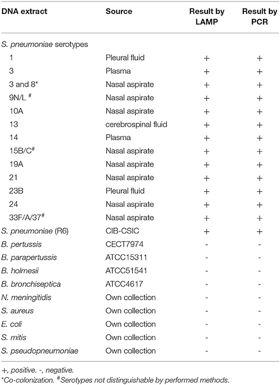
Table 1. Quality control panel performed with DNA extracts from direct clinical samples and bacterial strains.
LAMP intra- and inter-assay mean CV values across the linear range were 3.61 and 6.59%, respectively, as shown in Tables 2, 3. Between-run CV values ranged from 0.16% at 106 cp/mL to 5.68% at 108 cp/mL. Within-run CV values were higher at all concentrations tested, ranging from 6.23% at 106 cp/mL to 8.89% at 108 cp/mL.
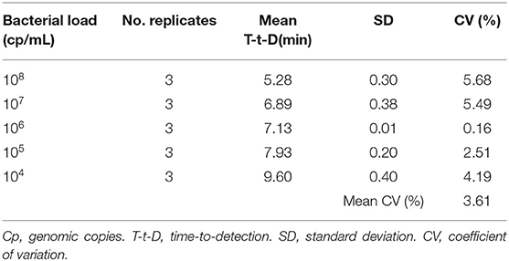
Table 2. Results of precision (intra-assay variability) for the LAMP assay.
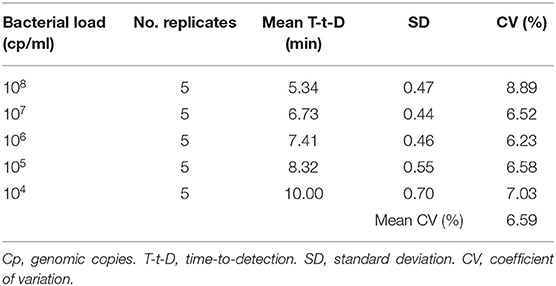
Table 3. Results of precision (inter-assay variability) for the LAMP assay.
Diagnostic Performance
During the study period, a total of 161 normally sterile samples (124 plasma fluids, 13 cerebrospinal fluids, 13 pleural fluids, and 11 synovial liquids) were collected. Thirteen (8.07%) were detected as positive by LAMP and 12 (7.45%) as negative by real-time PCR (Table 4) whereas there were 148 negatives by LAMP and 149 negatives by PCR. Diagnostic sensitivity and specificity of the LAMP assay compared to real-time PCR were 100% (95% CI, 75.8–100) and 99.3% (95% CI, 97.5–100), respectively. The discrepant sample, which corresponded to a pleural fluid, was repeated by both real-time PCR and culture and yielded a PCR-positive result and a negative result by culture. Of note, all positive results by both LAMP and PCR tests were negative by culture whereas culture yielded 17 positive results for bacteria different from pneumococcus that were negative by the two molecular methods. Specifically, bacteria identified by culture included Staphylococcus aureus (n = 6), Neisseria meningitidis (n = 3), Streptococcus viridans(n = 3), Kingella kingae (n = 2), Micrococcus spp. (n = 1), and Propionibcaterium acnes (n = 1). Positive PCR, LAMP, and culture results per type of specimen are detailed in Supplementary Tables 1, 2.
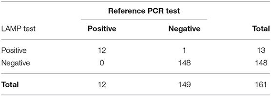
Table 4. Comparison of diagnostic results by LAMP vs. real-time PCR.
Pneumococcal DNA was amplified by LAMP within 30 min (median time: 15 min) while PCR amplification needed a maximum time of 103 min (median time: 79 min), which represents a median 5-fold decrease in time for DNA amplification.
Discussion
In the current study we developed a real-time LAMP assay targeting lytA gene, for S. pneumoniae detection and assessed analytical and diagnostic performance of the test against a reference real-time PCR. Since both lytA and ply targets have similar sensitivity but lytA gene contributes higher specificity, the latter was selected as the target gene of our LAMP reaction.
LOD of the LAMP assay (5·103 cp/mL 90 cp/rx) was found to be slightly higher though in the same order of magnitude as LOD of PCR (1·103 cp/mL, 20 cp/rx). These values are in contrast with values of analytical sensitivity reported to be 3-logs higher for LAMP than for conventional PCR in previous studies measuring LOD of PCR at around 104 cp/rx (Seki et al., 2005; Huy et al., 2012; Kim et al., 2012). Differences in PCR analytical sensitivity between studies could be due to the selected way of amplified DNA visualization (electrophoretic analysis). In this regard, it should be noted that we compared the LAMP assay with a real-time PCR with TaqMan probes. In fact other studies reported LOD values for PCR around 50 cp/rx (Nandakumar et al., 2008) and 22 cp/rx (Sheppard, 2004), which are in line with the LOD of 20 cp/rx obtained for the reference PCR of our study.
Other LAMP techniques targeting the lytA gene have determined similar values of analytical sensitivity (10 cp/rx) as those described our assay (Seki et al., 2005; Kim et al., 2012). Minor differences between LAMP sensitivities could be explained by the higher number of replicates that we used for LOD calculation, as suggested by guidelines (FDA, 2001), in comparison to the number of replicates considered in the referenced studies (<3). The LOD described in our study is also in concordance with the value of 100–500 CFU/mL (between 103 and 104 cp/mL) targeting the 16S rRNA gene (Huy et al., 2012) and the ply gene (Wang et al., 2018). Conversely, another published work established a LOD about 300 pg/μL (105 copies/μl) for a specific LAMP test based on SYBR Green fluorophore and using a portable tube scanner (Xia et al., 2014).
The new LAMP assay was able to detect specifically all the 14 S. pneumoniae serotypes tested. In addition, no cross-reactivity was detected either with human DNA or with the rest of tested bacteria, not even with genetically similar bacteria such as S. mitis or S. pseudoneumoniae. The use of lytA gene as the amplification target and the fact that LAMP reaction occurs only when six regions within a target DNA are recognized (Notomi, 2000) contributed high specificity to the new assay. Although multiple strains of S. pneumoniae and other pathogens and commensal bacteria of the nasopharynx have been tested, the modest size of the quality control panel might be a limitation of the study.
Intra- and inter-assay precision of the new test across the range of expected concentrations showed to be adequate. Mean CV values obtained did not exceed the recommended value of 15% for imprecision (FDA, 2001). Of note, linearity (108-104 cp/mL) was lower than that of PCR (108-103 cp/mL), which may indicate better suitability of the test for qualitative or semi-quantitative diagnosis than for quantification.
LAMP diagnostic sensitivity (100%) and specificity (99.3%) were optimal. Only a discrepant result was observed and after confirmatory PCR classified as a true positive result. The related samples showed a bacterial load under the LOD (Ct 39, 5; about 4 cp/rx) which could explain the initial discordance.
In addition, the LAMP assay proved to be a rapid method for detection of pneumococcus, allowing for a 5-fold median run time reduction in comparison with PCR. It should be noted that total time required for PCR amplification could be reduced with cycle alterations slightly below 1 h. However, we followed the PCR protocol recommended by the CDC to ensure adequate sensitivity and specificity while achieving much faster time of amplification with LAMP. The LAMP reaction is a single tube technique for DNA amplification that does not require thermal cyclers (Huy et al., 2012; Xia et al., 2014; Wang et al., 2018). In addition, amplification can be detected by turbidimetry or visual color change without the need for any other equipment (Huy et al., 2012). Even electricity-free and reusable devices heaters have been developed for DNA amplification using the LAMP technique (Sema et al., 2015). All these characteristics, together with LAMP high sensitivity and specificity, favor the use of this technique as a simple and inexpensive diagnostic assay, which would be especially useful in low and middle income countries (Geojith et al., 2011). In this sense, it should be mentioned that the LAMP assay described in this study could be integrated into a prototype of a rapid, simple, and cost-effective point-of-care detection of different respiratory pathogens including S. pneumoniae.
We performed a real-time PCR assay in samples collected from normally sterile sites, the majority of which were blood specimens, and considered this assay as a reference standard for validating the LAMP test. Pneumococcal real-time PCR in blood is widely used in hospital environments for routine diagnostics of pneumococcal infections and the European Union regulations have stated the adequacy of pneumococcal DNA detection for confirmation of cases with pneumococcal infection or IPD in normally sterile samples (Commission Implementing Decision EU 2018/945, 2018). However, there is some controversy about the consideration of blood pneumococcal PCR as a gold standard, since diverse studies have reported noticeable proportions of blood pneumococcal PCR-positive results in healthy children (Morpeth et al., 2017) and moderate values of sensitivity and specificity of PCR in blood samples extracted from children with IPD (Avni et al., 2010).
In conclusion, we designed and validated a real-time LAMP assay based on fluorescence for diagnosis of IPD. The test proved to be 5-fold faster than PCR for DNA amplification while maintaining similar levels of analytical and diagnostic performance. This novel LAMP assay could be useful for early detection of S. pneumoniae, especially if integrated into inexpensive easy-to-use point-of-care diagnostic devices not requiring the complex electronics associated to cyclic temperature changes.
Data Availability Statement
The datasets generated for this study are available on request to the corresponding author.
Author Contributions
CE performed the laboratory experiments. HP organized the database and wrote the first draft of the manuscript. PB performed the statistical analysis. HP and PB wrote sections of the manuscript. HP, PB, and CM-A contributed conception and design of the study. All authors contributed to manuscript revision, read and approved the submitted version.
Funding
The current work has been financially supported by the Seventh Framework Programme of the European Union (FP7-SME-2013, Grant No: 606488) and the Fondo de Investigaciones Sanitarias, Instituto de Salud Carlos III (PI13/01729). The funders had no role in study design, data collection, and interpretation, or the decision to submit the work for publication.
Conflict of Interest
CM-A reports research grants to her institution from Pfizer, BioFire Diagnostics, Roche Diagnostics, BioMérieux, Alere and Genomica SAU, and compensation fees from Qiagen, Roche Diagnostics and BioMérieux for scientific presentations in satellite symposiums outside the submitted work. PB reports compensation fees from Roche Diagnostics for scientific presentations in satellite symposiums outside the submitted work.
The remaining authors declare that the research was conducted in the absence of any commercial or financial relationships that could be construed as a potential conflict of interest.
Acknowledgments
The authors would like to thank Dr. Pedro García of the Molecular Microbiology and Infection Biology Department of CIB-CSIC for providing S. pneumoniae R6 strain. We would also like to thank the Hematology Department of Sant Joan de Déu Hospital for its support in tasks of measuring DNA concentration.
Supplementary Material
The Supplementary Material for this article can be found online at: https://www.frontiersin.org/articles/10.3389/fcimb.2020.00115/full#supplementary-material
References
Avni, T., Mansur, N., Leibovici, L., and Paul, M. (2010). PCR using blood for diagnosis of invasive pneumococcal disease: systematic review and meta-analysis. J. Clin. Microbiol. 48, 489–496. doi: 10.1128/JCM.01636-09
Bidet, P., Liguori, S., De Lauzanne, A., Caro, V., Lorrot, M., Carol, A., et al. (2008). Real-time PCR measurement of persistence of Bordetella pertussis DNA in nasopharyngeal secretions during antibiotic treatment of young children with pertussis. J. Clin. Microbiol. 46, 3636–3638. doi: 10.1128/JCM.01308-08
Bogaert, D., de Groot, R., and Hermans, P. (2004). Streptococcus pneumoniae colonisation: the key to pneumococcal disease. Lancet Infect. Dis. 4, 144–154. doi: 10.1016/S1473-3099(04)00938-7
Brotons, P., de Paz, H. D., Esteva, C., Latorre, I., and Muñoz-Almagro, C. (2016). Validation of a loop-mediated isothermal amplification assay for rapid diagnosis of pertussis infection in nasopharyngeal samples. Expert Rev. Mol. Diagn. 16, 125–130. doi: 10.1586/14737159.2016.1112741
Centers for Disease Control Prevention (CDC) (2015). Chapter 10: PCR for Detection and Characterization of Bacterial Meningitis Pathogens: Neisseria meningitidis, Haemophilus influenzae, and Streptococcus pneumoniae. Available on line at: https://www.cdc.gov/meningitis/lab-manual/chpt10-pcr.html (accessed March 10, 2020).
Chen, Z., Liao, Y., Ke, X., Zhou, J., Chen, Y., and Gao, L. (2011). Comparison of reverse transcription loop-mediated isothermal amplification, conventional PCR and real-time PCR assays for Japanese encephalitis virus. Mol. Biol. Rep. 38, 4063–4070. doi: 10.1007/s11033-010-0525-0
Commission Implementing Decision EU 2018/945 (2018). Commission Implementing Decision (EU) 2018/945 of 22 June 2018 on the communicable diseases and related special health issues to be covered by epidemiological surveillance as well as relevant case definitions. Official Journal of the European Union L170/1-170/73. Available online at: https://eur-lex.europa.eu/legal-content/EN/TXT/PDF/?uri=CELEX:32018D0945&from=EN (accessed January 7, 2020).
de Paz, H. D., Brotons, P., and Muñoz-Almagro, C. (2014). Molecular isothermal techniques for combating infectious diseases: towards low-cost point-of-care diagnostics. Expert Rev. Mol. Diagn. 14, 827–843. doi: 10.1586/14737159.2014.940319
Dellinger, R. P., Levy, M. M., Rhodes, A., Annane, D., Gerlach, H., Opal, S. M., et al. (2013). Surviving sepsis campaign. Crit. Care Med. 41, 580–637. doi: 10.1097/CCM.0b013e31827e83af
FDA (2001). FDA Guidance for Industry: Bioanalytical Method Validation Center for Drug Evaluation and Research. Rockville, MD.
Fierz, W. (2004). Basic problems of serological laboratory diagnosis. Methods Mol. Med. 94, 393–427. doi: 10.1385/1-59259-679-7:393
Geojith, G., Dhanasekaran, S., Chandran, S. P., and Kenneth, J. (2011). Efficacy of loop mediated isothermal amplification (LAMP) assay for the laboratory identification of Mycobacterium tuberculosis isolates in a resource limited setting. J. Microbiol. Methods 84, 71–73. doi: 10.1016/j.mimet.2010.10.015
Harboe, Z. B., Dalby, T., Weinberger, D. M., Benfield, T., Mølbak, K., Slotved, H. C., et al. (2014). Impact of 13-valent pneumococcal conjugate vaccination in invasive pneumococcal disease incidence and mortality. Clin. Infect. Dis. 59, 1066–1073. doi: 10.1093/cid/ciu524
Hess, A. S., Shardell, M., Johnson, J. K., Thom, K. A., Strassle, P., and Netzer, G. (2012). Methods and recommendations for evaluating and reporting a new diagnostic test. Eur. J. Clin. Microbiol. Infect. Dis. 31, 2111–2116. doi: 10.1007/s10096-012-1602-1
Huy, N. T., Hang, L. T. T., Boamah, D., Lan, N. T. P., Van Thanh, P., Watanabe, K., et al. (2012). Development of a single-tube loop-mediated isothermal amplification assay for detection of four pathogens of bacterial meningitis. FEMS Microbiol. Lett. 337, 25–30. doi: 10.1111/1574-6968.12002
Kim, D. W., Kilgore, P. E., Kim, E. J., Kim, S. A., Anh, D. D., Dong, B. Q., et al. (2012). The enhanced pneumococcal LAMP assay: a clinical tool for the diagnosis of meningitis due to Streptococcus pneumoniae. PLoS ONE 7:e42954. doi: 10.1371/journal.pone.0042954
Kosack, C. S., Page, A.-L., and Klatser, P. R. (2017). A guide to aid the selection of diagnostic tests. Bull. World Health Organ. 95, 639–645. doi: 10.2471/BLT.16.187468
Lai, C.-C., Lin, S.-H., Liao, C.-H., Sheng, W.-H., and Hsueh, P.-R. (2014). Decline in the incidence of invasive pneumococcal disease at a medical center in Taiwan, 2000–2012. BMC Infect. Dis. 14:76. doi: 10.1186/1471-2334-14-76
Morpeth, S. C., Deloria Knoll, M., Scott, J. A. G., Park, D. E., Watson, N. L., Bagget, H. C., et al. (2017). Detection of pneumococcal DNA in blood by polymerase chain reaction for diagnosing pneumococcal pneumonia in young children from low- and middle-income countries. Clin. Infect. Dis. 15(suppl_3), S347–356. doi: 10.1093/cid/cix145
Morpeth, S. C., Huggett, J. F., Murdoch, D. R., and Scott, J. A. G. (2014). Making standards for quantitative real-time pneumococcal PCR. Biomol. Detect Quantif. 2, 1–3. doi: 10.1016/j.bdq.2014.11.003
Muñoz-Almagro, C., Gala, S., Selva, L., Jordan, I., Tarrag,ó, D., and Pallares, R. (2011). DNA bacterial load in children and adolescents with pneumococcal pneumonia and empyema. Eur. J. Clin. Microbiol. Infect. Dis. 30, 327–335. doi: 10.1007/s10096-010-1086-9
Nandakumar, R., Whiting, J., and Fouad, A. F. (2008). Identification of selected respiratory pathogens in endodontic infections. Oral Surg. Oral Med. Oral Pathol. Oral Radiol. Endodontol. 106, 145–151. doi: 10.1016/j.tripleo.2008.01.036
Nilsson, A. C., Bjorkman, P., and Persson, K. (2008). Polymerase chain reaction is superior to serology for the diagnosis of acute Mycoplasma pneumoniae infection and reveals a high rate of persistent infection. BMC Microbiol. 8:93. doi: 10.1186/1471-2180-8-93
Notomi, T. (2000). Loop-mediated isothermal amplification of DNA. Nucleic Acids Res. 28:63e. doi: 10.1093/nar/28.12.e63
Obaro, S., and Adegbola, R. (2002). The pneumococcus: carriage, disease and conjugate vaccines. J. Med. Microbiol. 51, 98–104. doi: 10.1099/0022-1317-51-2-98
Parida, M., Shukla, J., Sharma, S., Ranghia Santhosh, S., Ravi, V., Mani, R., et al. (2011). Development and evaluation of reverse transcription loop-mediated isothermal amplification assay for rapid and real-time detection of the swine-origin influenza A H1N1 virus. J. Mol. Diagnostics 13, 100–107. doi: 10.1016/j.jmoldx.2010.11.003
Patel, J. C., Lucchi, N. W., Srivastava, P., Lin, J. T., Sug-aram, R., Aruncharus, S., et al. (2014). Field evaluation of a real-time fluorescence loop-mediated isothermal amplification assay, RealAmp, for the diagnosis of malaria in Thailand and India. J. Infect. Dis. 210, 1180–1187. doi: 10.1093/infdis/jiu252
Seki, M., Yamashita, Y., Torigoe, H., Tsuda, H., Sato, S., and Maeno, M. (2005). Loop-mediated isothermal amplification method targeting the lytA gene for detection of Streptococcus pneumoniae. J. Clin. Microbiol. 43, 1581–1586. doi: 10.1128/JCM.43.4.1581-1586.2005
Selva, L., Berger, C., Garcia-Garcia, J. J., de Paz, H., Nadal, D., and Munoz-Almagro, C. (2014). Direct identification of Streptococcus pneumoniae capsular types in pleural fluids by using multiplex PCR combined with automated fluorescence-based capillary electrophoresis. J. Clin. Microbiol. 52, 2736–2737. doi: 10.1128/JCM.00906-14
Selva, L., del Amo, E., Brotons, P., and Munoz-Almagro, C. (2012). Rapid and easy identification of capsular serotypes of Streptococcus pneumoniae by use of fragment analysis by automated fluorescence-based capillary electrophoresis. J. Clin. Microbiol. 50, 3451–3457. doi: 10.1128/JCM.01368-12
Sema, M., Alemu, A., Bayih, A., Getie, S., Getnet, G., Guelig, D., et al. (2015). Evaluation of non-instrumented nucleic acid amplification by loop-mediated isothermal amplification (NINA-LAMP) for the diagnosis of malaria in Northwest Ethiopia. Malar J. 14:44. doi: 10.1186/s12936-015-0559-9
Sheppard, C. L. (2004). Autolysin-targeted LightCycler assay including internal process control for detection of Streptococcus pneumoniae DNA in clinical samples. J. Med. Microbiol. 53, 189–195. doi: 10.1099/jmm.0.05460-0
Walker, C. L. F., Rudan, I., Liu, L., Nair, H., Theodoratou, E., Bhutta, Z. A., et al. (2013). Global burden of childhood pneumonia and diarrhoea. Lancet 381, 1405–1416. doi: 10.1016/S0140-6736(13)60222-6
Wang, Y., Wang, Y., Li, D., Xu, J., and Ye, C. (2018). Detection of nucleic acids and elimination of carryover contamination by using loop-mediated isothermal amplification and antarctic thermal sensitive uracil-DNA-glycosylase in a lateral flow biosensor: application to the detection of Streptococcus pneumoniae. Microchim. Acta 185:212. doi: 10.1007/s00604-018-2723-8
Xia, Y., Guo, X. G., and Zhou, S. (2014). Rapid detection of Streptococcus pneumoniae by real-time fluorescence loop-mediated isothermal amplification. J. Thorac. Dis. 6, 1193–1199. doi: 10.3978/j.issn.2072-1439.2014.07.29
Keywords: Streptococcus pneumoniae, loop-mediated isothermal amplification, invasive pneumococcal disease, rapid diagnosis, diagnostic accuracy
Citation: de Paz HD, Brotons P, Esteva C and Muñoz-Almagro C (2020) Validation of a Loop-Mediated Isothermal Amplification Assay for Rapid Diagnosis of Invasive Pneumococcal Disease. Front. Cell. Infect. Microbiol. 10:115. doi: 10.3389/fcimb.2020.00115
Received: 18 March 2019; Accepted: 02 March 2020;
Published: 24 March 2020.
Edited by:
John W. A. Rossen, University Medical Center Groningen, NetherlandsReviewed by:
Kevin Alby, The University of North Carolina at Chapel Hill, United StatesDaniel Angel Ortiz, Beaumont Health, United States
Copyright © 2020 de Paz, Brotons, Esteva and Muñoz-Almagro. This is an open-access article distributed under the terms of the Creative Commons Attribution License (CC BY). The use, distribution or reproduction in other forums is permitted, provided the original author(s) and the copyright owner(s) are credited and that the original publication in this journal is cited, in accordance with accepted academic practice. No use, distribution or reproduction is permitted which does not comply with these terms.
*Correspondence: Carmen Muñoz-Almagro, cma@sjdhospitalbarcelona.org
†These authors have contributed equally to this work