 Martina Becker1‡
 Steffen Güttler1†‡
 Annabell Bachem1
 Evelyn Hartung1
 Ahmed Mora1†Anika Jäkel1Andreas Hutloff1,2
 Volker Henn1Hans Werner Mages1Stephanie Gurka1
 Richard A. Kroczek1*
- 1Molecular Immunology, Robert Koch-Institute, Berlin, Germany
- 2German Rheumatism Research Centre, Berlin, Germany
In the past, lack of lineage markers confounded the classification of dendritic cells (DC) in the intestine and impeded a full understanding of their location and function. We have recently shown that the chemokine receptor XCR1 is a lineage marker for cross-presenting DC in the spleen. Now, we provide evidence that intestinal XCR1+ DC largely, but not fully, overlap with CD103+ CD11b− DC, the hypothesized correlate of “cross-presenting DC” in the intestine, and are selectively dependent in their development on the transcription factor Batf3. XCR1+ DC are located in the villi of the lamina propria of the small intestine, the T cell zones of Peyer’s patches, and in the T cell zones and sinuses of the draining mesenteric lymph node. Functionally, we could demonstrate for the first time that XCR1+/CD103+ CD11b− DC excel in the cross-presentation of orally applied antigen. Together, our data show that XCR1 is a lineage marker for cross-presenting DC also in the intestinal immune system. Further, extensive phenotypic analyses reveal that expression of the integrin SIRPα consistently demarcates the XCR1− DC population. We propose a simplified and consistent classification system for intestinal DC based on the expression of XCR1 and SIRPα.
Introduction
The intestinal immune system has to discriminate between harmless food proteins, commensal bacteria colonizing the gut, and dangerous pathogens. Dendritic cells (DC) play a central role in orchestrating the appropriate immune responses. Conventional DC reside in the lamina propria (LP) of the small and large intestine, in the scattered lymphoid follicles and Peyer’s patches (PP), and in lymph nodes draining the intestine, such as the mesenteric lymph nodes (MLN). In the past, any subdivision of intestinal DC into functional subpopulations was controversial because DC-specific markers were lacking and other surface molecules used for classification were found to be regulated or were also present on macrophages (1–6). A major step forward was the combined use of antibodies directed to the integrins CD103 and CD11b, which allowed to define four DC subsets, CD103+ CD11b−, CD103+ CD11b+, CD103− CD11b+, and CD103− CD11b− (7–9), which in the MLN were further grouped into resident and migratory DC (10). Because of open questions regarding the subdivision of DC in the intestine, only very few studies on antigen presentation were performed with intestinal DC.
Most of the work on the function of DC has been performed with splenic DC populations. These studies demonstrated that all DC can present exogenous antigen to CD4+ T cells (classical presentation), while only a subset excels in the cross-presentation of antigen to CD8+ T cells (11, 12). Cross-presentation is a central element in the activation of CD8+ T cells to cytotoxic effector cells, and thus of major importance in the defense of certain infections and in the elimination of tumors (13–15). Based on the commonly used classification of splenic DC, numerous studies established that CD8α+ DC excel over CD4+ DC and double-negative DC in their capacity to cross-present (cell-associated) antigen. Because of the different DC classification systems used, it remained unclear whether these splenic CD8α+ DC have a correlate among the intestinal DC.
A major advance for a unified classification of DC throughout the immune system was brought by analyses on the role of transcription factors (TF) in the differentiation of DC. These studies revealed that CD8α+ DC in the spleen and CD103+ CD11b− DC in the intestine and other organs were specifically absent in animals deficient for the TF Batf3 (16, 17). These findings strongly indicated that CD8α+ DC in the spleen and the lymphoid organs correspond to CD103+ CD11b− DC in tissues, and together represent a separate DC lineage of cross-presenting DC. The intestinal CD103+ CD11b− DC were thus subsequently also termed as “CD8α-like DC” or “Batf3-dependent DC.”
Recently, we and others have recognized that the chemokine receptor XCR1 is exclusively expressed on murine (18) and human DC (19, 20). In the murine system, expression of XCR1+ DC was found to be restricted to CD8α+ DC in the spleen and CD8α-like DC in peripheral organs (18, 21, 22). However, in a subsequent more detailed analysis it became apparent that splenic CD8α+ DC are not identical to XCR1+ DC, since these two populations only overlap (21). We could firmly establish that splenic DC expressing both XCR1 and CD8α+, or only XCR1, belong to the Batf3-dependent, antigen-cross-presenting DC (21). In contrast, CD8α+ DC lacking XCR1 on the cell surface are a clearly different population, which (i) is independent of Batf3, (ii) has a distinct gene expression program (23), and (iii) is functionally different, as it is unable to cross-present antigen (21). With this work, it became clear that the identified lineage of Batf3-dependent DC in the spleen (and possibly other organs) is truly represented by DC expressing XCR1. At the same time, this work determined that “CD8α+ DC” are in reality a phenotypically and functionally heterogeneous population.
With the present study, we analyzed the development, phenotype, localization, and function of XCR1+ cells in the intestinal immune system to find out whether they correspond to XCR1+ DC in the spleen. Although our work was focused on XCR1+ DC, most of the experiments also yielded information on XCR1− DC, which were used for comparison. The results demonstrate that the expression of XCR1 also in the intestinal immune system consistently demarcates the lineage of Batf3-dependent, antigen cross-presenting DC. At the same time, we show that intestinal XCR1+ DC strongly overlap, but are not fully congruent with CD103+ CD11b− DC.
Further, our extensive phenotypic studies demonstrate that XCR1 and SIRPα delineate two mutually exclusive DC populations, which together encompass essentially all conventional DC in the intestine. Based on these results, we propose a new and simplified classification system for conventional DC in the intestine based on only two surface markers, XCR1 and SIRPα.
Materials and Methods
Mice and Flt3-Ligand Treatment
Unless indicated otherwise, 8–10-week-old C57BL/6 female mice were used for cell isolation and immunohistological analyses. CX3CR1GFP (24), LangerinEGFP mice (25), B6.XCR1-lacZ+/+ (The Jackson Laboratories), and Batf3-deficient mice (16) were on the C57BL/6 background. OT-I TCR-transgenic mice were crossed onto the B6.PL background to allow identification of CD8+ T cells using the CD90.1 marker. For Flt3-ligand treatment, C57BL/6 mice were injected with 1 × 10⁶ B16 cells secreting Flt3-ligand (26) in 100 μl PBS s.c. All mice were bred under specific pathogen-free conditions in the animal facility of the Federal Institute for Risk Assessment (Berlin, Germany). All animal experiments were performed according to state guidelines and approved by the local animal welfare committee.
Antibodies
Hybridomas producing mAb recognizing CD4 (clone YTS 191.1), CD8α (53-6.72), CD11b (5C6), CD11c (N418), CD16/32 (2.4G2), CD24 (M1/69.16.11.HL), CD45R/B220 (RA3-6B2), DCIR2 (33D1), MHCII (M5/114.15.2), and NK1.1 (PK136) were obtained from ATCC, CD90.1 (OX-7) from ECACC. MAb to CD103 (M290), CD172a/SIRPα (P84), and CD11c (HL3) were from BD Biosciences, to CD69 (H1.2F3), CD45 (30F11), and CCR7 (4B12) from eBioscience, and to CD3 (17A2), F4/80 (BM8), and CD45R/B220 (RA3-6B2) from BioLegend. Anti-XCR1 [MARX10 (21)] and anti-Clec9A/DNGR-1 [clone 24/04-10B4 (27)] antibodies were used. Anti-CD3 (KT3) was generously provided by H. Savelkoul, anti-CD25 (2E4) by E. Shevach, and anti-DEC-205 (NLDC-145, CD205) by G. Kraal.
Cell Isolation
For isolation of small intestinal LP DC, the small intestine was freed from fat and PP, opened longitudinally, and stirred in PBS, 2% FCS, 1 mM EDTA, 1 mM DTT for 7 min at 37°C. After additional stirring under the same conditions without DTT, epithelial cells in solution were discarded, intestinal tissue was minced, and stirred in 500 μg/ml collagenase VIII (in some experiments, collagenase D was used instead in an attempt to improve staining of Clec9a, both from Sigma) and 20 μg/ml DNAse I (Roche) for 30 min at 37°C; thereafter, cells were mashed through a 70 μm nylon sieve (BD Falcon). For isolation of DC from lymphoid tissues, MLN and PP were ruptured and digested with collagenase D (500 μg/ml) and DNase I (20 μg/ml, both Roche) for 15 min at 37°C in RPMI 1640 containing 2% FCS (low endotoxin, Biochrom); EDTA (10 mM) was added for additional 5 min and cells were mashed through a 70 μm nylon sieve. For staining of DC from LP and PP, low density cells from these tissues were enriched by centrifugation over a 1.073 g/ml density gradient (NycoPrep, Axis-Shield). For flow sorting of DC from LP and MLN, low density cells were enriched and magnetically sorted with CD11c microbeads (Miltenyi Biotec). Splenocytes were obtained by mashing spleens through 70 μm cell sieves into PBS, followed by erythrocyte lysis with ACK Buffer (155 mM NH4Cl, 10 mM KHCO3, 0.1 mM EDTA).
Flow Cytometry and Flow Sorting
Antibodies were titrated for optimal signal-to-noise ratio. To block unspecific binding to Fc-receptors, cells were pre-incubated with 100 μg/ml 2.4G2 mAb for flow cytometry and in addition with 50 μg/ml purified rat Ig (Nordic) for flow sorting. Doublets and autofluorescent cells were excluded from the analysis. In all organs, DC were identified as CD11c+ MHCII+ Lin− F4/80− cells, in LP and PP DC were additionally defined using CD45; the lineage cocktail contained mAb directed to CD3 and B220. Standard staining with mAb was in PBS, 0.25% BSA, 0.1% NaN3 for 20 min on ice, staining for Clec9A was in the same buffer for 20 min at 37°C. For exclusion of dead cells, 4′,6-diamidino-2-phenylindole (DAPI) was added 5 min before measurement. Data were acquired on an LSRFortessa flow cytometer (BD Biosciences) and analyzed using FlowJo (Tree Star Inc.). For analysis of surface receptor expression, gates were set according to the appropriate isotype/FMO controls. Flow sorting of small intestinal DC (CD11c+ MHCII+ CD45+ Lin− F4/80−), migratory MLN DC (CD11c+ MHCIIhigh Lin− F4/80−), and resident MLN DC (CD11c+ MHCIIlow Lin− F4/80−) was based on their expression of CD103 and XCR1 and performed on a FACSAriaII (BD Biosciences).
Histology
For histological β-galactosidase analysis, tissues from homozygous B6.XCR1-lacZ+/+ mice and C57BL/6 mice were fixed with 0.1% glutaraldehyde plus 4% paraformaldehyde in PBS for 4 h at RT, immersed in 10% sucrose overnight, and snap-frozen in 0.9% NaCl. Cryostat sections (15 μm) were washed with cold PBS (pH 7.4) for 5 min after thawing, incubated with X-Gal staining solution (28) overnight at 37°C, washed with PBS, and counterstained with Neutral Red.
Cross-Presentation Assay
C57BL/6 mice were fed with 25 mg ovalbumin (OVA, Sigma-Aldrich) in 500 μl PBS by gavage. Seventeen hours after oral application of OVA, DC subsets were flow sorted to high purity (>98.5%). OT-I CD8+ T cells were enriched from spleens of OT-I mice by magnetically depleting cells expressing CD4, CD11b, CD11c, B220, or NK1.1 (Miltenyi Biotec). Preparations of resting OT-I T cells (confirmed by negativity for CD25 and CD69) were labeled with CFSE (Molecular Probes, 5 μM, 15 min, 37°C). For cross-presentation assays, 1 × 10⁵ CFSE-labeled OT-I T cells were co-cultured with titrated numbers (3,750–30,000) of DC subsets in 200 μl RPMI medium containing 10% FCS, 50 μM 2-mercaptoethanol, 1 mM sodium pyruvate, non-essential amino acids, and 100 μg/ml penicillin/streptomycin in 96-well round-bottomed plates (Nunc) for 2.5 days. Thereafter, proliferation of OT-I T cells was determined in the CFSE dilution assay after gating on CD90.1 cells. For positive control, sorted DC were incubated with 1 μM of the OVA peptide SIINFEKL, and co-cultured with CFSE-labeled OT-I T cells for 2.5 days.
Results
Phenotype of XCR1+ DC in the Lamina Propria, Peyer’s Patches, and Mesenteric Lymph Nodes
To determine the phenotype of XCR1+ DC in the intestinal immune system at steady state, mononuclear cells were obtained from the LP of the small intestine, PP, and MLN. Conventional DC, defined as lineage-negative, F4/80-negative, CD11chi MHCIIhi cells by flow cytometry, were then gated into four populations based on the expression of the integrins CD103 (Itgae) and CD11b. In the LP, essentially all CD103+ CD11b− DC expressed XCR1, while almost all CD103+ CD11b+ and CD103− CD11b+ DC were negative (Figure 1). Interestingly, a fraction (around 10%) of the small population of CD103− CD11b− (double-negative) DC found in the small intestine also expressed XCR1 (Figure 1). In the PP, a large majority (around 80%) of CD103+ CD11b− DC expressed XCR1, while all other populations resembled in their XCR1 expression profile LP cells (Figure 1). In MLN, expression levels of MHCII were used to further discriminate resident from migratory DC (10). Migratory DC subpopulations closely resembled in their XCR1 expression pattern LP DC (Figure 1). Different from LP, PP, and migratory MLN DC, the CD103− population in the resident MLN DC contained a substantial proportion of XCR1+ DC (Figure 1). Taken together, our results, based on the currently popular subdivision of intestinal DC using CD103 and CD11b as markers, demonstrated a large but clearly not full overlap between XCR1+ DC and CD103+ CD11b− DC.
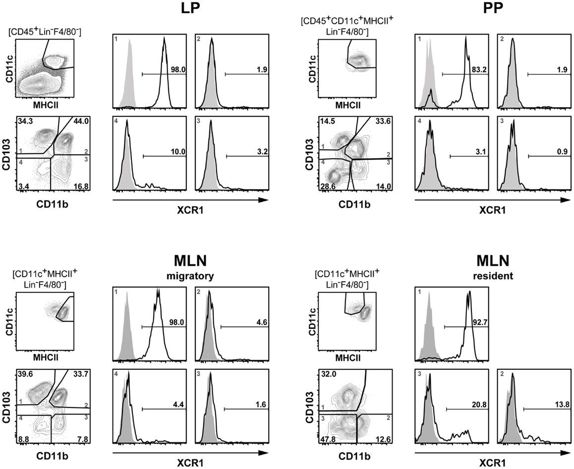
Figure 1. Expression of XCR1 on DC in the lamina propria, Peyer’s patches, and mesenteric lymph nodes. DC from the LP, PP, and MLN of C57BL/6 mice were enriched by digestion and density gradient centrifugation of the tissues, stained for CD11b and CD103, and counter-stained with XCR1-specific mAb MARX10. DC from MLN were separated into resident and migratory DC based on their MHCII expression levels. For analysis, the gates were set on live CD45+ Lin− F4/80− CD11c+ MHCII+ cells. Expression of XCR1 is shown on CD103+ CD11b− (left upper quadrants), CD103+ CD11b+ (right upper quadrants), CD103− CD11b+ (right lower quadrants), and CD103− CD11b− (left lower quadrants) DC. The background staining was determined with homozygous B6.XCR1-lacZ+/+ mice lacking XCR1 (gray). The results shown are representative of three experiments with three animals each.
To further define the phenotype of XCR1+ DC at the examined locations, expression of XCR1 was correlated to a greater number of surface molecules known to be expressed on DC in the intestine. In the steady state, expression of XCR1 was found to be highly correlated with CD8α on DC in the LP, PP, and MLN (Figure 2). Only a small population of resident MLN DC expressed CD8α, but was negative for XCR1 (Figure 2), as earlier found in the spleen (21). In tissues where expression of Clec9A/DNGR-1 on DC was detectable, it was highly correlated with XCR1. Surface presence of XCR1 corresponded well with CD205 in the LP and MLN, but less so in the PP. All XCR1+ DC were positive for CD24 and also the majority of XCR1− DC. XCR1+ DC usually co-expressed the integrin CD103, which in resident MLN DC was found to be partly downregulated. XCR1+ DC were low/negative for CX3CR1/fractalkine receptor, and with the exception of a population of resident MLN DC, also negative for CD207/langerin. In all instances, XCR1+ DC were negative for 33D1/DCIR2 and CD11b, but neither of these molecules was anti-correlated. Finally and importantly, only CD172a/SIRPα was clearly anti-correlated with XCR1 and also encompassed all XCR1− DC.
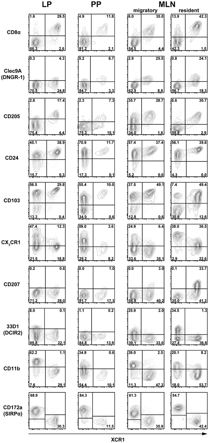
Figure 2. Correlation of XCR1 expression with different DC surface molecules. DC from the LP, PP, and MLN of C57BL/6 wt mice, and heterozygous CX3CR1GFP or LangerinEGFP (CD207) mice were enriched by digestion and density gradient centrifugation and double-stained for detection of XCR1 and the indicated surface molecules. For analysis, the gates were set on live CD45+ Lin− F4/80− CD11c+ MHCII+ cells. The results shown are representative of three experiments with three animals each.
In summary, none of the surface molecules examined was fully correlated with XCR1 on DC in all anatomical locations. The best overall correlation of XCR1 was seen with CD8α and with Clec9A/DNGR-1 (when detectable), indicating a functional link between these three receptors. Further, XCR1 was substantially, but not fully, correlated with CD205 in all tissues. On the other hand, a perfect anti-correlation could be observed between XCR1 and SIRPα in all anatomical sites, which was not the case between XCR1 and CD11b.
Expansion of Intestinal DC Populations Under the Influence of Flt3 Ligand
Flt3 ligand is a growth factor described to play a key role in the physiological expansion of classical CD8+ DC (29). We have previously observed that Flt3 ligand expanded XCR1+ DC in the spleen around 20-fold, while XCR1− DC were expanded only around 4-fold (21). In order to determine whether the same phenomenon can be observed in the intestinal immune system, mice were exposed to Flt3 ligand and the composition of the various DC populations was determined by flow cytometry. Under the influence of Flt3 ligand, both XCR1+ CD103+ and XCR1− CD103− increased in frequency, and this was accompanied by a substantial relative reduction of XCR1− CD103+ DC in all anatomical sites (Figure 3, flow cytometry histograms). In terms of absolute cell numbers, all DC populations increased under the influence of Flt3 ligand, with biggest changes in PP and MLN. There, XCR1+ CD103+ and XCR1− CD103− DC expanded both around 30-fold and thus more than XCR1− CD103+ DC (10-fold), while the most dramatic change was seen with the usually minute population of XCR1+ CD103− DC (up to 400-fold increase). Thus, the effects of Flt3 ligand on the expansion of intestinal DC subsets were more complex than in the spleen, with a massive expansion of XCR1+ CD103− DC, but a similar expansion of XCR1+ CD103+ and XCR1− CD103− DC.
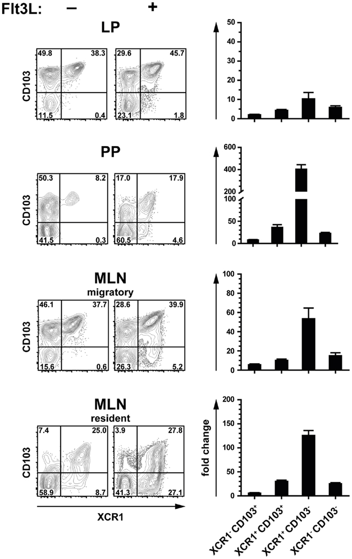
Figure 3. Expansion of intestinal XCR1+ DC by the growth factor Flt3 ligand. C57BL/6 mice were exposed to Flt3 ligand for 9 days in vivo. Thereafter, DC from the LP, PP, and MLN (CD45+ Lin− F4/80− CD11c+ MHCII+ cells) were analyzed for expression of CD103 and XCR1, and compared to unexposed controls (flow cytometry histograms). The bar graphs represent the fold increase in total numbers of the indicated DC subsets in relation to unexposed controls. The data shown are representative of two independent experiments (mean ± SEM; in each experimentn = 3).
XCR1+ DC in the Intestinal Immune System are Batf3-Dependent
The development of CD8α+ DC has originally been described to be dependent on the TF Batf3 (16). However, we recently found that in the spleen only CD8+ XCR1+ DC were dependent on this TF, but not the CD8α+ DC population negative for XCR1, which has a clearly different gene expression profile and function (21, 23). In order to determine the influence of Batf3 on the development of intestinal DC, we analyzed Batf3-deficient animals on the C57BL/6 background. In all anatomical sites, Batf3 deficiency essentially resulted in the absence of XCR1+ DC, clearly demonstrating that Batf3 is required for the development of XCR1+ DC in the gut (Figure 4).
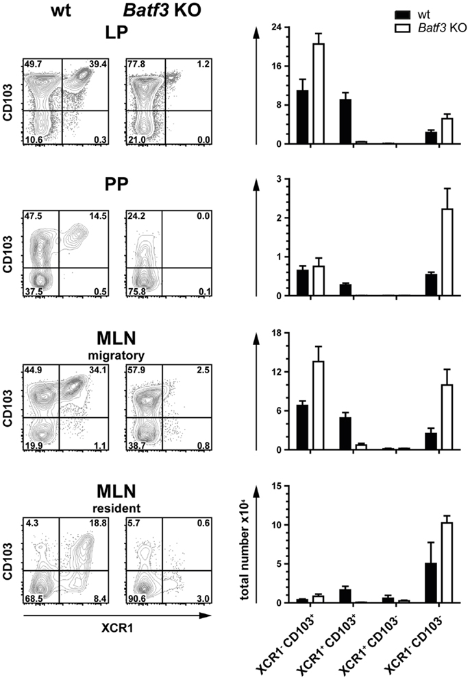
Figure 4. Development of intestinal XCR1+ DC is dependent on the transcription factor Batf3. DC (CD45+ Lin− F4/80− CD11c+ MHCII+) from LP, PP, and MLN cells of C57BL/6 wt controls and Batf3-deficient mice were stained for XCR1 and CD103 (left part of figure). Total numbers of the indicated DC subsets obtained from wt (black bars) and Batf3-deficient mice (open bars) are shown (right part of figure). The results shown are representative of two experiments (mean ± SEM; in each experiment, n = 3).
Positioning of XCR1+ DC in the Lamina Propria, Peyer’s Patches, and Mesenteric Lymph Nodes
Due to the absence of specific markers, an unequivocal localization of DC subsets in lymphoid tissues or organs was very challenging in the past. To overcome these difficulties, we attempted to use the XCR1-specific mAb MARX10 for histological analyses of gut tissues, but this approach gave a high background preventing a clear discrimination from false positive signals. In the next approach, we used B6.XCR1-lacZ+/+ mice, in which both XCR1 genes are replaced by LacZ reporter genes, to localize XCR1+ DC. Histochemical analysis of β-galactosidase activity in the small intestine gave signals in the LP of the villi (Figure 5A). The somewhat smaller multiple signals observed in the epithelial crypts of the LP (Figure 5A, arrows) were consistently also present in control stainings of wt tissues (Figure 5D, arrows) and have to be considered as false positive. In PP, cells with β-galactosidase activity could be found in the T cell zones, with some clustering in the interfollicular region (Figure 5B). Interestingly, no signals for XCR1 were detectable in the subepithelial dome of the PP, where CD11c+ cells are also known to localize (30). In the MLN, XCR1 signals were seen in the T cell zones and apparently in sinuses (Figure 5C), similar to the results obtained with axillary LN earlier (18). The positioning of XCR1+ DC could thus be determined in the absence of a functional XCR1 (which has been replaced by β-galactosidase). Whether the XCL1 ligand/XCR1 receptor axis influences the sublocalization of the XCR1+ DC in intestinal tissues has yet to be analyzed.
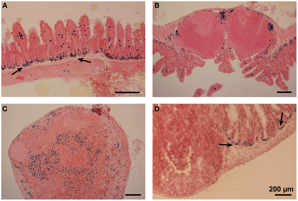
Figure 5. Positioning of XCR1-expressing cells in the lamina propria, Peyer’s patches, and mesenteric lymph nodes. Distribution of XCR1+ cells was determined in tissues of homozygous B6.XCR1-lacZ+/+ reporter mice using X-gal, a chromogenic substrate for β-galactosidase. (A) Lamina propria of the small intestine, (B) Peyer’s patch, and (C) mesenteric lymph node. (D) Represents a staining control using intestinal tissue from wt C57BL/6 mice containing a Peyer’s patch and adjacent portions of the small intestine. The arrows in (A,D) indicate false positive signals obtained in the epithelial crypts of the lamina propria.
No Apparent Involvement of the Chemokine Receptor XCR1 in the Migration of DC to Mesenteric Lymph Nodes
In steady state, intestinal DC constantly migrate from the gut to the MLN in a CCR7-dependent fashion (31, 32), and this migration is further increased under inflammatory conditions (9, 33, 34). Since XCR1 is also a chemokine receptor, we sought to determine any involvement of XCR1 in the migration of DC from the gut to the MLN, where the ligand XCL1 is secreted by NK cells at steady state and at high levels by activated CD8+ T cells, NK cells, and NKT cells (35, 36) (own unpublished data). In the first step, expression of XCR1 was correlated with CCR7 under various conditions. At steady state, CCR7 could not be detected on DC in the LP, PP, or resident DC in the MLN, but could be found on the majority of migratory DC (Figure 6). After intraperitoneal (i.p.) administration of LPS, CCR7 became detectable on around 70% of DC in the PP, but not on LP DC. Under inflammatory conditions, CCR7 was also present on 50–60% of MLN DC, which, due to their uniformly increased levels of MHCII, no longer could be subdivided into resident or migratory DC. In all instances in which CCR7 was detected, essentially all XCR1+ DC co-expressed CCR7, suggesting that XCR1 could be involved in the migration of XCR1+ DC into the MLN. We, therefore, performed a series of analyses comparing wt mice with mice deficient for XCL1 (18), the unique chemokine ligand of XCR1. In these experiments, absence of XCL1 did not change the relative representation of the various DC populations in the MLN at steady state or under inflammatory conditions, or their CCR7 expression (own unpublished data). The same observation was made with mice deficient for XCR1 (own unpublished data). These functional experiments largely excluded an involvement of the XCL1–XCR1 axis in the immigration of DC into the MLN.
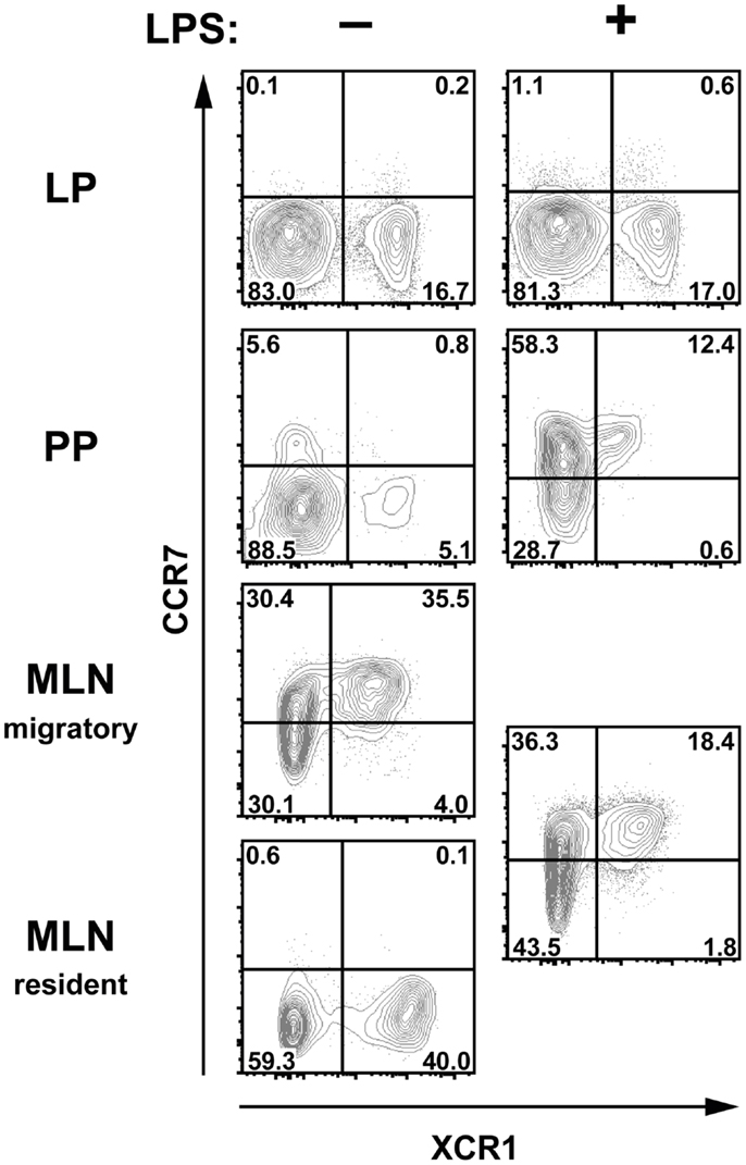
Figure 6. XCR1-expressing cells upregulate CCR7 after inflammation in Peyer’s patches and MLN. C57BL/6 mice were injected i.p. with LPS or with PBS for control, and the expression of CCR7 and XCR1 was compared 14 h later on DC (CD45+ Lin− F4/80− CD11c+ MHCII+ cells) in LP, PP, and MLN. Shown is one representative experiment out of three.
XCR1+ Migratory DC Excel in Cross-Presentation of Orally Applied Antigen
In order to test the ability of various intestinal DC populations to cross-present orally applied antigen, mice were fed with 25 mg of soluble OVA, sacrificed 17 h later, and the various intestinal DC subsets isolated to high purity (>98.5%). The DC subsets were then co-cultured at various ratios with OT-I T cells to test their capacity to cross-present the OVA-derived peptide SIINFEKL to CD8+ T cells. When the percentage of proliferating OT-I T cells was determined after 2.5 days of culture, MLN migratory XCR1+ CD103+ DC performed best in activating OT-I T cells in five out of five experiments, while migratory XCR1− DC, irrespective of their CD103 expression, were less effective (Figure 7). Resident MLN DC also had a low stimulatory effect on OT-I T cells (Figure 7). Interestingly, when LP DC subsets were tested in the same experiments and thus directly compared with MLN DC, essentially no proliferation of OT-I T cells was seen (Figure 7). Since these LP DC were fully capable to activate OT-I T cells when in vitro loaded with the peptide SIINFEKL, these results suggested that LP DC did not present sufficient antigen to activate OT-I T cells. Taken together, in all conditions, in which substantial cross-presentation could be observed, migratory XCR1+ DC outperformed all other DC populations in the cross-presentation of orally applied antigen.
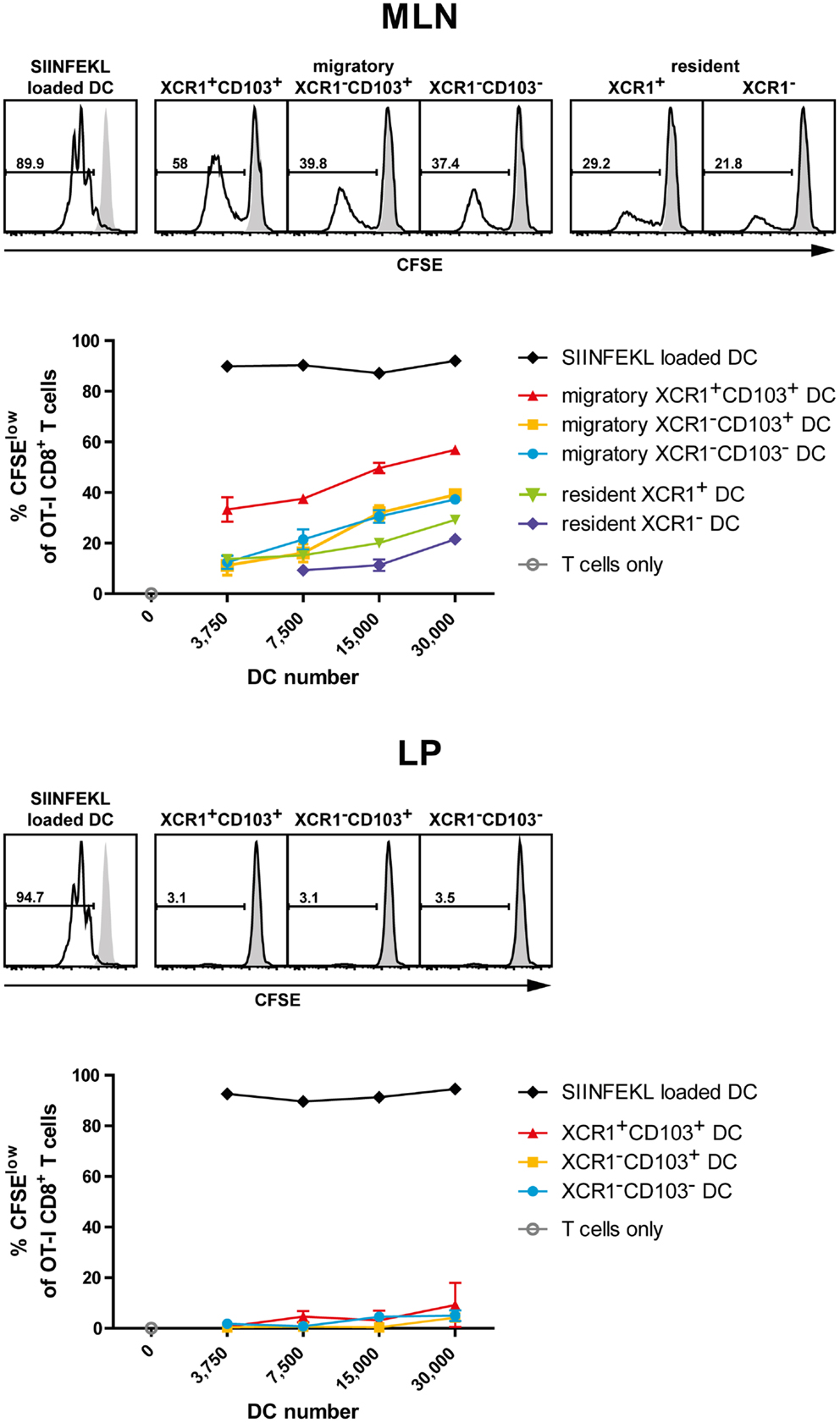
Figure 7. Intestinal XCR1+ migratory DC excel in cross-presentation of soluble antigen. C57BL/6 mice were fed with soluble OVA (25 mg), sacrificed 17 h later, and CD11c+ cells were enriched from MLN and LP by density gradient centrifugation and positive magnetic separation. The indicated DC subsets were flow sorted according to their expression of XCR1 and CD103 to purity (>98.5%). Titrated numbers of the respective DC subsets from MLN or LP were then co-cultured with 1 × 10⁵ CFSE-labeled OT-I T cells, DC loaded with SIINFEKL peptide in vitro served as positive controls. Shown is the CFSE dilution profile of the OT-I T cells (CD90.1+ CD8+) after 2.5 days of co-culture with the respective DC subsets (open histograms) or OT-I T cells alone (gray histograms). The results shown are representative of three experiments with LP and MLN, and two additional experiments with MLN only (mean ± SD).
Discussion
We have previously extensively characterized splenic XCR1+ DC using a variety of approaches. All results obtained were compatible with the notion that the surface expression of the chemokine receptor XCR1 characterizes the Batf3-dependent lineage of cross-presenting DC (21). In the present work, we sought to determine whether this finding can be extended to the intestinal immune system.
Several levels of evidence indicate that XCR1+ DC, also in the intestine, are a separate lineage of DC with a consistent phenotype and a special ability to cross-present antigen. First of all, in animals deficient for the TF Batf3 only XCR1+ DC were consistently absent, while DC bearing other surface markers were preserved. In particular, XCR1+ CD103+ and XCR1+ CD103− DC were absent, while XCR1− CD103+ and XCR1− CD103− DC remained present. Thus, in Batf3-deficient animals, only the XCR1+ DC population failed to differentiate from precursors, while the development of XCR1− DC remained intact.
Second, expression of XCR1 was in all instances closely correlated with the expression of CD8α and Clec9A/DNGR-1 on DC in all intestinal compartments and the draining MLN. Both CD8α and Clec9A/DNGR-1 have been identified in the past as integral parts of the gene expression programs of splenic cross-presenting DC and their putative correlates in the periphery (20). This high correlation between expression of XCR1, Clec9A/DNGR-1, and CD8α in various lymphoid tissues strongly suggests a cooperation of these molecules in the specific function of cross-presenting DC. While the contribution of CD8α in this process remains unclear, XCR1 functions as the only receptor for XCL1 (37), a chemokine mainly released by innate cells (NK, NKT) and CD8+ T cells (35, 36), and thus cells involved in the cytotoxic response, and more generally, in the type 1 immune defense. Clec9A/DNGR-1 has been described as a receptor of actin filaments expressed by necrotic cells, and is thought to contribute to the uptake of damaged cells into cross-presenting DC (38, 39). In spite of this close correlation between XCR1, CD8α, and Clec9A/DNGR-1 on (intestinal) DC, it has to be stated that only XCR1 is specifically expressed on conventional (intestinal) DC, while Clec9A/DNGR-1 is also present on plasmacytoid DC (pDC) (27, 40), and CD8α on pDC, T cells, NKT cells, and other cells.
Third, our data show that XCR1+ DC excel in cross-presentation also in the intestinal immune system. Only very few cross-presentation experiments have been performed with intestinal DC in the past, in particular, after application of antigen via the oral route. Chung et al. (41) reported that only CD11c+ DC isolated from the MLN were capable to cross-present orally applied OVA. At the same time, they somewhat surprisingly found that CD8− CD11b+ DC (i.e., XCR1−) rather than CD8+ DC (i.e., largely XCR1+) cross-present intestinal Ag. When Jaensson et al. (42) re-investigated this issue, only CD103+ DC isolated from MLN after oral administration of OVA induced proliferation of OT-I T cells in vitro. However, these authors did not further discriminate between CD103+ CD11b− (“CD8α-like,” “Batf3-dependent”) and CD103+ CD11b+ (“Batf3-independent”) DC in their study, and therefore their results, from today’s perspective, did not offer the required DC subset resolution. In our work, we have separately tested LP and both migratory and resident MLN DC, and also subdivided these populations into XCR1+ and XCR1− DC. DC originating from the LP usually did not give a signal in the cross-presentation assay; possibly the proportion of DC, which have taken up antigen was too low, or the isolation procedure introduced a bias into the DC population. When analyzing MLN populations, the migratory DC clearly outperformed the resident DC, which still gave a low signal. Within the migratory DC population, the XCR1+ DC consistently performed best in the cross-presentation of orally applied soluble antigen, but a lower signal was also obtained with XCR1− DC. These results were thus comparable to the data we have obtained earlier with splenic DC subsets. There, XCR1+ DC performed best, but not in a “unique fashion,” in the cross-presentation of soluble antigen (applied intravenously), but were “unique” in the cross-presentation of cell-bound antigen (21). We did not have the opportunity to test the uptake and presentation of cell-bound antigen in our current work on intestinal DC. Cerovic et al. (43) very recently published a study employing a transgenic mouse in which only intestinal epithelial cells (IEC) express non-secretable antigen. Using this model system, they could demonstrate that only CD103+ CD11b− DC (i.e., XCR1+ DC) migrating from the intestinal system, but not any other subset, were capable to cross-present this cell-associated antigen. Thus, combining the results of Cerovic et al. on cell-bound antigen with our own results on the presentation of orally applied antigen, one can clearly conclude that XCR1+/CD103+ CD11b− are the population of DC, which excels in antigen cross-presentation in the intestinal immune system. This stated, one should bear in mind that these two classification systems define largely overlapping, but not fully congruent DC populations; essentially all CD103+ CD11b− DC bear the XCR1 receptor, but XCR1 is also expressed on 10–15% of other DC in the CD103/CD11b classification system (compare Figure 1).
The specific expression of XCR1 allowed us to determine the anatomical localization of cross-presenting DC in the intestinal system. In the small intestine, cells expressing XCR1 were found in the LP of the villi. In PP, XCR1+ DC mainly clustered in the interfollicular region, and were absent from the subepithelial dome. The latter results are congruent with the findings of Iwasaki et al. (44), who, based on double-fluorescence studies using CD11c and CD8α as markers, described the presence of CD8α DC in the interfollicular region. Interestingly, XCR1+ signals were absent in the subepithelial dome of the PP, where CD11c+ CD11b+ cells can be found instead (44). This anatomical separation in the PP indicates a division of labor of DC subsets in this lymphoid organ, which at present is not fully understood. Finally, the distribution of XCR1+ DC in the draining MLN followed the pattern obtained with peripheral LN obtained earlier (18), with signals present in T cell zones and apparently sinuses, where incoming (cellular) material in the draining lymph can be taken up by DC. One should bear in mind that all of our results were obtained with homozygous B6.XCR1-lacZ+/+ mice lacking XCR1, so they cannot reflect any potential influence of locally secreted XCL1 on the sublocalization of XCR1+ in intestinal tissues. This aspect needs further examination in the future.
Our results on the phenotype, differentiation, function, and localization of DC demonstrate that XCR1+ DC are a homogenous population with a specific function in the intestinal immune system. With this understanding, we sought to determine, whether there are any surface molecules, which would fully demarcate XCR1− DC in a positive fashion. Interestingly, of the many markers tested, there were only two surface molecules, which in all instances and in all tissues were not expressed on XCR1+ DC, namely CD11b and CD172a/SIRPα. When analyzing DC for the expression of these two integrins, it became apparent that all of XCR1− DC express SIRPα, and some of them also express CD11b (compare Figure 2). Thus, only SIRPα showed a stringent anti-correlation with XCR1 on DC in the intestine, strongly suggesting that this surface molecule comprehensively characterizes the XCR1− DC population. Support for this conclusion comes from our studies in the spleen, where the same constellation was found (21), and our preliminary data indicate that this is also true in other body compartments (own unpublished results). Our data are fully compatible with earlier findings demonstrating a rather selective SIRPα expression on CD4+ and DN conventional DC using the older CD4/CD8 DC classification system (45).
SIRPα, an Ig-superfamily transmembrane protein, is in the immune system abundantly expressed on macrophages, DC, and neutrophils; outside of the immune system, it is present on neurons and weakly also on fibroblasts and endothelial cells (46, 47). Although the function of SIRPα is not fully understood, it has been implicated in the control of cell phagocytosis. Cells expressing CD47, the ligand for SIRPα, appear to be protected from engulfment by phagocytic cells (46). It is intriguing to note that Clec9A/DNGR-1 and SIRPα, which on DC are never co-expressed, both regulate cell phagocytosis. This functional feature possibly contributes to the division of labor between the XCR1+ and SIRPα+ DC populations.
Future work will determine whether SIRPα+ DC are a homogenous population or whether they have to be further split up into functional subsets. For practical purposes, it seems attractive now to classify DC in the immune system based on the expression of XCR1 and SIRPα, which greatly simplifies any phenotypical and functional analyses when compared with the DC classification systems currently in use. Figure 8 illustrates such an approach with DC populations from the various compartments of the intestine. Once separated into XCR1+ versus SIRPα+ positive DC, these subpopulations can be further analyzed for expression of other molecules, which are being actively regulated in various tissue compartments, e.g., CD103 (48, 49). It appears likely that this approach for the functional subdivision of DC can universally be applied throughout the murine immune system. Moreover, recent data indicate that a DC classification system based on the expression of XCR1 and SIRPα will also be useful in the human (50) and other species (51).
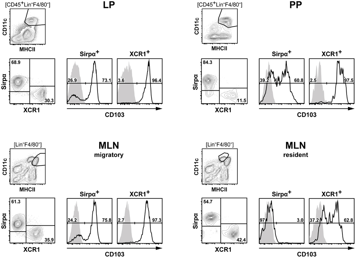
Figure 8. Classification of intestinal DC according to their expression of XCR1 and SIRPα. DC from the LP, PP, and MLN of C57BL/6 mice were enriched by digestion and density gradient centrifugation of the tissues, stained for XCR1 and SIRPα, DC from MLN were further separated into resident and migratory DC based on their MHCII expression levels. For analysis, the gates were set on live CD45+ Lin− F4/80− CD11c+ MHCII+ cells. Expression of CD103 is shown on XCR1+ versus SIRPα+ DC. The background staining was determined with homozygous B6.XCR1-lacZ+/+ mice lacking XCR1 (gray). The results shown are representative of two experiments.
Author Contributions
Martina Becker and Steffen Güttler designed and did experiments, analyzed and interpreted the data, and contributed to the writing of the manuscript; Annabell Bachem, Evelyn Hartung, Ahmed Mora, Anika Jäkel, Andreas Hutloff, Volker Henn, Hans Werner Mages, and Stephanie Gurka provided specialized expertise and/or assisted with some experiments; Richard A. Kroczek conceived the project, interpreted the data, and wrote the manuscript.
Conflict of Interest Statement
The authors declare that the research was conducted in the absence of any commercial or financial relationships that could be construed as a potential conflict of interest.
Acknowledgments
We are grateful to Maria Rescigno and her laboratory for introducing us to the methodology of cell isolation from intestinal tissues compartments. Batf3-deficient mice were kindly provided by H.-C. Probst, Mainz University, CX3CR1GFP mice by M. Gunzer (Magdeburg), and LangerinEGFP mice by P. Stoitzner (Innsbruck). Anti-murine Clec9A/DNGR-1 mAb were kindly provided by M. H. Lahoud and I. Caminschi (Melbourne) and C. Reis e Sousa (London). B16 cells secreting Flt3 ligand were a gift of S. Jung (Rehovot). We thank Reinhard Pabst, Hannover, for discussions on the positioning of XCR1+ DC in the intestinal immune tissues. This work was supported by the Fritz-Thyssen-Foundation, the Wilhelm Sander-Foundation, and the Deutsche Forschungsgemeinschaft (Kr 827/16-1 and 827/18-1).
References
1. Pabst O, Bernhardt G. The puzzle of intestinal lamina propria dendritic cells and macrophages. Eur J Immunol (2010) 40:2107–11. doi: 10.1002/eji.201040557
2. Milling S, Yrlid U, Cerovic V, MacPherson G. Subsets of migrating intestinal dendritic cells. Immunol Rev (2010) 234:259–67. doi:10.1111/j.0105-2896.2009.00866.x
3. Rescigno M. Intestinal dendritic cells. Adv Immunol (2010) 107:109–38. doi:10.1016/B978-0-12-381300-8.00004-6
4. Hashimoto D, Miller J, Merad M. Dendritic cell and macrophage heterogeneity in vivo. Immunity (2011) 35:323–35. doi:10.1016/j.immuni.2011.09.007
5. Mowat AM, Bain CC. Mucosal macrophages in intestinal homeostasis and inflammation. J Innate Immun (2011) 3:550–64. doi:10.1159/000329099
6. Bogunovic M, Mortha A, Muller PA, Merad M. Mononuclear phagocyte diversity in the intestine. Immunol Res (2012) 54:37–49. doi:10.1007/s12026-012-8323-5
7. Annacker O, Coombes JL, Malmstrom V, Uhlig HH, Bourne T, Johansson-Lindbom B, et al. Essential role for CD103 in the T cell-mediated regulation of experimental colitis. J Exp Med (2005) 202:1051–61. doi:10.1084/jem.20040662
8. Johansson-Lindbom B, Svensson M, Pabst O, Palmqvist C, Marquez G, Förster R, et al. Functional specialization of gut CD103+ dendritic cells in the regulation of tissue-selective T cell homing. J Exp Med (2005) 202:1063–73. doi:10.1084/jem.20051100
9. Schulz O, Jaensson E, Persson EK, Liu X, Worbs T, Agace WW, et al. Intestinal CD103+, but not CX3CR1+, antigen sampling cells migrate in lymph and serve classical dendritic cell functions. J Exp Med (2009) 206:3101–14. doi:10.1084/jem.20091925
10. Ohl L, Mohaupt M, Czeloth N, Hintzen G, Kiafard Z, Zwirner J, et al. CCR7 governs skin dendritic cell migration under inflammatory and steady-state conditions. Immunity (2004) 21:279–88. doi:10.1016/j.immuni.2004.06.014
11. den Haan JM, Lehar SM, Bevan MJ. CD8+ but not CD8− dendritic cells cross-prime cytotoxic T cells in vivo. J Exp Med (2000) 192:1685–96. doi:10.1084/jem.192.12.1685
12. Pooley JL, Heath WR, Shortman K. Cutting edge: intravenous soluble antigen is presented to CD4 T cells by CD8− dendritic cells, but cross-presented to CD8 T cells by CD8+ dendritic cells. J Immunol (2001) 166:5327–30. doi:10.4049/jimmunol.166.9.5327
14. Shen L, Rock KL. Priming of T cells by exogenous antigen cross-presented on MHC class I molecules. Curr Opin Immunol (2006) 18:85–91. doi:10.1016/j.coi.2005.11.003
15. Villadangos JA, Heath WR, Carbone FR. Outside looking in: the inner workings of the cross-presentation pathway within dendritic cells. Trends Immunol (2007) 28:45–7. doi:10.1016/j.it.2006.12.008
16. Hildner K, Edelson BT, Purtha WE, Diamond M, Matsushita H, Kohyama M, et al. Batf3 deficiency reveals a critical role for CD8α+ dendritic cells in cytotoxic T cell immunity. Science (2008) 322:1097–100. doi:10.1126/science.1164206
17. Edelson BT, Kc W, Juang R, Kohyama M, Benoit LA, Klekotka PA, et al. Peripheral CD103+ dendritic cells form a unified subset developmentally related to CD8α+ conventional dendritic cells. J Exp Med (2010) 207:823–36. doi:10.1084/jem.20091627
18. Dorner BG, Dorner MB, Zhou X, Opitz C, Mora A, Güttler S, et al. Selective expression of the chemokine receptor XCR1 on cross-presenting dendritic cells determines cooperation with CD8+ T cells. Immunity (2009) 31:823–33. doi:10.1016/j.immuni.2009.08.027
19. Bachem A, Güttler S, Hartung E, Ebstein F, Schaefer M, Tannert A, et al. Superior antigen cross-presentation and XCR1 expression define human CD11c+CD141+ cells as homologues of mouse CD8+ dendritic cells. J Exp Med (2010) 207:1273–81. doi:10.1084/jem.20100348
20. Crozat K, Guiton R, Contreras V, Feuillet V, Dutertre CA, Ventre E, et al. The XC chemokine receptor 1 is a conserved selective marker of mammalian cells homologous to mouse CD8α+ dendritic cells. J Exp Med (2010) 207:1283–92. doi:10.1084/jem.20100223
21. Bachem A, Hartung E, Güttler S, Mora A, Zhou X, Hegemann A, et al. Expression of XCR1 characterizes the Batf3-dependent lineage of dendritic cells capable of antigen cross-presentation. Front Immunol (2012) 3:214. doi:10.3389/fimmu.2012.00214
22. Crozat K, Tamoutounour S, Vu Manh TP, Fossum E, Luche H, Ardouin L, et al. Cutting edge: expression of XCR1 defines mouse lymphoid-tissue resident and migratory dendritic cells of the CD8α+ type. J Immunol (2011) 187:4411–5. doi:10.4049/jimmunol.1101717
23. Bar-On L, Birnberg T, Lewis KL, Edelson BT, Bruder D, Hildner K, et al. CX3CR1+ CD8α+ dendritic cells are a steady-state population related to plasmacytoid dendritic cells. Proc Natl Acad Sci U S A (2010) 107:14745–50. doi:10.1073/pnas.1001562107
24. Jung S, Aliberti J, Graemmel P, Sunshine MJ, Kreutzberg GW, Sher A, et al. Analysis of fractalkine receptor CX3CR1 function by targeted deletion and green fluorescent protein reporter gene insertion. Mol Cell Biol (2000) 20:4106–14. doi:10.1128/MCB.20.11.4106-4114.2000
25. Kissenpfennig A, Ait-Yahia S, Clair-Moninot V, Stossel H, Badell E, Bordat Y, et al. Disruption of the langerin/CD207 gene abolishes Birbeck granules without a marked loss of Langerhans cell function. Mol Cell Biol (2005) 25:88–99. doi:10.1128/MCB.25.1.88-99.2005
26. Mach N, Gillessen S, Wilson SB, Sheehan C, Mihm M, Dranoff G. Differences in dendritic cells stimulated in vivo by tumors engineered to secrete granulocyte-macrophage colony-stimulating factor or Flt3-ligand. Cancer Res (2000) 60:3239–46.
27. Caminschi I, Proietto AI, Ahmet F, Kitsoulis S, Shin TJ, Lo JC, et al. The dendritic cell subtype-restricted C-type lectin Clec9A is a target for vaccine enhancement. Blood (2008) 112:3264–73. doi:10.1182/blood-2008-05-155176
28. Sanes JR, Rubenstein JL, Nicolas JF. Use of a recombinant retrovirus to study post-implantation cell lineage in mouse embryos. EMBO J (1986) 5:3133–42.
29. Maraskovsky E, Brasel K, Teepe M, Roux ER, Lyman SD, Shortman K, et al. Dramatic increase in the numbers of functionally mature dendritic cells in Flt3 ligand-treated mice: multiple dendritic cell subpopulations identified. J Exp Med (1996) 184:1953–62. doi:10.1084/jem.184.5.1953
30. Kelsall BL, Strober W. Distinct populations of dendritic cells are present in the subepithelial dome and T cell regions of the murine Peyer’s patch. J Exp Med (1996) 183:237–47. doi:10.1084/jem.183.1.237
31. Jang MH, Sougawa N, Tanaka T, Hirata T, Hiroi T, Tohya K, et al. CCR7 is critically important for migration of dendritic cells in intestinal lamina propria to mesenteric lymph nodes. J Immunol (2006) 176:803–10. doi:10.4049/jimmunol.176.2.803
32. Worbs T, Bode U, Yan S, Hoffmann MW, Hintzen G, Bernhardt G, et al. Oral tolerance originates in the intestinal immune system and relies on antigen carriage by dendritic cells. J Exp Med (2006) 203:519–27. doi:10.1084/jem.20052016
33. MacPherson GG, Jenkins CD, Stein MJ, Edwards C. Endotoxin-mediated dendritic cell release from the intestine. Characterization of released dendritic cells and TNF dependence. J Immunol (1995) 154:1317–22.
34. Turnbull EL, Yrlid U, Jenkins CD, Macpherson GG. Intestinal dendritic cell subsets: differential effects of systemic TLR4 stimulation on migratory fate and activation in vivo. J Immunol (2005) 174:1374–84. doi:10.4049/jimmunol.174.3.1374
35. Dorner BG, Scheffold A, Rolph MS, Hüser MB, Kaufmann SH, Radbruch A, et al. MIP-1α, MIP-1β, RANTES, and ATAC/lymphotactin function together with IFN-γ as type 1 cytokines. Proc Natl Acad Sci U S A (2002) 99:6181–6. doi:10.1073/pnas.092141999
36. Dorner BG, Smith HR, French AR, Kim S, Poursine-Laurent J, Beckman DL, et al. Coordinate expression of cytokines and chemokines by NK cells during murine cytomegalovirus infection. J Immunol (2004) 172:3119–31. doi:10.4049/jimmunol.172.5.3119
37. Yoshida T, Imai T, Kakizaki M, Nishimura M, Takagi S, Yoshie O. Identification of single C motif-1/lymphotactin receptor XCR1. J Biol Chem (1998) 273:16551–4. doi:10.1074/jbc.273.26.16551
38. Ahrens S, Zelenay S, Sancho D, Hanc P, Kjaer S, Feest C, et al. F-actin is an evolutionarily conserved damage-associated molecular pattern recognized by DNGR-1, a receptor for dead cells. Immunity (2012) 36:635–45. doi:10.1016/j.immuni.2012.03.008
39. Zhang JG, Czabotar PE, Policheni AN, Caminschi I, San Wan S, Kitsoulis S, et al. The dendritic cell receptor Clec9A binds damaged cells via exposed actin filaments. Immunity (2012) 36:646–57. doi:10.1016/j.immuni.2012.03.009
40. Sancho D, Mourao-Sa D, Joffre OP, Schulz O, Rogers NC, Pennington DJ, et al. Tumor therapy in mice via antigen targeting to a novel, DC-restricted C-type lectin. J Clin Invest (2008) 118:2098–110. doi:10.1172/JCI34584
41. Chung Y, Chang JH, Kweon MN, Rennert PD, Kang CY. CD8α−11b+ dendritic cells but not CD8α+ dendritic cells mediate cross-tolerance toward intestinal antigens. Blood (2005) 106:201–6. doi:10.1182/blood-2004-11-4240
42. Jaensson E, Uronen-Hansson H, Pabst O, Eksteen B, Tian J, Coombes JL, et al. Small intestinal CD103+ dendritic cells display unique functional properties that are conserved between mice and humans. J Exp Med (2008) 205:2139–49. doi:10.1084/jem.20080414
43. Cerovic V, Houston SA, Westlund J, Utriainen L, Davison ES, Scott CL, et al. Lymph-borne CD8α+ dendritic cells are uniquely able to cross-prime CD8+ T cells with antigen acquired from intestinal epithelial cells. Mucosal Immunol (2014). doi:10.1038/mi.2014.40
44. Iwasaki A, Kelsall BL. Localization of distinct Peyer’s patch dendritic cell subsets and their recruitment by chemokines macrophage inflammatory protein (MIP)-3α, MIP-3β, and secondary lymphoid organ chemokine. J Exp Med (2000) 191:1381–94. doi:10.1084/jem.191.8.1381
45. Lahoud MH, Proietto AI, Gartlan KH, Kitsoulis S, Curtis J, Wettenhall J, et al. Signal regulatory protein molecules are differentially expressed by CD8− dendritic cells. J Immunol (2006) 177:372–82. doi:10.4049/jimmunol.177.1.372
46. Matozaki T, Murata Y, Okazawa H, Ohnishi H. Functions and molecular mechanisms of the CD47-SIRPα signalling pathway. Trends Cell Biol (2009) 19:72–80. doi:10.1016/j.tcb.2008.12.001
47. Nuvolone M, Kana V, Hutter G, Sakata D, Mortin-Toth SM, Russo G, et al. SIRPα polymorphisms, but not the prion protein, control phagocytosis of apoptotic cells. J Exp Med (2013) 210:2539–52. doi:10.1084/jem.20131274
48. Sathe P, Pooley J, Vremec D, Mintern J, Jin JO, Wu L, et al. The acquisition of antigen cross-presentation function by newly formed dendritic cells. J Immunol (2011) 186:5184–92. doi:10.4049/jimmunol.1002683
49. Zhan Y, Carrington EM, van Nieuwenhuijze A, Bedoui S, Seah S, Xu Y, et al. GM-CSF increases cross presentation and CD103 expression by mouse CD8+ spleen dendritic cells. Eur J Immunol (2011) 41:2585–95. doi:10.1002/eji.201141540
50. Watchmaker PB, Lahl K, Lee M, Baumjohann D, Morton J, Kim SJ, et al. Comparative transcriptional and functional profiling defines conserved programs of intestinal DC differentiation in humans and mice. Nat Immunol (2014) 15:98–108. doi:10.1038/ni.2768
Keywords: dendritic cells, XCR1, Batf3, SIRPα, cross-presentation
Citation: Becker M, Güttler S, Bachem A, Hartung E, Mora A, Jäkel A, Hutloff A, Henn V, Mages HW, Gurka S and Kroczek RA (2014) Ontogenic, phenotypic, and functional characterization of XCR1+ dendritic cells leads to a consistent classification of intestinal dendritic cells based on the expression of XCR1 and SIRPα. Front. Immunol. 5:326. doi: 10.3389/fimmu.2014.00326
Received: 29 April 2014; Accepted: 27 June 2014;
Published online: 28 July 2014.
Edited by:
Linda Sylvia Klavinskis, Kings College London, UKReviewed by:
Anne Hosmalin, Cochin Institute, FranceMarc Dalod, Centre National de la Recherche Scientifique, France
Pierre Guermonprez, King’s College London, UK
Copyright: © 2014 Becker, Güttler, Bachem, Hartung, Mora, Jäkel, Hutloff, Henn, Mages, Gurka and Kroczek. This is an open-access article distributed under the terms of the Creative Commons Attribution License (CC BY). The use, distribution or reproduction in other forums is permitted, provided the original author(s) or licensor are credited and that the original publication in this journal is cited, in accordance with accepted academic practice. No use, distribution or reproduction is permitted which does not comply with these terms.
*Correspondence: Richard A. Kroczek, Molecular Immunology, Robert Koch-Institute, Nordufer 20, Berlin 13353, Germany e-mail: kroczek@rki.de
†Present address: Steffen Güttler, TaconicArtemis, Cologne, Germany;
Ahmed Mora, Chemistry Department, Al-Azhar University, Cairo, Egypt
‡Martina Becker and Steffen Güttler have contributed equally to this work.