Article

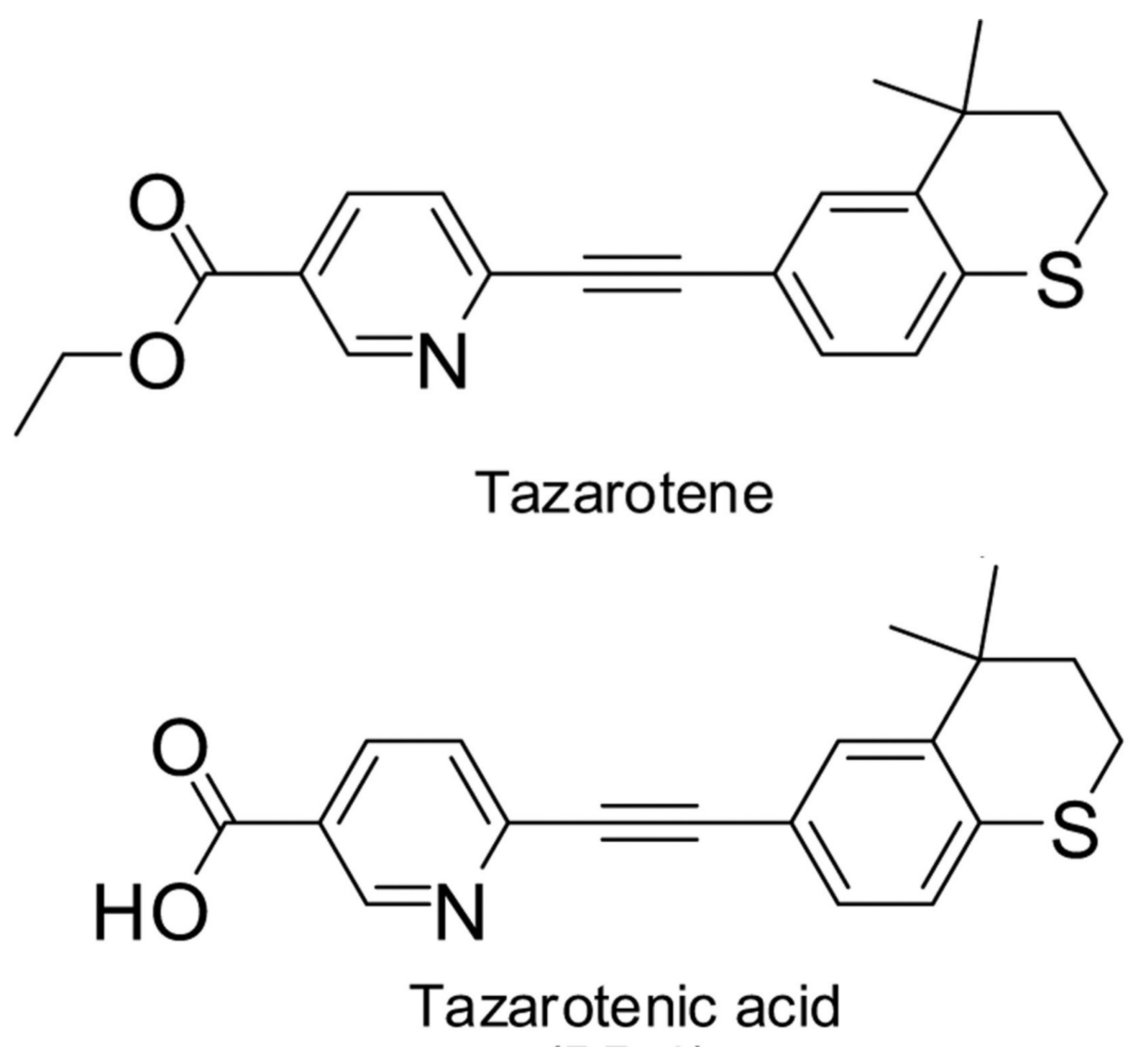
Controlled Release of Tazarotene from Magnetically Responsive Nanofiber Patch: Towards More Efficient Topical Therapy of Psoriasis

by
Natália Andrýsková
1,
Paul Sourivong
2,
Melánia Babincová
3,* and
Mária Šimaljaková
1
1
Department of Dermatovenereology, Faculty of Medicine, Comenius University and Bratislava University Hospital, Mickiewiczova 13, 813 69 Bratislava, Slovakia
2
Houston Methodist San Jacinto Hospital, Department of Radiation Oncology, 4021 Garth Road, Suite 105, Baytown, TX 77521, USA
3
Department of Nuclear Physics and Biophysics, Faculty of Mathematics, Physics and Informatics, Comenius University, Mlynská dolina F1, 842 48 Bratislava, Slovakia
*
Author to whom correspondence should be addressed.
Appl. Sci. 2021, 11(22), 11022; https://doi.org/10.3390/app112211022
Submission received: 3 November 2021 / Revised: 17 November 2021 / Accepted: 19 November 2021 / Published: 21 November 2021

Abstract

:
Electrospun polycaprolactone nanofibers with embedded magnetic nanoparticles were developed for use in the topical delivery of antipsoriatic drugs. To test a hydrophobic drug, a tazarotene has been used, which is an efficient retinoid derivative. Such a smart hyperthermia nanofiber system with self-generated heat from the incorporated magnetic nanoparticles induced drug release in response to on–off switching of alternating magnetic fields for the delivery of tazarotene through the skin, as quantified using Franz cells. This highly efficient external field-controllable system with minimal skin irritation could create a new avenue for the topical therapy of psoriasis.

1. Introduction

Psoriasis is a hereditary, chronic, and recurrent immune-based disease, which is characterized by the incomplete maturation and differentiation of skin cells. It affects about 2–4% of the European population. It occurs in the same percentage in women as in men. Morbidity varies based on geographical and ethnic conditions. The incidence of psoriasis is higher in the Nordic countries of Europe than in the southern countries. Psoriasis is practically absent in Inuit and South American Indian peoples. The etiopathogenesis of psoriasis is not yet fully understood. The genetic component plays an important role. Inheritance is polygenic. Family history is confirmed in approximately 30% of cases. Exogenous and endogenous factors play a significant role in the development of clinical manifestations of psoriasis, as they can induce a clinical manifestation of psoriasis in patients with a latent form of the disease. These factors include infections, inflammatory skin diseases, hormonal effects, poor lifestyle, physical and chemical effects, and other serious general diseases. In addition, commonly used drugs can trigger the disease, which include, for example: beta-blockers, non-steroidal anti-inflammatory drugs, ACE inhibitors, antimalarials, and lithium. Psoriasis treatment can be divided into topical and systemic types of treatment. Topical therapy is used by up to 90% of patients with psoriasis. The first step of treatment is to remove the scales by utilizing the keratolytic effect of salicylic acid. After the removal of the scales, corticosteroids in the form of solutions are most often used in topical therapy as creams and ointments. They have a non-specific anti-inflammatory, immunomodulatory, antiproliferative, and vasoconstrictive effects. The disadvantage of corticosteroids in long-term application is skin atrophy, hypertrichosis, and telangiectasia. The disease can become unstable and more difficult to manage. Other widely used topical antipsoriatic drugs include anthralin, coal tar, analogues of D3 vitamin, and retinoids. Local retinoids react with the nuclear receptors of cells and affect the transcription of genes. They have antiphlogistic, immunomodulatory, antiproliferative, and differentiating effects on keratinocytes, lymphocytes, and sebocytes [1]. the most popular topical therapy is tazarotene [2,3,4,5,6,7,8,9,10,11,12,13,14,15,16,17], which, chemically, is ethyl-[2-(4,4-dimethylthiochroman-6-yl)-ethynyl] nicotinate. Tazarotene is in organism rapidly hydrolysed by esterases to a biologically active form—tazarotenic acid 6-[2-(4,4-dimethylthiochroman-6-yl)ethynyl] nicotinic acid (Figure 1). Tazarotene is a retinoic-acid-specific receptor such as RARβ and RARγ, downregulating markers of keratinocyte differentiation and proliferation, as well as inflammation. The drug also upregulates three genes, TIG-1 (tazarotene-induced gene-1), TIG-2, and TIG-3, which can mediate antiproliferative activity.
For therapeutic purposes, several formulations (gel, cream, aerosol, and foam) of tazarotene are commonly used. As has been found recently, the most useful forms are microemulsions containing surfactant, as they display suitable skin permeation rates and skin uptake, which are appreciated in dermatology due to their transparency, stability, and minimal energy consumption and simplicity of preparation due to their spontaneous formation when oil, water, and surfactant are mixed together [18,19].
Nanotechnology, although only recently emerged, has already provided considerable opportunities to address the limitations of traditional drug delivery systems’ mechanisms, as well as with regard to biotechnology, biomedicine, and other fields. Nanocarriers with the appropriate characteristics can effectively target and release drugs, including retinoids, at the desired location [20]. Recently, we developed a smart hyperthermia nanofiber system with self-generated heat from the incorporated magnetic nanoparticles, which induced drug release in response to on–off switching of alternating magnetic fields for the delivery of water soluble antipsoriatics, e.g., methotrexate [21]. An electrospinning method was employed to fabricate the mesh. The treatment of psoriasis is usually prolonged (weeks or months) and may lead to adverse reactions such as pruritus, burning, and erythema. Low solubility limits tazarotene incorporation into drug carriers and often results in poor compliance in patients. Therefore, we investigated the possibility of a dressing for topical application prepared via the electrospinning of nanofibers. This dressing has the desired skin targeting properties that may reduce the occurrence of adverse reactions and biomimetic scaffolds that mimic the natural hierarchical structure of tissues, which may be used for a prolonged time, as has been already shown through the use of tretinoin-loaded polycaprolactone nanofibers as a potential anti-acne patch [22], antibacterial poly(ethylene oxide) nanofibers containing cinnamon [23], or 5-fluorouracil-loaded poly(vinyl alcohol) nanofibers for the treatment of skin cancer [24]. In this study, we address a more challenging task, namely the delivery of a drug with poor water solubility, tazarotene, using a nanofiber patch made of hydrophobic polycaprolactone polymer (Figure 2), an FDA-approved, biocompatible, and non-immunogenic semi-crystalline polymer with good biodegradability.

2. Materials and Methods
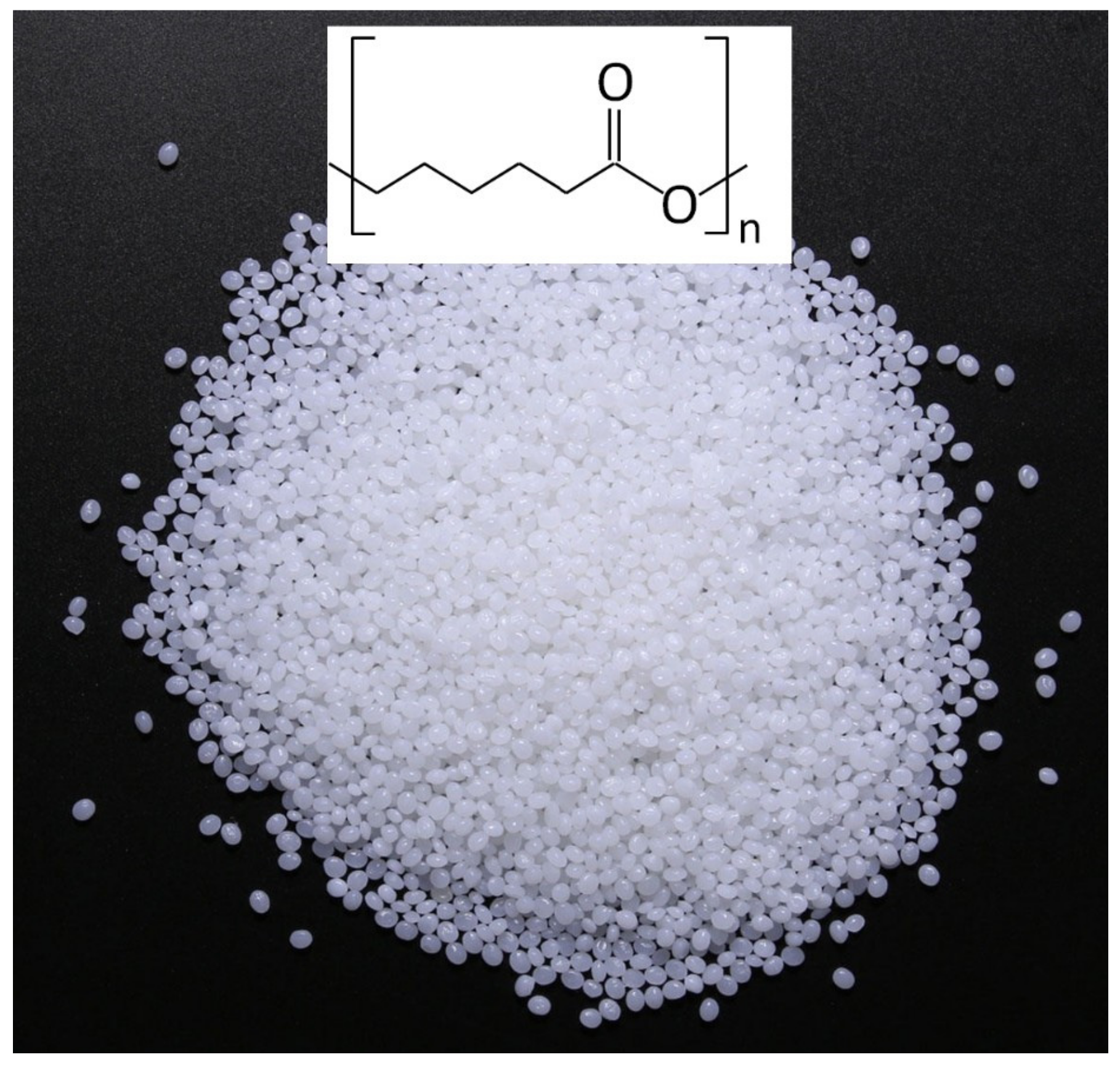
2.1. Materials

Polycaprolactone (PCL) (Mw = 80,000), tazarotene (99%), 1,1,1,2,2,2-hexafluoro-2-propanol (HFIP), and, if not otherwise stated, all other chemicals and material were obtained from Sigma-Aldrich company (Saint Louis, MO, USA). Franz cells were purchased from Across Barriers GmbH (Saarbruecken, Germany). XenoSkin H—Ex vivo dermatomed skin discs for permeation experiments using Franz cells—were obtained from Xenometrics AG (Allschwil, Switzerland). PCL-stabilized magnetic nanoparticles were prepared according to [25].

2.2. Electrospinning Process

Tazarotene (20 mg) and PCL (0.8 g) were dissolved in 10 mL of HFIP to obtain 8% (w/v) drug-mixed solution for use in spinning the drug-loaded fibrous patch. The mixed tazarotene solution, various amounts of PCL-stabilized magnetic nanoparticles, and the mixture were then stirred overnight in the dark (due to the photo-instability of tazarotene). This mixture filled a 10 mL syringe and was inserted with a 20 G stainless steel blunt needle. A Terumo TE-331 syringe pump (Terumo, Shibuya City, Tokyo, Japan) at a rate of flow 0.5 mL/h expelled the mixture from a syringe needle to which a voltage of 12 kV was connected; the other pole was attached to aluminum foil located 15 cm from the needle, or the nanofibers were deposited directly on the skin (Figure 3).

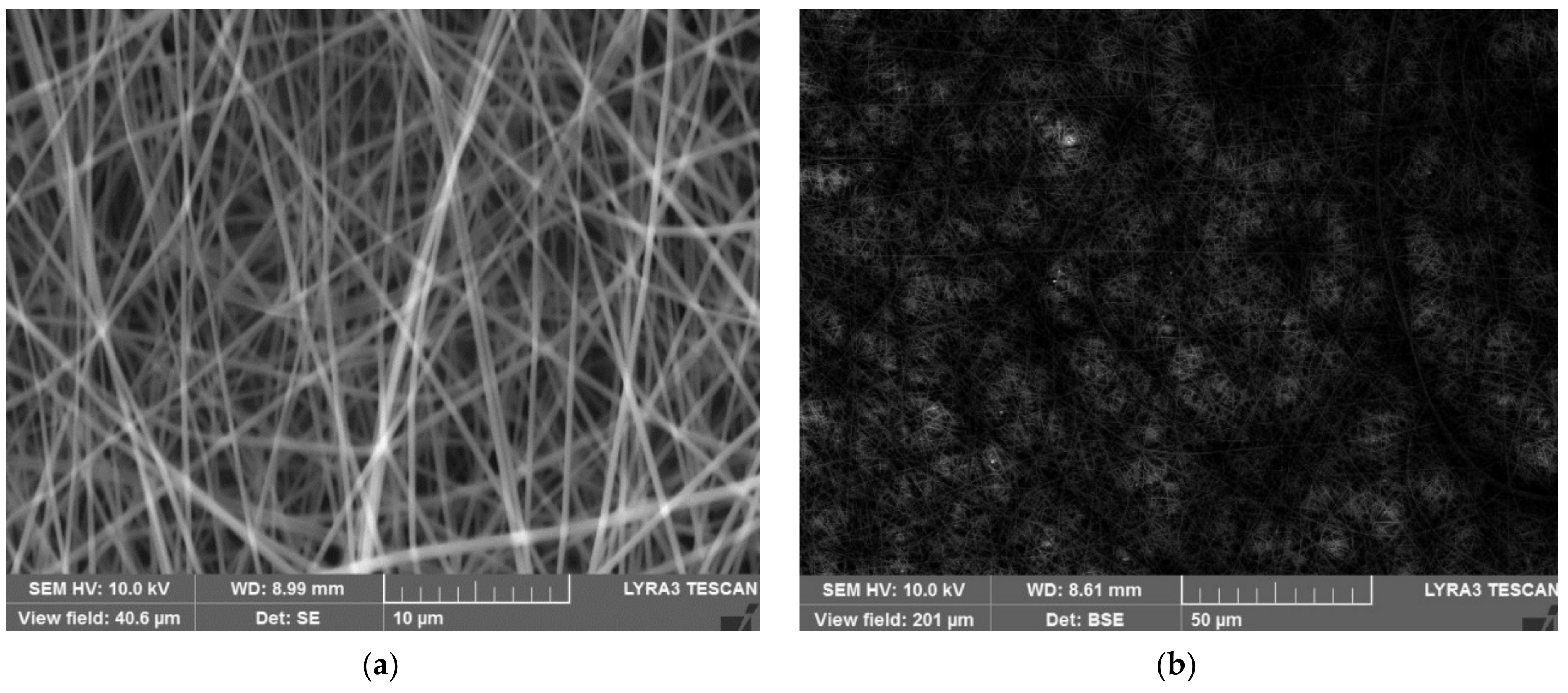
2.3. Structural Characterization of Nanofibers

Scanning electron microscopy (SEM) combines a large depth of field, high contrast, and spatial resolution to characterize nanofiber structures. Electron microscope images were taken on a Tescan Lyra3 (Brno, Czech Republic) SEM microscope. Since the prepared nanofibers were not conductive, they had to be metallized first, so a 20 nm layer of a mixture of platinum and gold (20/80) was applied to all fibers. Subsequently, samples were measured in the secondary electron emission mode in the working depth mode with at a distance of 15 mm and a voltage of 10–30 kV.

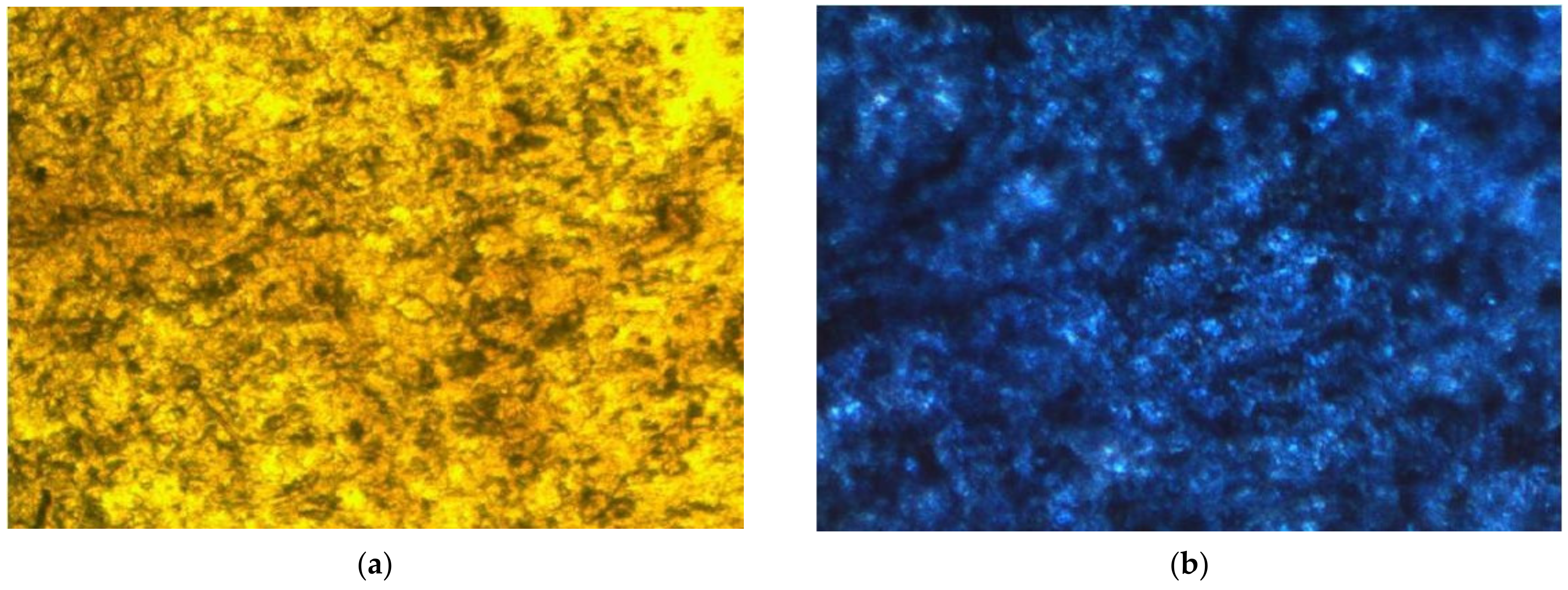
2.4. Colorimetric Assay of Iron

When drop of potassium ferrocyanide was added to a nanofiber patch containing ferric ions (Fe3+), insoluble crystals of with blue pigment (so-called Prussian blue) were formed.

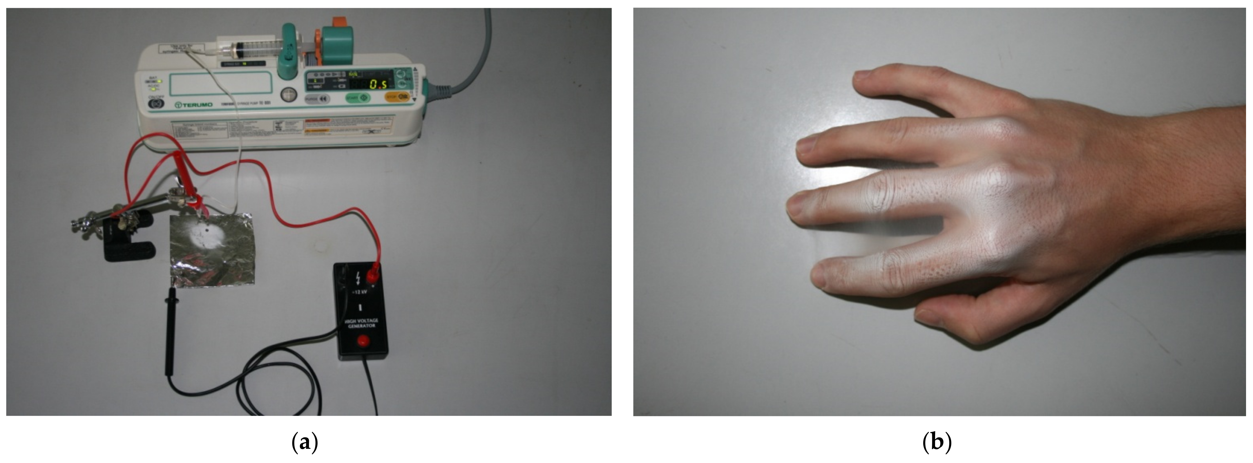
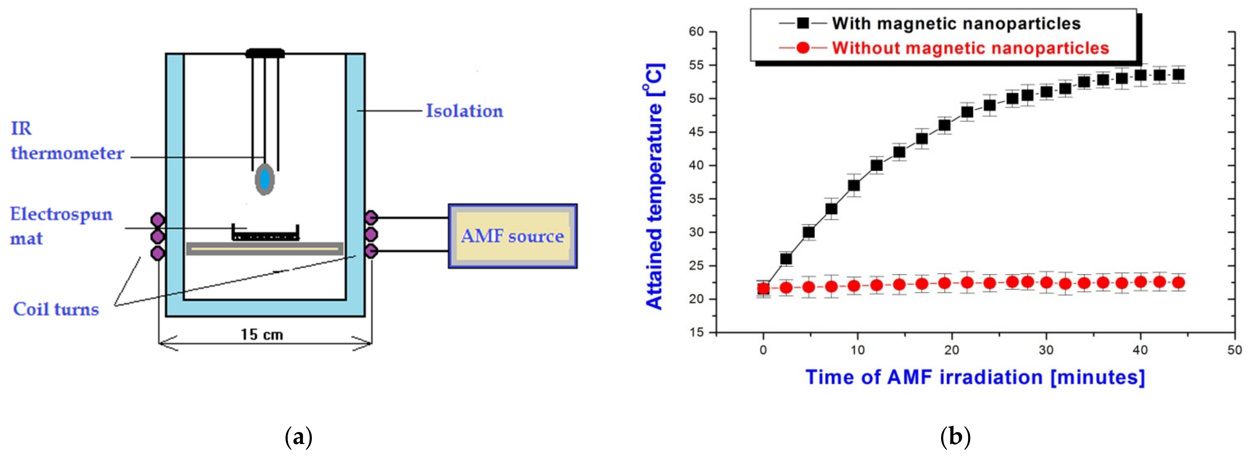
2.5. Application of Alternating Magnetic Field

When measuring the high-frequency heating of the samples, we used the apparatus based on the high-frequency generator GV6A (Závody elektrotepelných zařízení, Rychnov u Jablonce nad Nisou, Czech Republic) with a frequency of 3.5 MHz and an induction of 1.2 kAm−1 when water was cooled with three coil turns with a diameter of 15 cm (Figure 4).

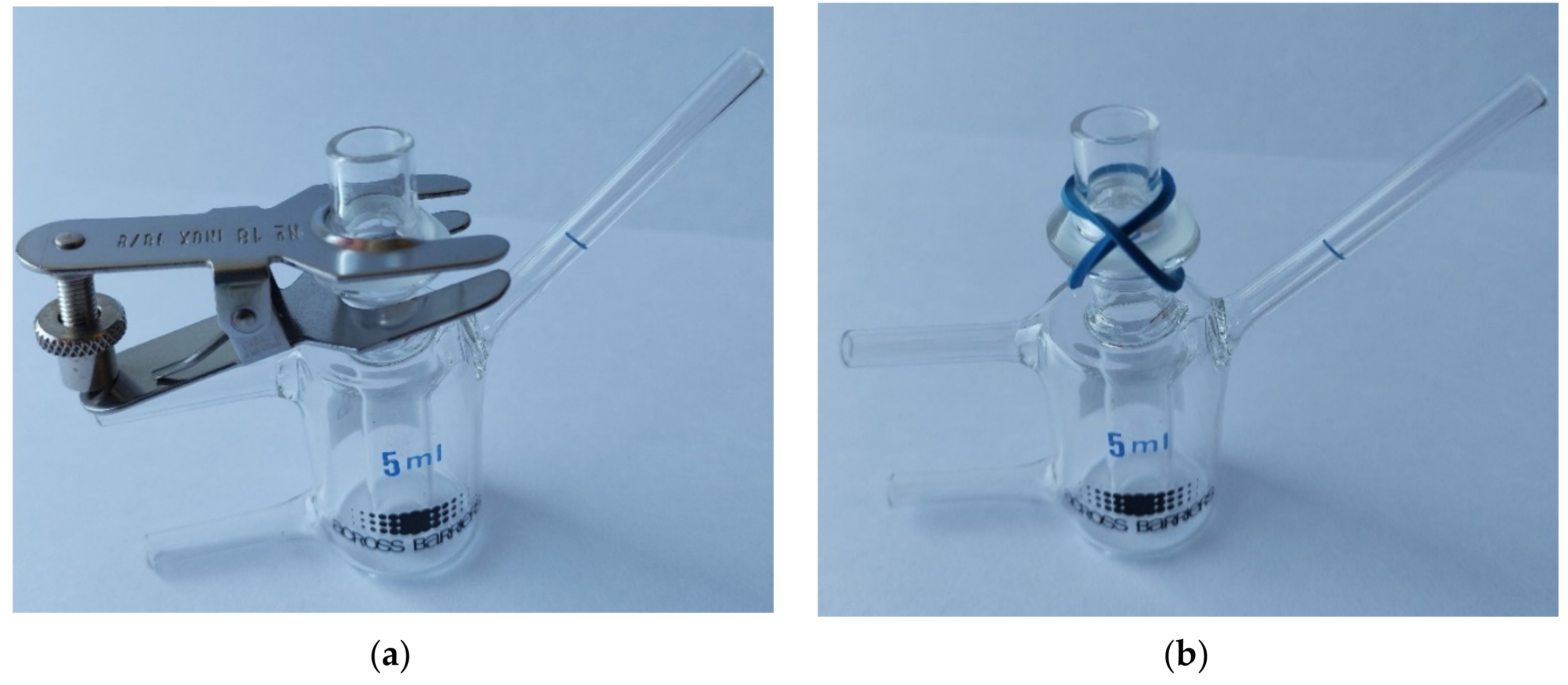
2.6. Ex Vivo Drug Permeation Study

The standard vertical Franz cell (Figure 5) method was used to quantify skin permeability for tazarotene. A cell with a receptor portion volume of 5 mL was used. Permeation testing was performed on circular sections of human abdominal skin firmly fastened between donor and acceptor chambers. After equilibration for 2 h with phosphate buffer (PBS, 1 mM, pH 7.4) at 32 °C (average skin surface temperature), the collection chamber was filled with 7.5 mL of ethanol/PBS solution (50:50 v/v), which maintained a descending position and was rotated continuously with a magnetic field (100 rpm). The tazarotene-containing nanofiber was attached to the skin and the tazarotene-free sample was used as a control. After sampling from the receptor compartment of the Franz cell, it was subsequently supplemented with an aliquot of the ethanol/PBS solution. The determination of tazarotene concentration in the receptor liquid was made spectrophotometrically (at 351 nm) using a UV–Vis spectrophotometer and the calibration curve (Figure 6). The experiments were performed by avoiding light exposure due to the inherent tazarotene photo-instability. For applications with alternating magnetic fields, it was not possible to use a metal clip (Figure 5a), which would have been extensively heated in this field; therefore, we used modified Franz cells without the metal parts (Figure 5b).
The actual amount of tazarotene in the nanofibrous mat (cut into rectangles 1.5 cm × 1.5 cm) was quantified by dissolving each sample in 4 mL of HFIP. Then, 0.5 mL of the solution was added to 8 mL of phosphate-buffered solution. The concentration of tazarotene was quantified using the calibration curve (Figure 6) obtained from absorbance (at wavelength 351 nm) measurement using a UV MINI 1240 UV–VIS spectrophotometer (Shimadzu, Kyoto, Japan). The tazarotene entrapment efficiency was calculated as:
Entrapment efficiency (%) = (tazarotene_fib/tazarotene_th) × 100
where tazarotene_fib is the amount of embedded magnetic nanoparticles in fibers, and tazarotene_th is the theoretical amount of tazarotene (used for their preparation) loaded into the fibers.

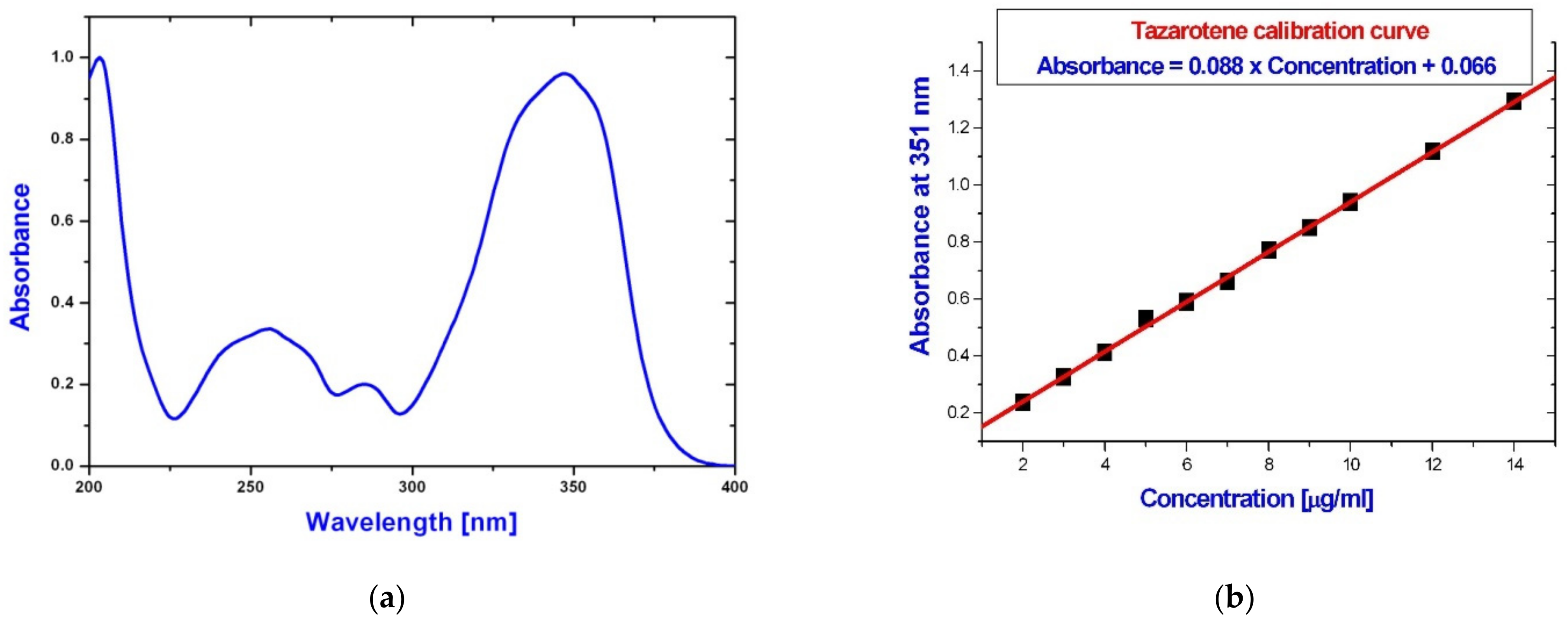
3. Results and Discussion

For efficient drug delivery and psoriasis treatment, sustained drug release for a prolonged time is important. Recently, a new medication concept called “dose-dense chemotherapy” has been introduced and successfully used, especially in cancer therapy, based on the technique of increasing dosage for a short time [26].
The smart nanofiber patch presented here has the potential to be used for temporal drug release triggered by external stimuli. For these purposes, we used nanofibers with embedded magnetic nanoparticles [27]. These nanoparticles are extensively heated when exposed to an alternating magnetic field [28,29,30,31,32,33].
The second key physical principle we used was the low melting temperature of PCL polymer (45–52 °C) [25] attainable by alternating magnetic-field-mediated hyperthermia. It is analogous to gel–liquid crystal phase transition in liposomes, exploited in liposomal temperature-sensitive drug delivery systems [32].
A key factor in the successful use of nanofibers for the controlled release of a drug is the need for its effective incorporation. We used tazarotene, whose water solubility is 0.75 mg/L, so it is a hydrophobic drug. Nevertheless, the solubility of another drug from the retinoid class—tretinoin—is only 0.025 mg/L. This was also a reason why we used the hydrophobic polymer, PCL, which is in contrast to our previous study [21], where we incorporated methotrexate—a drug well soluble in water—into polyvinyl alcohol, a hydrophilic polymer. By using PCL, we managed to achieve an incorporation efficiency of 96.7%. This makes it possible to use a smaller patch that can be applied for a longer time.
Scanning electron microscopy (Figure 7) was used to characterize the structure of the nanofibers. The fabricated nanofibers had diameters from 220 to 640 nm and a uniform smooth structure of their surface.
To effectively embed them, we used magnetic nanoparticles stabilized in PCL, which allowed us not only to incorporate them effectively into the structure of a nanofiber from the same polymer, but also to achieve their homogeneous distribution in nanofiber patches, as shown in Figure 8, using colorimetric Prussian blue staining.
For successful transdermal therapy, it is necessary for the drug to be concentrated rapidly on the skin surface in a therapeutically effective dose. To evaluate the ability of this formulation to cross the skin, we used human abdominal skin (obtained as frozen discs from Xenometrics) as a sample to measure tazarotene permeation using a vertical Franz cell (Figure 9). An analysis of the experimental data was carried out using the sigmoidal function proposed by Boltzmann in 1879 [34], which is based on the logistic sigmoidal equation, an appropriate starting point for the description of phase transition phenomena. As shown in Figure 9, the release of tazarotene is excellently described by this sigmoidal release pattern, which is therapeutically beneficial for timed release [35]. In addition to the phase transition of polycaprolactone, the release of tazarotene slightly increased, probably via the Arrhenius mechanism, in contrast to tazarotene permeation without heating with alternating magnetic field, where after 24 h, release of 70% tazarotene was observed. The sigmoidal curve reflects the experimental fact [33,36] that the phase transition temperature for polycaprolactone is 52.1 ± 2 °C, with an initial phase transition temperature of 45.4 ± 2 °C, which means that the melting point of the polycaprolactone nanofiber and the subsequent drug release can be easily achieved by means of hyperthermia induced by an alternating magnetic field (Figure 4b).
Moreover, the heat produced by AMF heating may also synergistically increase the efficiency of psoriasis therapy via superficial hyperthermia [37].

4. Conclusions

In this paper, we prepared an electrospun PCL nanofiber patch with embedded magnetic nanoparticles and the antipsoriatic drug, tazarotene. These fabrics were characterized by scanning electron microscopy and Prussian blue staining and tested with regard to drug delivery and permeation using Franz cells.
We showed that these electrospun nanofibers have promising potential as future candidates in remote-controlled drug delivery systems for the more efficient treatment of psoriasis. An attractive approach for their application would be a handheld electrospinning device activated at a short distance from psoriatic plaque, which is under investigation in our laboratory.

Author Contributions

Conceptualization, methodology, validation, formal analysis, investigation, data curation N.A., P.S., M.B. and M.Š.; writing—original draft preparation, N.A.; writing—review and editing, M.B., P.S. and M.Š. All authors have read and agreed to the published version of the manuscript.

Funding

This research was funded by the by Slovak Research and Development Agency APVV project No. APVV-16-0600, and Slovak Grant Agency VEGA project No. 1/0810/18.

Institutional Review Board Statement

Not applicable.

Informed Consent Statement

Not applicable.

Acknowledgments

We would like to thank O. Jirsák and P. Babinec for help in the preparation and characterization of the nanofibers.

Conflicts of Interest

The authors declare no conflict of interest.

References

  1. Šimaljaková, M.; Buchvald, D. Dermatovenereology; Comenius University Press: Bratislava, Slovakia, 2019. [Google Scholar]
  2. Lebwohl, M.G.; Tanghetti, E.A.; Stein Gold, L.; Del Rosso, J.Q.; Gilyadov, N.K.; Jacobson, A. Fixed-Combination Halobetasol Propionate and Tazarotene in the Treatment of Psoriasis: Narrative Review of Mechanisms of Action and Therapeutic Benefits. Dermatol. Ther. 2021, 11, 1157–1174. [Google Scholar] [CrossRef] [PubMed]
  3. He, C.; Jin, H.; Liu, X.; Hu, F.; Zhang, L.; Zhang, S.; He, Y.; Yang, X.; Chen, H.; Wang, X.; et al. Tazarotene/Betamethasone Dipropionate Cream in Patients with Plaque Psoriasis: Results of a Prospective, Multicenter, Observational Study. Dermatology 2021, 237, 603–610. [Google Scholar] [CrossRef] [PubMed]
  4. Baldwin, H.; Webster, G.; Stein Gold, L.; Callender, V.; Cook-Bolden, F.E.; Guenin, E. 50 Years of Topical Retinoids for Acne: Evolution of Treatment. Am. J. Clin. Dermatol. 2021, 22, 315–327. [Google Scholar] [CrossRef]
  5. Jyothi, S.L.; Krishna, K.L.; Ameena Shirin, V.K.; Sankar, R.; Pramod, K.; Gangadharappa, H.V. Drug delivery systems for the treatment of psoriasis: Current status and prospects. J. Drug Deliv. Sci. Technol. 2021, 62, 102364. [Google Scholar] [CrossRef]
  6. Ramanunny, A.K.; Wadhwa, S.; Thakur, D.; Singh, S.K.; Kumar, R. Treatment modalities of psoriasis: A focus on requisite for topical nanocarrier. Endocr. Metab. Immune Disord. Drug Targets 2021, 21, 418–433. [Google Scholar] [CrossRef]
  7. Jain, V.; Lovanshi, R.; Khan, A.I. Formulation development and evaluation of niosomal gel of tazarotene for treatment of psoriasis. J. Med. Pharm. Allied Sci. 2021, 10, 2664–2670. [Google Scholar] [CrossRef]
  8. Tanghetti, E.; Lebwohl, M.; Stein Gold, L. Tazarotene Revisited: Safety and Efficacy in Plaque Psoriasis and Its Emerging Role in Treatment Strategy. J. Drugs Dermatol. 2018, 17, 1280–1287. [Google Scholar]
  9. Dayal, S.; Kaura, R.; Sahu, P.; Jain, V.K. Tazarotene gel with narrow-band UVB phototherapy: A synergistic combination in psoriasis. An. Bras. Dermatol. 2018, 93, 385–390. [Google Scholar] [CrossRef] [Green Version]
  10. Prasad, V.; Chaurasia, S. Performance evaluation of non-ionic surfactant based tazarotene encapsulated proniosomal gel for the treatment of psoriasis. Mater. Sci. Eng. C 2017, 79, 168–176. [Google Scholar] [CrossRef] [PubMed]
  11. Jeon, S.-Y.; Ha, S.-M.; Ko, D.-Y.; Ku, B.-S.; Lee, C.-Y.; Song, K.-H.; Kim, K.-H. Tazarotene-induced gene 3 may affect inflammatory angiogenesis in psoriasis by downregulating placental growth factor expression. Ann. Dermatol. 2014, 26, 517–520. [Google Scholar] [CrossRef] [Green Version]
  12. Kumar, U.; Kaur, I.; Dogra, S.; De, D.; Kumar, B. Topical tazarotene vs. coal tar in stable plaque psoriasis: Clinical dermatology. Clin. Exp. Dermatol. 2010, 35, 482–486. [Google Scholar] [CrossRef]
  13. Veraldi, S.; Caputo, R.; Pacifico, A.; Peris, K.; Soda, R.; Chimenti, S. Short contact therapy with tazarotene in psoriasis vulgaris. Dermatology 2006, 212, 235–237. [Google Scholar] [CrossRef] [PubMed]
  14. Dando, T.M.; Wellington, K. Topical tazarotene: A review of its use in the treatment of plaque psoriasis. Am. J. Clin. Dermatol. 2005, 6, 255–272. [Google Scholar] [CrossRef]
  15. Su, Y.-H.; Fang, J.-Y. Drug delivery and formulations for the topical treatment of psoriasis. Expert Opin. Drug. Deliv. 2008, 5, 235–249. [Google Scholar] [CrossRef]
  16. Guenther, L. Tazarotene combination treatments in psoriasis. J. Am. Acad. Dermatol. 2000, 43, S36–S42. [Google Scholar] [CrossRef] [PubMed]
  17. Duvic, M.; Asano, A.T.; Hager, C.; Mays, S. The pathogenesis of psoriasis and the mechanism of action of tazarotene. J. Am. Acad. Dermatol. 1998, 39, S129–S133. [Google Scholar] [CrossRef]
  18. Nasr, M.; Abdel-Hamid, S. Optimizing the dermal accumulation of a tazarotene microemulsion using skin deposition modeling. Drug. Dev. Ind. Pharm. 2016, 42, 636–643. [Google Scholar] [CrossRef]
  19. Patel, M.R.; Patel, R.B.; Parikh, J.R.; Patel, B.G. Novel microemulsion-based gel formulation of tazarotene for therapy of acne. Pharm. Dev. Technol. 2016, 21, 921–932. [Google Scholar] [CrossRef]
  20. Zuccari, G.; Baldassari, S.; Alfei, S.; Marengo, B.; Valenti, G.E.; Domenicotti, C.; Ailuno, G.; Villa, C.; Marchitto, L.; Caviglioli, G. D-α-Tocopherol-Based Micelles for Successful Encapsulation of Retinoic Acid. Pharmaceuticals 2021, 14, 212. [Google Scholar] [CrossRef]
  21. Babincová, N.; Jirsák, O.; Babincová, M.; Babinec, P.; Šimaljaková, M. Remote magnetically controlled drug release from electrospun composite nanofibers: Design of a smart platform for therapy of psoriasis. Z. Naturforsch. 2020, 75, 587–591. [Google Scholar] [CrossRef]
  22. Khoshbakht, S.; Asghari-Sana, F.; Fathi-Azarbayjani, A.; Sharifi, Y. Fabrication and characterization of tretinoin-loaded nanofiber for topical skin delivery. Biomater. Res. 2020, 24, 8. [Google Scholar] [CrossRef] [PubMed] [Green Version]
  23. Lin, L.; Dai, Y.; Cui, H. Antibacterial poly(ethylene oxide) electrospun nanofibers containing cinnamon essential oil/beta-cyclodextrin proteoliposomes. Carbohydr. Polym. 2017, 15, 131–140. [Google Scholar] [CrossRef] [PubMed]
  24. Patel, G.; Yadav, B.K.N. Formulation, Characterization and In vitro Cytotoxicity of 5-Fluorouracil Loaded Polymeric Electrospun Nanofibers for the Treatment of Skin Cancer. Recent Pat. Nanotechnol. 2019, 13, 114–128. [Google Scholar] [CrossRef]
  25. Hedayatnasab, Z.; Dabbagh, A.; Abnisa, F.; Wan Daud, W.M.A. Polycaprolactone-coated superparamagnetic iron oxide nanoparticles for in vitro magnetic hyperthermia therapy of cancer. Eur. Polym. J. 2020, 133, 109789. [Google Scholar] [CrossRef]
  26. Katsumata, N. Dose-dense effect: Other contributor? Author’s reply. Lancet Oncol. 2013, 14, e489–e490. [Google Scholar] [CrossRef]
  27. Babinec, P.; Jirsák, O. Microwave absorbing nonwoven textile from electrospun magnetically responsive nanofibers. Optoelectron. Adv. Mat. 2008, 2, 474–477. [Google Scholar]
  28. Niiyama, E.; Uto, K.; Lee, C.M.; Sakura, K.; Ebara, M. Alternating Magnetic Field-Triggered Switchable Nanofiber Mesh for Cancer Thermo-Chemotherapy. Polymers 2018, 10, 1018. [Google Scholar] [CrossRef] [PubMed] [Green Version]
  29. Babincová, M. Microwave induced leakage of magnetoliposomes. Possible clinical implications. Bioelectrochem. Bioenerg. 1993, 32, 187–189. [Google Scholar] [CrossRef]
  30. Babincová, M.; Vrbovská, H.; Sourivong, P.; Babinec, P.; Durdík, Š. Application of albumin-embedded magnetic nanoheaters for release of etoposide in integrated chemotherapy and hyperthermia of U87-MG glioma cells. Anticancer Res. 2018, 38, 2683–2690. [Google Scholar] [CrossRef]
  31. Babincová, M.; Durdík, Š.; Babincová, N.; Sourivong, P.; Babinec, P. Application of cationized magnetoferritin for magnetic field-assisted delivery of short interfering RNA in vitro. Lasers Med. Sci. 2018, 33, 1807–1812. [Google Scholar] [CrossRef]
  32. Babincová, N.; Sourivong, P.; Babinec, P.; Bergemann, C.; Babincová, M.; Durdík, S. Applications of magnetoliposomes with encapsulated doxorubicin for integrated chemotherapy and hyperthermia of rat C6 glioma. Z. Naturforsch. C 2018, 73, 265–271. [Google Scholar] [CrossRef]
  33. Altanerova, U.; Babincova, M.; Babinec, P.; Benejova, K.; Jakubechova, J.; Altanerova, V.; Zduriencikova, M.; Repiska, V.; Altaner, C. Human mesenchymal stem cell-derived iron oxide exosomes allow targeted ablation of tumor cells via magnetic hyperthermia. Int. J. Nanomed. 2017, 12, 7923–7936. [Google Scholar] [CrossRef] [Green Version]
  34. Ravikumar, R.; Ganesh, M.; Senthil, V.; Ramesh, Y.V.; Jakki, S.L.; Choi, E.Y. Tetrahydro curcumin loaded PCL-PEG electrospun transdermal nanofiber patch: Preparation, characterization, and in vitro diffusion evaluations. J. Drug Deliv. Sci. Technol. 2018, 44, 342–348. [Google Scholar] [CrossRef]
  35. Navarro-Verdugo, A.L.; Goycoolea, F.M.; Romero-Meléndez, G.; Higuera-Ciapara, I.; Argüelles-Monal, W. A modified Boltzmann sigmoidal model for the phase transition of smart gels. Soft Matter 2011, 7, 5847–5853. [Google Scholar] [CrossRef]
  36. Kamaly, N.; Yameen, B.; Wu, J.; Farokhzad, O.C. Degradable Controlled-Release Polymers and Polymeric Nanoparticles: Mechanisms of Controlling Drug Release. Chem. Rev. 2016, 116, 2602–2663. [Google Scholar] [CrossRef] [PubMed] [Green Version]
  37. Boreham, D.R.; Gasmann, H.C.; Mitchel, R.E. Water bath hyperthermia is a simple therapy for psoriasis and also stimulates skin tanning in response to sunlight. Int. J. Hyperthermia 1995, 11, 745–754. [Google Scholar] [CrossRef]
Figure 1. Structure of the tazarotene and its main degradation product, tazarotenic acid.
Applsci 11 11022 g001
Figure 2. Polycaprolactone is a biodegradable polyester and ideal material for preparation of electrospun nanofibers.
Applsci 11 11022 g002
Figure 3. Electrospinning of polycaprolactone nanofibers: (a) experimental setup; (b) nanofibers can be deposited directly to the skin.
Applsci 11 11022 g003
Figure 4. Alternating magnetic field was used for heating of nanofibers with embedded magnetic nanoparticles: (a) experimental setup; (b) kinetics of temperature increase in nanofiber patch with magnetic nanoparticles.
Applsci 11 11022 g004
Figure 5. (a) Standard Franz cell with metal clip; (b) Franz cell fastened with an elastic band suitable for application with alternating magnetic field.
Applsci 11 11022 g005
Figure 6. (a) UV–Vis absorption spectra of tazarotene; (b) calibration curve at wavelength of 351 nm.
Applsci 11 11022 g006
Figure 7. Scanning electron microscopy of nanofibrous patch: (a) higher magnification; (b) lower magnification.
Applsci 11 11022 g007
Figure 8. Colorimetric assay of magnetic nanoparticles in nanofiber patch: (a) optical microscopy image of patch without Prussian blue; (b) patch immediately after staining when Prussian blue crystals are formed.
Applsci 11 11022 g008
Figure 9. In vitro permeation profile of tazarotene through the abdominal human skin form nanofiber patch and the effect of alternating magnetic field on time course of tazarotene permeation.
Applsci 11 11022 g009
Publisher’s Note: MDPI stays neutral with regard to jurisdictional claims in published maps and institutional affiliations.

