Anti-Bacterial and Anti-Candidal Activity of Silver Nanoparticles Biosynthesized Using Citrobacter spp. MS5 Culture Supernatant

 ,
,  , and
, and
Abstract
:
1. Introduction
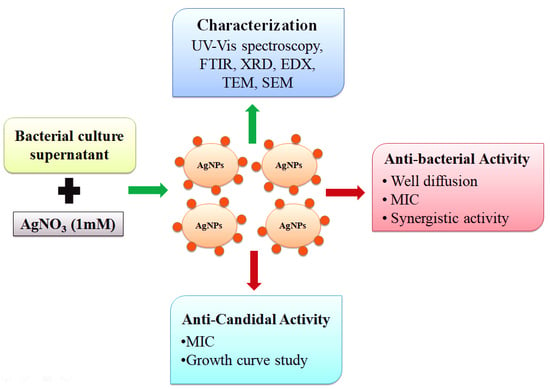
2. Methodology
2.1. Materials
2.2. Isolation and Screening of Bacterial Isolates for AgNPs Synthesis
2.3. Biosynthesis of AgNPs
2.4. Characterization
2.5. Isolation and Identification of Extended Spectrum β-Lactamase (ESBL) Producing Bacteria
2.6. Antibiotic Profiling of ESBL Producing Bacteria
2.7. Antibacterial Activity of AgNPs
2.8. Antifungal Activity of AgNPs
3. Results and Discussion
3.1. Isolation and Identification of the Bacteria
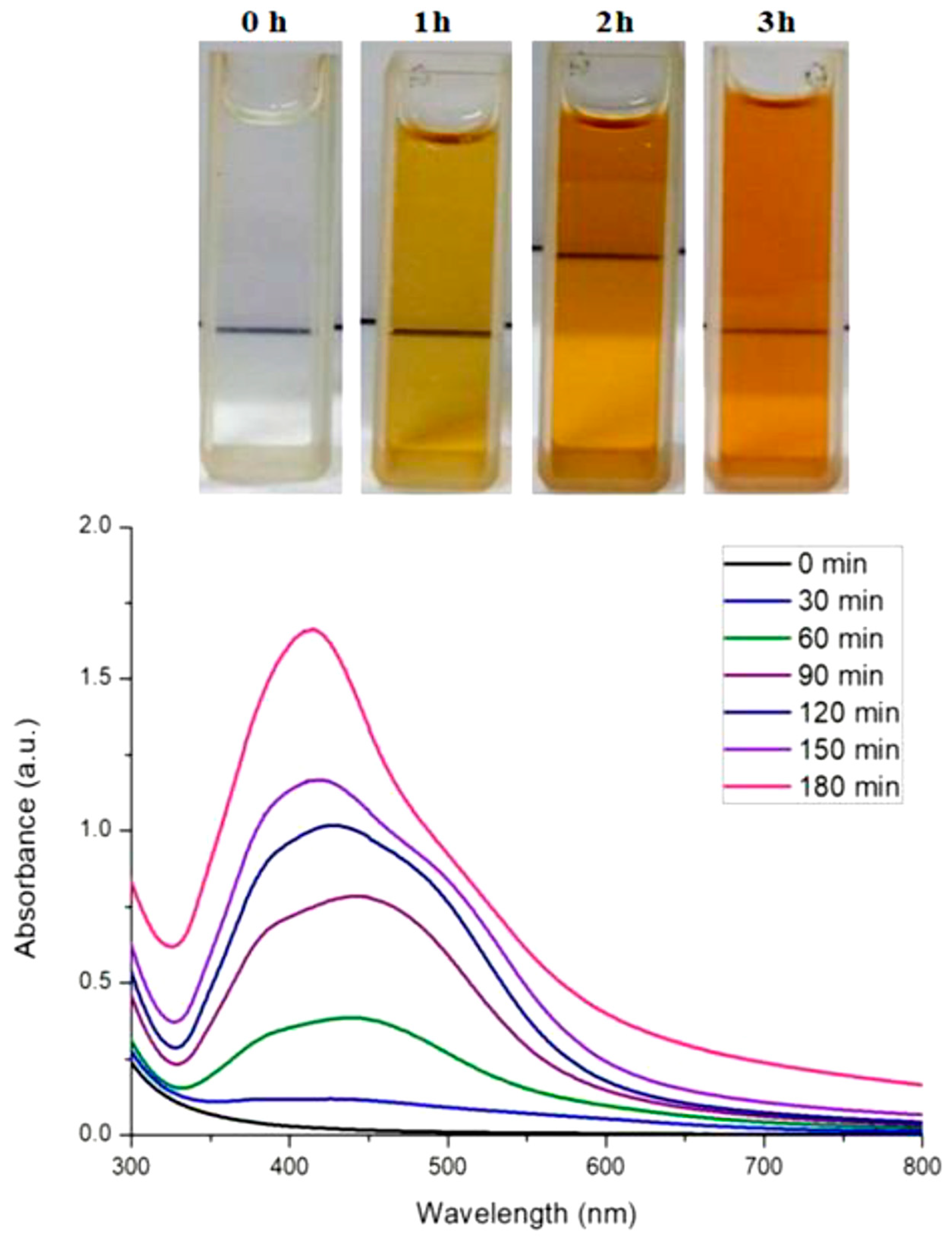
3.2. Biosynthesis of AgNPs
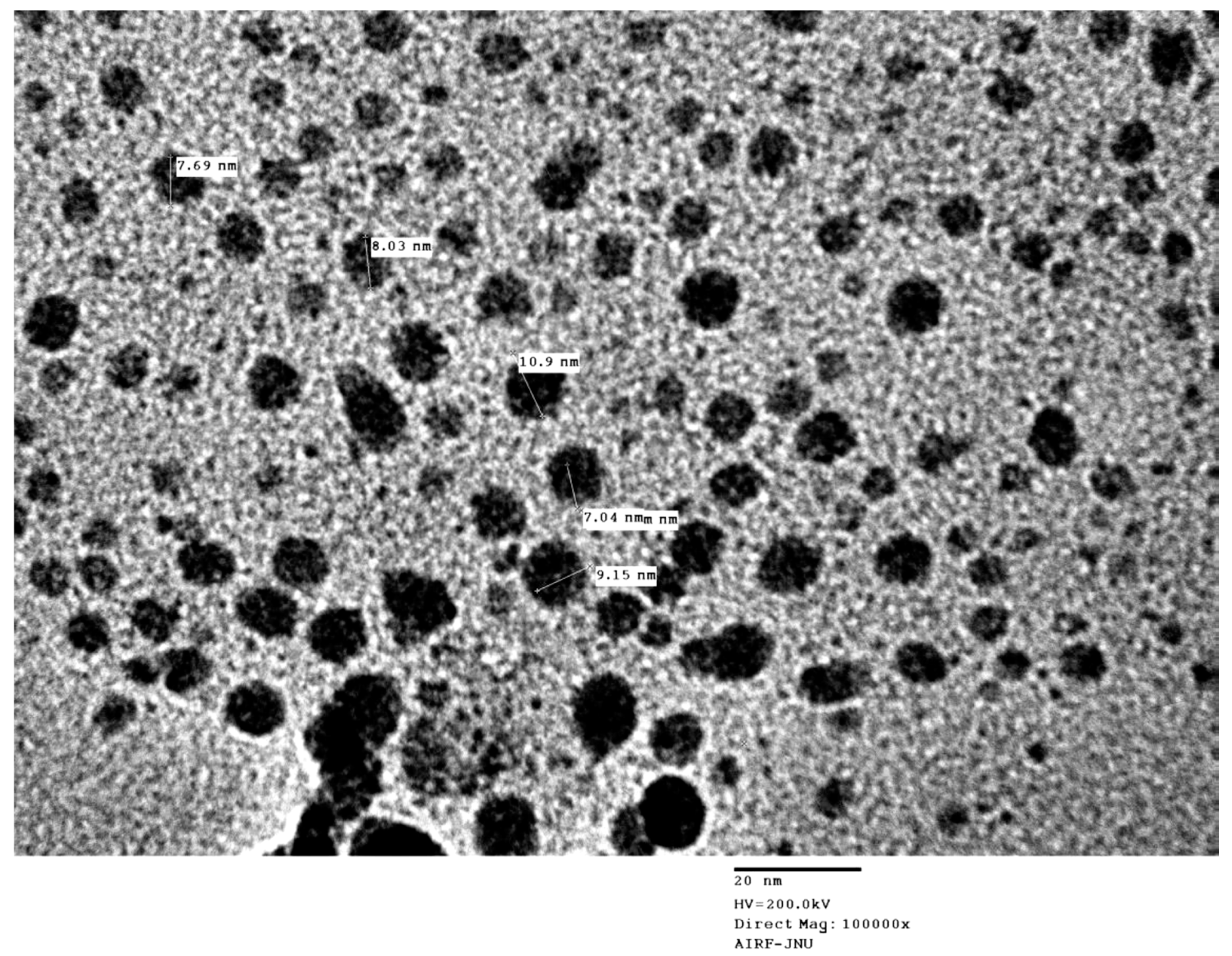
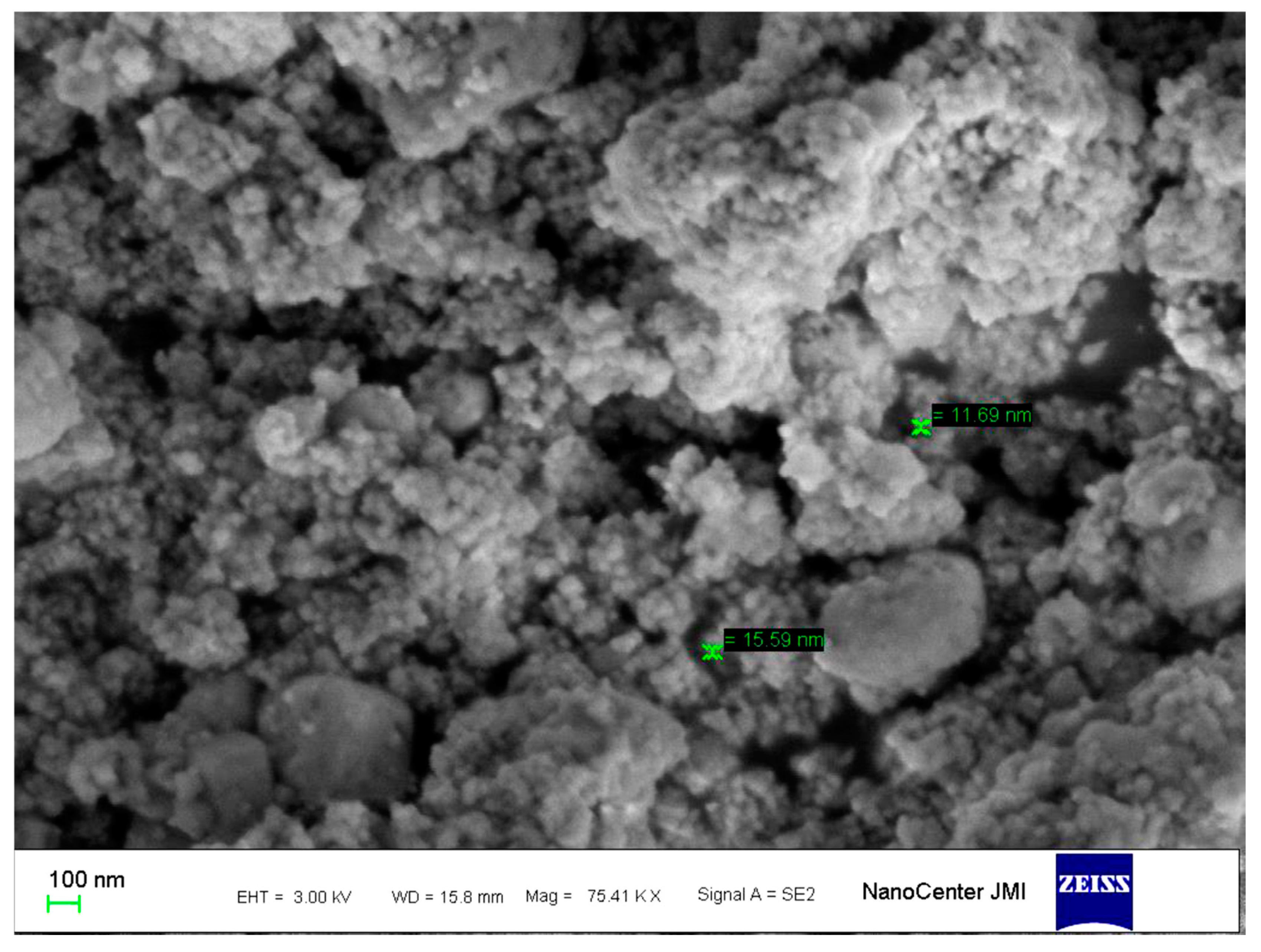
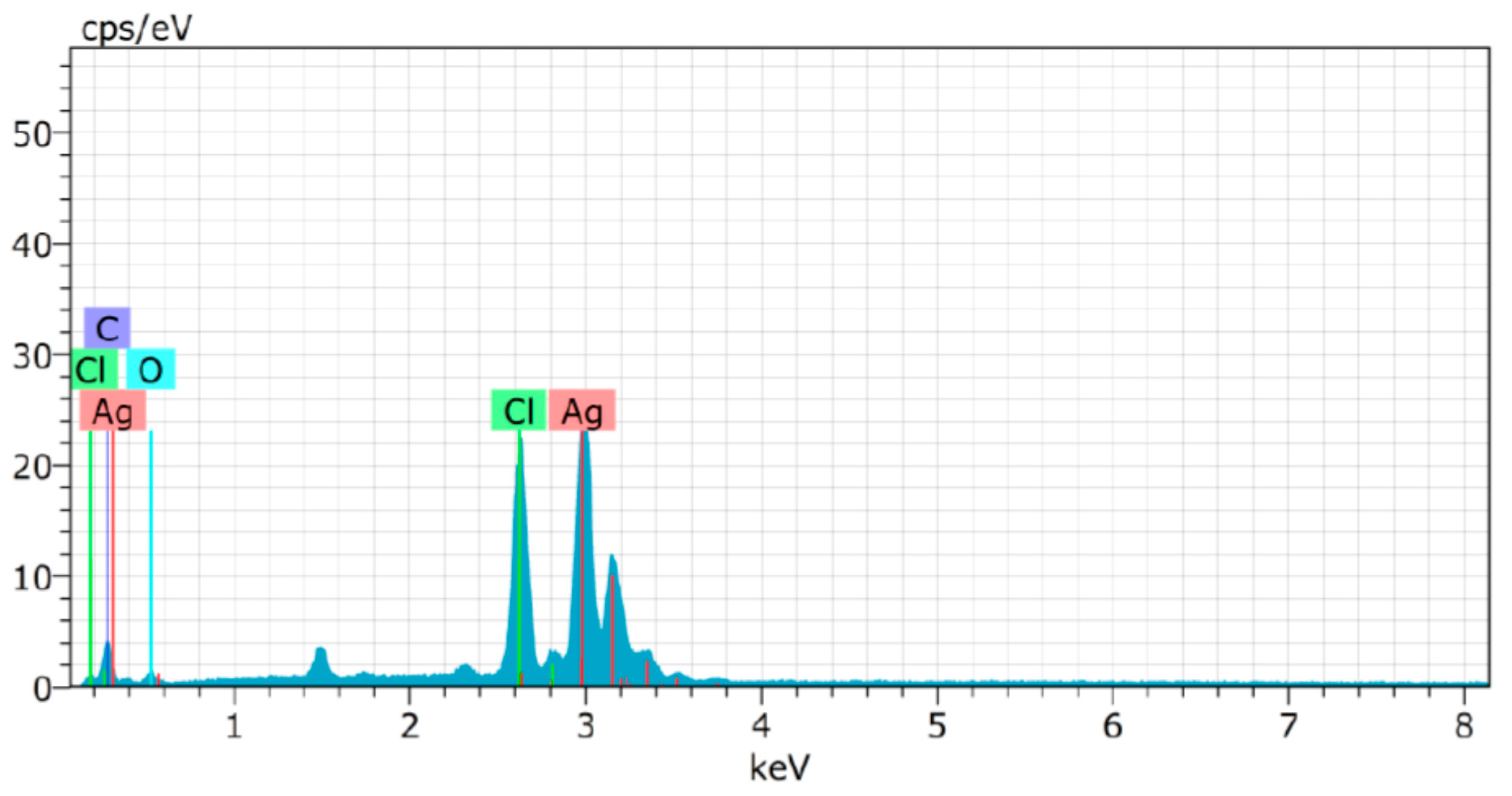
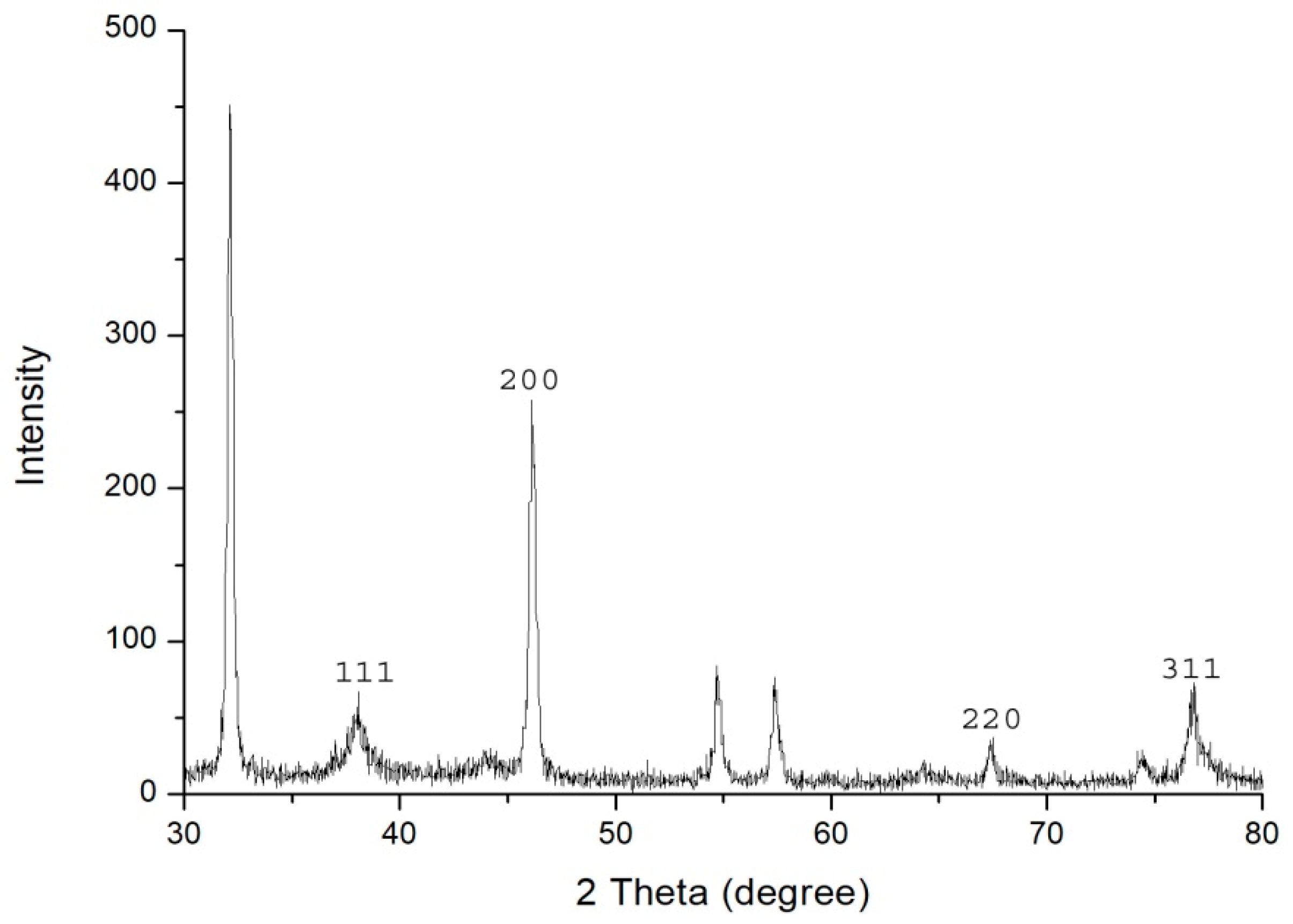
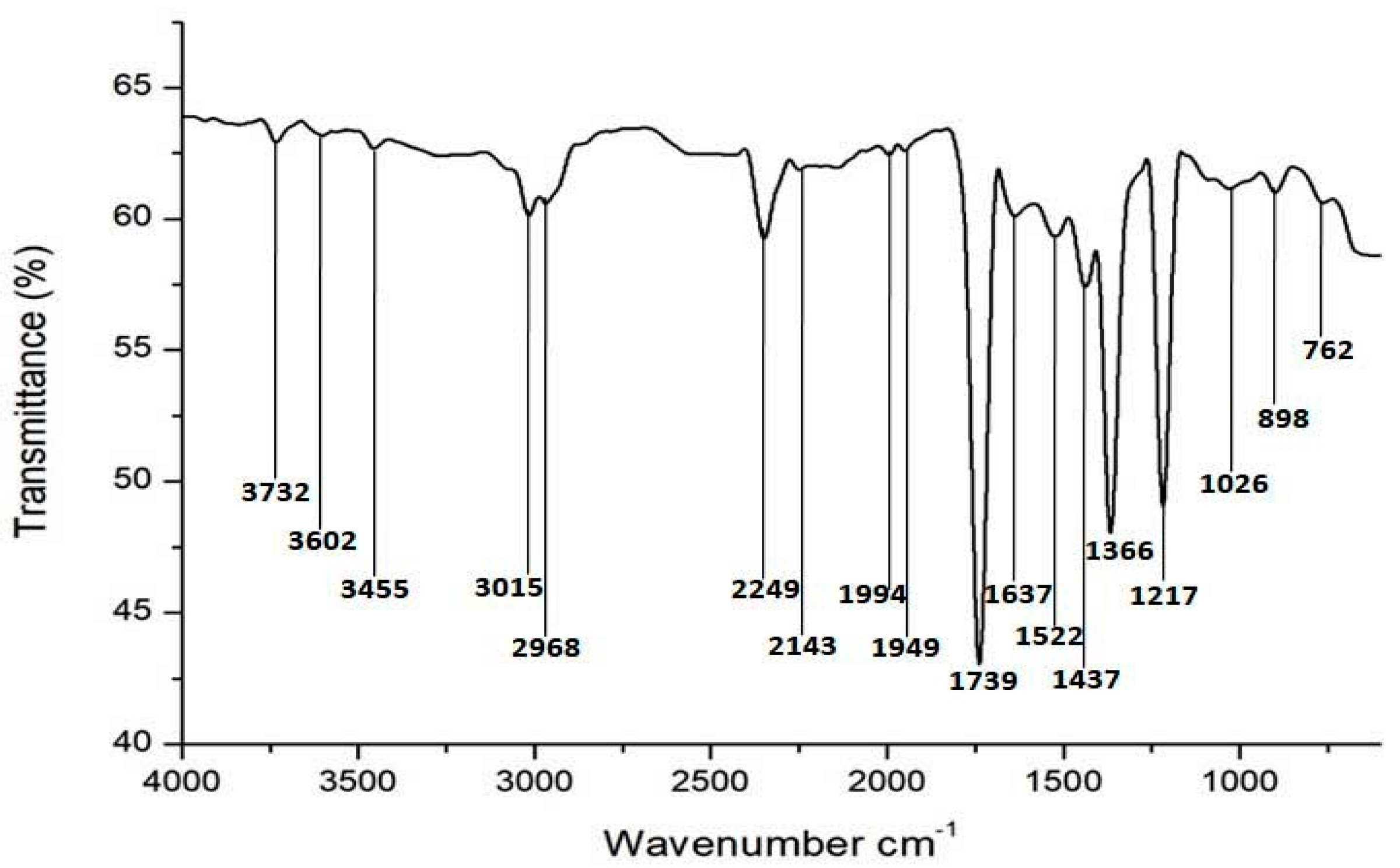
3.3. Characterization
3.4. Isolation, Identification, and Antibiotic Profiling of ESBL Producing Bacteria
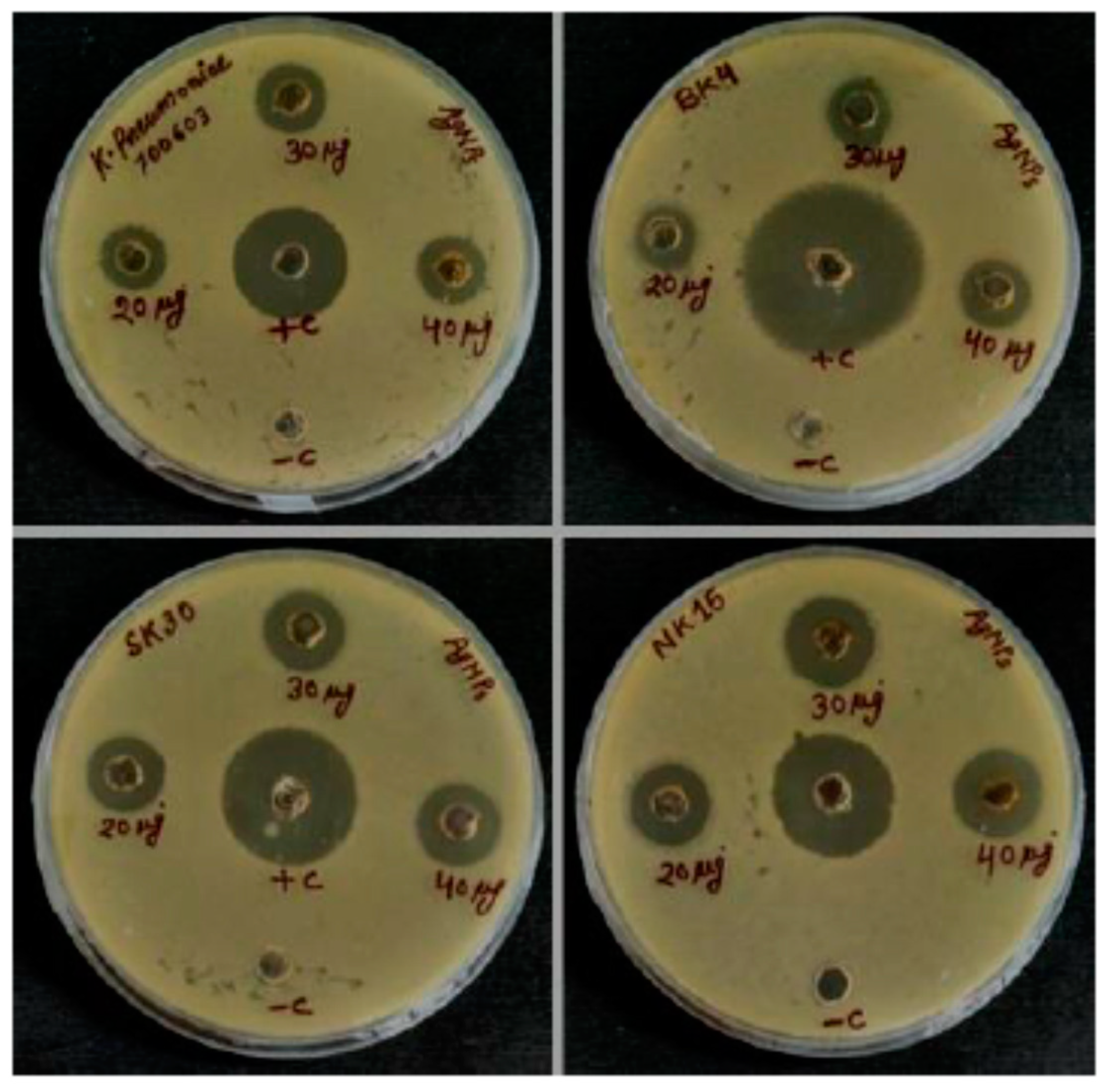
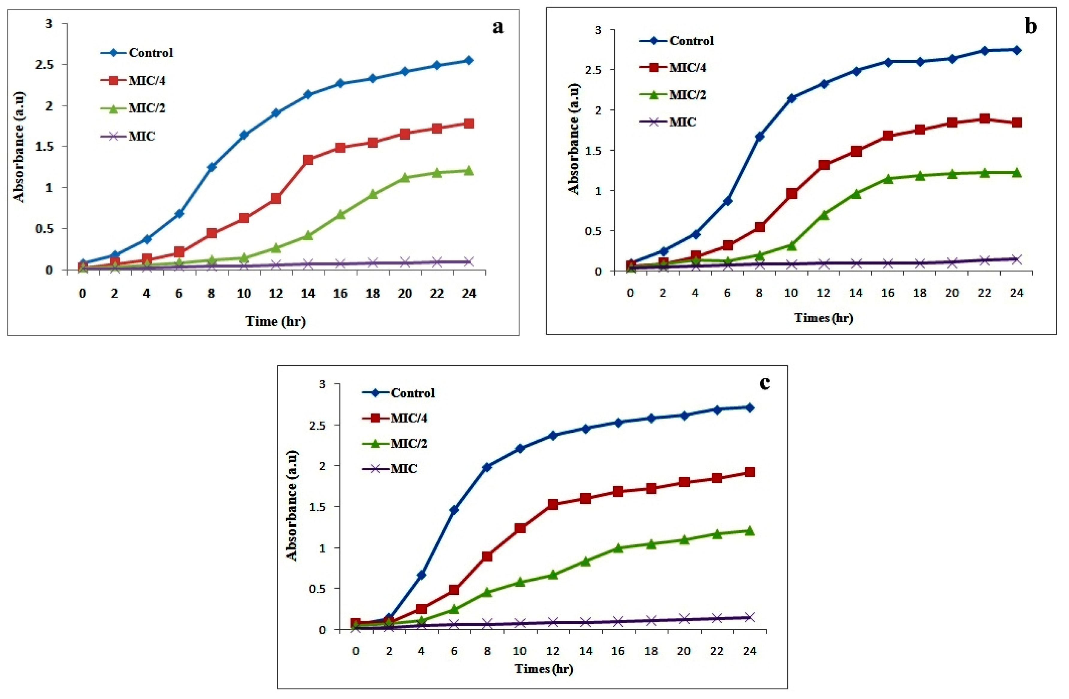
3.5. Antibacterial Activity of AgNPs
3.6. Antifungal Activity of AgNPs
4. Conclusions
Author Contributions
Funding
Acknowledgments
Conflicts of Interest
References
- Khan, I.; Saeed, K.; Khan, I. Nanoparticles: Properties, applications and toxicities. Arab. J. Chem. 2019, 12, 908–931. [Google Scholar] [CrossRef]
- Verma, N.; Upadhyay, L.S.B. Synthesis and characterization of cysteine functionalized silver nanoparticles for biomolecule immobilization. Bioprocess Biosyst. Eng. 2014, 37, 2139–2148. [Google Scholar]
- Burdusel, A.C.; Gherasim, O.; Grumezescu, A.M.; Mogoanta, L.; Ficai, A.; Andronescu, E. Biomedical applications of silver nanoparticles: An up-to-date overview. Nanomaterials 2018, 8, 681. [Google Scholar] [CrossRef] [Green Version]
- Rai, M.; Yadav, A.; Gade, A. Silver nanoparticles as a new generation of antimicrobials. Biotechnol. Adv. 2009, 27, 76–83. [Google Scholar] [CrossRef]
- Banu, A.; Rathod, V.; Ranganath, E. Silver nanoparticle production by rhizopus stolonifer and its antibacterial activity against extended spectrum β-lactamase producing (esbl) strains of enterobacteriaceae. Mater. Res. Bull. 2011, 46, 1417–1423. [Google Scholar] [CrossRef]
- Subashini, J.; Khanna, V.G.; Kannabiran, K. Anti-esbl activity of silver nanoparticles biosynthesized using soil streptomyces species. Bioprocess Biosyst. Eng. 2014, 37, 999–1006. [Google Scholar] [CrossRef]
- Paterson, D.L.; Bonomo, R.A. Extended-spectrum β-lactamases: A clinical update. Clin. Microbiol. Rev. 2005, 18, 657–686. [Google Scholar] [CrossRef] [Green Version]
- Adelowo, O.O.; Caucci, S.; Banjo, O.A.; Nnanna, O.C.; Awotipe, E.O.; Peters, F.B.; Fagade, O.E.; Berendonk, T.U. Extended spectrum beta-lactamase (esbl)-producing bacteria isolated from hospital wastewaters, rivers and aquaculture sources in nigeria. Environ. Sci. Pollut. Res. 2018, 25, 2744–2755. [Google Scholar] [CrossRef]
- Mondal, A.H.; Siddiqui, M.T.; Sultan, I.; Haq, Q.M.R. Prevalence and diversity of bla tem, bla shv and bla ctx-m variants among multidrug resistant klebsiella spp. From an urban riverine environment in india. Int. J. Environ. Health Res. 2019, 29, 117–129. [Google Scholar] [CrossRef]
- Kuralayanapalya, S.P.; Patil, S.S.; Hamsapriya, S.; Shinduja, R.; Roy, P.; Amachawadi, R.G. Prevalence of extended-spectrum beta-lactamase producing bacteria from animal origin: A systematic review and meta-analysis report from india. PLoS ONE 2019, 14, e0221771. [Google Scholar] [CrossRef]
- Hammerum, A.M.; Larsen, J.; Andersen, V.D.; Lester, C.H.; SkovgaardSkytte, T.S.; Hansen, F.; Olsen, S.S.; Mordhorst, H.; Skov, R.L.; Aarestrup, F.M. Characterization of extended-spectrum β-lactamase (esbl)-producing escherichia coli obtained from danish pigs, pig farmers and their families from farms with high or no consumption of third-or fourth-generation cephalosporins. J. Antimicrob. Chemother. 2014, 69, 2650–2657. [Google Scholar] [CrossRef] [PubMed]
- Bongomin, F.; Gago, S.; Oladele, R.O.; Denning, D.W. Global and multi-national prevalence of fungal diseases—Estimate precision. J. Fungi 2017, 3, 57. [Google Scholar] [CrossRef] [PubMed]
- Iravani, S.; Korbekandi, H.; Mirmohammadi, S.V.; Zolfaghari, B. Synthesis of silver nanoparticles: Chemical, physical and biological methods. Res. Pharm. Sci. 2014, 9, 385. [Google Scholar]
- Thakkar, K.N.; Mhatre, S.S.; Parikh, R.Y. Biological synthesis of metallic nanoparticles. Nanomed. Nanotechnol. Biol. Med. 2010, 6, 257–262. [Google Scholar] [CrossRef] [PubMed]
- Binupriya, A.; Sathishkumar, M.; Yun, S.-I. Biocrystallization of silver and gold ions by inactive cell filtrate of rhizopus stolonifer. Colloids Surf. B Biointerfaces 2010, 79, 531–534. [Google Scholar] [CrossRef] [PubMed]
- Narayanan, K.B.; Sakthivel, N. Biological synthesis of metal nanoparticles by microbes. Adv. Colloid Interface Sci. 2010, 156, 1–13. [Google Scholar] [CrossRef]
- Li, X.; Xu, H.; Chen, Z.-S.; Chen, G. Biosynthesis of nanoparticles by microorganisms and their applications. J. Nanomater. 2011, 2011. [Google Scholar] [CrossRef] [Green Version]
- Roy, A.; Bulut, O.; Some, S.; Mandal, A.K.; Yilmaz, M.D. Green synthesis of silver nanoparticles: Biomolecule-nanoparticle organizations targeting antimicrobial activity. RSC Adv. 2019, 9, 2673–2702. [Google Scholar] [CrossRef] [Green Version]
- Vaidyanathan, R.; Gopalram, S.; Kalishwaralal, K.; Deepak, V.; Pandian, S.R.K.; Gurunathan, S. Enhanced silver nanoparticle synthesis by optimization of nitrate reductase activity. Colloids Surf. B Biointerfaces 2010, 75, 335–341. [Google Scholar] [CrossRef]
- Gopinath, V.; Velusamy, P. Extracellular biosynthesis of silver nanoparticles using bacillus sp. Gp-23 and evaluation of their antifungal activity towards fusarium oxysporum. Spectrochim. Acta Part A Mol. Biomol. Spectrosc. 2013, 106, 170–174. [Google Scholar] [CrossRef]
- Siddiqi, K.S.; Husen, A.; Rao, R.A. A review on biosynthesis of silver nanoparticles and their biocidal properties. J. Nanobiotechnology 2018, 16, 14. [Google Scholar] [CrossRef] [PubMed]
- Kalpana, D.; Lee, Y.S. Synthesis and characterization of bactericidal silver nanoparticles using cultural filtrate of simulated microgravity grown klebsiella pneumoniae. Enzyme Microb. Technol. 2013, 52, 151–156. [Google Scholar] [CrossRef] [PubMed]
- Neelofar, K.; Shreaz, S.; Rimple, B.; Muralidhar, S.; Nikhat, M.; Khan, L.A. Curcumin as a promising anticandidal of clinical interest. Can. J. Microbiol. 2011, 57, 204–210. [Google Scholar] [CrossRef] [PubMed]
- Hamouda, R.A.; Hussein, M.H.; Abo-elmagd, R.A.; Bawazir, S.S. Synthesis and biological characterization of silver nanoparticles derived from the cyanobacterium oscillatoria limnetica. Sci. Rep. 2019, 9, 1–17. [Google Scholar] [CrossRef] [PubMed]
- Hossain, A.; Hong, X.; Ibrahim, E.; Li, B.; Sun, G.; Meng, Y.; Wang, Y.; An, Q. Green synthesis of silver nanoparticles with culture supernatant of a bacterium pseudomonas rhodesiae and their antibacterial activity against soft rot pathogen dickeya dadantii. Molecules 2019, 24, 2303. [Google Scholar] [CrossRef] [PubMed] [Green Version]
- Zhang, X.F.; Liu, Z.G.; Shen, W.; Gurunathan, S. Silver nanoparticles: Synthesis, characterization, properties, applications, and therapeutic approaches. Int. J. Mol. Med. Sci. 2016, 17, 1534. [Google Scholar] [CrossRef] [PubMed]
- Anandalakshmi, K.; Venugobal, J.; Ramasamy, V. Characterization of silver nanoparticles by green synthesis method using pedalium murex leaf extract and their antibacterial activity. Appl. Nanosci. 2016, 6, 399–408. [Google Scholar] [CrossRef] [Green Version]
- Kalimuthu, K.; Babu, R.S.; Venkataraman, D.; Bilal, M.; Gurunathan, S. Biosynthesis of silver nanocrystals by bacillus licheniformis. Colloids Surf. B Biointerfaces 2008, 65, 150–153. [Google Scholar] [CrossRef]
- Malarkodi, C.; Rajeshkumar, S.; Paulkumar, K.; Vanaja, M.; Jobitha, G.D.G.; Annadurai, G. Bactericidal activity of bio mediated silver nanoparticles synthesized by serratia nematodiphila. Drug Invent. Today 2013, 5, 119–125. [Google Scholar] [CrossRef]
- Rajamanickam, K.; Sudha, S.; Francis, M.; Sowmya, T.; Rengaramanujam, J.; Sivalingam, P.; Prabakar, K. Microalgae associated brevundimonas sp. Msk 4 as the nano particle synthesizing unit to produce antimicrobial silver nanoparticles. Spectrochim. Acta Part A Mol. Biomol. Spectrosc. 2013, 113, 10–14. [Google Scholar] [CrossRef]
- Uthayakumar, G.; Inbasekaran, S.; Sivasubramanian, A.; Jacob, S.J.P. Nanoparticle analysis for various medicinal drugs and human body saliva at macromolecular level. Appl. Nanosci. 2015, 5, 563–568. [Google Scholar] [CrossRef] [Green Version]
- Pawlak, A.; Mucha, M. Thermogravimetric and ftir studies of chitosan blends. Thermochim. Acta 2003, 396, 153–166. [Google Scholar] [CrossRef]
- Venugopal, K.; Rather, H.; Rajagopal, K.; Shanthi, M.; Sheriff, K.; Illiyas, M.; Rather, R.; Manikandan, E.; Uvarajan, S.; Bhaskar, M. Synthesis of silver nanoparticles (agnps) for anticancer activities (mcf 7 breast and a549 lung cell lines) of the crude extract of syzygium aromaticum. J. Photochem. Photobiol. B Biol. 2017, 167, 282–289. [Google Scholar] [CrossRef]
- Huang, J.; Li, Q.; Sun, D.; Lu, Y.; Su, Y.; Yang, X.; Wang, H.; Wang, Y.; Shao, W.; He, N. Biosynthesis of silver and gold nanoparticles by novel sundried cinnamomum camphora leaf. Nanotechnology 2007, 18, 105104. [Google Scholar] [CrossRef]
- Bansal, V.; Rautaray, D.; Bharde, A.; Ahire, K.; Sanyal, A.; Ahmad, A.; Sastry, M. Fungus-mediated biosynthesis of silica and titania particles. J. Mater. Chem. 2005, 15, 2583–2589. [Google Scholar] [CrossRef]
- Augustine, R.; Kalarikkal, N.; Thomas, S. A facile and rapid method for the black pepper leaf mediated green synthesis of silver nanoparticles and the antimicrobial study. Appl. Nanosci. 2014, 4, 809–818. [Google Scholar] [CrossRef] [Green Version]
- Fayaz, A.M.; Balaji, K.; Girilal, M.; Yadav, R.; Kalaichelvan, P.T.; Venketesan, R. Biogenic synthesis of silver nanoparticles and their synergistic effect with antibiotics: A study against Gram-positive and Gram-negative bacteria. Nanomedicine 2010, 6, 103–109. [Google Scholar] [CrossRef]
- Kumar, S.A.; Abyaneh, M.K.; Gosavi, S.; Kulkarni, S.K.; Pasricha, R.; Ahmad, A.; Khan, M. Nitrate reductase-mediated synthesis of silver nanoparticles from agno 3. Biotechnol. Lett. 2007, 29, 439–445. [Google Scholar] [CrossRef]
- Duran, N.; Marcato, P.D.; Alves, O.L.; Souza, G.I.; Esposito, E. Mechanistic aspects of biosynthesis of silver nanoparticles by several fusarium oxysporum strains. J. Nanobiotechnol. 2005, 3, 8. [Google Scholar] [CrossRef] [Green Version]
- Walsh, T.R.; Weeks, J.; Livermore, D.M.; Toleman, M.A. Dissemination of ndm-1 positive bacteria in the new delhi environment and its implications for human health: An environmental point prevalence study. Lancet Infect. Dis. 2011, 11, 355–362. [Google Scholar] [CrossRef]
- Magiorakos, A.P.; Srinivasan, A.; Carey, R.B.; Carmeli, Y.; Falagas, M.E.; Giske, C.G.; Harbarth, S.; Hindler, J.F.; Kahlmeter, G.; Olsson-Liljequist, B.; et al. Multidrug-resistant, extensively drug-resistant and pandrug-resistant bacteria: An international expert proposal for interim standard definitions for acquired resistance. Clin. Microbiol. Infect. 2012, 18, 268–281. [Google Scholar] [CrossRef] [PubMed] [Green Version]
- Naqvi, S.Z.; Kiran, U.; Ali, M.I.; Jamal, A.; Hameed, A.; Ahmed, S.; Ali, N. Combined efficacy of biologically synthesized silver nanoparticles and different antibiotics against multidrug-resistant bacteria. Int. J. Nanomed. 2013, 8, 3187–3195. [Google Scholar] [CrossRef] [Green Version]
- McShan, D.; Zhang, Y.; Deng, H.; Ray, P.C.; Yu, H. Synergistic antibacterial effect of silver nanoparticles combined with ineffective antibiotics on drug resistant salmonella typhimurium dt104. J. Environ. Sci. Health Part C Environ. Carcinog. Ecotoxicol. Rev. 2015, 33, 369–384. [Google Scholar] [CrossRef] [PubMed]
- Egorenkova, G.N.; Belov, A.P. Structural organization of the cell walls in yeasts of the genus candida. Mikrobiologiia 1984, 53, 300–304. [Google Scholar] [PubMed]
- Xue, B.; He, D.; Gao, S.; Wang, D.; Yokoyama, K.; Wang, L. Biosynthesis of silver nanoparticles by the fungus arthroderma fulvum and its antifungal activity against genera of candida, aspergillus and fusarium. Int. J. Nanomed. 2016, 11, 1899–1906. [Google Scholar] [CrossRef] [Green Version]
- Panáček, A.; Kolář, M.; Večeřová, R.; Prucek, R.; Soukupova, J.; Kryštof, V.; Hamal, P.; Zbořil, R.; Kvítek, L. Antifungal activity of silver nanoparticles against candida spp. Biomaterials 2009, 30, 6333–6340. [Google Scholar] [CrossRef] [PubMed]
- Jung, W.K.; Koo, H.C.; Kim, K.W.; Shin, S.; Kim, S.H.; Park, Y.H. Antibacterial activity and mechanism of action of the silver ion in staphylococcus aureus and escherichia coli. Appl. Environ. Microbiol. 2008, 74, 2171–2178. [Google Scholar] [CrossRef] [Green Version]
- Yun’an Qing, L.C.; Li, R.; Liu, G.; Zhang, Y.; Tang, X.; Wang, J.; Liu, H.; Qin, Y. Potential antibacterial mechanism of silver nanoparticles and the optimization of orthopedic implants by advanced modification technologies. Int. J. Nanomed. 2018, 13, 3311. [Google Scholar]
- Kim, T.H.; Kim, M.; Park, H.S.; Shin, U.S.; Gong, M.S.; Kim, H.W. Size-dependent cellular toxicity of silver nanoparticles. J. Biomed. Mater. Res. Part A 2012, 100, 1033–1043. [Google Scholar] [CrossRef]
- Ogar, A.; Tylko, G.; Turnau, K. Antifungal properties of silver nanoparticles against indoor mould growth. Sci. Total Environ. 2015, 521, 305–314. [Google Scholar] [CrossRef]


| Bacterial Strains | ZOI (mm) for Different Concentration of AgNPs | MIC (µg/mL) | ||||
|---|---|---|---|---|---|---|
| 20 µg | 30 µg | 40 µg | + ve Con. | − ve Con. | ||
| K. pneumoniae 700603 | 11 | 12 | 12 | 20 | 0 | 8 |
| K. pneumoniae BK4 | 11 | 12 | 13 | 30 | 0 | 8 |
| E. coli SK30 | 12 | 13 | 14 | 23 | 0 | 4 |
| Enterobacter hormaechei NK15 | 13 | 15 | 15 | 21 | 0 | 4 |
| Antibiotics | K. pneumoniae 700603 | K. pneumoniae BK4 | E. coli SK30 | Enterobacter hormaechei NK15 | Over All Synergistic Effect |
|---|---|---|---|---|---|
| A. CAZ AgNPs B. CAZ + AgNPs Increase in fold area * | 8 11 10 0.56 | 11 11 14 0.61 | 8 12 10 0.56 | 6 13 11 2.36 | 1.0 |
| A. CIP (5 µg) AgNPs B. CIP + AgNPs Increase in fold area | 25 11 27 0.16 | 25 11 27 0.16 | 20 12 22 0.21 | 11 13 13 0.39 | 0.23 |
| A. CL AgNPs B. CL + AgNPs Increase in fold area | 16 11 16 0 | 12 11 15 0.56 | 12 12 16 0.77 | 12 13 15 0.56 | 0.47 |
| A. C (30 µg) AgNPs B. C + AgNPs Increase in fold area | 20 11 21 0.10 | 24 11 27 0.26 | 26 12 28 0.16 | 27 13 28 0.07 | 0.14 |
| Candida Strains | Flucanazol (µg/mL) | AgNPs (µg/mL) |
|---|---|---|
| C. albicans 10261 | 16 | 100 |
| C. glabrata 90 | 16 | 150 |
| C. tropicalis 985 | >64 | 150 |
© 2020 by the authors. Licensee MDPI, Basel, Switzerland. This article is an open access article distributed under the terms and conditions of the Creative Commons Attribution (CC BY) license (http://creativecommons.org/licenses/by/4.0/).
Share and Cite
Mondal, A.H.; Yadav, D.; Ali, A.; Khan, N.; Jin, J.O.; Haq, Q.M.R. Anti-Bacterial and Anti-Candidal Activity of Silver Nanoparticles Biosynthesized Using Citrobacter spp. MS5 Culture Supernatant. Biomolecules 2020, 10, 944. https://doi.org/10.3390/biom10060944
Mondal AH, Yadav D, Ali A, Khan N, Jin JO, Haq QMR. Anti-Bacterial and Anti-Candidal Activity of Silver Nanoparticles Biosynthesized Using Citrobacter spp. MS5 Culture Supernatant. Biomolecules. 2020; 10(6):944. https://doi.org/10.3390/biom10060944
Chicago/Turabian StyleMondal, Aftab Hossain, Dhananjay Yadav, Asghar Ali, Neelofar Khan, Jun O Jin, and Qazi Mohd Rizwanul Haq. 2020. "Anti-Bacterial and Anti-Candidal Activity of Silver Nanoparticles Biosynthesized Using Citrobacter spp. MS5 Culture Supernatant" Biomolecules 10, no. 6: 944. https://doi.org/10.3390/biom10060944