
Smart Hydrogels Meet Carbon Nanomaterials for New Frontiers in Medicine


1
Chemical and Pharmaceutical Sciences Department, University of Trieste, 34127 Trieste, Italy
2
National Interuniversity Consortium of Materials Science and Technology (INSTM), University of Trieste, 34127 Trieste, Italy
*
Author to whom correspondence should be addressed.
Biomedicines 2021, 9(5), 570; https://doi.org/10.3390/biomedicines9050570
Submission received: 27 April 2021
/
Revised: 13 May 2021
/
Accepted: 15 May 2021
/
Published: 18 May 2021
(This article belongs to the Special Issue Hydrogels for Biomedical Application)
Abstract
:Carbon nanomaterials include diverse structures and morphologies, such as fullerenes, nano-onions, nanodots, nanodiamonds, nanohorns, nanotubes, and graphene-based materials. They have attracted great interest in medicine for their high innovative potential, owing to their unique electronic and mechanical properties. In this review, we describe the most recent advancements in their inclusion in hydrogels to yield smart systems that can respond to a variety of stimuli. In particular, we focus on graphene and carbon nanotubes, for applications that span from sensing and wearable electronics to drug delivery and tissue engineering.
1. Introduction
1.1. Hydrogels
Hydrogels are soft materials that retain high levels of water that are widely used for their adsorption and delivery properties in areas such as drug [1] or protein [2] release, tissue engineering [3,4], and wound healing [3,5]. They are typically composed of a three-dimensional network that traditionally arises from chains of polymers [6], polysaccharides [7], proteins [8], or other macromolecules. In recent years, supramolecular hydrogels, whose matrix is based on non-covalent interactions between small molecule building blocks, have attracted increasing attention to attain responsive materials [9] and 3D constructs [10]. One obvious advantage of this latter class is the possibility to easily break and reform the bonds that constitute the hydrogel, so that the material can be dynamic in response to a variety of stimuli [11]. This feature is particularly attractive for advanced applications such as targeted cancer therapy [12], regenerative medicine [13], and wound management [5]. Another advantage is the fine control that is possible to attain over hydrogel chemical constituents, contrarily to, for instance, macromolecular polymers that display a distribution of molecular weights and functional groups’ density [14].
Depending on the origin of the hydrogel components, they can be distinguished in natural and synthetic [15], or semi-synthetic [16]. Among their many properties, injectability, and self-healing are particularly sought after [17], as well as bioadhesion [18,19] and, obviously, biocompatibility [20]. Further, research is very active towards smart hydrogels that can adapt in response to changes in pH [21], temperature [22], light-irradiation [23], chemicals’ [24] or biomolecules’ [25] concentrations, as well as other physico-chemical stimuli [26,27]. A myriad of approaches has been developed to attain these properties, and, amongst them, a popular option consists of multi-component systems with nanofillers [28] to yield nanocomposite [29,30] or hybrid [31,32] hydrogels, so that new properties can emerge from the combination of the different constituents.
1.2. Carbon Nanomaterials
Carbon nanomaterials (Figure 1) are very diverse both in terms of their structure and morphology while being all composed of carbon atoms, which are, in the majority of cases, covalently bound in a sp2 hexagonal lattice [33]. A simple approach to understand their structure consists of considering the two-dimensional (2D)-sheet of graphene as a common building block, which can be folded in different ways to form the various carbon materials [34], just like a sheet of paper can be rolled into a nanotube or folded into a ball. In particular, 0D fullerenes [35] can be considered as soccer-ball structures, and 1D carbon nanotubes (CNTs) [36] have a tubular morphology. Other well-studied examples include nano-onions (CNOs) [37] that are formed by concentric fullerenes, and nanohorns (CNHs) [38] that are clusters of nanocones. More recently, carbon dots have attracted great attention for their ultrasmall size (<10 nm) that confers them with peculiar luminescent properties [39]. Nanodiamonds (NDs) differ for they contain a large portion of sp3 carbon atoms [40].
All these carbon nanomaterials display specific morphology, size, and reactivity; thus the resulting physico-chemical properties vary greatly from one another. Despite the vast literature on the topic, it is not always straightforward to anticipate which is the most suited for a specific application, especially in the complex context of biologically-relevant samples [43,44,45], or when interacting with biomolecules [46]. Nevertheless, it is possible to state that they all generally feature good electronic conductivity, low density, high mechanical strength, and the ability to be chemically functionalized to tailor their properties as required for the intended use [47]. Their high-surface area and hydrophobic nature can be convenient to non-covalently bind large amounts of bioactive compounds, for instance for drug delivery applications [48]. For all these reasons, they clearly hold a great innovative potential in challenging areas of medicine [49,50], such as the fight against micro-organisms [51] and cancer [52], owing to their targeting ability to reach the tumor microenvironment [53]. They are also very promising for tissue engineering [54], especially for bone [55], and for conductive cells, such as the cardiac [56,57] and nerve tissues [58,59]. Their potential uses in clinical applications [60] and sensing [61] have been recently reviewed. Therefore, this article will focus solely on the very latest developments regarding innovative biomedical uses of carbon nanomaterials in the form of smart hydrogels.
The biomedical application of carbon nanomaterials requires first a good understanding of their interactions with biomolecules, especially proteins [62] forming a corona on the nanocarbon surface [63], and thus affecting the biodistribution [64], the immune response [65], and the biodegradation [66,67] of the nanomaterials. Despite the great advances in all these sectors, concerns remain regarding the nanocarbons’ toxicity [68,69], and one further difficulty for its proper assessment is the great heterogeneity of this class of materials [70]. The lack of unified standards for their production and classification, which is a common issue for nanomaterials [71], is on the agenda of many committees that are working on initiatives to resolve it [72].
2. Recent Advancements on Hydrogels with Carbon Nanomaterials for Medicine
The inclusion of carbon nanomaterials in hydrogels is a promising strategy to attain advanced biomaterials for applications in medicine [73]. They are typically used to impart hydrogels with enhanced mechanical properties as well as conductivity, although conductive hydrogels can be attained also using conductive polymers, which are recently gaining momentum in biomedical research [74,75]. Furthermore, carbon nanomaterials’ antimicrobial properties are convenient for various applications in the health sector, ranging from water disinfection [76] to bioactive scaffolds and skin bandages [77]. Also, fabrication strategies, such as nanogel formulations, can maximize the benefits of the properties that arise by working at the nanoscale, especially related to optoelectronic activity and luminescence, for instance for biosensing, bioimaging, and multi-responsive drug delivery systems [78].
Despite the various types of carbon nanostructures available as described above, the vast majority of research studies on hydrogels with carbon nanomaterials focuses on graphene and, to a lesser extent, on carbon nanotubes (Figure 2). Therefore, this review will focus on these two types of carbon nanostructures. It is apparent that even though CNTs were discovered before graphene, they are lagging in terms of number of studies for this kind of applications. This is likely the result of the concerns raised over their morphological similarity to asbestos fibers, even though it is demonstrated that chemical functionalization [79] and fine control over the hydrogel stiffness [80] can alleviate their pathogenicity. Furthermore, given the large diversity of available CNT types [70], and the fact that their biocompatibility depends on a plethora of factors [81], it becomes apparent that alarming generalizations that pose innovation barriers should be avoided [82].
2.1. Hydrogels with Graphene-Based Materials
Graphene-based materials come in different forms and their classification is discussed elsewhere [83]. Graphene’s unique electronic and mechanical properties have stimulated scientists’ imagination for a myriad of biomedical applications, although unsolved challenges remain for its large-scale production on a global scale for implementation in the biomedical sector [84]. Graphene can display an exceptional Young modulus of 1 TPa, remarkable surface area as high as 2630 m2/g, and an extraordinary electrical conductivity of 6000 S/cm [85]. It is thus not surprising that numerous studies have focussed on the applications of such properties to open new horizons in the biomedical field as summarized in Table 1. Below, the most recent advances are briefly described divided by type of envisaged application.
2.1.1. Wearable Electronics and Artificial Skin
One of the most innovative uses of graphene’s properties focusses on the development of wearable electronics, given its high conductivity, stability, low density, and flexibility [103]. With the advent of smart phones and watches, the technology is getting ever closer to the human body, and it is moving in the direction of being incorporated into smart textiles, or even in skin patches and towards the generation of artificial skin as a replacement for irreversibly damaged tissue. Materials designed for this kind of purpose need to satisfy numerous demands in terms of mechanical properties, electrical performance, and biocompatibility. Therefore, new approaches are continuously sought to push the leading edge of research a step further. For instance, it was recently found that the inclusion of calcium hydroxide nanoparticles into a polyacrylamide-reduced graphene oxide (rGO) hydrogel led to the development of a strain sensor with good stretchability as required for wearable electronics [100]. The smart material responded to mechanical deformations by displaying changes in resistivity, which allowed the use as strain sensor [100]. Calcium ions also proved useful for the performance of a polyvinyl alcohol (PVA)-based hydrogel, where they served as cross-linkers; the material, with silver nanowires and graphene oxide (GO), was developed to create an artificial skin, with the ability to sense pressure variations [92]. Alternatively, borax was used as crosslinker for PVA which, combined with cellulose nanofibers and graphene, yielded a strain-sensing material with excellent self-healing ability (Figure 3), which was also envisaged for creating artificial skin [87]. Addition of glycerol provided a PVA-based hydrogel with anti-freezing properties, while a sandwich structure, with the polymer being sandwiched between graphene layers, notably improved the sensitivity of the strain-sensor envisaged for wearable electronics [88].
2.1.2. Nerve and Cardiac Tissue Regeneration
Graphene’s high conductivity, low density, and chemical stability, make it a highly researched component also for conductive-tissue regeneration [104,105], such as the nerve [59] and cardiac tissues [106]. To this end, natural biopolymers such as alginate [101] and biodegradable alternatives such as polylactic acid [44], are often preferred, although new synthetic materials are also being investigated. In particular, the whole class of 2D materials is a hot topic of research to advance the frontiers of tissue replacement, thanks to their low density and high mechanical resistance [107]. As an example, a titanium carbide (belonging to the family of 2D-materials called MXenes) was used to yield a hydrogel with rGO flakes (Figure 4) that demonstrated an excellent ability to sustain cell culture for the envisaged future application of heart actuators for cardiac repair [102]. Other approaches rely on multi-layered and multi-component structures to provide hydrogels with different spatio-temporally resolved responses, as needed to perform the complex task of tissue regeneration. For example, recent advancements included the design of a core-shell GO microfibrillar hydrogel with a chemoattractant, a growth factor, and nucleic acids, to perform the gene-transfection of endogenous stem cells that were effectively recruited and differentiated to reconstruct cutaneous nerves [94]. The hydrogel was 3D-printed as microfiber arrays, which were then crosslinked into a hydrogel chip. The smart material responded to recruited cells, since it displayed chemical bonds that were hydrolyzed by proteases secreted by the stem cells; as a result, the material released the gene vector and growth factor that induced differentiation into a neural-like lineage [94].
2.1.3. Protein Adsorption/Crosslinking for Bone/Muscle Repair or to Prepare Specialized Food Products
Another useful property of graphene is its high surface area, which can be conveniently exploited for the adsorption of biomolecules. To this end, a GO-loaded poly(N-isopropylacrylamide)-chitosan hydrogel was enriched with growth factors and, taking advantage of the thermo-responsive behavior of the synthetic polymer, demonstrated a good performance towards in-situ angiogenesis for regenerative medicine [91]. The gel precursor solution could be loaded with cells and growth factors into a syringe, so that, upon injection in vivo, the smart polymer responded to the body temperature by undergoing gelation into a scaffold to sustain neovascularization [91]. Similarly, the ability of GO to adsorb proteins was used to engineer a fibrin hydrogel for bone regeneration, which was loaded with hydroxyapatite to favor biomineralization, and with iron-oxide nanoparticles to render the system magneto-responsive [96]. Alternatively, bioactive proteins can be covalently bound to the large surface provided by graphene and stabilized through encapsulation within a hydrogel matrix. For example, galactosidase was crosslinked to GO flakes and formulated as alginate gel to prepare lactose-free food products [95].
As mentioned above, poly(N-isopropylacrylamide) is a convenient polymer that has gained a lot of attention for biomedical use to achieve smart hydrogels with graphene that respond to heating to human-body temperature through a physical collapse of the polymer chains, thus releasing entrapped drugs [108]. A laser-engraving system can be used to render these materials conductive through a so-called laser-induced graphitization [109]. Microfluidics proved to be effective for producing a hydrogel with GO, poly(N-isopropylacrylamide), and alginate to yield a thermo- and electro-responsive material with reversible bending properties for potential applications in the development of artificial muscles [110]. The synthetic polymer caused volume changes in response to temperature variations, while the natural polymer determined bending in response to electrical stimulation. This phenomenon occurs as a result of the fact that the polyelectrolyte macromolecules remain immobile, while their counterions move towards their counter electrodes. The consequent ionic concentration gradient that arises in the direction of the electric field determines a difference in osmotic pressure within the hydrogel that provides the driving force for bending [110].
2.1.4. Cartilage and Ligament Regeneration
Growth factors can be adsorbed onto the large surface area of graphene or GO in high amounts to direct stem cell differentiation. This approach was demonstrated for hydrogels based on GO and either collagen [111] or poly-D,L-lactic acid/polyethylene glycol [112], designed to repair the cartilage. Chondroitin sulfate in another biomolecule of interest to promote regeneration of this tissue, and to this end it was crosslinked with a chemically modified GO, to yield a hydrogel that effectively stimulated the deposition of a collagen matrix from mesenchymal stem cells [113]. Hydroxyapatite is another useful component of natural origin to yield biomaterials to repair the cartilage, as shown for a hydrogel with GO and polyvinyl alcohol that was 3D printed and displayed excellent biomechanical and bio-friction properties [114]. Indeed, the presence of GO can be beneficial to increase the fidelity and resolution of 3D printed scaffolds [115]. Further, GO can increase the lubrication properties of the biomaterial, as demonstrated on a multilayered system based on gellan gum and poly (ethylene glycol) diacrylate hydrogel [116]. Alternatively, modern plasma techniques can be used to generate radicals and crosslink hydrogels for cartilage reconstructive surgery, as applied to a gelatin-GO gel [117].
2.1.5. Eye Regeneration and Mimicry
Thermo-responsive polymers are widely applied to regenerate the eye and to create artificial bio-actuators for biomimicry. To this end, a hyperelastic poly(N-isopropylacrylamide) hydrogel was formulated with GO and shaped as an iris, with a hole in the middle. This actuator was designed to mimic the human iris’ action in response to light. Upon illumination, the photo-thermal conversion effect of GO led to a decrease in size of the inner hole, with a consequent decrease in transmitted light as the incident light intensity increased [118]. Methacrylate hydrogels have also been proposed as components to attain lenses, and polyvinylpyrrolidone and GO nanoparticles demonstrated high wettability as additives, allowing also for an ultraviolet shielding effect [119].
2.1.6. Drug Release
Hydrogels made of GO and poly(N-isopropylacrylamide) display also photo-responsiveness, because they can deform in response to infrared-light irradiation, thanks to GO’s presence [120]. The two components can be crosslinked for improved mechanical properties [121], and to achieve controlled drug release thanks to the dual responsiveness to both pH changes and electrical impulses [122]. Poly(N-isopropylacrylamide-co-methylacrylic acid copolymer-derived GO hydrogel was also successfully used to attain thermo- and pH-responsive membranes for controlled drug release [123]. Alternatively, reduced GO (rGO) can be used to enhance the material conductivity [124,125].
Electro-responsive properties have been envisaged also for skin bandages, so that drug release can be controlled upon application of a small voltage [89]. To this end, hydrogel films were prepared using acrylamide, polyethylene glycol, GO, gelatin or trypsin, and curcumin to attain antibacterial activity against methicillin-resistant Staphylococcus aureus [89]. Interestingly, when low voltage (12–24 V) was applied, the gels swelled as a result of the ionization of the proteins’ acidic groups above their isoelectric point, while when higher voltage (48 V) was used, the system shrunk because of the predominant effect determined by the osmotic pressure generated by mobile ions movement across the gel network [89].
Besides electro-responsiveness, mechanical responsiveness can also be exploited to attain drug release upon stretching of a hydrogel (Figure 5), as shown for a hybrid material composed of graphene and polyacrylamide [86]. Other convenient stimuli can be changes in pH or ionic strength, as demonstrated for a hydrogel made of polyacrylamide, carboxymethylcellulose, and GO [90]. Similarly, pH-responsive drug release was shown for the anti-cancer 5-fluorouracil loaded in a GO-pluronic hydrogel where the polymer was derivatized with oligolysines to bind GO through electrostatic interactions [126]. pH-sensitive drug release was shown also for a cellulose hydrogelator loaded with GO and prepared through a Pickering emulsion approach [127].
A field where targeted drug release is particularly important is obviously cancer therapy. Among the various hydrogelators, chitosan is widely used in virtue of its low cost, biocompatibility, and ease of derivatization thanks to the presence of reactive amine groups [128]. Upon suitable functionalization and crosslinking, smart hydrogels can be attained from this natural biopolymer [129]. Combined with polyethylene glycol and rGO, it yielded a photo-responsive hydrogel that was envisaged for cancer therapy, as it could be loaded with doxorubicin, which was released through various stimuli, including changes of pH. In particular, at the acidic pH of 6.5 as found in cancer cells, the drug release proceeded more efficiently relative to the use of physiological pH 7.4 as found in healthy tissues [99].
Self-assembling peptide hydrogels are also interesting candidates to attain drug release [130]. They have been combined with graphene-based materials for improved mechanical properties. For instance, a self-assembling tripeptide sequence, which proved useful as a vehicle for anticancer [131], anti-inflammatory [132], or antibiotic [133] drugs, displayed enhanced stiffness and mechanical resistance upon interaction with GO [46]. The mechanical resilience of multi-responsive bio-elastomers based on resilin polypeptides was also attained with GO [134]. Using a pseudopeptide gelator combined with graphene further yielded a thermo-responsive system [135]. These gelators are attractive not only for their molecular simplicity and ease of preparation, but also for their chirality-induced effects. For instance, a phenylalanine-derivative hydrogelator was shown to self-assemble into right-handed helical nanoribbons on the surface of GO flakes [98]. UV-irradiation determined the switching of helicity to left-handed, and this phenomenon was applied for the enantioselective adsorption and subsequent smart release of chiral drugs (Figure 6), such as ibuprofen, upon photo-stimulation [98].
2.1.7. Electrode Sensors for Disease Monitoring
Carbon-based materials are ideal components to build flexible sensors for instance to monitor physiological parameters [136]. The amino acid cysteine was employed to yield a graphene-hydrogel electrode that was envisaged to monitor pathological states such as diabetes and obesity [97]. The presence of a glucose transporter on cells’ surface could be detected upon recognition by a specific antibody coupled to carbon dots, thus leading to electrochemiluminescence [97]. Therefore, the system exploited both the electro-responsiveness of graphene for the electrode generation, and the photo-responsiveness of carbon dots for the visual detection.
2.2. Hydrogels with Carbon Nanotubes (CNTs)
The anisotropic structure of CNTs can offer additional advantages relative to graphene, as shown for instance in the superior performance of CNT-derived biomaterials to reconnect neurons, whose bioelectric activity is boosted when grown on CNT scaffolds [58]. CNTs’ elongated structure favors the alignment of electroactive cells, such as cardiomyocytes, which in turn increases their contractility along the long axis as in the cardiac tissue, thus offering good biomimicry for heart repair [57].
CNTs are popular additives to reinforce composite matrices [137] and can be spun into fibers for uses as conductive wires for wearable electronics [138] or implantable biosensors to monitor neural activity [139]. Furthermore, CNT fibers can easily be functionalized in a myriad of ways [140], including in the convenient waste-free gas-phase [141]. Many applications can be envisaged, especially when CNTs are embedded in smart hydrogels, and recent advances in this area are summarized in Table 2. It is apparent that the majority of studies focus on multi-walled CNTs, which consist of multiple sheets of graphene rolled up to form concentric nanotubes, and which are the easiest to handle and disperse in aqueous environments.
2.2.1. Wearable Electronics and Artificial Skin
As discussed above for graphene, CNTs find applications in the development of wearable electronics and artificial skin, often through similar approaches. A popular polymer for this kind of applications is PVA, alone or in combination with other polymers. Self-healing properties can be attained by introducing suitable dynamic cross-linkers, so that, through a combination of covalent and non-covalent interactions, the material can achieve a good balance between toughness and ability to self-heal. This was demonstrated for PVA combined with a poly(N,N-dimethyl acrylamide) copolymer derivative modified with pyrene and borate functional groups with well-dispersed CNTs and self-healing ability [159]. In particular, the pyrene moiety allowed for π-π interactions between the polymer and the CNTs, while dynamic boronate ester bonds crosslinked the two polymers and enabled self-healing [159]. Alternatively, glutaraldehyde proved to be an effective cross-linker for PVA and CNTs thus yielding a tough yet highly elastic and conductive hydrogel (Figure 7) whose applicability was demonstrated in wearable devices to detect finger motion, to monitor the pulse, and to record electromyograms [149]. Also, calcium divalent cations are effective cross-linkers, as shown on a PVA-alginate-CNT hydrogel whose piezoresistive and piezocapacitive performance allowed sensitive responses to subtle pressure changes in the human body, such as finger or knee flexion, and respiration, and was thus envisaged as integrated strain sensor for skin-like wearable electronics [161].
Fibrous components are very attractive for wearable electronics as mentioned above. Addition of nanocellulose fibers was reported as a useful strategy to improve the mechanical properties of CNT-polymer hydrogel strain sensors for potential applications in wearable electronics and artificial skin development [144,162]. Wet-spinning was used to prepare conductive microfibers from crosslinking of hyaluronic acid with CNTs, with good mechanical properties, electroactivity, and biocompatibility as revealed through implantation in vivo [156].
CNTs were recently envisaged also for the fabrication of aqueous batteries for smart contact lenses that operate in tears (Figure 8). To this end, a multilayered structure was necessary. In particular, nanocomposite flexible electrodes were formed by using CNTs and Prussian blue derived nanoparticles to obtain separate sections that acted as a cathode and an anode. The electrodes were encapsulated within a UV-polymerized hydrogel, which acted also as an ion-permeable separator. The power supply was sufficient to operate a low-power static random-access memory, and demonstrated good mechanical stability, biocompatibility, and compatibility with a contact lens cleaning solution [160].
2.2.2. Nerve and Cardiac Tissue Regeneration
CNTs are often added to hydrogels to improve the mechanical properties and impart conductivity, as shown for pH-responsive polymer microparticles that were envisaged for soft tissue repair [143]. Host-guest chemistry enabled the crosslinking of a hydrogel through polyethylene glycol encapsulation within cyclodextrins. Upon irradiation, the hydrogel, which also contained photo-responsive porphyrin and CNTs, disassembled in vivo and was thus proposed as a smart biomaterial scaffold that could respond to light as a stimulus [146].
Once responsive scaffolds are prepared, the next challenge to face is the integration with biological tissue. A popular approach consists of seeding cells prior to implantation, and to this end suitable protocols for the manipulation of hybrid systems with cells and scaffold must be developed. A recent advancement in this direction was provided by the combination of cell-laden methacrylated collagen, alginate matrix, and CNTs for cardiac patches [150]. Importantly, the derivatized collagen could be micro-patterned using a UV-crosslinking protocol in the presence of cells, whilst preserving their viability [150]. CNTs’ presence is important to ensure the scaffold is conductive to lead in-grown cardiomyocytes towards synchronous beating and avoid arrhythmias. This was the case for a recent CNT-hydrogel ingeniously made from pericardial matrix, which allowed the maturation of human-induced pluripotent stem cell-derived cardiomyocytes. The cells displayed enhanced alignment, contraction amplitude, and mature phenotype, with better response to electrical and pharmaceutical stimulation relative to controls [151].
Self-assembling peptides are gathering increasing attention as hydrogel building blocks for their biocompatibility and ability to convey biological messages to cells [163]. Inclusion of CNTs can impart them with self-healing ability, thanks to non-covalent interactions between the amphiphilic gelator and CNTs [46], which in turn favor CNT dispersibility in water [164]. This kind of soft materials has been applied for the regeneration of peripheral nerves and myelination, with good results especially when combined with electrical stimulation (Figure 9) [153].
2.2.3. Bone/Muscle Repair
CNT dispersibility in water could be promoted through adsorption of dopamine on their surface, so that a conductive hydrogel could be obtained with polyethylene glycol diacrylate. The material displayed self-rolling in response to irradiation or humidity. When bone-marrow derived stem cells were cultured on the scaffold, they displayed higher levels of osteogenic differentiation in response to higher levels of CNTs [145].
A strategy that is gaining momentum to control the structure of biomaterial scaffolds from the micro- to the macro-scale is 3D-printing. This fabrication technique was applied on a hydrogel obtained by crosslinking a negatively charged monomer (i.e., p-styrenesulfonate) and a positively charged monomer (i.e., 3-(methacryloylamino)propyl-trimethylammonium chloride) with 2-oxoglutaric acid [165]. After CNT addition to the precursor solution, the resulting polyionic gel was 3D-printed in various of micro-patterns for the culture of bone marrow-derived mesenchymal stem cells. The formation of mineralized matrix and the upregulation of osteogenesis-related genes confirmed a higher degree of osteogenic differentiation for cells grown in the presence of CNTs. Following experiments in a calvarial defect model of rats confirmed that the scaffolds promoted healing relative to controls, thus showing promise for biomedical use in bone tissue repair [166].
2.2.4. Cartilage and Ligament Regeneration and Mimicry
CNTs have been widely studied as useful additives to yield hydrogel scaffolds to repair the cartilage [167]. Besides tissue regeneration, CNTs have been applied on soft robots that mimic muscle and cartilage, through alignment and micropatterning [168]. Indeed, the anisotropic structure of CNTs can be beneficial to attain correct hierarchical organization to recapitulate natural tissues, which is an important aspect also for tendon regeneration [169]. The conductivity of CNTs can be exploited to this end also in the preparation of the hydrogel scaffold, for instance by using electrophoresis to align the CNTs within the biomaterial matrix [170].
2.2.5. Eye Regeneration
CNTs are also promising components for inclusion in retinal prosthetic devices. To this end, CNT electrodes successfully stimulated retinal ganglion cells (RGCs) in a mouse model of outer retinal degeneration. Electrophysiological recordings showed a progressive increase of coupling between cells and electrodes over days, thus providing evidence for the formation of viable bio-hybrids between CNTs and the retina [171]. CNTs were also successfully applied as delivery systems for non-viral neurotrophic factor gene therapy to treat glaucoma [172].
2.2.6. Drug Release
Acrylamide-based polymers are widely used to yield pH- and thermo-responsive matrices, as described in the previous sections. For instance, the different ionization state of acidic groups of acrylic acid at different pH values is convenient to attain materials that shrink as the pH is reduced, thus release their embedded components. Coupling an acrylamide-co-acrylic acid polymer with CNTs and chitosan as natural antimicrobial agent indeed yielded pH-responsive hydrogels with antibacterial activity [142].
Hydrogels of natural origin are also widely used. For instance, Matrigel® provided a scaffold for CNTs conjugated to mesoporous silica for the pulsatile drug delivery of an anticancer agent [146]. In particular, doxorubicin was released through a photo-thermal effect that was triggered by near-infrared light irradiation of the responsive CNTs [154]. Whey proteins were recently reported to facilitate CNT dispersion within their hydrogels, with CNTs leading to shortening of the protein fibrils that may have useful implications in the development of innovative therapies for amyloidoses [173].
2.2.7. Electrode Sensors for Disease Monitoring
This research area is widely studied, especially in light of the increasing numbers in terms of ageing population, which often requires monitoring of multiple chronic illnesses to ensure good health. An alanine-based amphiphile hydrogelator was used with redox-active viologen to obtain a hydrogel, and addition of CNTs rendered the system quasi redox reversible for applications in bioelectronics [155]. CNTs were also used to impart photo-responsiveness to a hydrogel that already displayed gel-to-sol transition upon ultrasonication, thus providing a multi-responsive system envisaged for bioelectronics [158]. A poly(N-isopropylacrylamide) thermo- and pH-responsive hydrogel embedding CNTs provided an artificial muscle which, upon inclusion of the glucose oxidase enzyme in the system, displayed responsiveness to glucose as a model biomolecule [147]. Glucose sensing for diabetes patients was also obtained by including glucose oxidase with CNTs in a hydrogel obtained by crosslinking mucin with the enzyme and albumin [152].
Pathogens’ detection is another urgent need that could benefit from CNTs’ inclusion in smart hydrogels. Recently, multiplexed nanosensors were developed by embedding suitably functionalized CNTs within a hydrogel matrix of polyethylene glycol and using the near-infrared fluorescence of CNTs as a means of detection (Figure 10). The CNTs were ingeniously modified with different biomolecules for pathogens’ recognition, so that their presence triggered fluorescence changes upon photoexcitation of the CNTs, thus allowing to detect and identify different types of bacteria [157].
3. Conclusions
This concise review discussed the most recent examples of the latest progress in the research pertaining graphene’s and CNTs’ inclusion in smart hydrogels to innovate in the health sector. The two materials share common properties and thus find similar types of applications, although their differing nanomorphology and consequent physicochemical differences should be taken into account when designing new materials. In particular, CNTs’ elongated structure appears to provide an additional benefit for the biomimicry of conductive tissues, and for the fabrication of fibrous structures, such as yarns and ropes, and conductive wires.
Among the various types of chemistries used, it is apparent that the combination of covalent and non-covalent chemistries can be a strategic choice to provide tough and durable materials (through covalent bonds) that can also be dynamic in nature, stretch, and self-repair (thanks to non-covalent linkages). Clearly, the most innovative research studies benefit form multi-disciplinary approaches, which can synergize from skills in chemistry and materials science to design smart hydrogels, as well as those in engineering and electronics specially to include a bio-electronic component. Finally, a good knowledge of the biological target of interest ensures appropriate assessment of biocompatibility and of the materials’ effects on cells in a physiological and pathological context. In particular, biosensors that can be integrated in medical implants and devices have significantly advanced and offer the potential to address unsolved challenges in tissue regeneration and biomedical monitoring of biological parameters.
It is worth noting that there are many other carbon nanomorphologies available for research (Figure 1) and that may hold undisclosed potential to innovate in medicine. Therefore, although CNTs and graphene are obvious key players in the development of smart hydrogels, it is certainly worth considering other structures that could provide morphology-related advantages that are yet to be discovered. In conclusion, it is apparent that carbon-based nanomaterials’ inclusion in smart hydrogels led to great leaps forward in terms of technological progress, but their innovative potential still holds unexplored areas that could open new horizons for the benefit of society.
Author Contributions
Writing—original draft preparation, S.A. and P.R.; writing—review and editing, S.M. All authors have read and agreed to the published version of the manuscript.
Funding
Our work in the field was supported by the Italian Ministry of University and Research (MIUR) PRIN2015 project number 2015TWP83Z. The authors would like to acknowledge COST Action EsSENce CA19118.
Institutional Review Board Statement
Not applicable.
Informed Consent Statement
Not applicable.
Data Availability Statement
Not applicable.
Acknowledgments
The authors acknowledge Francesca Prochilo for administrative assistance.
Conflicts of Interest
The authors declare no conflict of interest.
References
- Vigata, M.; Meinert, C.; Hutmacher, D.W.; Bock, N. Hydrogels as Drug Delivery Systems: A Review of Current Characterization and Evaluation Techniques. Pharmaceutics 2020, 12, 1188. [Google Scholar] [CrossRef] [PubMed]
- Lyu, Y.; Azevedo, H. Supramolecular Hydrogels for Protein Delivery in Tissue Engineering. Molecules 2021, 26, 873. [Google Scholar] [CrossRef] [PubMed]
- Jacob, S.; Nair, A.; Shah, J.; Sreeharsha, N.; Gupta, S.; Shinu, P. Emerging Role of Hydrogels in Drug Delivery Systems, Tissue Engineering and Wound Management. Pharmaceutics 2021, 13, 357. [Google Scholar] [CrossRef] [PubMed]
- Chyzy, A.; Plonska-Brzezinska, M.E. Hydrogel Properties and Their Impact on Regenerative Medicine and Tissue Engineering. Molecules 2020, 25, 5795. [Google Scholar] [CrossRef]
- Tavakoli, S.; Klar, A. Advanced Hydrogels as Wound Dressings. Biomolecules 2020, 10, 1169. [Google Scholar] [CrossRef]
- Chimisso, V.; Garcia, M.A.A.A.; Avsar, S.Y.Y.; Dinu, I.A.; Palivan, C.G. Design of Bio-Conjugated Hydrogels for Regenerative Medicine Applications: From Polymer Scaffold to Biomolecule Choice. Molecules 2020, 25, 4090. [Google Scholar] [CrossRef]
- Auriemma, G.; Russo, P.; Del Gaudio, P.; García-González, C.A.; Landín, M.; Aquino, R.P. Technologies and Formulation Design of Polysaccharide-Based Hydrogels for Drug Delivery. Molecules 2020, 25, 3156. [Google Scholar] [CrossRef]
- Ahn, W.; Lee, J.-H.; Kim, S.R.; Lee, J.; Lee, E.J. Designed protein- and peptide-based hydrogels for biomedical sciences. J. Mater. Chem. B 2021, 9, 1919–1940. [Google Scholar] [CrossRef]
- Eelkema, R.; Pich, A. Pros and Cons: Supramolecular or Macromolecular: What Is Best for Functional Hydrogels with Advanced Properties? Adv. Mater. 2020, 32, e1906012. [Google Scholar] [CrossRef]
- Gholami, A.; Hashemi, S.A.; Yousefi, K.; Mousavi, S.M.; Chiang, W.-H.; Ramakrishna, S.; Mazraedoost, S.; Alizadeh, A.; Omidifar, N.; Behbudi, G.; et al. 3D Nanostructures for Tissue Engineering, Cancer Therapy, and Gene Delivery. J. Nanomater. 2020, 2020, 1–24. [Google Scholar] [CrossRef]
- Vázquez-González, M.; Willner, I. Stimuli-Responsive Biomolecule-Based Hydrogels and Their Applications. Angew. Chem. Int. Ed. 2020, 59, 15342–15377. [Google Scholar] [CrossRef]
- Cai, Y.; Zheng, C.; Xiong, F.; Ran, W.; Zhai, Y.; Zhu, H.H.; Wang, H.; Li, Y.; Zhang, P. Recent Progress in the Design and Application of Supramolecular Peptide Hydrogels in Cancer Therapy. Adv. Heal. Mater. 2021, 10, 2001239. [Google Scholar] [CrossRef]
- Abdollahiyan, P.; Baradaran, B.; de la Guardia, M.; Oroojalian, F.; Mokhtarzadeh, A. Cutting-edge progress and challenges in stimuli responsive hydrogel microenvironment for success in tissue engineering today. J. Control. Release 2020, 328, 514–531. [Google Scholar] [CrossRef]
- Schuurmans, C.; Mihajlovic, M.; Hiemstra, C.; Ito, K.; Hennink, W.E.; Vermonden, T. Hyaluronic acid and chondroitin sulfate (meth)acrylate-based hydrogels for tissue engineering: Synthesis, characteristics and pre-clinical evaluation. Biomaterials 2021, 268, 120602. [Google Scholar] [CrossRef]
- Kamatar, A.; Gunay, G.; Acar, H. Natural and Synthetic Biomaterials for Engineering Multicellular Tumor Spheroids. Polymers 2020, 12, 2506. [Google Scholar] [CrossRef]
- Alves, T.; Morsink, M.; Batain, F.; Chaud, M.; Almeida, T.; Fernandes, D.; Da Silva, C.; Souto, E.; Severino, P. Applications of Natural, Semi-Synthetic, and Synthetic Polymers in Cosmetic Formulations. Cosmetics 2020, 7, 75. [Google Scholar] [CrossRef]
- Tu, Y.; Chen, N.; Li, C.; Liu, H.; Zhu, R.; Chen, S.; Xiao, Q.; Liu, J.; Ramakrishna, S.; He, L. Advances in injectable self-healing biomedical hydrogels. Acta Biomater. 2019, 90, 1–20. [Google Scholar] [CrossRef]
- Sosnik, A.; Seremeta, K.P. Polymeric Hydrogels as Technology Platform for Drug Delivery Applications. Gels 2017, 3, 25. [Google Scholar] [CrossRef] [Green Version]
- Zhang, C.; Wu, B.; Zhou, Y.; Zhou, F.; Liu, W.; Wang, Z. Mussel-inspired hydrogels: From design principles to promising applications. Chem. Soc. Rev. 2020, 49, 3605–3637. [Google Scholar] [CrossRef]
- Fan, M.; Tan, H. Biocompatible conjugation for biodegradable hydrogels as drug and cell scaffolds. Cogent Eng. 2020, 7, 1736407. [Google Scholar] [CrossRef]
- Bazban-Shotorbani, S.; Hasani-Sadrabadi, M.M.; Karkhaneh, A.; Serpooshan, V.; I Jacob, K.; Moshaverinia, A.; Mahmoudi, M. Revisiting structure-property relationship of pH-responsive polymers for drug delivery applications. J. Control. Release 2017, 253, 46–63. [Google Scholar] [CrossRef] [PubMed]
- Chatterjee, S.; Hui, P.C.-L.; Kan, C.-W. Thermoresponsive Hydrogels and Their Biomedical Applications: Special Insight into Their Applications in Textile Based Transdermal Therapy. Polymers 2018, 10, 480. [Google Scholar] [CrossRef] [PubMed] [Green Version]
- Li, L.; Scheiger, J.M.; Levkin, P.A. Design and Applications of Photoresponsive Hydrogels. Adv. Mater. 2019, 31, e1807333. [Google Scholar] [CrossRef] [PubMed] [Green Version]
- Gosecka, M.; Gosecki, M. Chemoresponsive polymer systems for selective molecular recognition of organic molecules in biological systems. Acta Biomater. 2020, 116, 32–66. [Google Scholar] [CrossRef] [PubMed]
- Sharifzadeh, G.; Hosseinkhani, H. Biomolecule-Responsive Hydrogels in Medicine. Adv. Heal. Mater. 2017, 6, 1700801. [Google Scholar] [CrossRef]
- Kralj, S.; Potrc, T.; Kocbek, P.; Marchesan, S.; Makovec, D. Design and Fabrication of Magnetically Responsive Nanocarriers for Drug Delivery. Curr. Med. Chem. 2017, 24, 454–469. [Google Scholar] [CrossRef]
- Jiang, Y.; Wang, Y.; Li, Q.; Yuheng, J.; Chu, W. Natural Polymer-based Stimuli-responsive Hydrogels. Curr. Med. Chem. 2020, 27, 2631–2657. [Google Scholar] [CrossRef]
- Vashist, A.; Kaushik, A.; Ghosal, A.; Bala, J.; Nikkhah-Moshaie, R.; Wani, W.A.; Manickam, P.; Nair, M. Nanocomposite Hydrogels: Advances in Nanofillers Used for Nanomedicine. Gels 2018, 4, 75. [Google Scholar] [CrossRef] [Green Version]
- Zhao, H.; Liu, M.; Zhang, Y.; Yin, J.; Pei, R. Nanocomposite hydrogels for tissue engineering applications. Nanoscale 2020, 12, 14976–14995. [Google Scholar] [CrossRef]
- Bhattacharya, S.; Samanta, S.K. Soft-Nanocomposites of Nanoparticles and Nanocarbons with Supramolecular and Polymer Gels and Their Applications. Chem. Rev. 2016, 116, 11967–12028. [Google Scholar] [CrossRef]
- Alcala-Orozco, C.R.; Cui, X.; Hooper, G.J.; Lim, K.S.; Woodfield, T.B. Converging functionality: Strategies for 3D hybrid-construct biofabrication and the role of composite biomaterials for skeletal regeneration. Acta Biomater. 2021. [Google Scholar] [CrossRef]
- Cai, M.-H.; Chen, X.-Y.; Fu, L.-Q.; Du, W.-L.; Yang, X.; Mou, X.-Z.; Hu, P.-Y. Design and Development of Hybrid Hydrogels for Biomedical Applications: Recent Trends in Anticancer Drug Delivery and Tissue Engineering. Front. Bioeng. Biotechnol. 2021, 9, 630943. [Google Scholar] [CrossRef]
- Georgakilas, V.; Perman, J.A.; Tucek, J.; Zboril, R. Broad Family of Carbon Nanoallotropes: Classification, Chemistry, and Applications of Fullerenes, Carbon Dots, Nanotubes, Graphene, Nanodiamonds, and Combined Superstructures. Chem. Rev. 2015, 115, 4744–4822. [Google Scholar] [CrossRef]
- Calvaresi, M.; Quintana, M.; Rudolf, P.; Zerbetto, F.; Prato, M. Rolling up a Graphene Sheet. ChemPhysChem 2013, 14, 3447–3453. [Google Scholar] [CrossRef]
- Mchedlov-Petrossyan, N.O. Fullerenes in Liquid Media: An Unsettling Intrusion into the Solution Chemistry. Chem. Rev. 2013, 113, 5149–5193. [Google Scholar] [CrossRef]
- Yang, F.; Wang, M.; Zhang, D.; Yang, J.; Zheng, M.; Li, Y. Chirality Pure Carbon Nanotubes: Growth, Sorting, and Characterization. Chem. Rev. 2020, 120, 2693–2758. [Google Scholar] [CrossRef]
- Camisasca, A.; Maffeis, V. Carbon Nano-onions: A Valuable Class of Carbon Nanomaterials in Biomedicine. Curr. Med. Chem. 2019, 26, 6915–6929. [Google Scholar] [CrossRef]
- Karousis, N.; Suarez-Martinez, I.; Ewels, C.P.; Tagmatarchis, N. Structure, Properties, Functionalization, and Applications of Carbon Nanohorns. Chem. Rev. 2016, 116, 4850–4883. [Google Scholar] [CrossRef]
- Liu, J.; Li, R.; Yang, B. Carbon Dots: A New Type of Carbon-Based Nanomaterial with Wide Applications. ACS Central Sci. 2020, 6, 2179–2195. [Google Scholar] [CrossRef]
- Basso, L.; Cazzanelli, M.; Orlandi, M.; Miotello, A. Nanodiamonds: Synthesis and Application in Sensing, Catalysis, and the Possible Connection with Some Processes Occurring in Space. Appl. Sci. 2020, 10, 4094. [Google Scholar] [CrossRef]
- Adorinni, S.; Cringoli, M.; Perathoner, S.; Fornasiero, P.; Marchesan, S. Green Approaches to Carbon Nanostructure-Based Biomaterials. Appl. Sci. 2021, 11, 2490. [Google Scholar] [CrossRef]
- Ugarte, D. Onion-like graphitic particles. In Carbon Nanotubes; Elsevier BV: Amsterdam, The Netherlands, 1996; pp. 163–167. [Google Scholar]
- Piovesana, S.; Iglesias, D.; Melle-Franco, M.; Kralj, S.; Cavaliere, C.; Melchionna, M.; Laganà, A.; Capriotti, A.L.; Marchesan, S. Carbon nanostructure morphology templates nanocomposites for phosphoproteomics. Nano Res. 2020, 13, 380–388. [Google Scholar] [CrossRef]
- Tonellato, M.; Piccione, M.; Gasparotto, M.; Bellet, P.; Tibaudo, L.; Vicentini, N.; Bergantino, E.; Menna, E.; Vitiello, L.; Di Liddo, R.; et al. Commitment of Autologous Human Multipotent Stem Cells on Biomimetic Poly-L-Lactic Acid-Based Scaffolds Is Strongly Influenced by Structure and Concentration of Carbon Nanomaterial. Nanomaterials 2020, 10, 415. [Google Scholar] [CrossRef] [Green Version]
- Vicentini, N.; Gatti, T.; Salerno, M.; Gomez, Y.S.H.; Bellon, M.; Gallio, S.; Marega, C.; Filippini, F.; Menna, E. Effect of different functionalized carbon nanostructures as fillers on the physical properties of biocompatible poly(l-lactic acid) composites. Mater. Chem. Phys. 2018, 214, 265–276. [Google Scholar] [CrossRef]
- Iglesias, D.; Melle-Franco, M.; Kurbasic, M.; Melchionna, M.; Abrami, M.; Grassi, M.; Prato, M.; Marchesan, S. Oxidized Nanocarbons-Tripeptide Supramolecular Hydrogels: Shape Matters! ACS Nano 2018, 12, 5530–5538. [Google Scholar] [CrossRef]
- Marchesan, S.; Melchionna, M.; Prato, M. Wire Up on Carbon Nanostructures! How To Play a Winning Game. ACS Nano 2015, 9, 9441–9450. [Google Scholar] [CrossRef] [Green Version]
- Zhuang, W.-R.; Wang, Y.; Cui, P.-F.; Xing, L.; Lee, J.; Kim, D.; Jiang, H.-L.; Oh, Y.-K. Applications of π-π stacking interactions in the design of drug-delivery systems. J. Control. Release 2019, 294, 311–326. [Google Scholar] [CrossRef]
- Marchesan, S.; Melchionna, M.; Prato, M. Carbon Nanostructures for Nanomedicine: Opportunities and Challenges. Full Nanotub. Carbon Nanostruct. 2014, 22, 190–195. [Google Scholar] [CrossRef]
- Panwar, N.; Soehartono, A.M.; Chan, K.K.; Zeng, S.; Xu, G.; Qu, J.; Coquet, P.; Yong, K.-T.; Chen, X. Nanocarbons for Biology and Medicine: Sensing, Imaging, and Drug Delivery. Chem. Rev. 2019, 119, 9559–9656. [Google Scholar] [CrossRef]
- Xin, Q.; Shah, H.; Nawaz, A.; Xie, W.; Akram, M.Z.; Batool, A.; Tian, L.; Jan, S.U.; Boddula, R.; Guo, B.; et al. Antibacterial Carbon-Based Nanomaterials. Adv. Mater. 2019, 31, e1804838. [Google Scholar] [CrossRef]
- Mehra, N.K.; Jain, A.K.; Nahar, M. Carbon nanomaterials in oncology: An expanding horizon. Drug Discov. Today 2018, 23, 1016–1025. [Google Scholar] [CrossRef]
- Saleem, J.; Wang, L.; Chen, C. Carbon-Based Nanomaterials for Cancer Therapy via Targeting Tumor Microenvironment. Adv. Heal. Mater. 2018, 7, e1800525. [Google Scholar] [CrossRef]
- Ku, S.H.; Lee, M.; Park, C.B. Carbon-Based Nanomaterials for Tissue Engineering. Adv. Heal. Mater. 2012, 2, 244–260. [Google Scholar] [CrossRef]
- Peng, Z.; Zhao, T.; Zhou, Y.; Li, S.; Li, J.; Leblanc, R.M. Bone Tissue Engineering via Carbon-Based Nanomaterials. Adv. Heal. Mater. 2020, 9, e1901495. [Google Scholar] [CrossRef]
- Marchesan, S.; Bosi, S.; Alshatwi, A.; Prato, M. Carbon nanotubes for organ regeneration: An electrifying performance. Nano Today 2016, 11, 398–401. [Google Scholar] [CrossRef]
- Amin, D.R.; Sink, E.; Narayan, S.P.; Abdel-Hafiz, M.; Mestroni, L.; Peña, B. Nanomaterials for Cardiac Tissue Engineering. Molecules 2020, 25, 5189. [Google Scholar] [CrossRef]
- Marchesan, S.; Ballerini, L.; Prato, M. Nanomaterials for stimulating nerve growth. Science 2017, 356, 1010–1011. [Google Scholar] [CrossRef] [Green Version]
- Aydin, T.; Gurcan, C.; Taheri, H.; Yilmazer, A. Graphene Based Materials in Neural Tissue Regeneration. Adv. Experiment. Med. Biol. 2018, 1107, 129–142. [Google Scholar] [CrossRef]
- Loh, K.P.; Ho, D.; Chiu, G.N.C.; Leong, D.T.; Pastorin, G.; Chow, E.K.-H. Clinical Applications of Carbon Nanomaterials in Diagnostics and Therapy. Adv. Mater. 2018, 30, e1802368. [Google Scholar] [CrossRef]
- Sainio, S.; Leppänen, E.; Mynttinen, E.; Palomäki, T.; Wester, N.; Etula, J.; Isoaho, N.; Peltola, E.; Koehne, J.; Meyyappan, M.; et al. Integrating Carbon Nanomaterials with Metals for Bio-sensing Applications. Mol. Neurobiol. 2019, 57, 179–190. [Google Scholar] [CrossRef] [Green Version]
- Marchesan, S.; Prato, M. Under the lens: Carbon nanotube and protein interaction at the nanoscale. Chem. Commun. 2015, 51, 4347–4359. [Google Scholar] [CrossRef] [PubMed]
- Pinals, R.L.; Yang, D.; Lui, A.; Cao, W.; Landry, M.P. Corona Exchange Dynamics on Carbon Nanotubes by Multiplexed Fluorescence Monitoring. J. Am. Chem. Soc. 2020, 142, 1254–1264. [Google Scholar] [CrossRef] [PubMed]
- Cai, K.; Wang, A.Z.; Yin, L.; Cheng, J. Bio-nano interface: The impact of biological environment on nanomaterials and their delivery properties. J. Control. Release 2017, 263, 211–222. [Google Scholar] [CrossRef]
- Marchesan, S.; Kostarelos, K.; Bianco, A.; Prato, M. The winding road for carbon nanotubes in nanomedicine. Mater. Today 2015, 18, 12–19. [Google Scholar] [CrossRef]
- Chen, M.; Qin, X.; Zeng, G. Biodegradation of Carbon Nanotubes, Graphene, and Their Derivatives. Trends Biotechnol. 2017, 35, 836–846. [Google Scholar] [CrossRef] [PubMed]
- Keshavan, S.; Calligari, P.; Stella, L.; Fusco, L.; Delogu, L.G.; Fadeel, B. Nano-bio interactions: A neutrophil-centric view. Cell Death Dis. 2019, 10, 1–11. [Google Scholar] [CrossRef] [PubMed] [Green Version]
- Madannejad, R.; Shoaie, N.; Jahanpeyma, F.; Darvishi, M.H.; Azimzadeh, M.; Javadi, H. Toxicity of carbon-based nanomaterials: Reviewing recent reports in medical and biological systems. Chem. Interactions 2019, 307, 206–222. [Google Scholar] [CrossRef] [PubMed]
- Gupta, N.; Rai, D.B.; Jangid, A.K.; Kulhari, H. A Review of Theranostics Applications and Toxicities of Carbon Nanomaterials. Curr. Drug Metab. 2019, 20, 506–532. [Google Scholar] [CrossRef] [PubMed]
- Fadeel, B.; Kostarelos, K. Grouping all carbon nanotubes into a single substance category is scientifically unjustified. Nat. Nanotechnol. 2020, 15, 164. [Google Scholar] [CrossRef] [Green Version]
- Gao, X.; Lowry, G.V. Progress towards standardized and validated characterizations for measuring physicochemical properties of manufactured nanomaterials relevant to nano health and safety risks. NanoImpact 2018, 9, 14–30. [Google Scholar] [CrossRef]
- Graphene Standards. Available online: https://www.thegraphenecouncil.org/page/GrapheneStandards (accessed on 13 May 2021).
- Iglesias, D.; Bosi, S.; Melchionna, M.; Da Ros, T.; Marchesan, S. The Glitter of Carbon Nanostructures in Hybrid/Composite Hydrogels for Medicinal Use. Curr. Top. Med. Chem. 2016, 16, 1976–1989. [Google Scholar] [CrossRef] [Green Version]
- Nezakati, T.; Seifalian, A.; Tan, A.; Seifalian, A.M. Conductive Polymers: Opportunities and Challenges in Biomedical Applications. Chem. Rev. 2018, 118, 6766–6843. [Google Scholar] [CrossRef]
- Walker, B.W.; Lara, R.P.; Mogadam, E.; Yu, C.H.; Kimball, W.; Annabi, N. Rational design of microfabricated electroconductive hydrogels for biomedical applications. Prog. Polym. Sci. 2019, 92, 135–157. [Google Scholar] [CrossRef] [Green Version]
- Wang, L.; Yuan, Z.; Karahan, H.E.; Wang, Y.; Sui, X.; Liu, F.; Chen, Y. Nanocarbon materials in water disinfection: State-of-the-Art and future directions. Nanoscale 2019, 11, 9819–9839. [Google Scholar] [CrossRef]
- Verma, S.; Mili, M.; Sharma, C.; Bajpai, H.; Pal, K.; Qureshi, D.; Hashmi, S.A.R.; Srivastava, A.K. Advanced X-ray shielding and antibacterial smart multipurpose fabric impregnated with polygonal shaped bismuth oxide nanoparticles in carbon nanotubes via green synthesis. Green Chem. Lett. Rev. 2021, 14, 271–284. [Google Scholar] [CrossRef]
- Wang, H.; Chen, Q.; Zhou, S. Carbon-based hybrid nanogels: A synergistic nanoplatform for combined biosensing, bioimaging, and responsive drug delivery. Chem. Soc. Rev. 2018, 47, 4198–4232. [Google Scholar] [CrossRef]
- Ali-Boucetta, H.; Nunes, A.; Sainz, R.; Herrero, M.A.; Tian, B.; Prato, M.; Bianco, A.; Kostarelos, K. Asbestos-like Pathogenicity of Long Carbon Nanotubes Alleviated by Chemical Functionalization. Angew. Chem. Int. Ed. 2013, 52, 2274–2278. [Google Scholar] [CrossRef]
- Wang, K.; Shi, L.; Linthicum, W.; Man, K.; He, X.; Wen, Q.; Rojanasakul, L.W.; Rojanasakul, Y.; Yang, Y.; Wang, L. Substrate Stiffness-Dependent Carbon Nanotube-Induced Lung Fibrogenesis. Nano Lett. 2019, 19, 5443–5451. [Google Scholar] [CrossRef]
- Aoki, K.; Saito, N. Biocompatibility and Carcinogenicity of Carbon Nanotubes as Biomaterials. Nanomaterials 2020, 10, 264. [Google Scholar] [CrossRef]
- Heller, D.A.; Jena, P.V.; Pasquali, M.; Kostarelos, K.; Delogu, L.G.; Meidl, R.E.; Rotkin, S.V.; Scheinberg, D.A.; Schwartz, R.E.; Terrones, M.; et al. Banning carbon nanotubes would be scientifically unjustified and damaging to innovation. Nat. Nanotechnol. 2020, 15, 164–166. [Google Scholar] [CrossRef]
- Wick, P.; Louw-Gaume, A.E.; Kucki, M.; Krug, H.F.; Kostarelos, K.; Fadeel, B.; Dawson, K.A.; Salvati, A.; Vázquez, E.; Ballerini, L.; et al. Classification Framework for Graphene-Based Materials. Angew. Chem. Int. Ed. 2014, 53, 7714–7718. [Google Scholar] [CrossRef] [PubMed] [Green Version]
- Reina, G.; González-Domínguez, J.M.; Criado, A.; Vázquez, E.; Bianco, A.; Prato, M. Promises, facts and challenges for graphene in biomedical applications. Chem. Soc. Rev. 2017, 46, 4400–4416. [Google Scholar] [CrossRef] [PubMed] [Green Version]
- Hernández, M.; Bernal, M.D.M.; Verdejo, R.; Ezquerra, T.A.; López-Manchado, M.A. Overall performance of natural rubber/graphene nanocomposites. Compos. Sci. Technol. 2012, 73, 40–46. [Google Scholar] [CrossRef]
- Gonzalez-Dominguez, J.M.; Martín, C.; Durá, Ó.J.; Merino, S.; Vázquez, E. Smart Hybrid Graphene Hydrogels: A Study of the Different Responses to Mechanical Stretching Stimulus. ACS Appl. Mater. Interfaces 2018, 10, 1987–1995. [Google Scholar] [CrossRef]
- Zheng, C.; Yue, Y.; Gan, L.; Xu, X.; Mei, C.; Han, J. Highly Stretchable and Self-Healing Strain Sensors Based on Nanocellulose-Supported Graphene Dispersed in Electro-Conductive Hydrogels. Nanomaterials 2019, 9, 937. [Google Scholar] [CrossRef] [Green Version]
- Wu, L.; Fan, M.; Qu, M.; Yang, S.; Nie, J.; Tang, P.; Pan, L.; Wang, H.; Bin, Y. Self-healing and anti-freezing graphene–hydrogel–graphene sandwich strain sensor with ultrahigh sensitivity. J. Mater. Chem. B 2021, 9, 3088–3096. [Google Scholar] [CrossRef]
- Di Luca, M.; Vittorio, O.; Cirillo, G.; Curcio, M.; Czuban, M.; Voli, F.; Farfalla, A.; Hampel, S.; Nicoletta, F.P.; Iemma, F. Electro-responsive graphene oxide hydrogels for skin bandages: The outcome of gelatin and trypsin immobilization. Int. J. Pharm. 2018, 546, 50–60. [Google Scholar] [CrossRef]
- Dai, H.; Zhang, Y.; Ma, L.; Zhang, H.; Huang, H. Synthesis and response of pineapple peel carboxymethyl cellulose-g-poly (acrylic acid-co-acrylamide)/graphene oxide hydrogels. Carbohydr. Polym. 2019, 215, 366–376. [Google Scholar] [CrossRef]
- Nie, L.; Chen, D.; Zhong, S.; Shi, Q.; Sun, Y.; Politis, C.; Shavandi, A. Injectable cell-laden poly(N-isopropylacrylamide)/chitosan hydrogel reinforced via graphene oxide and incorporated with dual-growth factors. Mater. Lett. 2020, 280, 128572. [Google Scholar] [CrossRef]
- Wang, S.; Li, Q.; Wang, B.; Hou, Y.; Zhang, T. Recognition of Different Rough Surface Based Highly Sensitive Silver Nanowire-Graphene Flexible Hydrogel Skin. Ind. Eng. Chem. Res. 2019, 58, 21553–21561. [Google Scholar] [CrossRef]
- Zhang, J.; Lu, N.; Peng, H.; Li, J.; Yan, R.; Shi, X.; Ma, P.; Lv, M.; Wang, L.; Tang, Z.; et al. Multi-triggered and enzyme-mimicking graphene oxide/polyvinyl alcohol/G-quartet supramolecular hydrogels. Nanoscale 2020, 12, 5186–5195. [Google Scholar] [CrossRef]
- Zhang, C.-Z.; Yuan, T.-J.; Tan, M.-H.; Xu, X.-H.; Huang, Y.-F.; Peng, L.-H. Smart graphene-based hydrogel promotes recruitment and neural-like differentiation of bone marrow derived mesenchymal stem cells in rat skin. Biomater. Sci. 2021, 9, 2146–2161. [Google Scholar] [CrossRef]
- Trusek, A.; Dworakowska, D.; Czyzewska, K. 3D enzymatic preparations with graphene oxide flakes and hydrogel to obtain lactose-free products. Food Bioprod. Process. 2020, 121, 224–229. [Google Scholar] [CrossRef]
- Pathmanapan, S.; Periyathambi, P.; Anandasadagopan, S.K. Fibrin hydrogel incorporated with graphene oxide functionalized nanocomposite scaffolds for bone repair—In vitro and in vivo study. Nanomed. Nanotechnol. Biol. Med. 2020, 29, 102251. [Google Scholar] [CrossRef]
- Liu, G.; Ma, C.; Jin, B.-K.; Chen, Z.; Cheng, F.-L.; Zhu, J.-J. Electrochemiluminescence Investigation of Glucose Transporter 4 Expression at Skeletal Muscle Cells Surface Based on a Graphene Hydrogel Electrode. Anal. Chem. 2019, 91, 3021–3026. [Google Scholar] [CrossRef]
- Zhang, Y.; Qin, M.; Xing, C.; Zhao, C.; Dou, X.; Feng, C. Redox-Driven In Situ Helix Reversal of Graphene-Based Hydrogels. ACS Nano 2020, 14, 17151–17162. [Google Scholar] [CrossRef]
- Liu, W.; Zhang, X.; Zhou, L.; Shang, L.; Su, Z. Reduced graphene oxide (rGO) hybridized hydrogel as a near-infrared (NIR)/pH dual-responsive platform for combined chemo-photothermal therapy. J. Colloid Interface Sci. 2019, 536, 160–170. [Google Scholar] [CrossRef]
- Wang, Q.; Li, L.; Lu, Z.; Hu, X.; Li, Z.; Sun, G. Highly Dispersed Graphene Network Achieved by using a Nanoparticle-Crosslinked Polymer to Create a Sensitive Conductive Sensor. ChemElectroChem 2019, 6, 5006–5013. [Google Scholar] [CrossRef]
- Hajishoreh, N.K.; Baheiraei, N.; Naderi, N.; Salehnia, M. Reduced graphene oxide facilitates biocompatibility of alginate for cardiac repair. J. Bioact. Compat. Polym. 2020, 35, 363–377. [Google Scholar] [CrossRef]
- Wychowaniec, J.K.; Litowczenko, J.; Tadyszak, K.; Natu, V.; Aparicio, C.; Peplińska, B.; Barsoum, M.W.; Otyepka, M.; Scheibe, B. Unique cellular network formation guided by heterostructures based on reduced graphene oxide - Ti3C2Tx MXene hydrogels. Acta Biomater. 2020, 115, 104–115. [Google Scholar] [CrossRef]
- Gao, Y.; Wan, Y.; Wei, B.; Xia, Z. Capacitive Enhancement Mechanisms and Design Principles of High-Performance Graphene Oxide-Based All-Solid-State Supercapacitors. Adv. Funct. Mater. 2018, 28, 1706721. [Google Scholar] [CrossRef]
- Bellet, P.; Gasparotto, M.; Pressi, S.; Fortunato, A.; Scapin, G.; Mba, M.; Menna, E.; Filippini, F. Graphene-Based Scaffolds for Regenerative Medicine. Nanomaterials 2021, 11, 404. [Google Scholar] [CrossRef]
- Raslan, A.; del Burgo, L.S.; Ciriza, J.; Pedraz, J.L. Graphene oxide and reduced graphene oxide-based scaffolds in regenerative medicine. Int. J. Pharm. 2020, 580, 119226. [Google Scholar] [CrossRef]
- Alagarsamy, K.N.; Mathan, S.; Yan, W.; Rafieerad, A.; Sekaran, S.; Manego, H.; Dhingra, S. Carbon nanomaterials for cardiovascular theranostics: Promises and challenges. Bioact. Mater. 2021, 6, 2261–2280. [Google Scholar] [CrossRef]
- Zheng, Y.; Hong, X.; Wang, J.; Feng, L.; Fan, T.; Guo, R.; Zhang, H. 2D Nanomaterials for Tissue Engineering and Regenerative Nanomedicines: Recent Advances and Future Challenges. Adv. Heal. Mater. 2021, 10, 2001743. [Google Scholar] [CrossRef]
- Czakkel, O.; Berke, B.; László, K. Effect of graphene-derivatives on the responsivity of PNIPAM-based thermosensitive nanocomposites – A review. Eur. Polym. J. 2019, 116, 106–116. [Google Scholar] [CrossRef]
- Dallinger, A.; Kindlhofer, P.; Greco, F.; Coclite, A.M. Multiresponsive Soft Actuators Based on a Thermoresponsive Hydrogel and Embedded Laser-Induced Graphene. ACS Appl. Polym. Mater. 2021, 3, 1809–1818. [Google Scholar] [CrossRef]
- Peng, L.; Liu, Y.; Gong, J.; Zhang, K.; Ma, J. Continuous fabrication of multi-stimuli responsive graphene oxide composite hydrogel fibres by microfluidics. RSC Adv. 2017, 7, 19243–19249. [Google Scholar] [CrossRef] [Green Version]
- Zhou, M.; Lozano, N.; Wychowaniec, J.K.; Hodgkinson, T.; Richardson, S.M.; Kostarelos, K.; Hoyland, J.A. Graphene oxide: A growth factor delivery carrier to enhance chondrogenic differentiation of human mesenchymal stem cells in 3D hydrogels. Acta Biomater. 2019, 96, 271–280. [Google Scholar] [CrossRef]
- Shen, H.; Lin, H.; Sun, A.X.; Song, S.; Wang, B.; Yang, Y.; Dai, J.; Tuan, R.S. Acceleration of chondrogenic differentiation of human mesenchymal stem cells by sustained growth factor release in 3D graphene oxide incorporated hydrogels. Acta Biomater. 2020, 105, 44–55. [Google Scholar] [CrossRef]
- Tang, C.; Holt, B.D.; Wright, Z.M.; Arnold, A.M.; Moy, A.C.; Sydlik, S.A. Injectable amine functionalized graphene and chondroitin sulfate hydrogel with potential for cartilage regeneration. J. Mater. Chem. B 2019, 7, 2442–2453. [Google Scholar] [CrossRef] [PubMed]
- Meng, Y.; Cao, J.; Chen, Y.; Yu, Y.; Ye, L. 3D printing of a poly(vinyl alcohol)-based nano-composite hydrogel as an artificial cartilage replacement and the improvement mechanism of printing accuracy. J. Mater. Chem. B 2020, 8, 677–690. [Google Scholar] [CrossRef] [PubMed]
- Olate-Moya, F.A.; Arens, L.; Wilhelm, M.; Mateos-Timoneda, M.A.; Engel, E.; Palza, H. Chondroinductive Alginate-Based Hydrogels Having Graphene Oxide for 3D Printed Scaffold Fabrication. ACS Appl. Mater. Interfaces 2020, 12, 4343–4357. [Google Scholar] [CrossRef] [PubMed]
- Trucco, D.; Vannozzi, L.; Teblum, E.; Telkhozhayeva, M.; Nessim, G.D.; Affatato, S.; Al-Haddad, H.; Lisignoli, G.; Ricotti, L. Graphene Oxide-Doped Gellan Gum–PEGDA Bilayered Hydrogel Mimicking the Mechanical and Lubrication Properties of Articular Cartilage. Adv. Heal. Mater. 2021, 10, 2001434. [Google Scholar] [CrossRef]
- Satapathy, M.K.; Manga, Y.B.; Ostrikov, K.; Chiang, W.-H.; Pandey, A.; R, L.; Nyambat, B.; Chuang, E.-Y.; Chen, C.-H. Microplasma Cross-Linked Graphene Oxide-Gelatin Hydrogel for Cartilage Reconstructive Surgery. ACS Appl. Mater. Interfaces 2019, 12, 86–95. [Google Scholar] [CrossRef]
- Kim, S.G.; Kim, D.; Kim, S.; Yoon, J.; Lee, H.S. Human-Iris-Like Aperture and Sphincter Muscle Comprising Hyperelastic Composite Hydrogels Containing Graphene Oxide. Macromol. Mater. Eng. 2018, 304, 1800560. [Google Scholar] [CrossRef]
- Kim, D.-H.; Seok, J.-W.; Sung, D.A.-Y. Effect of Graphene Nanoparticles on the Physical Properties of Ophthalmic Polymer Containing Pyrrolidone Group. J. Nanosci. Nanotechnol. 2019, 19, 6516–6523. [Google Scholar] [CrossRef]
- Peng, X.; Liu, T.-Q.; Shang, C.; Jiao, C.; Wang, H.-L. Mechanically strong Janus poly(N-isopropylacrylamide)/graphene oxide hydrogels as thermo-responsive soft robots. Chin. J. Polym. Sci. 2017, 35, 1268–1275. [Google Scholar] [CrossRef]
- Wang, Y.; Song, C.; Yu, X.; Liu, L.; Han, Y.; Chen, J.; Fu, J. Thermo-responsive hydrogels with tunable transition temperature crosslinked by multifunctional graphene oxide nanosheets. Compos. Sci. Technol. 2017, 151, 139–146. [Google Scholar] [CrossRef]
- Ganguly, S.; Ray, D.; Das, P.; Maity, P.P.; Mondal, S.; Aswal, V.; Dhara, S.; Das, N.C. Mechanically robust dual responsive water dispersible-graphene based conductive elastomeric hydrogel for tunable pulsatile drug release. Ultrason. Sonochemistry 2018, 42, 212–227. [Google Scholar] [CrossRef]
- Liu, H.; Zhu, J.; Hao, L.; Jiang, Y.; van der Bruggen, B.; Sotto, A.; Gao, C.; Shen, J. Thermo- and pH-responsive graphene oxide membranes with tunable nanochannels for water gating and permeability of small molecules. J. Membr. Sci. 2019, 587, 117163. [Google Scholar] [CrossRef]
- Yang, C.; Liu, Z.; Chen, C.; Shi, K.; Zhang, L.; Ju, X.-J.; Wang, W.; Xie, R.; Chu, L.-Y. Reduced Graphene Oxide-Containing Smart Hydrogels with Excellent Electro-Response and Mechanical Properties for Soft Actuators. ACS Appl. Mater. Interfaces 2017, 9, 15758–15767. [Google Scholar] [CrossRef]
- Li, B.; Wu, C.; Wang, C.; Luo, Z.; Cao, J. Fabrication of tough, self-recoverable, and electrically conductive hydrogels by in situ reduction of poly(acrylic acid) grafted graphene oxide in polyacrylamide hydrogel matrix. J. Appl. Polym. Sci. 2019, 137, 48781. [Google Scholar] [CrossRef]
- Li, P.; Dai, X.; Sui, Y.; Li, R.; Zhang, C. Thermally induced and physically cross-linked hydrogel doped with graphene oxide for controlled release. Soft Matter 2021, 17, 3664–3671. [Google Scholar] [CrossRef]
- Wang, X.; He, J.; Ma, L.; Yan, B.; Shi, L.; Ran, R. Self-assembling graphene oxide/modified amphipathic hydroxyethyl cellulose hybrid stabilized Pickering emulsion polymerization for functional hydrogel. Colloids Surfaces A: Physicochem. Eng. Asp. 2021, 610, 125742. [Google Scholar] [CrossRef]
- Shariatinia, Z.; Jalali, A.M. Chitosan-based hydrogels: Preparation, properties and applications. Int. J. Biol. Macromol. 2018, 115, 194–220. [Google Scholar] [CrossRef]
- Mu, M.; Li, X.; Tong, A.; Guo, G. Multi-functional chitosan-based smart hydrogels mediated biomedical application. Expert Opin. Drug Deliv. 2019, 16, 239–250. [Google Scholar] [CrossRef]
- Peng, F.; Zhang, W.; Qiu, F. Self-assembling Peptides in Current Nanomedicine: Versatile Nanomaterials for Drug Delivery. Curr. Med. Chem. 2020, 27, 4855–4881. [Google Scholar] [CrossRef]
- Parisi, E.; Garcia, A.M.; Marson, D.; Posocco, P.; Marchesan, S. Supramolecular Tripeptide Hydrogel Assembly with 5-Fluorouracil. Gels 2019, 5, 5. [Google Scholar] [CrossRef] [Green Version]
- Kurbasic, M.; Romano, C.D.; Garcia, A.M.; Kralj, S.; Marchesan, S. Assembly of a Tripeptide and Anti-Inflammatory Drugs into Supramolecular Hydrogels for Sustained Release. Gels 2017, 3, 29. [Google Scholar] [CrossRef] [Green Version]
- Marchesan, S.; Qu, Y.; Waddington, L.J.; Easton, C.D.; Glattauer, V.; Lithgow, T.J.; McLean, K.M.; Forsythe, J.S.; Hartley, P.G. Self-assembly of ciprofloxacin and a tripeptide into an antimicrobial nanostructured hydrogel. Biomaterials 2013, 34, 3678–3687. [Google Scholar] [CrossRef] [PubMed]
- Balu, R.; Dorishetty, P.; Mata, J.P.; Hill, A.J.; Dutta, N.K.; Choudhury, N.R. Tuning the Hierarchical Structure and Resilience of Resilin-like Polypeptide Hydrogels Using Graphene Oxide. ACS Appl. Bio Mater. 2020, 3, 8688–8697. [Google Scholar] [CrossRef]
- Giuri, D.; Barbalinardo, M.; Zanna, N.; Paci, P.; Montalti, M.; Cavallini, M.; Valle, F.; Calvaresi, M.; Tomasini, C. Tuning Mechanical Properties of Pseudopeptide Supramolecular Hydrogels by Graphene Doping. Molecules 2019, 24, 4345. [Google Scholar] [CrossRef] [PubMed] [Green Version]
- Li, S.; Xiao, X.; Hu, J.; Dong, M.; Zhang, Y.; Xu, R.; Wang, X.; Islam, J. Recent Advances of Carbon-Based Flexible Strain Sensors in Physiological Signal Monitoring. ACS Appl. Electron. Mater. 2020, 2, 2282–2300. [Google Scholar] [CrossRef]
- Zhang, S.; Ma, Y.; Suresh, L.; Hao, A.; Bick, M.; Tan, S.C.; Chen, J. Carbon Nanotube Reinforced Strong Carbon Matrix Composites. ACS Nano 2020, 14, 9282–9319. [Google Scholar] [CrossRef]
- Lee, S.-H.; Park, J.; Moon, S.Y.; Lee, S.Y.; Kim, S.M. Strong and Highly Conductive Carbon Nanotube Fibers as Conducting Wires for Wearable Electronics. ACS Appl. Nano Mater. 2021, 4, 3833–3842. [Google Scholar] [CrossRef]
- Feng, J.; Chen, C.; Sun, X.; Peng, H. Implantable Fiber Biosensors Based on Carbon Nanotubes. Accounts Mater. Res. 2021, 2, 138–146. [Google Scholar] [CrossRef]
- Kamran, U.; Heo, Y.-J.; Lee, J.W.; Park, S.-J. Functionalized Carbon Materials for Electronic Devices: A Review. Micromachines 2019, 10, 234. [Google Scholar] [CrossRef] [Green Version]
- Iglesias, D.; Senokos, E.; Alemán, B.; Cabana, L.; Navío, C.; Marcilla, R.; Prato, M.; Vilatela, J.J.; Marchesan, S. Gas-Phase Functionalization of Macroscopic Carbon Nanotube Fiber Assemblies: Reaction Control, Electrochemical Properties, and Use for Flexible Supercapacitors. ACS Appl. Mater. Interfaces 2018, 10, 5760–5770. [Google Scholar] [CrossRef]
- Bellingeri, R.; Mulko, L.; Molina, M.; Picco, N.; Alustiza, F.; Grosso, C.; Vivas, A.; Acevedo, D.F.; Barbero, C.A. Nanocomposites based on pH-sensitive hydrogels and chitosan decorated carbon nanotubes with antibacterial properties. Mater. Sci. Eng. C 2018, 90, 461–467. [Google Scholar] [CrossRef] [Green Version]
- Cui, Z.; Zhou, M.; Greensmith, P.J.; Wang, W.; Hoyland, J.A.; Kinloch, I.A.; Freemont, T.; Saunders, B.R. A study of conductive hydrogel composites of pH-responsive microgels and carbon nanotubes. Soft Matter 2016, 12, 4142–4153. [Google Scholar] [CrossRef] [Green Version]
- Jiao, Y.; Lu, K.; Lu, Y.; Yue, Y.; Xu, X.; Xiao, H.; Li, J.; Han, J. Highly viscoelastic, stretchable, conductive, and self-healing strain sensors based on cellulose nanofiber-reinforced polyacrylic acid hydrogel. Cellulouse 2021, 28, 4295–4311. [Google Scholar] [CrossRef]
- Jiang, J.; Huang, Y.; Wang, Y.; Xu, H.; Xing, M.; Zhong, W. Mussel-Inspired Dopamine and Carbon Nanotube Leading to a Biocompatible Self-Rolling Conductive Hydrogel Film. Mater. 2017, 10, 964. [Google Scholar] [CrossRef] [Green Version]
- Liang, J.; Dong, X.; Wei, C.; Ma, G.; Liu, T.; Kong, D.; Lv, F. A visible and controllable porphyrin-poly(ethylene glycol)/α-cyclodextrin hydrogel nanocomposites system for photo response. Carbohydr. Polym. 2017, 175, 440–449. [Google Scholar] [CrossRef]
- Lee, S.-H.; Kim, T.H.; Lima, M.D.; Baughman, R.H.; Kim, S.J. Biothermal sensing of a torsional artificial muscle. Nanoscale 2016, 8, 3248–3253. [Google Scholar] [CrossRef]
- Li, Z.; Tang, M.; Bai, W.; Bai, R. Preparation of Hydrophilic Encapsulated Carbon Nanotubes with Polymer Brushes and Its Application in Composite Hydrogels. Langmuir 2017, 33, 6092–6101. [Google Scholar] [CrossRef]
- Wang, H.; Lu, J.; Huang, H.; Fang, S.; Zubair, M.; Peng, Z. A highly elastic, Room-temperature repairable and recyclable conductive hydrogel for stretchable electronics. J. Colloid Interface Sci. 2021, 588, 295–304. [Google Scholar] [CrossRef]
- Izadifar, M.; Chapman, D.; Babyn, P.; Chen, X.; Kelly, M.E. UV-Assisted 3D Bioprinting of Nanoreinforced Hybrid Cardiac Patch for Myocardial Tissue Engineering. Tissue Eng. Part C: Methods 2018, 24, 74–88. [Google Scholar] [CrossRef]
- Roshanbinfar, K.; Mohammadi, Z.; Mesgar, A.S.-M.; Dehghan, M.M.; Oommen, O.P.; Hilborn, J.; Engel, F.B. Carbon nanotube doped pericardial matrix derived electroconductive biohybrid hydrogel for cardiac tissue engineering. Biomater. Sci. 2019, 7, 3906–3917. [Google Scholar] [CrossRef]
- Comba, F.N.; Romero, M.R.; Garay, F.S.; Baruzzi, A.M. Mucin and carbon nanotube-based biosensor for detection of glucose in human plasma. Anal. Biochem. 2018, 550, 34–40. [Google Scholar] [CrossRef] [Green Version]
- He, L.; Xiao, Q.; Zhao, Y.; Li, J.; Reddy, S.; Shi, X.; Su, X.; Chiu, K.; Ramakrishna, S. Engineering an Injectable Electroactive Nanohybrid Hydrogel for Boosting Peripheral Nerve Growth and Myelination in Combination with Electrical Stimulation. ACS Appl. Mater. Interfaces 2020, 12, 53150–53163. [Google Scholar] [CrossRef]
- Li, B.; Harlepp, S.; Gensbittel, V.; Wells, C.; Bringel, O.; Goetz, J.; Begin-Colin, S.; Tasso, M.; Begin, D.; Mertz, D. Near infrared light responsive carbon nanotubes@mesoporous silica for photothermia and drug delivery to cancer cells. Mater. Today Chem. 2020, 17, 100308. [Google Scholar] [CrossRef]
- Datta, S.; Bhattacharya, S. Carbon-Nanotube-Mediated Electrochemical Transition in a Redox-Active Supramolecular Hydrogel Derived from Viologen and an l -Alanine-Based Amphiphile. Chem. Eur. J. 2016, 22, 7524–7532. [Google Scholar] [CrossRef]
- Zheng, T.; Abadi, P.P.S.S.; Seo, J.; Cha, B.-H.; Miccoli, B.; Li, Y.-C.; Park, K.; Park, S.; Choi, S.-J.; Bayaniahangar, R.; et al. Biocompatible Carbon Nanotube-Based Hybrid Microfiber for Implantable Electrochemical Actuator and Flexible Electronic Applications. ACS Appl. Mater. Interfaces 2019, 11, 20615–20627. [Google Scholar] [CrossRef]
- Nißler, R.; Bader, O.; Dohmen, M.; Walter, S.G.; Noll, C.; Selvaggio, G.; Groß, U.; Kruss, S. Remote near infrared identification of pathogens with multiplexed nanosensors. Nat. Commun. 2020, 11, 1–12. [Google Scholar] [CrossRef]
- He, X.; Fan, J.; Zou, J.; Wooley, K.L. Reversible photo-patterning of soft conductive materials via spatially-defined supramolecular assembly. Chem. Commun. 2016, 52, 8455–8458. [Google Scholar] [CrossRef] [Green Version]
- Li, X.; Huang, X.; Mutlu, H.; Malik, S.; Theato, P. Conductive hydrogel composites with autonomous self-healing properties. Soft Matter. 2020, 16, 10969–10976. [Google Scholar] [CrossRef]
- Yun, J.; Zeng, Y.; Kim, M.; Gao, C.; Kim, Y.; Lu, L.; Kim, T.T.-H.; Zhao, W.; Bae, T.-H.; Lee, S.W. Tear-Based Aqueous Batteries for Smart Contact Lenses Enabled by Prussian Blue Analogue Nanocomposites. Nano Lett. 2021, 21, 1659–1665. [Google Scholar] [CrossRef]
- Wei, J.; Xie, J.; Zhang, P.; Zou, Z.; Ping, H.; Wang, W.; Xie, H.; Shen, J.Z.; Lei, L.; Fu, Z. Bioinspired 3D Printable, Self-Healable, and Stretchable Hydrogels with Multiple Conductivities for Skin-like Wearable Strain Sensors. ACS Appl. Mater. Interfaces 2021, 13, 2952–2960. [Google Scholar] [CrossRef]
- Xiao, G.; Wang, Y.; Zhang, H.; Zhu, Z.; Fu, S. Cellulose nanocrystal mediated fast self-healing and shape memory conductive hydrogel for wearable strain sensors. Int. J. Biol. Macromol. 2021, 170, 272–283. [Google Scholar] [CrossRef]
- Das, A.K.; Gavel, P.K. Low molecular weight self-assembling peptide-based materials for cell culture, antimicrobial, anti-inflammatory, wound healing, anticancer, drug delivery, bioimaging and 3D bioprinting applications. Soft Matter. 2020, 16, 10065–10095. [Google Scholar] [CrossRef] [PubMed]
- Mba, M.; Jiménez, A.I.; Moretto, A. Templating the Self-Assembly of Pristine Carbon Nanostructures in Water. Chem. Eur. J. 2014, 20, 3888–3893. [Google Scholar] [CrossRef]
- Zhu, F.; Cheng, L.; Yin, J.; Wu, Z.L.; Qian, J.; Fu, J.; Zheng, Q. 3D Printing of Ultratough Polyion Complex Hydrogels. ACS Appl. Mater. Interfaces 2016, 8, 31304–31310. [Google Scholar] [CrossRef] [PubMed]
- Cui, H.; Yu, Y.; Li, X.; Sun, Z.; Ruan, J.; Wu, Z.; Qian, J.; Yin, J. Direct 3D printing of a tough hydrogel incorporated with carbon nanotubes for bone regeneration. J. Mater. Chem. B 2019, 7, 7207–7217. [Google Scholar] [CrossRef] [PubMed]
- Xie, X.; Zhang, Q.; Zhou, T.; Ma, Q.; Liao, J. The Review of Nanomaterials Inducing the Differentiation of Stem Cells into Chondrocyte Phenotypes in Cartilage Tissue Engineering. Curr. Stem Cell Res. Ther. 2018, 13, 600–607. [Google Scholar] [CrossRef] [PubMed]
- Wang, T.; Migliori, B.; Miccoli, B.; Shin, S.R. Bioinspired Soft Robot with Incorporated Microelectrodes. J. Vis. Exp. 2020, 156, e60717. [Google Scholar] [CrossRef] [PubMed]
- Xu, Y.; Yin, H.; Chu, J.; Eglin, D.; Serra, T.; Docheva, D. An anisotropic nanocomposite hydrogel guides aligned orientation and enhances tenogenesis of human tendon stem/progenitor cells. Biomater. Sci. 2021, 9, 1237–1245. [Google Scholar] [CrossRef] [PubMed]
- Ramón-Azcón, J.; Ahadian, S.; Estili, M.; Liang, X.; Ostrovidov, S.; Kaji, H.; Shiku, H.; Ramalingam, M.; Nakajima, K.; Sakka, Y.; et al. Dielectrophoretically Aligned Carbon Nanotubes to Control Electrical and Mechanical Properties of Hydrogels to Fabricate Contractile Muscle Myofibers. Adv. Mater. 2013, 25, 4028–4034. [Google Scholar] [CrossRef]
- Eleftheriou, C.G.; Zimmermann, J.B.; Kjeldsen, H.D.; David-Pur, M.; Hanein, Y.; Sernagor, E. Carbon nanotube electrodes for retinal implants: A study of structural and functional integration over time. Biomaterials 2017, 112, 108–121. [Google Scholar] [CrossRef] [Green Version]
- Chen, D.-W.; Narsineni, L.; Foldvari, M. Multipotent stem cell-derived retinal ganglion cells in 3D culture as tools for neurotrophic factor gene delivery system development. Nanomed. Nanotechnol. Biol. Med. 2019, 21, 102045. [Google Scholar] [CrossRef]
- Kang, N.; Hua, J.; Gao, L.; Zhang, B.; Pang, J. The Interplay between Whey Protein Fibrils with Carbon Nanotubes or Carbon Nano-Onions. Materials 2021, 14, 608. [Google Scholar] [CrossRef]
Figure 1.
Carbon nanostructures (not to scale), reproduced from Adorinni, S. et. al. (2021) [41] under a Creative Commons license (https://creativecommons.org/licenses/by/4.0/). The nano-onion schematic structure is reproduced with permission from Ugarte, D. et al. (1996) [42], copyright ©1995 Elsevier.

Figure 2.
Results of a literature search (22 April 2021) on Scopus for documents over the last decade with the keywords “hydrogels” and one of the carbon nanomaterials as shown in the legend. The results pertaining nano-onions, nanohorns, or nanodiscs were <10 in total and are omitted.
Figure 2.
Results of a literature search (22 April 2021) on Scopus for documents over the last decade with the keywords “hydrogels” and one of the carbon nanomaterials as shown in the legend. The results pertaining nano-onions, nanohorns, or nanodiscs were <10 in total and are omitted.

Figure 3.
Self-healing GO-PVA hydrogel. (a,b) examples of the self-healing ability. (c) self-healing mechanism based on the use of borax as cross-linker between hydroxyl groups of PVA polymer. Reproduced from Zheng, C. et al. (2019) [87], under a Creative Commons license (https://creativecommons.org/licenses/by/4.0/).

Figure 4.
rGO-MXene hydrogels preparation (a) as scaffolds for cell networks (b). Adapted from Wychowaniec, J.K. et al. (2020) [102], under a Creative Commons license (https://creativecommons.org/licenses/by/4.0/).
Figure 4.
rGO-MXene hydrogels preparation (a) as scaffolds for cell networks (b). Adapted from Wychowaniec, J.K. et al. (2020) [102], under a Creative Commons license (https://creativecommons.org/licenses/by/4.0/).

Figure 5.
Example of mechanically responsive graphene hydrogel for drug release. Adapted with permission from Gonzalez-Dominguez, J.M. et al. (2018) [86] Copyright © 2021, American Chemical Society.
Figure 5.
Example of mechanically responsive graphene hydrogel for drug release. Adapted with permission from Gonzalez-Dominguez, J.M. et al. (2018) [86] Copyright © 2021, American Chemical Society.

Figure 6.
Photo-triggered drug release from a phenylalanine derived hydrogelator that self-assembles into helical nanoribbons on the surface of GO flakes and allows for enantioselective drug adsorption. Reprinted with permission from Zhang, Y. et al. (2020) [98], Copyright © 2021 American Chemical Society.

Figure 7.
(a) Schematic hydrogelation process. (b) SEM image of the hydrogel. (c) Stretching of the elastic material. Reproduced with permission from Wang, H. et al. (2020) [149] © 2021 Elsevier.
Figure 7.
(a) Schematic hydrogelation process. (b) SEM image of the hydrogel. (c) Stretching of the elastic material. Reproduced with permission from Wang, H. et al. (2020) [149] © 2021 Elsevier.

Figure 8.
Aqueous battery for smart contact lenses based on polyvinylidene fluoride polymer, CNTs, and Prussian blue analogue nanoparticles CuHCFe and FeHCFe as cathode and anode, respectively. Reproduced from Yun, J. (2021) [160] © 2021 American Chemical Society.
Figure 8.
Aqueous battery for smart contact lenses based on polyvinylidene fluoride polymer, CNTs, and Prussian blue analogue nanoparticles CuHCFe and FeHCFe as cathode and anode, respectively. Reproduced from Yun, J. (2021) [160] © 2021 American Chemical Society.

Figure 9.
Neuronal axon myelination after 30-day cultivation within peptide-CNT hydrogels. (a–c) Immunostaining with anti-S100/anti-NF200 antibodies to probe the interactions between Schwann cells and axons and (d–f) with anti-MBP/anti-NF200 antibodies to probe myelination. (g) 3D structure of bundled axons within the hydrogel under electrical stimulation. The asterisk-marked region in (g) was investigated by analyzing (h-i) higher-magnification sections with axons and myelinated segments colored green and red, respectively. Adapted with permission from He, L. (2020) [153], copyright © 2021, American Chemical Society.

Figure 10.
(1) Nanosensors based on near-infrared fluorescent CNTs. (2) Array of eight hydrogel nanosensors and one reference. (3) Bacteria growth changes the sensor array fingerprint, which allows us to differentiate important pathogens. (4) Multiple sensors can be spectrally encoded and used for hyperspectral differentiation of bacteria. Reproduced from Nißler, R. et al. (2020) [157].


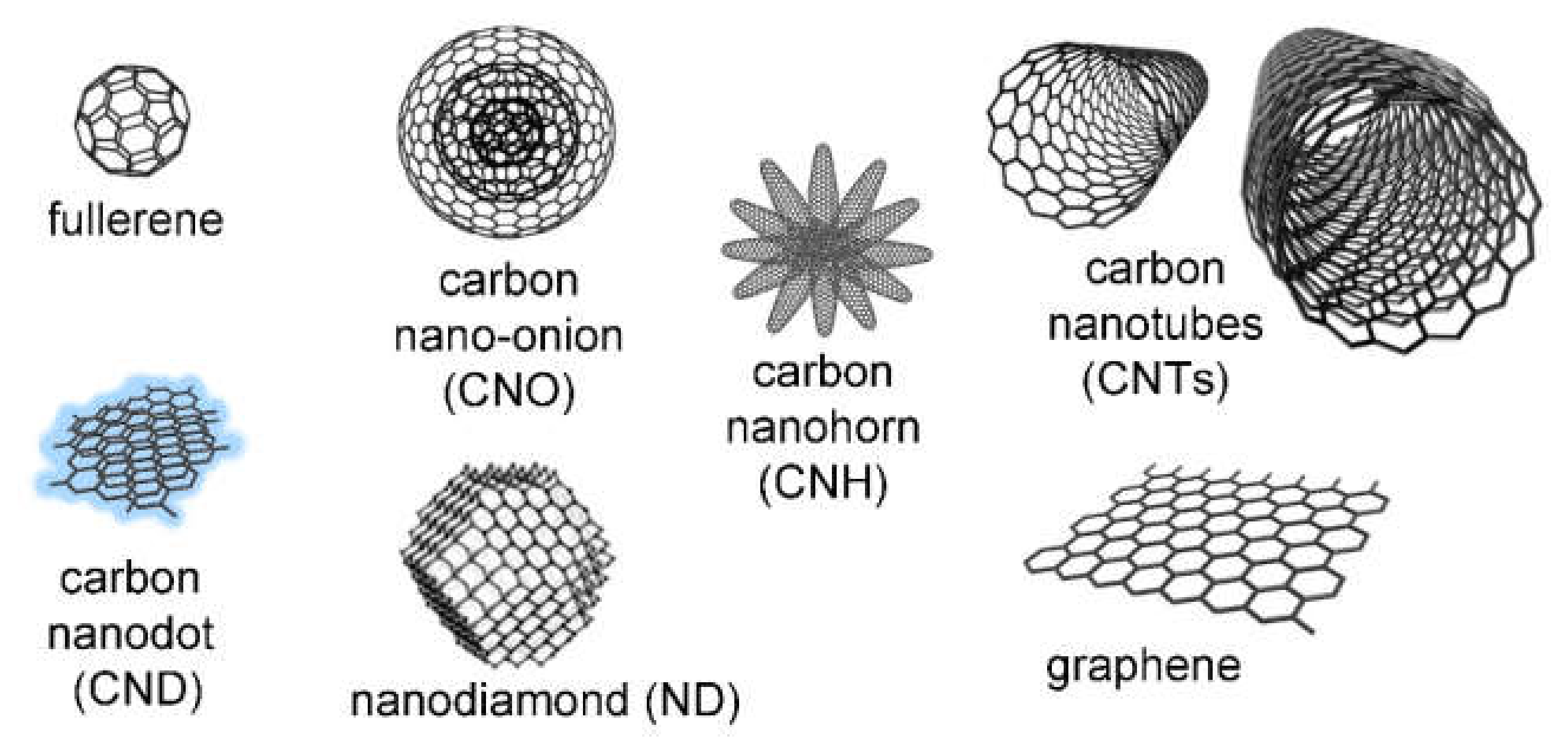
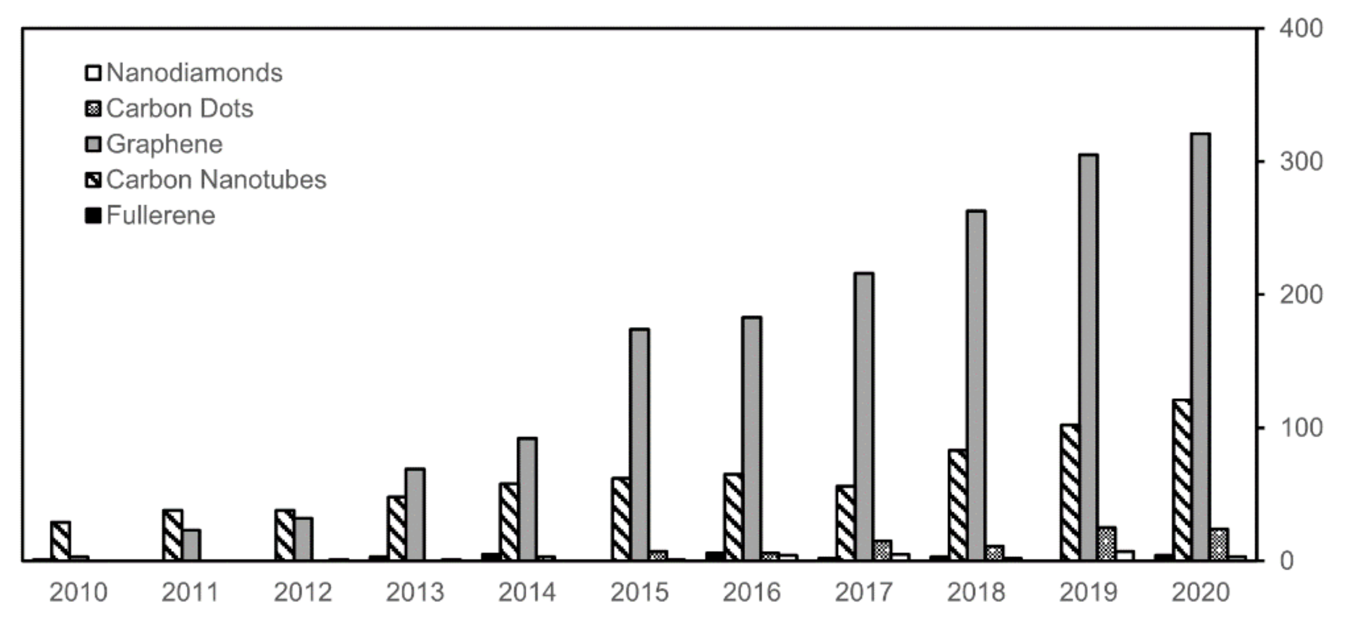
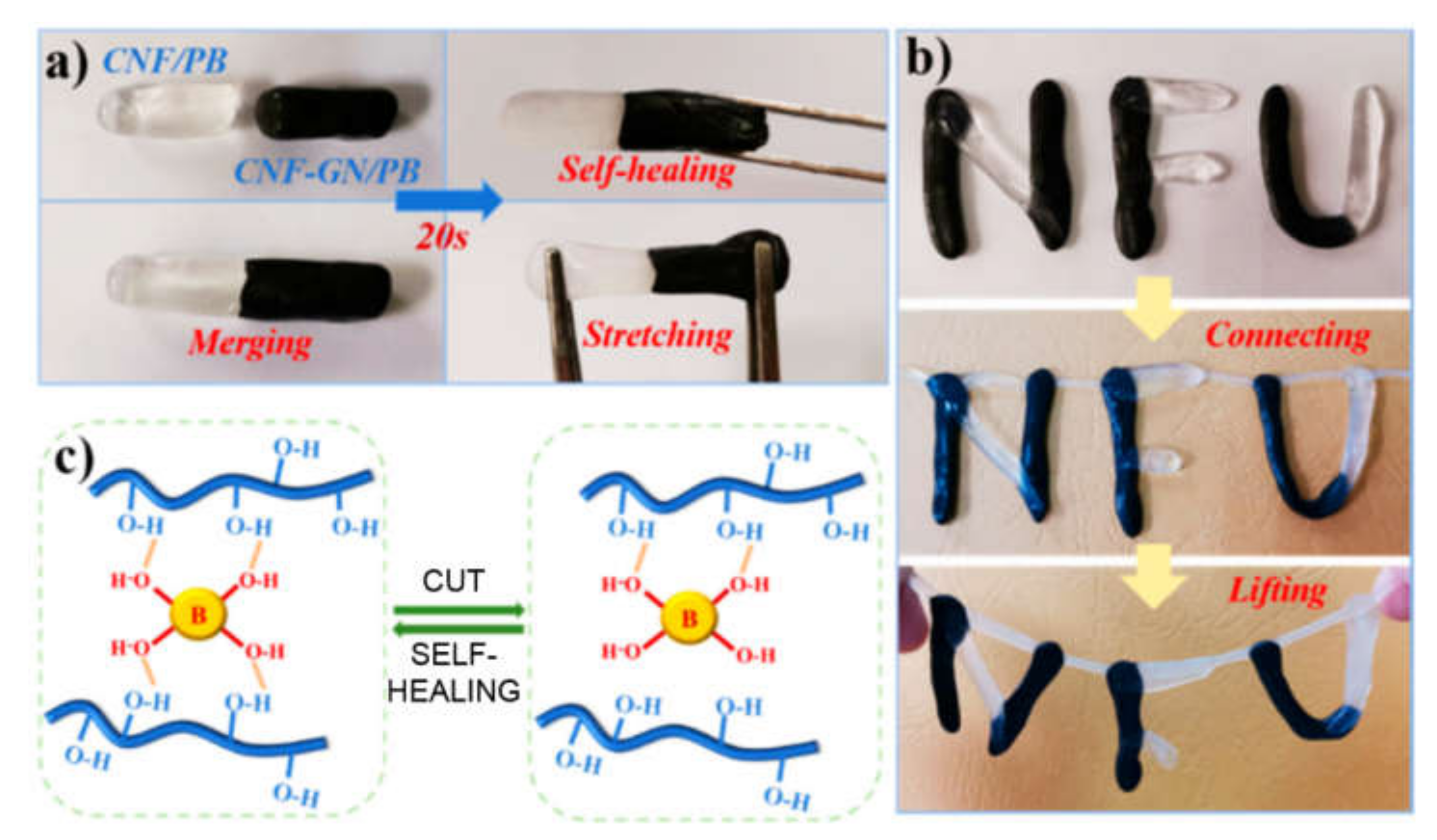
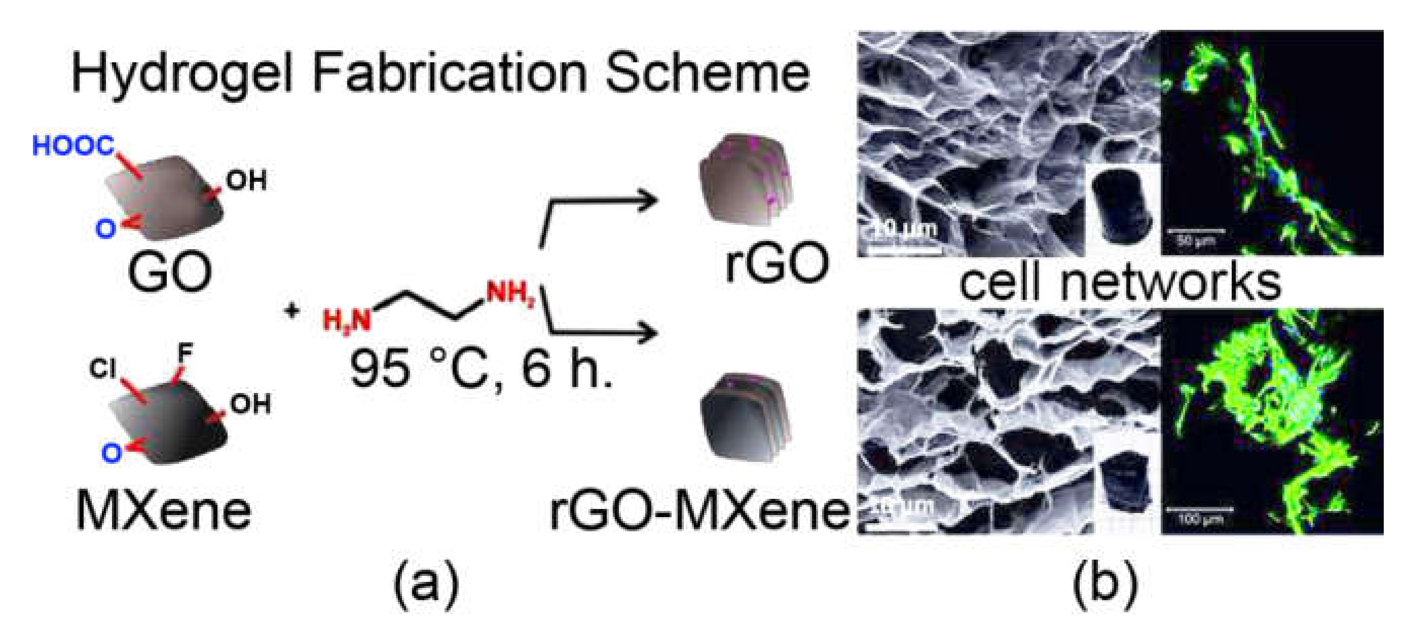
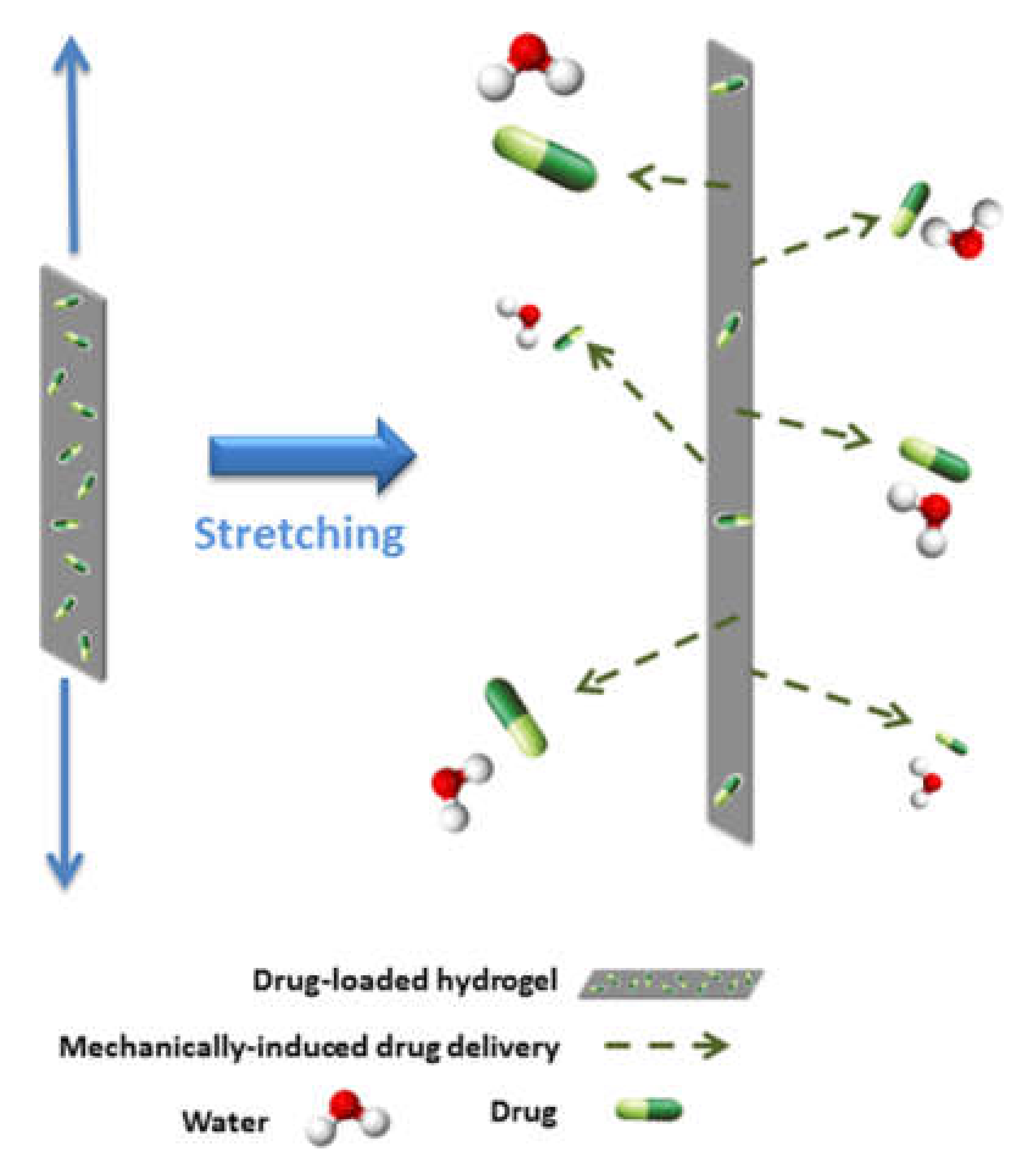
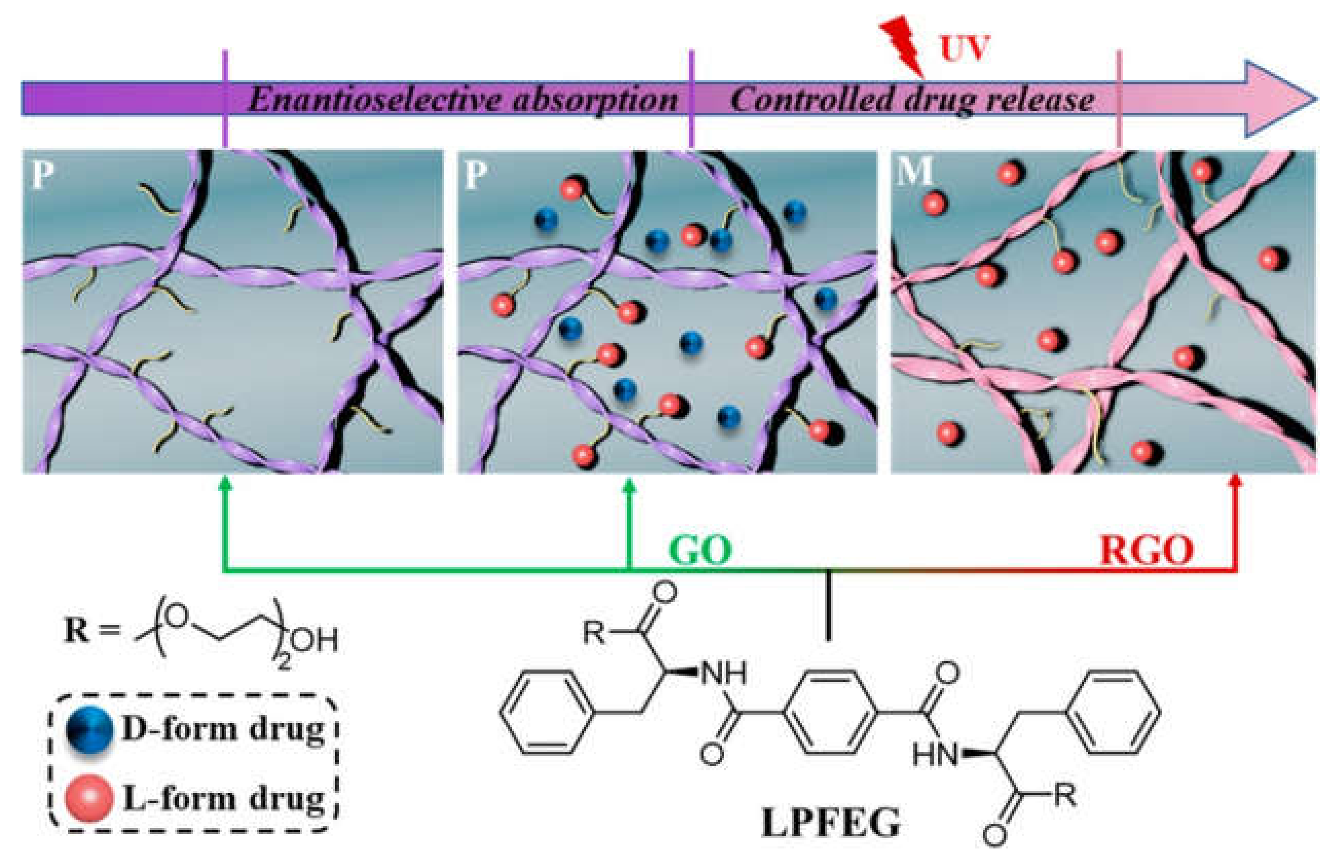
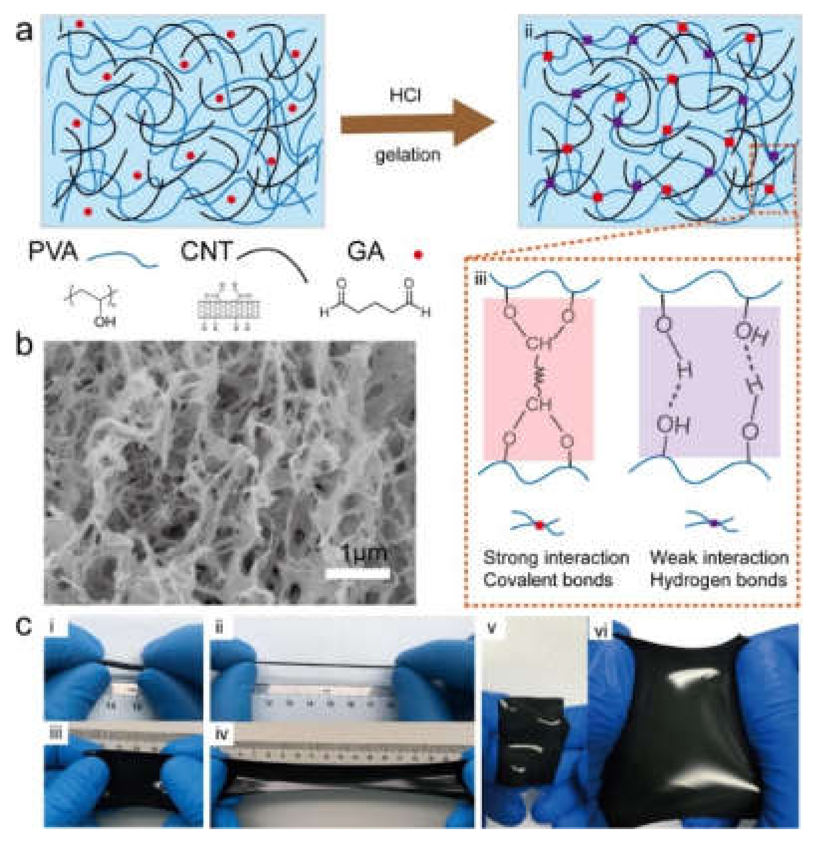
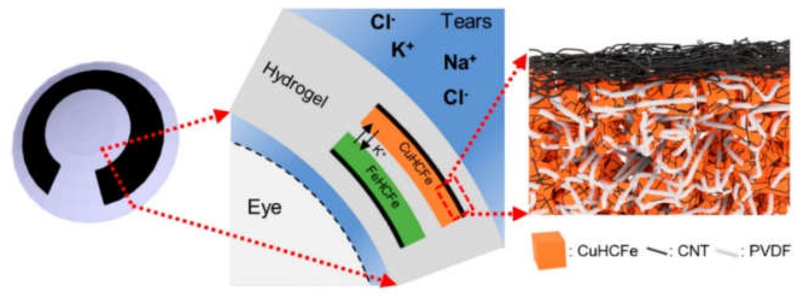
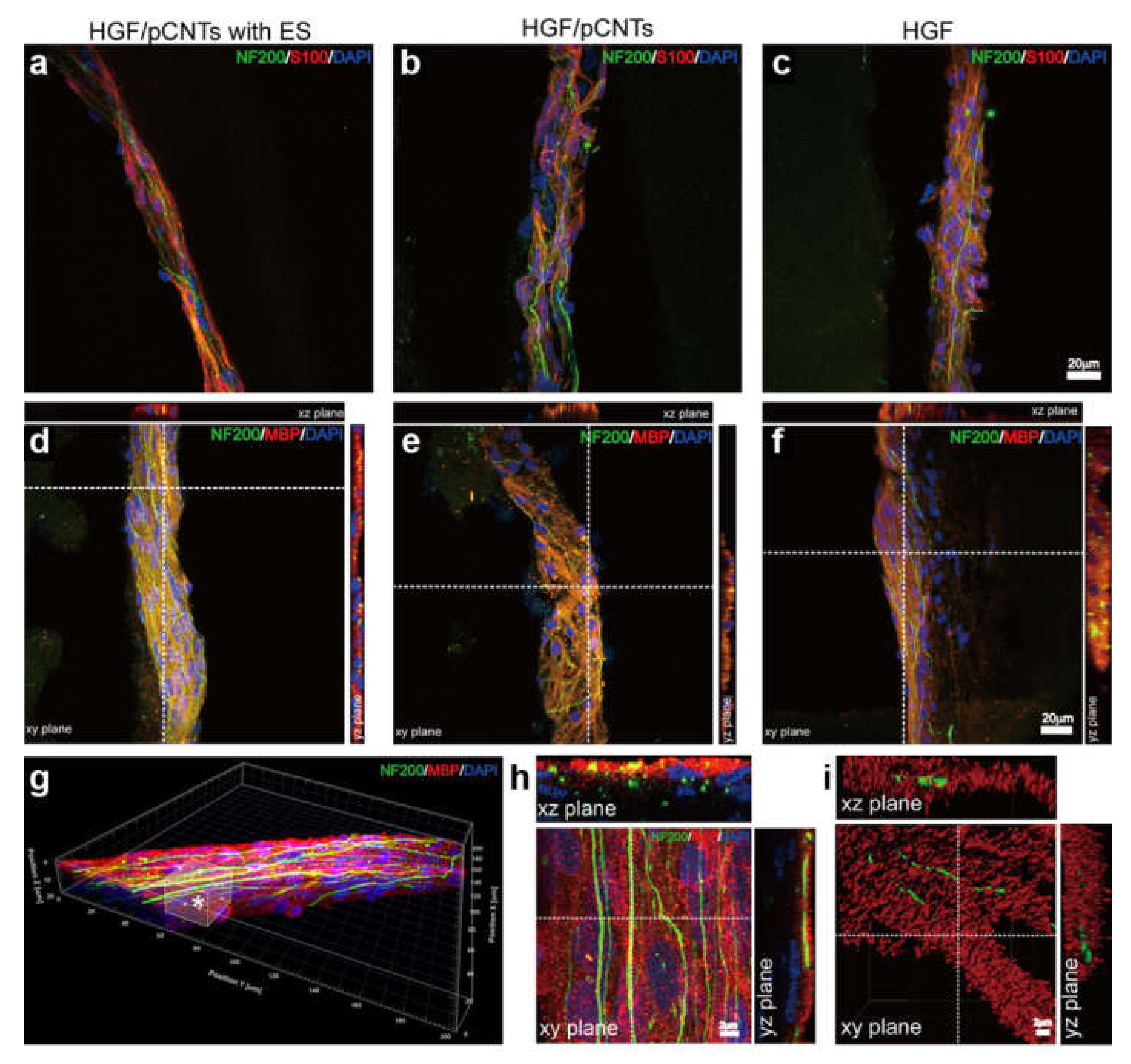
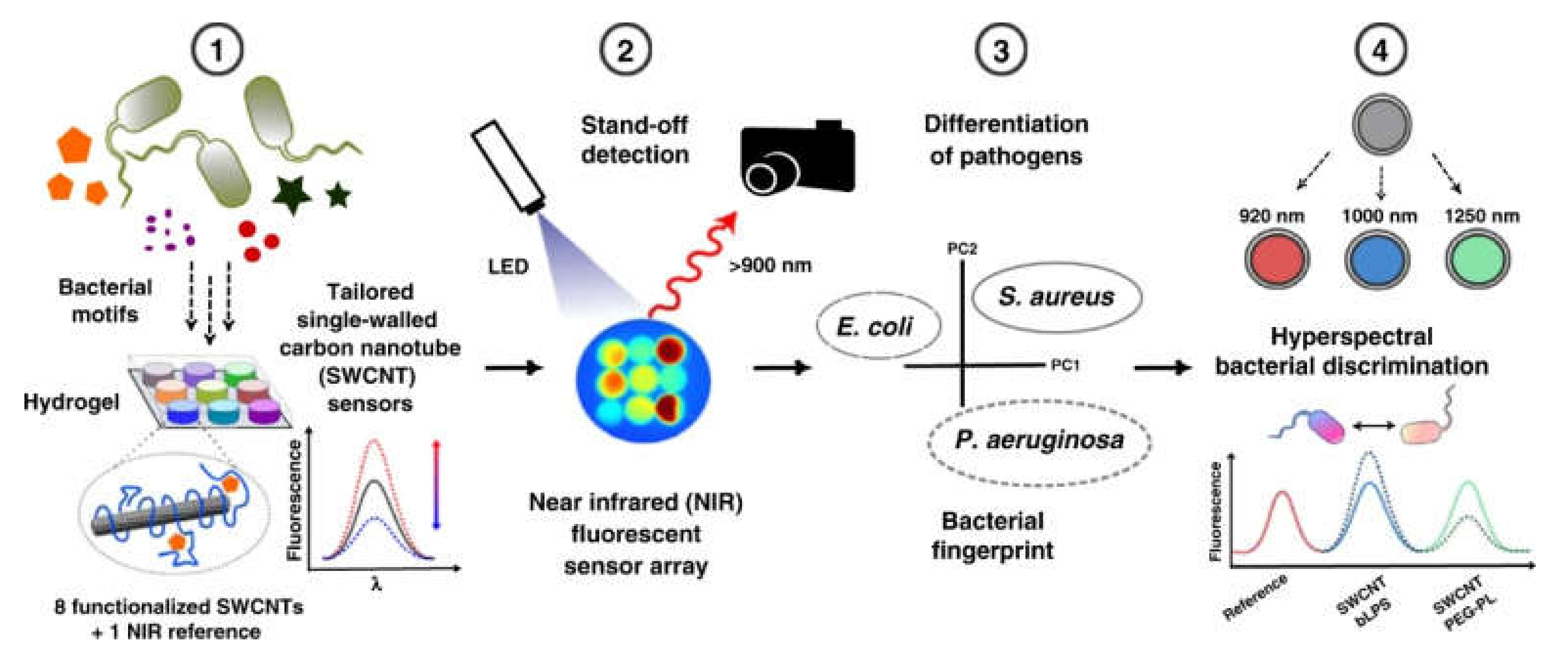
Table 1.
Recent examples of smart hydrogels with graphene for biomedical applications. GO = graphene oxide, rGO = reduced GO.
Table 1.
Recent examples of smart hydrogels with graphene for biomedical applications. GO = graphene oxide, rGO = reduced GO.
| Graphene Type | Gelator and Additives | Responsiveness | Application | Ref. |
|---|---|---|---|---|
| Graphene | Polyacrylamide | Drug release upon stretching | Wound healing | [86] |
| Graphene | Polyvinyl alcohol-borax, nanocellulose | Strain sensor | Artificial skin | [87] |
| Graphene | Polyvinyl alcohol, polyacrylic acid, glycerol | Electro-active | Wearable electronics | [88] |
| GO | Polyacrylamide, polyethylene glycol dimethacrylate | Electro-responsive drug release | Skin bandage | [89] |
| GO | Polyacrylamide, polyacrylic acid, carboxymethylcellulose | Salts/pH (de)swelling | Drug release | [90] |
| GO | Poly(N-isopropylacrylamide), chitosan, growth factors | Thermo-sensitive | Angiogenesis for tissue regeneration | [91] |
| GO | Polyvinyl alcohol, Ca++ ions, silver nanowires | Pressure sensor | Artificial skin | [92] |
| GO | Polyvinyl alcohol, G-quartet/hemin | Electro-active | Biosensors | [93] |
| GO | Cross-linked polyethylenimine | Enzyme-responsive check | Tissue regeneration | [94] |
| GO | Alginate, galactosidase | Lactose | Lactose-free Foodstuff | [95] |
| GO | Fibrin, iron-oxide nanoparticles, Hydroxyapatite | Magneto-responsive | Bone regeneration | [96] |
| GO | Cysteine | Electrochemi- Luminescence | Electrode sensor for diabetes | [97] |
| GO | Phenylalanine-derivative | Photo-active | Drug delivery | [98] |
| rGO | Carboxymethylchitosan, polyethylene glycol | Photo-thermal drug release | Cancer therapy | [99] |
| rGO | Polyacrylamide, calcium hydroxide nanoparticles | Strain sensor | Wearable electronics | [100] |
| rGO | Alginate | Electro-active | Heart repair | [101] |
| rGO | MXene (metal carbide) | Electro-active | Artificial heart actuators | [102] |
Table 2.
Recent examples of smart hydrogels with CNTs for biomedical applications. MW = multi-walled. SW = single-walled.
Table 2.
Recent examples of smart hydrogels with CNTs for biomedical applications. MW = multi-walled. SW = single-walled.
| CNT Type | Gelator and Additives | Responsiveness | Application | Ref. |
|---|---|---|---|---|
| MW | Polyacrylamide-co-acrylic acid, Chitosan | pH | Biomaterial | [142] |
| MW | Poly(ethyl acrylate-co-methacrylic acid-co-1,4-butanediol diacrylate) | pH | Soft tissue repair | [143] |
| MW | Polyacrylic acid, nanocellulose | Self-healing | Biosensors | [144] |
| MW | Polyethylene glycol diacrylate, Dopamine | Photo-triggered self-rolling | Bone regeneration | [145] |
| MW | Polyethylene glycol, α-cyclodextrin, Porphyrin | Photo-triggered Disassembly | Tissue engineering | [146] |
| MW | Poly(N-isopropylacrylamide), glucose oxidase | Glucose | Biosensors | [147] |
| MW | Poly(N-isopropylacrylamide), polyacrylic acid | Thermo-responsive | Biomaterials | [148] |
| MW | Polyvinyl alcohol, glutaraldehyde | Self-healing | Biosensors | [149] |
| MW | Methacrylated collagen, alginate | Electro-active | Cardiac patch | [150] |
| MW | Pericardial matrix | Electro-active | Cardiac patch | [151] |
| MW | Mucin, albumin, glutaraldehyde, glucose oxidase | Glucose | Biosensors | [152] |
| MW | Self-assembling tripeptide | Self-healing | Biomaterial | [46] |
| MW | Self-assembling 16-mer peptide | Electro-active | Nerve repair | [153] |
| MW | Matrigel, mesoporous silica, isobutyramide, doxorubicin, albumin | Photo-active | Drug release | [154] |
| SW | Alanine-based amphiphile, Viologen | Electro-active | Bioelectronics | [155] |
| SW | Hyaluronic acid, cross-linkers | Electro-active | Bioelectronics | [156] |
| SW | Polyethylene glycol, biomolecules | Photo-active | Biosensors | [157] |
| SW | Poly(ethylene glycol)-block-poly(D,L-allylglycine) | Photo-active | Bioelectronics | [158] |
| SW | Poly(vinyl alcohol), poly(N,N-dimethyl acrylamide) copolymer derivative with pyrene and borate groups | Self-healing | Biomaterials | [159] |
| SW | Polyvinylidene fluoride, CuHCFe, FeHCFe | Electro-active | Contact-lens battery | [160] |
Publisher’s Note: MDPI stays neutral with regard to jurisdictional claims in published maps and institutional affiliations. |
© 2021 by the authors. Licensee MDPI, Basel, Switzerland. This article is an open access article distributed under the terms and conditions of the Creative Commons Attribution (CC BY) license (https://creativecommons.org/licenses/by/4.0/).
Share and Cite
MDPI and ACS Style
Adorinni, S.; Rozhin, P.; Marchesan, S. Smart Hydrogels Meet Carbon Nanomaterials for New Frontiers in Medicine. Biomedicines 2021, 9, 570. https://doi.org/10.3390/biomedicines9050570
AMA Style
Adorinni S, Rozhin P, Marchesan S. Smart Hydrogels Meet Carbon Nanomaterials for New Frontiers in Medicine. Biomedicines. 2021; 9(5):570. https://doi.org/10.3390/biomedicines9050570
Chicago/Turabian StyleAdorinni, Simone, Petr Rozhin, and Silvia Marchesan. 2021. "Smart Hydrogels Meet Carbon Nanomaterials for New Frontiers in Medicine" Biomedicines 9, no. 5: 570. https://doi.org/10.3390/biomedicines9050570
Note that from the first issue of 2016, this journal uses article numbers instead of page numbers. See further details here.