Clinical Theragnostic Relationship between Drug-Resistance Specific miRNA Expressions, Chemotherapeutic Resistance, and Sensitivity in Breast Cancer: A Systematic Review and Meta-Analysis

 ,
,  ,
,  ,
,  ,
,
Abstract
:1. Introduction
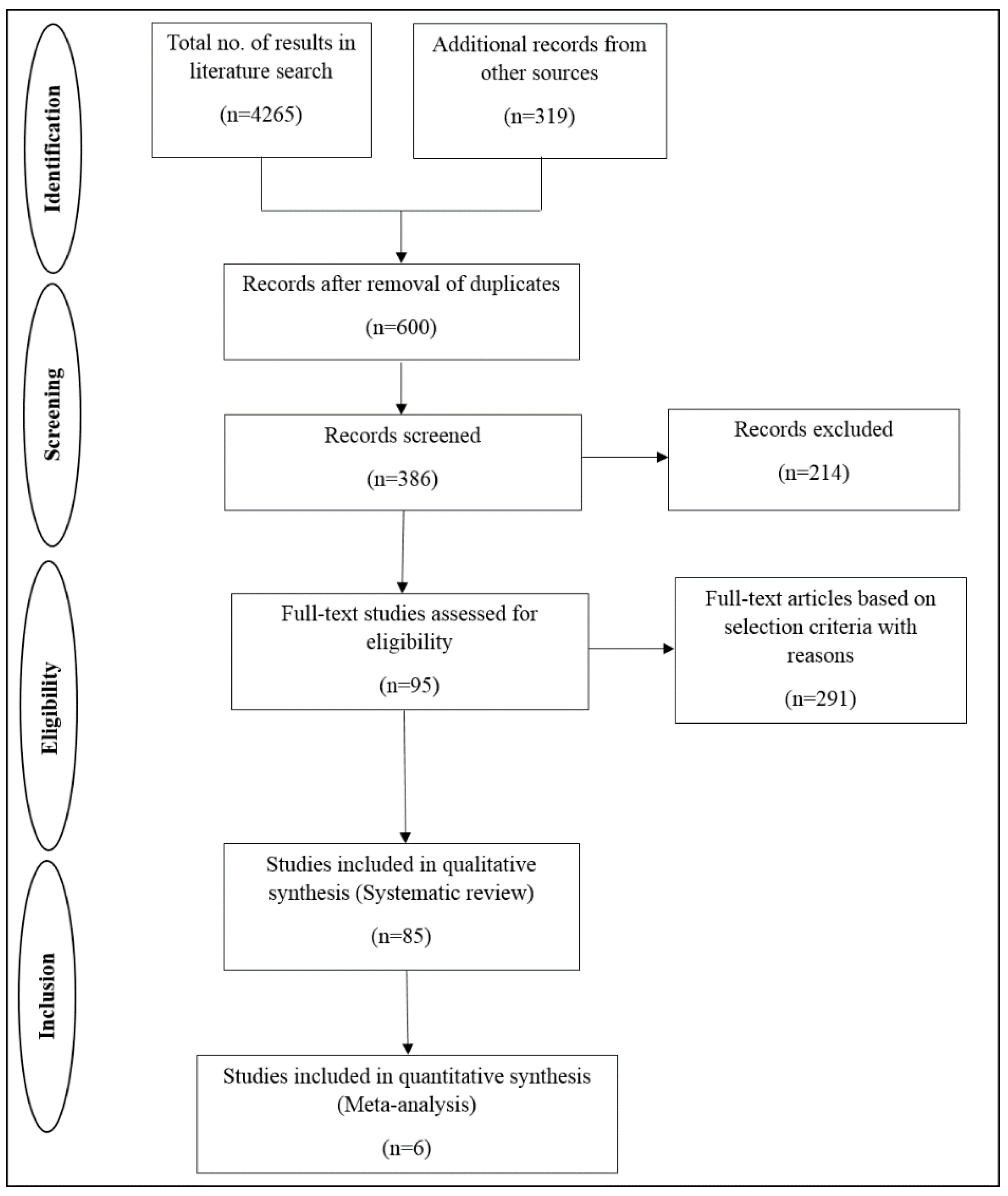
2. Methods
2.1. Selection Criteria
- An analysis of the association between miRNA and breast cancer;
- Studies with both breast cancer patients as well as in vitro studies with cell lines;
- Studies that focused on cancer tissues that had resistance to some form of therapy;
- Reporting of miRNA profiling platforms;
- Information about the genes or pathways involved in chemotherapeutic resistance or sensitivity;
- Inclusion of some in vitro assays to analyse the expression of miRNA or gene-related studies.
2.2. Data Analysis
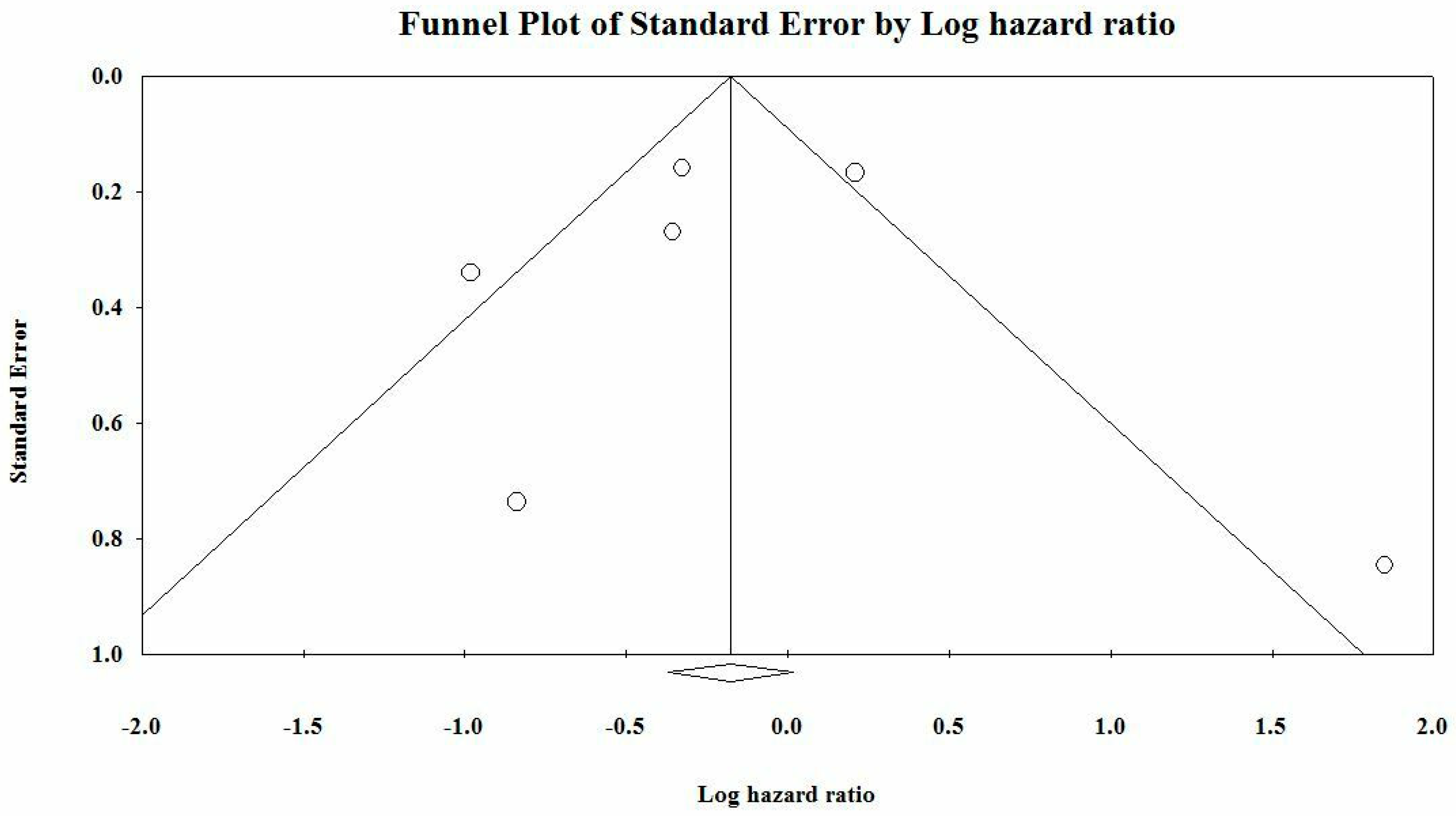
2.3. Publication Bias
2.4. Statistical Analysis
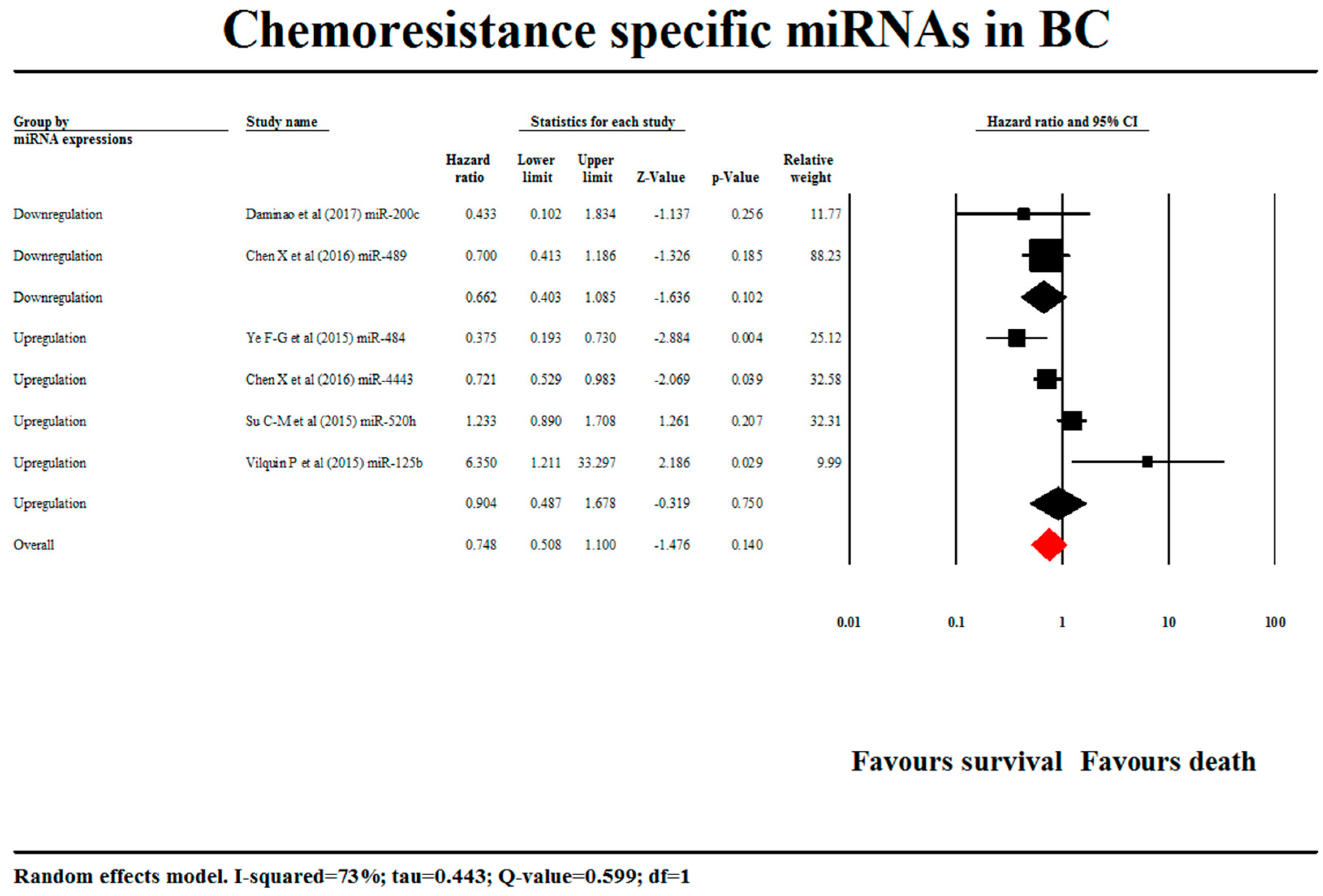
3. Results
miRNA Pathway Relation
4. Discussion
4.1. Role of miRNAs in Guiding Diagnosis and Prognosis
4.2. Current Challenges
Author Contributions
Funding
Acknowledgments
Conflicts of Interest
References
- Society, A.C. Cancer Facts & Figures 2017. Available online: https://www.cancer.org/research/cancer-facts-statistics/all-cancer-facts-figures/cancer-facts-figures-2017.html (accessed on 12 June 2018).
- Berry, J. Worldwide statistics on breast cancer. Available online: https://www.medicalnewstoday.com/articles/317135.php (accessed on 13 January 2019).
- Malvia, S.; Bagadi, S.A.; Dubey, U.S.; Saxena, S. Epidemiology of breast cancer in Indian women. Asia-Pac. J. Clin. Oncol. 2017, 13, 289–295. [Google Scholar] [CrossRef] [PubMed]
- Research, I.C.o.M. Three-Year Report of Population Based Cancer Registries 2012–2014. Available online: http://www.ncdirindia.org/ncrp/ALL_NCRP_REPORTS/PBCR_REPORT_2012_2014/ALL_CONTENT/PDF_Printed_Version/Preliminary_Pages_Printed.pdf (accessed on 13 January 2019).
- Research, I.C.o.M. Consolidated Report of Hospital Based Cancer Registries 2012–2014. Available online: http://www.ncdirindia.org/ncrp/ALL_NCRP_REPORTS/HBCR_REPORT_2012_2014/ALL_CONTENT/PDF_Printed_Version/Preliminary_Pages.pdf (accessed on 13 January 2019).
- Weigelt, B.; Geyer, F.C.; Reis-Filho, J.S. Histological types of breast cancer: How special are they? Mol. Oncol. 2010, 4, 192–208. [Google Scholar] [CrossRef] [PubMed] [Green Version]
- Cheang, M.C.; Chia, S.K.; Voduc, D.; Gao, D.; Leung, S.; Snider, J.; Watson, M.; Davies, S.; Bernard, P.S.; Parker, J.S. Ki67 index, HER2 status, and prognosis of patients with luminal B breast cancer. J. Natl. Cancer. Inst. 2009, 101, 736–750. [Google Scholar] [CrossRef] [PubMed]
- Gärtner, R.; Jensen, M.-B.; Nielsen, J.; Ewertz, M.; Kroman, N.; Kehlet, H.J.J. Prevalence of and factors associated with persistent pain following breast cancer surgery. JAMA 2009, 302, 1985–1992. [Google Scholar] [CrossRef]
- Early Breast Cancer Trialists’ Collaborative Group. Effects of radiotherapy and of differences in the extent of surgery for early breast cancer on local recurrence and 15-year survival: An overview of the randomised trials. Lancet 2005, 366, 2087–2106. [Google Scholar] [CrossRef]
- Fournier, A.; Berrino, F.; Clavel-Chapelon, F. Unequal risks for breast cancer associated with different hormone replacement therapies: Results from the E3N cohort study. Breast Cancer Res. Treat. 2008, 107, 103–111. [Google Scholar] [CrossRef]
- Higgins, M.J.; Baselga, J. Targeted therapies for breast cancer. J. Clin. Invest. 2011, 121, 3797–3803. [Google Scholar] [CrossRef]
- Miles, D.; Towlson, K.; Graham, R.; Reddish, M.; Longenecker, B.; Taylor-Papadimitriou, J.; Rubens, R.D. A randomised phase II study of sialyl-Tn and DETOX-B adjuvant with or without cyclophosphamide pretreatment for the active specific immunotherapy of breast cancer. Brit. J. Cancer 1996, 74, 1292–1296. [Google Scholar] [CrossRef]
- Hassan, M.; Ansari, J.; Spooner, D.; Hussain, S.J.O. Chemotherapy for breast cancer. Oncol. Rep. 2010, 24, 1121–1131. [Google Scholar] [CrossRef]
- Kovalchuk, O.; Filkowski, J.; Meservy, J.; Ilnytskyy, Y.; Tryndyak, V.P.; Vasyl’F, C.; Pogribny, I.P. Involvement of microRNA-451 in resistance of the MCF-7 breast cancer cells to chemotherapeutic drug doxorubicin. Mol. Cancer Therap. 2008, 7, 2152–2159. [Google Scholar] [CrossRef] [Green Version]
- Buzdar, A.U.; Valero, V.; Ibrahim, N.K.; Francis, D.; Broglio, K.R.; Theriault, R.L.; Pusztai, L.; Green, M.C.; Singletary, S.E.; Hunt, K.K. Neoadjuvant therapy with Paclitaxel followed by 5-fluorouracil, epirubicin, and cyclophosphamide chemotherapy and concurrent trastuzumab in human epidermal growth factor receptor 2–positive operable breast cancer: An update of the initial randomized study population and data of additional patients treated with the same regimen. Clin. Cancer Res. 2007, 13, 228–233. [Google Scholar] [PubMed]
- Jones, S.; Erban, J.; Overmoyer, B.; Budd, G.; Hutchins, L.; Lower, E.; Laufman, L.; Sundaram, S.; Urba, W.; Pritchard, K.I. Randomized phase III study of docetaxel compared with Paclitaxel in metastatic breast cancer. J. Clin. Oncol. 2005, 23, 5542–5551. [Google Scholar] [CrossRef] [PubMed]
- Robert, N.; Leyland-Jones, B.; Asmar, L.; Belt, R.; Ilegbodu, D.; Loesch, D.; Raju, R.; Valentine, E.; Sayre, R.; Cobleigh, M. Randomized phase III study of trastuzumab, Paclitaxel, and carboplatin compared with trastuzumab and Paclitaxel in women with HER-2–overexpressing metastatic breast cancer. J. Clin. Oncol. 2006, 24, 2786–2792. [Google Scholar] [CrossRef] [PubMed]
- Joensuu, H.; Bono, P.; Kataja, V.; Alanko, T.; Kokko, R.; Asola, R.; Utriainen, T.; Turpeenniemi-Hujanen, T.; Jyrkkiö, S.; Möykkynen, K. Fluorouracil, epirubicin, and cyclophosphamide with either docetaxel or vinorelbine, with or without trastuzumab, as adjuvant treatments of breast cancer: Final results of the FinHer Trial. J. Clin. Oncol. 2009, 27, 5685–5692. [Google Scholar] [CrossRef] [PubMed]
- Hanahan, D.; Bergers, G.; Bergsland, E. Less is more, regularly: Metronomic dosing of cytotoxic drugs can target tumor angiogenesis in mice. J. Clin. Investig. 2000, 105, 1045–1047. [Google Scholar] [CrossRef]
- Gonzalez-Angulo, A.M.; Morales-Vasquez, F.; Hortobagyi, G.N. Overview of resistance to systemic therapy in patients with breast cancer. Adv. Exp. Med. Biol. 2007, 608, 1–22. [Google Scholar]
- Early Breast Cancer Trialists’ Collaborative Group. Systemic treatment of early breast cancer by hormonal, cytotoxic, or immune therapy: 133 randomised trials involving 31 000 recurrences and 24 000 deaths among 75 000 women. Lancet 1992, 339, 1–15. [Google Scholar]
- Niero, E.L.; Rocha-Sales, B.; Lauand, C.; Cortez, B.A.; de Souza, M.M.; Rezende-Teixeira, P.; Urabayashi, M.S.; Martens, A.A.; Neves, J.H.; Machado-Santelli, G.M.; et al. The multiple facets of drug resistance: One history, different approaches. J Exp. Clin. Cancer Res.. 2014. [Google Scholar] [CrossRef]
- Coley, H.M. Mechanisms and strategies to overcome chemotherapy resistance in metastatic breast cancer. Cancer Treat. Rev. 2008, 34, 378–390. [Google Scholar] [CrossRef]
- Baguley, B.C. Multiple drug resistance mechanisms in cancer. Mol. Biotechnol. 2010, 46, 308–316. [Google Scholar] [CrossRef]
- Hu, Y.; Guo, R.; Wei, J.; Zhou, Y.; Ji, W.; Liu, J.; Zhi, X.; Zhang, J.J. Effects of PI3K inhibitor NVP-BKM120 on overcoming drug resistance and eliminating cancer stem cells in human breast cancer cells. Cell Death Dis. 2015. [Google Scholar] [CrossRef] [PubMed]
- Tanei, T.; Morimoto, K.; Shimazu, K.; Kim, S.J.; Tanji, Y.; Taguchi, T.; Tamaki, Y.; Noguchi, S. Association of breast cancer stem cells identified by aldehyde dehydrogenase 1 expression with resistance to sequential Paclitaxel and epirubicin-based chemotherapy for breast cancers. Clin. Cancer Res. 2009, 15, 4234–4241. [Google Scholar] [CrossRef] [PubMed]
- Teixeira, C.; Reed, J.C.; Pratt, M.C. Estrogen promotes chemotherapeutic drug resistance by a mechanism involving Bcl-2 proto-oncogene expression in human breast cancer cells. Cancer Res. 1995, 55, 3902–3907. [Google Scholar]
- Garofalo, M.; Croce, C.M. MicroRNAs as therapeutic targets in chemoresistance. Drug Resist. Updat. 2013, 16, 47–59. [Google Scholar] [CrossRef] [PubMed] [Green Version]
- Yang, W.; Ma, J.; Zhou, W.; Cao, B.; Zhou, X.; Yang, Z.; Zhang, H.; Zhao, Q.; Fan, D.; Hong, L. Molecular mechanisms and theranostic potential of miRNAs in drug resistance of gastric cancer. Exp. Opinion Therap. Tar. 2017, 21, 1063–1075. [Google Scholar] [CrossRef]
- Lin Teoh, S.; Das, S. The role of MicroRNAs in diagnosis, prognosis, metastasis and resistant cases in breast cancer. Curr. Pharmaceut. Des. 2017, 23, 1845–1859. [Google Scholar] [CrossRef] [PubMed]
- Zhang, W.; Zhou, J.; Zhu, X.; Yuan, H.J.G. MiR-126 reverses drug resistance to TRAIL through inhibiting the expression of c-FLIP in cervical cancer. Gene 2017, 627, 420–427. [Google Scholar] [CrossRef]
- Zhang, Y.; Wang, J. MicroRNAs are important regulators of drug resistance in colorectal cancer. Biol. Chem. 2017, 398, 929–938. [Google Scholar] [CrossRef] [Green Version]
- Võsa, U.; Vooder, T.; Kolde, R.; Vilo, J.; Metspalu, A.; Annilo, T. Meta-analysis of microRNA expression in lung cancer. Internat. J. Cancer 2013, 132, 2884–2893. [Google Scholar] [CrossRef]
- Zhuang, Z.; Hu, F.; Hu, J.; Wang, C.; Hou, J.; Yu, Z.; Wang, T.T.; Liu, X.; Huang, H. MicroRNA-218 promotes cisplatin resistance in oral cancer via the PPP2R5A/Wnt signaling pathway. Oncol. Rep. 2017, 38, 2051–2061. [Google Scholar] [CrossRef]
- Tung, S.; Huang, W.; Hsu, F.; Yang, Z.; Jang, T.; Chang, J.; Chuang, C.; Lai, C.; Wang, L.J.O. miRNA-34c-5p inhibits amphiregulin-induced ovarian cancer stemness and drug resistance via downregulation of the AREG-EGFR-ERK pathway. Oncogenesis 2017. [Google Scholar] [CrossRef] [PubMed]
- Amponsah, P.S.; Fan, P.; Bauer, N.; Zhao, Z.; Gladkich, J.; Fellenberg, J.; Herr, I. microRNA-210 overexpression inhibits tumor growth and potentially reverses gemcitabine resistance in pancreatic cancer. Cancer Lett. 2017, 388, 107–117. [Google Scholar] [CrossRef] [PubMed]
- Armstrong, C.M.; Liu, C.; Lou, W.; Lombard, A.P.; Evans, C.P.; Gao, A.C. MicroRNA-181a promotes docetaxel resistance in prostate cancer cells. Prostate 2017, 77, 1020–1028. [Google Scholar] [CrossRef] [PubMed]
- Fattore, L.; Sacconi, A.; Mancini, R.; Ciliberto, G.J.C. MicroRNA-driven deregulation of cytokine expression helps development of drug resistance in metastatic melanoma. Cytokine Growth Factor Rev. 2017, 36, 39–48. [Google Scholar] [CrossRef]
- Zhao, G.; Li, Y.; Wang, T.J.B. Potentiation of docetaxel sensitivity by miR-638 via regulation of STARD10 pathway in human breast cancer cells. Biochem. Biophys. Res. Commun. 2017, 487, 255–261. [Google Scholar] [CrossRef]
- Miller, T.E.; Ghoshal, K.; Ramaswamy, B.; Roy, S.; Datta, J.; Shapiro, C.L.; Jacob, S.; Majumder, S. MicroRNA-221/222 confers tamoxifen resistance in breast cancer by targeting p27Kip1. J. Biol. Chem. 2008, 283, 29897–29903. [Google Scholar] [CrossRef]
- Liang, Z.; Wu, H.; Xia, J.; Li, Y.; Zhang, Y.; Huang, K.; Wagar, N.; Yoon, Y.; Cho, H.T.; Scala, S. Involvement of miR-326 in chemotherapy resistance of breast cancer through modulating expression of multidrug resistance-associated protein 1. Biochem. Pharmacol. 2010, 79, 817–824. [Google Scholar] [CrossRef] [Green Version]
- Shi, W.; Gerster, K.; Alajez, N.M.; Tsang, J.; Pintilie, M.; Hui, A.B.; Sykes, J.; P’ng, C.; Miller, N.; McCready, D. MicroRNA-301 mediates proliferation and invasion in human breast cancer. Cancer Res. 2011, 71, 2926–2937. [Google Scholar] [CrossRef]
- Jung, E.J.; Santarpia, L.; Kim, J.; Esteva, F.J.; Moretti, E.; Buzdar, A.U.; Di Leo, A.; Le, X.F.; Bast Jr, R.C.; Park, S.T. Plasma microRNA 210 levels correlate with sensitivity to trastuzumab and tumor presence in breast cancer patients. Cancer 2012, 118, 2603–2614. [Google Scholar] [CrossRef]
- Ao, X.; Nie, P.; Wu, B.; Xu, W.; Zhang, T.; Wang, S.; Chang, H.; Zou, Z. Decreased expression of microRNA-17 and microRNA-20b promotes breast cancer resistance to taxol therapy by upregulation of NCOA3. Cell Death Dis. 2016. [Google Scholar] [CrossRef]
- Moher, D.; Liberati, A.; Tetzlaff, J.; Altman, D.G. Preferred reporting items for systematic reviews and meta-analyses: The PRISMA statement. Ann. Intern. Med. 2009, 151, 264–269. [Google Scholar] [CrossRef] [PubMed]
- Madhav, M.R.; Nayagam, S.G.; Biyani, K.; Pandey, V.; Kamal, D.G.; Sabarimurugan, S.; Ramesh, N.; Gothandam, K.M.; Jayaraj, R. Epidemiologic analysis of breast cancer incidence, prevalence, and mortality in India: Protocol for a systematic review and meta-analyses. Medicine 2018. [Google Scholar] [CrossRef] [PubMed]
- Poddar, A.; Aranha, R.R.; Muthukaliannan, G.K.; Nachimuthu, R.; Jayaraj, R. Head and neck cancer risk factors in India: Protocol for systematic review and meta-analysis. BMJ Open 2018. [Google Scholar] [CrossRef] [PubMed]
- Jayaraj, R.; Kumarasamy, C.; Piedrafita, D. Systematic review and meta-analysis protocol for Fasciola DNA vaccines. J. Vet. Res. 2018, 22, 517–524. [Google Scholar]
- Stroup, D.F.; Berlin, J.A.; Morton, S.C.; Olkin, I.; Williamson, G.D.; Rennie, D.; Moher, D.; Becker, B.J.; Sipe, T.A.; Thacker, S.B. Meta-analysis of observational studies in epidemiology: A proposal for reporting. JAMA 2000, 283, 2008–2012. [Google Scholar] [CrossRef] [PubMed]
- Jayaraj, R.; Kumarasamy, C.; Madhav, M.R.; Pandey, V.; Sabarimurugan, S.; Ramesh, N.; Gothandam, K.M.; Baxi, S. Comment on “Systematic Review and Meta-Analysis of Diagnostic Accuracy of miRNAs in Patients with Pancreatic Cancer”. Dis. Markers 2018. [Google Scholar] [CrossRef]
- Jayaraj, R.; Kumarasamy, C. Comment on ’Prognostic biomarkers for oral tongue squamous cell carcinoma: A systematic review and meta-analysis’. Br. J. Cancer 2018. [Google Scholar] [CrossRef]
- Jayaraj, R.; Kumarasamy, C. Comment on,” Survival for HPV-positive oropharyngeal squamous cell carcinoma with surgical versus non-surgical treatment approach: A systematic review and meta-analysis”. J. Oral Oncol. 2018, 90, 137–138. [Google Scholar] [CrossRef]
- Jayaraj, R.; Kumarasamy, C.; Sabarimurugan, S.; Baxi, S. Commentary: Blood-Derived microRNAs for Pancreatic Cancer Diagnosis: A Narrative Review and Meta-Analysis. Front. Physiol. 2018. [Google Scholar] [CrossRef]
- Jayaraj, R.; Kumarasamy, C. Conceptual interpretation of analysing and reporting of results on systematic review and meta-analysis of optimal extent of lateral neck dissection for well-differentiated thyroid carcinoma with metastatic lateral neck lymph nodes. Oral Oncol. 2019, 89, 153–154. [Google Scholar] [CrossRef]
- Jayaraj, R.; Kumarasamy, C.; Gothandam, K.M. Letter to the editor “Prognostic value of microRNAs in colorectal cancer: A meta-analysis”. Cancer Manag. Res. 2018, 10, 3501–3503. [Google Scholar] [CrossRef] [PubMed]
- Jayaraj, R.; Kumarasamy, C. Letter to the Editor about the Article: “Performance of different imaging techniques in the diagnosis of head and neck cancer mandibular invasion: A systematic review and meta-analysis”. J. Oral Oncol. 2018, 89, 159–160. [Google Scholar] [CrossRef] [PubMed]
- Jayaraj, R.; Kumarasamy, C.; Sabarimurugan, S.; Baxi, S. Letter to the Editor in response to the article, “The epidemiology of oral human papillomavirus infection in healthy populations: A systematic review and meta-analysis”. Oral Oncol. 2018, 84, 121–122. [Google Scholar] [CrossRef] [PubMed]
- Jayaraj, R.; Kumarasamy, C.; Samiappan, S.; Swaminathan, P. Letter to the Editor regarding, “The prognostic role of PD-L1 expression for survival in head and neck squamous cell carcinoma: A systematic review and meta-analysis”. Oral Oncol. 2019, 90, 139–140. [Google Scholar] [CrossRef] [PubMed]
- Jayaraj, R.; Kumarasamy, C.; Madurantakam Royam, M.; Devi, A.; Baxi, S. Letter to the editor: Is HIF-1alpha a viable prognostic indicator in OSCC? A critical review of a meta-analysis study. World J. Surg. Oncol. 2018. [Google Scholar] [CrossRef] [PubMed]
- Kumarasamy, C.; Devi, A.; Jayaraj, R. Prognostic value of microRNAs in head and neck cancers: A systematic review and meta-analysis protocol. Syst. Rev. 2018. [Google Scholar] [CrossRef]
- Jayaraj, R.; Kumarasamy, C. Systematic review and meta-analysis of cancer studies evaluating diagnostic test accuracy and prognostic values: Approaches to improve clinical interpretation of results. Cancer Manag. Res. 2018, 10, 4669–4670. [Google Scholar] [CrossRef]
- Jayaraj, R.; Kumarasamy, C.; Ramalingam, S.; Devi, A. Systematic review and meta-analysis of risk-reductive dental strategies for medication related osteonecrosis of the jaw among cancer patients: Approaches and strategies. Oral Oncol. 2018, 86, 312–313. [Google Scholar] [CrossRef]
- Sabarimurugan, S.; Madurantakam Royam, M.; Das, A.; Das, S.; Gothandam, K.M.; Jayaraj, R. Systematic Review and Meta-analysis of the Prognostic Significance of miRNAs in Melanoma Patients. Mol. Diagn. Ther. 2018, 22, 653–669. [Google Scholar] [CrossRef]
- Rosenthal, R. The file drawer problem and tolerance for null results. Psychol. Bull. 1979, 86, 638–641. [Google Scholar] [CrossRef]
- Orwin, R.G. A fail-safe N for effect size in meta-analysis. J. Educ. Stat. 1983, 8, 157–159. [Google Scholar] [CrossRef]
- Duval, S.; Tweedie, R. Trim and fill: A simple funnel-plot–based method of testing and adjusting for publication bias in meta-analysis. Biometrics 2000, 56, 455–463. [Google Scholar] [CrossRef] [PubMed]
- Higgins, J.P.; Thompson, S.G. Quantifying heterogeneity in a meta-analysis. Stat. Med. 2002, 21, 1539–1558. [Google Scholar] [CrossRef] [PubMed]
- DerSimonian, R.; Laird, N. Meta-analysis in clinical trials. Control. Clin. Trials 1986, 7, 177–188. [Google Scholar] [CrossRef]
- Lin, X.; Chen, W.; Wei, F.; Zhou, B.P.; Hung, M.-C.; Xie, X. Nanoparticle Delivery of miR-34a Eradicates Long-term-cultured Breast Cancer Stem Cells via Targeting C22ORF28 Directly. Theranostics 2017, 7, 4805–4824. [Google Scholar] [CrossRef] [Green Version]
- Nakano, M.; Fukami, T.; Gotoh, S.; Nakajima, M. A-to-I RNA editing up-regulates human dihydrofolate reductase in breast cancer. JBC 2017. [Google Scholar] [CrossRef]
- Miao, Y.; Zheng, W.; Li, N.; Su, Z.; Zhao, L.; Zhou, H.; Jia, L. MicroRNA-130b targets PTEN to mediate drug resistance and proliferation of breast cancer cells via the PI3K/Akt signaling pathway. Sci. Rep. 2017. [Google Scholar] [CrossRef]
- Chen, M.-J.; Cheng, Y.-M.; Chen, C.-C.; Chen, Y.-C.; Shen, C.-J. MiR-148a and miR-152 reduce tamoxifen resistance in ER+ breast cancer via downregulating ALCAM. Biochem. Biophys. Res. Commun. 2017, 483, 840–846. [Google Scholar] [CrossRef]
- Yang, F.; Luo, L.-j.; Zhang, L.; Wang, D.-d.; Yang, S.-j.; Ding, L.; Li, J.; Chen, D.; Ma, R.; Wu, J.-z. MiR-346 promotes the biological function of breast cancer cells by targeting SRCIN1 and reduces chemosensitivity to docetaxel. Gene 2017, 600, 21–28. [Google Scholar] [CrossRef]
- Gong, J.P.; Yang, L.; Tang, J.W.; Sun, P.; Hu, Q.; Qin, J.W.; Xu, X.M.; Sun, B.C.; Tang, J.H. Overexpression of microrna-24 increases the sensitivity to Paclitaxel in drug-resistant breast carcinoma cell lines via targeting abcb9. Oncol. Lett. 2016, 12, 3905–3911. [Google Scholar] [CrossRef]
- Zhu, J.; Zou, Z.; Nie, P.; Kou, X.; Wu, B.; Wang, S.; Song, Z.; He, J. Downregulation of microRNA-27b-3p enhances tamoxifen resistance in breast cancer by increasing NR5A2 and CREB1 expression. Cell Death Dis. 2016, 7, e2454. [Google Scholar] [CrossRef] [PubMed]
- Chen, X.; Lu, P.; Wang, D.-d.; Yang, S.-j.; Wu, Y.; Shen, H.-Y.; Zhong, S.-l.; Zhao, J.-h.; Tang, J.-h. The role of miRNAs in drug resistance and prognosis of breast cancer formalin-fixed paraffin-embedded tissues. Gene 2016, 595, 221–226. [Google Scholar] [CrossRef] [PubMed]
- Damiano, V.; Brisotto, G.; Borgna, S.; di Gennaro, A.; Armellin, M.; Perin, T.; Guardascione, M.; Maestro, R.; Santarosa, M. Epigenetic silencing of miR-200c in breast cancer is associated with aggressiveness and is modulated by ZEB1. Gene. Chromosome. Cancer 2017, 56, 147–158. [Google Scholar] [CrossRef] [PubMed]
- Jana, S.; Sengupta, S.; Biswas, S.; Chatterjee, A.; Roy, H.; Bhattacharyya, A.J.B. miR-216b suppresses breast cancer growth and metastasis by targeting SDCBP. Biochem. Biophys. Res. Commun. 2017, 482, 126–133. [Google Scholar] [CrossRef]
- Wang, D.-d.; Yang, S.-j.; Chen, X.; Shen, H.-Y.; Luo, L.-j.; Zhang, X.-h.; Zhong, S.-l.; Zhao, J.-h.; Tang, J.-h. miR-222 induces Adriamycin resistance in breast cancer through PTEN/Akt/p27 kip1 pathway. Tumor Biol. 2016, 37, 15315–15324. [Google Scholar] [CrossRef]
- Xu, X.; Lv, Y.-g.; Yan, C.-y.; Yi, J.; Ling, R. Enforced expression of hsa-miR-125a-3p in breast cancer cells potentiates docetaxel sensitivity via modulation of BRCA1 signaling. Biochem. Biophys. Res. Commun. 2016, 479, 893–900. [Google Scholar] [CrossRef]
- Chen, X.; Zhong, S.-l.; Lu, P.; Wang, D.-d.; Zhou, S.-y.; Yang, S.-j.; Shen, H.-y.; Zhang, L.; Zhang, X.-h.; Zhao, J.-h. miR-4443 Participates in the Malignancy of Breast Cancer. PLoS ONE 2016. [Google Scholar] [CrossRef]
- Gao, M.; Miao, L.; Liu, M.; Li, C.; Yu, C.; Yan, H.; Yin, Y.; Wang, Y.; Qi, X.; Ren, J. miR-145 sensitizes breast cancer to doxorubicin by targeting multidrug resistance-associated protein-1. Oncotarget 2016, 7, 59714–59726. [Google Scholar] [CrossRef] [Green Version]
- Thakur, S.; Grover, R.K.; Gupta, S.; Yadav, A.K.; Das, B.C. Identification of specific miRNA signature in paired sera and tissue samples of Indian women with triple negative breast cancer. PLoS ONE 2016. [Google Scholar] [CrossRef]
- Hu, Y.; Qiu, Y.; Yagüe, E.; Ji, W.; Liu, J.; Zhang, J. miRNA-205 targets VEGFA and FGF2 and regulates resistance to chemotherapeutics in breast cancer. Cell Death Dis. 2016. [Google Scholar] [CrossRef]
- Sha, L.; Zhang, Y.; Wang, W.; Sui, X.; Liu, S.; Wang, T.; Zhang, H. MiR-18a upregulation decreases Dicer expression and confers Paclitaxel resistance in triple negative breast cancer. Eur. Rev. Med. Pharmacol. Sci. 2016, 20, 2201–2208. [Google Scholar] [PubMed]
- Chen, X.; Wang, Y.W.; Xing, A.Y.; Xiang, S.; Shi, D.B.; Liu, L.; Li, Y.X.; Gao, P. Suppression of SPIN1-mediated PI3K–Akt pathway by miR-489 increases chemosensitivity in breast cancer. J. Pathol. 2016, 239, 459–472. [Google Scholar] [CrossRef] [PubMed]
- Venturutti, L.; Russo, R.C.; Rivas, M.A.; Mercogliano, M.F.; Izzo, F.; Oakley, R.; Pereyra, M.; De Martino, M.; Proietti, C.; Yankilevich, P. MiR-16 mediates trastuzumab and lapatinib response in ErbB-2-positive breast and gastric cancer via its novel targets CCNJ and FUBP1. Oncogene 2016, 35, 6189–6202. [Google Scholar] [CrossRef] [PubMed]
- Gu, X.; Xue, J.-Q.; Han, S.-J.; Qian, S.-Y.; Zhang, W.-H. Circulating microRNA-451 as a predictor of resistance to neoadjuvant chemotherapy in breast cancer. Cancer Biomarkers 2016, 16, 395–403. [Google Scholar] [CrossRef]
- Zhong, S.; Chen, X.; Wang, D.; Zhang, X.; Shen, H.; Yang, S.; Lv, M.; Tang, J.; Zhao, J. MicroRNA expression profiles of drug-resistance breast cancer cells and their exosomes. Oncotarget 2016, 7, 19601–19609. [Google Scholar] [CrossRef]
- Zhang, B.; Zhao, R.; He, Y.; Fu, X.; Fu, L.; Zhu, Z.; Fu, L.; Dong, J.-T. Micro RNA 100 sensitizes luminal A breast cancer cells to Paclitaxel treatment in part by targeting mTOR. Oncotarget 2016, 7, 5702–5714. [Google Scholar] [CrossRef]
- Shen, R.; Wang, Y.; Wang, C.-X.; Yin, M.; Liu, H.-L.; Chen, J.-P.; Han, J.-Q.; Wang, W.-B. MiRNA-155 mediates TAM resistance by modulating SOCS6-STAT3 signalling pathway in breast cancer. Am. J. Transl. Res. 2015, 7, 2115–2126. [Google Scholar]
- Yu, X.; Luo, A.; Liu, Y.; Wang, S.; Li, Y.; Shi, W.; Liu, Z.; Qu, X. MiR-214 increases the sensitivity of breast cancer cells to tamoxifen and fulvestrant through inhibition of autophagy. Mol. Cancer 2015. [Google Scholar] [CrossRef]
- Zhou, S.; Huang, Q.; Zheng, S.; Lin, K.; You, J.; Zhang, X. miR-27a regulates the sensitivity of breast cancer cells to cisplatin treatment via BAK-SMAC/DIABLO-XIAP axis. Tumor Biol. 2016, 37, 6837–6845. [Google Scholar] [CrossRef]
- Zheng, Y.; Lv, X.; Wang, X.; Wang, B.; Shao, X.; Huang, Y.; Shi, L.; Chen, Z.; Huang, J.; Huang, P. MiR-181b promotes chemoresistance in breast cancer by regulating Bim expression. Oncol. Rep. 2016, 35, 683–690. [Google Scholar] [CrossRef]
- Ye, Z.; Hao, R.; Cai, Y.; Wang, X.; Huang, G. Knockdown of miR-221 promotes the cisplatin-inducing apoptosis by targeting the BIM-Bax/Bak axis in breast cancer. Tumor Biol. 2016, 37, 4509–4515. [Google Scholar] [CrossRef] [PubMed]
- De Mattos-Arruda, L.; Bottai, G.; Nuciforo, P.G.; Di Tommaso, L.; Giovannetti, E.; Peg, V.; Losurdo, A.; Pérez-Garcia, J.; Masci, G.; Corsi, F. MicroRNA-21 links epithelial-to-mesenchymal transition and inflammatory signals to confer resistance to neoadjuvant trastuzumab and chemotherapy in HER2-positive breast cancer patients. Oncotarget 2015, 6, 37269–37280. [Google Scholar] [CrossRef] [PubMed]
- Lu, L.; Ju, F.; Zhao, H.; Ma, X. MicroRNA-134 modulates resistance to doxorubicin in human breast cancer cells by downregulating ABCC1. Biotechnol. Lett. 2015, 37, 2387–2394. [Google Scholar] [CrossRef] [PubMed]
- Sun, D.-w.; Mao, L.; Zhang, J.; Jiang, L.-h.; Li, J.; Wu, Y.; Ji, H.; Chen, W.; Wang, J.; Ma, R. MiR-139-5p inhibits the biological function of breast cancer cells by targeting Notch 1 and mediates chemosensitivity to docetaxel. Biochem. Biophys. Res. Commun. 2015, 465, 702–713. [Google Scholar]
- He, H.; Tian, W.; Chen, H.; Jiang, K. MiR-944 functions as a novel oncogene and regulates the chemoresistance in breast cancer. Tumor Biol. 2016, 37, 1599–1607. [Google Scholar] [CrossRef] [PubMed]
- Ikeda, K.; Horie-Inoue, K.; Ueno, T.; Suzuki, T.; Sato, W.; Shigekawa, T.; Osaki, A.; Saeki, T.; Berezikov, E.; Mano, H. miR-378a-3p modulates tamoxifen sensitivity in breast cancer MCF-7 cells through targeting GOLT1A. Sci. Rep. 2015. [Google Scholar] [CrossRef]
- Wu, J.; Li, S.; Jia, W.; Deng, H.; Chen, K.; Zhu, L.; Yu, F.; Su, F. Reduced Let-7a is associated with chemoresistance in primary breast cancer. PLoS ONE 2015. [Google Scholar] [CrossRef]
- Takahashi, R.-u.; Miyazaki, H.; Takeshita, F.; Yamamoto, Y.; Minoura, K.; Ono, M.; Kodaira, M.; Tamura, K.; Mori, M.; Ochiya, T. Loss of microRNA-27b contributes to breast cancer stem cell generation by activating ENPP1. Nature Commun. 2015. [Google Scholar] [CrossRef]
- Niu, J.; Xue, A.; Chi, Y.; Xue, J.; Wang, W.; Zhao, Z.; Fan, M.; Yang, C.H.; Shao, Z.; Pfeffer, L.M. Induction of miRNA-181a by genotoxic treatments promotes chemotherapeutic resistance and metastasis in breast cancer. Oncogene 2016, 35, 1302–1313. [Google Scholar] [CrossRef]
- Su, C.-M.; Wang, M.; Hong, C.; Chen, H.-A.; Su, Y.-H.; Wu, C.-H.; Huang, M.-T.; Chang, Y.W.; Jiang, S.S.; Sung, S.-Y. miR-520h is crucial for DAPK2 regulation and breast cancer progression. Oncogene 2016, 35, 1134–1142. [Google Scholar] [CrossRef]
- Boulbes, D.R.; Chauhan, G.B.; Jin, Q.; Bartholomeusz, C.; Esteva, F.J. CD44 expression contributes to trastuzumab resistance in HER2-positive breast cancer cells. Breast Cancer Res. Treat 2015, 151, 501–513. [Google Scholar] [CrossRef] [PubMed]
- Manvati, S.; Mangalhara, K.C.; Kalaiarasan, P.; Srivastava, N.; Bamezai, R. miR-24-2 regulates genes in survival pathway and demonstrates potential in reducing cellular viability in combination with docetaxel. Gene 2015, 567, 217–224. [Google Scholar] [CrossRef] [PubMed]
- Kang, L.; Mao, J.; Tao, Y.; Song, B.; Ma, W.; Lu, Y.; Zhao, L.; Li, J.; Yang, B.; Li, L. Micro RNA-34a suppresses the breast cancer stem cell-like characteristics by downregulating Notch 1 pathway. Cancer Sci. 2015, 106, 700–708. [Google Scholar] [CrossRef] [PubMed]
- Lü, M.; Ding, K.; Zhang, G.; Yin, M.; Yao, G.; Tian, H.; Lian, J.; Liu, L.; Liang, M.; Zhu, T. MicroRNA-320a sensitizes tamoxifen-resistant breast cancer cells to tamoxifen by targeting ARPP-19 and ERRγ. Sci. Rep. 2015. [Google Scholar] [CrossRef] [PubMed]
- Ye, F.-G.; Song, C.-G.; Cao, Z.-G.; Xia, C.; Chen, D.-N.; Chen, L.; Li, S.; Qiao, F.; Ling, H.; Yao, L. Cytidine deaminase axis modulated by miR-484 differentially regulates cell proliferation and chemoresistance in breast cancer. Cancer Res. 2015, 75, 1504–1515. [Google Scholar] [CrossRef] [PubMed]
- Vilquin, P.; Donini, C.F.; Villedieu, M.; Grisard, E.; Corbo, L.; Bachelot, T.; Vendrell, J.A.; Cohen, P.A. MicroRNA-125b upregulation confers aromatase inhibitor resistance and is a novel marker of poor prognosis in breast cancer. Breast Cancer Res. 2015. [Google Scholar] [CrossRef]
- Ujihira, T.; Ikeda, K.; Suzuki, T.; Yamaga, R.; Sato, W.; Horie-Inoue, K.; Shigekawa, T.; Osaki, A.; Saeki, T.; Okamoto, K. MicroRNA-574-3p, identified by microRNA library-based functional screening, modulates tamoxifen response in breast cancer. Sci. Rep. 2015. [Google Scholar] [CrossRef]
- Cui, J.; Yang, Y.; Li, H.; Leng, Y.; Qian, K.; Huang, Q.; Zhang, C.; Lu, Z.; Chen, J.; Sun, T. MiR-873 regulates ERα transcriptional activity and tamoxifen resistance via targeting CDK3 in breast cancer cells. Oncogene 2015, 34, 3895–3907. [Google Scholar] [CrossRef]
- Lv, J.; Xia, K.; Xu, P.; Sun, E.; Ma, J.; Gao, S.; Zhou, Q.; Zhang, M.; Wang, F.; Chen, F. miRNA expression patterns in chemoresistant breast cancer tissues. Biomed. Pharmacother. 2014, 68, 935–942. [Google Scholar] [CrossRef]
- He, X.; Xiao, X.; Dong, L.; Wan, N.; Zhou, Z.; Deng, H.; Zhang, X. MiR-218 regulates cisplatin chemosensitivity in breast cancer by targeting BRCA1. Tumor Biol. 2015, 36, 2065–2075. [Google Scholar] [CrossRef]
- Winsel, S.; Mäki-Jouppila, J.; Tambe, M.; Aure, M.; Pruikkonen, S.; Salmela, A.; Halonen, T.; Leivonen, S.; Kallio, L.; Børresen-Dale, A. Excess of miRNA-378a-5p perturbs mitotic fidelity and correlates with breast cancer tumourigenesis in vivo. Brit. J. Cancer 2014, 111, 2142–2151. [Google Scholar] [CrossRef] [PubMed] [Green Version]
- Hu, J.; Xu, J.; Wu, Y.; Chen, Q.; Zheng, W.; Lu, X.; Zhou, C.; Jiao, D. Identification of microRNA-93 as a functional dysregulated miRNA in triple-negative breast cancer. Tumor Biol. 2015, 36, 251–258. [Google Scholar] [CrossRef] [PubMed]
- He, D.-X.; Gu, X.-T.; Jiang, L.; Jin, J.; Ma, X. A methylation-based regulatory network for microRNA 320a in chemoresistant breast cancer. Mol. Pharmacol. 2014, 86, 536–547. [Google Scholar] [CrossRef] [PubMed]
- He, D.X.; Gu, X.T.; Li, Y.R.; Jiang, L.; Jin, J.; Ma, X. Methylation-regulated miR-149 modulates chemoresistance by targeting Glc NA c N-deacetylase/N-sulfotransferase-1 in human breast cancer. FEBS J. 2014, 281, 4718–4730. [Google Scholar] [CrossRef]
- Ouyang, M.; Li, Y.; Ye, S.; Ma, J.; Lu, L.; Lv, W.; Chang, G.; Li, X.; Li, Q.; Wang, S. MicroRNA profiling implies new markers of chemoresistance of triple-negative breast cancer. PLoS ONE 2014. [Google Scholar] [CrossRef]
- Luo, M.-L.; Gong, C.; Chen, C.-H.; Lee, D.Y.; Hu, H.; Huang, P.; Yao, Y.; Guo, W.; Reinhardt, F.; Wulf, G. Prolyl isomerase Pin1 acts downstream of miR200c to promote cancer stem–like cell traits in breast cancer. Cancer Res. 2014, 74, 3603–3616. [Google Scholar] [CrossRef]
- Jiang, L.; He, D.; Yang, D.; Chen, Z.; Pan, Q.; Mao, A.; Cai, Y.; Li, X.; Xing, H.; Shi, M. MiR-489 regulates chemoresistance in breast cancer via epithelial mesenchymal transition pathway. FEBS Lett. 2014, 588, 2009–2015. [Google Scholar] [CrossRef]
- Ye, X.-M.; Zhu, H.-Y.; Bai, W.-D.; Wang, T.; Wang, L.; Chen, Y.; Yang, A.-G.; Jia, L.-T. Epigenetic silencing of miR-375 induces trastuzumab resistance in HER2-positive breast cancer by targeting IGF1R. BMC Cancer 2014. [Google Scholar] [CrossRef]
- Zhu, Y.; Wu, J.; Li, S.; Ma, R.; Cao, H.; Ji, M.; Jing, C.; Tang, J. The function role of miR-181a in chemosensitivity to adriamycin by targeting Bcl-2 in low-invasive breast cancer cells. Cell. Physiol. Biochem. 2013, 32, 1225–1237. [Google Scholar] [CrossRef]
- Yang, G.; Wu, D.; Zhu, J.; Jiang, O.; Shi, Q.; Tian, J.; Weng, Y. Upregulation of miR-195 increases the sensitivity of breast cancer cells to Adriamycin treatment through inhibition of Raf-1. Oncol. Rep. 2013, 30, 877–889. [Google Scholar] [CrossRef]
- Pichiorri, F.; Palmieri, D.; De Luca, L.; Consiglio, J.; You, J.; Rocci, A.; Talabere, T.; Piovan, C.; Lagana, A.; Cascione, L. In vivo NCL targeting affects breast cancer aggressiveness through miRNA regulation. J. Exp. Med. 2013, 210, 951–968. [Google Scholar] [CrossRef] [PubMed]
- Wang, H.J.; Guo, Y.Q.; Tan, G.; Dong, L.; Cheng, L.; Li, K.J.; Wang, Z.Y.; Luo, H.F. miR-125b regulates side population in breast cancer and confers a chemoresistant phenotype. J. Cell. Biochem. 2013, 114, 2248–2257. [Google Scholar] [CrossRef] [PubMed]
- Ji, S.; Shao, G.; Lv, X.; Liu, Y.; Fan, Y.; Wu, A.; Hu, H. Downregulation of mi RNA-128 sensitises breast cancer cell to chemodrugs by targeting Bax. Cell Biol. Int. 2013, 37, 653–658. [Google Scholar] [CrossRef] [PubMed]
- Hu, H.; Li, S.; Cui, X.; Lv, X.; Jiao, Y.; Yu, F.; Yao, H.; Song, E.; Chen, Y.; Wang, M. The overexpression of hypomethylated miR-663 induces chemotherapy resistance in human breast cancer cells by targeting heparin sulfate proteoglycan 2 (HSPG2). Int. J. Biol. Chem. 2013, 288, 10973–10985. [Google Scholar] [CrossRef] [PubMed]
- Masuda, M.; Miki, Y.; Hata, S.; Takagi, K.; Sakurai, M.; Ono, K.; Suzuki, K.; Yang, Y.; Abe, E.; Hirakawa, H. An induction of microRNA, miR-7 through estrogen treatment in breast carcinoma. J. Transl. Med. 2012. [Google Scholar] [CrossRef]
- Li, X.-j.; Ji, M.-h.; Zhong, S.-l.; Zha, Q.-b.; Xu, J.-j.; Zhao, J.-h.; Tang, J.-h. MicroRNA-34a modulates chemosensitivity of breast cancer cells to adriamycin by targeting Notch 1. Arch. Med. Res. 2012, 43, 514–521. [Google Scholar] [CrossRef]
- Lv, K.; Liu, L.; Wang, L.; Yu, J.; Liu, X.; Cheng, Y.; Dong, M.; Teng, R.; Wu, L.; Fu, P. Lin28 mediates Paclitaxel resistance by modulating p21, Rb and Let-7a miRNA in breast cancer cells. PLoS ONE 2012. [Google Scholar] [CrossRef]
- Wang, H.; Tan, G.; Dong, L.; Cheng, L.; Li, K.; Wang, Z.; Luo, H. Circulating MiR-125b as a marker predicting chemoresistance in breast cancer. PLoS ONE 2012. [Google Scholar] [CrossRef]
- Chen, J.; Tian, W.; Cai, H.; He, H.; Deng, Y. Down-regulation of microRNA-200c is associated with drug resistance in human breast cancer. Med. Oncol. 2012, 29, 2527–2534. [Google Scholar] [CrossRef]
- Zhu, Y.; Yu, F.; Jiao, Y.; Feng, J.; Tang, W.; Yao, H.; Gong, C.; Chen, J.; Su, F.; Zhang, Y. Reduced miR-128 in breast tumor–initiating cells induces chemotherapeutic resistance via Bmi-1 and ABCC5. Clin. Cancer Res. 2011, 17, 7105–7115. [Google Scholar] [CrossRef]
- Zhao, Y.; Deng, C.; Lu, W.; Xiao, J.; Ma, D.; Guo, M.; Recker, R.R.; Gatalica, Z.; Wang, Z.; Xiao, G.G. let-7 microRNAs induce tamoxifen sensitivity by downregulation of estrogen receptor α signaling in breast cancer. Mol. Med. 2011, 17, 1233–1241. [Google Scholar] [CrossRef] [PubMed]
- Gong, C.; Yao, Y.; Wang, Y.; Liu, B.; Wu, W.; Chen, J.; Su, F.; Yao, H.; Song, E. Up-regulation of miR-21 mediates resistance to trastuzumab therapy for breast cancer. J. Biol. Chem. 2011, 286, 19127–19137. [Google Scholar] [CrossRef] [PubMed]
- Cittelly, D.M.; Das, P.M.; Spoelstra, N.S.; Edgerton, S.M.; Richer, J.K.; Thor, A.D.; Jones, F.E. Downregulation of miR-342 is associated with tamoxifen resistant breast tumors. Mol. Cancer 2010. [Google Scholar] [CrossRef] [PubMed]
- Maillot, G.; Lacroix-Triki, M.; Pierredon, S.; Gratadou, L.; Schmidt, S.; Bénès, V.; Roché, H.; Dalenc, F.; Auboeuf, D.; Millevoi, S. Widespread estrogen-dependent repression of micrornas involved in breast tumor cell growth. Cancer Res. 2009, 69, 8332–8340. [Google Scholar] [CrossRef]
- Iorio, M.V.; Casalini, P.; Piovan, C.; Di Leva, G.; Merlo, A.; Triulzi, T.; Ménard, S.; Croce, C.M.; Tagliabue, E. microRNA-205 regulates HER3 in human breast cancer. Cancer Res. 2009, 69, 2195–2200. [Google Scholar] [CrossRef]
- Yu, F.; Yao, H.; Zhu, P.; Zhang, X.; Pan, Q.; Gong, C.; Huang, Y.; Hu, X.; Su, F.; Lieberman, J. let-7 regulates self renewal and tumorigenicity of breast cancer cells. Cell 2007, 131, 1109–1123. [Google Scholar] [CrossRef]
- Li, G.; Wu, X.; Qian, W.; Cai, H.; Sun, X.; Zhang, W.; Tan, S.; Wu, Z.; Qian, P.; Ding, K. CCAR1 5′ UTR as a natural miRancer of miR-1254 overrides tamoxifen resistance. Cell Res. 2016, 26, 655–673. [Google Scholar] [CrossRef]
- Yu, S.-J.; Yang, L.; Hong, Q.; Kuang, X.-Y.; Di, G.-H.; Shao, Z.-M. MicroRNA-200a confers chemoresistance by antagonizing TP53INP1 and YAP1 in human breast cancer. BMC Cancer 2018. [Google Scholar] [CrossRef]
- Lee, J.W.; Guan, W.; Han, S.; Hong, D.K.; Kim, L.S.; Kim, H. Micro RNA-708-3p mediates metastasis and chemoresistance through inhibition of epithelial-to-mesenchymal transition in breast cancer. Cancer Sci. 2018, 109, 1404–1413. [Google Scholar] [CrossRef]
- Si, W.; Shen, J.; Du, C.; Chen, D.; Gu, X.; Li, C.; Yao, M.; Pan, J.; Cheng, J.; Jiang, D. A miR-20a/MAPK1/c-Myc regulatory feedback loop regulates breast carcinogenesis and chemoresistance. Cell Death Differ. 2018, 25, 406–420. [Google Scholar] [CrossRef]
- Cheng, S.; Huang, Y.; Lou, C.; He, Y.; Zhang, Y.; Zhang, Q. FSTL1 enhances chemoresistance and maintains stemness in breast cancer cells via integrin β3/Wnt signaling under miR-137 regulation. Cancer Biol. Ther. 2019, 20, 328–337. [Google Scholar] [CrossRef] [PubMed]
- Hu, G.; Zhao, X.; Wang, J.; Lv, L.; Wang, C.; Feng, L.; Shen, L.; Ren, W. miR-125b regulates the drug-resistance of breast cancer cells to doxorubicin by targeting HAX-1. Oncol. Lett. 2018, 15, 1621–1629. [Google Scholar] [CrossRef] [PubMed]
- Sabarimurugan, S.; Kumarasamy, C.; Baxi, S.; Devi, A.; Jayaraj, R. Systematic review and meta-analysis of prognostic microRNA biomarkers for survival outcome in nasopharyngeal carcinoma. PLoS ONE 2019, 14, e0209760. [Google Scholar] [CrossRef] [PubMed]


| Author | Ethnicity (Patient) | Period of Study | Drug(s) | Clinical Stages | No. of Samples (Cancer/Normal) | miRNA | miRNA Profiling Platform | ||||
|---|---|---|---|---|---|---|---|---|---|---|---|
| Total stages | I | II | III | IV | |||||||
| Lin X et al. (2017) [69] | Chinese | 2001 to 2006 and 2015 | docetaxel | 2 stages (I–II and III) | 74 | 4 | 60 | 0 | 138/83 | 34a | GeneSpring GX (Agilent Technologies, Capital Biochip Corporation) |
| Zhao G et al. (2017) [39] | Chinese | January 2012 to November 2015 | docetaxel | NM | NM | NM | NM | NM | 78/78 | 638 | qRT-PCR- SYBR Premix ExTaqTM (Takara, USA) |
| Nakano M et al. (2017) [70] | Japanese | NM | methotrexate | 3 stages (I, I–II, II, II–III) | 1 | 21 | 1 | NM | 19/19 | 25-3p and 125a-3p | Mx3000P (Stratagene, La Jolla, CA) |
| Miao Y et al. (2017) [71] | Chinese | January 2014 to March 2016 | doxorubicin | NM | NM | NM | NM | NM | 29/29 | 130b | SYBR Green qRT-PCR master mix (TaKaRa, Otsu, Shiga, Japan) |
| Chen M-J et al. (2017) [72] | Taiwanese | NM | tamoxifen | NM | NM | NM | NM | NM | 36a | 148a, 152 | ABI 7900 and SYBR® Select Master Mix (Applied Biosystems). |
| Yang F et al. (2017) [73] | Chinese | 2012–2015 | docetaxel | NM | NM | NM | NM | NM | 24/24 | 346 | ABI 7300 real-time PCR machine (Applied Biosystems, USA) |
| Gong J-P et al. (2016) [74] | Chinese | July 2010 to June 2014 | Paclitaxel | NM | NM | NM | NM | NM | 40a | 24 | TaqMan™ MicroRNA Assays (Applied Biosystems; Thermo Fisher Scientific, Inc.) |
| Ao X et al. (2016) [44] | Chinese | 2009–2011 | taxol | 3 stages (II, III and III-IV) | 0 | 12 | 18 | 25 | 55/55 | 17 and 20b | SYBR on the CFX96 system (Bio-Rad). |
| Zhu J et al. (2016) [75] | Chinese | 2005–2009 | tamoxifen | 3 stages (II, III and III–IV) | 0 | 8 | 22 | 22 | 73/19 | 27b-3p | SYBR on the CFX96 system (Bio-Rad) |
| Chen X et al. (2016) [76] | Chinese | January 2010 to February 2015 | docetaxel, epirubicin and vinorelbine | NM | NM | NM | NM | NM | 55/26 | 29a, 34a, 90b, 130a, 138, 139, 140, 149, 197, 200b, 210, 222, 423, 452, 574, 671, 744, 1246, 1268a, 3178, 3613, 4258, 4298, 4644, 6780b, 7107 and 7847 | SYBR® Advantage® qPCR Premix, Light cycler system (Roche, Australia) |
| Damiano V et al. [77] | Italian | 2000–2010 | anthracycline, anthracycline + taxane and CMF | 2 stages (I–II and III) | 2 | 48 | 0 | 51a | 200c | TaqMan normalizer (Applied Biosystems, ThermoFisher Scientific) | |
| Jana S et al. (2016) [78] | Indian | NM | NM | NM | NM | NM | NM | NM | 35/35 | 216b | SYBR green detection system |
| Wang D et al. (2016) [79] | Chinese | 2010–2015 | doxorubicin | NM | NM | NM | NM | NM | 21a | 222 | SYBR Premix Ex Taq system (Roche, Australia) |
| Xu X et al. (2016) [80] | NM | 2011–2014 | docetaxel | NM | NM | NM | NM | NM | 37/37 | 125a-3p | SYBR Premix ExTaqTM (Takara, USA) |
| Chen X et al. (2016) [81] | Chinese | January 2010 to February 2015 | epirubicin | 3 stages (I, II and III) | 10 | 32 | 4 | 0 | 76a | 4443 | MiR-X miRNA qRT-PCR SYBR Kit (638314; Clontech Laboratories, USA) |
| Gao M et al. (2016) [82] | Chinese | NM | doxorubicin | NM | NM | NM | NM | NM | 55/21 | 145 | NCode VILO miRNA cDNA Synthesis Kit and the EXPRESS SYBR GreenER miRNA qRT-PCR Kit, respectively (Invitrogen, Carlsbad, CA, USA) |
| Thakur S et al. (2016) [83] | Indian | NM | NM | 2 stages (I–II and III–IV) | 47 | 38 | 100/100 | 21, 145, 195, 210, 221 and Let-7a | TaqMan Universal Master Mix kit (Applied Biosystems, USA) | ||
| Hu Y et al. (2016) [84] | Chinese | June 2014 to June 2015 | docetaxel, doxorubicin and cyclophosphamide | 3 stages (II, III and III–IV) | 0 | 7 | 19 | 4 | 30a | 205 | TaqMan assays (Life Technologies) |
| Sha L-Y et al. (2016) [85] | Chinese | NM | epirubicin plus Paclitaxel | NM | NM | NM | NM | NM | 20/20 | 18a | TaqMan MicroRNA Assay Kit (Applied Biosystems) |
| Chen X et al. (2016) [86] | Chinese | 2008–2013 | doxorubicin | 4 stages (I, II, III and IV) | 37 | 64 | 12 | 3 | 114/114 | 489 | SYBR Primescript miRNA RT PCR Kit (TaKaRa, Dalian, China) |
| Venturutti L et al. (2016) [87] | Argentinians | 2008–2014 | trastuzumab and lapatinib | 4 stages (I, II, III and IV) | 5 | 9 | 3 | 2 | 19a | 16 | TaqMan® MicroRNA assay (Ambion) |
| Gu X et al. (2016) [88] | Chinese | January 2010 to December 2013 | epirubicin and docetaxel | 2 stages (II and III) | NM | NM | NM | NM | 82/60 | 451 | miScript SYBR Green PCR Kit (QIAGEN, Hilden, Germany) and a real-time LightCycler PCR (Roche Molecular Biochemicals, Mannheim, Germany) |
| Zhong S et al. (2016) [89] | Chinese | January 2010 to February 2015 | docetaxel, epirubicin and vinorelbine | 3 stages (I, II and III) | 6 | 8 | 9 | 0 | 23a | 138-5p, 139-5p, 140-3p, 149-3p, 197-3p, 210-3p, 423-5p, 574-3p, 744-5p, 1246, 1268a, 3178, 4258, 4298, 4443, 4644, 6780b-3p, 7107-5p and 7847-3p | Affymetrix GeneChip miRNA 4.0 Array |
| Zhang B et al. (2015) [90] | Chinese | NM | Paclitaxel | NM | NM | NM | NM | NM | 36/36 | 100 | Realplex Real-time PCR Detection System (Eppendorf, Beijing, China) |
| Shen R et al. (2015) [91] | Chinese | Between January 2006 to December 2011 | tamoxifen | NM | NM | NM | NM | NM | 18a | 155 | SYBR Green PCR master mix (TaKaRa) on the ABI 7500HT System |
| Yu X et al. (2015) [92] | Chinese | NM | tamoxifen and fulvestrant | NM | NM | NM | NM | NM | 20/20 | 214 | MiScript SYBR Green PCR kit (Qiagen) |
| Zhou S et al. (2015) [93] | Chinese | March 2014 to June 2015 | cisplatin | NM | NM | NM | NM | NM | 40/40 | 27a | FastStart Universal STBR Green Master (Roche, Switzerland) |
| Zheng Y et al. (2015) [94] | Chinese | NM | doxorubicin | NM | NM | NM | NM | NM | 30/30 | 181b | TaqMan MicroRNA assays kit (Applied Biosystems, USA) |
| Ye Z et al. (2015) [95] | Chinese | NM | cisplatin | NM | NM | NM | NM | NM | 85/85 | 221 | SYBR Green (Takara) |
| Mattos-Arruda L-D et al. (2015) [96] | Spaniards | 2005–2011 | trastuzumab, anthracyclines, taxanes | NM | NM | NM | NM | NM | 85a | 21 | LightCycler 480 Real-Time PCR System (Roche) |
| Lu L et al. (2015) [97] | Chinese | Not mentioned | doxorubicin, cyclophosphamide and fluorouracil | 2 stages (II–III) | NM | NM | NM | NM | 40a | 134 | SYBR PrimeScript miRNA RT-PCR Kit (Takara, Japan) |
| Zhang H-d et al. (2015) [98] | Chinese | 2012–2015 | docetaxel | 2 stages (I–II and III) | 18 | 17 | 0 | 35a | 139 | TaqMan MicroRNA Assay Kit (assay ID: miR-139-5p: 002289, and RNU6B: 001093), (Applied Biosystems, Life Technologies) | |
| He H et al. (2015) [99] | Chinese | October 2012 to January 2015 | cisplatin | NM | NM | NM | NM | NM | 70/70 | 944 | ABI PRISM 7900 Sequence Detection System (Applied Biosystems) with SYBR Green (TaKaRa, Japan) |
| Ikeda K et al. (2015) [100] | Japanese | Not mentioned | tamoxifen | NM | NM | NM | NM | NM | 40/16 | 378a-3p | TaqMan microRNA assays (Applied Biosystems, CA, USA) |
| Wu J et al. (2015) [101] | Chinese | January 2005 to December 2006 | before therapy | NM | NM | NM | NM | NM | 39a | Let7a | Real-time quantitative reverse transcription PCR (qRT-PCR) |
| January 2008 to December 2009 | epirubicin | NM | NM | NM | NM | NM | 31a | ||||
| Takahashi R et al. (2015) [102] | Japanese | 1996–2000 | docetaxel | 1 stage (II–III) | NM | 26 | NM | 26/9 | 27b | TaqMan MicroRNA Assays (Applied Biosystems) | |
| Niu J et al. (2015) [103] | Chinese | 1 January 2009 to 31 December 2010 | doxorubicin | 2 stages (I–II and III–IV) | 49 | 13 | 62a | 181a | MyiQ Real-Time PCR Detection System (Bio-Rad) | ||
| Su C-M et al. (2015) [104] | Taiwanese | NM | Paclitaxel | 2 stages (I and I–II) | 36 | 110 | NM | NM | 146a | 520h | Applied Biosystems 7900 Fast Real-Time PCR |
| Boulbes D et al. (2015) [105] | American | NM | trastuzumab, fluorouracil, epirubicin and cyclophosphamide | NM | NM | NM | NM | NM | 50a | has-520b-5p, 532-3p, 548n and 34a-3p | miRNA microarray (version 4.0, microRNACHIPv4) |
| Manvati S et al. (2015) [106] | Indian | NM | docetaxel | 3 stages (I, II and III) | NM | NM | NM | NM | 46/46 | 24-2 | TaqMan microRNA assays (Applied Biosystems) |
| Kang L et al. (2015) [107] | Chinese | NM | Paclitaxel | 4 stages (I, II, III and IV) | 11 | 18 | 12 | 4 | 45a | 34a | TaqMan MicroRNA Assay kit (Applied Biosystems, Foster City, CA, USA) |
| Lu M et al. (2015) [108] | Chinese | 2009–2010 | tamoxifen | NM | NM | NM | NM | NM | 31/27 | 320a | Applied Biosystems Step One real-time PCR system using an SYBR Premix Ex Taq II Kit (Takara Bio, Inc., Shiga, Japan) |
| Ye F-G et al. (2015) [109] | Chinese | September 2013 | gemcitabine | 3 stages (I, II and III) | 159 | 32 | NM | 400/243 | 484 | SYBR Premix Ex Taq System (TaKaRa) | |
| Vilquin P et al. (2015) [110] | French | NM | letrozole, anastrazole, tamoxifen and fulvestrant | 3 stages (I, II and III) | 4 | 18 | 23 | 0 | 65/65 | 125b | ExiLENT SYBR Green Master Mix and CFX96 (BioRad, Marne-laCoquette, France) |
| Ujihira T et al. (2015) [111] | Japanese | NM | tamoxifen | NM | NM | NM | NM | NM | 19a | 574-3p | triplicate TaqMan microRNA assays (Applied Biosystems, CA, USA) |
| Cui J et al. (2014) [112] | Chinese | NM | tamoxifen | NM | NM | NM | NM | NM | NM | 873 | RNeasy Mini kit (Qiagen, Hilden, Germany) or TRIzol (Invitrogen) reagent. SYBR Green PCR Master Mix reagents using an ABI Prism 7700 Sequence Detection System (Applied Biosystems, Foster City, CA, USA) |
| Lv J et al. (2014) [113] | Chinese | 2008–2009 | doxorubicin | NM | NM | NM | NM | NM | NM | 31, 125b-1, 141, 145, 196b, 200a, 200c, 370, 429, 491-3p, 576, 760, 765 and Let-7a | ABI 7900 PCR System (Applied Biosystems, USA) using Power SYBR Green PCR Master Mix (2X, Applied Biosystems) |
| He X et al. (2014) [114] | Chinese | NM | cisplatin | 4 stages (I, II, III and IV) | 15 + 17 | 15 + 17 | 30 + 23 | 30 + 23 | 85a | 218 | TRIzol reagent (Invitrogen) miRNA microarray chip (v.10.0, Exiqon, Vedbaek, Denmark) |
| Winsel S et al. (2014) [115] | Norwegians | May 1995 to December 1998 | taxol | NM | NM | NM | NM | NM | 101a | 378a-3p | RNeasy Mini Kit (Qiagen) TaqMan Universal Master Mix II, no PNG (Applied Biosystems, Foster City, CA, USA) |
| Hu J et al. (2014) [116] | Chinese | NM | NM | 4 stages (I, II, III and IV) | 20 | 25 | 31 | 4 | 119a | 93 | TRIzol Reagent (Invitrogen) and the miRNeasy Mini Kit (QIAGEN) |
| He DX et al. (2014) [117] | Chinese | NM | doxorubicin, Paclitaxel | NM | NM | NM | NM | NM | NM | 320a | All-in-One miRNA qRT-PCR detection kit (GeneCopoeia, Rockville, MD, USA) |
| He DX et al. (2014) [118] | Chinese | NM | doxorubicin, Paclitaxel | NM | NM | NM | NM | NM | NM | 149 | All-in-One miRNA qRT-PCR detection kit (GeneCopoeia, Rockville, MD, USA). Briefly, total RNA was extracted from MCF-7/WT and ADM cells with TRIzol (Invitrogen, Carlsbad, CA, USA) |
| Ouyang M et al. (2014) [119] | Chinese | 2011 (January–October) | doxorubicin | NM | NM | NM | NM | NM | NM | 10b-5p, 21-3p, 31-5p, 125b-3p, 130a-3p, 155-5p, 181a-5p, 181b-5p, 183-5p, 195-5p and 451a | Total RNA was harvested using TRIzol (Invitrogen) and miRNAeasy mini kit (QIAGEN). SYBR Premix EX TaqTM II kit (Takara, Dalian, China) |
| Luo ML et al. (2014) [120] | Chinese | NM | PiB | NM | NM | NM | NM | NM | NM | 200 | Total RNA was isolated from miRNeasy kit (Qiagen) and reversely transcribed by miScript PCR starter kit |
| Jiang L et al. (2014) [121] | Chinese | NM | doxorubicin | NM | NM | NM | NM | NM | NM | 489 | Total RNA was prepared using TRIzol (Beyotime, China) according to the manufacturer’s instructions. |
| Ye XM et al. (2014) [122] | Chinese | NM | trastuzumab/Herceptin | NM | NM | NM | NM | NM | NM | 375 | Total RNA was extracted from each cell line using TRIzol reagent (Invitrogen, USA) |
| Zhu Y et al. (2013) [123] | Chinese | NM | doxorubicin | 2 stages (I and II) | 34 | 9 | NM | NM | 43a | 181a | Total RNA was extracted from each cell line using TRIzol reagent (Invitrogen, Carlsbad, CA, USA) |
| Ye X et al. (2014) [122] | Chinese | NM | trastuzumab | NM | NM | NM | NM | NM | NM | 221 | Total RNA from each cell line was extracted by TRIzol reagent (Invitrogen, USA) |
| Yang G et al. (2013) [124] | Chinese | NM | doxorubicin | 2 stages (I and II) | 9 | 8 | NM | NM | 30a | 195 | Total cellular RNA from tissues and cultured cells were isolated using a TRIzol Reagent (Invitrogen) |
| Pichiorri F et al. (2013) [125] | Americans | NM | fulvestrant | NM | NM | NM | NM | NM | 183/57 | 21, 103, 221 and 222 | TaqMan PCR kit (Applied Biosystems) and 7900HT Sequence Detection System (Applied Biosystems) |
| Wang H-J et al. (2013) [126] | Chinese | January 2010 to December 2011 | Paclitaxel, 5-FU, epirubicin and cyclophosphamide | NM | NM | NM | NM | NM | 19/19 | 125b | ABI 7900HT system (Applied Biosystems) |
| Ji S et al. (2013) [127] | Chinese | 2007–2009 | taxol + doxorubicin + cyclophosphamide | NM | NM | NM | NM | NM | 67/67 | 128 | QRT-PCR |
| Hu H et al. (2013) [128] | Chinese | October 2003 to July 2010 | topotecan, etoposide, doxorubicin, docetaxel and cyclophosphamide | NM | NM | NM | NM | NM | 39/39 | 663 | Conventional TaqMan PCR (Bio-Rad) |
| Masuda M et al. (2011) [129] | Japanese | NM | estradiol (E2) | NM | NM | NM | NM | NM | 41a | 7 | PCR was performed in ABI7500 Real-Time PCR System (Applied Biosystems, Foster city, CA, USA) |
| Li X et al. (2012) [130] | Chinese | 2008–2010 | doxorubicin, cyclophosphamide (CTX) and 5-fluorouracil (5-FU) | 1 stage (II) | 0 | 38 | 0 | 0 | 38/38 | 34a | SYBR Green PCR Master Mix (Applied Biosystems, Foster City, CA, USA) |
| Lv K et al. (2012) [131] | Chinese | 2002–2010 | Paclitaxel, vincristine | NM | NM | NM | NM | NM | 9/9 | Lin28 | Real-time PCR was performed using the TaqMan MicroRNA Reverse Transcription Kit and the Fast Real-Time PCR System (Applied Biosystems, Carlsbad, CA, USA) |
| Wang H et al. (2012) [132] | Chinese | 2009–2010 | 5-FU (5-fluorouracil) | 2 stages (II and III) | 0 | 35 | 21 | 0 | 56/10 | 10b, 34a, 125b and 155 | miRNA-specific TaqMan MicroRNA Assays (Applied Biosystems) |
| Jung E-J et al. (2012) [43] | Americans, Koreans | NM | trastuzumab, Paclitaxel, fluorouracil, cyclophosphamide and epirubicin | 3 stages (I, II and III) | 33 | 31 | 8 | 0 | 72/72 | 21, 29a, 126 and 210 | TaqMan MicroRNA Assay kit (Applied Biosystems, Foster City, Calif) |
| Chen J et al. (2011) [133] | Chinese | 2007–2011 | doxorubicin | NM | NM | NM | NM | NM | 39a | 200c | Real-time PCR was performed using SYBR Green PCR Master Mix (Applied Biosystems, USA) on the Stepone plus system (Applied Biosystems, USA) |
| Zhu Y et al. (2011) [134] | Chinese | 2004–2011 | NM | 3 stages (II, III and IV) | NM | 44 | 29 | 4 | 77a | 128 | Mature miRNA expression analysis was conducted using a TaqMan MicroRNA Assays (Applied Biosystems) |
| Zhao Y et al. (2011) [135] | NM | NM | tamoxifen | NM | NM | NM | NM | NM | 29/15 | Let-7 | mirVana miRNA isolation kit (Ambion Inc., Austin, TX, USA) or from FFPE tissues using the miRNeasy FFPE Kit (Qiagen, Valencia, CA, USA) |
| Gong C et al. (2011) [136] | Chinese | 2008–2009 | trastuzumab (Herceptin) | NM | NM | NM | NM | NM | 32a | 21 | Total RNA was harvested using TRIzol (Invitrogen) and the RNeasy minikit (Qiagen) according to the manufacturer’s instructions. |
| Shi W et al. (2011) [42] | NM | NM | NM | 3 stages (I, II and III) | 8 | 33 | 30 | NM | 71a | 301 | Standard TaqMan MicroRNA Assay (Applied Biosystems) |
| Cittelly D et al. (2010) [137] | Americans | 1978–1993 | tamoxifen | 3 stages (I, II and III) | 72 | 346 | 322 | NM | 791a | 342 | miRVANA RNA Isolation System (Ambion) |
| Liang Z et al. (2010) [41] | Americans | NM | VP-16, mitoxantrone | 3 stages (I, III and IV) | 5 | NM | 10 (III and IV) | 10 (III and IV) | 35a | 326 | Total RNA was extracted from 70% to 85% confluence of MCF-7 and MCF-7/VP cells with TRIzol (Invitrogen, Carlsbad, CA, USA) |
| Maillot G et al. (2009) [138] | NM | NM | tamoxifen | 2 stages (III and IV) | NM | NM | 5 | 10 | 15a | 21, 23b, 26a, 26b, 27b, 181a, 181b and 200c | miRNA microarray analysis was performed as described by Castoldi and colleagues |
| Iorio M et al. (2009) [139] | Italians | NM | NM | NM | NM | NM | NM | NM | NM | 205 | TaqMan MicroRNA Reverse Transcription kit and TaqMan MicroRNA Assay were used to detect and quantify mature microRNA-205 (Applied Biosystems) |
| Miller T et al. (2008) [40] | Americans | NM | tamoxifen | NM | NM | NM | NM | NM | 76a | 221 and 222 | The miRNA microarray was performed at the Ohio State University Comprehensive Cancer Center Microarray Core Facility |
| Yu F et al. (2007) [140] | Chinese | NM | epirubicin | NM | NM | NM | NM | NM | 25a | Let-7 | NM |
| Li G et al. (2016) [141] | Chinese | 2001–2002 | tamoxifen | NM | NM | NM | NM | NM | 57/57 | 1254 | mirVana miRNA isolation kit (Ambion) using stem-loop RT primers and analysed by qPCR (TaqMan, TaKaRa) |
| Yu S-J et al. (2018) [142] | Chinese | 2003–2009 | Paclitaxel and carboplatin | 2 stages (II and III) | NM | 28 | 44 | NM | 110/110 | 200a-5p | 7900HT Fast Real-Time PCR System (Applied Biosystems) |
| Lee J-W et al. (2017) [143] | South Korean | NM | doxorubicin | 2 stages (I–II and III–IV) | 28 | NM | 21 | NM | 50/50 | 708-3p | High-Capacity cDNA Reverse Transcription Kit (Life Technologies) |
| Si W et al. (2018) [144] | Chinese | NM | Paclitaxel | 3 stages (I, II and III) | 15 | 38 | 53 | 0 | 106/106 | 20a | SYBR Premix Ex Taq (TaKaRa, RR420A) |
| Cheng S et al. (2018) [145] | Chinese | NM | cisplatin and doxorubicin | NM | NM | NM | NM | NM | 57/31 | 137 | ABI Prism 7900HT thermal cycler (Applied Biosystems, Foster City, CA, USA) |
| Hu G et al. (2018) [146] | Chinese | August 2013 to December 2015 | doxorubicin | NM | NM | NM | NM | NM | 30a | 125b | ABI PRISM 7900 Sequence Detection system (Applied Biosystems) |
| Downregulated | Upregulated | ||||
|---|---|---|---|---|---|
| Drug | miRNA | Gene/Pathway | Drug | miRNA | Gene/Pathway |
| 5-FU | 134 | ABCC1 | 5-FU | 125b | EMT |
| anastrozole | 424 | Akt/mTOR pathway | 5-FU | 125b | Transcription factor E2F3 |
| anthracycline | 200c | ZEB1 | anthracycline | 21 | IL-6/STAT3/NF-κB/PI3K pathway. |
| anthracycline + taxane | 200c | ZEB1 | cisplatin | 944 | Bcl2/BNIP3 |
| CMF | 200c | ZEB1 | cisplatin and doxorubicin | 137 | FSTL1/integrin β3/Wnt |
| CTX | 134 | ABCC1 | CTX | 125b | EMT |
| docetaxel | 451 | NM | CTX | 663 | HSPG2 |
| docetaxel | 24-2 | YWHAZ, TP53, SMAD3, ESR1 and CREBBP | docetaxel | 663 | HSPG2 |
| doxorubicin | 145 | MRP1 | doxorubicin | 130b | PTEN/PI3K/Akt |
| doxorubicin | 320a | TRPC5, NFATC3 and ETS-1 gene | doxorubicin | 222 | PTEN/Akt/cyclin-dependent kinase (p27) pathway |
| doxorubicin | 149 | GlcNAc-NDST1 | doxorubicin | 181b | MMP/caspase pathway |
| doxorubicin | 103 | NCL | doxorubicin | 663 | HSPG2 |
| doxorubicin | 222 | NCL | doxorubicin | 31 | MAPK signalling pathway, cytokine–cytokine receptor interaction |
| doxorubicin | 134 | ABCC1 | doxorubicin | 141 | MAPK signalling pathway, cytokine–cytokine receptor interaction |
| doxorubicin | 181a | STAT3/NF-kB/MSK1 | doxorubicin | 200c | MAPK signalling pathway, cytokine–cytokine receptor interaction |
| doxorubicin | 10b-5p | PTEN/Akt, MAPK, RhoA, FOXO3 and PDCD4 genes | doxorubicin | 181b-5p | PTEN/Akt, MAPK, RhoA, FOXO3 and PDCD4 genes |
| doxorubicin | 125b-3p | PTEN/Akt, MAPK, RhoA, FOXO3 and PDCD4 genes | doxorubicin | 183-5p | PTEN/Akt, MAPK, RhoA, FOXO3 and PDCD4 genes |
| doxorubicin | 155-5p | PTEN/Akt, MAPK, RhoA, FOXO3 and PDCD4 genes | doxorubicin | 195-5p | PTEN/Akt, MAPK, RhoA, FOXO3 and PDCD4 genes |
| doxorubicin | 181a-5p | PTEN/Akt, MAPK, RhoA, FOXO3 and PDCD4 genes | doxorubicin | 21-3p | PTEN/Akt, MAPK, RhoA, FOXO3 and PDCD4 genes |
| doxorubicin | 31-5p | PTEN/Akt, MAPK, RhoA, FOXO3 and PDCD4 genes | E2 | 124 | EGFR |
| doxorubicin | 200c | MDR1 mRNA | E2 | 29a | EGFR |
| doxorubicin | 708-3p | ZEB1/CDH2/vimentin | E2 | 21 | EGFR |
| doxorubicin | 125b | HAX-1 | E2 | 181d | EGFR |
| E2 | 301a | EGFR | E2 | 34c-5p | EGFR |
| E2 | 20a | EGFR | epirubicin | 4443 | TIMP2 |
| E2 | 149 | EGFR | epirubicin + Paclitaxel | 18a | Dicer |
| E2 | 17 | EGFR | epirubicin | 125b | EMT |
| E2 | 25 | EGFR | etoposide | 663 | HSPG2 |
| E2 | 191 | EGFR | fulvestrant | 125b | Akt/mTOR pathway |
| E2 | 27b | EGFR | letrozole | 205 | Akt/mTOR pathway |
| E2 | 148a | EGFR | Paclitaxel | 520h | DAPK2 |
| E2 | 210 | EGFR | Paclitaxel | Lin28 | p21, RB, cyclin B1, Akt and Let-7 miRNA |
| E2 | 7 | EGFR | Paclitaxel | 125b | EMT |
| epirubicin | Let7a | H-RAS/HMGA2 | Paclitaxel and carboplatin | 200a-5p | TP53INP1/YAP1 |
| epirubicin | Let7a | H-RAS/HMGA2 | tamoxifen | 222 | p27Kip1 |
| epirubicin | 451 | NM | tamoxifen | 221 | p27Kip1 |
| fulvestrant | 21 | NCL | taxanes | 21 | IL-6/STAT3/NF-κB/PI3K pathway |
| methotrexate | 25-3p | ADAR1/DHFR | taxol | 378a-3p | Triggered receptor tyrosine kinase–MAP kinase pathway signalling, suppression of Aurora B kinase |
| methotrexate | 125a-3p | ADAR1/DHFR | topotecan | 663 | HSPG2 |
| Paclitaxel | 320a | TRPC5 gene; NFATC3gene; ETS-1 gene | trastuzumab | 21 | IL-6/STAT3/NF-κB/PI3K pathway |
| Paclitaxel | 149 | GlcNAc-NDST1 | trastuzumab | 221 | PTEN |
| Paclitaxel | 20a | MAPK1/c-Myc | trastuzumab | 21 | PTEN |
| tamoxifen | 574-3p | CLTC | vincristine | Lin28 | p21, RB, cyclin B1 |
| tamoxifen | 873 | CDK3, Erα | |||
| tamoxifen | 424 | Akt/mTOR pathway | |||
| taxol | 17 | NCOA3 | |||
| taxol | 20b | NCOA3 | |||
| trastuzumab | 221 | NCL | |||
| trastuzumab | 375 | IGF1R | |||
| Downregulation | Upregulation | ||||
|---|---|---|---|---|---|
| Drug | miRNA | Gene/Pathway | Drug | miRNA | Gene/Pathway |
| CTX | 205 | VEGF/FGF2 | 5-FU | 34a | Notch 1 |
| cisplatin | 218 | BRCA1 | CTX | 34a | Notch 1 |
| doxorubicin | 489 | Smad3, EMT | cisplatin | 27a | BAK-SMAC/DIABLO-XIAP Pathway |
| doxorubicin | 181a | Bcl-2 | cisplatin | 221 | BIM/Bcl-2/Bax/Bak |
| docetaxel | 34a | C22ORF28 | docetaxel | 346 | SRCIN1 |
| docetaxel | 638 | STARD10 | doxorubicin | 196b | MAPK signalling pathway, cytokine–cytokine receptor interaction |
| docetaxel | 125a-3p | BRCA1 | doxorubicin | 200a | MAPK signalling pathway, cytokine–cytokine receptor interaction |
| doxorubicin | 195 | Raf-1 | doxorubicin | 34a | Notch 1 |
| docetaxel | 139 | Notch 1 | doxorubicin | 451a | PTEN/Akt, MAPK, RhoA, FOXO3 and PDCD4 genes |
| docetaxel | 27b | ENPP1 | doxorubicin | 429 | MAPK signalling pathway, cytokine–cytokine receptor interaction |
| docetaxel | 205 | VEGF/FGF2 | gemcitabine | 484 | CDA/Cyclin-dependent kinase |
| doxorubicin | 145 | MAPK signalling pathway, cytokine–cytokine receptor interaction | lapatinib | 16 | CCNJ/FUBP1 |
| doxorubicin | 370 | MAPK signalling pathway, cytokine–cytokine receptor interaction | tamoxifen | 148a | ALCAM |
| doxorubicin | 576-3p | MAPK signalling pathway, cytokine–cytokine receptor interaction | tamoxifen | 152 | ALCAM |
| doxorubicin | 760 | MAPK signalling pathway, cytokine–cytokine receptor interaction | tamoxifen | Let-7 | MAPK/Akt, ER-α36 |
| doxorubicin | 765 | MAPK signalling pathway, cytokine–cytokine receptor interaction | tamoxifen | 155 | SOCS6-STAT3 signalling pathway |
| doxorubicin | 125b-1 | MAPK signalling pathway, cytokine–cytokine receptor interaction | taxol + doxorubicin + cyclophosphamide | 128 | Bax |
| doxorubicin | Let-7a | MAPK signalling pathway, cytokine–cytokine receptor interaction | trastuzumab | 16 | CCNJ/FUBP1 |
| doxorubicin | 130a-3p | PTEN/Akt, MAPK, RhoA, FOXO3 and PDCD4 genes | |||
| doxorubicin | 205 | VEGF/FGF2 | |||
| epirubicin | Let-7 | HMGA2 | |||
| fulvestrant | 214 | UCP2/PI3K-Akt-mTOR pathway | |||
| mitoxantrone | 326 | MRP-1 | |||
| Paclitaxel | 24 | ABCB9 | |||
| Paclitaxel | 34a | Notch 1 | |||
| Paclitaxel | 100 | mTOR | |||
| PiB | 200 | Pin1 | |||
| tamoxifen | 342 | Cyclin B1, p53, BRCA1 gene | |||
| tamoxifen | 27b-3p | NR5A2/CREB1 | |||
| tamoxifen | 378a-3p | GOLT1A | |||
| tamoxifen | 320a | ARPP-19/ERRᵧ, c-Myc, Cyclin D1 | |||
| tamoxifen | 21 | Estrogen-dependent cellular functions | |||
| tamoxifen | 181a | Estrogen-dependent cellular functions | |||
| tamoxifen | 181b | Estrogen-dependent cellular functions | |||
| tamoxifen | 200c | Estrogen-dependent cellular functions | |||
| tamoxifen | 23b | Estrogen-dependent cellular functions | |||
| tamoxifen | 26a | Estrogen-dependent cellular functions | |||
| tamoxifen | 26b | Estrogen-dependent cellular functions | |||
| tamoxifen | 27b | Estrogen-dependent cellular functions | |||
| tamoxifen | 1254 | CCAR1 | |||
| tamoxifen | 214 | UCP2/PI3K-Akt-mTOR pathway | |||
| VP-16 | 326 | MRP-1 | |||
© 2019 by the authors. Licensee MDPI, Basel, Switzerland. This article is an open access article distributed under the terms and conditions of the Creative Commons Attribution (CC BY) license (http://creativecommons.org/licenses/by/4.0/).
Share and Cite
Jayaraj, R.; Madhav, M.R.; Nayagam, S.G.; Kar, A.; Sathyakumar, S.; Mohammed, H.; Smiti, M.; Sabarimurugan, S.; Kumarasamy, C.; Priyadharshini, T.; et al. Clinical Theragnostic Relationship between Drug-Resistance Specific miRNA Expressions, Chemotherapeutic Resistance, and Sensitivity in Breast Cancer: A Systematic Review and Meta-Analysis. Cells 2019, 8, 1250. https://doi.org/10.3390/cells8101250
Jayaraj R, Madhav MR, Nayagam SG, Kar A, Sathyakumar S, Mohammed H, Smiti M, Sabarimurugan S, Kumarasamy C, Priyadharshini T, et al. Clinical Theragnostic Relationship between Drug-Resistance Specific miRNA Expressions, Chemotherapeutic Resistance, and Sensitivity in Breast Cancer: A Systematic Review and Meta-Analysis. Cells. 2019; 8(10):1250. https://doi.org/10.3390/cells8101250
Chicago/Turabian StyleJayaraj, Rama, Madurantakam Royam Madhav, Sankaranarayanan Gomathi Nayagam, Ananya Kar, Shubhangi Sathyakumar, Hina Mohammed, Maria Smiti, Shanthi Sabarimurugan, Chellan Kumarasamy, T. Priyadharshini, and et al. 2019. "Clinical Theragnostic Relationship between Drug-Resistance Specific miRNA Expressions, Chemotherapeutic Resistance, and Sensitivity in Breast Cancer: A Systematic Review and Meta-Analysis" Cells 8, no. 10: 1250. https://doi.org/10.3390/cells8101250