Next Article in Journal
Study of the Annealing Effect on the γ-Phase Aluminum Oxide Films Prepared by the High-Vacuum MOCVD System
Next Article in Special Issue
A Potential Role of Apelin-13 against Hepatic Injury and Metabolic Disorders in Preeclampsia Induced by L-NAME
Previous Article in Journal
Modification of the Properties of Al2O3/TZ-3YS Thermal Barrier Coating by the Addition of Silicon Carbide Particles and Fructose
Previous Article in Special Issue
Therapeutic and Ameliorative Effects of Active Compounds of Combretum molle in the Treatment and Relief from Wounds in a Diabetes Mellitus Experimental Model
 
 
Expression of Concern published on 15 September 2021, see Coatings 2021, 11(9), 1114.
Retraction published on 16 August 2022, see Coatings 2022, 12(8), 1185.
Article

Utilizing of (Zinc Oxide Nano-Spray) for Disinfection against “SARS-CoV-2” and Testing Its Biological Effectiveness on Some Biochemical Parameters during (COVID-19 Pandemic)—”ZnO Nanoparticles Have Antiviral Activity against (SARS-CoV-2)”

by
Samy M. El-Megharbel
1,*,
Mohammed Alsawat
1,
Fawziah A. Al-Salmi
2 and
Reham Z. Hamza
2
1
Department of Chemistry, College of Science, Taif University, P.O. Box 11099, Taif 21944, Saudi Arabia
2
Department of Biology, College of Science, Taif University, P.O. Box 11099, Taif 21944, Saudi Arabia
*
Author to whom correspondence should be addressed.
Coatings 2021, 11(4), 388; https://doi.org/10.3390/coatings11040388
Submission received: 14 March 2021 / Revised: 20 March 2021 / Accepted: 23 March 2021 / Published: 29 March 2021 / Retracted: 16 August 2022
(This article belongs to the Special Issue Biomedical Application of Natural Plant Extracts)

Abstract

:
A newly synthesized zinc (II) oxide nanoparticle (ZnO-NPs) has been used as a disinfectant Nano-spray for the emerging corona virus (SARS-CoV-2). The synthesized obtained nanomaterial of (ZnO) was fully chemically characterized by using different spectroscopic analysis (FT-IR, UV and XRD) and surface analysis techniques. ZnO-Nps surface morphology and chemical purity has been investigated by transmission electron microscope (TEM), high resolution transmission electron microscope (HR-TEM), scanning electron microscopy (SEM) as well as energy dispersive X-ray analysis (EDX), Additionally Zeta potential and Zeta size distribution were measured and evaluated to confirm its nano-range scale. The synthesized Zno-NPs have been tested using 10% DMSO and ddH2O for estimation of antiviral activity against (SARS-CoV-2) by using cytotoxicity assay (CC50) and inhibitory concentration (IC50). The results revealed that (Zno-NPs) has high anti-SARS-CoV-2 activity at cytotoxic concentrations in vitro with non-significant selectivity index (CC50/IC50 ≤ 1). The current study results demonstrated the (ZnO-NPs) has potent antiviral activity at low concentration (IC50 = 526 ng/mL) but with some cytotoxic effect to the cell host by (CC50 = 292.2 ng/mL). We recommend using of (ZnO-NPs) as potent disinfectant against (SARS-Cov-2), but there are slight side effects on the cellular host, so we recommend more prospective studies on complexation of other compounds with (ZnO-NPs) in different concentrations to reduce its cellular toxicity and elevate its antiviral activity against SARS-CoV-2 activities.

1. Introduction

Viruses are very small proteins containing genetic information. In the case of enveloped viruses, additional lipid layers surround the virus and can spread via two distinct routes, either by direct cell contact or through aqueous environment [1].
Coronaviruses (CoVs) are considered as part of the RNA viruses. They cause many diseases in mammals and birds that have been isolated and are considered as a major pathogen [2]. CoVs at first infect the upper respiratory tract of mammals and birds. Cough and fever are considered as major symptoms of CoV infection in humans, and breathing difficulty is also found as a result of SARS-CoV-2. COVID-19′s clinical symptoms include severe respiratory disorders caused by SARS-CoV-2, such as excessive inflammation and oxidation [3].
COVID-19 has a wide spectrum of clinical symptoms. The majority of persons infected with COVID-19 are asymptomatic or with mild symptoms such as: hyposmia, dysgeusia and anosmia, shortness of breath and cough. Fatigue and fever were suggested as the initial classic symptoms of COVID-19. However, it has been demonstrated that these symptoms are found in only 15% of cases, although fever has been proposed to be one of the most common symptoms present in almost all (98%) of infected cases accompanied by fatigue and myalgia [4].
COVID-19 and the SARS virus are closely related to each other and appeared in 2002 and 2003. About 8000 were people infected by SARS-CoV and about 800 killed. COVID-19 differs from other types of COVID virus (CoVs) in its effects and SARS-CoV-2 is spreading widely in humans. COVID-19 is considered as a pandemic with millions of patients worldwide infected in one year [5].
Emerging diseases caused by corona viruses have been considered as a major worldwide concern [5]. Currently, SARS-CoV-2 viruses are generally spherical in shape, and the diameter varies from 70-140 nm. SARS-CoV-2 surfaces are covered by spikes [6].
SARS-CoV-2 can be transmitted from infected persons by exhalation or aerosol particles, which then attach to surfaces and are touched by the receiver [6].
Zinc is considered as one of the most abundant trace metals within the human body, at about 2–3 g, 90%, in bone and muscle, and 10% in the other organs [1]. Zinc plays a great role in the cells. Zn has low cytotoxicity and acts on several molecular regulators of cellular death [1]. Zinc induces antiviral activity and may enhance lymphocyte response to the gene transcription or bio-molar function [1]. Zinc oxide with binding energy of ∼3.2 eV and 60 meV, respectively, has a wide band gap that belongs to the group of II-IV semi-conductors. Zinc oxide nanoparticles can block UV rays and has many antibacterial applications, anti-cancer, and wound healing properties [7,8,9]. Triphenylmethane compounds like malachite green are used as dye in the pigment industry and as an antibacterial, but controversial agent, in water areas. Oomycete Saprolegnia, which infects fish eggs, is treated with malachite green in humans through the food chain (zoonotic) [10,11]. MG has ready availability, efficiency and economical value so is used in fisheries [12,13].
Viral pathogenesis is the process by which an infection leads to viral diseases and this process includes viral entry and shedding of diseases like COVID-19 [14]. Additionally, Zn induced Zn2+ ions may share in the regulation of viral growth and thus may lead eventually to viral death in the host cells during the pathogenesis process.
Among nanoparticle types, ZnONPs have received high attention due to their advantage of having good biocompatibility with human cells. ZnO-NPs have direct antiviral activity against many viruses, Zn has anti-viral activity [1] as it can prevent viral entry, viral replication and spreading to organs and eventually can trigger reactive oxygen species leading to oxidative injury and viral death [1].
Zn has anti-viral effects and may act against various species of respiratory viruses, including SARS-CoV-2. Therefore, the including of vitamins and minerals in diets can be used as adjuvant therapy with antiviral medicines in the management of COVID-19 disease [15]. Due to the anti-viral properties of zinc, it may be supportive against SARS-CoV-2. It has been suggested that zinc supplementation may increase the efficacy of other treatments currently under investigation such as hydroxychloroquine [16].
ZnO-NPs inhibit Herpes virus infection of cultured corneas [17]. ZnO-NPs inhibited the rate of the virus by 92% and also reduced virus titer [18]. A ZnO-NP modified surface could alter the infection potential of viruses via neutralizing the virus rather than through interfering with cellular targets, by electrostatic interference of H-ZNPs with the virus rather than the hydrophobic interaction of ZNPs [19]. Ghaffari et al. [20] determined that Zn2+ ions potentially inhibited viral replication and elevated the concentration of the intracellular Zn2+ that can efficiently impair the replication of a variety of RNA viruses.
The capability of nanoparticles as a new class of antibacterial and antiviral is considered; however, new studies against bacterial pathogens and gram positive and gram-negative nanoparticles are current [21,22,23]. Greener nanoparticles assaying combination is of recent interest in standard drugs to clarify antimicrobial resistance [24,25,26]. Various metal oxide nanoparticles are of importance in recent studies, especially zinc oxide as considered a safe substance by USFDA and as an element for the synthesis of nucleic acid and hemopoiesis along tissues of the body.
The current study is designed to evaluate the antiviral activity of synthesized ZnO-NPs against SARS-CoV-2 through employing the chemical characterization of ZNO nanoparticles and to assess its using as a nano-spray against SARS-CoV-2 infection, along with estimation of cytotoxicity and suggested prospective studies to higher the antiviral activity of ZnO-NPs.

2. Materials and Methods

2.1. Ethical Approval

The biological antiviral activity of Zno nanoparticles was carried out at the Center of Scientific Excellence for Influenza Viruses, National Research Centre, 12,311 Dokki, Giza on (SARS-CoV-2) (hCoV-19/Egypt/NRC-03/2020 (Accession Number on GSAID: EPI_ISL_430820) as shown in the graphic for the experimental work (Figure 1).

2.2. Synthesis of Zinc Oxide Nanostructure

Direct precipitation was used for preparation of zinc oxide nanoparticles by employing Zn (NO3)2 as a potent precursor and potassium hydroxide as a precipitator. Aqueous solution of 0.2 M zinc nitrate and 0.4 M KOH solution was synthesized at first, then a 0.4 M solution of potassium hydroxide was added slowly to the solution of 0.2 M zinc nitrate stirring at 25 °C, and a precipitate with white color was collected by centrifugation, then dried under a vacuum oven for 4 h at 70 °C [27]. To produce zinc oxide with different sizes of nano crystallite, zinc oxide was annealed for 4 h at various temperatures from 100 to 650 °C. The size and shape of zinc oxide nanoparticles were characterized using SEM, TEM and HRTEM images. Elemental analysis was produced using EDX. Electronic spectrums of ZnO–NPs were analyzed using UV–Visible spectroscopy. The FT-IR analysis was performed, mixing zinc oxide powder with KBr. The pH effect on the size of particles and the zeta potential distributions of zinc oxide nanoparticles occurred using pH from 5 to 12 by drop-wise addition of solutions of 0.1 M sodium hydroxide or hydrochloric acid.

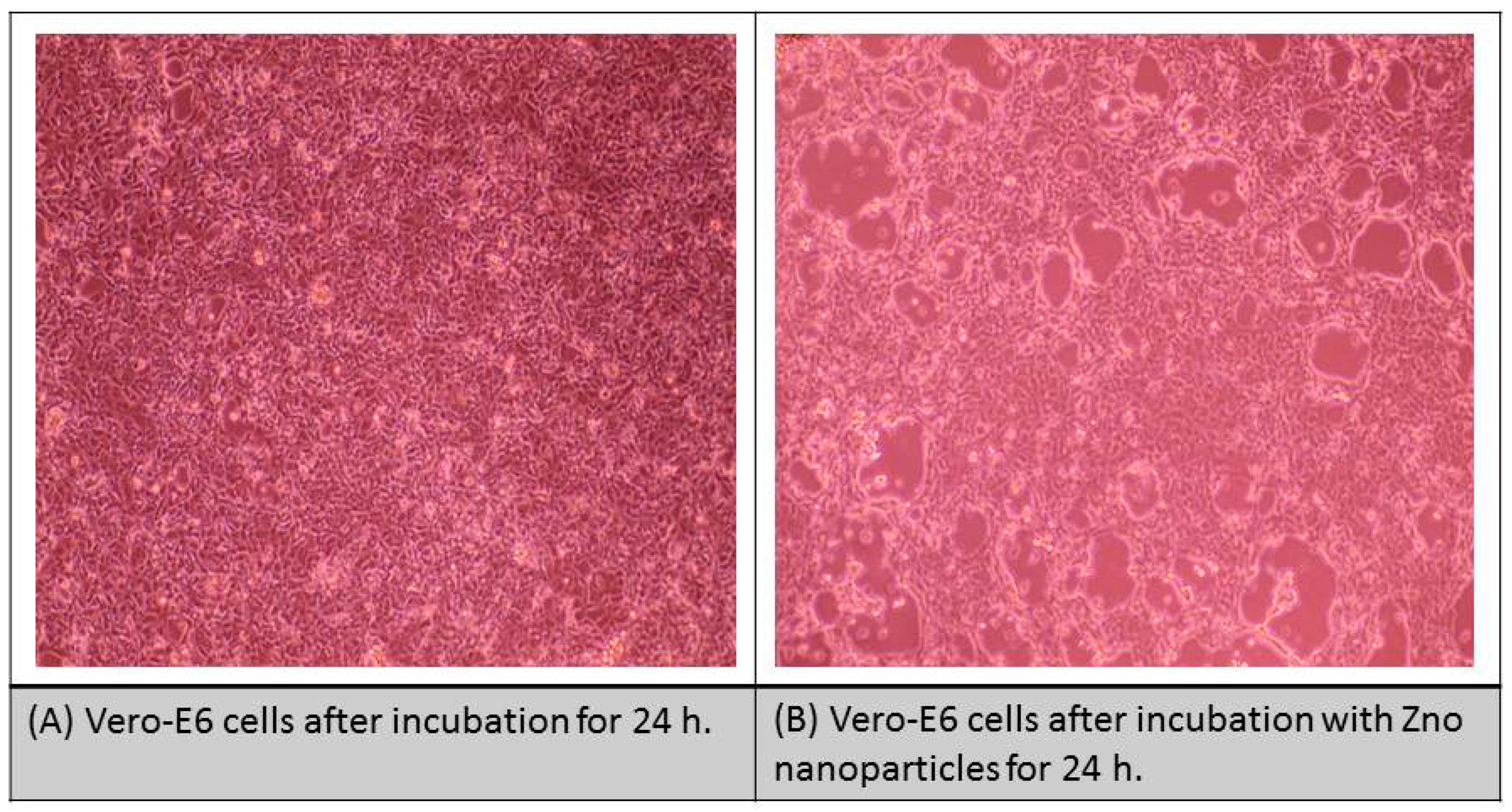
2.3. Cytotoxicity Assay

To evaluate the cytotoxic maximal half concentration (CC50) of any compound, the stock solution of ZnO-NPs was prepared in a concentration of 10% Di-methyl sulfoxide (DMSO) in di-dist. H2O and diluted to the DMEM working solutions. The cytotoxic activity of ZnO-NPs was tested in VERO-E6 cells by using the MTT method. Briefly, VERO-E6 cells were seeded in a concentration of 100 µL/well at a density 3 × 10⁵ cells/mL in 96 well-plates and then incubated at 37 °C for 24 h in CO2 (5%). After about 24 h, VERO-E6 cells were treated with different ZnO-NPs concentrations in triplicates. After 24 h, the cellular supernatant was completely discarded, and the cellular monolayers were successively washed with sterile (PBS) phosphate buffer saline three times and MTT solution (20 µL of 5 mg/mL of ZnO-NPs solution) was added to each well and incubated at a temperature of 37 °C for approximately 4 h, followed by the medium aspiration. The well-formed formazan crystals in each well of the 96-well plate were then dissolved with 200 µL of isopropanol treated with HCl (0.04 M HCl with isopropanol = 0.073 mL HCL in 50 mL isopropanol). Absorbance of formazan solutions was measured at λ max 540 nm with 620 nm as a reference λ by using a multi-well plate reader. The % of cytotoxicity as compared to the untreated cells was determined with the following equation.
The % cytotoxicity against sample concentration was used to calculate the concentration which exhibited 50% of the cellular cytotoxicity (CC50).
% Cytotoxicity = (absorbance of cells without treatment − absorbance of cells with treatment) × 100/absorbance of cells without treatment.

2.4. Determination of Inhibitory Concentration 50% (IC50)

In 96-well plates for the tissue cultures, 2.4 × 10⁴ Vero-E6 cells were distributed in the wells and they were incubated overnight at a humidified 37 °C incubator under 5% CO2. The cellular monolayers were then washed twice with PBS “1x” and subjected to virus adsorption (hCoV-19/Egypt/NRC-03/2020 (Accession Number on GSAID: EPI_ISL_430820) for about 1 h at 37 °C. The cellular monolayers were overlaid with approximately 100 µL of DMEM containing different concentrations of ZnO-NPs. After incubation at 37 °C in 5% CO2 incubator for about 72 h, VERO-E6 cells were treated with 100 µL of 4% paraformaldehyde for about 20 min and stained with 0.1% crystal violet stain in dist. H2O for about 15 min at 37 °C. 100 µL methanol (100%)/each well was used for dissolving the crystal violet dye and the color optical intensity of ZnO-NPs was measured at 570 nm by using plate reader (Anthos Zenyth 200rt, Anthos Labtec Instruments, Heerhugowaard, The Netherlands). The IC50 of ZnO-NPs is needed to decrease the SARS-CoV-2-induced cytopathic effect by about 50%, relative to control SRAS-CoV-2.

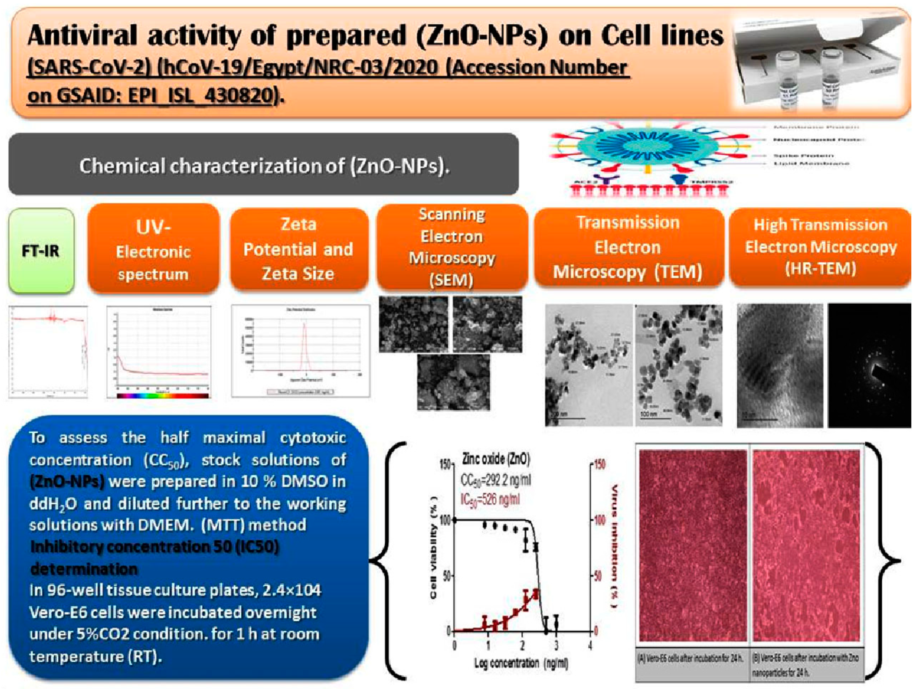
3. Results and Discussion

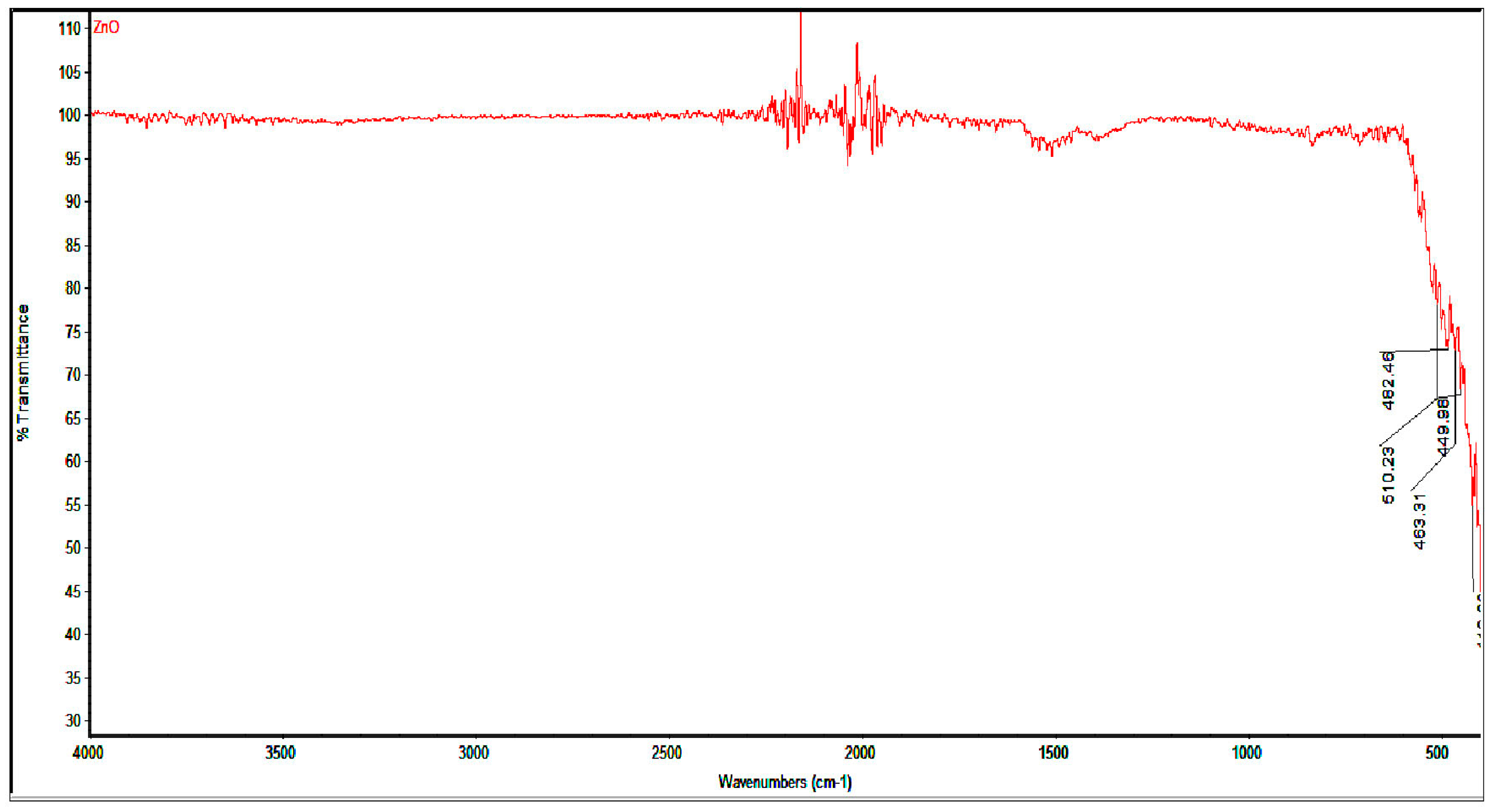
3.1. Infrared Spectrum of Synthesized ZnO-NPs

The IR spectrm of ZnO-NPs shows a peak at 510 and 449 cm−1 that is characteristic for absorption of the Zn–O bond (Figure 2) [28].

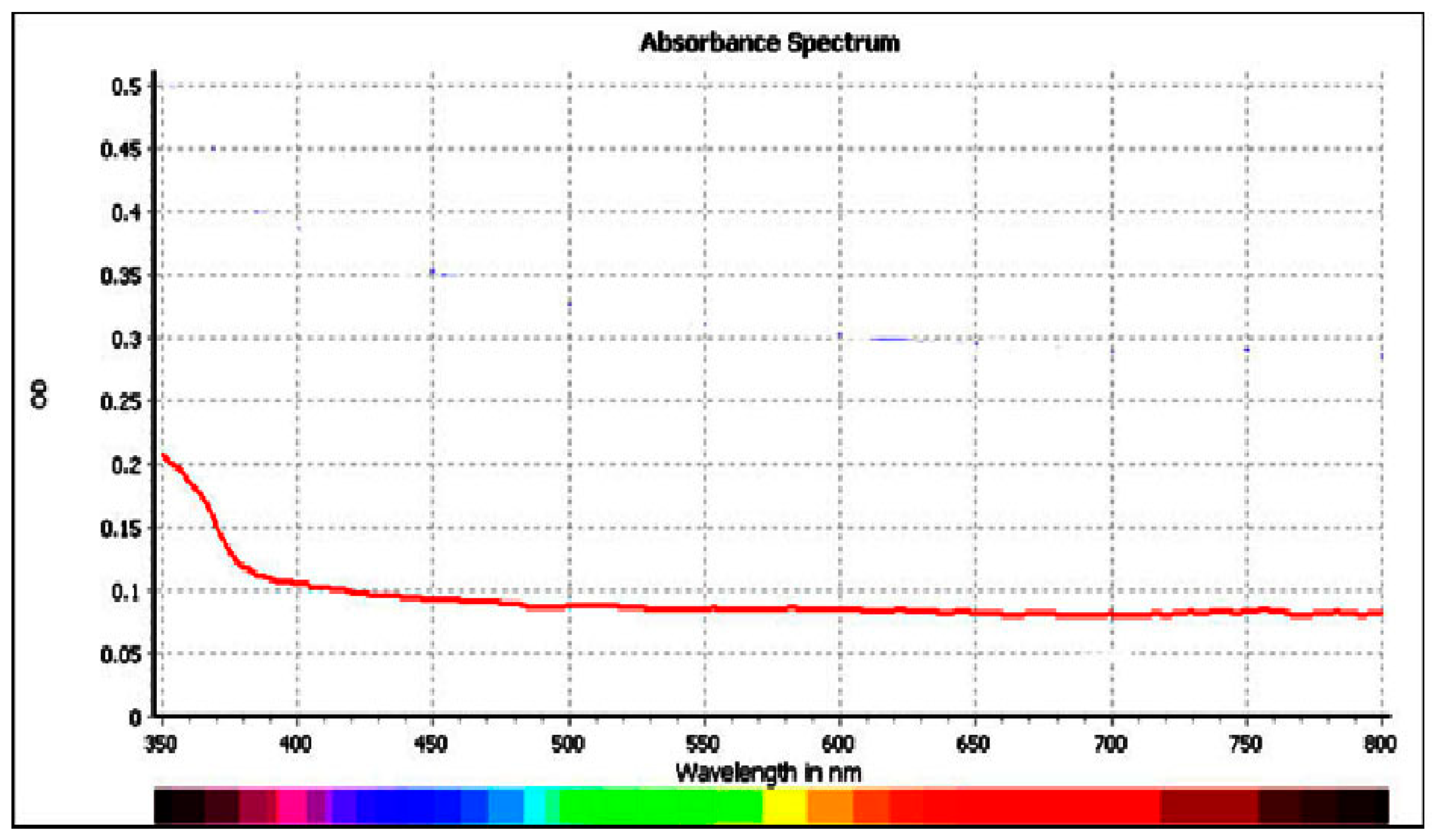
3.2. Electronic Spectrum of ZnO-NPs

The UV–Vis absorption spectra of synthesized ZnO give a broad absorption spectrum that appears at 350 nm wavelength. These results are in accord with those in the literature [29]. Figure 3 gives the electronic spectrum of ZnO. The peak refers to zinc oxide produced by heating at 100 °C giving a wavelength of 350 nm with a strong peak. This is due to the absorption band intrinsic gap for zinc oxide produced due to excitation of electrons from valence to conduction band [30]. The energy of the band gap (Eg) of zinc oxide is estimated by equation Eg = hc/λ, where the speed of light is c and equal to (3.0 × 10⁸ m/s); h is a plank constant = (6.626 × 10⁻³⁴ Js), and wavelength λ (m) [31,32]. Band gap energy is equal to 3.23 eV.

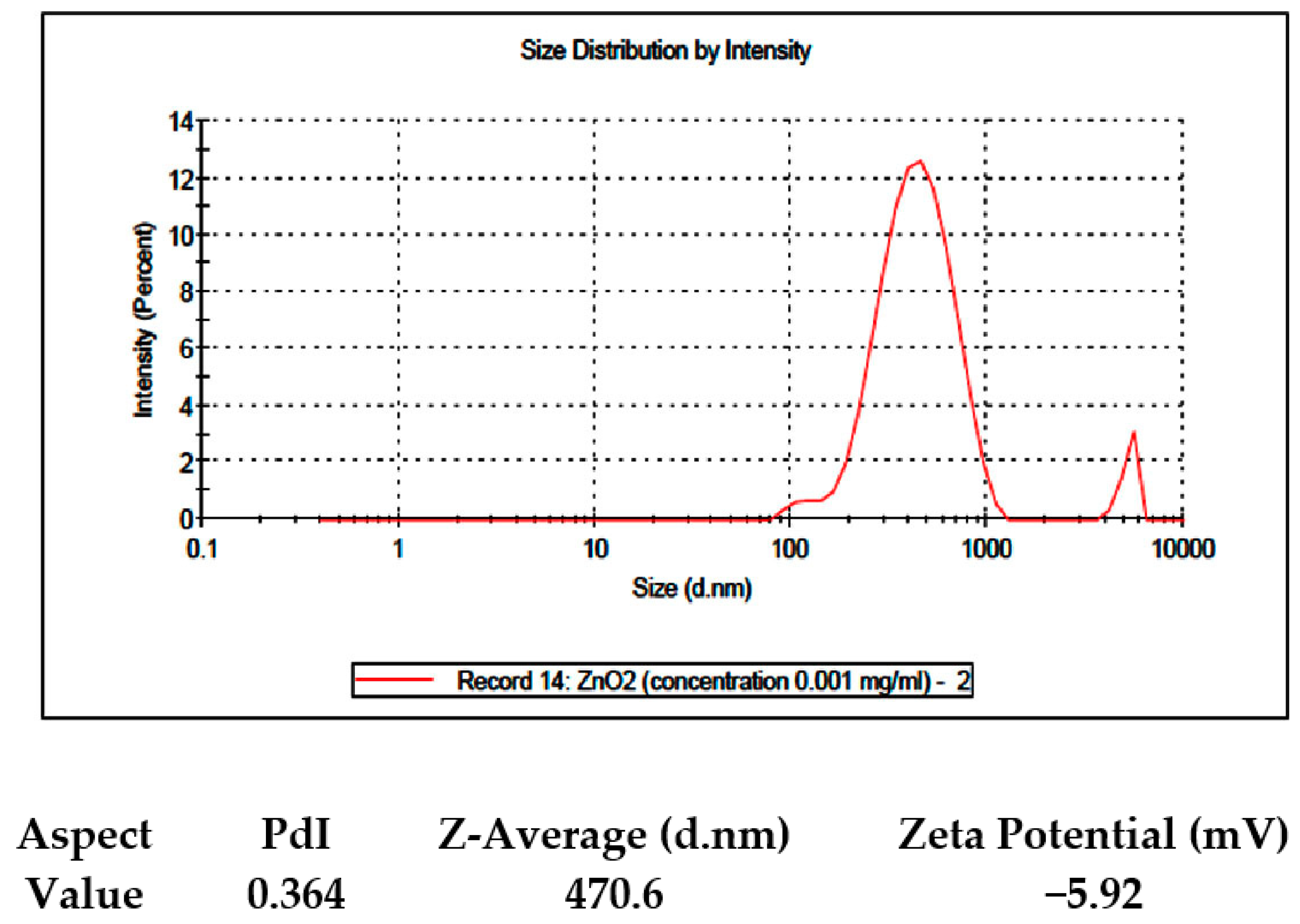
3.3. Zeta Potential and Size Distribution Intensity

The diameter of zinc oxide nanoparticles and Zeta potential (Figure 4) in distilled water at pH value of ~7.36 was determined by ultrasonication with DLS of prepared dispersions using 0.2 mg of zinc oxide in 10 mL of deionized H2O. ZnO-NPs particles’ diameter average was 470.6 nm with zeta potential of −5.92 mV and pdI = 0.364. Zeta potential and size of zinc oxide nanoparticles were determined using pH 6−12, and zeta potential decreases while the size of particles increases by increasing the pH. Measurements of ZnO-NPs’ surface area attributed to various zinc oxide burning temperatures occurred by N2-adsorption of N2 at 77 K. As temperature of calcination elevated from (29 m2·g−1) for 82 ± 10 nm ZnO-NPs at 100 °C to (7 m2·g−1) for 265 ± 8 nm at 600 °C, surface area of zinc oxide decreased, which is in accord with a previous study [30].

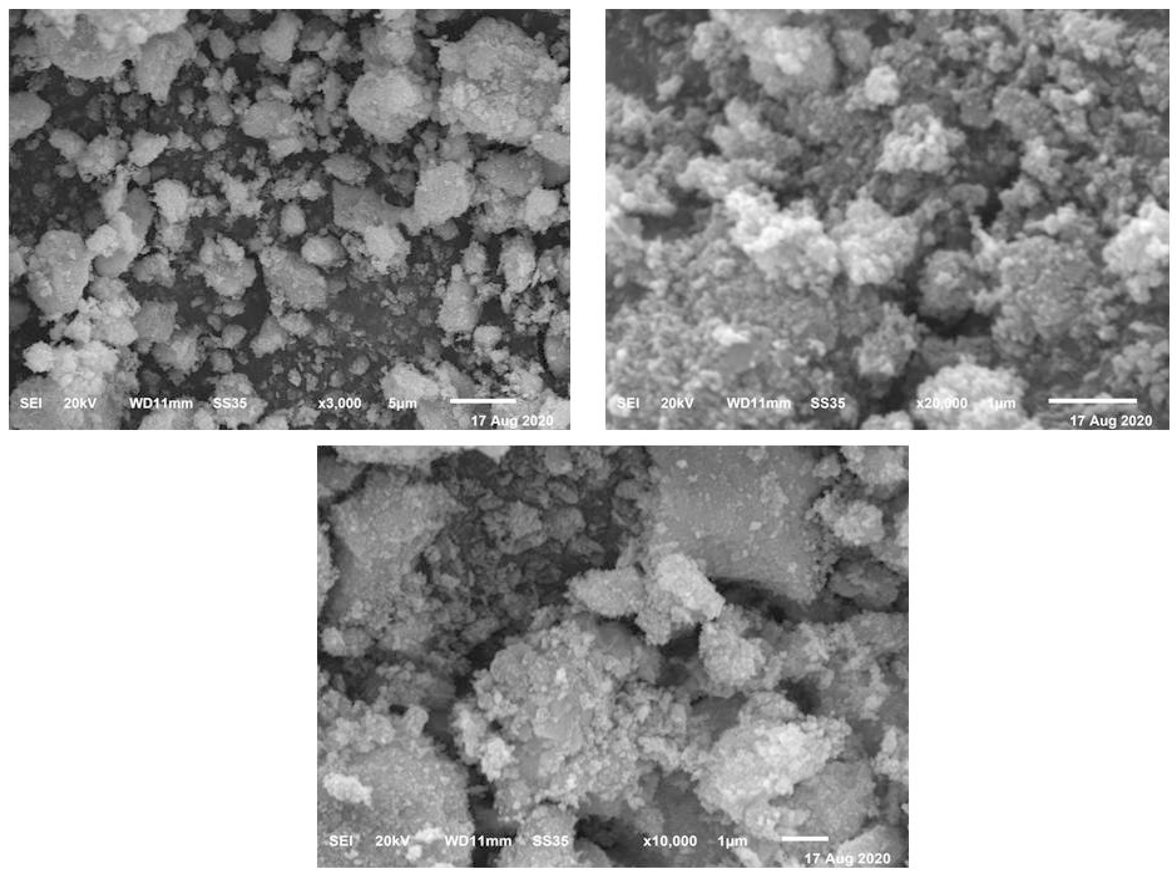
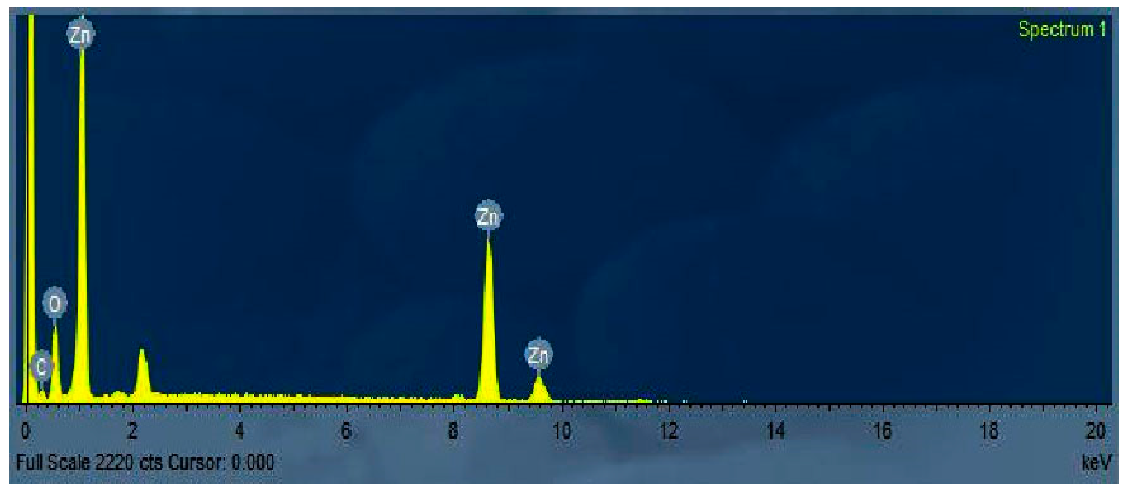
3.4. Scanning Electron Microscope (SEM)

Figure 5 represents a scanning electron microscopy photo SEM (JEOL SEM-6400, Jeol, Tokyo, Japan) of ZnO NPs. The crystallinity of the ZnO nanoparticles was associated, and the size of grains showed nearly uniform morphology. The morphologies’ surface of prepared ZnO powders was measured using the SEM analytical technique, showing a crushed-ice shape with homogenous size distributions for ZnO NPs. Chemical composition analysis by EDX (Figure 6) confirms that Zn and O peaks in the ZnO nanoparticle. The sample elemental analysis gave 79% of Zn and 20% of O, which confirms the high purity of ZnO [31,32].

3.5. XRD

X-ray diffraction of zinc oxide nanoparticles was produced by the heating of ZnO at temperatures ranging from 100−650 °C in a furnace, to study the effect of temperature on zinc oxide nanoparticles. The main peaks for zinc oxide nanoparticles did not agree with previous studies [33]. In the X-ray diffraction pattern, no data referred to impurities, confirming the high purity of the prepared zinc oxide. The data reported that there is a change in zinc oxide nanoparticles’ crystallinity when temperatures rise from 100 to 650 °C. The size of the zinc oxide nanoparticles was determined using the Scherer equation.

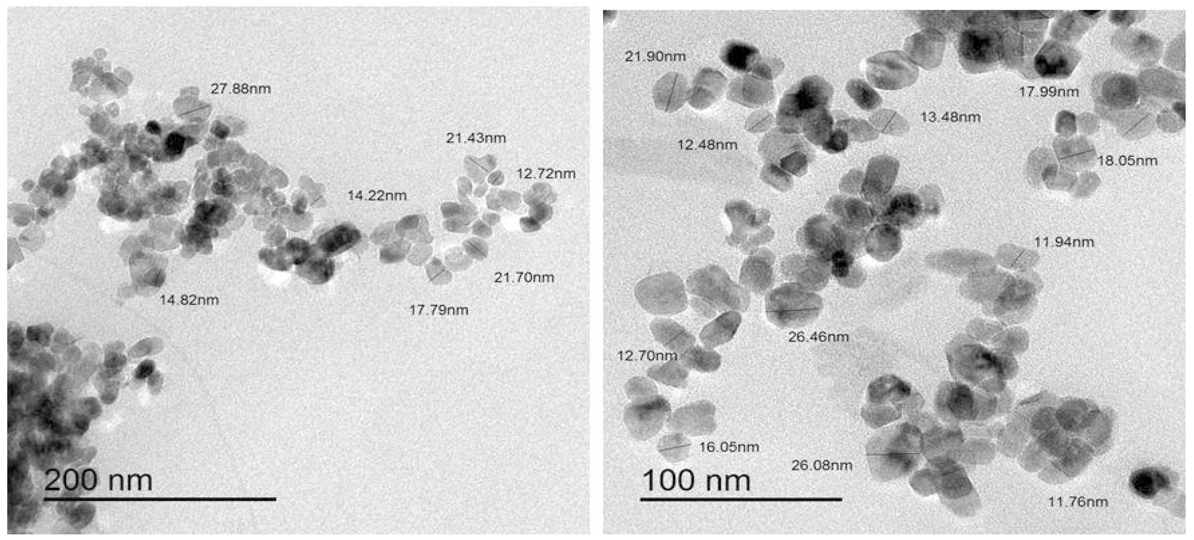
3.6. Transmission Electron Microscope (TEM) and High Resolution Transmittance Electron Micrographs (HRTEM) of ZnO

The morphology of the ZnO sample was determined by the TEM system (JEOL JEM-3300, Tokyo, Japan). Figure 7 shows a typical TEM image of the ZnO. The size and morphological of the zinc oxide nanoparticles are estimated by HRTEM micrographs. The ZnO nanoparticles were found in range of 40–60 nm with a spherical medium granule-like shape. HRTEM.
(Figure 8) shows a characteristic lattice space of 0.23 for the ZnO planes.

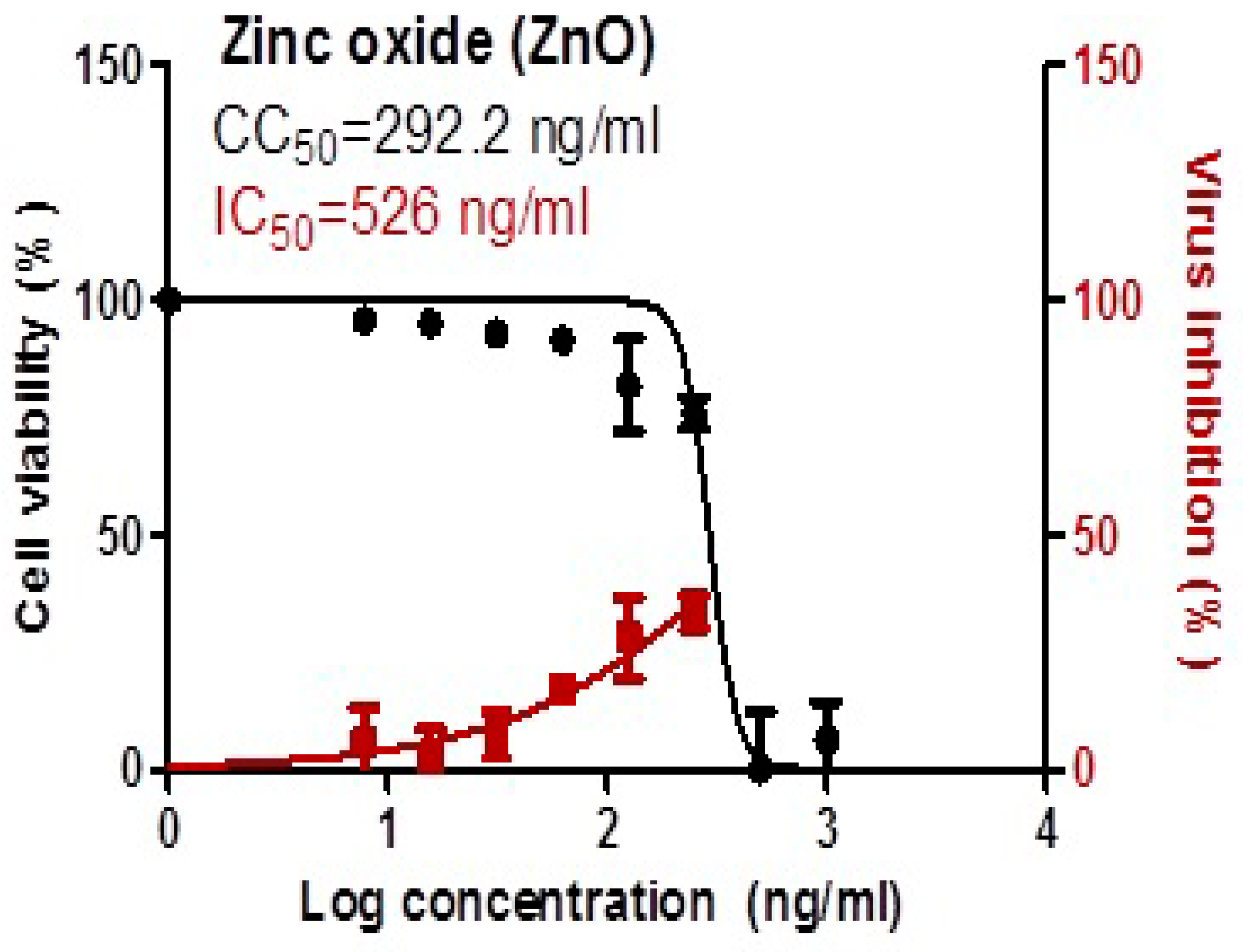
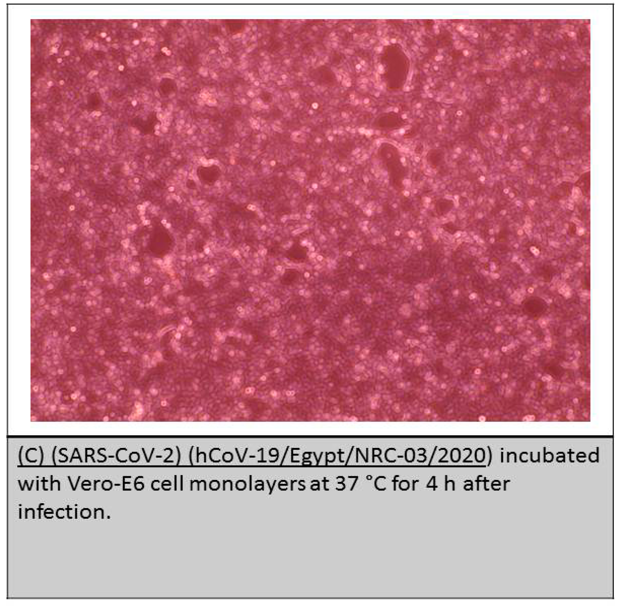
3.7. Anti-Viral Activity and Cytotoxicity of Zinc Oxide Nanoparticles and Inhibition Activity against SARS-CoV-2

ZnO-NPs showed potent antiviral activity against SARS-CoV-2 in very low concentration of 526 ng/mL. The MTT assay was used to evaluate potential cell cytotoxicity of ZnO-NPs. The results showed that 292.2 ng/mL of ZnO-NPs significantly reduced the viability of VERO-E6 cells (Figure 9) and Images of Vero-6 cell after incubation with SARS-CoV-2 (after infection) which showed potent anti-viral activity of ZnO-NPs against SARS–Cov-2, the sheet of cells treated with ZnO-NPs showing enlarged patches around SRAS-CoV-2 cells as shown in (Figure 10).
* Cytotoxicity concentration 50 (CC50): on Vero E6 cells.
* Inhibitory concentration 50 (IC50): Antiviral activity against Severe Acute.
Respiratory Syndrome Coronavirus 2 (SARS-CoV-2) (hCoV-19/Egypt/NRC-03/2020.

4. Discussion

Viral prevention is still a vital and major objective for human health. The most attractive approach to prevention is inhibition of viral replication. The present antiviral drugs, especially during the COVID-19 pandemic, are currently weakened due to major adverse effects and increasing drug resistance [34] and so development of new effective anti-COVID-19 measures is urgently required.
There are major benefits of using nanoparticles as therapeutics as nanoparticles are effective in very low concentration and also may possess potent antiviral activity against drug-resistant viruses. Nanoparticles also have very important characteristics such as is their suitability as different coating types [35,36,37,38].
The current study revealed that ZnO-NPs has anti-SARS-CoV-2 activity with low cytotoxicity as indicated by SI (IC50/CC50 ≤ 1). These finding are consistent with [39] who revealed that ZnO-NPs have stronger antiviral activity but with low cytotoxicity when used alongside PE-Glyated-NPs, as compared to ZNo-NPs alone, and this suggests a new path for the current study to propose ZNO-NPs as disinfecting agent against SARS-CoV-2 with low cytotoxicity. We suggest using ZnO-NPs coated with PEGlyated (Polyethylene Glyated) which plays a major role in enhancing and elevating antiviral activity against the SARS-CoV-2 virus and may reduce ZnO-NPs’ cellular cytotoxicity to the host cells.
More confirmation of our findings and our suggestion for coating ZnO-NPs can be found in the previous study of Ghaffari et al. [20] who reported that ZnO-NPs show cytotoxicity on breast cells higher than PEGlyated Zno-NPs. Our explanation is that ZnO-NPs produce Zn2+ ions and different types of reactive oxygen (ROS) and these free radicals can damage proteins, lipids, carbohydrates and DNA and eventually lead to apoptosis [19]. Therefore, we suggest that coating of ZnO-NPs with polyethylene glycol can lead to high anti-COVID-19 activity to reduce cytotoxicity and prevent the release of reactive oxygen by masking of ZNO–NPs.
Recently, metal nanoparticles such as ZnO-NPs have been shown to be efficient against different ranges of pathogens including viruses [20]. ZnO-NPs have been demonstrated to show activities against various pathogens that infect humans [40]. However, most studies on ZnO-NPs have focused on the inhibitory actions of ZnO-NPs on bacterial infections, and there are very limited studies on the interaction between ZnO-NPs and viruses. In a recent work by our group, we found that ZnO-NPs possess strong inhibitory effects on SARS-CoV-2, which is amongst the most challenging virus threatening human health.
ZnO-NPs have antiviral activity that has been evaluated previously in virus infected corneal tissues [17]. ZnO-NPs efficiently trap virions via a novel virostatic mechanism making them unable to enter corneal fibroblast cells. ZnO-NPs significantly block the entry and also stop the spread of Herpes virus into target cells. It is considered that ZnO-NPs exhibit their antiviral activity by an ability to neutralize the virus [40]. Another explanation for the antiviral activity of ZnO-NPs is that it may induce and enhance the immune system against viruses and thus provide potent therapeutic effects [41]. This approach is very important in fighting against COVID-19.
ZnO-NPs inhibited the virus titer [18]. ZnO-NPs’ modified surface could defend against the infection potential of viruses via neutralizing the virus rather than through interfering with cellular targets [42]. Ghaffari et al. [20] determined that Zn2+ ions potentially inhibited viral replication, elevating concentration of the intracellular Zn2+ that can efficiently impair the replication of a variety of RNA viruses. All these finding confirmed that obtained in the current study, proving the ability of ZnO-NPs to act as disinfectant against SARS-CoV-2.
ZnO-NPs are clinically important as a previous study of Antoine et al. [43] demonstrated that ZnO-NPs employ a micro-bivac concept using a virus trapping agent which provides immediate protection by trapping virus particles and then presenting them for mucosal immunity development. ZnO-NPs prevented viral entry by inhibition of the interaction between virus and cellular receptors and thus priming the immune system, so can be utilized to provide viral protection.
Previous data suggested that ZnO-NPs can be of benefit as an efficient adjuvant. Confirming the current obtained results, Antoine et al. [43] proved that splenocytes of mice primed with ZnO-NPs showed significantly higher cytotoxicity towards Herpes virus-infected cells, as compared to those from control infected untreated mice. ZnO-NPs showed a higher enhanced cytotoxic T cell response, indicating a strong immunogenicity-enhancing property, which may be used in combination with natural active compounds as existing adjuvants to achieve synergism, as we suppose in our prospective studies.
The current finding broadly supports the work of [17] who revealed that ZnO-NPs are metals containing nanoparticles that revealed a significant activity against different types of microorganism, including viruses.
As indicated in previous studies [44]. ZnO-NPs exhibit anti-viral activity against Herpes simplex virus type-2 (HSV-2) and stopped the viral spread. ZnO-NPs may reveal activity against viral infection by their capability to neutralize HSV-2 and greatly reduce its high infectivity.
The finding of [45] revealed that incubation of HSV-2 with ZNO-NPs inhibits the viral ability to infect the vaginal tissues of female mice and blocks viral shedding in the cellular host, stimulating its immune response.
This study produced results which concur with the findings of [43] who demonstrated that a negatively charged ZnO-NP can simply trap the herpes virus and inhibit its attachment to the host cells.
Contrary to the current findings, Sunada et al. [15] found that culturing of ZnO with viruses elevated cellular survival by over 50%, while in the current finding cellular survival decreased, which opens the gate for further study of different concentrations of ZnO-NPs to obtain an effective ratio against SARS-CoV-2 with low cytotoxicity.
It is somewhat surprising to obtain such data, which confirms the anti-COVID-19 activity of ZnO-NPs at very low cytotoxicity and at very low concentration (526 ng/mL), with somewhat low cytotoxicity which can be alleviated by coating ZNO-NPs with polymeric compounds such as PEGlyated which may decrease the particles’ diameter and reduce the triggering side effects of ROS on cellular component, achieving high infective action against SARS-CoV-2 at a very low concentration, which is why we consider these finding promising in the fight against the COVID-19 pandemic.

5. Conclusions

ZnO-NPs were fully chemically characterized using different spectroscopic tools. ZnO-NPs have a potent antiviral activity against SARS-CoV-2 at a very low concentration (IC50 = 526 ng/mL) and can trigger many free radicals that may cause oxidative stress to SARS-CoV-2 and induce severe damage to SRAS-CoV-2 cellular membranes, which is a very important result in fighting against SARS-CoV-2; but we found that ZnO-NPs have a cytotoxic level (CC50 = 292.2 ng/mL) against VERO-E6 cells, and thus we recommend more prospective studies applied to ZnO-NPs immobilization with natural active compounds that may reduce this cytotoxicity and elevate its antiviral activity against SARS-CoV-2.

Author Contributions

All authors contributed equally in this work. Conceptualization, S.M.E.-M., M.A., R.Z.H. and F.A.A.-S.; methodology, S.M.E.-M., M.A. and R.Z.H.; software, S.M.E.-M., M.A. and R.Z.H.; validation, S.M.E.-M., M.A., R.Z.H. and F.A.A.-S.; formal analysis, S.M.E.-M., M.A., R.Z.H. and F.A.A.-S.; investigation, S.M.E.-M., M.A., R.Z.H. and F.A.A.-S.; resources, S.M.E.-M., M.A., R.Z.H. and F.A.A.-S.; data curation, S.M.E.-M., M.A. and R.Z.H.; writing—original draft preparation, S.M.E.-M., M.A., R.Z.H. and F.A.A.-S.; writing—review and editing, S.M.E.-M., M.A., R.Z.H. and F.A.A.-S.; visualization, S.M.E.-M., M.A., R.Z.H. and F.A.A.-S.; supervision, S.M.E.-M., M.A., R.Z.H. and F.A.A.-S.; project administration, S.M.E.-M., M.A., R.Z.H. and F.A.A.-S.; funding acquisition, S.M.E.-M., M.A. and F.A.A.-S. All authors have read and agreed to the published version of the manuscript.

Funding

The authors thanked the Deanship of Scientific Research for funding this research on behalf of COVID-19 projects. This work was supported by the Deanship of the scientific research, Taif University, Saudi Arabia, under project number 1-441-65.

Institutional Review Board Statement

The study was conducted at the Center of Scientific Excellence for Influenza Viruses, National Research Centre, 12, 311 Dokki, Giza on (SARS-CoV-2) (hCoV-19/Egypt/NRC-03/2020 (Accession No. on GSAID: EPI_ISL_430820) and approved by the National Research Centre Review Board.

Data Availability Statement

The raw data supporting the conclusions of this article will be made available by the corresponding author.

Conflicts of Interest

The authors declare that there are no conflicts of interest.

References

  1. Tsuneo, I. Anti-viral vaccine activity of Zinc (II) for viral prevention, entry, replication, and spreading during pathogenesis process. Curr. Trends Biomed. Eng. Biosci. 2019, 19, 1–6. [Google Scholar]
  2. Perlman, S.; Netland, J. Coronaviruses post-SARS: Update on replication and pathogenesis. Nat. Rev. Microbiol. 2009, 7, 439–450. [Google Scholar] [CrossRef]
  3. Zhang, B.; Zhou, X.; Zhu, C.; Feng, F.; Qiu, Y.; Feng, J.; Jia, Q.; Song, Q.; Zhu, B.; Wang, J. Immune phenotyping based on neutrophil-to-lymphocyte ratio and IgG predicts disease severity and outcome for patients with COVID-19. Front. Mol. Biosci. 2020, 7, 157. [Google Scholar] [CrossRef] [PubMed]
  4. Tsatsakis, A.; Calina, D.; Falzone, L.; Petrakis, D.; Mitrut, R.; Siokas, V.; Pennisi, M.; Lanza, G.; Libra, M.; Doukas, S.G.; et al. ARS-CoV-2 pathophysiology and its clinical implications: An integrative overview of the pharmacotherapeutic management of COVID-19. Food Chem. Toxicol. 2020, 146, 111769. [Google Scholar] [CrossRef]
  5. Hamza, R.Z.; Al-Talhi, T.; Gobouri, A.; Al-Sanie, W.; El-Megharbel, S.M. Are Favipiravir and Acyclovir with IgG Injections Supplemented with Vitamin D “Suggested Therapeutic Option” Can Fight Against COVID-19? Adv. Anim. Vet. Sci. 2021, 9, 549–554. [Google Scholar]
  6. Zhou, J.; Hu, Z.; Zabihi, F.; Chen, Z.; Zhu, M. Progress and perspective of antiviral protective material. Adv. Fiber Mater. 2020, 2, 123–139. [Google Scholar] [CrossRef]
  7. Zhou, P.; Yang, X.L.; Wang, X.G.; Hu, B.; Zhang, L.; Zhang, W.; Si, H.R.; Zhu, Y.; Li, B.; Huang, C.L.; et al. A pneumonia outbreak associated with a new coronavirus of probable bat origin. Nature 2020, 579, 270. [Google Scholar] [CrossRef] [PubMed]
  8. Mishra, P.K.; Mishra, H.; Ekielski, A.; Talegaonkar, S.; Vaidya, B. Zinc oxide nanoparticles: A promising nanomaterial for biomedical applications. Drug Discov. Today 2017, 22, 1825–1834. [Google Scholar] [CrossRef] [PubMed]
  9. Kołodziejczak-Radzimska, A.; Jesionowski, T. Zinc oxide—from synthesis to application: A review. Materials 2014, 7, 2833–2881. [Google Scholar] [CrossRef] [PubMed]
  10. Kalyani, R.L.; Pammi, S.V.N.; Vijay Kumar, P.P.N.; Swamy, P.V.; Ramana Murthy, K.V. Antibiotic potentiation and anti-cancer competence through bio-mediated ZnO nanoparticles. Mater. Sci. Eng. C 2019, 103, 109756. [Google Scholar]
  11. Sudova, E.; Machova, J.; Svobodova, Z.; Vesely, T. Negative effects of malachite green and possibilities of its replacement in the treatment of fish eggs and fish: A review. Vet. Med. 2007, 52, 527–539. [Google Scholar] [CrossRef]
  12. Srivastava, S.; Sinha, R.; Roy, D. Toxicological effects of malachite green. Aquat. Toxicol. 2004, 66, 319–329. [Google Scholar] [CrossRef]
  13. Culp, S.J.; Beland, F.A. Malachite Green: A Toxicological Review. J. Am. Coll. Toxicol. 1996, 15, 219–238. [Google Scholar] [CrossRef]
  14. Vijay Kumar, P.P.N.; Pammi, S.V.N.; Kollu, P.; Satyanarayana, K.V.V.; Shameem, U. Green synthesis and characterization of silver nanoparticles using Boerhaavia diffusa plant extract and their anti-bacterial activity. Ind. Crops Prod. 2014, 52, 562–566. [Google Scholar] [CrossRef]
  15. Sunada, K.; Minoshima, M.; Hashimoto, K. Highly efficient antiviral and antibacterial activities of solid-state cuprous compounds. J. Hazard. Mater. 2012, 235, 265. [Google Scholar] [CrossRef] [PubMed]
  16. Islam, M.T.; Quispe, C.; Martorell, M.; Docea, A.O.; Salehi, B.; Calina, D.; Reiner, Ž.; Sharifi-Rad, J. Dietary supplements, vitamins and minerals as potential interventions against viruses: Perspectives for COVID-19. Int. J. Vitam. Nutr. Res. 2021. [Google Scholar] [CrossRef] [PubMed]
  17. Gurunathan, S.; Qasim, M.; Choi, Y.; Do, J.T.; Park, C.; Hong, K.; Kim, J.H.; Song, H. Antiviral potential of nanoparticles—can nanoparticles fight against coronaviruses? Nanomaterials 2020, 10, 1645. [Google Scholar] [CrossRef] [PubMed]
  18. Tavakoli, A.; Ataei-Pirkooh, A.; Sadeghi, G.M.M.; Bokharaei-Salim, F.; Sahrapour, P.; Kiani, S.J.; Moghoofei, M.; Farahmand, M.; Javanmard, D.; Monavari, S.H. Polyethylene glycol-coated zinc oxide nanoparticle: An efficient nanoweapon to fight against herpes simplex virus type 1. Nanomedicine 2018, 13, 2675–2690. [Google Scholar] [CrossRef]
  19. Farouk, F.; Sgebl, R.I. Comparing surface chemical modifications of zinc oxide nanoparticles for modulating their antiviral activity against herpes simplex virus type-1. Int. J. Nanopart. Nanotechnol. 2018, 4, 21. [Google Scholar] [CrossRef]
  20. Ghaffari, H.; Tavakoli, A.; Abdolvahab, M.; Alijan, T.; Bokharaei-Salim, F.; Masoumeh, Z.; Mohammad, F.; Davod, J.; Seyed, J.K.; Esghaei, M.; et al. Inhibition of H1N1 influenza virus infection by zinc oxide nanoparticles: Another emerging application of nanomedicine. J. Biomed. Sci. 2019, 26, 70. [Google Scholar] [CrossRef]
  21. Baron, S.; Fons, M.; Albrecht, T. Viral Pathogenesis, General Concepts. In Medical Microbiology, 4th ed.; Baron, S., Ed.; University of Texas Medical Branch: Galveston, TX, USA, 1996; Chapter 45; pp. 1–7. [Google Scholar]
  22. Ruddaraju, L.K.; Pammi, S.V.N.; Sankar, G.G.; Padavala, V.S.; Kolapalli, V.R.M. A review on anti-bacterials to combat resistance: From ancient era of plants and metals to present and future perspectives of green nano technological combinations. Asian J. Pharm. Sci. 2019. [Google Scholar] [CrossRef]
  23. Raghunath, A.; Perumal, E. Metal oxide nanoparticles as antimicrobial agents: A promise for the future. Int. J. Antimicrob. Agents 2017, 49, 137–152. [Google Scholar] [CrossRef] [PubMed]
  24. Wang, L.; Hu, C.; Shao, L. The antimicrobial activity of nanoparticles: Present situation and prospects for the future. Int. J. Nanomed. 2017, 12, 1227–1249. [Google Scholar] [CrossRef] [PubMed]
  25. Baker, S.; Pasha, A.; Satish, S. Biogenic nanoparticles bearing antibacterial activity and their synergistic effect with broad spectrum antibiotics: Emerging strategy to combat drug resistant pathogens. Saudi Pharm. J. 2017, 25, 44–51. [Google Scholar] [CrossRef]
  26. Fayaz, A.M.; Balaji, K.; Girilal, M.; Yadav, R.; Kalaichelvan, P.T.; Venketesan, R. Biogenic synthesis of silver nanoparticles and their synergistic effect with antibiotics: A study against gram-positive and gram-negative bacteria. Nanomed. Nanotechnol. Biol. Med. 2010, 6, 103–109. [Google Scholar] [CrossRef]
  27. Thirumurugan, G.; Seshagiri Rao, J.V.L.N.; Dhanaraju, M.D. Elucidating pharmacodynamic interaction of silver nanoparticle-topical deliverable antibiotics. Sci. Rep. 2016, 6, 1–11. [Google Scholar] [CrossRef]
  28. Ghorbani, H.R.; Mehr, F.P.; Pazoki, H.; Rahmani, B.M. Synthesis of ZnO Nanoparticles by Precipitation Method. Orient. J. Chem. 2015, 31, 1219–1221. [Google Scholar] [CrossRef]
  29. Hoffmann, M.R.; Martin, S.T.; Choi, W.; Bahnemann, D.W. Environmental applications of semiconductor photocatalysis. Chem. Rev. 1995, 95, 69–96. [Google Scholar] [CrossRef]
  30. Bagheri, S.; Chandrappa, K.; Hamid, S.B.A. Facile synthesis of nano-sized ZnO by direct precipitation method. Pharm. Chem. 2013, 5, 265–270. [Google Scholar]
  31. Bagabas, A.; Alshammari, A.; Aboud, M.F.; Kosslick, H. Roomtemperature synthesis of zinc oxide nanoparticles in different media and their application in cyanide photodegradation. Nanoscale Res. Lett. 2013, 8, 1. [Google Scholar] [CrossRef]
  32. Nagarajan, S.; Kuppusamy, K.A. Extracellular synthesis of zinc oxide nanoparticle using seaweeds of gulf of Mannar, India. J. Nanobiotechnol. 2013, 11, 1. [Google Scholar] [CrossRef]
  33. Raj, L.F.A.; Jayalakshmy, E. Biosynthesis and characterization of zinc oxide nanoparticles using root extract of Zingiber officinale. Orient. J. Chem. 2015, 31, 51–56. [Google Scholar] [CrossRef]
  34. Salahuddin, N.A.; El-Kemary, M.; Ibrahim, E.M. Synthesis and Characterization of ZnO Nanoparticles via Precipitation Method: Effect of Annealing Temperature on Particle Size. Nanosci. Nanotechnol. 2015, 5, 82–88. [Google Scholar]
  35. Makau, J.N.; Watanabe, K.; Mohammed, M.M.; Nishida, N. Antiviral activity of peanut (Arachis hypogaea L.) skin extract against human influenza viruses. J. Med. Food 2018, 21, 777–784. [Google Scholar]
  36. Hamza, R.Z.; Diab, A.A. Testicular protective and antioxidant effects of selenium nanoparticles on Monosodium glutamate-induced testicular structure alterations in male mice. Toxicol. Rep. 2020, 7, 254–260. [Google Scholar] [CrossRef] [PubMed]
  37. Reed, L.J.; Muench, H. A simple method of estimating fifty per cent endpoints. Am. J. Epidemiol. 1938, 27, 493–497. [Google Scholar] [CrossRef]
  38. Zhou, J.; Zhao, F.; Wang, Y.; Zhang, Y.; Yang, L. Size-controlled synthesis of ZnO nanoparticles and their photoluminescence properties. J. Lumin. 2007, 122, 195–197. [Google Scholar] [CrossRef]
  39. Khoshhesab, Z.M.; Sarfaraz, M.; Asadabad, M.A. Preparation of ZnO nanostructures by chemical precipitation method. Synth. React. Inorg. Metal-Org. Nano-Metal Chem. 2011, 41, 814–819. [Google Scholar] [CrossRef]
  40. Refat, M.S.; Hamza, R.Z.; Adam, A.A.; Saad, H.A.; Gobouri, A.A.; Al-Harbi, F.S.; Al-Salmi, F.A.; Altalhi, T.; El-Megharbel, S.M. Quercetin/Zinc complex and stem cells: A new drug therapy to ameliorate glycometabolic control and pulmonary dysfunction in diabetes mellitus: Structural characterization and genetic studies. PLoS ONE 2021, 3, e0246265. [Google Scholar]
  41. Siddiqi, K.S.; Rahman, A.; Husen, A. Properties of zinc oxide nanoparticles and their activity against microbes. Nanoscale Res. Lett. 2018, 13, 141. [Google Scholar] [CrossRef]
  42. Refat, M.S.; Hamza, R.Z.; Adam, A.M.A.; Saad, H.A.; Gobouri, A.A.; Al-Salmi, F.A.; Altalhi, T.; El-Megharbel, S.M. Synthesis of N,N′-bis(1,5-dimethyl-2-phenyl-1,2-dihydro-3-oxopyrazol-4-yl) sebacamide that ameliorate osteoarthritis symptoms and improve bone marrow matrix structure and cartilage alterations induced by monoiodoacetate in the rat model: “Suggested potent anti-inflammatory agent against COVID-19”. Hum. Exp. Toxicol. 2021, 2, 325–341. [Google Scholar]
  43. Antoine, T.E.; Mishra, Y.K.; Trigilio, J.; Tiwari, V.; Adelung, R.; Shukla, D. Prophylactic, therapeutic and neutralizing effects of zinc oxide tetrapod structures against herpes simplex virus type-2 infection. Antivir. Res. 2012, 96, 363–375. [Google Scholar] [CrossRef] [PubMed]
  44. Agelidis, A.; Koujah, L.; Suryawanshi, R.; Yadavalli, T.; Mishra, Y.K.; Adelung, R.; Shukla, D. An intra-vaginal zinc oxide tetrapod nanoparticles (ZOTEN) and genital herpesvirus cocktail can provide a novel platform for live virus vaccine. Front. Immunol. 2019, 10, 500. [Google Scholar] [CrossRef] [PubMed]
  45. Mishra, Y.; Adelung, R.; Roehl, C.; Shukla, D.; Spors, F.; Tiwari, V. Virostatic potential of micro-nano filopodia-like ZnO structures against herpes simplex virus-1. Antivir. Res. 2011, 92, 305. [Google Scholar] [CrossRef] [PubMed]
Figure 1. Graphical abstract for the experimental work.
Coatings 11 00388 g001
Figure 2. FT-IR of synthesized ZnO-NPs.
Coatings 11 00388 g002
Figure 3. UV spectra of ZnO-NPs.
Coatings 11 00388 g003
Figure 4. Zeta potential and Size distribution of the synthesized ZnO-NPs.
Coatings 11 00388 g004aCoatings 11 00388 g004b
Figure 5. Scanning electron microscopy (SEM) of ZnO nanoparticles (NPs) shows that the size of grains has nearly uniform morphology and appear with a crushed ice-like shape with homogenous size distributions for ZnO NPs.
Coatings 11 00388 g005
Figure 6. Energy dispersive X-ray analysis (EDX) for (ZnO-NPs) confirm the high purity of ZnO NPs (79% of Zn and 20% of O).
Coatings 11 00388 g006
Figure 7. TEM with 100 and 200 nm scale of ZnO-NPs estimated the size and morphology of the zinc oxide nanoparticles that were found in a range of 40–60 nm, with spherical medium granule-like-shape.
Coatings 11 00388 g007
Figure 8. High Resolution Transmittance Electron Micrographs (HRTEM) images of ZnO-NPs shows a characteristic lattice space of 0.23 for the ZnO planes.
Coatings 11 00388 g008
Figure 9. Cytotoxicity and inhibitory concentration of Zno-NPs against SARS-CoV-2.
Coatings 11 00388 g009
Figure 10. Images of Vero-6 cell after incubation with SARS-CoV-2 (after infection) which showed potent anti-viral activity of ZnO-NPs against SARS–Cov-2, the sheet of cells treated with ZnO-NPs showing enlarged patches around SRAS-CoV-2 cells, thus demonstrating the ability of ZnO-NPs to disinfect SARS-CoV-2 and offer a high level of inhibition to its growth.
Coatings 11 00388 g010aCoatings 11 00388 g010b
Publisher’s Note: MDPI stays neutral with regard to jurisdictional claims in published maps and institutional affiliations.

