Next Article in Journal
Simulating the Growth of Dual-Phase Boride Layer on AISI M2 Steel by Two Kinetic Approaches
Next Article in Special Issue
Antioxidant, Antigenotoxic, and Hepatic Ameliorative Effects of Quercetin/Zinc Complex on Cadmium-Induced Hepatotoxicity and Alterations in Hepatic Tissue Structure
Previous Article in Journal
Naphthoquinones and Their Derivatives: Emerging Trends in Combating Microbial Pathogens
Previous Article in Special Issue
A Potential Role of Apelin-13 against Hepatic Injury and Metabolic Disorders in Preeclampsia Induced by L-NAME
 
 
Retraction published on 16 August 2022, see Coatings 2022, 12(8), 1189.
Article

Neurological Alterations and Testicular Damages in Aging Induced by D-Galactose and Neuro and Testicular Protective Effects of Combinations of Chitosan Nanoparticles, Resveratrol and Quercetin in Male Mice

by
Reham Z. Hamza
*,
Mohammad S. Al-Harbi
and
Munirah A. Al-Hazaa
Department of Biology, College of Sciences, Taif University, P.O. Box 11099, Taif 21944, Saudi Arabia
*
Author to whom correspondence should be addressed.
Coatings 2021, 11(4), 435; https://doi.org/10.3390/coatings11040435
Submission received: 6 March 2021 / Revised: 28 March 2021 / Accepted: 6 April 2021 / Published: 9 April 2021 / Retracted: 16 August 2022
(This article belongs to the Special Issue Biomedical Application of Natural Plant Extracts)

Abstract

:
Aging is a neurological disease that is afforded by incidence of oxidative stress. Chitosan has received global interests due to its wide medical uses. Quercetin (Q) is a bioflavonoid and widely distributed in vegetables and fruits. Resveratrol is considered as a potent antioxidant and is a component of a wide range of foods. The using of either chitosan nanopartciles (CH-NPs), querectin (Q), and resveratrol (RV) to reduce the oxidative stress and biochemical alterations on brain and testicular tissues induced by D-galactose (DG) (100 mg/Kg) were the aim of the present study. This study investigated the probable protective effects of CH-NPs in two doses (140,280 mg/Kg), Q (20 mg/Kg) and RV (20 mg/Kg), against DG induced aging and neurological alterations. Brain antioxidant capacity as malonaldehyde (MDA), catalase (CAT), and glutathione reductase (GRx), as well as histopathological damages of the brain and testicular tissues were measured. The DG treated group had significantly elevated the oxidative stress markers by 96% and 91.4% in brain and testicular tissues respectively and lower significantly the antioxidant enzyme activities of both brain and testicular tissues than those of the control group by 86.95%, 69.27%, 83.07%, and 69.43%. Groups of DG that treated with a combination of CH-NPs in two doses, Q and RV, the levels of oxidative stress marker declined significantly by 68.70%, 76.64% in brain tissues and by 74.07% and 76.61% in testicular tissues, and the enzymatic antioxidants increased significantly by 75.55%, 79.24%, 62.32%, and 61.97% as compared to the DG group. The present results indicate that CH-NPs, Q, and RV have protective effects against DG-induced brain and testis tissue damage at the biochemical and histopathological levels. Mechanisms of this protective effect of used compounds against neurological and testicular toxicity may be due to the enhanced brain and testis antioxidant capacities.

1. Introduction

Oxidative stress is involved in the genesis of nervous system disorders during the aging. Reactive oxygen species play a vital role in simplifying the incidence of neurodegenerative diseases in aging [1]. For replication, the pathological alterations which are observed in aging (chemicals such as D-galactose (DG)) have been extremely used. DG is a reduced sugar found in the human diets; when its level is released in the body, it is oxidized by galactose oxidase to aldehydes and hydrogen peroxide [1].
Rodents injected with DG showed deterioration progressively of memory properties and learning outcomes and increased the free radicals production in the brain [2].
Chitosan is a polysaccharide derived from the deacetylation of chitin, which is mainly found in crustacean shells, parts of fungi, and insect exoskeleton [3]. Chitosan has been receiving attention in drug delivery applications. It succeeds mainly due to its unique physicochemical characters, as it has different and wide biological properties [4]. The chitosan nanoparticles (CH-NPs) are of major interest in nanomedicine field due to its diminished pharmacological toxicity[5].
Chitosan has attracted more attention due to its wide biomedical activity. It has a wide range of biological activities, such as antitumor agent, anti-inflammatory capacities, and free radical scavenging activities [6].
Chitosan is a polysaccharide composing of various amounts of glucosamine. Chitosan nanoparticles (CH-NPs) display more superior actions as these particles have multiple immune-ameliorative effect and antimicrobial activities. Additionally, CH-NPs possess a potent surface warp, as compared to the large particles; this produces more decay pressure with an analogous increment in saturation solubility. The elevated saturation solubility elevates an increment in concentration between the cells in the intestine and the mesenteric circulation which is of high medical importance.
Quercetin (Q) (3, 5, 7, 39, 49-pentahydroxyflavone) is one of the most common flavonol in plants that contains a lot of beneficial effects to the human health, and can be found in capers, onions, tomatos, and lettuce. Q is a part of the polyphenols that is considered as a strong antioxidant agent to prevent oxidant harm and cell death, which could lead to many health problems [7,8]. Nowadays, Q has antioxidant properties that helped in improving learning abilities and memory [9].
Sperms are particularly not easily attacked by the reactive oxygen species or free radicals, as sperms’ membranes are rich in a lot of polyunsaturated fatty acid. Studying the effect of natural compounds which has antioxidant properties is attractive as it can offer the therapeutic options possibility for male infertility and for the development of new strategies of media supplementation in vitro used for semen handling [9]. A comprehensive review of recent literature agrees in a protective effect of Q on testicular damage induced by several toxicants in animal models [10].
Q is a flavonoid present in food, beverages, and plants that has been demonstrated to have a role in the prevention of neurodegenerative diseases. In neuronal culture, quercetin elevates survival against oxidative stress. Q also regulates the activity of kinases, changing the phosphorylation state of target molecules, resulting in modulation of genes and cellular function. Higher Concentrations of Q showed cytotoxic and apoptotic effects by its autoxidation and generation of toxic quinones [11]; thus, using safe doses of Q, according to previous studies, are of great interest in our study.
The beneficial effects of Q and related flavonoids have been attributed mainly to their antioxidant capacity, including their direct free radical scavenger and their metal chelating activity properties [11]. Additionally, Q can modulate antioxidant enzyme activities. Earlier work already demonstrated that the in vitro free radical scavenger activity of Q depends on the arrangement of functional groups on its core structure [12].
Resveratrol (RV) is related to compounds called polyphenols with antioxidant properties and it is found in many foods, especially grapes, and is also found in berries and peanuts and is extracted from natural resources [13]. Recently, RV has possessed a wide spectrum of pharmacologic characteristics such as anti-aging, antioxidant activity, anti-inflammatory, and anti-carcinogenic activities besides cardioprotective and neuroprotective effects [14]. RV has multi-protective activities that can be assigned due to its antioxidant actions [15]. Recent studies have shown that RV has protective effects on spermatogenesis against lipid peroxidation and increases the sperm viability and motility [16]. Additionally, RV is more efficient in reducing the DNA damage than other known antioxidants [17]. The aim of the mentioned study was to approve the RV protective effects against reproductive toxicity induced by cisplatin with reference to spermatogenesis and epididymal oxidative toxicity and sperm characteristics.
Resveratrol, an antioxidant found in many foods, including grapes and blueberries, has been shown to elevate the latency to fall on the rotarod [18]. One of the limitations to the use of resveratrol is that it has limited bioavailability [19]. Therefore, methylated resveratrol analogs with greater bioavailability have gained interest as potential antioxidant treatments. One such analog has been shown to protect neurons against oxidative stress in vitro and in vivo [20]. So, it’s use in the safe doses as determined by previous studies is required for safety and efficacy.
The supplementation of RV to rats leading to the reinforcement of hypothalamic-pituitary-gonad axis activity related to improving sperms quality [10]. However, there is no report on whether resveratrol has any ameliorative effects on type one diabetes mellitus—which afforded DNA damage and abnormalities in sperms [11]. RV has multi-protective activities that can be assigned due to its antioxidant actions [12].
RV has a beneficial effect on the blood vessels and thus prevents the atherosclerotic plaques formation [13]. Therefore, this study assessed the probable neuroprotective and testicular protective effects of CH-NPs in two doses and RV against DG induced neurological damages in male mice.
Therefore, this study assessed the probable protective effects of CH-NPs, Q, and RV against DG induced brain damage testicular oxidative injury in male mice.

2. Materials and Methods

2.1. Chemicals

D-galactose, Chitosan particles, and Quercetin (purity ≥ 98%) were purchased from Sigma-Aldrich (St. Louis, MO, USA), while resveratrol was purchased from china company (Xi’an Natural Field Bio-Technique Co., Xi’an, China) for chemicals under code number 430075000. Male mice were injected intraperitoneally daily by DG at a dose of 100 mg/kg per day [14] for 30 days, as previously demonstrated. Continuous administration of D-galactose for successive days induces alterations that mimic the aging cases in animals. Quercetin was administrated intraperitoneally in a dose of 20 mg/kg [21] for 30 successive days. Resveratrol was administrated intraperitoneally in a dose of 20 mg/kg [22] for 30 days.

2.2. Preparation of Chitosan Nanoparticles (CNPs)

Twenty-five mg of chitosan was dissolved in 30 mL of acetic acid. A 25 mL of sodium tripolyphosphate was used drop by drop with continuous gentle stirring. Chitosan nanoparticles were collected as suspension [23].

2.3. Characterization of Chitosan

Sizes of CH-NPs were analyzed using a special analyzer. CH-NPS were put into various sizes. Chitosan nanoparticles were given to male mice in doses of 140 and 280 mg/kg (low dose and high dose) according to El-Denshary et al. [13]. Chitosan nanoparticles were characterized by transmission electron microscope (TEM, JEOL, Tokyo, Japan).

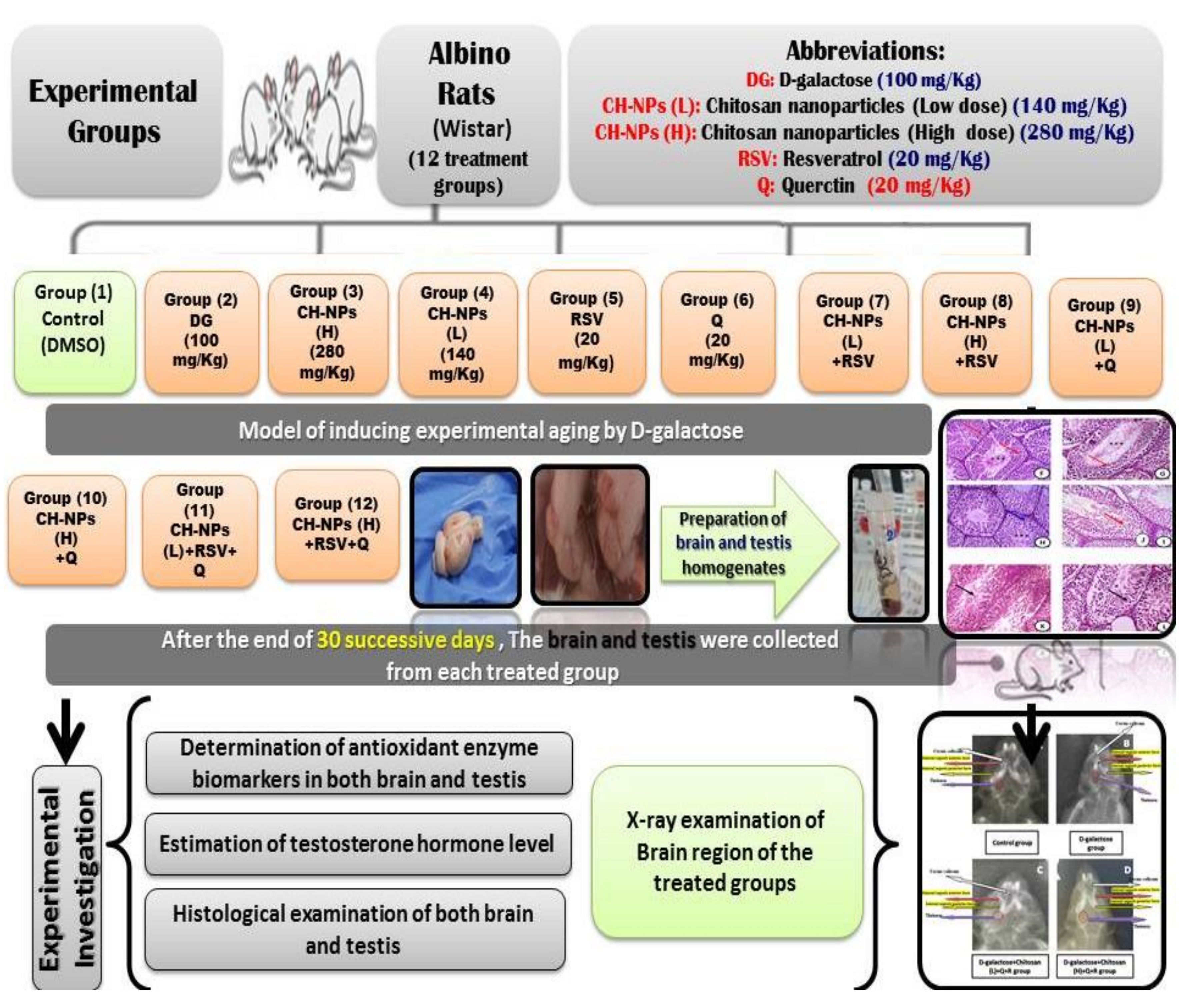
2.4. Experimental Animals

Sixty-nine adult male mice, their weight ranging from 40–50 g, were kept under standard laboratory conditions, while following European rules (86/609/EEC) on animal care. The experimental studies were approved by the ethical committee of Deanship of scientific research in Taif University. Then, accommodation occurred for two weeks before the experiment started under approval number: 39-31-0043.

2.5. Experimental Design

The male mice were split into 12 groups, eight rats within each group as follows: The first control group was given 1 mg/kg of DMSO (1%) as a vehicle; the second group was given a dose of D-galactose (100 mg/kg) dissolved in the vehicle (DMSO). The third group was treated with chitosan nanoparticles (high dose) (280 mg/kg) dissolved in the vehicle (DMSO). The fourth group was treated with chitosan nanoparticles (low dose) (140 mg/kg) dissolved in the vehicle (DMSO). The fifth group received a dose of resveratrol (20 mg/kg). The sixth group was treated with a dose of querectin (20 mg/kg). The seventh group was given a dose of chitosan nanoparticles (low dose) (140 mg/kg) followed by administration of resveratrol (20 mg/kg). The eighth group was given a dose of chitosan nanoparticles (high dose) (280 mg/kg) followed by administration of resveratrol (20 mg/kg). The ninth group was given a dose of chitosan nanoparticles (low dose) (140 mg/kg) followed by administration of quercetin (20 mg/kg). The tenth group was given a dose of chitosan nanoparticles (high dose) (280 mg/kg) followed by administration of quercetin (20 mg/kg). The eleventh group was treated with chitosan nanoparticles (low dose) (140 mg/kg) followed by administration of resveratrol (20 mg/kg) and then followed by quercetin (20 mg/kg). The twelfth group was treated with a dose of chitosan nanoparticles (high dose) (280 mg/kg) followed by administration of resveratrol (20 mg/kg) and then followed by quercetin (20 mg/kg). Treatment for all groups was intraperitoneally for 30 successive days as shown in experimental protocol ( Figure 1).

2.6. Preparation of Brain and Testis Homogenates for Estimation of Redox State

Brain tissues (0.23 g) and testicular tissues (0.25 g) were used for the determination of oxidative damage markers. They washed with saline for the removal of blood. Tissues were immersed with a sodium phosphate buffer (pH 7.4) in an ice medium. Then, tissues were homogenized and were centrifuged at 6000 r.p.m for 1/4 h. The homogenates supernatant was preserved at −80 °C untill it was used for measurement of catalase (CAT), GR, and malonaldehyde (MDA) [24,25].

2.7. Determination of Oxidative/Antioxidant Biomarkers

The lipid peroxidation end product (MDA) level was determined by Ohkawa et al. [26] and expressed as µmol/g tissue. Catalase (CAT) activity was measured according to Aebi [27]. CAT activity was measured in tissues by assaying the hydrolysis of H2O2 and the resulting decrease in absorbance at 240 nm over a 3 min period at 25 °C and expressed as mmol/mg. Glutathione reductase (GRX) activity was determined by commercial kits. GPx activity was determined in the brain tissues. The peroxide substrate (ROOH), glutathione reductase (GRx), and nicotinamide adenine dinucleotide phosphate (NADPH) are included.

2.8. Determination of the Testosterone Hormone Levels

Testosterone levels were measured by Enzyme-linked immunosorbent assay (ELISA) test according to the method of Wheeler [28]. Briefly, working solutions of the testosterone-HRP conjugate and wash buffer were prepared, then 50 μL of each calibrator, control, and specimen sample were pipetted into correspondingly labeled wells in duplicate. One hundred μL of the conjugate working solution was pipetted into each well and incubated on a plate shaker (approximately 200 rpm) for 1 h at 25 °C. Fifty μL of stop solution was pipetted into each well at the same timed intervals. The plate was read on a microwell plate reader at 450 nm within 20 min after addition of the stop solution.

2.9. Histological Evaluation

A part of brain tissues were fixed in 10% neutral buffered formalin as described by Gabe [29]. Brain tissue pieces were fixed in 10% formalin, then it was removed by washing the samples with tap water overnight. The tissue was dehydrated using a series of alcohols and were embedded in paraffin. Sections were cut using a microtome at 6 μm thickness. The thin sections were stained with hematoxylin and eosin. The slides were examined by light microscope and photographed by a digital camera.

2.10. X-ray Examination

Some of selected mice from some treated groups were given light anesthesia by sodium thiopental and then were examined by X-ray unit under special control in Kingdome Faisel Hosiptal in Taif City, Saudi Arabia.

2.11. Statistical Analysis

Data are expressed as mean values ± SE (n = 8). Statistical analysis was performed by using (ANOVA). For each significant effect, the Duncan’s Test [30] and post hoc Tukey’s test were used.

3. Results

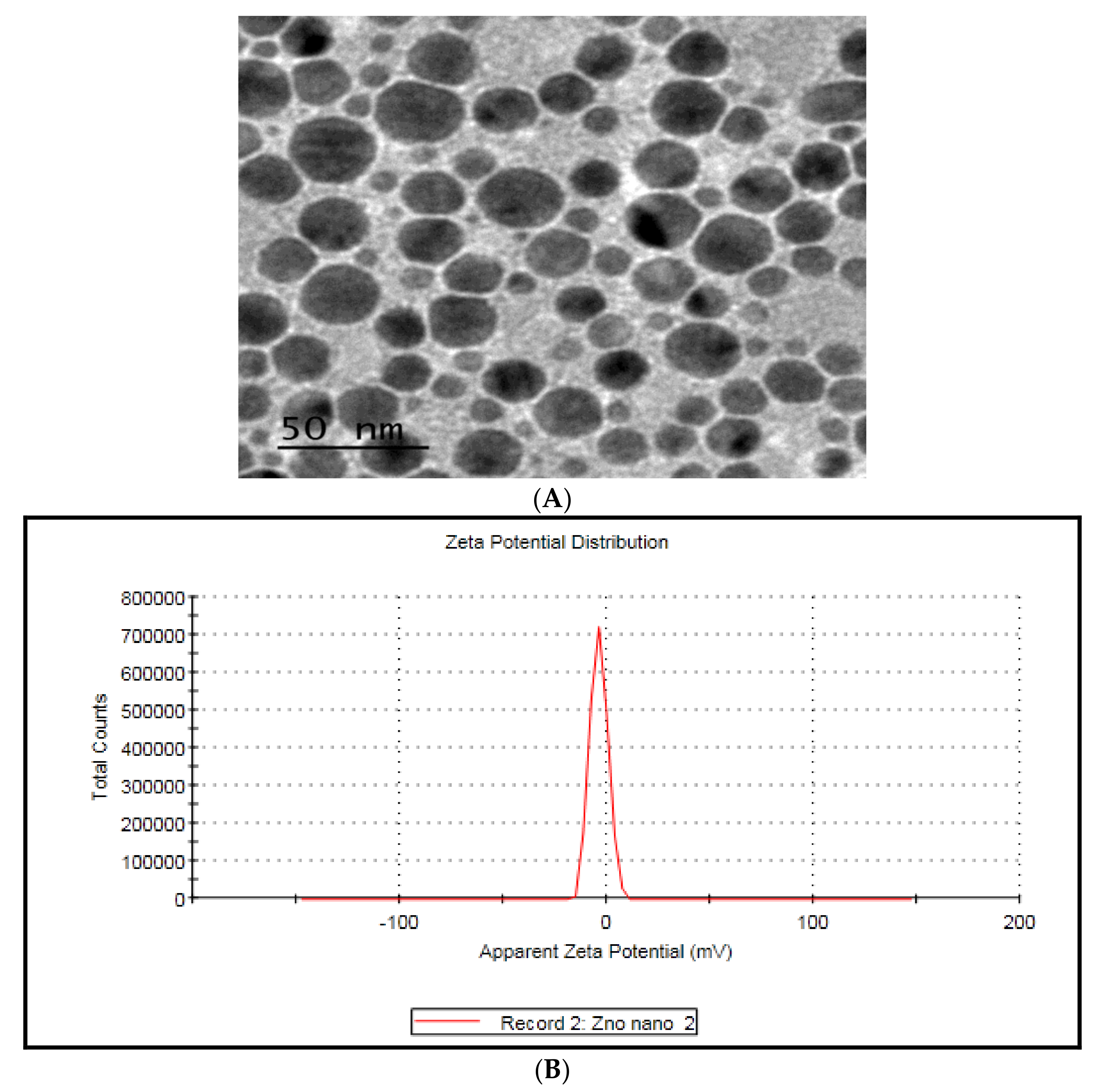
3.1. TEM Characterization of Chitosan Nanoparticles

Spherical form of chitosan nanoparticles within the scale of nanoparticles (50 nm) Zeta potential confirmed its nano-range scale Zeta Potential (mV): −3.77 ( Figure 2).

3.2. Oxidative Stress Biomarkers

The data in Table 1 and Table 2 demonstrated that there is no significant change in catalase (CAT) and glutathione reductase (GRx) of brain and testis homogenates in control rats and those treated with CH-NPs, RV, and Q and their combinations. In contrast, a decrease from all the previously mentioned antioxidant enzymes was recorded in the brain and testis of animals treated with DG for 30 days. The administration, both of the CH-NPs in two doses, RV, and Q combined with DG induced a significant increment in antioxidant enzymes as compared to its related group of DG.
The neuro-and testicular oxidative parameters (MDA) levels were significantly elevated by DG (Table 1). Either CH-NPs, Q, and RV declined MDA in brain and testis tissue homogenates which is the end product marker of lipid peroxidation as compared to the control group. The decrease in MDA levels was observed in combination groups of CH-NPs, RV, and Q with DG as compared to DG treated group.

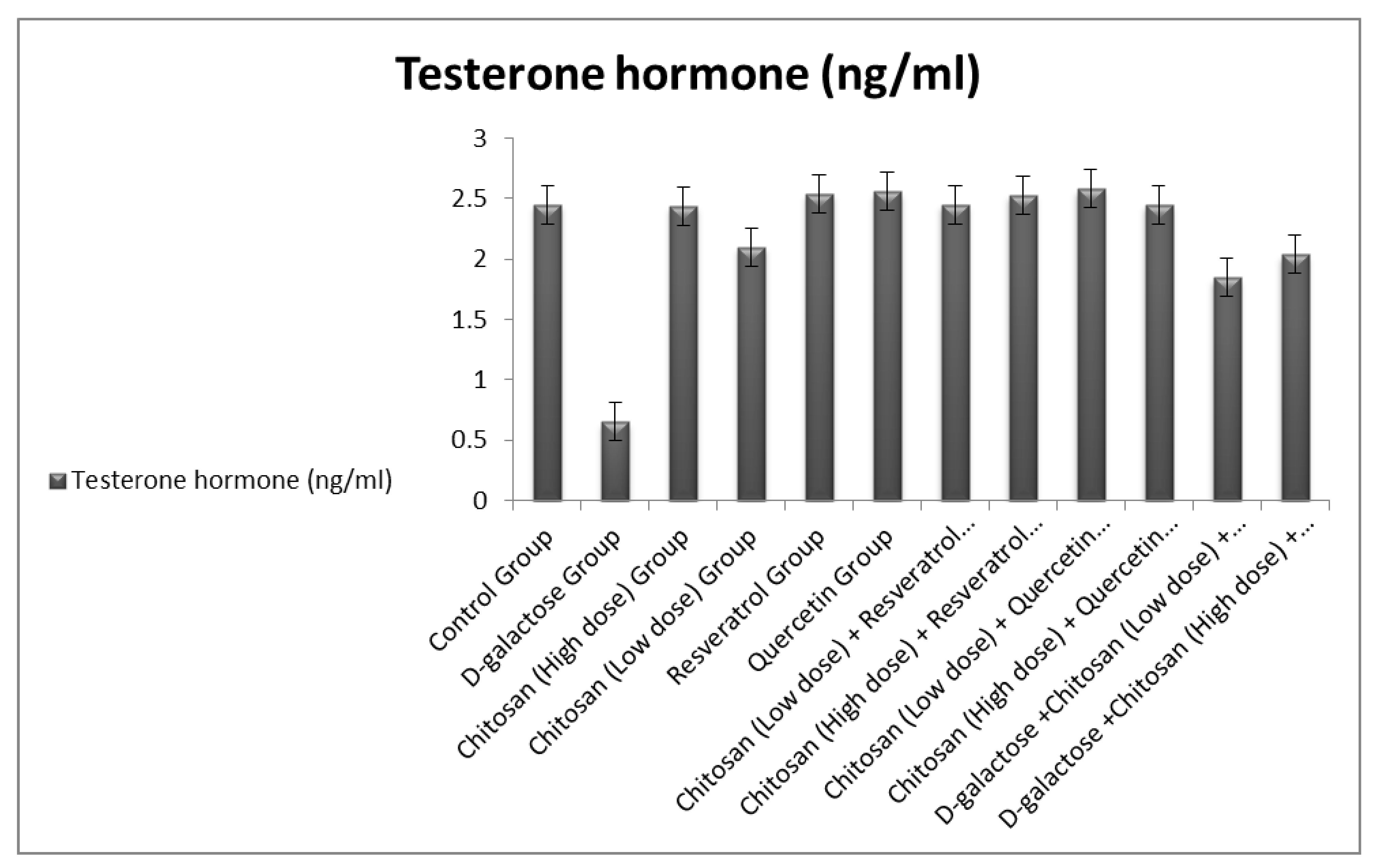
3.3. Testosterone Hormone Levels

The level of testosterone hormone in chitosan nanoparticles, quercetin, and resveratrol groups elicited a significant increase as compared to the normal control group. The level of testosterone decreased only in animals that were treated with D-galactose. The chitosan nanoparticles, querectin, and resveratrol improved the level of testosterone when given with D-galactose as compared with D-galactose treated group (Figure 3).

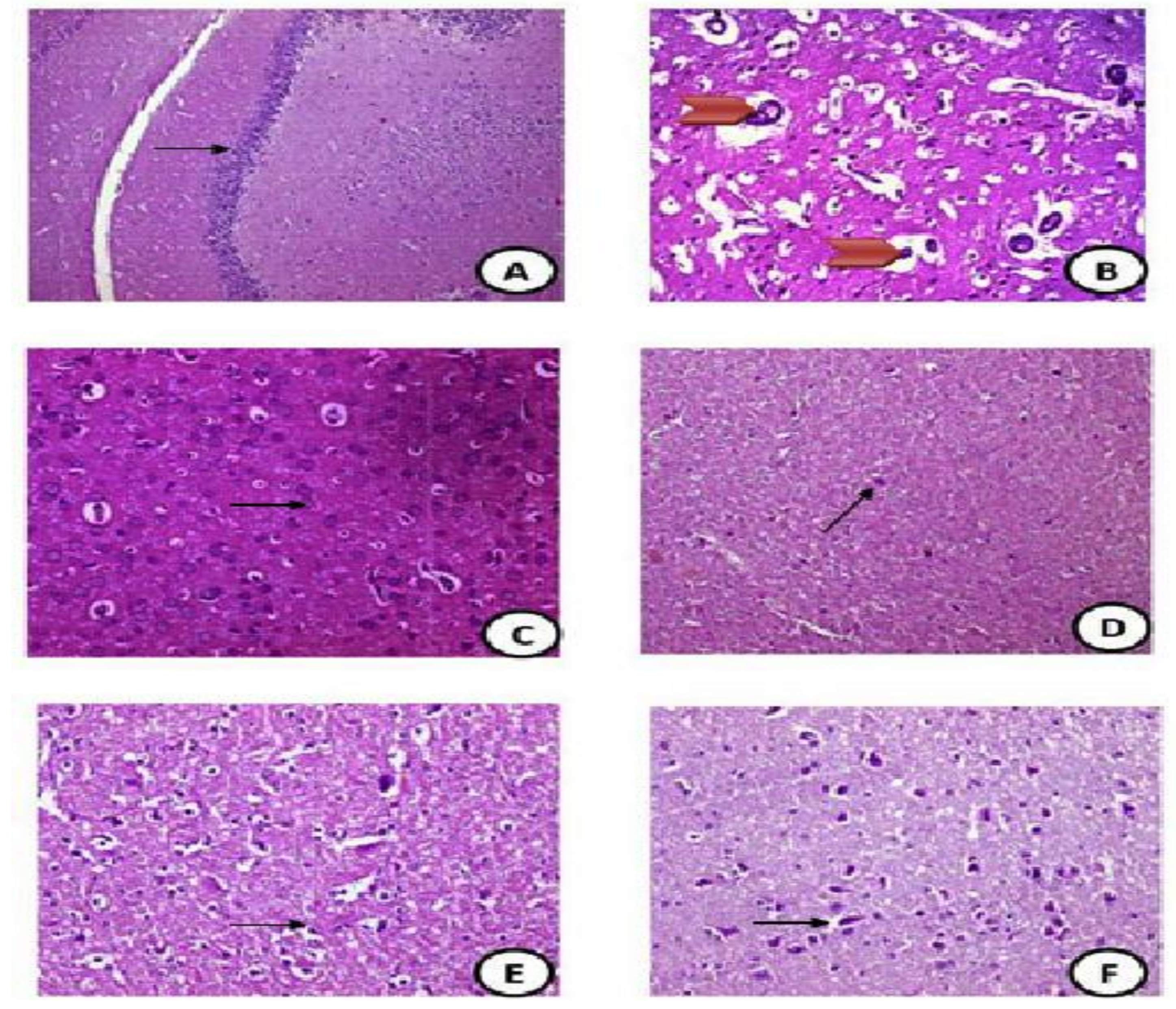
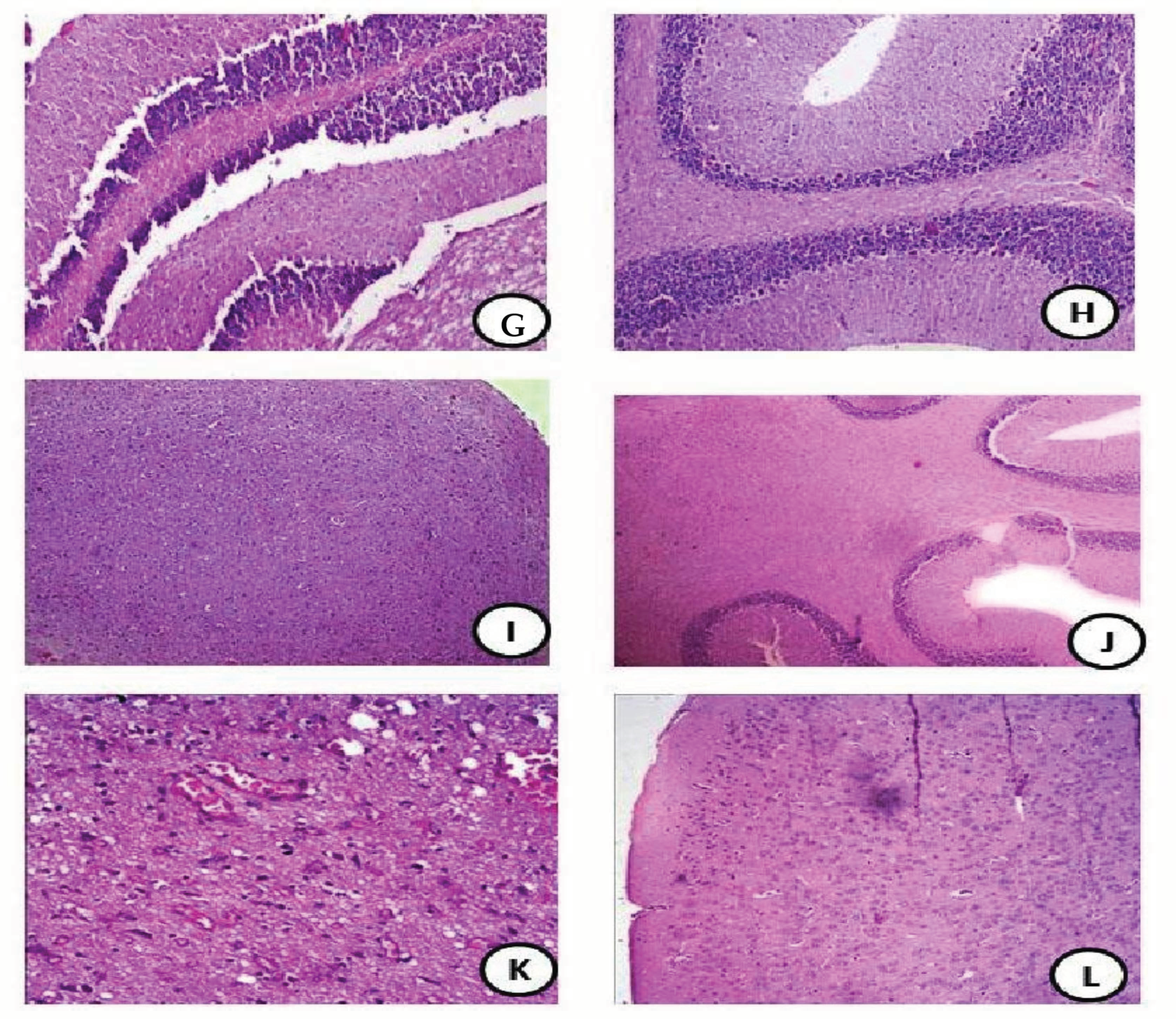
3.4. Histopathology Evaluation of Brain Tissues

The brain tissues from control group showed normal cerebral cortex with normal pyramidal cells with normal sized nuclei (black arrow) (H&EX100) (Figure 4A). The DG treated group showed disorganized pyramidal cells (red head arrow) with small dark nuclei with granular cells showing apoptotic nuclei (H&EX400) (Figure 4B). CH-NPs (high dose) showed normal cerebral cortex with normal pyramidal cells with normal sized nuclei (black arrow) and normal blood vessels (H&EX400) (Figure 4C). The CH-NPs (low dose) group showed normal cerebral cortex with normal glial cells with normal sized nuclei (black arrow) (H&EX400) (Figure 4D). The RV group showed normal cerebral cortex with normal pyramidal cells with normal sized nuclei (black arrow) (H&EX400) (Figure 4E). The Q group showed normal cerebral cortex with normal pyramidal cells with normal sized nuclei (black arrow) (H&EX400) (Figure 4F). The CH-NPs (low dose) + resveratrol group showed normal cerebral cortex with normal pyramidal cells with normal sized nuclei (black arrow) (H&EX400) (Figure 4G). The CH-NPs (High dose) + RV group showed normal cerebral cortex with normal pyramidal cells with normal sized nuclei (black arrow) (H&EX100) (Figure 4H). The CH-NPs (low dose) + Q group showed normal cerebral cortex with normal pyramidal cells with normal sized nuclei (black arrow) (H&EX100) (Figure 4I). CH-NPs (high dose) + Q group showed normal cerebral cortex with normal pyramidal cells with normal sized nuclei (black arrow) (H&EX100) (Figure 4J). DG + CH-NPs (Low dose) + RV+ Q showed mild congested blood vessels (red arrow) and neural cells showing small dark stained nuclei (H&EX400) (Figure 4K). DG + CH-NPs (High dose) + RV + Q showed the brain with highly ameliorative effect than DG group with few scattered apoptotic cells (**) with dark stained nuclei (black arrow) (H&EX400) (Figure 4L).

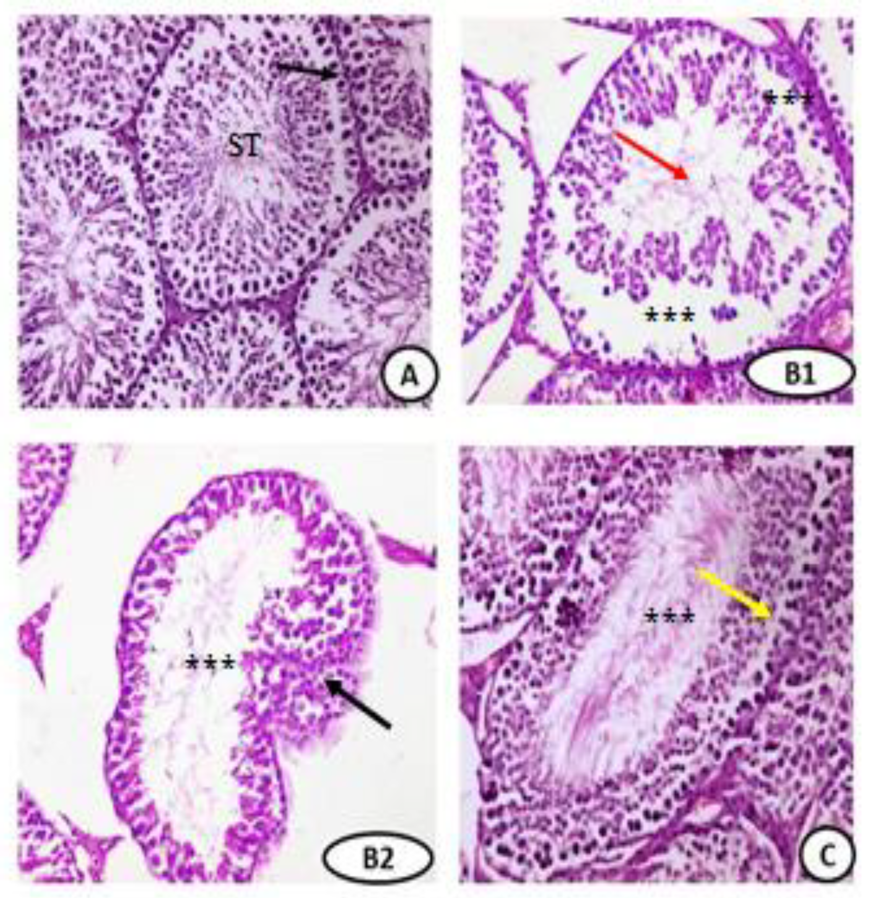
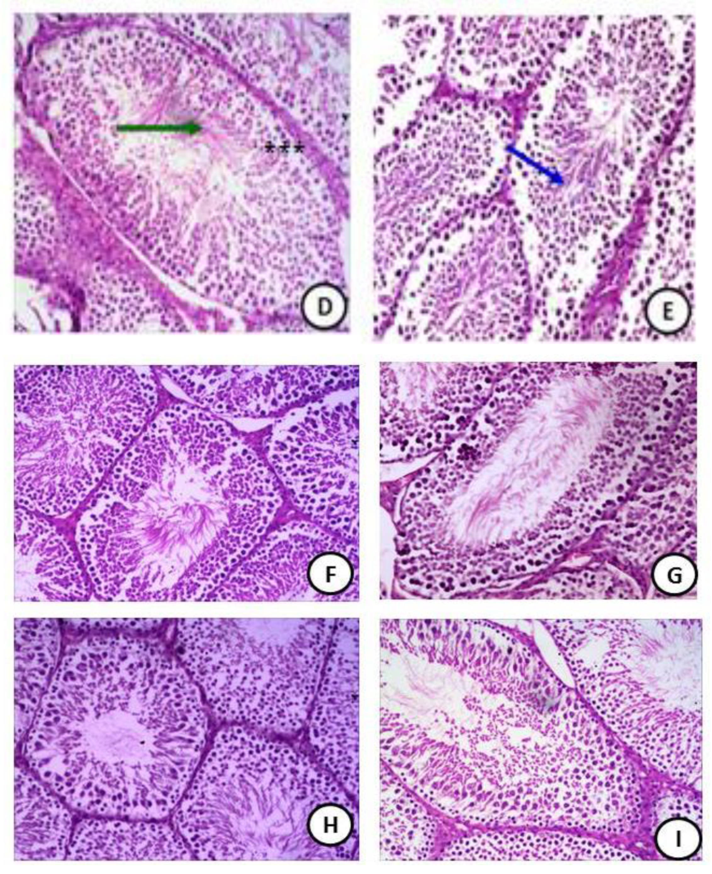
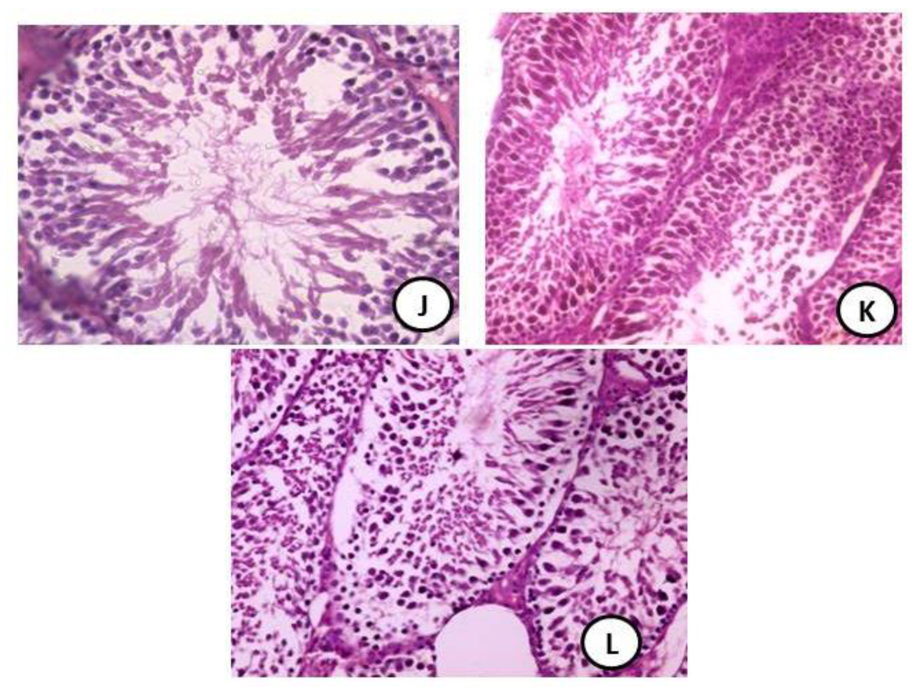
3.5. Histopathology Evaluation of Testicular Tissues

The testicular tissues from the control group was in a normal appearance and showed normal seminiferous tubules (ST) with normal spermatogenic cells (black arrow) and an appearance of plenty of sperms (H&EX400) (Figure 5A). The DG group showed a testis section of a group treated with DG showing degeneration of most parts of the seminiferous tubules (***) with oligospermia (red arrow) and hyperplasia of spermatogonia with high disintegration (black arrow) (H&EX400) (Figure 5(B1,B2)). The CH-NPs (high dose) group showed a normal spermatogonia (yellow arrow) with a lot of sperms (***) and an appearance of plenty of sperms (H&EX400) (Figure 5C). The CH-NPs (low dose) group showed a normal spermatogonia (***) with a lot of sperms (green arrow) and an appearance of plenty of sperms (H&EX400) (Figure 5D). The RV group showed a normal spermatogonia (blue arrow) and normal seminiferous tubules and ledyig cells (H&EX400) (Figure 5E). The Q group showed a normal spermatogonia (red arrow) with plenty of sperms (***) (H&EX400) (Figure 5F). The CH-NPs (low dose) + RV group showed a normal spermatogonia (red arrow) with plenty of sperms (***) and ledyig cells (H&EX400) (Figure 5G). The CH-NPs (high dose) + RV group showed a normal spermatogonia (blue arrow) with plenty of sperms (***) (H&EX400) (Figure 5H). CH-NPs (low dose) + Q group showing a normal spermatogonia (red arrow) (H&EX400) (Figure 5I). The CH-NPs (high dose) + Q group showed a normal spermatogonia (red arrow) (H&EX400) (Figure 5J). The DG + CH-NPs (low dose) + RV + Q showed restoration of spermatogonia layers (black arrow) and plenty of sperms (H&EX400) (Figure 5K). DG + CH-NPs (high dose) + RV + Q showed high restoration of spermatogonia layers (black arrow) and plenty of sperms (H&EX400) (Figure 5L).

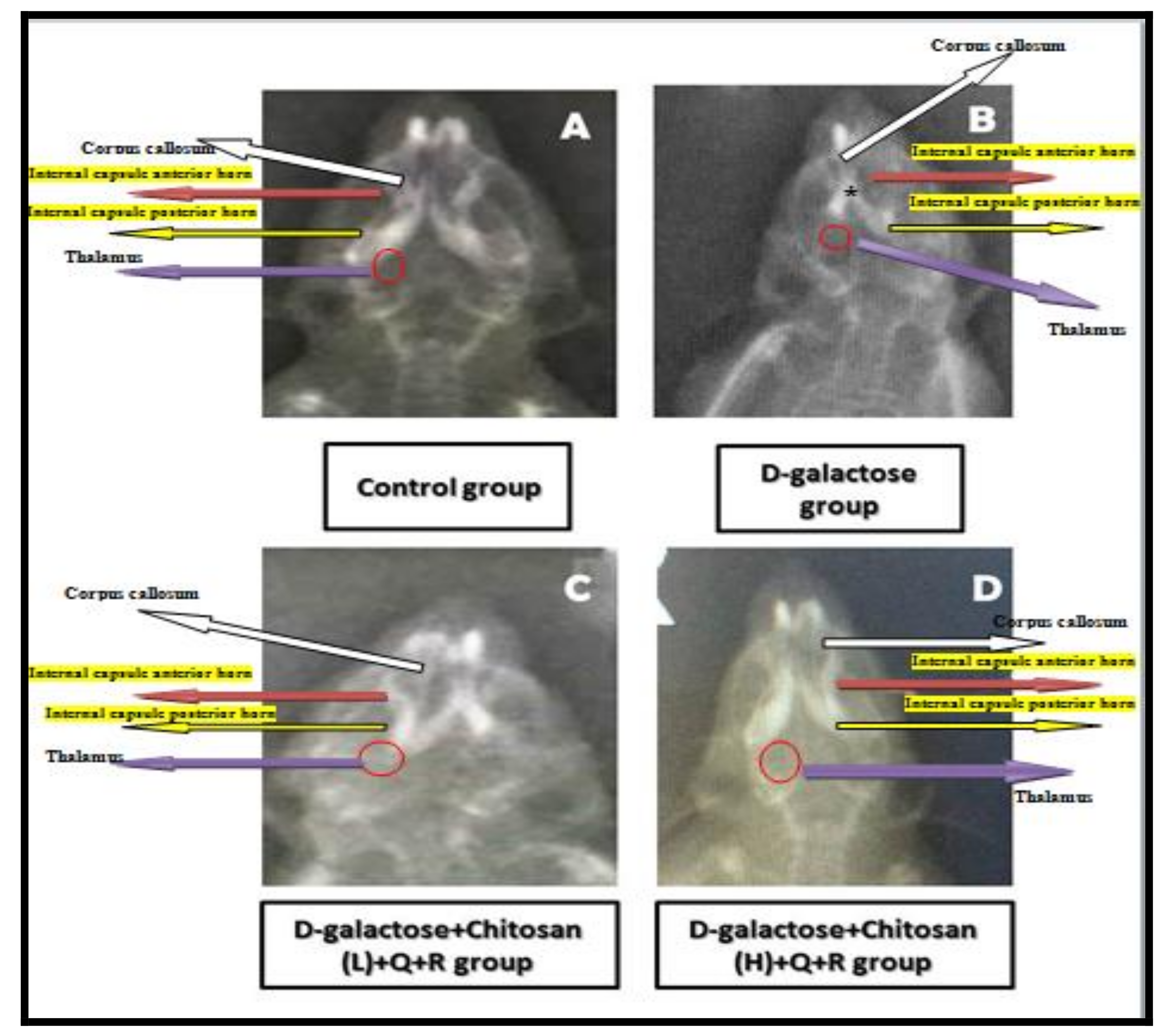
3.6. X-ray Alterations in Brain Structure of Some Selected Groups

Control group: Appearance of normal structure of the brain of control group showing appearance of Corpus callosum with normal size and appearance of two equal sized internal horns both interior and posterior with normal sized thalamus (Figure 6A).
D-galactose group: Appearance of the diminished Corpus callosum with reduced size as a spot and very reduced horns, both anterior and posterior with unequal sizes and reduced thalamus (Figure 6B).
D-galactose + Chitosan (Low dose) + Quercetin Resveratrol group: Appearance of mild ameliorative effect on internal horns, both anterior and posterior with mild restoration of thalamus and normal sized Corpus callosum (Figure 6C).
D-galactose + Chitosan (High dose) + Quercetin Resveratrol group: Appearance of highly ameliorative effect on brain structure x-ray with restoration of both horns the normal appearance with a mid-sized thalamus (Figure 6D).

4. Discussion

In the present study, the evaluation of the protective role of chitosan nanoparticles in two doses, resveratrol and quercetin against the toxicity resulting from the administration of D-galactose in male mice, was considered. Therefore, the main goals of the current study were to determine the neurological toxic effect of D-galactose for the long term by measuring the oxidative/antioxidant status as well as the brain histological and X-ray changes.
There is no specific scientific report that assayed the protective role of chitosan nanoparticles alone or combined with either quercetin and/or resveratrol against neurological alterations induced by D-galactose, chitosan, Quercetin, and resveratrol. They have potent antioxidant effects, which decline lipid peroxides production and increase antioxidant enzyme activities.
The study was undertaken to estimate the protective effect of Quercetin separately or in combination with either chitosan nanoparticles in two doses and resveratrol against D-galactose-afforded oxidative stress in different tissues of male mice. There is no literature report which is available on the efficacy of the three combinations of Quercetin, Resveratrol, and Chitosan nanoparticles in cases of D-galactose exposure in male mice. In this respect, the present study is original.
The study on D-galactose showed an increase in lipid peroxodation levels in brain tissues with a significant decrease in superoxide dismutase (SOD), Catalase (CAT), and Glutathione reductase (GRx) activities. These changes indicated the excessive production of oxygen free radicals in the brain tissues of D-galactose treated male mice, and such an imbalance is referred to as oxidative stress [31].
The obtained results reported that the balance between the oxidative system and antioxidant system in the male mice was damaged during D-galactose exposure.
Oxidative stress is elaborated through enzymatic and non-enzymatic mechanisms, which form the cellular defense mechanism. The antioxidant enzymes SOD and CAT play an important role in these mechanisms. SOD converts the superoxide radicals into hydrogen peroxide (H2O2), and CAT is accountable for the breakdown of H2O2 into water and oxygen. Therefore, the effects of these two enzymes are highly complementary to each other [31].
Some studies suggest the use of antioxidants and antioxidant-rich foods for the management of oxidative damage [32] and also for the beneficial effects of antioxidants as antidotes for any toxicity [33,34].
Interestingly, Quercetin could markedly renew the activities of glutathione in all tissues, SOD level, and other enzymes in other tissues of D-galactose-treated male mice as well as significantly enhance the antioxidant capacity of the body, and this is parallel to the present finding as these enzymes were significantly declined in the D-galactose treated group. Meanwhile, there was an increase in the groups treated with either chitosan nanoparticles, resveratrol and quercetin either with or without D-galactose.
The bioactivity of Quercetin is donating to its specific molecular structure, in which the oxygen active group, phenolic hydroxyls and 2,3-unsaturated double bond, give Quercetin strong antioxidant ability not from accepting oxygen free radicals but also by forming metal chelation compounds, and thus the metals excessive levels can be reduced [35] and this can help in maintaining brain tissue’s viability and maintenance of nerve cells.
Q acts as an antioxidant by prohibiting the oxidative enzymes such as xanthine oxidase and lipoxygenase. Inhibition of these enzymes is also responsible for the attenuation of oxidative stress as they play key roles in the free radical initial process which induced cellular damage [36]. Further, it has been reported that Q metabolites can also inhibit some chemical compounds oxidation, similar to free Q [37].
In the present study, treatment with RV either separately or combined with Q after DG administration caused normalization of levels of oxidative/antioxidant parameters in brain tissues. It also caused significant improvement in inflammatory biomarkers in different tissues histopathological and changes as histological sections of the brain and changes in X-ray examination.
Resveratrol proved its antioxidant ability in our study by preserving SOD, CAT, and glutathione reduced as well as reducing MDA levels in the D-galactose combined with resveratrol and CH-NPs combined with RV and Q groups compared to the DG treated group.
Jiang et al. [38] found that resveratrol possesses potent antioxidant activity that may decline oxidative stress and inhibit free radicals. Results from the current study are in agreement with previous studies that reported the lowest level of serum lipid peroxide products in animals fed with resveratrol [39,40]. Toklu et al. [41] found that RV ameliorated the antioxidant status and decreased oxidative damage to the heart and brain of rats with induced hypertension and this is parallel with the current investigation.
Resveratrol is both a free radical scavenger and a potent antioxidant because of its ability to promote the activities of a variety of antioxidant enzymes, as previous studies found that resveratrol could function as SOD1 and GPx1. These findings supported our results that the LPO levels were significantly lower in the brain homogenates of groups treated with a combination of D-galactose with resveratrol while antioxidant enzymes increased.
The resveratrol antioxidant activity could be due to its capability to prohibit reductase in the ribonucleotide and cyclooxygenase-2 as previously reported. Others reported the resveratrol antioxidant effects that could be due to its capability to reduce the oxidative chain reactions of reactive oxygen species in comparable with coenzyme Q, remove the superoxide radicals from the mitochondria, and thus inhibit lipid peroxidation, and this is very important for brain tissues.
Our results promoted with previous findings. These histological alterations could be related to the biochemical alterations, especially contents of reactive oxygen species in the brain and liver that were markedly increased in male mice due to D-galactose administration.
Administration of either Q, RV, and CH-NPs resulted in improvement partially in all histopathological changes noticed from DG in a lot of tissues of mice. This may be due to antioxidative properties of Q polyphenols or active compounds of resveratrol. They are a part of the polyphenols group that have strong electrophilic centers, a property that provides an opportunity for them to react with compounds like DG, thus diminishing DG toxic effects.
Juan et al. [42] found that repeated trans-RV consumption at a dose of 20 mg/kg/day does not affect adversely on the hematologic or biochemical parameters as well as the histopathologic examination of different organs, but in the current study, the administration of RV induced neuroprotective effect in groups treated with a combination of DG combined with RV, Q, and CH-NPs.
The study on DG showed a significant increase in lipid peroxidation levels in the tested testis tissues and a decrease of SOD, CAT, and GRx activities. These alterations indicated the presence of free radicals excessive production in DG treated male mice, and this imbalance is considered as oxidative stress [43]. The antioxidant enzymes as CAT have a vital role in these mechanisms. CAT is accountable for the breakdown of H2O2 produced by superoxide radicals [43].
The current aim of the study is to prove the ameliorating effect of Q or RV and/or CH-NPs or their combination on DG-induced oxidative imbalance. No scientific reports have been published to describe the protective role of the used compounds against DG -induced testicular oxidative stress.
Interestingly, Q could greatly regenerate the activities of glutathione in all tissues, SOD level, and other enzymes in other tissues of D-galactose-treated male mice as well as significantly reinforced the antioxidant capacities.
Quercetin acts as an antioxidant by prohibiting the oxidative enzymes. Inhibition of these enzymes is responsible for the attenuation of oxidative stress by inhibiting the free radicals which induced cellular damage [44]. Further, it has been demonstrated that Q can inhibit some compound’s oxidation [45].
Jiang et al. [38] reported that resveratrol possesses potent antioxidant activity that may inhibit free radicals. Results from the current study are in agreement with studies that reported the lower level of serum lipid peroxides products in animals treated with resveratrol [46,47].
Our results promoted with previous findings. These histological alterations could be related to the biochemical alterations, especially contents of reactive oxygen species in testis that were markedly increased in male mice due to DG administration. According to DG dose regimen for male mice used in the current study, marked alterations in the histology of their testis taken, including denudation of seminiferous tubules that cause disturbance of spermatogenesis.
Administration of either Q, RV, and CH-NPs resulted in improvement partially in all histopathological changes noticed from D-galactose in a lot of tissues of mice. This may be due to antioxidative properties of Q polyphenols or active compounds of resveratrol. They are part of the polyphenols group that have strong electrophilic centers, a property that provides an opportunity for them to react with compounds like DG, thus diminishing DG toxic effects.
Additionally, the current study found that testis tissues structures were normal in the resveratrol treated groups, contrary to the DG group. These results were parallel to previous studies who reported the potent antioxidant activities of resveratrol [48].
The resveratrol administration is followed by DG ameliorated different histological injury, which was concerned with ameliorating some of the biochemical parameters. All the findings were proved by the histopathological examination, which confirmed the resveratrol capability to keep functionally active testicular tissues either histological sections or TEM sections in testis tissues.
Additionally, the current study found that brain tissue structures were generally normal in the groups combined with RV, Q, and two doses of CH-NPs contrary to the damage changes which are seen in DG group. These results were parallel to reports of Xiao, 2015 [48].

5. Conclusions

D-galactose induced neurotoxicity in the brain and testis of male mice. Neuro-ameliorative properties of CH-NPs, RV, and Q may be related to their ability to increase the endogenous antioxidant enzymes by decreasing the oxidative stress. Therefore, we want to pass through the light regarding the deleterious effects of oxidative stress, especially in brain and testicular tissues and their causes which leads to aging and reducing the antioxidant capacities of the brain and testicular tissues, thus reducing its general criteria through either learning or memory. So, the neuroprotective and testicular synergistic protective effects of these used antioxidant compounds are of great importance, especially for children to overcome the problems of delay in learning and reducing learning abilities. So, we recommend using CH-NPs in combination with both RV and Q to alleviate signs of aging and give protection to the tissues from sever oxidative stress.

Author Contributions

Conceptualization and methodology, R.Z.H.; software, R.Z.H., M.S.A.-H., and M.A.A.-H.; validation, R.Z.H., M.S.A.-H., and M.A.A.-H.; formal analysis and investigation, R.Z.H.; resources, R.Z.H. and M.S.A.-H.; data curation, R.Z.H., M.S.A.-H., and M.A.A.-H.; writing—original draft preparation, R.Z.H., M.S.A.-H., and M.A.A.-H.; writing—review and editing, R.Z.H., M.S.A.-H., and M.A.A.-H.; visualization and funding acquisition, R.Z.H. All authors have read and agreed to the published version of the manuscript.

Funding

The authors also acknowledge to Taif University Researchers supporting project number (TURSP-2020/21), Taif University, Taif, Saudi Arabia.

Institutional Review Board Statement

Not applicable.

Informed Consent Statement

Not applicable.

Data Availability Statement

The raw data supporting the conclusions of this article will be made available by the corresponding author.

Conflicts of Interest

TThe author declares that there are not any conflict of interest.

Abbreviations

D-galactose (DG), Chitosan nanoparticles (CH-NPs), Quercetin (Q), Resveratrol (RV), Catalase (CAT), Malondialdhyde (MDA), Glutathione reductase (GRx).

References

  1. Banji, D.; Banjia, O.F.; Dasaroju, S.; CH, K.K. Curcumin and piperine abrogate lipid and protein oxidation induced by D-galactose in rat brain. Brain Res. 2013, 1515, 1–11. [Google Scholar] [CrossRef] [PubMed]
  2. Cui, X.; Zuo, P.; Zhang, Q.; Li, X.; Hu, Y.; Long, J. Chronic systemic D-galactose exposure induces memory loss, neurodegeneration, and oxidative damage in mice: Protective effects of R-alpha-lipoic acid. J. Neurosci. Res. 2006, 83, 1584–1590. [Google Scholar] [CrossRef] [PubMed]
  3. Park, J.; Ramanathan, R.; Pham, L.; Woodrow, K.A. Chitosan enhances nanoparticle delivery from the repro-ductive tract to target draining lymphoid organs. Nanomed. Nanotechnol. Biol. Med. 2017, 13, 2015–2025. [Google Scholar] [CrossRef]
  4. Bugnicourt, L.; Ladavière, C. A close collaboration of chitosan with lipid colloidal carriers for drug delivery applications. J. Control. Release 2017, 256, 121–140. [Google Scholar] [CrossRef]
  5. Ghadi, A.; Mahjoub, S.; Tabandeh, F.; Talebnia, F. Synthesis and optimization of chitosan nanoparticles: Potential applications in nanomedicine and biomedical engineering. Casp. J. Intern. Med. 2014, 5, 156–161. [Google Scholar]
  6. Al-Baqami1, N.M.; Hamza, R.Z. Synergistic antioxidant capacities of vanillin and chitosan nanoparticlesn against re-active oxygen species, hepatotoxicity, and genotoxicity induced by aging in male Wistar rats. Hum. Exp. Toxicol. 2021, 40, 183–202. [Google Scholar] [CrossRef]
  7. Dajas, F.; Abin-Carriquiry, J.A.; Arredondo, F.; Blasina, F.; Echeverry, C.; Martínez, M.; Rivera, F.; Vaamonde, L. Quercetin in brain diseases: Potential and limits. Neurochem. Int. 2015, 89, 140–148. [Google Scholar] [CrossRef]
  8. Zini, A.; Al-Hathal, N. Antioxidant therapy in male infertility: Fact or fiction? Asian J. Androl. 2011, 13, 374–381. [Google Scholar] [CrossRef]
  9. Bhutada, P.; Mundhada, Y.; Bansod, K.; Bhutada, C.; Tawari, S.; Dixit, P.; Mundhada, D. Neurobiology of learning and memory ameliorative effect of quercetin on memory dysfunction in streptozotocin-induced diabetic rats. Neurobiol. Learn. Mem 2010, 94, 293–302. [Google Scholar] [CrossRef] [PubMed]
  10. Juan, M.E.; González-Pons, E.; Munuera, T.; Ballester, J.; Rodríguez-Gil, J.E.; Planas, J.M. Trans-Resveratrol, a natural antioxidant from grapes, increases sperm output in healthy rats. J. Nutr. 2005, 135, 757–760. [Google Scholar] [CrossRef] [PubMed]
  11. Hamza, R.Z.; El-Shenawy, N.S.; Ismail, H.A.A. Protective effects of blackberry and quercetin on sodium fluoride-induced oxidative stress and histological changes in the hepatic, renal, testis and brain tissue of male rat. J. Basic Clin. Physiol. Pharmacol. 2015, 26, 237–251. [Google Scholar] [CrossRef]
  12. Refat, M.S.; Hamza, R.Z.; Adam, A.A.; Saad, H.A.; Gobouri, A.A.; Al-Harbi, F.S.; Al-Salmi, F.A.; Altalhi, T.; El-Megharbel, S.M. Quercetin/Zinc complex and stem cells: A new drug therapy to ameliorate glycometabolic control and pulmonary dysfunction in diabetes mellitus: Structural characterization and genetic studies. PLoS ONE 2021, 16, e0246265. [Google Scholar] [CrossRef] [PubMed]
  13. El-Denshary, E.S.; Aljawish, A.; El Nekeety, A.A.; Hassan, N.S.; Saleh, R.H.; Rihn, B.H.; Abdel-Wahaab, M.A. Pos-sible synergistic effect ioxidant properties of chitosan nanoparticles and quercetin against carbon tetrachloride induce hepatotoxicity in rats. Soft Nanosci. Lett. 2015, 5, 36–51. [Google Scholar] [CrossRef]
  14. Agarwal, A.; Mahfouz, R.Z.; Sharma, R.K.; Sarkar, O.; Mangrola, D.; Mathur, P.P. Potential biological role of poly (ADP-ribose) polymerase (PARP) in male gametes. Reprod. Biol. Endocrinol. 2009, 7, 143. [Google Scholar] [CrossRef] [PubMed]
  15. Reddy, P.K.; Madhu, P.; Reddy, S.P. Protective effects of resveratrol against cisplatin-induced testicular and epi-didymal toxicity in rats. Food Chem. Toxicol. 2016, 91, 65–72. [Google Scholar] [CrossRef] [PubMed]
  16. Wici’nskia, M.; Leis, K.; Szyperski, P.; Eclewicz, M.M.W.; Mazura, E.; Pawlak-Osinska, K. Impact of resveratrol on exercise performance: A review. Sci. Sports 2018, in press. [Google Scholar]
  17. Prakash, A.; Kumar, A. Pioglitazone alleviates the mitochondrial apoptotic pathway and mito-oxidative damage in the d -galactose-induced mouse model. Clin. Exp. Pharmacol. Physiol. 2013, 40, 644–651. [Google Scholar] [CrossRef]
  18. Hamza, R.Z.; Al-Thubaiti, E.H.; Omar, A.S. The antioxidant activity of quercetin and its effect on acrylamide hepatotoxicity in liver of rats. Lat. Am. J. Pharm. 2019, 38, 2057–2062. [Google Scholar]
  19. Allen, E.N.; Potdar, S.; Tapias, V.; Parmar, M.; Cassia, S.; Rimando, M.A.; Cavanaugh, J.E. Resveratrol and pinostilbene confer neuroprotection against aging-related deficits through an ERK1/2-dependent mecha-nism. J. Nutr. Biochem. 2018, 54, 77–86. [Google Scholar] [CrossRef]
  20. Dal-Pan, A.; Terrien, J.; Pifferi, F.; Botalla, R.; Hardy, I.; Marchal, J.; Zahariev, A.; Chery, I.; Zizzari, P.; Perret, M.; et al. Caloric restriction or resveratrol supplementation and ageing in a non-human primate: First-year outcome of the RESTRIKAL study in Microcebus murinus. AGE 2011, 33, 15–31. [Google Scholar] [CrossRef]
  21. Al-Otaibi, S.S.; Arafah, M.M.; Sharma, B.; Alhomida, A.S.; Siddiqi, N.J. Synergistic effect of quercetin and 𝛼-lipoic acid on aluminium chloride induced neurotoxicity in rats. J. Toxicol. 2018, 2018, 2817036. [Google Scholar] [CrossRef] [PubMed]
  22. Türedi, S.; Yuluğ, E.; Alver, A.; Kutlu, Ö.; Kahraman, C. Effects of resveratrol on doxorubicin induced testicular damage in rats. Exp. Toxicol. Pathol. 2015, 67, 229–235. [Google Scholar] [CrossRef] [PubMed]
  23. Tang, Z.-X.; Qian, J.-Q.; Shi, L.-E. Preparation of chitosan nanoparticles as carrier for immobilized enzyme. Appl. Biochem. Biotechnol. 2007, 136, 77–96. [Google Scholar] [CrossRef]
  24. Hamza, R.Z.; Al-Hazmi, M.A.; Rawi, S.M. Biochemical, histological, and neuro-physiological effects of long-term aluminum chloride exposure in rats. Metab. Brain Dis. 2021, 36, 429–436. [Google Scholar] [CrossRef]
  25. Habig, W.H.; Pabst, M.J.; Jakoby, W.B. Glutathione S-transferases. The first enzymatic step in mercapturic acid formation. J. Biol. Chem. 1974, 249, 7130–7139. [Google Scholar] [CrossRef]
  26. Ohkawa, H.; Ohishi, N.; Yagi, K. Assay for lipid peroxides in animal tissues by thiobarbituric acid reaction. Anal. Biochem. 1979, 95, 351–358. [Google Scholar] [CrossRef]
  27. Aebi, H. Catalase in vitro. Method Enzymol. 1984, 105, 121–126. [Google Scholar]
  28. Gabe, M. Techniques Histologiques; Masson Publisher: Paris, France, 1968. [Google Scholar]
  29. Duncan, D.B. Multiple range and multiple F-test. Biometrics 1955, 11, 1–42. [Google Scholar] [CrossRef]
  30. Halliwell, B. Biochemistry of oxidative stress. Biochem. Soc. Trans. 2007, 35, 1147–1150. [Google Scholar] [CrossRef]
  31. Susheel, A.K. Fluorosis management programme in India by D-galactose in mice. Curr. Sci. 1999, 77, 1250–1256. [Google Scholar]
  32. Chinoy, N.J.; Memon, M.R. Beneficial effects of some vitamins and quercetin and quercetin-5′,8-disulfonate against carbon tetrachloride-caused oxidative liver injury in mice. Molecules 2001, 19, 291–305. [Google Scholar]
  33. Reddy, G.B.; Khandare, A.L.; Reddy, P.Y.; Rao, G.S.; Balakrishna, N. Antioxidant defense system and lipid peroxida-tion in patients with skeletal fluorosis and in fluoride-intoxicated rabbits. Toxicol. Sci. 2003, 72, 363–3688. [Google Scholar] [CrossRef] [PubMed]
  34. El-Megharbel, S.M.; Alsawat, M.; Al-Salmi, F.A.; Hamza, R.Z. Utilizing of (zinc oxide nano-spray) for disinfection against “SARS-CoV-2” and testing its biological effectiveness on some biochemical parameters during (COVID-19 pandemic)— “ZnO nanoparticles have antiviral activity against (SARS-CoV-2)”. Coatings 2021, 11, 388. [Google Scholar] [CrossRef]
  35. Day, J.A.; Canada, J.F.; Diaz, C.J.; Kroon, A.P.; Mclauchlan, R.; Faulds, B.C. Dietary flavonoid and isoflavone glyco-sides are hydrolysed D-galactose-induced mouse model. Clin. Exp. Pharmacol. Physiol. 2000, 40, 644–651. [Google Scholar]
  36. Klotz, L.O.; Sies, H. Defenses against peroxynitrite: Seleno compounds and flavonoids. Toxicol. Lett. 2003, 140, 125–132. [Google Scholar] [CrossRef]
  37. Jiang, Y.-G.; Peng, T.; Luo, Y.; Li, M.-C.; Lin, Y.-H. Resveratrol reestablishes spermatogenesis after testicular injury in rats caused by 2,5-hexanedione. Chin. Med. J. 2008, 121, 1204–1209. [Google Scholar] [CrossRef]
  38. Kasdallah-Grissa, A.; Mornagui, B.; Aouani, E.; Hammami, M.; Gharbi, N.; Kamoun, A.; El-Fazaa, S. Protective effect of resveratrol on ethanol-induced lipid peroxidation in rats. Alcohol Alcohol. 2006, 41, 236–239. [Google Scholar] [CrossRef] [PubMed]
  39. Silan, C. The effects of chronic resveratrol treatment on vascular responsiveness of streptozotocin-induced diabetic rats. Biol. Pharm. Bull. 2008, 31, 897–902. [Google Scholar] [CrossRef]
  40. Toklu, H.Z.; Sehirli, O.; Ersahin, M.; Süleymanoğ, S.; Yiğiner, O.; Emekli-Alturfan, E.; Yarat, A.; Yeğen, B.C.; Sener, G. Resveratrol improves cardiovascular function and reduces oxidative organ damage in the renal, cardiovascular and cerebral tissues of two-kidney, one clip hypertensive rats. J. Pharm. Pharmacol. 2010, 62, 1784–1793. [Google Scholar] [CrossRef] [PubMed]
  41. Juan, M.E.; Vinardell, M.P.; Planas, J.M. The daily oral administration of high doses of trans-resveratrol to rats for 28 days is not harmful. J. Nutr. 2002, 132, 257–260. [Google Scholar] [CrossRef]
  42. Hamza, R.Z.; El-Shenawy, N.S. Anti-inflammatory and antioxidant role of resveratrol on nicotine-induced lung changes in male rats. Toxicol. Rep. 2017, 4, 399–407. [Google Scholar] [CrossRef]
  43. Refat, M.S.; Hamza, R.Z.; Adam, A.A.; Saad, H.A.; Gobouri, A.A.; Al-Salmi, F.A.; Altalhi, T.A.; El-Megharbel, S.M. Potential therapeutic effects of new ruthenium (III) complex with quercetin: characterization, structure, gene regulation, and antitumor and anti-inflammatory studies (RuIII/Q novel complex is a potent immunoprotective agent). Crystals 2021, 11, 367. [Google Scholar] [CrossRef]
  44. Altintas, R.; Ciftci, O.; Aydin, M.; Akpolat, N.; Oguz, F.; Beytur, A. Quercetin prevents docetaxel-induced testicular damage in rats. Asian J. Androl. 2014, 47, 248–256. [Google Scholar] [CrossRef] [PubMed]
  45. Zhao, H.; Li, N.; Wang, Q.; Cheng, X.; Li, X.; Liu, T. Resveratrol decreases the insoluble Aβ1–42 level in hippocampus and protects the integrity of the blood–brain barrier in AD rats. Neuroscience 2015, 310, 641–649. [Google Scholar] [CrossRef]
  46. Hamza, R.Z.; Al-Talhi, T.; Gobouri, A.A.; Al-Yasi, H.M.; Diab, A.A.; El-Megharbel, S.M. Resveratrol and nicotine toxicity. Toxicology 2021, 505–517. [Google Scholar] [CrossRef]
  47. Bucak, M.N.; Ataman, M.B.; Başpınar, N.; Uysal, O.; Taşpınar, M.; Bilgili, A. Lycopene and resveratrol improve post thaw bull sperm parameters, sperm motility, mitochondrial activity and DNA integrity. Andrologia 2017, 47, 545–552. [Google Scholar] [CrossRef]
  48. Xiao, N.-N. Effects of resveratrol supplementation on oxidative damage and lipid peroxidation induced by strenuous exercise in rats. Biomol. Ther. 2015, 23, 374–378. [Google Scholar] [CrossRef]
Figure 1. Experimental protocol.
Coatings 11 00435 g001
Figure 2. Transmission electron microscope (TEM) and Zeta potential of Chitosan nanoparticles: (A) TEM characterization for chitosan nanoparticles; (B) Zata potential for Chitosan nanoparticles.
Coatings 11 00435 g002
Figure 3. Effect of D-galactose, Chitosan nanoparticles, correcting and Resveratrol on testosterone hormone level (ng/mL) in male mice.
Coatings 11 00435 g003
Figure 4. Cross sections of mice brain tissues from the treated groups showing structure alterations. H & E stain × 400: (A) Control group showed normal cerebral cortex with normal pyramidal cells (H&EX100); (B) DG treated group showed disorganized pyramidal cells (red head arrow) with small dark nuclei with granular cells showing apoptotic nuclei (H&EX400); (C) CH-NPs (high dose) showed normal cerebral cortex with normal pyramidal cells with normal sized nuclei (H&EX400); (D) CH-NPs (low dose) group showed normal cerebral cortex with normal glial cells with normal sized nuclei (H&EX400); (E) RV group showed normal cerebral cortex with normal pyramidal cells with normal sized nuclei (H&EX400); (F) Q group showed normal cerebral cortex with normal pyramidal cells with normal sized nuclei (H&EX400); (G) CH-NPs (low dose) + resveratrol group showed normal cerebral cortex with normal pyramidal cells with normal sized nuclei (black arrow) (H&EX400); (H) CH-NPs (High dose) + RV group showed normal cerebral cortex with normal pyramidal cells with normal sized nuclei (H&EX100); (I) CH-NPs (low dose) + Q group showed normal cerebral cortex with normal pyramidal cells with normal sized nuclei (black arrow) (H&EX100); (J) CH-NPs (high dose) + Q group showed normal cerebral cortex with normal pyramidal cells with normal sized nuclei (black arrow) (H&EX100); (K) DG + CH-NPs (Low dose) + RV + Q showed mild congested blood vessels (red arrow) and neural cells showing small dark stained nuclei (H&EX400); (L) DG + CH-NPs (High dose) + RV + Q showed the brain with highly ameliorative effect than DG group (H&EX400).
Coatings 11 00435 g004aCoatings 11 00435 g004b
Figure 5. Cross sections of mice testis tissues from the treated groups showing structure alterations H & E stain X400: (A) The testicular tissues from the control group were in a normal appearance and showed normal seminiferous tubules (ST) with normal spermatogenic cells (black arrow) (H&EX400); (B1,B2) DG group showed a testis section of a group treated with DG showing degeneration of most parts of the seminiferous tubules (***) with oligospermia (red arrow) and hyperplasia of spermatogonia (H&EX400); (C) CH-NPs (high dose) group showed a normal spermatogonia (yellow arrow) with a lot of sperms (***); (D) CH-NPs (low dose) group showed a normal spermatogonia (***) with a lot of sperms (green arrow) and an appearance of plenty of sperms (H&EX400); (E) RV group showed a normal spermatogonia (blue arrow) and normal seminiferous tubules and ledyig cells (H&EX400); (F) Q group showed a normal spermatogonia (red arrow) with plenty of sperms (***) (H&EX400); (G) CH-NPs (low dose) + RV group showed a normal spermatogonia (red arrow) with plenty of sperms (***) and ledyig cells (H&EX400); (H) CH-NPs (high dose) + RV group showed a normal spermatogonia (blue arrow) with plenty of sperms (***) (H&EX400); (I) CH-NPs (low dose) + Q group showing a normal spermatogonia (red arrow) (H&EX400) ; (J) CH-NPs (high dose) + Q group showed a normal spermatogonia (red arrow) (H&EX400); (K) DG + CH-NPs (low dose) + RV + Q showed restoration of spermatogonia layers (black arrow) and plenty of sperms (H&EX400); (L) DG + CH-NPs (high dose) + RV + Q showed high restoration of spermatogonia layers (black arrow) and plenty of sperms (H&EX400).
Coatings 11 00435 g005aCoatings 11 00435 g005bCoatings 11 00435 g005c
Figure 6. X-ray changes in brain structure in different treated groups with D-galactose, chitosan nanoparticles, Resveratrol, Quercetin: (A) Control group: Appearance of normal structure of the brain; (B) D-galactose group: Appearance of the diminished Corpus callosum with reduced size as a spot with very reduced horns; (C) D-galactose + Chitosan (Low dose) + Quercetin Resveratrol group: Appearance of mild ameliorative effect on internal horns, both anterior and posterior with mild restoration of thalamus and normal sized Corpus callosum; (D) D-galactose + Chitosan (High dose) + Quercetin Resveratrol group: Appearance of highly ameliorative effect on brain structure x-ray with restoration of both horns the normal appearance with a mid-sized thalamus.
Coatings 11 00435 g006
Table 1. Effect of D-galactose, Chitosan nanoparticles, Resveratrol, Quercetin, and their combination on antioxidant enzyme: Catalase, Glutathione reductase, and Malondialdhyde (in Brain homogenates) of male mice (mean ± SE).
Groups Glutathione Reductase
(GRx)
(U/g)
Catalase
(CAT)
(U/g)
Malondialdhyde
(MDA)
(U/g)
Group 1 Control 0.65 ± 0.09b 1.15 ± 0.56b 2.21 ± 0.79e
Group 2 D-galactose 0.11 ± 0.02e 0.15 ± 0.02e 52.06 ± 4.69a
Group 3 Chitosan (High dose) 0.65 ± 0.08b 1.30 ± 0.28ab 2.36 ± 0.86de
Group 4 Chitosan (Low dose) 0.68 ± 0.04b 1.20 ± 0.78b 2.33 ± 0.18de
Group 5 Resveratrol 0.61 ± 0.06b 1.23 ± 0.58b 2.38 ± 0.65de
Group 6 Quercetin 0.63 ± 0.05b 1.27 ± 0.58b 2.10 ± 0.77e
Group 7 Chitosan (Low dose) + Resveratrol 0.77 ± 0.09ab 1.21 ± 0.55b 2.62 ± 0.26de
Group 8 Chitosan (High dose) + Resveratrol 0.76 ± 0.09ab 1.26 ± 0.74b 2.38 ± 0.25de
Group 9 Chitosan (Low dose) + Quercetin 0.64 ± 0.05b 1.32 ± 0.56ab 2.47 ± 0.48de
Group 10 Chitosan (High dose) + Quercetin 0.64 ± 0.09b 1.35 ± 0.47ab 2.49 ± 0.58de
Group 11 D-galactose +Chitosan (Low dose) + Resveratrol + Quercetin 0.45 ± 0.02d 1.18 ± 0.81bc 16.29 ± 2.81b
Group 12 D-galactose + Chitosan (High dose) + Resveratrol + Quercetin 0.53 ± 0.05c 1.01 ± 0.52d 12.16 ± 1.18c
Groups Post Hoc Power Analysis
Groups 1 versus 2 100% 100% 100%
Groups 2 versus 3 100% 14.95% 100%
Groups 3 versus 4 100% 80% 70.12%
Groups 4 versus 5 57.85% 13.92% 50.47%
Groups 5 versus 6 49.98% 51.25% 98.76%
Groups 6 versus 7 100% 24.36% 100%
Groups 7 versus 8 90% 100% 90%
Groups 8 versus 9 81.57% 52.58% 49.13%
Groups 9 versus 10 99.93% 100% 88.4%
Groups 10 versus 11 97.93% 99.94% 44.18%
Groups 11 versus 12 97.93% 95.94% 44.18%
Means within the same column in each category carrying different litters are significant at (p ≤ 0.05).
Table 2. Effect of D-galactose, Chitosan nanoparticles, Resveratrol, Quercetin, and their combinations on antioxidant enzymes: Catalase, Glutathione reductase, and Malondialdhyde (in testis homogenates) of male mice (mean ± SE).
Groups Glutathione Reductase
(GRx)
(U/g)
Catalase
(CAT)
(U/g)
Malondialdhyde
(MDA)
(U/g)
Group 1 Control 2.65 ± 0.59ab 3.32 ± 0.44b 4.01 ± 0.89d
Group 2 D-galactose 0.81 ± 0.12e 1.01 ± 0.04d 40.46 ±3.69a
Group 3 Chitosan (High dose) 2.35 ± 0.58bc 3.48 ± 0.84ab 3.06 ± 0.58e
Group 4 Chitosan (Low dose) 2.28 ± 0.54bc 3.45 ± 0.57ab 3.13 ± 0.88e
Group 5 Resveratrol 2.51 ± 0.96b 3.43 ± 0.25ab 3.48 ± 0.85e
Group 6 Quercetin 2.73 ± 0.85ab 3.47 ± 0.68ab 3.20 ± 0.67e
Group 7 Chitosan (Low dose) + Resveratrol 2.57 ± 0.89ab 3.41 ± 0.25b 3.10 ± 0.56e
Group 8 Chitosan (High dose) + Resveratrol 2.56 ± 0.29ab 3.46 ± 0.87ab 3.48 ± 0.75e
Group 9 Chitosan (Low dose) + Quercetin 2.54 ± 0.45ab 3.52 ± 0.26ab 3.37 ± 0.38e
Group 10 Chitosan (High dose) + Quercetin 2.68 ± 0.99ab 3.55 ± 0.97ab 3.59 ± 0.58e
Group 11 D-galactose +Chitosan (Low dose) + Resveratrol + Quercetin 2.15 ± 0.52d 3.28 ± 0.51b 10.49 ± 1.01bc
Group 12 D-galactose +Chitosan (High dose) + Resveratrol + Quercetin 2.13 ± 0.35d 3.00 ± 0.52c 9.36 ± 1.58c
Groups Post Hoc Power Analysis
Groups 1 versus 2 100% 100% 100%
Groups 2 versus 3 100% 85% 100%
Groups 3 versus 4 100% 80% 100%
Groups 4 versus 5 100% 13.92% 100%
Groups 5 versus 6 95% 100% 98.76%
Groups 6 versus 7 100% 95% 100%
Groups 7 versus 8 100% 100% 90%
Groups 8 versus 9 100% 100% 98%
Groups 9 versus 10 100% 100% 95%
Groups 10 versus 11 98% 99.94% 100%
Groups 11 versus 12 100% 95.94% 98%
Means within the same column in each category (mean ± SE and n = 10) carrying different letters are significant at p ≤ 0.05 using Duncan’s multiple range test, where the highest mean value has the symbol (a) and decreasing in value were assigned alphabetically.
Publisher’s Note: MDPI stays neutral with regard to jurisdictional claims in published maps and institutional affiliations.

