Springtime Bark-Splitting of Acer pseudoplatanus in Germany
by
 ,
,
Ernst Detlef Schulze
1,*, 
Fritz Schweingruber
2,
Martin M. Gossner
2,
Angela Günther
1,
Ulrich Weber
1,
Burkhardt Stumpf
3 and
Ewald Komor
3 1
Max Planck Institute for Biogeochemistry, PO Box 100164, 07701 Jena, Germany
2
Swiss Federal Research Institute WSL, Zürcherstrasse 111, 8903 Birmensdorf, Switzerland
3
Fachgruppe Biologie, Univeresity Bayreuth, 95440 Bayreuth, Germany
*
Author to whom correspondence should be addressed.
Forests 2019, 10(12), 1106; https://doi.org/10.3390/f10121106
Submission received: 29 July 2019
/
Revised: 14 November 2019
/
Accepted: 27 November 2019
/
Published: 3 December 2019
(This article belongs to the Special Issue Forest Management and Silviculture to Address Phytopathological Threats)
Abstract
:A large-scale regional event of springtime bark-splitting in Acer pseudoplatanus was observed in Germany in May 2018, where bark dissected from the wood. In young trees, an average of about 30% of the circumference was affected by cracks that were up to 8 m long. The damage occurred on the south-facing side of the trees after a warm period in March, followed by an extreme cold spell and warm temperatures. In this study, we investigated the possible causes of this damage. The damage occurred in the expanding xylem with cambial cells remaining in the bark. These cells-initiated growths of new, bark-based stems. The unprotected xylem was attacked by several fungi and wood-boring bark beetles. The mode of damage-recovery suggested that this event will eventually lead to a frost-scar-like structure in the future that will not totally heal, because the new stems attached to the old bark were in the process of forming new bark. Due to the increase in variation of springtime temperatures observed over the past 40 years, such damage may become more common in the future.
1. Introduction
An increasing number of plant diseases have been observed over the past years, most likely initiated by increasing summer temperatures and drought events throughout the past decade [1]. Besides this accumulating number of biotic pests and diseases, in May 2018, a new phenomenon was observed in Acer, which was reported by a forest engineer from the forest enterprise Große Hermannsberg, Thuringia, Germany (longitude, 50.7123; latitude 10.6035) at 700 m above sea level, who stated that “Acer has a blood pressure that is too high. The bark cracks along the trees”. Despite much experience in this area, for the first time in his life, he witnessed an event involving virtually hundreds of young Acer trees experiencing major bark damage. The bark cracked open in a single long stripe (see Figure 1A). In some trees, the crack emerged from the base to the crown. To our knowledge, this type of damage has not previously been described [2,3]. It is possible that this damage was assumed to be frost-damage in the past, but this damage incident became apparent during warm temperatures of May.
The following sections discuss possible causes of this event. As a main hypothesis, we considered late frost-damage to the expanding xylem cells next to the cambial zone, which initiated growth before the frost event. Desiccation of the active growth layer at the same time that xylem sap flow and bleeding began, but when the phloem was still dormant, could have interacted with the late-frost event. It emerged that the cambium and cortex were more frost- and drought-tolerant than the expanding xylem cells.
2. Materials and Methods
2.1. Weather Conditions in Spring 2018
We based our climatic information on the records of two national weather service stations, namely, the station at “Schmuecke”, which was 12 km away from the site but at a higher elevation (937m above sea level), and “Meiningen”, which was 18 km away but at a lower elevation (450 m above sea level).
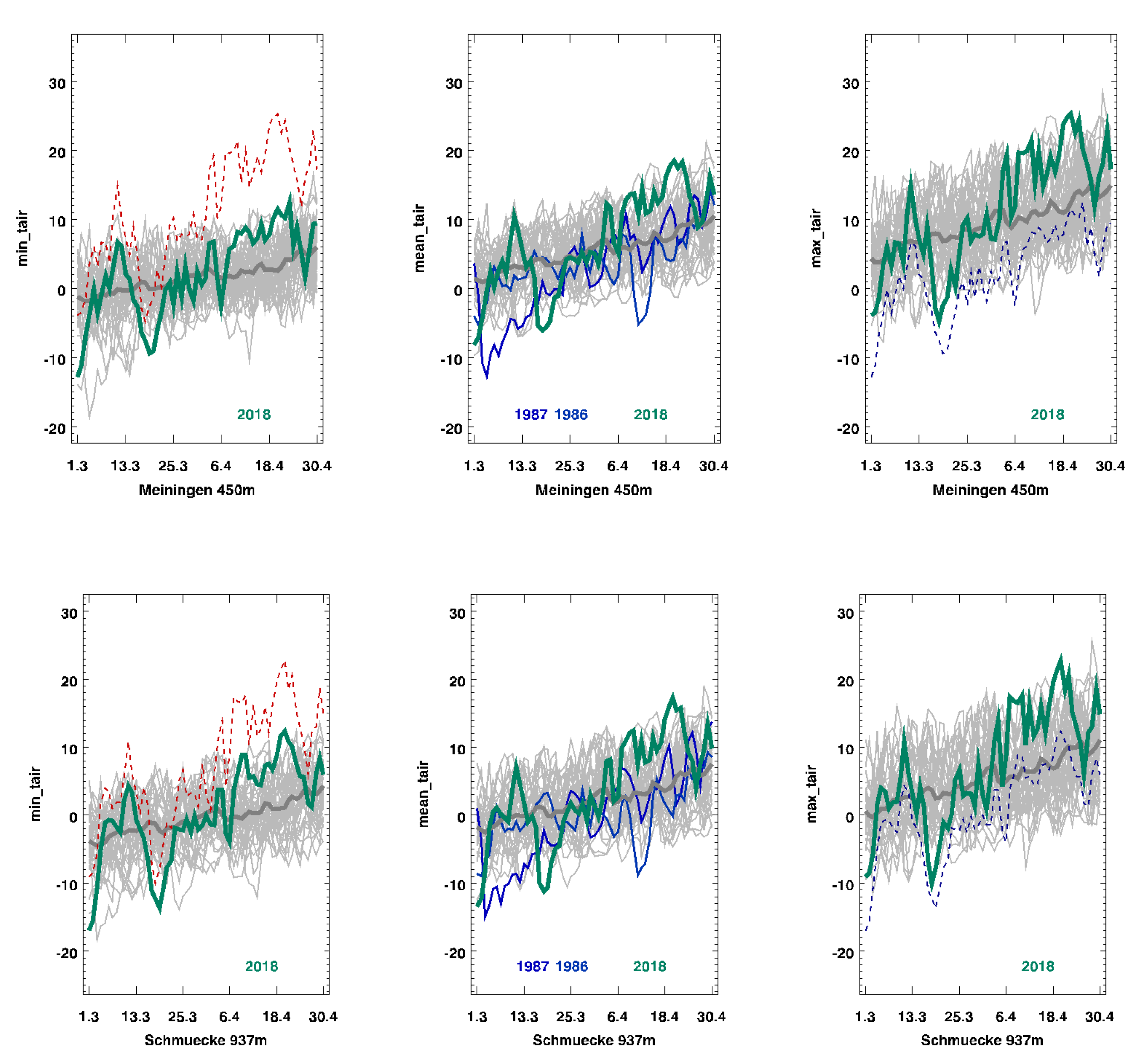
In 2018, following a relatively mild winter with snow cover, the soil was not frozen. There was a short cold period of 12 ice-days at Schmuecke and 8 ice-days at Meiningen, with maximum temperatures below −5 °C in late February and early March (Figure 2, green line). The minimum temperatures were between −11 °C (Meiningen) and −14 °C (Schmuecke) above the snow cover. This frost event was followed by a warm period, with maximum daily air temperatures above 10 °C (Figure 2, green line). No xylem bleeding was observed in February before the frost event. However, the xylem sap flow (bleeding from cut xylem) in Acer began during the warm period in early March following the frost event, as observed while testing whether it was time for pruning. This warm period was followed by a second cold event in late March, when the minimum temperatures dropped to −6 °C (Meiningen) and −11 °C (Schmuecke) for 3–4 days (Figure 2, green line). The weather became warm again a week later, when temperatures once again reached more than 10 °C, and Acer started to resume bleeding (Table 1).
The five-year average daily temperatures during March to April increased by 1.8 °C between 1979 and 2018. The standard deviation of the average daily temperature in March and April was 4.2 at Meiningen and 4.1 at Schmuecke, as averaged over the 40 years of observation. Compared to this average, 2018 showed the highest standard deviation of daily temperatures during the observation time, specifically, 7.0 at Meiningen and 7.5 at Schmuecke. It seems possible that with ongoing climate change, this pattern of a very warm March followed by cold events in April may become more pronounced in the future.
2.2. Study Site
The observations were carried out at the forest enterprise “Forstbetrieb Hermannsberg”, which consists of an area of about 200 ha covering a carboniferous volcano (867 m above sea level; longitude 50.7123; latitude: 10.6035) that produces acidic bedrock of quartz-porphyry and tuff. The upper elevation (< 700 m elevation) is covered by coniferous forest. The lower elevation (550–700 m) is covered by broad-leaved forest dominated by Acer pseudoplatanus, Sorbus aucuparia, Fraxinus excelsior, Populus tremula, Prunus avium, Betula pendula, Quercus robur, and Fagus sylvatica, with Acer being of greatest abundance and Fagus the least.
Observations were made on the north and northwest sides of the mountain, where old growth Acer stands (>200-year-old trees) exist from natural succession and where meadows were afforested by Acer (2.5 m × 2 m planting distance) about 15 years ago. The damage occurred in stands of natural succession as well as in hedge rows in meadows and afforestations, mainly in stems that were reached by solar radiation. Selected trees were pruned as future trees a year before the damage. In the year of the damage, single branches were pruned in late winter to see if xylem bleeding had started in order to allow for further pruning of twin stems. None of the damaged trees were pruned in 2018.
It should be noted that following this damage, we were informed that bark damage also occurred on calcareous sites (limestone) and sandstone. The damage appeared to be independent of geological conditions and management, but it was related to sun exposure (stands with low levels of cover or at the forest edges).
The observations in this study were made in 2019 at the Hermannsberg site, where the damage was quantified in the field using measurements of the dimensions of the affected trees and their damage. Tissue microscopy of wood samples was carried out at the Swiss Federal Research Institute WSL, Birmensdorf, Switzerland, following instructions by Schweingruber et al. [4,5]. Fungi were identified microscopically by the morphology of conidia and conidiophores (Ascomycetes [6,7]), by the morphology of basiodiocarps (Basiodiomycetes [8,9,10,11,12]), and by DNA sequencing of fungal barcodes (Cladosporum, Neocucurbitaria). Bark beetle galleries were analysed and identified by experts at WSL, Birmensdorf and remainings of individuals were identified by barcoding. Climate data were retrieved from the National Weather Service database (https://opendata.dwd.de/climate_environment/CDC/observations_germany/climate/daily/kl/).
3. Results
3.1. The Damage
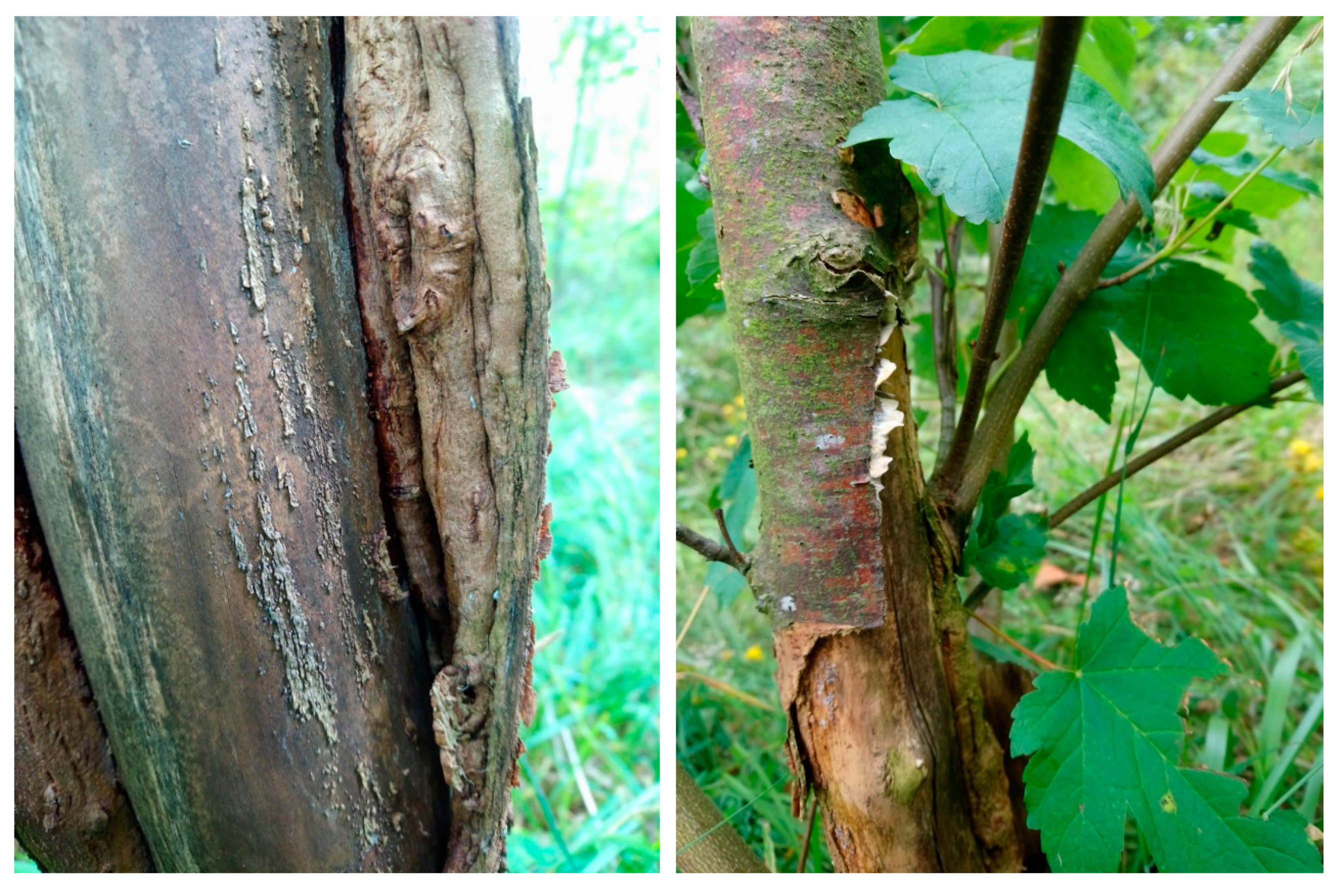
For all affected trees, a single crack was visible (Figure 1A). The bark dissected from the wood, thereby affecting a large proportion of the circumference, which only became visible upon removal of the dissected bark (Figure 1B). There was no damage visible in the radial direction of the xylem.
The damage occurred in young trees (<17 cm diameter at breast height, DBH) only. Based on measurements of 100 damaged trees, the average height of the trees was 6 ± 3 m, with an average diameter at breast height (DBH) of 5.7 ± 3.5 cm (maximum DBH 17.2 cm). The average length of the cracks was highly variable, ranging from 0.1 to 8 m, with an average length of damage of 1 ± 2 m. The width of the damage was, on average, 8 ± 4 cm, which was about 30% of the circumference. As expected, tree height and breast height diameter were closely correlated with the length and width of the damage. Even though 2018 was an unusually dry year, the affected trees healed the crack by 2 cm (on one side) after one year, but about 10% of the trees died, with some of them losing stability and breaking. The damage occurred in > 90% of the observations on the south-facing side of the stem, starting at the base of the tree. This was the location where the temperatures of the stem were expected to be highest when exposed to solar radiation and where growth was expected to initiate. Also, the damage was more pronounced in open stands and in trees that were selected and pruned as future high-quality trees, but it also occurred in naturally regenerated trees. In young stands, up to 100% of the Acer trees with the above-mentioned dimensions were damaged.
3.2. Anatomical Evidence
After one year, all of the damaged trees showed wood regrowth along the damage site, but regrowth was, in all cases, irregular. In many trees, the regrowth of the wood was an unorganized callus, or wood growth occurred along the dissected bark and not along the remaining xylem (Figure 3).
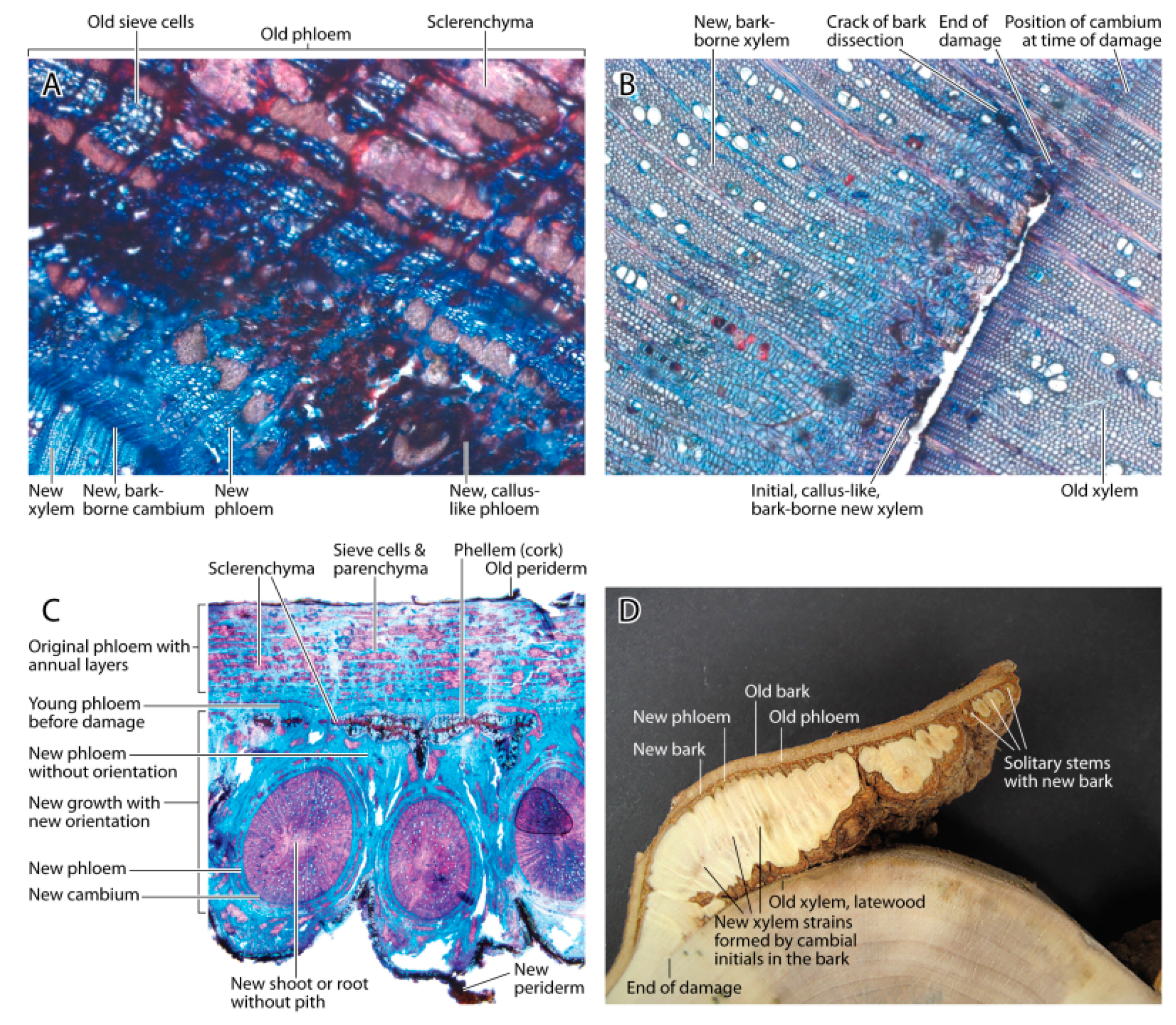
An anatomical investigation of the wood growing on the dissected bark indicated that the cambial mother cells ([13,14], see also Figure 5) of Acer were located in the inner bark, not on the visible surface of the xylem. Thus, in cases where the bark remained alive, these cells continued to form phloem and xylem cells. These cells differentiated into a callus-like, lignified structure on the phloem (Figure 4A) and xylem sides (Figure 4B) of the secondary vascular cambium. One year after the damage, cambial cell division resumed and resulted in the formation of a functional sieve and other xylem elements, thereby leading to the formation of new round stems attached to the bark. These stems had no pith, thus resembling more of a root structure than a stem (Figure 4A).
The independent stems sometimes merged with the increasing growth (Figure 4D). However, bark (periderm) sometimes formed around the stem as an outer layer of the independent phloem and the bark cambium. The differentiated xylem cells in the main stem showed no damage and no healing. There was no sign of fungal activity at the very end of the bark dissection crack.
In July 2019, when the new growth of 2019 terminated, the bark-borne wood structures showed an irregular, ribbon-like structure, which developed into new epicormic shoots (Figure 5).
In order to further understand the causes of this event, we measured the osmotic concentration in the bleeding sap when the bleeding started in May 2019.The osmotic concentration was 137 ± 10 mmol L−1 (average ± standard deviation of five trees).
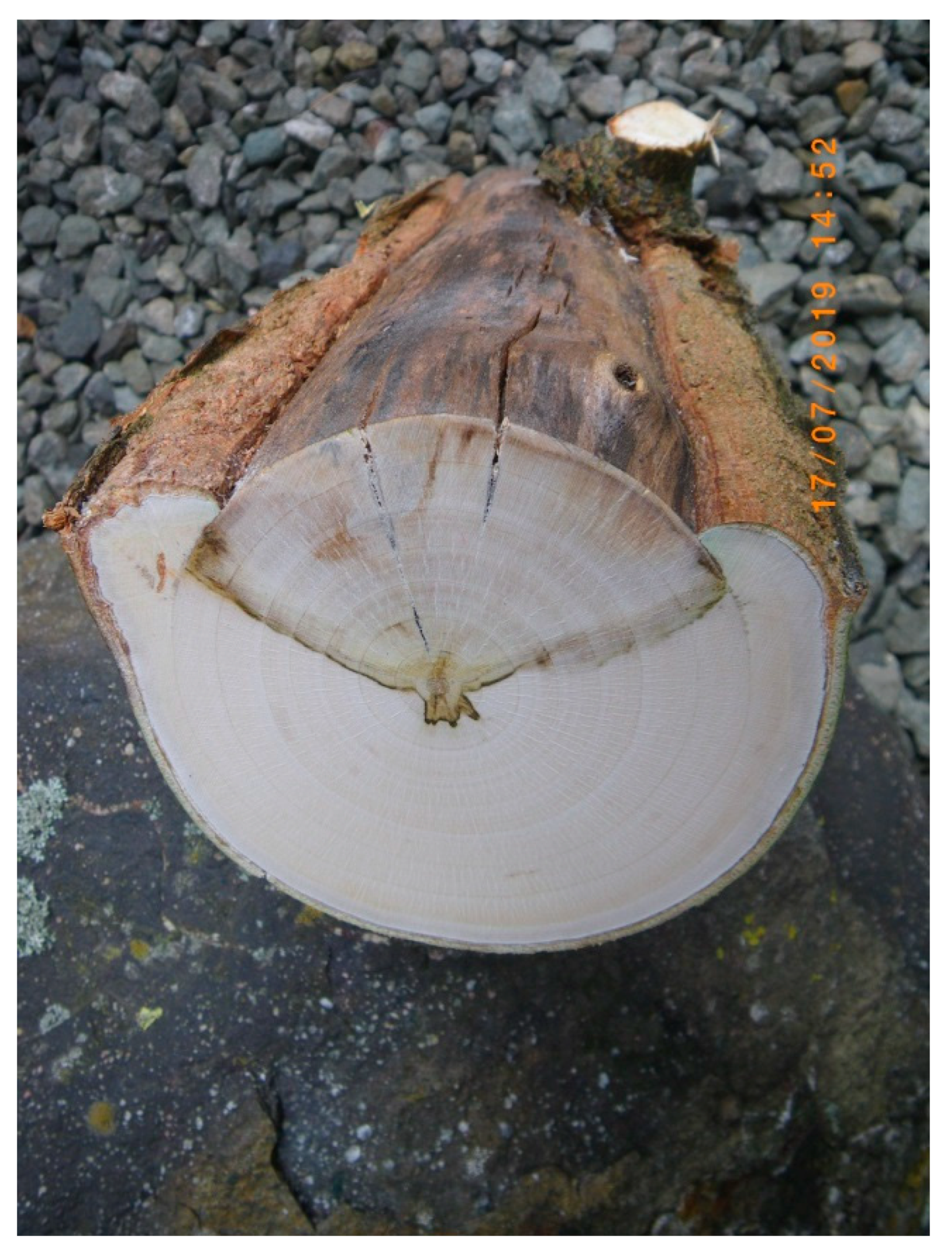
The xylem below the wound died and separated from the remaining living xylem along the ray cells (Figure 6), therefore showing that the damage extended inside the stem up to the pith. After a year, the wood below the wound dried and showed secondary splits. The dark border between the living and dead xylem appeared to have been produced by the intact part of the xylem, even in the absence of saprophytic fungi. Later, fungal hyphae were found along this line up to the pith.
3.3. Colonization by Fungi and Insects
The exposed xylem below the bark damage was an entry point for numerous wood damaging fungi.
After one year, the following fungi were observed:
- (1)
- Ascomycetes: Cladosporium cladiosporoides complex, Nectria cinnabarina in its anamorphic form, which affected about 5% of the trees, causing them to break, and Neocucurbitaria acerina;
- (2)
- Basidiomycetes: Bjerkandera adusta, a white-rot that became established on wounded trees, Schizophyllum commune, a white-rot that also became established on wounded surfaces, Schizopora paradoxa, a white-rot which was typically found on dead wood, and Stereum ochraceoflavum, a white-rot also typically found on dead wood.
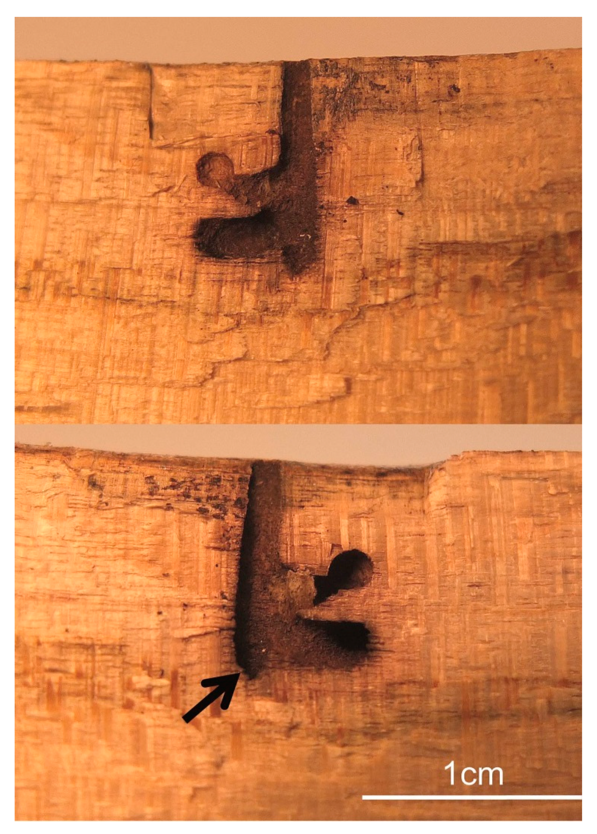
About 10% of the damaged trees showed boreholes of wood-breeding bark beetles. These holes were established in the bark-free cracked zone of the tree. The dissection of twelve insect galleries suggested that these were most likely caused by the non-native ambrosia beetle Xylosandrus germanus (Figure 7). Only one gallery of the fan-bearing wood-borer Ptilinus pectinicornis was detected, in which also a head of this species was found and identified by barcoding. As secondary colonizer of bark beetle galleries, a larvae of the horned black wasp Passaloecus corniger (identified by barcoding), was found in a Xylosandrus-gallery. These generalist wasps are known to build nests in tunnels of wood-boring beetles [15].
4. Discussion
In this study, we reported a phenomenon of bark-cracking in spring following a very cold event after xylem sap flow had already started during warm temperatures prior to the cold event.
The event of the spring bark-splitting of Acer indicated that climate change may not only mean a rise in temperatures or a change in precipitation, but also an increase in the variation of springtime temperatures. The temperate zone of Europe will always be in the reach of arctic air masses, especially in spring, when the air masses across the Atlantic tend to be unstable. Thus, late and severe frost may limit growth starting earlier. This may not only be important for Acer, but also for other deciduous, broad-leaved tree species, in which we observed similar bark damage (mainly oak and ash). Frost-ribs in other species was previously interpreted as damage resulting from winter temperatures and radial cracking of the xylem rather than damage by late frost, which usually affects a narrow cell layer in a tangential direction [2].
It remains uncertain if the damage was caused by frost per se, or by an interaction of high osmotic concentrations in the bark and the cambium followed by a desiccation of the expanding xylem cells after the frost event. Most likely, the osmotic effect of the bleeding sap in Acer trees was not sufficient to cause the damage in the cambial cells. These may have an osmotic concentration of about one molar [16], however, expanding xylem cells could well be affected. Thus, a frost event with temperatures below −5 °C remains the most likely factor to cause this damage. No fungal cells were detected at the end-point of the damage.
It might also be possible that pruning enhanced the damage. At our site, the pruning of selected trees took place a year prior to the damage. In the year of damage, only a few branches were cut to inspect the early timing of xylem sap flow. However, no pruning took place in 2018 due to early bleeding. The damage also took place in unpruned trees. Thus, pruning may have enhanced the damage via microclimatic effects, allowing solar radiation to reach stems that were previously shaded.
We are aware that our interpretations of this observation are retrospective, but it is difficult to investigate this damage experimentally due to the apparent relationship between the damage and the activation of growth prior to the frost event. Temperatures in March 2018 following a warm winter activated xylem flow, and this period was followed by a cold spell with temperatures below −10 °C. We know from inquiries by several forest managers that the damage occurred regionally on a wide scale, covering North Bavaria and Southern Thuringia (500 km × 500 km), but no systematic investigation regarding the regional extent of the observation was made.
Based on the anatomical evidence, the damage occurred in the expanding xylem cells and not in the primary cambium. The cambium and the cortex appeared to have higher frost tolerances due to higher osmotic cell concentrations of the solutes than the expanding xylem cells. The surviving cambial cells remained attached to the bark rather than the xylem, and these cells were the basis for wound healing and new wood growth. The range of cell types formed after this damage was surprisingly large. Initially, callus-like lignified cells formed after cell division on the phloem and xylem sides of the new secondary cambium. It remains unclear if the cambial cells attached to the bark initially received growth hormones mainly from the shoot, because is it possible that phloem-flow was reversed at the time of growth initiation [17]. The functional phloem and xylem elements were formed only after water and hormonal transport resumed from the root. Thus, the balance of growth hormones produced by the shoot and roots cause the observed recovery of wood formation [4,18].
The damage resembled the well-known observation of bark-cracking and “frost-plates” in fruit trees. In orchards, stems are stained white by liming to reduce solar irradiance [19]. In orchards and ornamental tree nurseries, this kind of damage was explained by the desiccation of the bark at a time of inactive xylem water-flow. However, the time of the damage observed in orchards coincides with the initiation of growth. Thus, also in these cases, the damage could be due to a combination of desiccation and late frost, which was reduced by liming the stems.
This damage caused major disorder in the stem. In fact, after one year, the damage extended deeper into the wood than was visible from the outside. Apparently, the tree isolated the segment of wood where the ray cells were no longer connected to the phloem. The black border in the wood is generally assumed to be produced in the presence of fungi [20], but in this case it appeared to be a plant response to separate the active and inactive xylem [5,20,21,22,23]. We found phenolic deposits in ray cells in the absence of fungal hyphae.
Since the bark initiated the growth of independent stems, where each stem has the opportunity to form its own bark, it is possible that these stems may never fully merge again. Thus, we expect a frost-rib-like feature to remain visible in older trees until they reach a late age. It seems unlikely that these trees will ever be able to produce stems of veneer-quality.
The more severe damage to the tree was observed in the wedge-like separation of the damaged wood segment from the living wood. The secondary drying cracks of this “dead” part of the wood were entry points for several fungi, which became established near the pith in the tree center. Even if the wounds close in the future, the trees will probably continue to rot in the center. It is most likely that these trees will break in a few years.
In terms of management, it may be necessary to cut these damaged trees and start again with a new sprout, if high-quality timber is the objective of management. Since the damage occurred in young trees only, and since the variation in springtime temperatures appears to have increased, starting with new sprouts may not even be a solution. Since the damage exposed the differentiated xylem, there were no wound-responses of defense against fungal and bark beetle attacks by the wood. Thus, these trees could become “habitat-trees” for saproxylics in managed forests. In the future, we may have to live with frost-scar-like structures in several types of heartwood tree or avoid exposure of the stem to solar radiation by establishing dense stands.
Author Contributions
Conceptualization, E.D.S.; methodology, F.S., B.S., and E.K.; investigation, M.M.G., A.G., U.W., and B.S.; writing—original draft preparation, E.D.S., F.S., M.M.G., A.G., and E.K.; writing—review and editing, E.D.S. and E.K.
Funding
This research received no external funding.
Acknowledgments
We acknowledge the help with the barcoding of two Ascomycetae by Grit Walther (HKI, Jena) and of insects by Nina Feddern (WSL, Birmensdorf), the help in identifying bark beetle galleries by Beat Forster (WSL, Birmensdorf), the help in deissecting the galleries by Dieter Trummer (WSL), and the artwork of Annett Boerner (www.dn.com.au/annett-boerner.html).
Conflicts of Interest
The authors declare no conflict of interest.
References
- Horst, D.; Jörg, G.; Reinhold, J.; Gregor, S.; Jan, W. Übersicht Waldschutz 2018/2019. AFZ-Der Wald 2019, 7, 22–25. [Google Scholar]
- Schwerdtfeger, F. Pflanzenkrankheiten; 2. Aufl; Paul Parey Verlag: Hamburg, Germany, 1957. [Google Scholar]
- Horst, R.K. Field Manual of Diseases on Trees and Shrubs; Springer Verlag: Berlin/Heidelberg, Germany, 2013. [Google Scholar]
- Schweingruber, F.H.; Börner, A.; Schulze, E.D. Atlas of Stem Anatomy in Herbs, Shrubs and Trees; Springer: Berlin/Heidelberg, Germany, 2012; Volume 2, p. 377. [Google Scholar]
- Schweingruber, F.H.; Boerner, A.; Schulze, E.-D. Atlas of Woody Plant. Stems; Evolution, Structure, and Environmental Modifications; Springer Verlag: Berlin/Heidelberg, Germany, 2006; p. 229. [Google Scholar]
- Breitenbach, J.; Kranzlin, F. Pilze der Schweiz, 2nd ed.; Verlag Mycologia: Luzern, Switzzerland, 1984; Volume 1. [Google Scholar]
- Ellis, M.B.; Ellis, J.P. Microfungi on Land Plants; Enlarged ed.; Richmond Publishing: Slough, UK, 1997; p. 868S. [Google Scholar]
- Breitenbach, J.; Kranzlin, F. Pilze der Schweiz; Nichtblatterpilze; Verlag Mycologia: Luzern, Switzerland, 1986; Volume 2. [Google Scholar]
- Krieglsteiner, G.J. (Ed.) Die Großpilze Baden-Württembergs; Allgemeiner Teil. Ständerpilze: Gallert-, Rinden-, Stachel- und Porenpilze; Ulmer: Stuttgart, Germany, 2000; Volume 1. [Google Scholar]
- Jahn, H. Stereoide Pilze in Europa (Stereaceae Pil. emend. Parm. u.a., Hymenochaete), mit besonderer Berücksichtigung ihres Vorkommens in der Bundesrepublik Deutschland. Westfälische Pilzbriefe 1971, 8, 69–176. [Google Scholar]
- Jülich, W. Die Nichtblatterpilze, Gallertpilze und Bauchpilze (Aphyllophorales, Heterobasidiomycetes, Gastromycetes). Kleine Kryptogamenflora Band II b/1; Gustav Fischer Verlag: Stuttgart, Germany; New York, NY, USA, 1984. [Google Scholar]
- Ryvarden, L.; Melo, I. Poroid Fungi of Europe. 2. Aufl. Synopsis Fungorum 37; Fungiflora: Oslo, Norway, 2017; p. 370S. [Google Scholar]
- Schweingruber, H. Wood Structure and Environment; Springer Verlag: Berlin/Heidelberg, Germany, 2007. [Google Scholar]
- Schweingruber, F.H.; Steiger, P.; Börner, A. Bark Anatomy of Trees and Shrubs in the Northen Hemisphere; Springer: Berlin/Heidelberg, Germany, 2019; p. 394. [Google Scholar]
- Corbet, S.A.; Backhouse, M. Aphid-hunting wasps: a field study of Passaloecus. Trans. R. Entomol. Soc. Lond. 1975, 127, 11–30. [Google Scholar] [CrossRef]
- Komor, E. Regulation by futile cycles: The transport of carbon and nitrogen in plants. In Flux Control in Biological Systems; Schulze, E.D., Ed.; Academic Press: San Diego, CA, USA, 1994. [Google Scholar]
- Aloni, R. The induction of vascular tissues by auxin and cytokinin. In Plant Hormones: Physiology, Biochemistry, and Molecular Biology, 2nd ed.; Davis, P.J., Ed.; Kluwer Academic Publisher: Dordrecht, The Netherlands, 2004. [Google Scholar]
- Bresinsky, A.; Körner, C.; Kadereit, J.W.; Neuhaus, G.; Sonnewald, U. Strasburger: Lehrbuch der Botanik; Spektrum Verlag: Berlin/Heidelberg, Germany, 2008; 36. Aufl. [Google Scholar]
- Lucas, E. Vollständiges Handbuch der Obstkultur; Eugen Ulmer: Stuttgart, Germany, 1887; p. 121. [Google Scholar]
- Butler, M.J.; Day, A.W. Fungal melanins: a review. Can. J. Micobiol. 1998, 44, 1115–1136. [Google Scholar] [CrossRef]
- Khuo, M.J.; Alexander, M. Inhibition of the lysis of fungi by melanins. J. Bacteriol. 1967, 94, 624–629. [Google Scholar]
- Bell, A.A.; Wheeler, M.H. Biosynthesis and functions of fungal melanins. Ann. Rev. Phytopathol. 1986, 24, 411–451. [Google Scholar] [CrossRef]
- Morris, H.; Hietala, A.M.; Jansen, S.; Ribera, J.; Rosner, S.; Salmeica, K.A.; Schwarze, F.W.M.R. Using the CODIT model to explain secondary metabolites of xylem in defense systems of temperate trees against decay fungi. Annals Botany 2019, 20, 2–20. [Google Scholar]
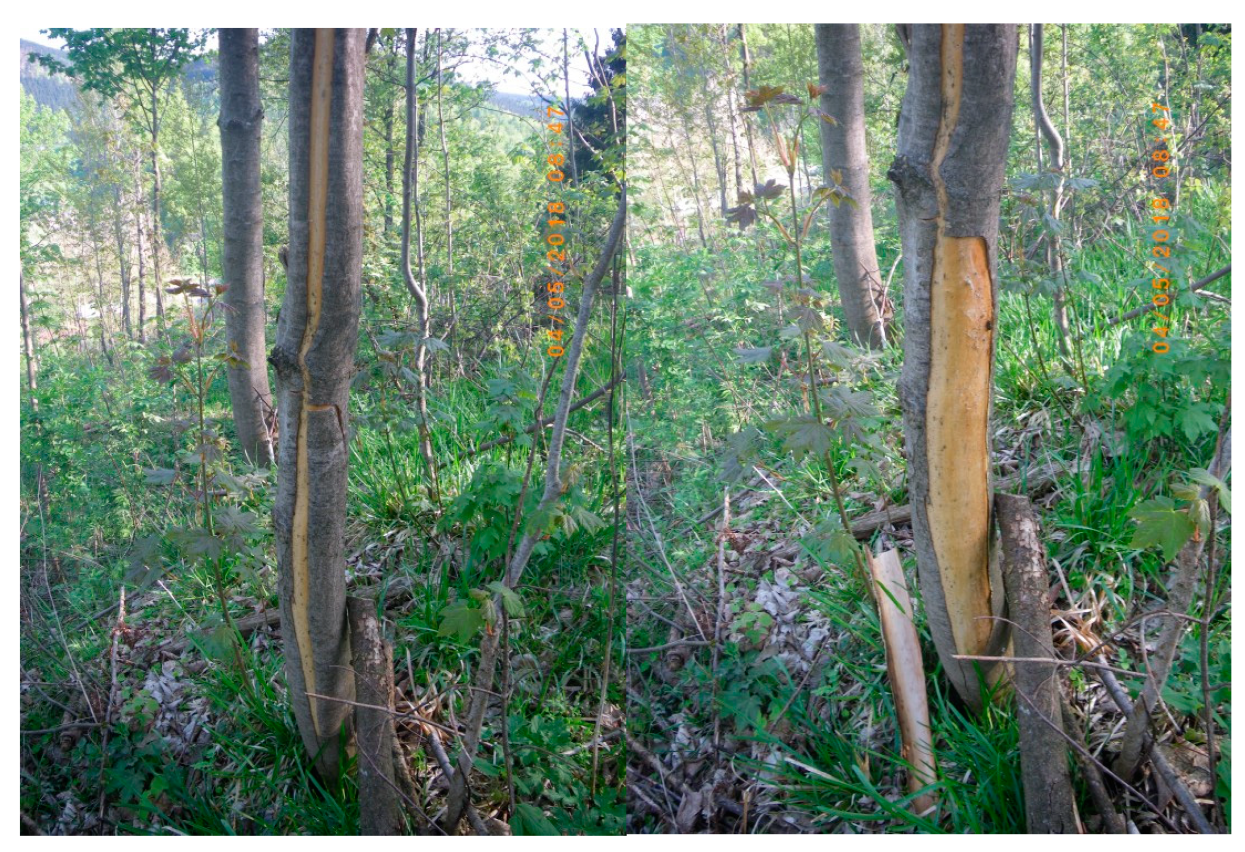
Figure 1.
(Left) In May 2018, single cracks in the bark of young Acer trees became apparent when the bark dissected from the wood. (Right) The same tree as shown in the Left, but after removal of part of the dissected bark. The total damage was larger than the visible crack. In some cases, the bark around the whole circumference of the tree was dissected.

Figure 2.
Minimum (min), average (mean), and maximum (max) air temperatures (tair) measured at two German Weather Service meteorological stations, (Upper panel) Meiningen (450 m above sea level) and (Lower panel) Schmuecke (937 m a.s.l.), between 1 March and 30 April for the period of 1979–2018 (one minor grey line per year). The thick grey line shows the average trend. The thick green line is the year 2018. The dotted red lines in the left panels show the maximum temperatures as comparisons. The dotted blue lines in the right panels show the minimum temperatures as comparisons. The solid blue lines in the middle panels are the years 1987 and 1986, when very cold temperatures occurred in March and April in a generally cold spring. Both weather stations were within 12–18 km distance from the observation site.

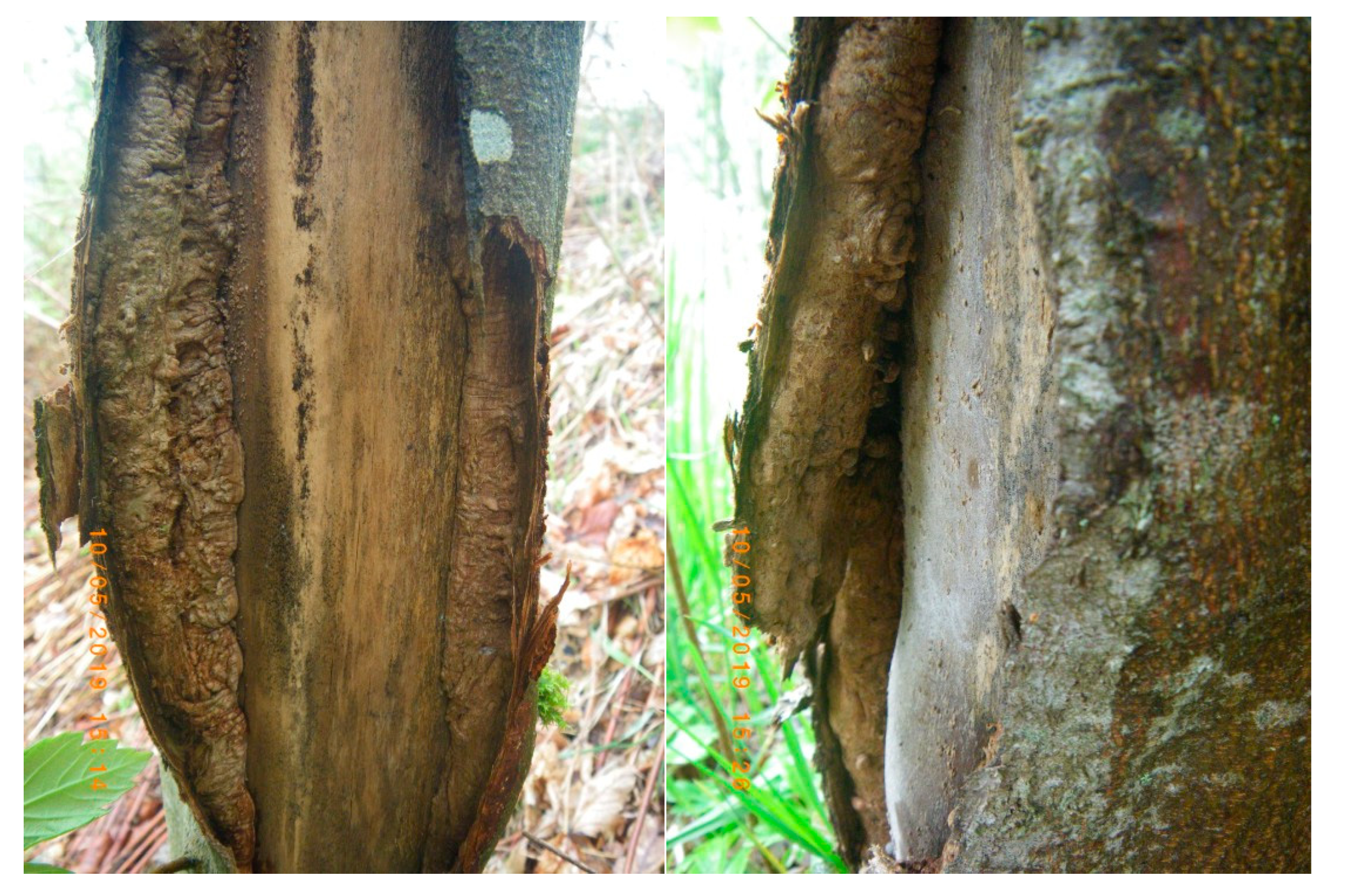
Figure 3.
Regeneration and healing of the damage. (Left) Callus-like growth oriented along the dissected bark; (Right) wood growth along the dissected bark.
Figure 3.
Regeneration and healing of the damage. (Left) Callus-like growth oriented along the dissected bark; (Right) wood growth along the dissected bark.

Figure 4.
(A) Regeneration of the phloem side of the secondary vascular cambium. A callus-like phloem initially formed, which then resumed the formation of sieve cells. (B) Regeneration of the xylem side of the secondary cambium. A callus-like xylem formed, which then resumed the formation of the xylem elements. (C) Regrowth of newly-formed stems initiated by individual cambial cells located in the dissected bark. (D) New wood formed by individual stems, either via the merging of wood initials, as indicated by the light-colored wood without bark, or by the formation of bark. The anatomical sections were prepared as described by Schweingruber et al. [4] and stained with Astrablue and Safranin, which stain lignified cells red color unlignified cells blue [4].

Figure 5.
(Left) Irregular structure of bark-borne wood strings 15 months after damage. The white fungal basiodiocarps were formed by Bjerkandera adusta. The black coating consisted of unidentified Ascomycetes. (Right) Development of new epicormic shoots from the callus-like wood structure.
Figure 5.
(Left) Irregular structure of bark-borne wood strings 15 months after damage. The white fungal basiodiocarps were formed by Bjerkandera adusta. The black coating consisted of unidentified Ascomycetes. (Right) Development of new epicormic shoots from the callus-like wood structure.

Figure 6.
Wedge-like separation of the living xylem from the wood below the area of damage. The radial cracks developed over the year following the damage.
Figure 6.
Wedge-like separation of the living xylem from the wood below the area of damage. The radial cracks developed over the year following the damage.

Figure 7.
Example of the entrance to an insect gallery of Xylosandrus germanus cf. The dark color of the gallery was caused by an Ambrosia fungus, which the beetle established as food-source in the tunnel. The black arrow indicates the continuation of the tunnel following the annual ring.
Figure 7.
Example of the entrance to an insect gallery of Xylosandrus germanus cf. The dark color of the gallery was caused by an Ambrosia fungus, which the beetle established as food-source in the tunnel. The black arrow indicates the continuation of the tunnel following the annual ring.


Table 1.
Maximum and minimum temperatures in spring 2018.
| Date | Meiningen 450 m NN | Schmuecke 937 m NN | |
|---|---|---|---|
| 11 March 2018 | Tmax | 15.0 | 10.9 |
| 18 March 2018 | Tmin | −6.0 | −10.0 |
| 19 April 2018 | Tmax | 21.4 | 17.6 |
© 2019 by the authors. Licensee MDPI, Basel, Switzerland. This article is an open access article distributed under the terms and conditions of the Creative Commons Attribution (CC BY) license (http://creativecommons.org/licenses/by/4.0/).
Share and Cite
MDPI and ACS Style
Schulze, E.D.; Schweingruber, F.; Gossner, M.M.; Günther, A.; Weber, U.; Stumpf, B.; Komor, E. Springtime Bark-Splitting of Acer pseudoplatanus in Germany. Forests 2019, 10, 1106. https://doi.org/10.3390/f10121106
AMA Style
Schulze ED, Schweingruber F, Gossner MM, Günther A, Weber U, Stumpf B, Komor E. Springtime Bark-Splitting of Acer pseudoplatanus in Germany. Forests. 2019; 10(12):1106. https://doi.org/10.3390/f10121106
Chicago/Turabian StyleSchulze, Ernst Detlef, Fritz Schweingruber, Martin M. Gossner, Angela Günther, Ulrich Weber, Burkhardt Stumpf, and Ewald Komor. 2019. "Springtime Bark-Splitting of Acer pseudoplatanus in Germany" Forests 10, no. 12: 1106. https://doi.org/10.3390/f10121106
Note that from the first issue of 2016, this journal uses article numbers instead of page numbers. See further details here.