
Overview of Rapid Detection Methods for Salmonella in Foods: Progress and Challenges

College of Life Science, Shandong Normal University, Jinan 250014, China
*
Author to whom correspondence should be addressed.
Foods 2021, 10(10), 2402; https://doi.org/10.3390/foods10102402
Submission received: 31 August 2021
/
Revised: 6 October 2021
/
Accepted: 8 October 2021
/
Published: 11 October 2021
(This article belongs to the Special Issue Research on Isolation and Intelligent Detection Methods of Foodborne Pathogens)
Abstract
:Salmonella contamination in food production and processing is a serious threat to consumer health. More and more rapid detection methods have been proposed to compensate for the inefficiency of traditional bacterial cultures to suppress the high prevalence of Salmonella more efficiently. The contamination of Salmonella in foods can be identified by recognition elements and screened using rapid detection methods with different measurable signals (optical, electrical, etc.). Therefore, the different signal transduction mechanisms and Salmonella recognition elements are the key of the sensitivity, accuracy and specificity for the rapid detection methods. In this review, the bioreceptors for Salmonella were firstly summarized and described, then the current promising Salmonella rapid detection methods in foodstuffs with different signal transduction were objectively summarized and evaluated. Moreover, the challenges faced by these methods in practical monitoring and the development prospect were also emphasized to shed light on a new perspective for the Salmonella rapid detection methods applications.
1. Introduction
The contamination of bacteria is a problem that cannot be ignored in food production and processing, and which may cause inestimable health damages to consumers [1,2,3,4]. Every year, more than half of the food-borne diseases in the world are caused by food-borne pathogens. As one of the major food-borne pathogens, Salmonella caused hundreds of thousands of deaths every year [5,6,7]. In the United States, the annual economic losses caused by Salmonella exceed $3 billion, which does not include the cases that were not reported [8,9,10].
Salmonella, a species of Gram-negative bacteria with more than 2500 serotypes, is responsible for a food-borne illness in humans and animals. Meanwhile, it can easily contaminate meat, eggs, milk and other foods, which leads to its strong transmission and difficulties of prevention and control [11,12]. Moreover, only one colony-forming unit (CFU) of Salmonella, the extremely low pathogenicity limit, can cause human infection, which put forward higher requirements for the prevention and control of food Salmonella contaminants [13]. Considering the serious threat of Salmonella to human health, the effective identification and rapid detection of Salmonella is of importance to prevent outbreaks of food-borne disease [14,15,16]. Nowadays, the conventional culture method is known as the “gold standard method” for Salmonella detection in food enterprises and testing companies. However, the long waiting time of the standard method is an enormous drawback for the immediate detection of Salmonella, which would greatly affect the transportation of foods to the markets [17]. With the development of molecular biology, a variety of specific Salmonella bioreceptors, such as antibody and aptamer, have been obtained [18,19,20], and more and more rapid detection methods of Salmonella based on specific bioreceptors have also been presented recently, which is of innovative significance for the real-time Salmonella detection in foods [21,22,23,24,25].
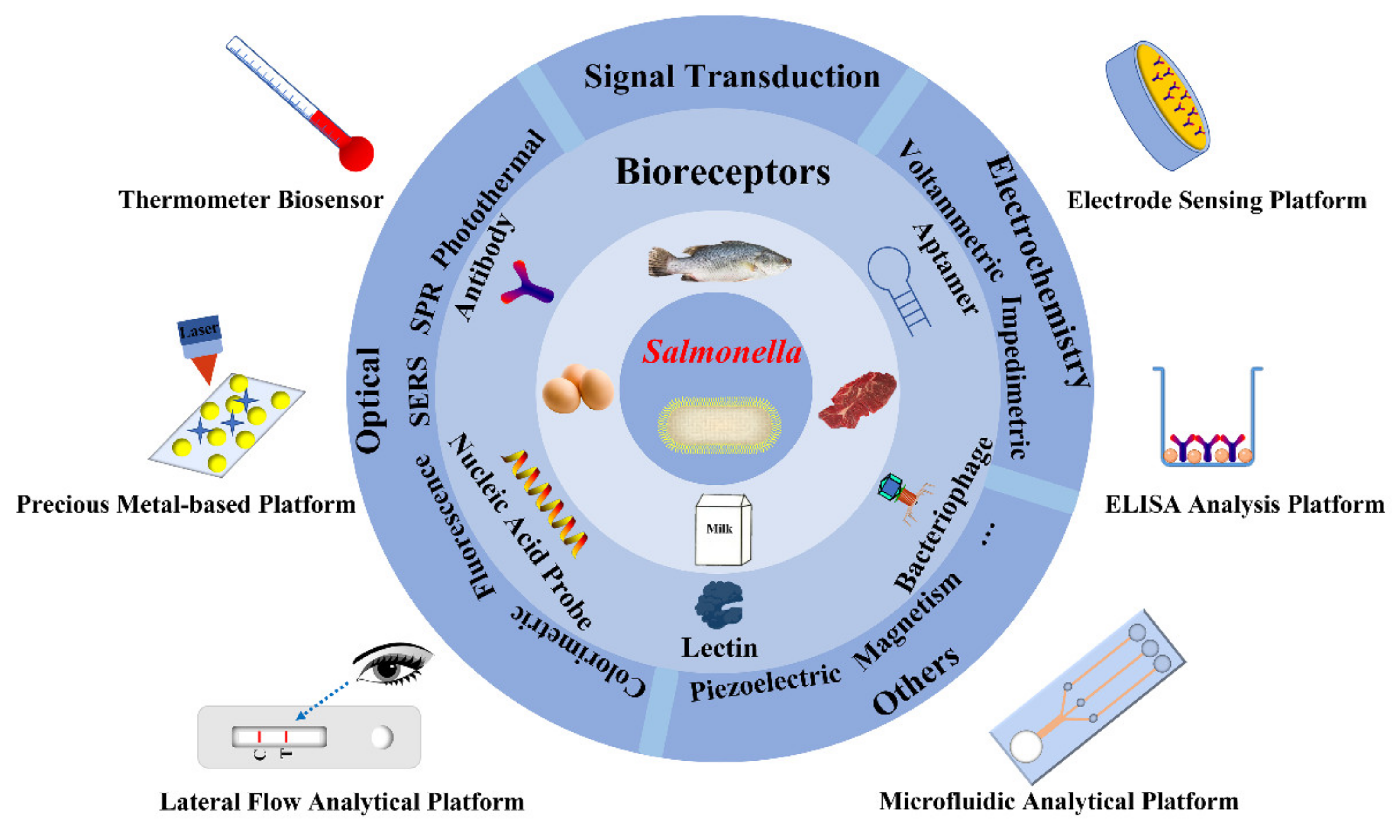
In this review, the bioreceptor of Salmonella which is the key factor affecting the sensitivity and specificity of rapid detection was described in detail as shown in Figure 1. Then, some popular Salmonella rapid detection methods applied in foods were emphatically descripted. Besides, the current trends of Salmonella’s rapid detection methods, the existing challenges and the application perspectives were also reviewed and discussed.
2. Gold Standard Method
Most countries and regions have set strict standards to ensure the absence of Salmonella in foodstuffs (as in China and the European Union), which shows the necessity of a sensitive detection of Salmonella [26,27]. Nowadays, microbial culture is still the gold standard strategy for the identification of Salmonella in many countries [28]. This method usually employs selective and differential media containing different nutrients and specific pH conditions to isolate Salmonella [13,29]. To achieve an accurate Salmonella identification in samples with a small amount and low residual, it is usually necessary to enrich the bacteria in the enrichment medium, which prolongs the detection time. Including subsequent bacterial counting and biochemical experiments, the whole detection process may take at least 5 days. Moreover, the type of testing applied is dictated by the existing legal requirements that food business operators should conform to, but the long waiting time is painful for products like fresh foods that need to be rapidly served to the market [30]. Therefore, these practical requirements also emphasize the need for the development of ideal rapid Salmonella detection methods in foods.
3. Bioreceptors for Salmonella
The rapid detection of Salmonella could not be separated from its effective identification. Recognizing the Salmonella molecule is one of the most important conditions for its rapid detection, which affects the efficiency of the separation and the direct signal output. Bioreceptor is a general term for the biological recognition molecule that can recognize the target [31,32,33]. After sorting out the published articles on Salmonella detection in recent years, the commonly used bioreceptors were summarized, including antibody, aptamer, nucleic acid probe, bacteriophage and lectin. The comprehensive comparison of the five bioreceptors is presented in Table 1.
3.1. Antibody
Antibody is a kind of immunoglobulin (Ig) generally produced by the mammalian immune system. IgG, one kind of Ig, has a strong affinity for its target, which is commonly used as a bioreceptor to identify the target in food contaminants detection [34,35]. The typical IgG molecule possesses a basic “Y” structure and consists of two heavy chains and two light chains, including the fragment crystallizable (FC) region for immune response activation and the fragment antigen-binding (Fab) region for antigen recognition.
The carboxyl group (-COOH) on the lower side of the Fab site is conducive to the fixation of the antibody on the surface of the sensor to realize the detection of the recognition signals. Moreover, the antibody exhibits a high selectivity to antigens and a strong anti-interference ability due to the unique epitope. Therefore, the antibody has been recognized as the standard recognition factor of commercial rapid detection products used in food safety detection, especially in immunochromatographic lateral flow strips and the enzyme-linked immunosorbent assay (ELISA) [36,37,38]. However, antibodies need to be produced by mammals or animal cells, which greatly slows down their production and leads to their high price. Furthermore, the activity of antibodies is vulnerable to organism and external environments, which weakens their stability [39,40]. Therefore, there are also some limitations for the powerful antibody.
Following the good performance of the antibody, a new type of antibody, called nanobody, is being developed to reduce the limitations of the traditional antibody. The nanobody, with an affinity to the antigen similar to that of the antibody, can be produced abundantly and has an extremely small size (12–15 kDa), so that it can bind to epitopes of antigens more conveniently than traditional antibodies (150–160 kDa) [41]. Although the nanobody has many advantages, the preparation of immune libraries is still difficult, and the development for the detection of food contaminants is still immature. However, nanobodies have been developed by pioneers to detect Salmonella. He et al. prepared the Salmonella nanobody library and verified the feasibility of its application in Salmonella detection by a mature ELISA, which also laid the foundation for the further development of the nanobody [42].
3.2. Aptamer
Broadly speaking, aptamers are single-stranded nucleotides or short-peptide molecules. Only the nucleic acid aptamer is introduced here. Aptamer, a single-stranded DNA or RNA selected by Systematic Evolution of Ligands by Exponential Enrichment (SELEX, a selection technology of nucleic acid aptamer) in vitro, is considered as a good substitute for the antibody and has been used as bioreceptor in various biosensors for the detection of food contaminants [19,43]. The short aptamer can form a stable and complex three-dimensional structure to recognize antigens of Salmonella specifically. Meanwhile, because of the aptamer’s miniaturization, it can also recognize special sites which are difficult to recognize by traditional antibodies [44].
Aptamer, a non-immunogenic recognition molecule with thermal stability and chemical modifiability, is unchallenged by antibodies and can be synthesized in large quantities in vitro at a low cost [45]. Aptamers are employed for the detection of inorganic molecules, proteins, cells and other molecules by transmitting the signal changes of conformational aptamers which combined with the target. One of the powerful obstacles in the aptamer sensing strategy is the immobilization of the aptamer onto the surface of the sensor while ensuring the proper functioning of the aptamer analysis. In general, the direct absorption aptamer by biotin/streptavidin binding is a suitable choice for the immobilization of aptamers on the carrier surface, which has the least impact on the specificity and affinity of the aptamer. Based on this, the special aptamer-based biosensors (aptasensors), including those used for Salmonella detection, have become an independent detection platform. In Bayraç’s study, for example, the aptamer of Salmonella Enteritidis was selected by Cell-SELEX, and the colorimetric detection method was established by a capillary tube, which proved the feasibility of the aptamer for the naked-eye detection of Salmonella for the first time, paving the way for the development of the subsequent method [18]. The astonishing performance of the aptamer in the detection field has really won the favor of many people.
3.3. Nucleic Acid Probe
Macromolecular nucleic acid sequences are composed of two kinds of nucleotides usually present in cells: deoxyribonucleic acid (DNA), which stores genetic material, and ribonucleic acid (RNA), which produces proteins. The genetic sequences of different strains or individual microorganisms are specific. Thus, the nucleic acid sequence can provide biological information on food-borne pathogens, and then the determination of its sequence is critical in the detection of food-borne pathogens. The nucleic acid probe is the complementary sequence of the special commentary strand of Salmonella, which can be cheaply synthesized and is relatively stable, like an aptamer. Through its high affinity and the specificity of base pairing, the nucleic acid probe can identify the presence of the bacteria and realize the transmission and measurement of special recognition signals [46,47]. Generally, nucleic acid probes rely on the signals generated by special groups that were modified with the signal molecule (such as fluorophore groups), and beyond that, through a modification on the surface of the sensor by the nucleic acid probe, which outputs the detection signals. However, the nucleic acid probe is always limited by the recognition of the target nucleic acid sequence, and the signal sensing of the methods generally depends on gene amplification and laborious gene extraction.
3.4. Bacteriophage
Bacteriophage-based biosensors are also the research focus of Salmonella detection in recent years [48]. A bacteriophage, a kind of virus, is a powerful bioreceptor for bacterial recognition, which can specifically infect living bacteria with high host selectivity. A bacteriophage can only replicate in viable hosts, which makes it a potential tool for distinguishing living and dead bacteria. The identification of living Salmonella is interesting and was demonstrated by Fernandes et al. They successfully distinguished viable, dormant and dead Salmonella Enteritidis by using a bacteriophage as a bioreceptor [49]. It is of great significance for Salmonella control in foods, by identifying the contaminations of viable and viable but non-culturable bacteria. Various biosensors have been developed based on a variety of reported Salmonella bacteriophages (including M13, PRD1, P22), which benefit from the strong resistance of bacteriophages to harsh conditions. The bacteriophage-based Salmonella detection platforms have been successfully commercialized because of their good detection results [50,51,52]. However, along with the good prospect of phages, the lysis of the Salmonella extracted from phages will lead to the decrease of the capture efficiency and the reduction or loss of recognition signals, which is also a key challenge faced by researchers in this field. Nevertheless, the bacteriophage-based detection method is still the breakthrough for the rapid and accurate identification of living bacteria, and the more innovative bacteriophage biosensing strategy would be an expected demonstration in the future.
3.5. Lectin
The O-antigen, constituting the lipopolysaccharide (LPS) on the cell wall of Gram-negative bacteria, can be used for strain distinction. The LPS O-antigens can exhibit a high specific lectin binding affinity that indicates the recognition ability of lectin to Salmonella. As a non-enzyme and non-antibody protein, lectin has a higher stability, but a lower price compared with antibodies. At present, biosensors using lectins as recognition molecules have emerged one after another, and the more commonly used lectins are concanavalin A and wheat germ agglutinin [53]. Dao et al. enriched Salmonella by using concanavalin A as the recognition molecule, combined with microfluidic technology and isothermal amplification for target DNA detection, with the limit of detection (LOD) as low as 5 CFU/mL [54]. Then, much work has been devoted to optical and electrochemical bacterial biosensors utilizing lectin as a recognition element. With the continuous development and application of nanomaterials, the detection methods combined with various materials are also booming. It is believed that lectin will have a promising role in the field of Salmonella detection.
4. Rapid Detection Methods of Salmonella in Foods
With the manufacture of advanced instruments and the continuous development of nanomaterials, various rapid detection methods for Salmonella are constantly being developed, based on the above bioreceptors. However, a different signal transduction also determines the complexity and sensitivity of the rapid detection method. Here, rapid detection methods of Salmonella based mainly on optical sensing and electrochemical identification methods were introduced.
4.1. Optical Sensing
Optical sensing, one of the fastest developing methods of Salmonella detection, converts the biological and chemical reactions that occur between bioreceptors and targets into optical signals through transducers or detection instruments [55]. Optical sensing mainly includes colorimetry, fluorescence analysis, surface-enhanced Raman spectral (SERS) detection, surface plasmon resonance (SPR) determination and photothermal detection. The optical sensing methods of Salmonella reported in recent years are shown in Table 2.
4.1.1. Colorimetry
The most important feature of the colorimetric method is that the signal response can be observed by the naked eye. Meanwhile, the whole colorimetric experiment is simple, without the need for complicated instruments, so it is one of the ideal methods for a rapid in-field detection of Salmonella [56]. There are two main types of colorimetry: one is to produce a color change through the optical or chemical properties of the probe itself; the other is to obtain the color change of the chromogenic substrate through enzymatic or similar catalytic-like reactions. Here, classic studies were chosen to introduce these color-rendering methods.

Table 2.
Optical sensors reported for Salmonella detection.
| Detection Methods | Bioreceptor | Linear Range | Limit of Detection (LOD) | Detection Time | Real Sample Application | Reference |
|---|---|---|---|---|---|---|
| Colorimetry | Nucleic acid probe | 100 to 109 CFU/mL | 16 CFU/mL | / | Milk | [57] |
| Antibody | 103 to 108 CFU/mL | 103 CFU/mL | 14 min | Cabbage and drinking water | [58] | |
| Antibody | 1.88 × 10⁴ to 1.88 × 10⁷ CFU/mL | 1.88 × 10⁴ CFU/mL | / | Milk | [59] | |
| Antibody | 0 to 108 CFU/mL | 500 CFU/mL | 60 min | Milk | [60] | |
| Antibody | 0 to 107 CFU/mL | 34 CFU/mL | / | Milk | [61] | |
| Antibody | 100 to 104 CFU/mL | 100 CFU/mL | 90 min | Milk | [62] | |
| Fluorometry | Antibody | 5 × 10⁴ to 107 CFU/mL | 5 × 10³ CFU/mL | 12 min | Broth | [9] |
| Antibody | 500 to 5 × 10⁷ CFU/mL | 60 CFU/mL | 60 min | Milk | [63] | |
| Antibody | 40 to 4 × 10⁶ CFU/mL | 40 CFU/mL | 120 min | Chicken | [64] | |
| Aptamer | 10 to 107 CFU/mL | 10 CFU/mL | / | Meat, milk and chicken | [65] | |
| Aptamer | 50 to 106 CFU/mL | 35 CFU/mL | / | Chicken and shrimp | [66] | |
| Aptamer | 12 to 5 × 10⁵ CFU/mL | 11 CFU/mL | / | Milk | [67] | |
| SERS | Aptamer | 10 to 104 CFU/mL | 4 CFU/mL | / | Chicken and milk | [68] |
| Nucleic acid probe | 27 to 2.7 × 10⁶ CFU/mL | 27 CFU/mL | 30 min | Milk, chicken breast and beef | [69] | |
| Aptamer | 100 to 107 CFU/mL | 50 CFU/mL | / | Milk | [70] | |
| Lectin | 10 to 104 CFU/mL | 10 CFU/mL | / | / | [71] | |
| Aptamer | 0 to 107 CFU/mL | 25 CFU/mL | / | Milk, orange juice, and tap water | [72] | |
| SPR | Nucleic acid probe | 0.01 to 100 ng/mL | 10 pg/mL | 60 min | / | [73] |
| Antibody | / | 7.4 × 10³ CFU/mL | 80 min | Cucumber and hamburger | [74] | |
| Antibody | 100 to 106 CFU/mL | 103 CFU/mL | 60 min | Powdered milk | [75] | |
| Photothermal | Antibody | 300 to 103 CFU/mL | 300 CFU/mL | 90 min | / | [76] |
| Antibody | 100 to 107 CFU/mL | 100 CFU/mL | 25 min | Milk and grape juice | [77] | |
| Antibody | 104 to 108 CFU/mL | 104 CFU/mL | 20 min | Milk and grape juice | [78] | |
| Antibody | 103 to 109 CFU/mL | 103 CFU/mL | 15 min | Milk and grape juice | [79] | |
| Antibody | 5 to 5 × 10³ CFU/mL | 70.7 CFU/mL | 36 min | / | [80] |
The commonly used colorimetric materials in non-enzymatic colorimetry are gold nanoparticles (Au NPs), which are excellent materials for identifying the presence of the target based on its wine red and obvious blue-purple color after aggregation. The antibodies and aptamers adsorbed on the surface of Au NPs prevent the surface charge of Au NPs from being destroyed by the salt solution, which slows down the solution color change caused by the aggregation of Au NPs [81]. Yi et al. developed an agglomerated Au NP colorimetry for Salmonella detection based on an aptamer. The aptamer was immobilized on chitosan and bonded to the surface of the Au NPs by electrostatic adsorption, and then the Salmonella combined with the aptamer and led to the loss of protection of aptamer for Au NPs. With the addition of the salt solution, the agglomeration of Au NPs would result in a color change, which can be recorded by an ultraviolet spectrophotometer for Salmonella detection. Moreover, the recoveries of Salmonella from spiked milk samples, between 92.4 to 97.2%, also confirmed the feasibility of this method in actual sample detection [57]. Among the paper-based colorimetric detection methods, the lateral flow test strip has always been the most popular detection platform. Based on the van der Waals forces of Salmonella on agglomerated Au NPs, Ren et al. realized the label-free sensitive determination of Salmonella by the naked eye through an immunochromatographic strip. The visual LOD of 103 CFU/mL based on salt-induced aggregated Au NPs in the developed immunochromatographic strip was 100-fold lower than the method based on cationic AuNPs, and the test strip also exhibited an excellent recovery in cabbage and other food samples [58]. In addition to Au NPs, many kinds of nanomaterials with conspicuous colors make a great contribution to the colorimetric detection of Salmonella [59]
The other colorimetric method is an enzymatic or enzyme-like catalytic reaction based on ELISA, and the chromogenic substrate used is generally tetramethylbenzidine (TMB). The color of TMB from colorless to blue catalyzed by horseradish peroxidase (HRP) is a classical colorimetric method for Salmonella detection. Chen et al. enriched Salmonella by magnetic separation and then used Au NPs and HRP to catalyze TMB for signal enhancement, which was used for the specific determination of Salmonella in a food matrix [60]. Due to the low stability, low environmental tolerance and high cost of HRP, many mimic enzymes have been used in the colorimetric detection of Salmonella after Fe3O4 nanoparticles proved with peroxidase-like activity [82]. Cheng et al. synthesized a Fe-MOF nanozyme, which remained in the solution after magnetic separation was used to catalyze TMB for the Salmonella colorimetric analysis. The use of a nanometer enzyme caused the LOD of Salmonella to be as low as 34 CFU/mL, with a good storage stability and detection potential in milk samples [61]. Besides Fe-based nanozymes, graphene, molybdenum disulfide and other nanomaterials also play an important role in the study of the colorimetric detection of Salmonella by catalyzing TMB. In addition to TMB, there are a variety of chromogenic substrates for Salmonella detection. Srisa-Art et al. developed a paper-based colorimetric device combined with magnetic separation to visually detect Salmonella using chlorophenol red-β-d-galactopyranoside colorimetry based on β-galactosidase (Figure 2A). The white paper provides a good background for color generation, which obtained a LOD of 100 CFU/mL without any complex processing or enrichment of the samples, and has a good performance in whole milk [62]
With the development of detection technology and the more intelligent portable equipment, the colorimetric analysis method that judges the bacterial residue in foods according to the color change has become increasingly popular. The real-time monitoring of Salmonella is an important means to ensure the safety of consumers. The colorimetric method with a convenient measurement and simple operation is undoubtedly convenient for sampling inspections by non-professionals, which are of great value for food safety prevention and control in remote areas. However, the interference of the background of food samples in the colorimetric method is also a difficult problem that affects its accuracy, and hence a challenge that needs to be faced.
4.1.2. Fluorescence Analysis
Fluorescence analysis is one of the optical sensing methods to quantify the concentration of the target according to the fluorescence intensity of the labeled materials, which can solve the color interference of a food matrix when using colorimetric methods. The generated fluorescence signal can be visually observed by a fluorescence observer or measured by a fluorescence spectrometer [83,84]. Fluorescence analysis has two main means of fluorescence generation; one mean is the direct measurement of the fluorescence intensity from fluorescent materials; the other mean is the indirect measurement of fluorescence quenching to reflect the amount of Salmonella.
The detection signal of sandwich fluorescence sensing is directly obtained from a fluorescent material such as quantum dots (QDs), which is the most common fluorescence detection method for Salmonella [85]. QDs with a high fluorescence yield and a long fluorescence lifetime have always been popular in fluorescence sensors. Hu et al. developed lateral flow immunoassay strips based on the silicon shell protected quantum dot nanospheres and realized the rapid detection of Salmonella. The whole detection process can be completed in 10 min, and obvious fluorescence signals can be observed with the naked eye, which provides a new opportunity for the prevention and control of Salmonella in foods [9]. Immunomagnetic beads are also commonly used for sample concentration to improve the detection sensitivity of fluorescence analysis. In the work of Yin et al., the immunomagnetic beads and QDs were combined to realize the low background and high sensitivity detection of Salmonella through the reverse assaying strategy, and the LOD could reach 60 CFU/mL, which led to a 50-fold improvement of the sensitivity compared with a conventional QD-based immunosensor. Moreover, the immunosensor can also achieve an accurate quantification of Salmonella in milk samples within 1 h [63]. Xue’s group proposed an innovative work: they loaded QDs with MnO2 nanoflowers and separated the part that bound to the bacteria by immunomagnetic beads. Then, the addition of glutathione reduced MnO2 to Mn2+, which caused the QDs to be released. The LOD of Salmonella with 40 CFU/mL was obtained by measuring the fluorescence intensity of the released QDs, and the method was successfully verified in chicken samples (Figure 2B) [64]. More and more fluorescent materials with excellent performance have been used in Salmonella detection through sandwich-format-based fluorescence analysis, such as upconversion nanoparticles, organic fluorophores, fluorescent microspheres and so on [86,87].
The fluorescence analysis methods designed based on fluorescence resonance energy transfer (FRET) are also popular [65]. Compared with sandwich fluorescence sensing, FRET can achieve a signal output without washing steps. “Turn-off” and “turn-on” are the two detection models of fluorescence signal in FRET. The “turn-off” model relies on the close proximity of fluorescent materials and the quencher in the process of target recognition, which leads to FRET and weakens the fluorescence signals, while the “turn-on” model realizes the signal measurement through the fluorescence recovery caused by the fluorescent materials being separated from the fluorescent acceptors after being combined with the target. Fluorescence observation with a dark background in FRET may be a more sensible choice due to its better signal-to-noise ratio, which may also be the reason for the higher number of studies on the “turn-on” model [88]. Duan et al. prepared a fluorescent acceptor by modifying the aptamers on the surface of QDs and blocking the fluorescence emission using carbon nanoparticles. In the presence of Salmonella, QDs separated from the carbon nanoparticles through the aptamer-bacteria recognition and re-emitted fluorescence. At this point, the fluorescence intensity of the solution reflected the amount of Salmonella, and the good practicability and stability of this method were also confirmed in shrimp and chicken samples [66]. Cheng at al. have synthesized a TM upconversion nanoparticle by doped Mn2+ into NaYF4: Yb and used the “turn-on” model, which constructs a fluorescence donor with the emission peak at 807 nm and, subsequently, a couple of aptamers. Then, the Au nanorods can lead to the fluorescence quenching of upconversion nanomaterials, while the presence of Salmonella prevented the energy transfer and the fluorescence recoveries. The application performance of the method has been verified in milk samples, and the LOD was as low as 11 CFU/mL under optimal conditions [67].
Fluorescence analysis can obviously solve the problems of color interference in the food matrix and has a lower detection limit, which is important for the detection of traces Salmonella compared with colorimetric detection. However, although the ubiquity of portable fluorescence detectors has accelerated the commercialization of fluorescence analysis, its inherent disadvantages are also obvious. The uncontrollable fluorescence bleaching caused by complex food substrates has always been a great problem in fluorescence analysis, and the application of new materials with better stability and optical properties is also a key measure to solve the pain point.
4.1.3. Surface-Enhanced Raman Spectroscopy Detection
The Raman spectrum is a scattering spectrum which provides unique fingerprint information on different substances according to molecular vibration, and the Raman shift can describe the spectral information of different molecules [68]. SERS is an optical sensing technology that uses roughened metal surfaces or special nanomaterials to enhance Raman scattering and improve the detection sensitivity. Precious metals are the classic materials used for SERS, in which taking silver and gold as enhanced substrates is the most interesting option [69]. The earliest SERS for Salmonella detection can be traced back to 2003, with the unsatisfactory LOD of 106 CFU/mL, which was limited by the detection equipment and single colloidal gold particles. However, the latest research progress has been beyond comparison [89]. Ma et al. designed a dimer probe that combined aptamer-modified Au NPs with silver nanoparticles. Then, Salmonella was enriched by magnetic beads and identified by the dimer probe to form a sandwich complex. The enhanced Raman scattering induced by gold and silver nanoparticles significantly reduced the LOD of Salmonella to 50 CFU/mL, and the method not only had a good selectivity for Salmonella but also confirmed its actual detection performance in milk samples [70]. Compounds with specific Raman shifts have also been used for Salmonella detection. Three kinds of pathogenic bacteria have been detected simultaneously by Kearns et al. using the SERS strategy based on magnetic separation. As shown in Figure 2C, magnetic materials coated with silver-binding lectin provided support for bacterial isolation and the enhancement of the Raman scattering, while a 7-dimethylamino-4-methylcoumarin-3-isothiocyanate binding antibody provided the specific signal production site of Salmonella. This method can not only realize the simultaneous determination of different pathogens in the same matrix, but also obtain a lower LOD, of 10 CFU Salmonella per mL [71].
Compared with the free precious metal substrate, the fixed precious metal surface also shows a promising Raman scattering enhancement effect to detect Salmonella. Using aptamer-modified silver nanorod arrays as substrates, Chen et al. successfully found that the Raman signals of Salmonella were different from the blank and control groups, which realized the signal amplification of Salmonella. SERS-active silver nanorod array substrates can amplify the specific Raman signals of Salmonella captured by the aptamer, and the existence of Salmonella can be verified according to noticeable spectral changes [90]. In addition, the vancomycin-coated silver nanoparticle array designed by Liu et al. also provides a reference for SERS analysis of Salmonella in foods [91]
SERS detection can obtain accurate Salmonella detection results with a lower amount of sample consumption, which has a good application prospect in the prevention and control of food safety. However, the sensitivity of the miniaturized Raman detector is not satisfactory, which is also a worrying aspect of SERS detection.
4.1.4. Surface Plasma Resonance (SPR) Determination
The SPR sensor, a kind of optical sensor, can monitor multi-component interactions in real time and in situ through refractive index changes without labeling [92]. The basis of SPR is the sensing interface composed of a metal substrate. Similar to SERS, the materials dominate the detection performance of SPR, and the classic gold chips is also the preferred substrate for SPR [73]. Singh et al. presented an SPR nucleic acid sensor constructed by fixing ssDNA on the surface of the gold plate, which has carried out a sensitive, label-free and real-time monitoring of Salmonella conservative genes [93].
The label detection of Salmonella by SPR is also developing rapidly [94]. The SPR labeling detection designed by Vaisocherová-Lísalová et al. used Au NPs as the labeling material, and then the thickness of substrate materials was studied to evaluate the recognition ability of the sensor. The acceptable LOD of Salmonella at 7.4 × 10³ CFU/mL in cucumber was acquired by virtue of the superior surface resistance after Au NPs labeling. However, such sensitivity has great limitations in actual detections, which is not conducive to the rapid identification of low concentrations of Salmonella [74]. Enhancing the detection signal through an enzyme-catalyzed reaction in SPR has also been developed. Farka et al. used HRP-conjugated secondary antibodies as Salmonella markers to improve the detection sensitivity by catalyzing the conversion of 4-chloro-1-naphthol into insoluble precipitates. The sensitivity of this precipitation enhancement method was 40 times higher than that of the label-free approach. However, the LOD of 103 CFU/mL in powdered milk is still lower than that of fluorescence and Raman detection [75].
The low sample consumption and real-time monitoring of SPR attracted investors in the commercialization process. Although SPR sensor miniaturization has been achieved, it still faces a low sensitivity. How to improve its stability in sample testing and the auxiliary of the precision instrument are the questions that researchers should think about.
4.1.5. Photothermal Detection
Photothermal detection is an innovative and rapidly developing method for the detection of contaminants in recent years [95,96]. When some nanomaterials are irradiated by photon energy with different wavelengths, the vibration inside the atom converts light energy into heat energy, resulting in a photothermal effect, and this is the cornerstone of photothermal detection [97]. Common nanomaterials such as Au NPs, graphene oxide, black phosphorus, molybdenum disulfide and Prussian blue are used in photothermal detection frequently [98,99,100,101,102].
In general, photothermal detection shows the concentration of the targets through the photothermal temperature of the intercepted nanomaterials. The Au NPs with good photothermal properties are commonly used in immune analysis. The first study on improving the sensitivity of colloidal gold test strips by photothermal detection was carried out by Qin et al. [103]. This motivated the use of the photothermal properties of colloidal gold in many rapid detection methods. Especially in Zhang’s group, a variety of immunochromatographic test strips assays based on photothermal nanomaterials have been developed for Salmonella detection in recent years. They performed photothermal tests on commercial test strips based on Au NPs using a self-built portable sensor device to confirm the feasibility of improving the sensitivity of the test strip [104]. In order to achieve a more sensitive Salmonella detection, more and more nanomaterials with a higher photothermal conversion efficiency have been used in rapid detection methods to amplify the detection signal. The immunomagnetic beads which can enrich bacteria were firstly selected. Zhang et al. realized the capture, detection and killing of Salmonella using the immunomagnetic beads in 1.5 h, and a LOD as low as 300 CFU/mL was obtained. Then, they verified the detection performance of this method in drinking water, which provides a reference for the development of Salmonella detection methods in food [76]. Lu et al. not only used MoS2@Au nanomaterials with a higher photothermal conversion efficiency, but also oxidized TMB through its good catalytic performance. The nanomaterial and oxidized TMB were combined for photothermal detection, which made the LOD of Salmonella reach 100 CFU/mL. In addition, the application of this method has been validated in milk and grape juice samples, and the detection time of less than 25 min also shows its good prospect in the detection of Salmonella in foods [77]
Besides photothermal materials, the choice of temperature reading device is also related to the convenience and sensitivity of the methods. The thermal imager commonly used in the above methods is expensive, bulky and often used with a computer, which limits its popularity [105]. Therefore, many new works highlight the low cost and practicality of temperature reading devices. A card reader designed and manufactured by Zhang et al. has specially been used for the test strip to further increase its quantitative sensitivity, which is a strong boost to the standardization and commercialization of rapid photothermal detection [104]. Wang et al. focused on the detection of Salmonella in the changing environment using a resistance temperature sensor in a graphene oxide-based immunofiltration strip. The small volume of the sensor and the short detection time (within 20 min) contributed to the field prevention and control of Salmonella [78]. The following work innovatively integrated the temperature reading device and detection carrier, which greatly simplified the detection process. Du et al. immobilized the anti-Salmonella antibody on the mercury head of a glass thermometer, and the bacteria in the test solution was recognized by immune-graphene and then connected to the surface of the mercury head based on a sandwich immunoassay. The mercury head combined with immune-graphene was irradiated with laser light, and the temperature change can be shown directly on the thermometer. The 15-min detection time and the successful detection of Salmonella in milk and grape juice samples indicates the good application potential of this novel immune-thermometer. However, this novel method is also faced with the limitation of its low sensitivity (LOD of 103 CFU/mL), which needs to be improved [79]. Instead of using laser irradiation to trigger the photothermal temperature, Guo et al. developed a new temperature generation strategy [106]. They designed nanomaterials with the activity of peroxidase to decompose hydrogen peroxide and produce oxygen, which forced the pre-placed calcium oxide to react with water and generate heat by increasing the pressure in the space. Then, the temperature change was measured by a temperature sensor carried on the mobile phone to judge the number of bacteria. This temperature generation strategy made the detection more automatic and reduced the manual operation steps. Although this work is not a temperature detection strategy triggered by a laser, this temperature detection method is coincident with photothermal detection.
Aside from the direct measurement of the photothermal temperature, the photothermal effect is also applied to Salmonella detection in other forms. Kim et al. used the photothermal temperature of gold nanorods to lytic bacteria directionally, and then measured the adenosine triphosphate bioluminescence in the lysis bacteria for a qualitative and a quantitative analysis of the pathogenic bacteria (Figure 2D). In this study, the photothermal effect was used for bacterial lysis, which greatly simplified the operation process and achieved a low LOD of 70.7 CFU/mL in 36 min. Moreover, the successful application of this method on plastic cutting boards also indicates its application potential in food sample detection [80]
With the development of photothermal detection, the more intuitive display of target concentration becomes more and more unique, which provides a new idea for the photothermal detection of Salmonella. Despite more than a decade of development, photothermal detection still has a long way to go before its formal commercialization, which is limited by its instability. The addition of more stable photothermal materials and the development of accurate detection instruments are crucial for its follow-up development.
4.2. Electrochemical Detection
Electrochemical sensing has always played an important role in bacterial detection due to its extremely low LOD and high application potential [107]. Electrochemical identification is mainly divided into voltammetry and impedimetry. Table 3 summarizes the electrochemical detection methods of Salmonella.
4.2.1. Voltammetry
Voltammetry has always been a hot topic in electrochemical detection, and is still enduring after decades of development [119,120]. Voltammetry generally monitors current changes by applying different voltages to reflect the analyte concentration. There are three main types of voltammetry sensors for the monitoring of Salmonella in foods: cyclic voltammetry, differential pulse voltammetry and square-wave anodic stripping voltammetry. Silva et al. used ethylenediaminetetraacetic acid disodium salt as the reducing agent to form Au NPs on a polyvinyl chloride (PVC) membrane to construct a polymer electrode. The design of the polymer membrane significantly amplified the electrochemical signals and confirmed the Salmonella residues through the potential shift caused by the recognition of antigens and antibodies. What is impressive is that the polymeric ion selective electrodes can achieve an astounding LOD of 6 Salmonella cells within 1 h and were validated in apple juice samples [108].
The use of metal electrodes and enhancing electrical signals by depositing nanomaterials is also popular. Singh et al. deposited the composite of graphene oxide and multiwalled carbon nanotubes (GO-cMWCNTs) on an indium tin oxide electrode and then sealed the aperture with polydimethylsiloxane. The GO-cMWCNTs composited on the electrode provided sufficient antibody loading sites and a high electron transfer rate to the electrode, which made the recognition performance of Salmonella reach 0.376 CFU/mL. Such a low LOD for this method can detect Salmonella contamination earlier and respond effectively [109]. Nguyet et al. synthesized core-shell cerium oxide nanorods for electrode modification and covalently attached ss-DNA to the surface of the microelectrodes. The nucleic acid probe biosensor has obtained a LOD of 0.084 nM due to the deposition of the core-shell nanorods [121].
While a label-free electrochemical detection shows great allure, voltammetric sensors labeled with enzymes or nanomaterials are also personally involved in improving sensitivity [122,123]. The Au NPs deposited on the screen-printed electrode were used for enhancing the electrochemical signal and connecting antibodies to capture Salmonella in Fei et al.’s work. Then, HRP-linked secondary antibodies were added to form a sandwich structure, and the signal of newly added thionine oxidized by HRP was measured by cyclic voltammetry. The number of bacteria, in the range of 104 to 106 CFU/mL, showed a good linear relationship with the intensity of the electrical signals, and the LOD was 3.0 × 10³ CFU/mL [110]. In addition, Freitas and Viswanathan et al. also detected Salmonella by labeling cadmium sulfide and copper sulfide on the surface of Salmonella, respectively. With the enhancement of the electrical signals by the two compounds and the use of square-wave anodic stripping voltammetry, they obtained low detection limits of 13 cells/mL and 400 cells/mL, respectively, and the suitability of the methods in the determination of real samples has been tested in milk samples [111,112].
Voltammetry, one of the pioneers of electrochemical detection, has experienced many vicissitudes in life, but still faced threats such as the stability of electrodes and labeling materials. The electrical activity of the detection electrode and the stability of the label material always restrict the further upgrading of voltammetry. Nowadays, the rapid development of biometric technologies and advanced nanomaterials promotes the progress of voltammetry, which is expected to become the normative standard for high sensitivity and specificity detection of Salmonella in foods in the future.
4.2.2. Impedimetry
Impedance, expressed in terms of current-voltage ratio, reflects the resistance of the circuit under the change of electric field, and is generally measured by electrochemical impedance spectroscopy (EIS) [124]. The ultra-low LOD provided by impedance biosensors is outstanding, and one of the main reasons for its rapid development. Impedance biosensors generally consist of a three-electrode system (working electrode, auxiliary electrode and reference electrode), which can realize label-free detection, although label detection is also very important for improving sensitivity [119]. For the sake of improving the sensitivity, Bagheryan et al. fixed the diazonium-supporting layer to the electrode and connected the amino-modified aptamer to the surface of the electrode for capturing Salmonella. The LOD of the impedance biosensor can reach an amazing 6 CFU/mL by a denser aptamer layer formed on the electrode after the immobilization of the diazonium-grafting layer. Moreover, the good recovery of the sensor in an apple juice sample also demonstrated its applicability for real sample detection [113]. The work of Ranjbar et al. has lowered the LOD of Salmonella to the extreme. By electrochemical synthesis, they synthesized porous gold on the surface of a gold electrode and immobilized the aptamer to capture Salmonella. The nano-porous gold synthesized by an Au-Cu alloy greatly improved the detection sensitivity of the method, which can lower the LOD of Salmonella to 1 CFU/mL. Furthermore, the designed aptasensor can be successfully applied to the detection of egg samples and be capable of distinguishing living and dead bacteria [114]. In addition, the modification of electrodes by other nanomaterials with excellent electrochemical properties has also greatly promoted the improvement of the detection sensitivity (Figure 3) [115,116].
Label detection is also popular in the construction of impedance sensors. In the work of Xue et al., a specific ring magnetic bead network was formed using the immunomagnetic beads under the action of a high gradient magnetic field, and the Salmonella bacteria were captured in the specific location of the network. Then, the antibody modified MnO2 nanoflowers were added. After the formation of the three-layer sandwich structure, the resistance was increased by the injection of hydrogen peroxide and then reduced by the production of Mn2+ decomposed from MnO2. The addition of a microelectrode made the impedance measurement more convenient, and the LOD of 19 CFU/mL was obtained. The Salmonella capture efficiency of this method was more than 60%, which also provided a feasible scheme for direct bacteria separation from large-volume food samples [117]. Wang et al. also used hydrogen peroxide to reduce MnO2 to realize the impedance change of the solution to detect Salmonella, which abandoned the operation of modifying bioreceptors on the electrode. Magnetic separation was mediated by immunomagnetic beads and formed the complex with Salmonella and SiO2@MnO2 nanocomposites, making it fully applicable in milk samples. The chip designed in a lab also laid the foundation for the commercialization of impedance sensors [118].
The great success of impedimetry in the detection field is inseparable from its short time consumption, high sensitivity and miniaturized device. However, the differences in detection signals caused by different batches of electrodes and the uncertain status of the probes need to be improved. The development of new electrodes, the preparation of special materials and the new screen-printed means are undoubtedly huge pushes for the popularization of impedance sensors. In particular, the standardization and unification of impedance signals may be a fundamental turning point for improving the reliability of detection results.
4.3. Other Signal Transduction Methods
Other rapid Salmonella detection methods are also driving advances in Salmonella control in food, such as mass-based sensors and magnetic biosensors [125,126]. The piezoelectric biosensors and the magnetic relaxation switching will now be introduced.
4.3.1. Piezoelectric Biosensors
The piezoelectric sensor depends on the piezoelectric effect generated by the external force on the dielectric of the sensor surface, and the detection signal is measured by the quartz crystal microbalance (QCM) [127]. For example, Fulgione et al. used QCM to obtain a LOD of Salmonella in chicken of less than 1 CFU/mL with a pre-enrichment step for 2 h, and the LOD in chicken samples was also less than 100 CFU/mL [128]. In recent years, there are relatively few studies on QCM for Salmonella detection, but its portability and real-time signal monitoring still require further attention.
4.3.2. Magnetic Relaxation Biosensors
A magnetic relaxation biosensor is a kind of magnetic sensor, which is a method to detect the concentration of targets by the changes of the relaxation time of magnetic materials in nuclear magnetic resonance (NMR) [129]. Magnetic relaxation sensing generally uses transverse relaxation time (T2) to judge the amount of Salmonella [130]. Wang et al. designed a paramagnetic ion-mediated magnetic relaxation sensor based on Mn(VII)/Mn(II) interconversion. Alkaline phosphatase combined with immune-magnetic spheres was used for Salmonella separation, and then nonreducing ascorbic acid phosphate was catalyzed to ascorbic acid, which led to the reduction of Mn(VII). The change of T2 induced by the Mn(VII) reduction predicted the presence of Salmonella and could be numerically calculated, and the LOD was as low as 20 CFU/mL, with a good recovery of milk samples [131]. Apart from metal ions, hydrogels are also used for magnetic relaxation switching. Chen’s group evaluated Salmonella residues through the change of the T2 signal generated by the sol-gel transformation of mediated sodium alginate. This innovative study also rids the magnetic relaxation biosensor of the limitation of metal ions, and this method’s LOD of 50 CFU/mL and good applicability in milk samples also offers a brand-new platform for Salmonella detection in foods [132]. The high sensitivity and low operational difficulty of magnetic relaxation detection have opened a new door for Salmonella detection. However, the miniaturization and popularization of detection instruments are important ways of improving the recognition of this method.
5. Challenges and Trends of Rapid Detection Methods
The popular rapid detection methods for Salmonella detection in recent years and their shortcomings have been summarized and analyzed. Here, the reasons why many current rapid detection methods are difficult to use in Salmonella prevention and control were summarized, along with realistic problems that need to be solved. At present, the application obstacles of various rapid detection methods come mainly from the following aspects: complex sample pretreatment, low stability and difficulties of non-destructive testing and in-field application.
5.1. Sample Pretreatment
The complexity of food matrixes is unmatched by other actual samples (such as waste water and soil samples), and the reliable pretreatment schemes with less loss of targets are even more critical than the excellent detection methods. A qualified sample pretreatment can remove the complex components in food matrixes and enrich Salmonella, which is very important for the successful implementation of the subsequent detection process. Filtration and centrifugation are the earliest methods of Salmonella enrichment, which are simple and in-expensive [133]. But the non-specific enrichment easily misses the target bacteria and reduces the accuracy of Salmonella detection. Nowadays, many kinds of Salmonella sorting technologies have been developed, such as magnetic separation, electrophoresis, microfluidic screening and so on (Figure 4A) [72,134,135,136]. Immunomagnetic separation is the widely recognized method for the isolation and enrichment of Salmonella. However, in magnetic separation the aggregation and sedimentation caused by special salt ions in a food matrix need to be overcome. Therefore, the development of screening and identification methods integrating pretreatment and detection, such as microfluidic technology integrating target capture and signal transduction, is one of the ways to prevent and control Salmonella efficiently. Therefore, the developed methods with resistance to matrix interference can solve the above problems perfectly [137,138].
5.2. Non-Destructive Testing
The detection method that needs sample pretreatment indicates a destructive detection, which will undoubtedly damage foods and miss the targets. Especially for trace target detection, the rapidity and non-actual matrix dependence of non-destructive testing will reduce the difficulty of the testing operation, which can help manufacturers to control the cost and supply the products as soon as possible, also avoiding post-market recalls [140]. At present, non-destructive testing in foods mainly depends on spectral scanning, which is often used for the determination of food components and is difficult for pathogenic bacteria detection [141,142]. Moreover, it is hard to find the intrinsic harmful factors merely by detecting the shallow layer of food surface, which is also a great hidden danger. It is undoubtedly a subversive progress to develop automatic, intelligent and efficient non-destructive testing technology for pathogenic bacteria in foods [143,144].
5.3. In-Field Application and Stability Problems
The realization of real-time and in-field detection is the most significant application of the developed rapid detection methods. The most common limitation of advanced detection methods with ultra-high sensitivity are complicated and precise instruments, which are bulky and non-portable. Although simple and intuitive methods such as colorimetry are more suitable for in-field rapid screening, the low sensitivity is a major limitation. However, the development of portable instruments makes optical and electrochemical sensors more convenient to use. Furthermore, the stability of the biosensors may be more important for in-field detection, except for the dependence on instruments. The changing environmental temperature and complex food matrix seriously affect the performance of the sensors by impairing the function of bioreceptors (such as antibodies and other recognition proteins) [145]. To improve the detection stability, recognition elements with high anti-interference ability, such as aptamers, are widely used in the establishment of detection methods, which is also one of the mainstream research directions at present [146,147]. The broad way to accelerate the industrialization of rapid detection products is screening aptamers with high affinity and specificity. Meanwhile, the study of the recognition mechanism of aptamers is also of great significance.
In addressing practicality and stability, user-friendly Salmonella detection methods supported by functional nanomaterials, portable sensing platforms and advanced technologies also guide the future development. The development of new materials and nanotechnology is the key to signal generation and to promote the progress of the detection methods. Especially, materials (precious metal-based materials, carbon-based materials, metal organic frame materials, QDs, upconversion materials, etc.) with good optical, electrical and biological compatibility and enzyme activity simulation can greatly improve the sensitivity and stability of the sensor [148,149]. Even though the new materials have been applied in Salmonella detection explosively, improving the properties of materials is still the most relevant topic.
Nanomaterials are the source of signal transduction, but signal reception and processing could not be separated from various instruments. The volume and energy consumption of instruments are the main influencing factors of in-filed detection, so more portable and intelligent instruments are being commercialized. With the development of optical and electrochemical sensing technology, the instruments used in rapid detection are being miniaturized and rendered more accurate and portable to be used specifically in colorimetric, fluorescent, photothermal and electrochemical signal reading instruments [150,151]. A typical popular detection tool is a smartphone with an intelligent operating system, which has been used as an advanced sensing platform assembling optical and electrical sensing devices. The most intensively studied sensing strategies of smartphones are fluorescence detection and temperature measurement, which have achieved fruitful results in rapid detection (Figure 4B) [139,152].
In addition to signal collection, the choice of detection platform is also an important issue. ELISA and lateral flow test strips have been commercialized on a large scale and are the most recognized detection platforms [153,154]. New detection platforms such as microfluidic devices have also been actively developed. They amount to miniature laboratories that can integrate sample pretreatment, target separation and detection, which has the advantages of miniaturization, standardization and automation. Their modular detection steps and high-throughput screening properties are ideal choices for the in-field prevention and control of Salmonella [155,156,157].
6. Conclusions
The high incidence of Salmonella contamination in foods and its serious threat to human health have led to an explosive development of Salmonella rapid detection methods in a decade. The advantages and disadvantages of different bioreceptors for Salmonella recognition in rapid detection were discussed. With the development and utilization of various recognition receptors, the accuracy of rapid Salmonella detection methods has grown gradually closer to that of the traditional methods of culture and gene sequencing. Moreover, the diversity of recognition receptors have also broadened the design ideas of detection methods and accelerated the popularization of different sensing strategies. Then, the most popular methods for the rapid detection of Salmonella were summarized and classified according to different signal transduction mechanisms. The classical optical and electrochemical biosensors for Salmonella detection based on bioreceptors were reviewed in detail. Although colorimetry is easily conditioned by the color of the samples, it is still the simplest and most effective method for the preliminary identification of Salmonella. The high sensitivity of the fluorescence method depends on the fluorescence spectrophotometer under a high background noise from the matrix. The strong anti-matrix interference ability of SERS will evolve further after the development of more accurate and smaller instruments. However, the sensitivity of SPR needs to be improved, although it can achieve micro-detection and real-time monitoring. Due to the development of small-scale detection equipment and instruments, photothermal detection has recently created a new vision for Salmonella prevention and control. However, the unstable and insensitive detection results of photothermal detection are still the dominant limitations. The electrochemical biosensor with the ultra-high sensitivity is a practical detection method with a great potential. However, the complex food matrix is very troublesome for electrochemical biosensors, and the sample pretreatment must be sufficient.
The ideal rapid detection methods for Salmonella should have great resistance to the interference of the food matrix and a complex external environment for reducing the manual operation steps as much as possible. Sample pretreatment mainly includes target enrichment and separation, such as immunomagnetic separation, to guarantee the performance of the subsequent sensors. As a smartphone was successfully applied in the rapid detection, the miniaturization and automation of the detection equipment is also a major trend, which may be the most competitive detection strategy in the future. Restricted by the current detection technology, the combination of multiple sensing methods may be a more effective prevention and control strategy. In particular, a more attention-focused microfluidic technology can integrate sample processing, advanced nanomaterials and sensing devices to realize the innovative promotion of detection methods. The integration of new detection technologies for Salmonella, especially in the combination with information technology and big data analysis, needs to be further adjusted and improved to monitor Salmonella in real time in the whole food industry chain and provide an effective early warning for the relevant departments.
Author Contributions
Writing—original draft preparation, M.W. and Y.Z.; writing—review and editing, F.T. and S.D.; visualization, X.L.; resources and supervision, G.R. All authors have read and agreed to the published version of the manuscript.
Funding
This research was funded by the National Natural Science Foundation of China (31871874) and the Key Program of the Natural Science Foundation of Shandong Province (ZR202010290039).
Institutional Review Board Statement
Not applicable.
Informed Consent Statement
Not applicable.
Data Availability Statement
Not applicable.
Conflicts of Interest
The authors declare no conflict of interest.
References
- Aworh, O.C. Food safety issues in fresh produce supply chain with particular reference to sub-Saharan Africa. Food Control 2021, 123, 107737. [Google Scholar] [CrossRef]
- Hou, Y.; Zhu, L.; Hao, H.; Zhang, Z.; Ding, C.; Zhang, G.; Bi, J.; Yan, S.; Liu, G.; Hou, H. A novel photoelectrochemical aptamer sensor based on rare-earth doped Bi2WO6 and Ag2S for the rapid detection of Vibrio parahaemolyticus. Microchem. J. 2021, 165, 106132. [Google Scholar] [CrossRef]
- Jia, M.; Liu, Z.; Wu, C.; Zhang, Z.; Ma, L.; Lu, X.; Mao, Y.; Zhang, H. Detection of Escherichia coli O157:H7 and Salmonella enterica serotype Typhimurium based on cell elongation induced by beta-lactam antibiotics. Analyst 2019, 144, 4505–4512. [Google Scholar] [CrossRef] [PubMed]
- Scallan, E.; Hoekstra, R.M.; Angulo, F.J.; Tauxe, R.V.; Widdowson, M.A.; Roy, S.L.; Jones, J.L.; Griffin, P.M. Foodborne illness acquired in the United States-major pathogens. Emerg. Infect. Dis. 2011, 17, 7–15. [Google Scholar] [CrossRef] [PubMed]
- Kim, M.-J.; Kim, H.-J.; Kim, H.-Y. Direct triplex loop-mediated isothermal amplification assay for the point-of-care molecular detection of Salmonella genus, subspecies I, and serovar Typhimurium. Food Control 2021, 120, 107504. [Google Scholar] [CrossRef]
- Shen, Y.; Xu, L.; Li, Y. Biosensors for rapid detection of Salmonella in food: A review. Compr. Rev. Food Sci. Food Saf. 2021, 20, 149–197. [Google Scholar] [CrossRef] [PubMed]
- Yang, Q.; Domesle, K.J.; Ge, B. Loop-Mediated Isothermal Amplification for Salmonella Detection in Food and Feed: Current Applications and Future Directions. Foodborne Pathog. Dis. 2018, 15, 309–331. [Google Scholar] [CrossRef] [PubMed] [Green Version]
- Lin, L.; Zheng, Q.; Lin, J.; Yuk, H.-G.; Guo, L. Immuno- and nucleic acid-based current technique for Salmonella detection in food. Eur. Food Res. Technol. 2020, 246, 373–395. [Google Scholar] [CrossRef]
- Hu, J.; Tang, F.; Jiang, Y.Z.; Liu, C. Rapid screening and quantitative detection of Salmonella using a quantum dot nanobead-based biosensor. Analyst 2020, 145, 2184–2190. [Google Scholar] [CrossRef] [PubMed]
- Zhuang, L.; Gong, J.; Ji, Y.; Tian, P.; Kong, F.; Bai, H.; Gu, N.; Zhang, Y. Lateral flow fluorescent immunoassay based on isothermal amplification for rapid quantitative detection of Salmonella spp. Analyst 2020, 145, 2367–2377. [Google Scholar] [CrossRef]
- Nadi, Z.R.; Salehi, T.Z.; Tamai, I.A.; Foroushani, A.R.; Sillanpaa, M.; Dallal, M.M.S. Evaluation of antibiotic resistance and prevalence of common Salmonella enterica serovars isolated from foodborne outbreaks. Microchem. J. 2020, 155, 104660. [Google Scholar] [CrossRef]
- Teh, C.S.J.; Lau, M.Y.; Chong, C.W.; Ngoi, S.T.; Chua, K.H.; Lee, W.S.; Thong, K.L. One-step differential detection of Salmonella enterica serovar Typhi, serovar Paratyphi A and other Salmonella spp. by using a quadruplex real-time PCR assay. J. Microbiol. Meth. 2021, 183, 106184. [Google Scholar] [CrossRef] [PubMed]
- Silva, N.F.D.; Magalhaes, J.; Freire, C.; Delerue-Matos, C. Electrochemical biosensors for Salmonella: State of the art and challenges in food safety assessment. Biosens. Bioelectron. 2018, 99, 667–682. [Google Scholar] [CrossRef] [PubMed] [Green Version]
- Crump, J.A.; Sjolund-Karlsson, M.; Gordon, M.A.; Parry, C.M. Epidemiology, Clinical Presentation, Laboratory Diagnosis, Antimicrobial Resistance, and Antimicrobial Management of Invasive Salmonella Infections. Clin. Microbiol. Rev. 2015, 28, 901–937. [Google Scholar] [CrossRef] [PubMed] [Green Version]
- LaRock, D.L.; Chaudhary, A.; Miller, S.I. Salmonellae interactions with host processes. Nat. Rev. Microbiol. 2015, 13, 191–205. [Google Scholar] [CrossRef] [PubMed]
- Postollec, F.; Falentin, H.; Pavan, S.; Combrisson, J.; Sohier, D. Recent advances in quantitative PCR (qPCR) applications in food microbiology. Food Microbiol. 2011, 28, 848–861. [Google Scholar] [CrossRef] [PubMed]
- Lee, K.-M.; Runyon, M.; Herrman, T.J.; Phillips, R.; Hsieh, J. Review of Salmonella detection and identification methods: Aspects of rapid emergency response and food safety. Food Control 2015, 47, 264–276. [Google Scholar] [CrossRef]
- Bayrac, C.; Eyidogan, F.; Avni Oktem, H. DNA aptamer-based colorimetric detection platform for Salmonella enteritidis. Biosens. Bioelectron. 2017, 98, 22–28. [Google Scholar] [CrossRef] [PubMed]
- Majdinasab, M.; Hayat, A.; Marty, J.L. Aptamer-based assays and aptasensors for detection of pathogenic bacteria in food samples. TrAC Trend. Anal. Chem. 2018, 107, 60–77. [Google Scholar] [CrossRef]
- Shan, S.; Lai, W.; Xiong, Y.; Wei, H.; Xu, H. Novel strategies to enhance lateral flow immunoassay sensitivity for detecting foodborne pathogens. J. Agric. Food Chem. 2015, 63, 745–753. [Google Scholar] [CrossRef]
- Zhao, X.; Lin, C.W.; Wang, J.; Oh, D.H. Advances in rapid detection methods for foodborne pathogens. J. Microbiol. Biotechnol. 2014, 24, 297–312. [Google Scholar] [CrossRef] [PubMed] [Green Version]
- Paniel, N.; Noguer, T. Detection of Salmonella in Food Matrices, from Conventional Methods to Recent Aptamer-Sensing Technologies. Foods 2019, 8, 371. [Google Scholar] [CrossRef] [PubMed] [Green Version]
- Arshad, R.; Pal, K.; Sabir, F.; Rahdar, A.; Bilal, M.; Shahnaz, G.; Kyzas, G.Z. A review of the nanomaterials use for the diagnosis and therapy of Salmonella typhi. J. Mol. Struct. 2021, 1230, 129928. [Google Scholar] [CrossRef]
- Park, S.H.; Aydin, M.; Khatiwara, A.; Dolan, M.C.; Gilmore, D.F.; Bouldin, J.L.; Ahn, S.; Ricke, S.C. Current and emerging technologies for rapid detection and characterization of Salmonella in poultry and poultry products. Food Microbiol. 2014, 38, 250–262. [Google Scholar] [CrossRef] [PubMed]
- Wu, S.; Hulme, J.P. Recent Advances in the Detection of Antibiotic and Multi-Drug Resistant Salmonella: An Update. Int. J. Mol. Sci. 2021, 22, 3499. [Google Scholar] [CrossRef]
- GB 29921-2021, National Food Safety Standards—Limit of Pathogenic Bacteria in Prepackaged Food; National Standards of the People’s Republic of China: Beijing, China, 2021.
- EC No 2160/2003. The Control of Salmonella and Other Specified Food-Borne Zoonotic Agents. Regulation of the European Parliament and of the Council. Available online: https://eur-lex.europa.eu/legal-content/EN/ALL/?uri=celex:32003R2160 (accessed on 1 August 2021).
- GB 4789.4-2016. National Food Safety Standards—Food Microbiology Testing—Salmonella Test. National Standards of the People’s Republic of China. Available online: http://tradechina.dairyaustralia.com.au/wp-content/uploads/2018/08/GB-4789.4-2016-Safety-Standard-Food-Microbiological-Examination-Salmonella.pdf (accessed on 1 August 2021).
- De Boer, E.; Beumer, R.R. Methodology for detection and typing of foodborne microorganisms. Int. J. Food Microbiol. 1999, 50, 119–130. [Google Scholar] [CrossRef]
- Zheng, Q.; Miks-Krajnik, M.; Yang, Y.; Lee, S.M.; Lee, S.C.; Yuk, H.G. Evaluation of real-time PCR coupled with immunomagnetic separation or centrifugation for the detection of healthy and sanitizer-injured Salmonella spp. on mung bean sprouts. Int. J. Food Microbiol. 2016, 222, 48–55. [Google Scholar] [CrossRef]
- Jayan, H.; Pu, H.B.; Sun, D.W. Recent development in rapid detection techniques for microorganism activities in food matrices using bio-recognition: A review. Trends Food Sci. Tech. 2020, 95, 233–246. [Google Scholar] [CrossRef]
- Vanegas, D.C.; Gomes, C.L.; Cavallaro, N.D.; Giraldo-Escobar, D.; McLamore, E.S. Emerging Biorecognition and Transduction Schemes for Rapid Detection of Pathogenic Bacteria in Food. Compr. Rev. Food Sci. Food Saf. 2017, 16, 1188–1205. [Google Scholar] [CrossRef] [Green Version]
- Hoyos-Nogues, M.; Gil, F.J.; Mas-Moruno, C. Antimicrobial Peptides: Powerful Biorecognition Elements to Detect Bacteria in Biosensing Technologies. Molecules 2018, 23, 1683. [Google Scholar] [CrossRef] [Green Version]
- Crivianu-Gaita, V.; Thompson, M. Aptamers, antibody scFv, and antibody Fab’ fragments: An overview and comparison of three of the most versatile biosensor biorecognition elements. Biosens. Bioelectron. 2016, 85, 32–45. [Google Scholar] [CrossRef] [PubMed]
- Goulet, D.R.; Atkins, W.M. Considerations for the Design of Antibody-Based Therapeutics. J. Pharm. Sci. 2020, 109, 74–103. [Google Scholar] [CrossRef] [Green Version]
- Schenk, F.; Weber, P.; Vogler, J.; Hecht, L.; Dietzel, A.; Gauglitz, G. Development of a paper-based lateral flow immunoassay for simultaneous detection of lipopolysaccharides of Salmonella serovars. Anal. Bioanal. Chem. 2018, 410, 863–868. [Google Scholar] [CrossRef] [PubMed]
- Wang, B.; Park, B. Immunoassay Biosensing of Foodborne Pathogens with Surface Plasmon Resonance Imaging: A Review. J. Agric. Food Chem. 2020, 68, 12927–12939. [Google Scholar] [CrossRef] [PubMed]
- Bu, S.J.; Wang, K.Y.; Liu, X.; Ma, L.; Wei, H.G.; Zhang, W.G.; Liu, W.S.; Wan, J.Y. Ferrocene-functionalized nanocomposites as signal amplification probes for electrochemical immunoassay of Salmonella typhimurium. Mikrochim. Acta 2020, 187, 600. [Google Scholar] [CrossRef]
- Ilhan, H.; Tayyarcan, E.K.; Caglayan, M.G.; Boyaci, I.H.; Saglam, N.; Tamer, U. Replacement of antibodies with bacteriophages in lateral flow assay of Salmonella enteritidis. Biosens. Bioelectron. 2021, 189, 113383. [Google Scholar] [CrossRef] [PubMed]
- Bruce, V.J.; McNaughton, B.R. Evaluation of Nanobody Conjugates and Protein Fusions as Bioanalytical Reagents. Anal. Chem. 2017, 89, 3819–3823. [Google Scholar] [CrossRef] [PubMed] [Green Version]
- Hassanzadeh-Ghassabeh, G.; Devoogdt, N.; De Pauw, P.; Vincke, C.; Muyldermans, S. Nanobodies and their potential applications. Nanomedicine 2013, 8, 1013–1026. [Google Scholar] [CrossRef] [Green Version]
- He, Y.; Ren, Y.; Guo, B.; Yang, Y.; Ji, Y.; Zhang, D.; Wang, J.; Wang, Y.; Wang, H. Development of a specific nanobody and its application in rapid and selective determination of Salmonella Enteritidis in milk. Food Chem. 2020, 310, 125942. [Google Scholar] [CrossRef] [PubMed]
- Wang, M.; Wang, Q.; Li, X.; Lu, L.; Du, S.; Zhang, H. Selection and identification of diethylstilbestrol-specific aptamers based on magnetic-bead SELEX. Microchem. J. 2020, 159, 105354. [Google Scholar] [CrossRef]
- Mukama, O.; Sinumvayo, J.P.; Shamoon, M.; Shoaib, M.; Mushimiyimana, H.; Safdar, W.; Bemena, L.; Rwibasira, P.; Mugisha, S.; Wang, Z. An Update on Aptamer-Based Multiplex System Approaches for the Detection of Common Foodborne Pathogens. Food Anal. Method. 2017, 10, 2549–2565. [Google Scholar] [CrossRef]
- Kim, Y.S.; Raston, N.H.; Gu, M.B. Aptamer-based nanobiosensors. Biosens. Bioelectron. 2016, 76, 2–19. [Google Scholar] [CrossRef] [PubMed]
- Lee, S.Y.; Chen, F.; Lee, T.Y. Tryptamine-functionalized magnetic nanoparticles for highly sensitive detection of Salmonella typhimurium. Analyst 2021, 146, 2559–2566. [Google Scholar] [CrossRef] [PubMed]
- Kim, K.H.; Hwang, A.; Song, Y.; Lee, W.S.; Moon, J.; Jeong, J.; Bae, N.H.; Jung, Y.M.; Jung, J.; Ryu, S.; et al. 3D Hierarchical Nanotopography for On-Site Rapid Capture and Sensitive Detection of Infectious Microbial Pathogens. ACS Nano 2021, 15, 4777–4788. [Google Scholar] [CrossRef]
- Hussain, W.; Ullah, M.W.; Farooq, U.; Aziz, A.; Wang, S. Bacteriophage-based advanced bacterial detection: Concept, mechanisms, and applications. Biosens. Bioelectron. 2021, 177, 112973. [Google Scholar] [CrossRef] [PubMed]
- Fernandes, E.; Martins, V.C.; Nobrega, C.; Carvalho, C.M.; Cardoso, F.A.; Cardoso, S.; Dias, J.; Deng, D.; Kluskens, L.D.; Freitas, P.P.; et al. A bacteriophage detection tool for viability assessment of Salmonella cells. Biosens. Bioelectron. 2014, 52, 239–246. [Google Scholar] [CrossRef]
- Moon, J.S.; Choi, E.J.; Jeong, N.N.; Sohn, J.R.; Han, D.W.; Oh, J.W. Research Progress of M13 Bacteriophage-Based Biosensors. Nanomaterials 2019, 9, 1448. [Google Scholar] [CrossRef] [PubMed] [Green Version]
- Olsson, A.L.; Wargenau, A.; Tufenkji, N. Optimizing Bacteriophage Surface Densities for Bacterial Capture and Sensing in Quartz Crystal Microbalance with Dissipation Monitoring. ACS Appl. Mater. Inter. 2016, 8, 13698–13706. [Google Scholar] [CrossRef] [PubMed]
- Phongtang, W.; Choi, G.P.; Chukeatirote, E.; Ahn, J. Bacteriophage control of Salmonella typhimurium in milk. Food Sci. Biotechnol. 2019, 28, 297–301. [Google Scholar] [CrossRef] [PubMed]
- Mi, F.; Guan, M.; Hu, C.; Peng, F.; Sun, S.; Wang, X. Application of lectin-based biosensor technology in the detection of foodborne pathogenic bacteria: A review. Analyst 2021, 146, 429–443. [Google Scholar] [CrossRef]
- Dao, T.N.T.; Yoon, J.; Jin, C.E.; Koo, B.; Han, K.; Shin, Y.; Lee, T.Y. Rapid and sensitive detection of Salmonella based on microfluidic enrichment with a label-free nanobiosensing platform. Sens. Actuat. B Chem. 2018, 262, 588–594. [Google Scholar] [CrossRef]
- Khansili, N.; Rattu, G.; Krishna, P.M. Label-free optical biosensors for food and biological sensor applications. Sens. Actuat. B Chem. 2018, 265, 35–49. [Google Scholar] [CrossRef]
- Wu, Y.; Wu, M.; Liu, C.; Tian, Y.; Fang, S.; Yang, H.; Li, B.; Liu, Q. Colloidal gold immunochromatographic test strips for broad-spectrum detection of Salmonella. Food Control 2021, 126, 108052. [Google Scholar] [CrossRef]
- Yi, J.; Wu, P.; Li, G.; Xiao, W.; Li, L.; He, Y.; He, Y.; Ding, P.; Chen, C. A composite prepared from carboxymethyl chitosan and aptamer-modified gold nanoparticles for the colorimetric determination of Salmonella typhimurium. Mikrochim. Acta 2019, 186, 711. [Google Scholar] [CrossRef] [PubMed]
- Ren, Y.; Wei, J.; He, Y.; Wang, Y.; Bai, M.; Zhang, C.; Luo, L.; Wang, J.; Wang, Y. Ultrasensitive label-free immunochromatographic strip sensor for Salmonella determination based on salt-induced aggregated gold nanoparticles. Food Chem. 2021, 343, 128518. [Google Scholar] [CrossRef] [PubMed]
- Hu, J.; Jiang, Y.Z.; Tang, M.; Wu, L.L.; Xie, H.Y.; Zhang, Z.L.; Pang, D.W. Colorimetric-Fluorescent-Magnetic Nanosphere-Based Multimodal Assay Platform for Salmonella Detection. Anal. Chem. 2019, 91, 1178–1184. [Google Scholar] [CrossRef] [PubMed]
- Chen, Y.; Xie, M. A colorimetric and ultrasensitive immunosensor for one-step pathogen detection via the combination of nanoparticle-triggered signal amplification and magnetic separation. RSC Adv. 2015, 5, 100633–100637. [Google Scholar] [CrossRef]
- Cheng, N.; Zhu, C.; Wang, Y.; Du, D.; Zhu, M.-J.; Luo, Y.; Xu, W.; Lin, Y. Nanozyme Enhanced Colorimetric Immunoassay for Naked-Eye Detection of Salmonella Enteritidis. J. Anal. Test. 2018, 3, 99–106. [Google Scholar] [CrossRef]
- Srisa-Art, M.; Boehle, K.E.; Geiss, B.J.; Henry, C.S. Highly Sensitive Detection of Salmonella typhimurium Using a Colorimetric Paper-Based Analytical Device Coupled with Immunomagnetic Separation. Anal. Chem. 2018, 90, 1035–1043. [Google Scholar] [CrossRef]
- Yin, B.F.; Wang, Y.; Dong, M.L.; Wu, J.; Ran, B.; Xie, M.X.; Joo, S.W.; Chen, Y.P. One-step multiplexed detection of foodborne pathogens: Combining a quantum dot-mediated reverse assaying strategy and magnetic separation. Biosens. Bioelectron. 2016, 86, 996–1002. [Google Scholar] [CrossRef] [PubMed] [Green Version]
- Xue, L.; Huang, F.C.; Hao, L.; Cai, G.Z.; Zheng, L.Y.; Li, Y.B.; Lin, J.H. A sensitive immunoassay for simultaneous detection of foodborne pathogens using MnO2 nanoflowers-assisted loading and release of quantum dots. Food Chem. 2020, 322, 126719. [Google Scholar] [CrossRef] [PubMed]
- Renuka, R.M.; Maroli, N.; Achuth, J.; Ponmalai, K.; Kadirvelu, K. Highly adaptable and sensitive FRET-based aptamer assay for the detection of Salmonella paratyphi A. Spectrochim. Acta A 2020, 243, 118662. [Google Scholar] [CrossRef]
- Duan, N.; Wu, S.J.; Dai, S.L.; Miao, T.T.; Chen, J.; Wang, Z.P. Simultaneous detection of pathogenic bacteria using an aptamer based biosensor and dual fluorescence resonance energy transfer from quantum dots to carbon nanoparticles. Microchim. Acta 2015, 182, 917–923. [Google Scholar] [CrossRef]
- Cheng, K.Y.; Zhang, J.G.; Zhang, L.P.; Wang, L.; Chen, H.Q. Aptamer biosensor for Salmonella typhimurium detection based on luminescence energy transfer from Mn2+-doped NaYF4:Yb, Tm upconverting nanoparticles to gold nanorods. Spectrochim. Acta A 2017, 171, 168–173. [Google Scholar] [CrossRef] [PubMed]
- Yang, E.; Li, D.; Yin, P.; Xie, Q.; Li, Y.; Lin, Q.; Duan, Y. A novel surface-enhanced Raman scattering (SERS) strategy for ultrasensitive detection of bacteria based on three-dimensional (3D) DNA walker. Biosens. Bioelectron. 2021, 172, 112758. [Google Scholar] [CrossRef] [PubMed]
- Liu, H.B.; Du, X.J.; Zang, Y.X.; Li, P.; Wang, S. SERS-Based Lateral Flow Strip Biosensor for Simultaneous Detection of Listeria monocytogenes and Salmonella enterica Serotype Enteritidis. J. Agric. Food Chem. 2017, 65, 10290–10299. [Google Scholar] [CrossRef] [PubMed]
- Ma, X.; Lin, X.; Xu, X.; Wang, Z. Fabrication of gold/silver nanodimer SERS probes for the simultaneous detection of Salmonella typhimurium and Staphylococcus aureus. Mikrochim. Acta 2021, 188, 202. [Google Scholar] [CrossRef]
- Kearns, H.; Goodacre, R.; Jamieson, L.E.; Graham, D.; Faulds, K. SERS Detection of Multiple Antimicrobial-Resistant Pathogens Using Nanosensors. Anal. Chem. 2017, 89, 12666–12673. [Google Scholar] [CrossRef] [PubMed] [Green Version]
- Zhou, Z.; Xiao, R.; Cheng, S.; Wang, S.; Shi, L.; Wang, C.; Qi, K.; Wang, S. A universal SERS-label immunoassay for pathogen bacteria detection based on Fe3O4@Au-aptamer separation and antibody-protein A orientation recognition. Anal. Chim. Acta 2021, 1160, 338421. [Google Scholar] [CrossRef] [PubMed]
- Melaine, F.; Saad, M.; Faucher, S.; Tabrizian, M. Selective and High Dynamic Range Assay Format for Multiplex Detection of Pathogenic Pseudomonas aeruginosa, Salmonella typhimurium, and Legionella pneumophila RNAs Using Surface Plasmon Resonance Imaging. Anal. Chem. 2017, 89, 7802–7807. [Google Scholar] [CrossRef] [PubMed]
- Vaisocherova-Lisalova, H.; Visova, I.; Ermini, M.L.; Springer, T.; Song, X.C.; Mrazek, J.; Lamacova, J.; Scott Lynn, N., Jr.; Sedivak, P.; Homola, J. Low-fouling surface plasmon resonance biosensor for multi-step detection of foodborne bacterial pathogens in complex food samples. Biosens. Bioelectron. 2016, 80, 84–90. [Google Scholar] [CrossRef] [PubMed]
- Farka, Z.; Jurik, T.; Pastucha, M.; Skladal, P. Enzymatic Precipitation Enhanced Surface Plasmon Resonance Immunosensor for the Detection of Salmonella in Powdered Milk. Anal. Chem. 2016, 88, 11830–11836. [Google Scholar] [CrossRef]
- Zhang, Z.; Wang, Q.; Han, L.; Du, S.; Yu, H.; Zhang, H. Rapid and sensitive detection of Salmonella typhimurium based on the photothermal effect of magnetic nanomaterials. Sens. Actuat. B Chem. 2018, 268, 188–194. [Google Scholar] [CrossRef]
- Lu, L.; Ge, Y.; Wang, X.; Lu, Z.; Wang, T.; Zhang, H.; Du, S. Rapid and sensitive multimode detection of Salmonella typhimurium based on the photothermal effect and peroxidase-like activity of MoS2@Au nanocomposite. Sens. Actuat. B Chem. 2021, 326, 128807. [Google Scholar] [CrossRef]
- Wang, Y.; Su, S.; Du, S.; Gao, Y.; Xu, Z.; Zhang, H. Rapid Detection for Salmonella typhimurium by Conventional Resistive Temperature Sensor Based on Photothermal Effect of Carboxylated Graphene Oxide. Food Anal. Method. 2020, 13, 1755–1763. [Google Scholar] [CrossRef]
- Du, S.; Wang, Y.; Liu, Z.; Xu, Z.; Zhang, H. A portable immune-thermometer assay based on the photothermal effect of graphene oxides for the rapid detection of Salmonella typhimurium. Biosens. Bioelectron. 2019, 144, 111670. [Google Scholar] [CrossRef] [PubMed]
- Kim, S.U.; Jo, E.J.; Noh, Y.; Mun, H.; Ahn, Y.D.; Kim, M.G. Adenosine Triphosphate Bioluminescence-Based Bacteria Detection Using Targeted Photothermal Lysis by Gold Nanorods. Anal. Chem. 2018, 90, 10171–10178. [Google Scholar] [CrossRef] [PubMed]
- Ma, X.; Song, L.; Zhou, N.; Xia, Y.; Wang, Z. A novel aptasensor for the colorimetric detection of S. typhimurium based on gold nanoparticles. Int. J. Food Microbiol. 2017, 245, 1–5. [Google Scholar] [CrossRef] [PubMed]
- Gao, L.; Zhuang, J.; Nie, L.; Zhang, J.; Zhang, Y.; Gu, N.; Wang, T.; Feng, J.; Yang, D.; Perrett, S.; et al. Intrinsic peroxidase-like activity of ferromagnetic nanoparticles. Nat. Nanotechnol. 2007, 2, 577–583. [Google Scholar] [CrossRef] [PubMed]
- Fang, S.; Song, D.; Zhuo, Y.; Chen, Y.; Zhu, A.; Long, F. Simultaneous and sensitive determination of Escherichia coli O157:H7 and Salmonella typhimurium using evanescent wave dual-color fluorescence aptasensor based on micro/nano size effect. Biosens. Bioelectron. 2021, 185, 113288. [Google Scholar] [CrossRef] [PubMed]
- Hao, L.; Xue, L.; Huang, F.; Cai, G.; Qi, W.; Zhang, M.; Han, Q.; Wang, Z.; Lin, J. A Microfluidic Biosensor Based on Magnetic Nanoparticle Separation, Quantum Dots Labeling and MnO2 Nanoflower Amplification for Rapid and Sensitive Detection of Salmonella typhimurium. Micromachines 2020, 11, 281. [Google Scholar] [CrossRef] [PubMed] [Green Version]
- Kamal, Z.; Ghobadi, M.Z.; Mohseni, S.M.; Ghourchian, H. High-performance porphyrin-like graphene quantum dots for immuno-sensing of Salmonella typhi. Biosens. Bioelectron. 2021, 188, 113334. [Google Scholar] [CrossRef] [PubMed]
- Kurt, H.; Yuce, M.; Hussain, B.; Budak, H. Dual-excitation upconverting nanoparticle and quantum dot aptasensor for multiplexed food pathogen detection. Biosens. Bioelectron. 2016, 81, 280–286. [Google Scholar] [CrossRef] [PubMed]
- Kim, S.; Romero-Lozano, A.; Hwang, D.S.; Yoon, J.Y. A guanidinium-rich polymer as a new universal bioreceptor for multiplex detection of bacteria from environmental samples. J. Hazard. Mater. 2021, 413, 125338. [Google Scholar] [CrossRef] [PubMed]
- Guo, J.B.A.; Chan, E.W.C.; Chen, S.; Zeng, Z.L. Development of a Novel Quantum Dots and Graphene Oxide Based FRET Assay for Rapid Detection of invA Gene of Salmonella. Front. Microbiol. 2017, 8, 8. [Google Scholar] [CrossRef] [PubMed]
- Montoya, J.R.; Armstrong, R.L.; Smith, G.B. Detection of Salmonella using Surfaced Enhanced Raman Scattering. Chem. Biol. Sens. IV 2003, 5085, 144–152. [Google Scholar] [CrossRef]
- Chen, J.; Park, B.; Huang, Y.W.; Zhao, Y.P.; Kwon, Y. Label-free SERS detection of Salmonella Typhimurium on DNA aptamer modified AgNR substrates. J. Food Meas. Charact. 2017, 11, 1773–1779. [Google Scholar] [CrossRef]
- Liu, T.Y.; Tsai, K.T.; Wang, H.H.; Chen, Y.; Chen, Y.H.; Chao, Y.C.; Chang, H.H.; Lin, C.H.; Wang, J.K.; Wang, Y.L. Functionalized arrays of Raman-enhancing nanoparticles for capture and culture-free analysis of bacteria in human blood. Nat. Commun. 2011, 2, 538. [Google Scholar] [CrossRef] [PubMed]
- Chen, J.; Park, B. Label-free screening of foodborne Salmonella using surface plasmon resonance imaging. Anal. Bioanal. Chem. 2018, 410, 5455–5464. [Google Scholar] [CrossRef] [PubMed]
- Singh, A.; Verma, H.N.; Arora, K. Surface plasmon resonance based label-free detection of Salmonella using DNA self assembly. Appl. Biochem. Biotechnol. 2015, 175, 1330–1343. [Google Scholar] [CrossRef]
- Bhandari, D.; Chen, F.C.; Bridgman, R.C. Detection of Salmonella Typhimurium in Romaine Lettuce Using a Surface Plasmon Resonance Biosensor. Biosensors 2019, 9, 94. [Google Scholar] [CrossRef] [PubMed] [Green Version]
- Zhou, W.; Hu, K.; Kwee, S.; Tang, L.; Wang, Z.; Xia, J.; Li, X. Gold Nanoparticle Aggregation-Induced Quantitative Photothermal Biosensing Using a Thermometer: A Simple and Universal Biosensing Platform. Anal. Chem. 2020, 92, 2739–2747. [Google Scholar] [CrossRef] [PubMed]
- Huang, L.; Chen, J.; Yu, Z.; Tang, D. Self-Powered Temperature Sensor with Seebeck Effect Transduction for Photothermal-Thermoelectric Coupled Immunoassay. Anal. Chem. 2020, 92, 2809–2814. [Google Scholar] [CrossRef] [PubMed]
- Hou, X.; Tao, Y.; Pang, Y.; Li, X.; Jiang, G.; Liu, Y. Nanoparticle-based photothermal and photodynamic immunotherapy for tumor treatment. Int. J. Cancer 2018, 143, 3050–3060. [Google Scholar] [CrossRef] [PubMed] [Green Version]
- Jia, M.; Liu, J.; Zhang, J.; Zhang, H. An immunofiltration strip method based on the photothermal effect of gold nanoparticles for the detection of Escherichia coli O157:H7. Analyst 2019, 144, 573–578. [Google Scholar] [CrossRef]
- Zhang, H.; Zhang, Z.; Wang, Y.; Wu, C.; Li, Q.; Tang, B. Rapid and Sensitive Detection of Cancer Cells Based on the Photothermal Effect of Graphene Functionalized Magnetic Microbeads. ACS Appl. Mater. Inter. 2016, 8, 29933–29938. [Google Scholar] [CrossRef]
- Lu, L.; Wang, M.; Zhang, D.; Zhang, H. Establishment of an immunofiltration strip for the detection of 17beta-estradiol based on the photothermal effect of black phosphorescence. Analyst 2019, 144, 6647–6652. [Google Scholar] [CrossRef]
- Du, S.; Lu, Z.; Gao, L.; Ge, Y.; Xu, X.; Zhang, H. Salmonella typhimurium detector based on the intrinsic peroxidase-like activity and photothermal effect of MoS2. Mikrochim. Acta 2020, 187, 627. [Google Scholar] [CrossRef] [PubMed]
- Zhou, W.; Fu, G.; Li, X. Detector-Free Photothermal Bar-Chart Microfluidic Chips (PT-Chips) for Visual Quantitative Detection of Biomarkers. Anal. Chem. 2021, 93, 7754–7762. [Google Scholar] [CrossRef]
- Qin, Z.; Chan, W.C.; Boulware, D.R.; Akkin, T.; Butler, E.K.; Bischof, J.C. Significantly improved analytical sensitivity of lateral flow immunoassays by using thermal contrast. Angew. Chem. 2012, 51, 4358–4361. [Google Scholar] [CrossRef] [PubMed] [Green Version]
- Zhang, D.; Du, S.; Su, S.; Wang, Y.; Zhang, H. Rapid detection method and portable device based on the photothermal effect of gold nanoparticles. Biosens. Bioelectron. 2019, 123, 19–24. [Google Scholar] [CrossRef]
- Tao, Y.; Luo, F.; Lin, Y.; Dong, N.; Li, C.; Lin, Z. Quantitative gold nanorods based photothermal biosensor for glucose using a thermometer as readout. Talanta 2021, 230, 122364. [Google Scholar] [CrossRef] [PubMed]
- Guo, R.; Xue, L.; Cai, G.; Qi, W.; Liu, Y.; Lin, J. Fe-MIL-88NH2 Metal–Organic Framework Nanocubes Decorated with Pt Nanoparticles for the Detection of Salmonella. ACS Appl. Nano Mater. 2021, 4, 5115–5122. [Google Scholar] [CrossRef]
- Cinti, S.; Volpe, G.; Piermarini, S.; Delibato, E.; Palleschi, G. Electrochemical Biosensors for Rapid Detection of Foodborne Salmonella: A Critical Overview. Sensors 2017, 17, 1910. [Google Scholar] [CrossRef] [PubMed]
- Silva, N.F.D.; Magalhaes, J.; Barroso, M.F.; Oliva-Teles, T.; Freire, C.; Delerue-Matos, C. In situ formation of gold nanoparticles in polymer inclusion membrane: Application as platform in a label-free potentiometric immunosensor for Salmonella typhimurium detection. Talanta 2019, 194, 134–142. [Google Scholar] [CrossRef] [PubMed]
- Singh, C.; Ali, M.A.; Reddy, V.; Singh, D.; Kim, C.G.; Sumana, G.; Malhotra, B.D. Biofunctionalized graphene oxide wrapped carbon nanotubes enabled microfluidic immunochip for bacterial cells detection. Sens. Actuat. B Chem. 2018, 255, 2495–2503. [Google Scholar] [CrossRef]
- Fei, J.; Dou, W.; Zhao, G. A sandwich electrochemical immunosensor for Salmonella pullorum and Salmonella gallinarum based on a screen-printed carbon electrode modified with an ionic liquid and electrodeposited gold nanoparticles. Microchim. Acta 2015, 182, 2267–2275. [Google Scholar] [CrossRef]
- Viswanathan, S.; Rani, C.; Ho, J.A. Electrochemical immunosensor for multiplexed detection of food-borne pathogens using nanocrystal bioconjugates and MWCNT screen-printed electrode. Talanta 2012, 94, 315–319. [Google Scholar] [CrossRef] [PubMed] [Green Version]
- Freitas, M.; Viswanathan, S.; Nouws, H.P.; Oliveira, M.B.; Delerue-Matos, C. Iron oxide/gold core/shell nanomagnetic probes and CdS biolabels for amplified electrochemical immunosensing of Salmonella typhimurium. Biosens. Bioelectron. 2014, 51, 195–200. [Google Scholar] [CrossRef] [Green Version]
- Bagheryan, Z.; Raoof, J.B.; Golabi, M.; Turner, A.P.F.; Beni, V. Diazonium-based impedimetric aptasensor for the rapid label-free detection of Salmonella typhimurium in food sample. Biosens. Bioelectron. 2016, 80, 566–573. [Google Scholar] [CrossRef] [PubMed] [Green Version]
- Ranjbar, S.; Shahrokhian, S.; Nurmohammadi, F. Nanoporous gold as a suitable substrate for preparation of a new sensitive electrochemical aptasensor for detection of Salmonella typhimurium. Sens. Actuat. B Chem 2018, 255, 1536–1544. [Google Scholar] [CrossRef]
- Soares, R.R.A.; Hjort, R.G.; Pola, C.C.; Parate, K.; Reis, E.L.; Soares, N.F.F.; McLamore, E.S.; Claussen, J.C.; Gomes, C.L. Laser-Induced Graphene Electrochemical Immunosensors for Rapid and Label-Free Monitoring of Salmonella enterica in Chicken Broth. ACS Sens. 2020, 5, 1900–1911. [Google Scholar] [CrossRef] [PubMed]
- Singh, C.; Ali, M.A.; Kumar, V.; Ahmad, R.; Sumana, G. Functionalized MoS2 nanosheets assembled microfluidic immunosensor for highly sensitive detection of food pathogen. Sens. Actuat. B-Chem. 2018, 259, 1090–1098. [Google Scholar] [CrossRef]
- Xue, L.; Guo, R.; Huang, F.; Qi, W.; Liu, Y.; Cai, G.; Lin, J. An impedance biosensor based on magnetic nanobead net and MnO2 nanoflowers for rapid and sensitive detection of foodborne bacteria. Biosens. Bioelectron. 2020, 173, 112800. [Google Scholar] [CrossRef]
- Wang, S.; Peng, T.; Meng, Q.; Zhu, X.; Guo, L.; Yao, K.; Wang, Z.; Zheng, P.; Ren, Z.; He, Z.; et al. Rapid and ultrasensitive detection of Salmonella typhimurium using a novel impedance biosensor based on SiO2@MnO2 nanocomposites and interdigitated array microelectrodes. Sens. Actuat. B Chem. 2020, 324, 128654. [Google Scholar] [CrossRef]
- Riu, J.; Giussani, B. Electrochemical biosensors for the detection of pathogenic bacteria in food. TrAC Trend. Anal. Chem. 2020, 126, 115863. [Google Scholar] [CrossRef]
- Pei, Q.; Song, X.; Liu, S.; Wang, J.; Leng, X.; Cui, X.; Yu, J.; Wang, Y.; Huang, J. A facile signal-on electrochemical DNA sensing platform for ultrasensitive detection of pathogenic bacteria based on Exo III-assisted autonomous multiple-cycle amplification. Analyst 2019, 144, 3023–3029. [Google Scholar] [CrossRef] [PubMed]
- Nguyet, N.T.; Yen, L.T.H.; Doan, V.Y.; Hoang, N.L.; Van Thu, V.; Lan, H.; Trung, T.; Pham, V.H.; Tam, P.D. A label-free and highly sensitive DNA biosensor based on the core-shell structured CeO2-NR@Ppy nanocomposite for Salmonella detection. Mater. Sci. Eng. C Mater. Biol. Appl. 2019, 96, 790–797. [Google Scholar] [CrossRef] [PubMed]
- Muniandy, S.; Dinshaw, I.J.; Teh, S.J.; Lai, C.W.; Ibrahim, F.; Thong, K.L.; Leo, B.F. Graphene-based label-free electrochemical aptasensor for rapid and sensitive detection of foodborne pathogen. Anal. Bioanal. Chem. 2017, 409, 6893–6905. [Google Scholar] [CrossRef] [PubMed]
- Dinshaw, I.J.; Muniandy, S.; Teh, S.J.; Ibrahim, F.; Leo, B.F.; Thong, K.L. Development of an aptasensor using reduced graphene oxide chitosan complex to detect Salmonella. J. Electroanal. Chem. 2017, 806, 88–96. [Google Scholar] [CrossRef] [Green Version]
- Lopez-Tellez, J.; Sanchez-Ortega, I.; Hornung-Leoni, C.T.; Santos, E.M.; Miranda, J.M.; Rodriguez, J.A. Impedimetric Biosensor Based on a Hechtia argentea Lectin for the Detection of Salmonella spp. Chemosensors 2020, 8, 115. [Google Scholar] [CrossRef]
- Pohanka, M. Overview of Piezoelectric Biosensors, Immunosensors and DNA Sensors and Their Applications. Materials 2018, 11, 448. [Google Scholar] [CrossRef] [PubMed] [Green Version]
- Li, T.; Jin, L.; Feng, K.; Yang, T.; Yue, X.; Wu, B.; Ding, S.; Liang, X.; Huang, G.; Zhang, J. A novel low-field NMR biosensor based on dendritic superparamagnetic iron oxide nanoparticles for the rapid detection of Salmonella in milk. LWT 2020, 133, 110149. [Google Scholar] [CrossRef]
- Makhneva, E.; Farka, Z.; Skládal, P.; Zajíčková, L. Cyclopropylamine plasma polymer surfaces for label-free SPR and QCM immunosensing of Salmonella. Sens. Actuat. B Chem. 2018, 276, 447–455. [Google Scholar] [CrossRef]
- Fulgione, A.; Cimafonte, M.; Della Ventura, B.; Iannaccone, M.; Ambrosino, C.; Capuano, F.; Proroga, Y.T.R.; Velotta, R.; Capparelli, R. QCM-based immunosensor for rapid detection of Salmonella Typhimurium in food. Sci. Rep. 2018, 8, 16137. [Google Scholar] [CrossRef] [Green Version]
- Xianyu, Y.; Dong, Y.; Wang, Z.; Xu, Z.; Huang, R.; Chen, Y. Broad-Range Magnetic Relaxation Switching Bioassays Using Click Chemistry-Mediated Assembly of Polystyrene Beads and Magnetic Nanoparticles. ACS Sens. 2019, 4, 1942–1949. [Google Scholar] [CrossRef] [PubMed]
- Jin, L.; Li, T.; Yang, T.; Liang, X.; Wu, B.; Zou, D.; Hu, L.; Huang, G.; Zhang, J. NMR rapid detection of Salmonella in milk based on ultra-small iron oxide nanobiosensor. Int. Dairy J. 2020, 110, 104807. [Google Scholar] [CrossRef]
- Wang, Z.; Xianyu, Y.; Zhang, Z.; Guo, A.; Li, X.; Dong, Y.; Chen, Y. Background Signal-Free Magnetic Bioassay for Food-Borne Pathogen and Residue of Veterinary Drug via Mn(VII)/Mn(II) Interconversion. ACS Sens. 2019, 4, 2771–2777. [Google Scholar] [CrossRef] [PubMed]
- Wei, L.; Wang, Z.; Feng, C.; Xianyu, Y.; Chen, Y. Direct Transverse Relaxation Time Biosensing Strategy for Detecting Foodborne Pathogens through Enzyme-Mediated Sol-Gel Transition of Hydrogels. Anal. Chem. 2021, 93, 6613–6619. [Google Scholar] [CrossRef] [PubMed]
- Hoszowski, A.; Fraser, A.D.; Brooks, B.W.; Riche, E.M. Rapid detection and enumeration of Salmonella in chicken carcass rinses using filtration, enrichment and colony blot immunoassay. Int. J. Food Microbiol. 1996, 28, 341–350. [Google Scholar] [CrossRef]
- Wang, Z.; Cai, R.; Gao, Z.; Yuan, Y.; Yue, T. Immunomagnetic separation: An effective pretreatment technology for isolation and enrichment in food microorganisms detection. Compr. Rev. Food Sci. Food Saf. 2020, 19, 3802–3824. [Google Scholar] [CrossRef]
- Nguyen, V.D.; Nguyen, H.V.; Bui, K.H.; Seo, T.S. Smart phone-powered capillary electrophoresis on a chip for foodborne bacteria detection. Sens. Actuat. B Chem. 2019, 301, 127108. [Google Scholar] [CrossRef]
- Srbova, J.; Krulisova, P.; Holubova, L.; Pereiro, I.; Bendali, A.; Hamiot, A.; Podzemna, V.; Macak, J.; Dupuy, B.; Descroix, S.; et al. Advanced immunocapture of milk-borne Salmonella by microfluidic magnetically stabilized fluidized bed. Electrophoresis 2018, 39, 526–533. [Google Scholar] [CrossRef] [PubMed]
- Kim, S.E.; Tieu, M.V.; Hwang, S.Y.; Lee, M.H. Magnetic Particles: Their Applications from Sample Preparations to Biosensing Platforms. Micromachines 2020, 11, 302. [Google Scholar] [CrossRef] [PubMed] [Green Version]
- Lu, Y.; Yang, Q.; Wu, J. Recent advances in biosensor-integrated enrichment methods for preconcentrating and detecting the low-abundant analytes in agriculture and food samples. TrAC Trend. Anal. Chem. 2020, 128, 115914. [Google Scholar] [CrossRef]
- Bueno, D.; Muñoz, R.; Marty, J.L. Fluorescence analyzer based on smartphone camera and wireless for detection of Ochratoxin A. Sens. Actuat. B Chem. 2016, 232, 462–468. [Google Scholar] [CrossRef]
- Budagovskaya, O.N.; Budagovsky, A.V. Nondestructive laser testing of fruit. Russ. J. Nondestruct. Test. 2015, 51, 236–244. [Google Scholar] [CrossRef]
- Fu, X.; Chen, J. A Review of Hyperspectral Imaging for Chicken Meat Safety and Quality Evaluation: Application, Hardware, and Software. Compr. Rev. Food Sci. Food Saf. 2019, 18, 535–547. [Google Scholar] [CrossRef] [PubMed] [Green Version]
- Liu, L.; Wang, Y.; Gao, C.; Huan, H.; Zhao, B.; Yan, L. Photoacoustic Spectroscopy as a Non-destructive Tool for Quantification of Pesticide Residue in Apple Cuticle. Int. J. Thermophys. 2014, 36, 868–872. [Google Scholar] [CrossRef]
- Zidane, F.; Lanteri, J.; Marot, J.; Brochier, L.; Joachimowicz, N.; Roussel, H.; Migliaccio, C. Nondestructive Control of Fruit Quality via Millimeter Waves and Classification Techniques: Investigations in the Automated Health Monitoring of Fruits. IEEE Antenn. Propag. M. 2020, 62, 43–54. [Google Scholar] [CrossRef]
- Tian, Y.G.; Zhang, Z.N.; Tian, S.Q. Nondestructive Testing for Wheat Quality with Sensor Technology Based on Big Data. J. Anal. Methods Chem. 2020, 2020, 8851509. [Google Scholar] [CrossRef]
- Rohde, A.; Hammerl, J.A.; Boone, I.; Jansen, W.; Fohler, S.; Klein, G.; Dieckmann, R.; Al Dahouk, S. Overview of validated alternative methods for the detection of foodborne bacterial pathogens. Trends Food Sci. Tech. 2017, 62, 113–118. [Google Scholar] [CrossRef]
- Sharifi, S.; Vahed, S.Z.; Ahmadian, E.; Dizaj, S.M.; Eftekhari, A.; Khalilov, R.; Ahmadi, M.; Hamidi-Asl, E.; Labib, M. Detection of pathogenic bacteria via nanomaterials-modified aptasensors. Biosens. Bioelectron. 2020, 150, 111933. [Google Scholar] [CrossRef] [PubMed]
- Li, D.; Liu, L.; Huang, Q.; Tong, T.; Zhou, Y.; Li, Z.; Bai, Q.; Liang, H.; Chen, L. Recent advances on aptamer-based biosensors for detection of pathogenic bacteria. World J. Microbiol. Biotechnol. 2021, 37, 45. [Google Scholar] [CrossRef] [PubMed]
- Pourakbari, R.; Shadjou, N.; Yousefi, H.; Isildak, I.; Yousefi, M.; Rashidi, M.R.; Khalilzadeh, B. Recent progress in nanomaterial-based electrochemical biosensors for pathogenic bacteria. Mikrochim. Acta 2019, 186, 820. [Google Scholar] [CrossRef] [PubMed]
- Zhang, R.; Belwal, T.; Li, L.; Lin, X.; Xu, Y.; Luo, Z. Nanomaterial-based biosensors for sensing key foodborne pathogens: Advances from recent decades. Compr. Rev. Food Sci. Food Saf. 2020, 19, 1465–1487. [Google Scholar] [CrossRef] [PubMed]
- Kim, S.; Lee, M.H.; Wiwasuku, T.; Day, A.S.; Youngme, S.; Hwang, D.S.; Yoon, J.Y. Human sensor-inspired supervised machine learning of smartphone-based paper microfluidic analysis for bacterial species classification. Biosens. Bioelectron. 2021, 188, 113335. [Google Scholar] [CrossRef]
- Eyvazi, S.; Baradaran, B.; Mokhtarzadeh, A.; Guardia, M.d.l. Recent advances on development of portable biosensors for monitoring of biological contaminants in foods. Trends Food Sci. Tech. 2021, 114, 712–721. [Google Scholar] [CrossRef]
- Cai, G.; Yu, Z.; Tong, P.; Tang, D. Ti3C2 MXene quantum dot-encapsulated liposomes for photothermal immunoassays using a portable near-infrared imaging camera on a smartphone. Nanoscale 2019, 11, 15659–15667. [Google Scholar] [CrossRef] [PubMed]
- Bu, T.; Wang, J.; Huang, L.; Dou, L.; Zhao, B.; Li, T.; Zhang, D. New Functional Tracer-Two-Dimensional Nanosheet-Based Immunochromatographic Assay for Salmonella Enteritidis Detection. J. Agric. Food Chem. 2019, 67, 6642–6649. [Google Scholar] [CrossRef] [PubMed]
- Bu, T.; Yao, X.; Huang, L.; Dou, L.; Zhao, B.; Yang, B.; Li, T.; Wang, J.; Zhang, D. Dual recognition strategy and magnetic enrichment based lateral flow assay toward Salmonella Enteritidis detection. Talanta 2020, 206, 120204. [Google Scholar] [CrossRef] [PubMed]
- Yu, J.; Wu, H.; He, L.; Tan, L.; Jia, Z.; Gan, N. The universal dual-mode aptasensor for simultaneous determination of different bacteria based on naked eyes and microfluidic-chip together with magnetic DNA encoded probes. Talanta 2021, 225, 122062. [Google Scholar] [CrossRef] [PubMed]
- Zhao, X.; Li, M.; Liu, Y. Microfluidic-Based Approaches for Foodborne Pathogen Detection. Microorganisms 2019, 7, 381. [Google Scholar] [CrossRef] [PubMed] [Green Version]
- Puiu, M.; Bala, C. Microfluidics-integrated biosensing platforms as emergency tools for on-site field detection of foodborne pathogens. TrAC Trends in Anal. Chem. 2020, 125, 115831. [Google Scholar] [CrossRef]
Figure 1.
Overview of the bioreceptors and rapid detection methods for Salmonella.

Figure 2.
Optical sensing for Salmonella detection. (A) A paper-based colorimetric detection device [62]. (B) Fluorescence immunoassay for Salmonella by the loading and release of quantum dots [64]. (C) Label-based signal detection strategy based on the surface-enhanced Raman spectral scattering [71]. (D) A sensing strategy of targeting lytic Salmonella by the photothermal effect of Au NPs [80].

Figure 3.
Schematic diagram of a label-free electrochemical immunosensor for Salmonella detection. (a) label-free laser-induced graphene processing, (b) working electrode, (c) passivation of the working electrode, (d) SEM image showing, (e) antibodies immobilization, (f) Salmonella binding to the electrode [115].

Figure 4.
(A) Enhancement of specific and sensitive sensing of Salmonella by the pretreatment of magnetic separation and enrichment [72]. (B) Fluorescence detection strategy based on a smartphone [139].

Table 1.
Summary of bioreceptors for Salmonella recognition.
| Bioreceptors | Description | Advantages | Limitations |
|---|---|---|---|
| Antibody | Specific recognition proteins produced by immune cells stimulated by antigens | High affinity and specificity | Time-consuming and low output; poor resistant to high temperature and acid and alkali |
| Aptamer | A single stranded nucleic acid | Simple synthesis, strong anti-interference and high affinity | Special 3-dimensional structure is required to identify the target |
| Nucleic acid probe | Nucleotide sequences complementary to bacterial genes | Simple synthesis and high affinity | Special immobilization |
| Bacteriophage | A virus that can infect and replicate in bacteria | Identification of living and dead bacteria | Lysis of bacteria |
| Lectin | A class of non-enzyme and non-antibody proteins that can recognize carbohydrate chemicals | High stability and low cost | Limited selectivity, less practical types |
Table 3.
Electrochemical sensors reported for Salmonella detection.
| Detection Methods | Bioreceptor | Linear Range | Limit of Detection (LOD) | Detection Time | Real Sample Application | Reference |
|---|---|---|---|---|---|---|
| Voltammetry | Antibody | 13 to1.3 × 106 cells/mL | 6 cells/mL | 60 min | Apple juice | [108] |
| Antibody | 10 to 107 CFU/mL | 0.37 CFU/mL | / | / | [109] | |
| Antibody | 104 to 109 CFU/mL | 3.0 × 10³ CFU/mL | / | Egg, chicken and meat | [110] | |
| Antibody | 103 to 5 × 10⁵ cells/mL | 400 cells/mL | / | Milk | [111] | |
| Antibody | 10 to 106 cells/mL | 13 cells/mL | 60 min | Milk | [112] | |
| Impedimetry | Aptamer | 10 to 108 CFU/mL | 6 CFU/mL | / | Apple juice | [113] |
| Aptamer | 650 to 6.5 × 10⁸ CFU/mL | 65 CFU/mL | / | Egg | [114] | |
| Antibody | 10 to 105 CFU/mL | 13 CFU/mL | 22 min | Chicken broth | [115] | |
| Antibody | 10 to 107 CFU/mL | 10 CFU/mL | / | / | [116] | |
| Antibody | 30 to 3.0 × 10⁶ CFU/mL | 19 CFU/mL | 90 min | Chicken | [117] | |
| Antibody | 20 to 2.0 × 10⁵ CFU/mL | 21 CFU/mL | / | Milk | [118] |
Publisher’s Note: MDPI stays neutral with regard to jurisdictional claims in published maps and institutional affiliations. |
© 2021 by the authors. Licensee MDPI, Basel, Switzerland. This article is an open access article distributed under the terms and conditions of the Creative Commons Attribution (CC BY) license (https://creativecommons.org/licenses/by/4.0/).
Share and Cite
MDPI and ACS Style
Wang, M.; Zhang, Y.; Tian, F.; Liu, X.; Du, S.; Ren, G. Overview of Rapid Detection Methods for Salmonella in Foods: Progress and Challenges. Foods 2021, 10, 2402. https://doi.org/10.3390/foods10102402
AMA Style
Wang M, Zhang Y, Tian F, Liu X, Du S, Ren G. Overview of Rapid Detection Methods for Salmonella in Foods: Progress and Challenges. Foods. 2021; 10(10):2402. https://doi.org/10.3390/foods10102402
Chicago/Turabian StyleWang, Minglu, Yilun Zhang, Fangyuan Tian, Xiaoyu Liu, Shuyuan Du, and Guocheng Ren. 2021. "Overview of Rapid Detection Methods for Salmonella in Foods: Progress and Challenges" Foods 10, no. 10: 2402. https://doi.org/10.3390/foods10102402
Note that from the first issue of 2016, this journal uses article numbers instead of page numbers. See further details here.