Development of Natural Product-Conjugated Metal Complexes as Cancer Therapies
by
 , and
, and
Dik-Lung Ma
1,*, 
Chun Wu
1,
Sha-Sha Cheng
2,
Fu-Wa Lee
3,
Quan-Bin Han
4 and
Chung-Hang Leung
2,* 1
Department of Chemistry, Hong Kong Baptist University, Kowloon, Hong Kong 999077, China
2
State Key Laboratory of Quality Research in Chinese Medicine, Institute of Chinese Medical Sciences, University of Macau, Taipa, Macao 999078, China
3
College of International Education, School of Continuing Education, Hong Kong Baptist University, Shek Mun, Hong Kong 999077, China
4
School of Chinese Medicine, Hong Kong Baptist University, Kowloon, Hong Kong 999077, China
*
Authors to whom correspondence should be addressed.
Int. J. Mol. Sci. 2019, 20(2), 341; https://doi.org/10.3390/ijms20020341
Submission received: 18 December 2018
/
Revised: 10 January 2019
/
Accepted: 11 January 2019
/
Published: 15 January 2019
(This article belongs to the Section Bioinorganic Chemistry)
Abstract
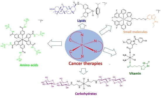
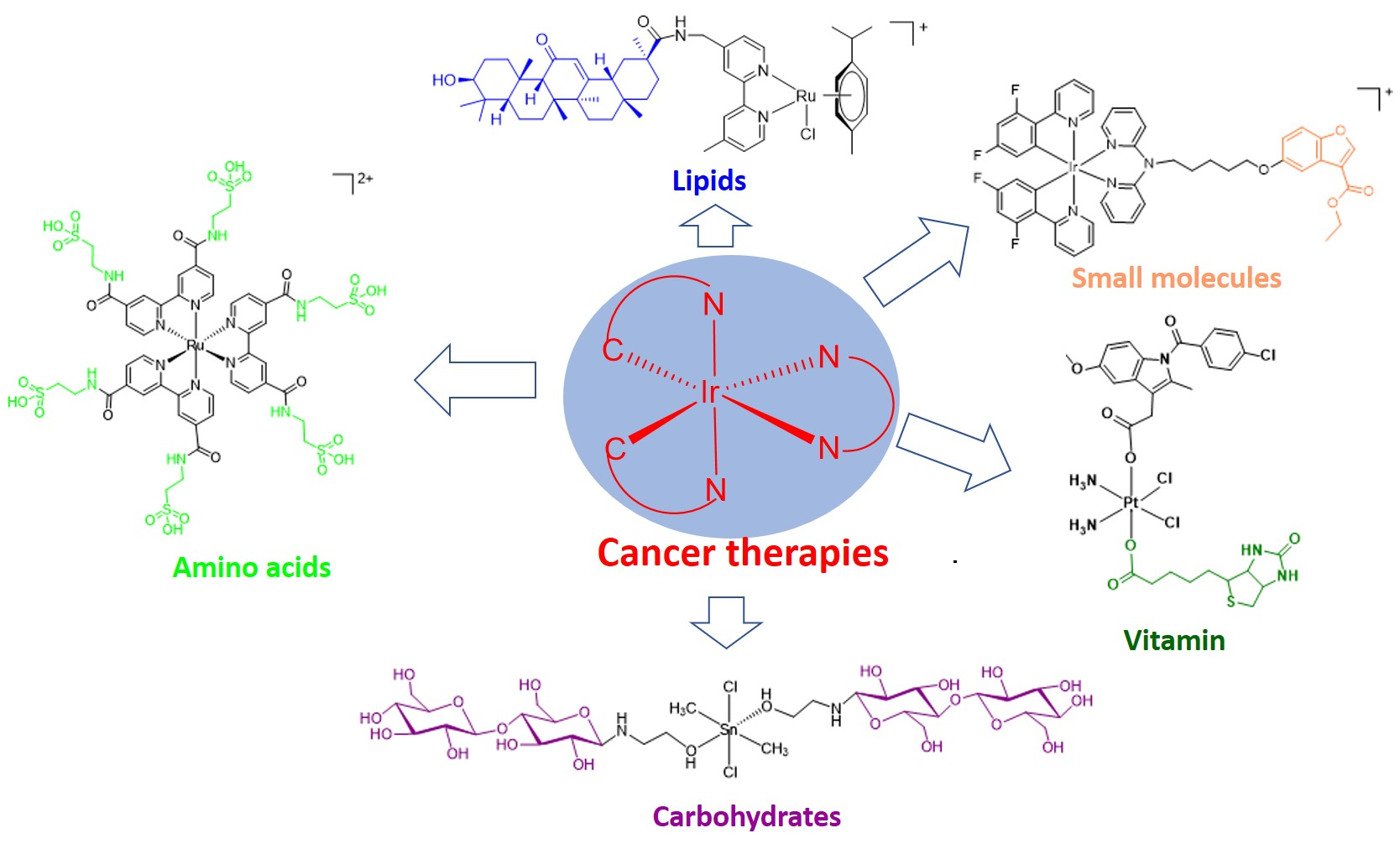
:Platinum-based drugs have revolutionized cancer care, but are unfortunately associated with various adverse effects. Meanwhile, natural product scaffolds exhibit multifarious bioactivities and serve as an attractive resource for cancer therapy development. Thus, the conjugation of natural product scaffolds to metal complexes becomes an attractive strategy to reduce the severe side effects arising from the use of metal bearing drugs. This review aims to highlight the recent examples of natural product-conjugated metal complexes as cancer therapies with enhanced selectivity and efficacy. We discuss the mechanisms and features of different conjugate complexes and present an outlook and perspective for the future of this field.
1. Introduction
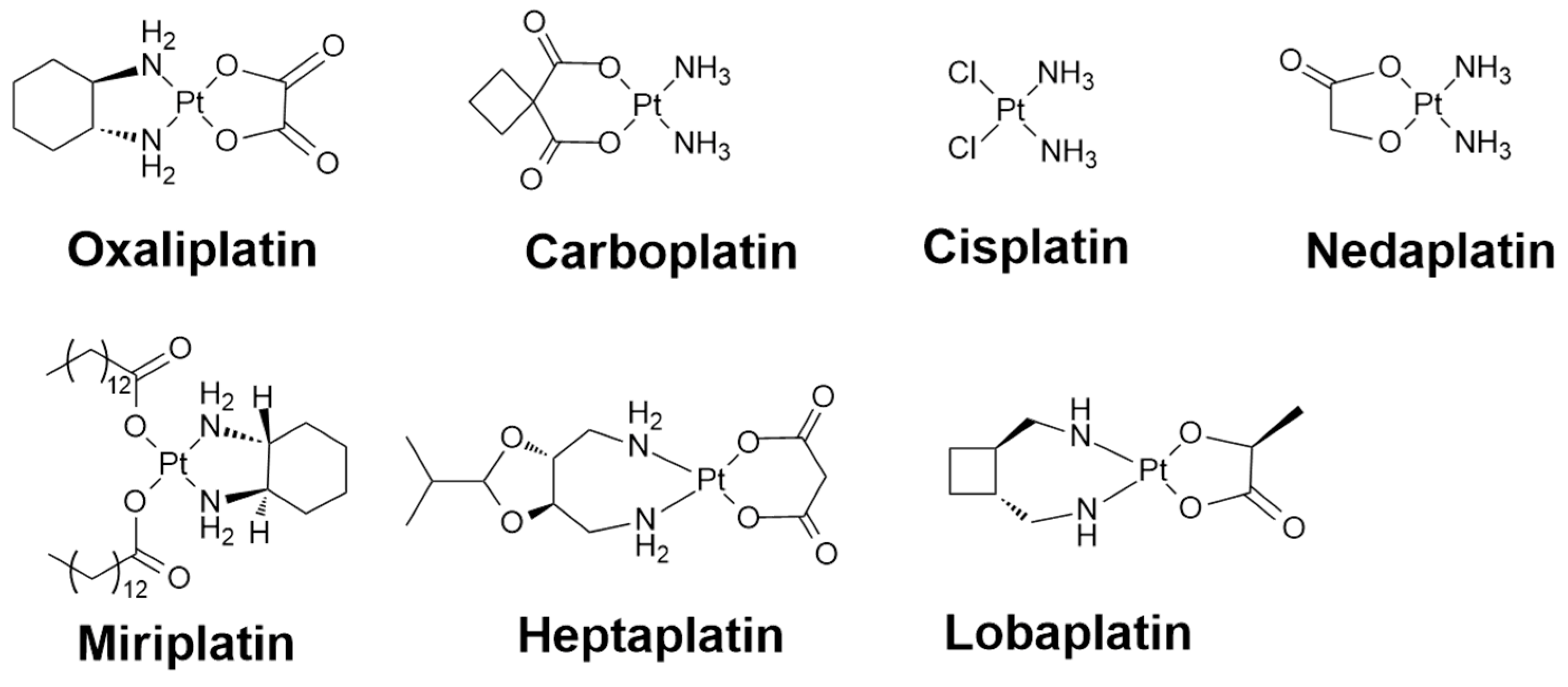
Since the serendipitous discovery of cisplatin, platinum-based drugs have emerged and have become one of the most widely-used class of chemotherapeutic drugs against various human tumors, such as testicular cancer, ovarian cancer, lung, head and neck, and advanced bladder cancer [1]. Three platinum(II)-based drugs (oxaliplatin, carboplatin, and cisplatin) are officially approved in the United States, while regionally approved platinum(II) and platinum(IV) drugs are also available in Japan (nedaplatin and miriplatin), Korea (heptaplatin), and China (lobaplatin) (Figure 1) [2,3]. Platinum-based chemotherapies mainly function by entering the target cells under the assistance of copper transporters [4]. Upon entering a cell, aquation of the metal complexes firstly takes place as a result of a low osmotic pressure environment, and the complexes may undergo further functional group hydrolysis for activity activation, particularly in the form of losing labile moiety such as carboxylate and chloride. The metal-based molecules become highly reactive after activation and readily display a positive interaction with the intracellular targeting sites notably embedded in the proteins/peptides or nuclear DNA, especially for redox active sulfur residues from methionine or cysteine [5,6]. Despite the clinical success of platinum-based drugs, their use remains limited by their systemic toxicity, which results in bone marrow suppression, hair loss, vomiting, nausea, and so on [7,8].
2. Side Effects of Platinum-Based Cancer Therapies
Platinum-based cancer therapies possess inevitable side effects owing to their limited selectivity for cancer cells or tissues over the surrounding healthy ones. The side effects might be induced by the same high nutrition requirements of cancerous cells versus other fast-growing healthy cells, such as cells of the mucous membranes, bone marrow, and hair follicles [9,10]. Around 40 specific side effects have been documented from the use of platinum-based drugs, which can be classified into the following sub-types: nephrotoxicity, ototoxicity, gastrointestinal toxicity, hepatotoxicity, hematological toxicity, cardiotoxicity, and neurotoxicity [11]. The symptoms generated from the severe side effects mainly include asthenia, anorexia, cachexia, alopecia, pain, stomatitis, mucositis, diarrhea, vomiting, nausea, cytopenias, anaphylaxis, and so on. Hence, patients taking platinum-based chemotherapeutics require additional medical interference and monitoring in order to achieve normal physiological maintenance of the body [12]. Additionally, ancillary drugs are commonly co-prescribed with platinum-centered cancer therapies to minimize unwanted side effects, which can include antioxidants, antibody cytokine blockers, monoclonal, magnesium supplements, saline hyperhydration, propafenone, mannitol, myeloid growth factors, antibiotics, and antiemetics [13,14].
Another important issue for controlling the side effects from platinum-based therapies is to avoid a superfluous aquation reaction, which typically occurs during the drug preparation and administration process. The use of cisplatin is regarded as a pattern metal-based cancer therapy, and cisplatin is commonly formulated in a stabilizer solution containing 0.9% sodium chloride, to ease the problem of losing the chloride ligand from the cisplatin scaffold. However, the sodium chloride solution used in the formulation would lead to the premature structure conversion and activation of parent drugs, like carboplatin or oxaliplatin, into more reactive but less soluble cisplatin or PtCl2(R,R-dach) (dach = diaminocyclohexane), respectively. Therefore, a 5% glucose solution, instead of a 0.9% sodium chloride solution, is normally adopted in the carboplatin and oxaliplatin formulation [9,15]. Moreover, it has been well documented that the toxicity of platinum-based cancer therapy is highly related to its level of binding reactivity towards functional target sites, which is determined mostly by the stability of the binding/leaving moieties on the parent drugs. The parent metal-based scaffolds bearing more labile moieties tend to be more active and generate more undesirable side effects at equivalent doses to other platinum drugs [16,17]. For instance, introducing a bis-carboxylate ligand into cisplatin to replace the already conjugated chloride ligands can reduce the aquation reactivity. As a result, a reduction of toxicity is demonstrated [16,18,19].
3. Strategies for Reducing the Toxicity of Metal-Based Cancer Therapies
Targeted cancer therapy is a challenging strategy that uses drugs or other substances to more accurately identify and attack cancer cells. One of the approaches is directing the therapeutic molecules selectively to the targeted cancer cells or tissues, and thus promoting the curing efficacy and minimizing the unwanted side effects simultaneously [20,21]. Therefore, the development of efficiently targeted drugs with minimal side effects and toxicity is important for anticancer therapies [20].
Cancer cells/tissues always possess similar characteristics to the homologous generated healthy cells/tissues, rendering selective treatment challenging. The bearing ligands on a metal-centered scaffold play a significant role in tuning the corresponding efficiency and toxicity properties of the anticancer therapy. The modification of the ligands benefits the regulation of the associated substitutional hydrophilicity/hydrophobicity, inertness/oxidation reactivity, and systematic/target biocompatibility according to specific treatment requirements and application conditions [22,23,24]. A hydrolysis process often takes place for the activation of premature metal therapies to allow for further binding to target the intercellular biomolecules [25,26]. Therefore, a high affinity and specificity to the reception targeting sites are always of primary consideration for ligand modification in metal-centered therapy development. Moreover, metal centers might experience a redox reaction for activation [27]. For example, platinum(IV) conjugated prodrugs can undergo reduction and generate platinum(II) complexes after delivering to intra-/inter-cellular target sites. As the platinum(IV)-based complexes are kinetically inert, they will not be as likely to cause side reactions until they enter into the cellular environment. Upon entry into the cell, they undergo reduction to generate the active platinum(II) species that is capable of forming platinum(II)-DNA adducts. The redox mechanism is thought to involve the initial binding of the platinum(IV) complex to the N7 site of the guanosine (G) moiety. Subsequently, the 5′-phosphate or 5′-hydroxyl group attacks the C8 site of the G moiety, resulting in a two-electron transfer process that produces cyclic (5′-O-C8)-G and a platinum(II) complex. The platinum(II)–DNA adducts induce the distortion of DNA structures and other biological dysfunctions, leading to cancer cell death [28]. The further modification of auxiliary ligands tunes the hydrophobicity and stability of platinum(IV) conjugated drugs to maintain intact structures before arrival at the cancer cells, with only selective redox reaction on target sites, thus reducing the undesirable toxicity to nearby healthy cells/tissues [29].
Moreover, the replacement of alternative metal centers, such as ruthenium(II), gold(III), palladium(II), iridium(III), rhodium(III), iron(III), osmium(IV), cobalt(II), and tin(II) [30,31,32,33,34], offers promising options to ease drug resistance to cisplatin, and to achieve diverse anticancer activities with reduced sides effect [35,36,37,38]. Ruthenium(II)-centered complexes are of particular interest among these metal centers in virtue of their moderate side effects with their reduced toxicity, excellent selectivity over healthy cells/tissues, and high efficiency of anticancer activity [39], along with their diverse functional mechanisms through different coordination geometries, coordination types, and oxidation states [13,40]. As octahedral ruthenium(II) complexes with three bidentate (usually polypyridyl) ligands are typically propeller-shaped, they possess two enantiomeric configurations that can differ in their biological activity and selectivity against target chiral biomolecules, such as DNA. Generally, the “left” configuration of the ruthenium(II) complexes is more able to bind to the minor groove of the DNA, while the “right” configuration is more likely to intercalate into the DNA molecule [41,42]. Moreover, ruthenium(II) complexes have also been developed that target the major groove of DNA, thereby avoiding the most common mechanisms of drug resistance from platinum-centered cancer therapies [43].
4. Natural Product-Conjugated Metal Complex Cancer Therapies
Natural product scaffolds exhibit multifarious bioactivities and serve as an attractive resource for cancer therapy development. The conjugation of natural product scaffolds to metal complexes serves as an attractive strategy to reduce the severe side effects of metal bearing drugs [44,45,46]. In the following sections, representative examples of the conjugation of natural product molecules to metal complexes will be illustrated (Table 1). Natural products possess inherent benefits as medicinal scaffolds, including an abundant structural diversity, intrinsic bioactivity, and excellent biocompatibility. The attachment of natural product moieties onto metal-centered complexes can confer selectivity for intercellular/intracellular target sites and as well as overcome typical mechanisms of drug resistance exhibited by cancer cells against platinum-based drugs. Moreover, the fine-tuning of either the metal centers or the conjugated natural products allows for the optimization of pharmacological characteristics, including enhanced biological potency and reduced side effects, depending on the mechanism of action of the natural product-conjugated metal complex [39,47,48].
4.1. Small Molecules
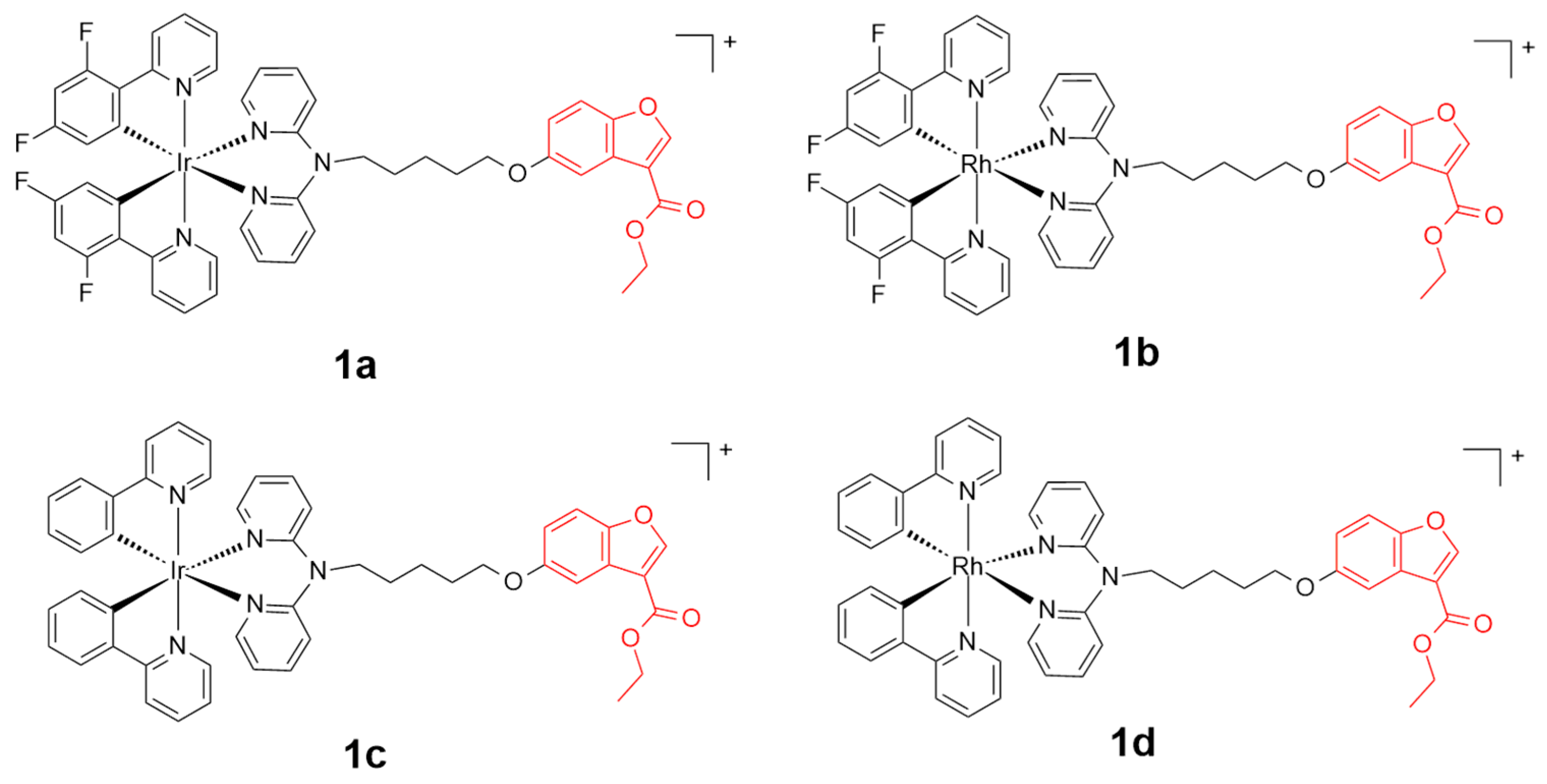
Benzofuran and its derivatives widely exist in nature and display significant inhibition activities upon comprehensive interaction towards cancer related biomarkers such as β-amyloid, mPTPB (protein tyrosine phosphatase B), mTOR (mammalian target of rapamycin), and PI3 (phosphatidylinositide 3) kinase [49,50]. A series of benzofuran-conjugated rhodium(III) or iridium(III)-centered metal scaffolds 1a–1d (Figure 2) have been reported as anti-prostate cancer agents by Ma and coworkers [51]. Complex 1a exhibited the best inhibition efficiency among the four scaffolds towards either the TNF-α-induced NF-κB pathway (TNF = tumor necrosis factor; NF-κB = nuclear factor kappa light chain enhancer of activated B cells) or IL-6-induced STAT3 pathway (IL-6 = Interleukin 6; STAT3 = Signal transducer and activator of transcription 3) in DU145 prostate cancer cells. Complex 1a regulated the protein expression and permutation by the inhibition of the cytoplasm translocation of NF-κB and STAT3 to the nucleus. Additionally, complex 1a displayed a selective toxicity against DU145 cells and a suppression activity against tumors in a prostate cancer xenograft mouse model. Complex 1a, with the half maximal inhibitory concentration (IC50) at ca. 4.34 µM, showed a higher toxicity against DU145 cancer cell lines than the reference cisplatin and doxorubicin (IC50 > 30 µM), probably by disrupting the plasma membrane integrity. Importantly, complex 1a exhibited a selective inhibition activity for cancer cells over healthy cells, with a relatively lower cytotoxicity against the healthy cells, including HEK293 (IC50 = ca. 32.34 µM) and LO2 cells (IC50 = ca. 29.21 µM).
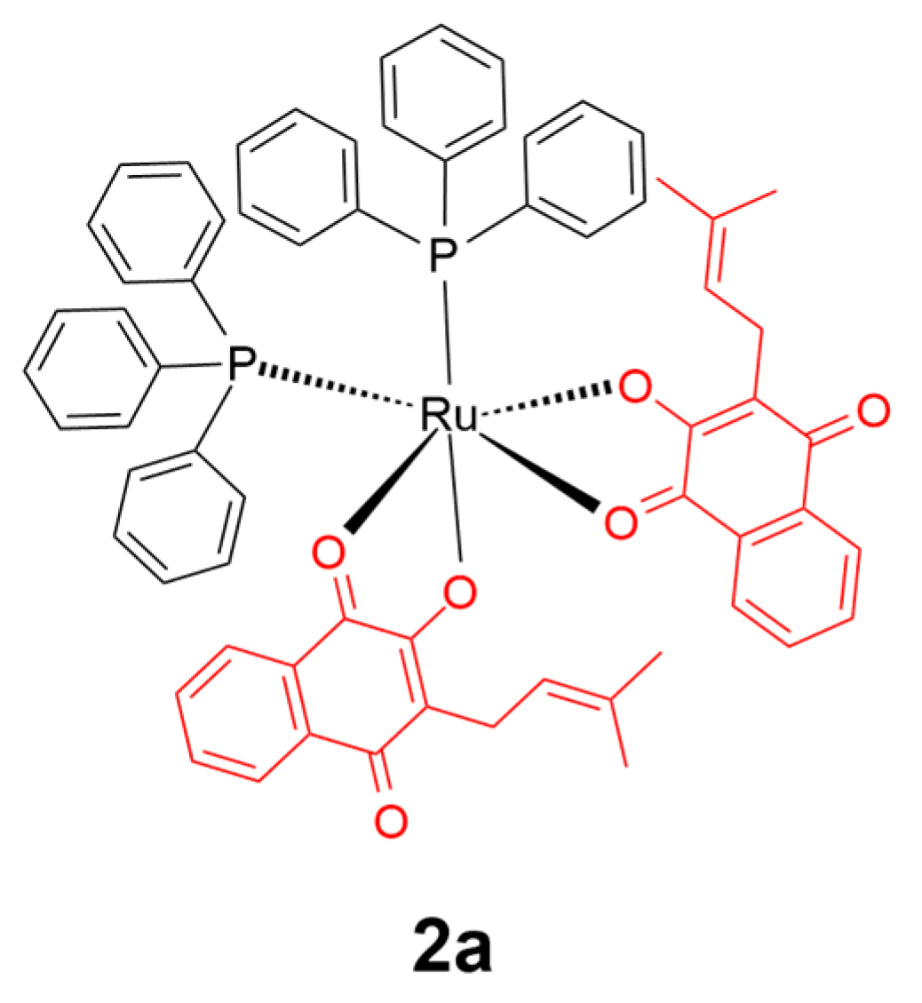
Lapachol is a naphthoquinone natural product mainly derived from Bignoniaceae plants with diverse biological activities including anticancer, antimicrobial, antiparasitic, antifungal, and antiviral activities [46,52]. In 2017, a lapachol conjugated ruthenium(II) (2a) scaffold was reported (Figure 3) [53]. Fluorescence measurements suggested that the cis configuration of complex 2a showed stronger binding upon both bovine serum albumin (BSA) and human serum albumin (HSA) proteins compared to the trans configuration one. However, cytotoxicity assays against healthy lung cells (V79) and breast/lung cancer cells (MDA-MB-231/A549) indicated that trans-2a was more active and selective towards cancer cells over healthy cells than both the cis isomer and the reference drug cisplatin, which was attributed to the weaker affinity of trans-2a to engage in non-selective interactions with non-target proteins such as albumin.
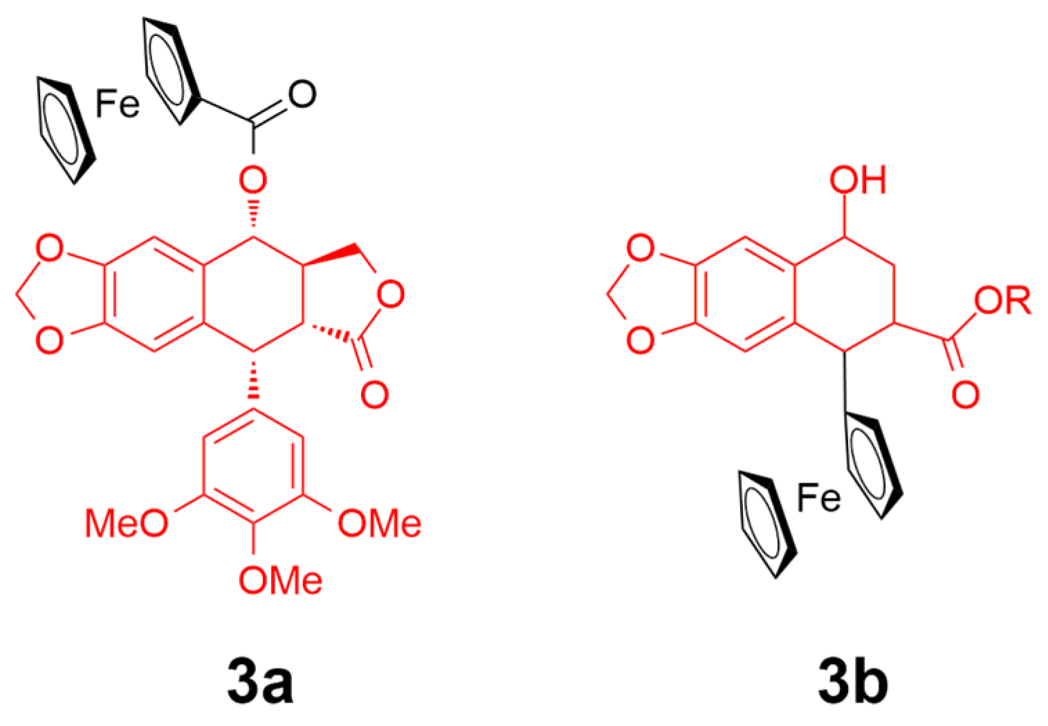
Podophyllotoxin (PPT) is a root-derived natural product from Podophyllum peltatum that exhibits a tubulin/antimitotic targeting activity, resulting in the interference of cell division or even cell death [54]. PPT possesses a high cytotoxicity and demonstrates severe side effects, such as vomiting, diarrhea, and nausea, and a poor selectivity over targeting cells [55]. Recently, conjugated ferrocenyl–podophyllotoxin analogues have been reported as breast cancer inhibitors (Figure 4) [56]. Podophyllotoxin alone shows a high activity against cancer cells MDA-MB-231 and MCF-7 (IC50 = 0.01 µM). Upon the conjugation of PPT to ferrocenyl complexes, the cytotoxicity of 3a was decreased with an IC50 value of 0.43 and 0.93 µM against the MDA-MB-231 and MCF-7 cells, respectively. However, the PPT analogue-conjugated 3b was found to be much less potent than 3a. On the basis of the reversible redox behavior of the ferrocenyl moiety, which might affect the cellular oxidative environment and facilitate the generation of reactive oxygen species (ROS), the authors suggested that the metal-complex–PPT conjugate could show superior selectivity for cancer cells over healthy ones.
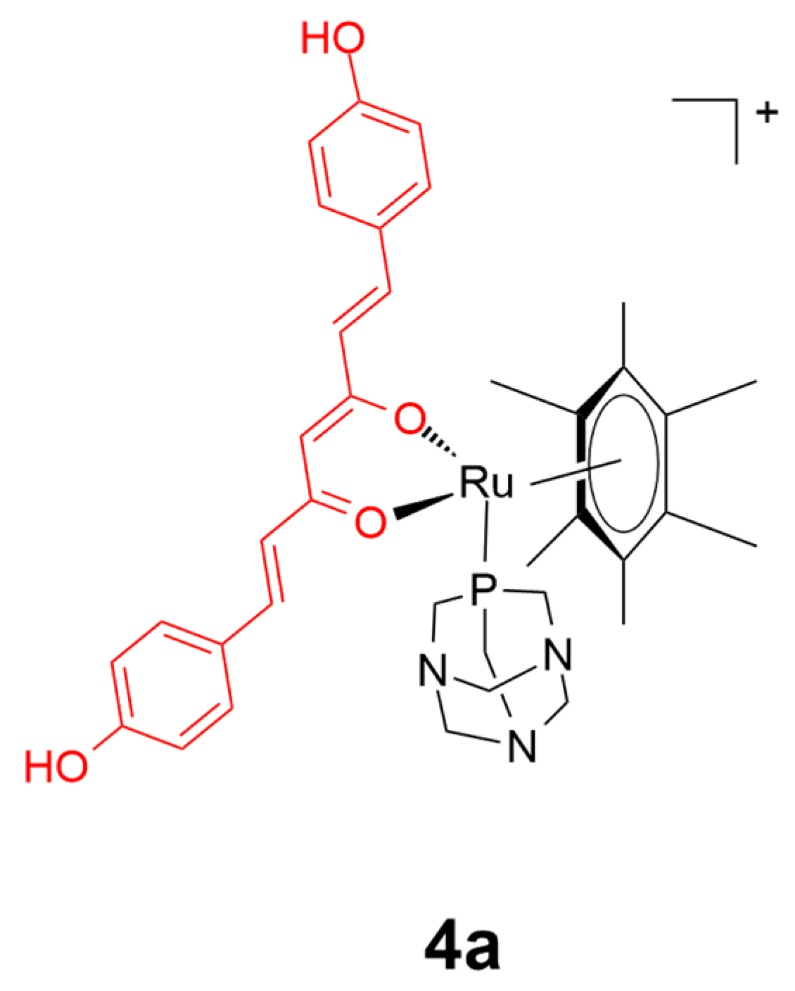
Curcumin is the principal active ingredient from the herbal plant Curcuma longa. Curcumin has been extensively studied as a pharmacological agent because of its broad range of biological activities, including its anticancer, antioxidant, and anti-inflammatory effects [57,58]. However, as curcumin itself is both unstable and not readily bioavailable [59,60], researchers have investigated whether the attachment of curcumin to metal scaffolds could allow for the favorable medicinal properties of both curcumin and the metal complex to be maintained, and yet retaining the desirable biocompatibility. The first curcumin-based metal complex was reported in 1987, which was a gold(III) conjugate that displayed moderate anti-arthritic activity [61]. Subsequently, curcumin complexes based on vanadyl(II), nickel(II), cobalt(II), copper(II), ruthenium(II), palladium(II), and zinc(II) have been reported [45,62]. Among these, the ruthenium(II) conjugates have shown the greatest potential as anticancer agents. For instance, a series of novel ruthenium(II) complexes have been developed by Dyson, Pettinari, and coworkers, using the conjugation of 1,3,5-triaza-7-phosphaadamantane (RAPTA) and curcumin as auxiliary ligands [63]. All of the curcumin complexes showed both an improved solubility and high selectivity for the tumor cell lines (A2780 and A2780cisR) over the non-tumorous HEK293 cell line. Particularly, compound 4a (Figure 5) showed the most promising activity profile over cisplatin, with around a 70-fold higher inhibition activity against the cancer cell lines (IC50 < 0.27 μM) over the normal HEK cell line (IC50 = 13.0 μM). The replacement of the chloride ligand present in most platinum drugs with the RAPTA ligand was thought to allow the complexes to bypass drug resistance mechanisms for cisplatin. Moreover, the more rapid dissociation of the bisdemethoxycurcumin moiety from complex 4a greatly enhanced its activity and selectivity against targeted cancer cells compared with the parent curcumin-containing complex.
4.2. Amino Acids
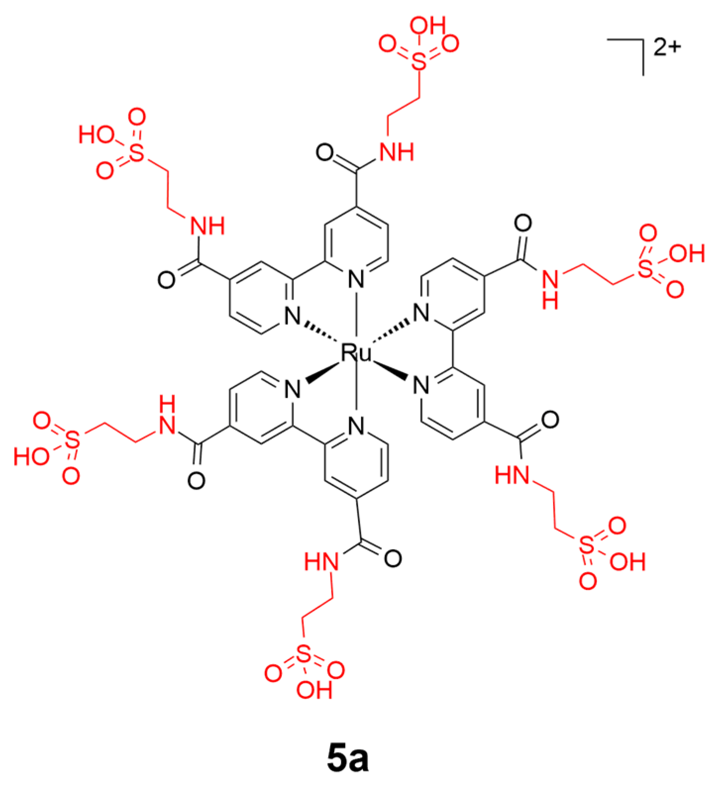
Taurine is a typical amino acid existing in the brain and is involved in central nervous system (CNS) signaling. A taurine-bearing ruthenium(II) compound (5a) has been reported by Zhang and coworkers for targeting brain cancer cells (Figure 6) [64]. Complex 5a showed lysosome-specific intracellular accumulation in cancer cells. The symmetrical introduction of taurine moieties to the ruthenium(II) scaffold benefits the enhancement of the emission and further releases of ROS, which renders the taurine-conjugated molecule a reactive photosensitizer for photodynamic therapy to target tumor cells selectively.
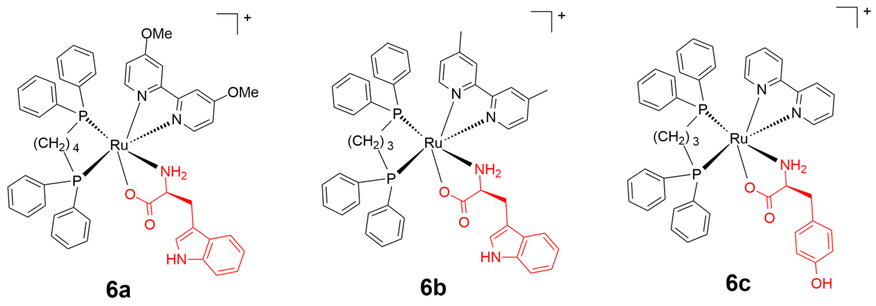
Ruthenium(II) complexes bearing proteinogenic α-amino acids exhibit potentially reduced cytotoxicity over cancer cells in contrast to anticancer platinum complexes [64]. In 2018, Santos and coworkers [65] developed thirteen amino acid conjugated ruthenium(II) complexes as cancer therapeutic drugs, including l-methionine (Met), l-histidine (His), l-tryptophan (Trp), l-tyrosine (Tyr), l-valine (Val), l-alanine (Ala), and glycine (Gly). Human breast cancer cells and healthy breast cells (MDA-MB-231 and MCF-10A, respectively) were applied for in vitro cytotoxicity investigation, and cisplatin was used as the reference drug. All of the scaffolds exhibited inhibition activity and selectivity to MDA-MB-231 over MCF-10A with promising IC50 values contrasted to the reference drug cisplatin. Complexes 6a and 6b containing l-Trp residue showed the best combination of activity and selectivity (Figure 7). Particularly, complex 6b (IC50 = 3.0 and 29.9 µM, respectively) showed better anticancer selectivity to cancer cells over normal cell lines, and a higher anticancer efficiency over non-amino acid conjugated ruthenium(II) complexes (IC50 = 15.6 and 17.0 µM, respectively). Moreover, this group developed similar amino acid-based ruthenium(II) complexes and evaluated their activity against MDA-MB-231 cells. These conjugates demonstrated a better activity against breast cancer cells compared to the reference drug cisplatin and the non-amino acid conjugate ruthenium(II) complex, with an IC50 ranging from 3.04 to 7.44 µM. Among all of the developed compounds, complex 5c bearing the Tyr residue exhibited the best activity [66].
4.3. Lipids
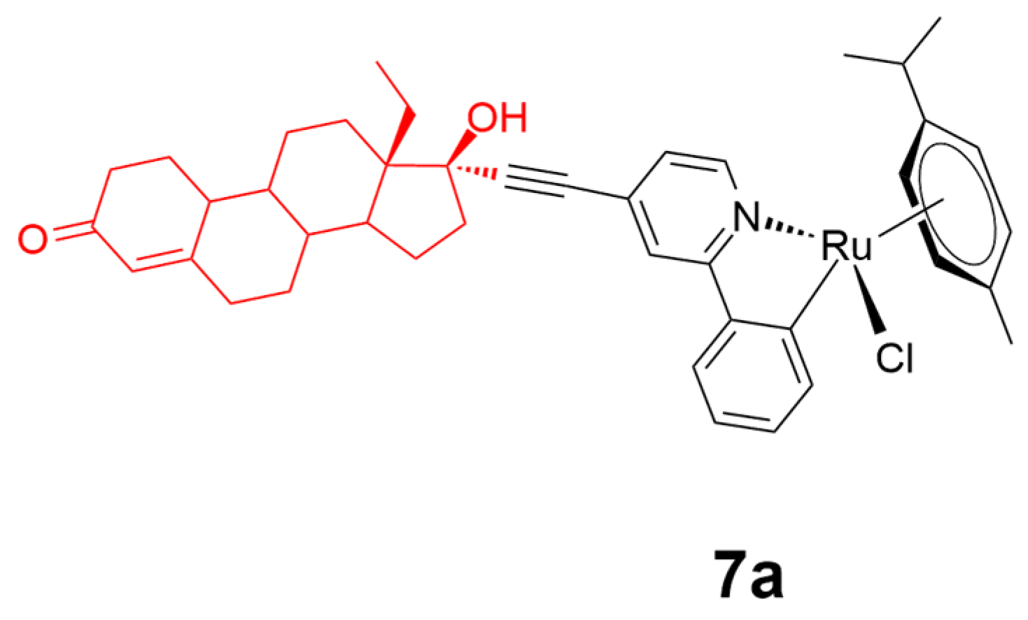
The binding affinity towards targeted active sites is of great importance for cancer therapy development based on the similar hydrophilic–lipophilic ability of the treatment molecule and the corresponding targeting receptors [66]. Ruiz and coworkers recently developed a lipophilic levonorgestrel-conjugated ruthenium(II) complex 7a (Figure 8) against breast cancer [67]. Complex 7a showed an enhanced inhibition activity against T47D breast cancer cells over nonsteroidal analogues with an eight-fold activity enhancement over the reference cisplatin. The conjugation of the levonorgestrel motif to ruthenium(II) scaffold generated a tunable synergy between the steroidal axial-accessories and the ruthenium(II) metal center, thus benefiting the improved activity of the comprehensive scaffold. Theoretical density functional theory calculations on complex 7a suggested that the lipophilic steroidal moiety increased the lability of the Ru–Cl bond, allowing for the easier formation of a stronger Ru–N bond upon substitution of Cl by N-nucleophiles. The calculations also revealed the interaction of the guanine/phenylpyridine/steroid moiety towards the reception targets at the lowest energy located in pseudocavity.
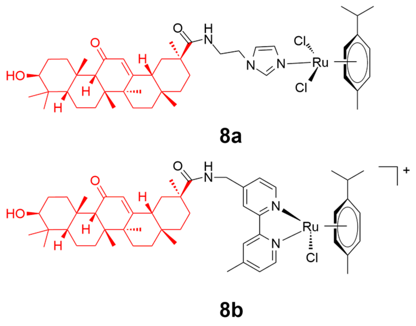
Glycyrrhetinic acid (GA) widely exists in Glycyrrhiza glabra and possesses outstanding anticancer activity through multiple mechanisms, including ROS production [68], mitochondria targeting [68], acting via protein receptors [68], or affecting the microenvironment of the tumor cell [69]. Liu and coworkers reported the conjugation of 18β-glycyrrhetinic acid on ruthenium(II)-arene scaffolds to generate conjugates 8a and 8b (Figure 9) [70]. Complex 8b containing the N,N-chelating moiety rather than the imidazole moiety in 8a displayed a higher stability and better solubility. Complex 8b experienced a higher rate of hydrolysis than 8a, with only one chloride leaving motif within the scaffold. Additionally, complex 8b also exhibited a better inhibition towards human cancer cells. This might be attributed to the primary role that complex 8b participated in the alteration of secondary structure of B-DNA by hydrolysis, and in the enhancement of intracellular ROS concentrations, which is likely to cause the disruption of cell metabolism and/or cell death.
4.4. Carbohydrates
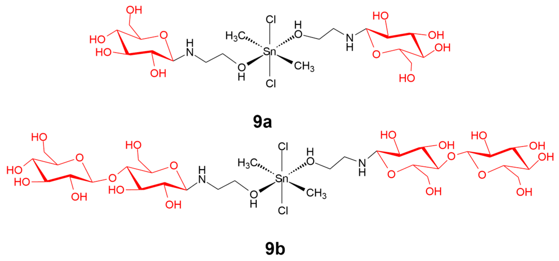
Carbohydrates are widely distributed natural compounds and possess multiple tunable functional groups for modulation according to their desired properties. In living systems, carbohydrates play an important role in mediating carbohydrate–protein interactions, which are crucial in cell–cell recognition and adhesion phenomena during cancer growth and progression [71]. Tabassum and coworkers [72] recently described carbohydrate-linked organotin(IV) complex 9a and 9b, as a human topoisomerase Iα inhibitor against human carcinoma cells (Figure 10). Both of the complexes showed a strong Topo I inhibition activity in contrast to the reference drug cisplatin at 30–35 µM. The complexes also showed good antiproliferative activity against human carcinoma cells. The complexes significantly suppressed the expression of MMP-2 mRNA levels, suggesting that their antiproliferative activity was mediated through inducing morphological transformations and further cell apoptosis.
4.5. Vitamin
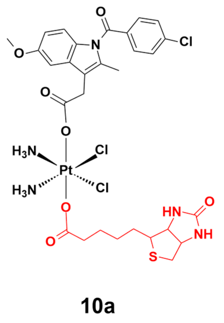
Biotin is a water-soluble vitamin that is involved in the metabolism of amino acids, carbohydrates, and fats, both in humans and in other organisms. Gou and coworkers recently reported biotin-conjugated platinum(IV) complex 10a, which selectively targets cancer cells expressing enhanced levels of biotin receptor (Figure 11) [73]. Complex 10a was activated by endogenous reductants in the cellular environment, to release indomethacin and cisplatin moieties to inhibit cancer cell activity. In in vitro studies, complex 10a displayed a remarkable activity against cisplatin-resistant gastric cancer cells (SGC7901/CDDP), as well as five other cancer cell lines, including gastric cancer cells (SGC7901), umbilical vein endothelial cells (EA. hy926), prostate carcinoma cells (PC-3), hepatocellular carcinoma cells (HepG-2), and colorectal cancer cells (HCT-116). Notably, complex 10a also alleviated inflammatory symptoms in cancer cells via the inhibition of cyclooxygenases. Moreover, complex 10a perturbed the formation of capillary-like tubes in EA. hy 926 cells and weakened the invasiveness of the highly aggressive PC-3 cell line.
5. Conclusions
Most cancer-oriented metal therapies attract significant interest in the activity levels against cancer cells/tissues. Focus on the toxicity and selectivity over healthy surroundings, including cells, tissues, or organs, is somehow neglected and rendered as secondary factors for consideration, which is, however, the primary concern for patients suffering from severe cancer sickness. In 2016, about nine million people worldwide were reported to have died from diverse species of cancers. Hence, growing efforts have been contributed to give a closer look and a more comprehensive understanding of the function mechanisms as well as the corresponding side effects from cancer therapies, particularly for metal-conjugated complexes bearing inherent toxicity from the metals. In search of side-effect reduced metal therapies, tremendous improvements in terms of the sensitivity, efficiency, selectivity, and stability have been achieved, capitalizing on the unique characteristics of metal centers, including distinguished spectroscopic properties and isotopic patterns.
Side effects from metal-centered cancer therapies often differ from species to species, owing to their diversity of the conjugated ligands and mechanisms of action. To date, platinum drugs are still the most important class of metal-based compounds used in the clinic, however, resistance and adverse side effects are limiting factors against their more widespread use. In this context, strategies for the modification of alternative metal centers (such as ruthenium(II), iridium(III), osmium(IV), gold(III), palladium(II), rhodium(III), iron(III), cobalt(II), and tin(II)) or axial conjugating ligands (either based on synthesized organic moiety or natural product-based motif) continue to be sought in order to further the development of novel metal-based drugs with tunable medicinal characteristics, improved potency, and reduced side effects. Particularly, natural products possess inherent benefits with abundant structure diversity; effortless access; and, most importantly, splendid bioactivity with excellent biocompatibility, mainly based on hydrolysis and redox processes upon binding with intercellular/intracellular target sites. Except for small molecule-based natural product moieties, such as naturally derived active ingredients, amino acids, lipids, carbohydrates, or vitamins, larger natural biomolecules, including peptides [74,75,76], antibodies [77,78] and oligonucleotides, particularly aptamers [79,80,81], have also been introduced into metal-based complexes as potential cancer therapies. Linking biomolecules to metal complexes can both reduce their cytotoxicity and enhance the bioavailability of the conjugates to the specific target organelles/cells/tissues, owing to their similar physicochemical properties and steric configurations to the target biomolecules. However, as larger biomolecules are extremely sensitive to the surrounding environment, the maintenance of their original biological structure and function upon grafting to a metal scaffold can be rather difficult compared with small molecule-based metal complexes. Looking forward, we speculate that more efforts by pioneers will be contributed to the improvement of function efficiency and selectivity over targeted cancer cells/tissues, based on natural product-conjugated metal complexes. In this case, unwanted side effects can be very likely to be reduced with enhanced cancer targeting and anticancer efficiency.
Author Contributions
D.-L.M. proposed the topic and instructed the writing process of this manuscript. C.-H.L. provided valuable suggestions and instructed the writing process of this manuscript. C.W. helped to draft this manuscript. S.-S.C. collected and studied the source literatures for this manuscript. F.-W.L. made contribution to the critical revision of this manuscript. Q.-B.H. participated in the critical revision of this manuscript.
Funding
This work is supported by Hong Kong Baptist University (FRG2/16-17/007 and FRG2/17-18/003), the Health and Medical Research Fund (HMRF/14150561), the Research Grants Council (HKBU/12301115), the National Natural Science Foundation of China (21575121 and 21775131), the Hong Kong Baptist University Century Club Sponsorship Scheme 2018, the Interdisciplinary Research Matching Scheme (RC-IRMS/16-17/03), Interdisciplinary Research Clusters Matching Scheme (RC-IRCs/17-18/03), Innovation and Technology Fund (ITS/260/16FX), Collaborative Research Fund (C5026-16G), Matching Proof of Concept Fund (MPCF-001-2017/18), SKLEBA and HKBU Strategic Development Fund (SKLP_1718_P04), the Science and Technology Development Fund, Macao SAR (0072/2018/A2), and the University of Macau (MYRG2018-00187-ICMS and MYRG2016-00151-ICMS-QRCM).
Conflicts of Interest
The authors declare no conflicts of interest.
Abbreviations
| BSA | bovine serum albumin |
| HSA | human serum albumin |
| PPT | podophyllotoxin |
| ROS | reactive oxygen species |
| CNS | central nervous system |
| GA | glycyrrhetinic acid |
References
- Ndagi, U.; Mhlongo, N.; Soliman, M.E. Metal complexes in cancer therapy—An update from drug design perspective. Drug Des. Dev. Ther. 2017, 11, 599–616. [Google Scholar] [CrossRef] [PubMed]
- Pasetto, L.M.; D’Andrea, M.R.; Brandes, A.A.; Rossi, E.; Monfardini, S. The development of platinum compounds and their possible combination. Crit. Rev. Oncol. Hematol. 2006, 60, 59–75. [Google Scholar] [CrossRef] [PubMed]
- Apps, M.G.; Choi, E.H.; Wheate, N.J. The state-of-play and future of platinum drugs. Endocr. Relat. Cancer. 2015, 22, R219–R233. [Google Scholar] [CrossRef] [PubMed] [Green Version]
- Kalayda, G.V.; Wagner, C.H.; Jaehde, U. Relevance of copper transporter 1 for cisplatin resistance in human ovarian carcinoma cells. J. Inorg. Biochem. 2012, 116, 1–10. [Google Scholar] [CrossRef] [PubMed]
- Chin, C.F.; Tian, Q.; Setyawati, M.I.; Fang, W.; Tan, E.S.Q.; Leong, D.T.; Ang, W.H. Tuning the activity of platinum(IV) anticancer complexes through asymmetric acylation. J. Med. Chem. 2012, 55, 7571–7582. [Google Scholar] [CrossRef] [PubMed]
- Johnstone, T.C.; Suntharalingam, K.; Lippard, S.J. The next generation of platinum drugs: Targeted Pt(II) agents, nanoparticle delivery, and Pt(IV) prodrugs. Chem. Rev. 2016, 116, 3436–3486. [Google Scholar] [CrossRef] [PubMed]
- Kapdi, A.R.; Fairlamb, I.J. Anti-cancer palladium complexes: A focus on PdX2L2, palladacycles and related complexes. Chem. Soc. Rev. 2014, 43, 4751–4777. [Google Scholar] [CrossRef] [PubMed]
- Hartmann, J.T.; Lipp, H.-P. Toxicity of platinum compounds. Expert Opin. Pharmacother. 2003, 4, 889–901. [Google Scholar] [CrossRef]
- Oun, R.; Moussa, Y.E.; Wheate, N.J. The side effects of platinum-based chemotherapy drugs: A review for chemists. Dalton Trans. 2018, 47, 6645–6653. [Google Scholar] [CrossRef]
- Ma, L.; Lin, X.; Li, C.; Xu, Z.; Chan, C.-Y.; Tse, M.-K.; Shi, P.; Zhu, G. A cancer cell-selective and low-toxic bifunctional heterodinuclear Pt(IV)–Ru(II) anticancer prodrug. Inorg. Chem. 2018, 57, 2917–2924. [Google Scholar] [CrossRef]
- Arjmand, F.; Parveen, S.; Tabassum, S.; Pettinari, C. Organotin antitumor compounds: Their present status in drug development and future perspectives. Inorg. Chim. Acta. 2014, 423, 26–37. [Google Scholar] [CrossRef]
- Machiels, J.-P.H.; Haddad, R.I.; Fayette, J.; Licitra, L.F.; Tahara, M.; Vermorken, J.B.; Clement, P.M.; Gauler, T.; Cupissol, D.; Grau, J.J. Afatinib versus methotrexate as second-line treatment in patients with recurrent or metastatic squamous-cell carcinoma of the head and neck progressing on or after platinum-based therapy (LUX-Head & Neck): An open-label, randomised phase 3 trial. Lancet Oncol. 2015, 16, 583–594. [Google Scholar] [PubMed]
- Allardyce, C.S.; Dyson, P.J. Ruthenium in medicine: Current clinical uses and future prospects. Platin. Met. Rev. 2001, 45, 62–69. [Google Scholar]
- Landier, W. Ototoxicity and cancer therapy. Cancer 2016, 122, 1647–1658. [Google Scholar] [CrossRef] [Green Version]
- Margiotta, N.; Marzano, C.; Gandin, V.; Osella, D.; Ravera, M.; Gabano, E.; Platts, J.A.; Petruzzella, E.; Hoeschele, J.D.; Natile, G. Revisiting [PtCl2(cis-1, 4-DACH)]: An underestimated antitumor drug with potential application to the treatment of oxaliplatin-refractory colorectal cancer. J. Med. Chem. 2012, 55, 7182–7192. [Google Scholar] [CrossRef] [PubMed]
- Dasari, S.; Tchounwou, P.B. Cisplatin in cancer therapy: Molecular mechanisms of action. Eur. J. Pharmacol. 2014, 740, 364–378. [Google Scholar] [CrossRef] [PubMed] [Green Version]
- Bruijnincx, P.C.; Sadler, P.J. New trends for metal complexes with anticancer activity. Curr. Opin. Chem. Biol. 2008, 12, 197–206. [Google Scholar] [CrossRef] [Green Version]
- Dreisbach, L.; Ho, M.; Reid, E.; Siegel, J. Effects of oxaliplatin, carboplatin, and cisplatin across treatment on high-frequency objective and subjective auditory measures in adults. Perspect ASHA Spec Interest Groups 2017, 2, 17–36. [Google Scholar] [CrossRef]
- Abu-Surrah, A.S.; Kettunen, M. Platinum group antitumor chemistry: Design and development of new anticancer drugs complementary to cisplatin. Curr. Med. Chem. 2006, 13, 1337–1357. [Google Scholar] [CrossRef]
- Kumar, A.; Zhang, X.; Liang, X.-J. Gold nanoparticles: Emerging paradigm for targeted drug delivery system. Biotechnol. Adv. 2013, 31, 593–606. [Google Scholar] [CrossRef]
- Ruoslahti, E.; Bhatia, S.N.; Sailor, M.J. Targeting of drugs and nanoparticles to tumors. J. Cell Biol. 2010, 188, 759–768. [Google Scholar] [CrossRef] [PubMed] [Green Version]
- Mital, M.; Ziora, Z. Biological applications of Ru(II) polypyridyl complexes. Coord. Chem. Rev. 2018, 375, 434–458. [Google Scholar] [CrossRef]
- Thota, S.; Rodrigues, D.A.; Crans, D.C.; Barreiro, E.J. Ru(II) compounds: Next-generation anticancer metallotherapeutics? J. Med. Chem. 2018, 61, 5805–5821. [Google Scholar] [CrossRef] [PubMed]
- Galanski, M.; Jakupec, M.A.; Keppler, B.K. Update of the preclinical situation of anticancer platinum complexes: Novel design strategies and innovative analytical approaches. Curr. Med. Chem. 2005, 12, 2075–2094. [Google Scholar] [CrossRef] [PubMed]
- Meier-Menches, S.M.; Gerner, C.; Berger, W.; Hartinger, C.G.; Keppler, B.K. Structure–activity relationships for ruthenium and osmium anticancer agents–towards clinical development. Chem. Soc. Rev. 2018, 47, 909–928. [Google Scholar] [CrossRef] [PubMed]
- Coverdale, J.P.; Romero-Canelón, I.; Sanchez-Cano, C.; Clarkson, G.J.; Habtemariam, A.; Wills, M.; Sadler, P.J. Asymmetric transfer hydrogenation by synthetic catalysts in cancer cells. Nat. Chem. 2018, 10, 347–354. [Google Scholar] [CrossRef] [PubMed]
- Foltinová, V.; Švihálková Šindlerová, L.; Horváth, V.; Sova, P.; Hofmanova, J.; Janisch, R.; Kozubík, A. Mechanisms of effects of platinum(II) and platinum(IV) complexes. Comparison of cisplatin and oxaliplatin with satraplatin and LA-12, new Pt(IV)-based drugs. A Minireview. Scr. Med. 2008, 81, 105–116. [Google Scholar]
- Choi, S.; Vastag, L.; Larrabee, Y.C.; Personick, M.L.; Schaberg, K.B.; Fowler, B.J.; Sandwick, R.K.; Rawji, G. Importance of platinum(II)-assisted platinum(IV) substitution for the oxidation of guanosine derivatives by platinum(IV) complexes. Inorg. Chem. 2008, 47, 1352–1360. [Google Scholar] [CrossRef]
- Hall, M.D.; Amjadi, S.; Zhang, M.; Beale, P.J.; Hambley, T.W. The mechanism of action of platinum(IV) complexes in ovarian cancer cell lines. J. Inorg. Biochem. 2004, 98, 1614–1624. [Google Scholar] [CrossRef]
- Yang, G.; Zhong, H.-J.; Ko, C.-N.; Wong, S.-Y.; Vellaisamy, K.; Ye, M.; Ma, D.-L.; Leung, C.-H. Identification of a Rhodium(III) complex as a Wee1 inhibitor against TP53-mutated triple-negative breast cancer cells. Chem. Commun. 2018, 54, 2463–2466. [Google Scholar] [CrossRef]
- Liu, L.-J.; Wang, W.; Huang, S.-Y.; Hong, Y.; Li, G.; Lin, S.; Tian, J.; Cai, Z.; Wang, H.-M.D.; Ma, D.-L. Inhibition of the Ras/Raf interaction and repression of renal cancer xenografts in vivo by an enantiomeric iridium(III) metal-based compound. Chem. Sci. 2017, 8, 4756–4763. [Google Scholar] [CrossRef] [PubMed]
- Yang, C.; Wang, W.; Liang, J.-X.; Li, G.; Vellaisamy, K.; Wong, C.-Y.; Ma, D.-L.; Leung, C.-H. A rhodium(III)-based inhibitor of lysine-specific histone demethylase 1 as an epigenetic modulator in prostate cancer cells. J. Med. Chem. 2017, 60, 2597–2603. [Google Scholar] [CrossRef] [PubMed]
- Yang, C.; Wang, W.; Chen, L.; Liang, J.; Lin, S.; Lee, M.-Y.; Ma, D.-L.; Leung, C.-H. Discovery of a VHL and HIF1α interaction inhibitor with in vivo angiogenic activity via structure-based virtual screening. Chem. Commun. 2016, 52, 12837–12840. [Google Scholar] [CrossRef] [PubMed]
- Zhong, H.-J.; Lu, L.; Leung, K.-H.; Wong, C.C.; Peng, C.; Yan, S.-C.; Ma, D.-L.; Cai, Z.; Wang, H.-M.D.; Leung, C.-H. An iridium(III)-based irreversible protein–protein interaction inhibitor of BRD4 as a potent anticancer agent. Chem. Sci. 2015, 6, 5400–5408. [Google Scholar] [CrossRef] [PubMed]
- Liang, J.-X.; Zhong, H.-J.; Yang, G.; Vellaisamy, K.; Ma, D.-L.; Leung, C.-H. Recent development of transition metal complexes with in vivo antitumor activity. J. Inorg. Biochem. 2017, 177, 276–286. [Google Scholar] [CrossRef] [PubMed]
- Wu, M.X.; Yang, Y.W. Metal–organic framework (MOF)-based drug/cargo delivery and cancer therapy. Adv. Mater. 2017, 29, 1606134. [Google Scholar] [CrossRef] [PubMed]
- Zhang, P.; Sadler, P.J. Redox-active metal complexes for anticancer therapy. Eur. J. Inorg. Chem. 2017, 27, 1541–1548. [Google Scholar] [CrossRef]
- Lan, G.; Ni, K.; Lin, W. Nanoscale metal–organic frameworks for phototherapy of cancer. Coord. Chem. Rev. 2019, 379, 65–81. [Google Scholar] [CrossRef]
- Ma, D.L.; He, H.Z.; Leung, K.H.; Chan, D.S.H.; Leung, C.H. Bioactive luminescent transition-metal complexes for biomedical applications. Angew. Chem. Int. Ed. Engl. 2013, 52, 7666–7682. [Google Scholar] [CrossRef]
- Schmitt, F.; Kasparkova, J.; Brabec, V.; Begemann, G.; Schobert, R.; Biersack, B. New arene ruthenium(II) complexes of 4-aryl-4H-naphthopyrans with anticancer and anti-vascular activities. J. Inorg. Biochem. 2018, 184, 69–78. [Google Scholar] [CrossRef]
- Choi, S.-D.; Kim, M.-S.; Kim, S.K.; Lincoln, P.; Tuite, E.; Nordén, B. Binding mode of [ruthenium(II)(1, 10-phenanthroline)2L]2+ with poly (dT* dA-dT) triplex. Ligand size effect on third-strand stabilization. Biochemistry 1997, 36, 214–223. [Google Scholar] [CrossRef] [PubMed]
- Barton, J.K.; Danishefsky, A.; Goldberg, J. Tris (phenanthroline) ruthenium(II): Stereoselectivity in binding to DNA. J. Am. Chem. Soc. 1984, 106, 2172–2176. [Google Scholar] [CrossRef]
- Pascu, G.I.; Hotze, A.C.; Sanchez-Cano, C.; Kariuki, B.M.; Hannon, M.J. Dinuclear Ruthenium (II) Triple-Stranded Helicates: Luminescent Supramolecular Cylinders That Bind and Coil DNA and Exhibit Activity against Cancer Cell Lines. Angew. Chem. Int. Ed. 2007, 46, 4374–4378. [Google Scholar] [CrossRef] [PubMed]
- Caruso, F.; Rossi, M.; Benson, A.; Opazo, C.; Freedman, D.; Monti, E.; Gariboldi, M.B.; Shaulky, J.; Marchetti, F.; Pettinari, R. Ruthenium–arene complexes of curcumin: X-ray and density functional theory structure, synthesis, and spectroscopic characterization, in vitro antitumor activity, and DNA docking studies of (p-cymene)-Ru-(curcuminato)-chloro. J. Med. Chem. 2012, 55, 1072–1081. [Google Scholar] [CrossRef]
- Banerjee, S.; Chakravarty, A.R. Metal complexes of curcumin for cellular imaging, targeting, and photoinduced anticancer activity. Acc. Chem. Res. 2015, 48, 2075–2083. [Google Scholar] [CrossRef]
- Kandioller, W.; Balsano, E.; Meier, S.M.; Jungwirth, U.; Göschl, S.; Roller, A.; Jakupec, M.A.; Berger, W.; Keppler, B.K.; Hartinger, C.G. Organometallic anticancer complexes of lapachol: Metal centre-dependent formation of reactive oxygen species and correlation with cytotoxicity. Chem. Commun. 2013, 49, 3348–3350. [Google Scholar] [CrossRef] [PubMed]
- Man, B.Y.-W.; Chan, H.-M.; Leung, C.-H.; Chan, D.S.-H.; Bai, L.-P.; Jiang, Z.-H.; Li, H.-W.; Ma, D.-L. Group 9 metal-based inhibitors of β-amyloid (1–40) fibrillation as potential therapeutic agents for Alzheimer′s disease. Chem. Sci. 2011, 2, 917–921. [Google Scholar] [CrossRef]
- Wenzel, M.; Bertrand, B.; Eymin, M.-J.; Comte, V.; Harvey, J.A.; Richard, P.; Groessl, M.; Zava, O.; Amrouche, H.; Harvey, P.D. Multinuclear cytotoxic metallodrugs: Physicochemical characterization and biological properties of novel heteronuclear gold–titanium complexes. Inorg. Chem. 2011, 50, 9472–9480. [Google Scholar] [CrossRef]
- Leung, C.-H.; Yang, H.; Ma, V.P.-Y.; Chan, D.S.-H.; Zhong, H.-J.; Li, Y.-W.; Fong, W.-F.; Ma, D.-L. Inhibition of Janus kinase 2 by cyclometalated rhodium complexes. MedChemComm 2012, 3, 696–698. [Google Scholar] [CrossRef]
- He, Y.; Xu, J.; Yu, Z.-H.; Gunawan, A.M.; Wu, L.; Wang, L.; Zhang, Z.-Y. Discovery and evaluation of novel inhibitors of mycobacterium protein tyrosine phosphatase B from the 6-Hydroxy-benzofuran-5-carboxylic acid scaffold. J. Med. Chem. 2013, 56, 832–842. [Google Scholar] [CrossRef]
- Kang, T.-S.; Wang, W.; Zhong, H.-J.; Dong, Z.-Z.; Huang, Q.; Mok, S.W.F.; Leung, C.-H.; Wong, V.K.W.; Ma, D.-L. An anti-prostate cancer benzofuran-conjugated iridium(III) complex as a dual inhibitor of STAT3 and NF-κB. Cancer Lett. 2017, 396, 76–84. [Google Scholar] [CrossRef]
- Tabrizi, L.; Chiniforoshan, H. Ruthenium(II) p-cymene complexes of naphthoquinone derivatives as antitumor agents: A structure−activity relationship study. J. Organomet. Chem. 2016, 822, 211–220. [Google Scholar] [CrossRef]
- Oliveira, K.M.; Corrêa, R.S.; Barbosa, M.I.; Ellena, J.; Cominetti, M.R.; Batista, A.A. Ruthenium(II)/triphenylphosphine complexes: An effective way to improve the cytotoxicity of lapachol. Polyhedron 2017, 130, 108–114. [Google Scholar] [CrossRef]
- Yousefzadi, M.; Sharifi, M.; Behmanesh, M.; Moyano, E.; Bonfill, M.; Cusido, R.M.; Palazon, J. Podophyllotoxin: Current approaches to its biotechnological production and future challenges. Eng. Life Sci. 2010, 10, 281–292. [Google Scholar] [CrossRef]
- Xu, H.; Lv, M.; Tian, X. A review on hemisynthesis, biosynthesis, biological activities, mode of action, and structure-activity relationship of podophyllotoxins. Curr. Med. Chem. 2009, 16, 327–349. [Google Scholar] [CrossRef] [PubMed]
- Beauperin, M.; Polat, D.; Roudesly, F.; Top, S.; Vessières, A.; Oble, J.; Jaouen, G.; Poli, G. Approach to ferrocenyl-podophyllotoxin analogs and their evaluation as anti-tumor agents. J. Organomet. Chem. 2017, 839, 83–90. [Google Scholar] [CrossRef]
- Maheshwari, R.K.; Singh, A.K.; Gaddipati, J.; Srimal, R.C. Multiple biological activities of curcumin: A short review. Life Sci. 2006, 78, 2081–2087. [Google Scholar] [CrossRef]
- Anand, P.; Thomas, S.G.; Kunnumakkara, A.B.; Sundaram, C.; Harikumar, K.B.; Sung, B.; Tharakan, S.T.; Misra, K.; Priyadarsini, I.K.; Rajasekharan, K.N. Biological activities of curcumin and its analogues (Congeners) made by man and Mother Nature. Biochem. Pharmacol. 2008, 76, 1590–1611. [Google Scholar] [CrossRef]
- Manolova, Y.; Deneva, V.; Antonov, L.; Drakalska, E.; Momekova, D.; Lambov, N. The effect of the water on the curcumin tautomerism: A quantitative approach. Spectrochim. Acta A Mol. Biomol. Spectrosc. 2014, 132, 815–820. [Google Scholar] [CrossRef] [Green Version]
- Nelson, K.M.; Dahlin, J.L.; Bisson, J.; Graham, J.; Pauli, G.F.; Walters, M.A. The essential medicinal chemistry of curcumin: Miniperspective. J. Med. Chem. 2017, 60, 1620–1637. [Google Scholar] [CrossRef] [PubMed]
- Sharma, K.; Chandra, S.; Basu, D. Synthesis and antiarthritic study of a new orally active diferuloyl methane (curcumin) gold complex. Inorg. Chim. Acta 1987, 135, 47–48. [Google Scholar] [CrossRef]
- Wanninger, S.; Lorenz, V.; Subhan, A.; Edelmann, F.T. Metal complexes of curcumin–synthetic strategies, structures and medicinal applications. Chem. Soc. Rev. 2015, 44, 4986–5002. [Google Scholar] [CrossRef] [PubMed]
- Pettinari, R.; Marchetti, F.; Condello, F.; Pettinari, C.; Lupidi, G.; Scopelliti, R.; Mukhopadhyay, S.; Riedel, T.; Dyson, P.J. Ruthenium(II)–Arene RAPTA Type Complexes Containing Curcumin and Bisdemethoxycurcumin Display Potent and Selective Anticancer Activity. Organometallics 2014, 33, 3709–3715. [Google Scholar] [CrossRef]
- Du, E.; Hu, X.; Roy, S.; Wang, P.; Deasy, K.; Mochizuki, T.; Zhang, Y. Taurine-modified Ru(II)-complex targets cancerous brain cells for photodynamic therapy. Chem. Commun. 2017, 53, 6033–6036. [Google Scholar] [CrossRef] [PubMed]
- Dos Santos, E.R.; Graminha, A.E.; Schultz, M.S.; Correia, I.; Selistre-de-Araújo, H.S.; Corrêa, R.S.; Ellena, J.; Elisângela de Paula, S.L.; Pessoa, J.C.; Batista, A.A. Cytotoxic activity and structural features of Ru (II)/phosphine/amino acid complexes. J. Inorg. Biochem. 2018, 182, 48–60. [Google Scholar] [CrossRef]
- Dos Santos, E.R.; Corrêa, R.S.; Pozzi, L.V.; Graminha, A.E.; Selistre-de-Araújo, H.S.; Pavan, F.R.; Batista, A.A. Antitumor and anti-Mycobacterium tuberculosis agents based on cationic ruthenium complexes with amino acids. Inorg. Chim. Acta. 2017, 463, 1–6. [Google Scholar] [CrossRef]
- Ruiz, J.; Rodríguez, V.; Cutillas, N.; Espinosa, A.; Hannon, M.J. A potent ruthenium(II) antitumor complex bearing a lipophilic levonorgestrel group. Inorg. Chem. 2011, 50, 9164–9171. [Google Scholar] [CrossRef]
- Lin, K.-W.; Huang, A.-M.; Hour, T.-C.; Yang, S.-C.; Pu, Y.-S.; Lin, C.-N. 18β-Glycyrrhetinic acid derivatives induced mitochondrial-mediated apoptosis through reactive oxygen species-mediated p53 activation in NTUB1 cells. Bioorg. Med. Chem. 2011, 19, 4274–4285. [Google Scholar] [CrossRef]
- Vicker, N.; Su, X.; Lawrence, H.; Cruttenden, A.; Purohit, A.; Reed, M.J.; Potter, B.V. A novel 18β-glycyrrhetinic acid analogue as a potent and selective inhibitor of 11β-hydroxysteroid dehydrogenase. Bioorg. Med. Chem. Lett. 2004, 14, 3263–3267. [Google Scholar]
- Kong, Y.; Chen, F.; Su, Z.; Qian, Y.; Wang, F.-x.; Wang, X.; Zhao, J.; Mao, Z.-W.; Liu, H.-K. Bioactive ruthenium(II)-arene complexes containing modified 18β-glycyrrhetinic acid ligands. J. Inorg. Biochem. 2018, 182, 194–199. [Google Scholar] [CrossRef]
- Nangia-Makker, P.; Conklin, J.; Hogan, V.; Raz, A. Carbohydrate-binding proteins in cancer, and their ligands as therapeutic agents. Trends Mol. Med. 2002, 8, 187–192. [Google Scholar] [CrossRef]
- Khan, R.A.; Yadav, S.; Hussain, Z.; Arjmand, F.; Tabassum, S. Carbohydrate linked organotin(IV) complexes as human topoisomerase Iα inhibitor and their antiproliferative effects against the human carcinoma cell line. Dalton Trans. 2014, 43, 2534–2548. [Google Scholar] [CrossRef] [PubMed]
- Hu, W.; Fang, L.; Hua, W.; Gou, S. Biotin-Pt(IV)-indomethacin hybrid: A targeting anticancer prodrug providing enhanced cancer cellular uptake and reversing cisplatin resistance. J. Inorg. Biochem. 2017, 175, 47–57. [Google Scholar] [CrossRef] [PubMed]
- Vellaisamy, K.; Li, G.; Wang, W.; Leung, C.-H.; Ma, D.-L. A long-lived peptide-conjugated iridium (iii) complex as a luminescent probe and inhibitor of the cell migration mediator, formyl peptide receptor 2. Chem. Sci. 2018, 9, 8171–8177. [Google Scholar] [CrossRef]
- Copeland, K.D.; Lueras, A.M.; Stemp, E.D.; Barton, J.K. DNA cross-linking with metallointercalator− peptide conjugates. Biochemistry 2002, 41, 12785–12797. [Google Scholar] [CrossRef]
- Copeland, K.D.; Fitzsimons, M.P.; Houser, R.P.; Barton, J.K. DNA hydrolysis and oxidative cleavage by metal-binding peptides tethered to rhodium intercalators. Biochemistry 2002, 41, 343–356. [Google Scholar] [CrossRef] [PubMed]
- Alexis, F.; Pridgen, E.; Molnar, L.K.; Farokhzad, O.C. Factors affecting the clearance and biodistribution of polymeric nanoparticles. Mol. Pharmaceutics 2008, 5, 505–515. [Google Scholar] [CrossRef] [PubMed]
- Bendas, G. Immunoliposomes. BioDrugs 2001, 15, 215–224. [Google Scholar] [CrossRef]
- Kim, D.; Jeong, Y.Y.; Jon, S. A drug-loaded aptamer−gold nanoparticle bioconjugate for combined CT imaging and therapy of prostate cancer. ACS Nano 2010, 4, 3689–3696. [Google Scholar] [CrossRef]
- Dhar, S.; Gu, F.X.; Langer, R.; Farokhzad, O.C.; Lippard, S.J. Targeted delivery of cisplatin to prostate cancer cells by aptamer functionalized Pt(IV) prodrug-PLGA–PEG nanoparticles. Proc. Natl. Acad. Sci. USA 2008. [Google Scholar] [CrossRef]
- Dhar, S.; Daniel, W.L.; Giljohann, D.A.; Mirkin, C.A.; Lippard, S.J. Polyvalent oligonucleotide gold nanoparticle conjugates as delivery vehicles for platinum(IV) warheads. J. Am. Chem. Soc. 2009, 131, 14652–14653. [Google Scholar] [CrossRef] [PubMed]
Figure 1.
Chemical structure of approved platinum(II) and platinum(IV)based drugs.

Figure 2.
Chemical structure of complexes 1a, 1b, 1c, and 1d. The benzofuran motif is highlighted in red. Reprinted figure with permission from Copyright (2017) Elsevier B.V.
Figure 2.
Chemical structure of complexes 1a, 1b, 1c, and 1d. The benzofuran motif is highlighted in red. Reprinted figure with permission from Copyright (2017) Elsevier B.V.

Figure 3.
Chemical structure of complex 2a. The lapachol moiety is highlighted in red. Reprinted figure with permission from Copyright (2017) Elsevier Ltd.
Figure 3.
Chemical structure of complex 2a. The lapachol moiety is highlighted in red. Reprinted figure with permission from Copyright (2017) Elsevier Ltd.

Figure 4.
Chemical structure of complexes 3a and 3b. The podophyllotoxin moiety and analogue are highlighted in red. Reprinted figure with permission from Copyright (2017) Elsevier B.V.
Figure 4.
Chemical structure of complexes 3a and 3b. The podophyllotoxin moiety and analogue are highlighted in red. Reprinted figure with permission from Copyright (2017) Elsevier B.V.

Figure 5.
Chemical structure of complex 4a. The curcumin moiety is highlighted in red. Reprinted figure with permission from Copyright (2014) American Chemical Society.
Figure 5.
Chemical structure of complex 4a. The curcumin moiety is highlighted in red. Reprinted figure with permission from Copyright (2014) American Chemical Society.

Figure 6.
Chemical structure of complex 5a. The taurine motif is highlighted in red. Reprinted figure with permission from Copyright (2017) Royal Society of Chemistry.
Figure 6.
Chemical structure of complex 5a. The taurine motif is highlighted in red. Reprinted figure with permission from Copyright (2017) Royal Society of Chemistry.

Figure 7.
Chemical structure of complexes 6a, 6b, and 6c. The amino acid residues are highlighted in red. Reprinted figure with permission from Copyright (2017) Elsevier Inc. and Copyright (2017) Elsevier B.V.
Figure 7.
Chemical structure of complexes 6a, 6b, and 6c. The amino acid residues are highlighted in red. Reprinted figure with permission from Copyright (2017) Elsevier Inc. and Copyright (2017) Elsevier B.V.

Figure 8.
Chemical structure of complex 7a. The levonorgestrel group is highlighted in red. Reprinted figure with permission from Copyright (2011) American Chemical Society.
Figure 8.
Chemical structure of complex 7a. The levonorgestrel group is highlighted in red. Reprinted figure with permission from Copyright (2011) American Chemical Society.

Figure 9.
Chemical structure of complexes 8a and 8b. The glycyrrhetinic acid motif is highlighted in red. Reprinted figure with permission from Copyright (2018) Elsevier Inc.
Figure 9.
Chemical structure of complexes 8a and 8b. The glycyrrhetinic acid motif is highlighted in red. Reprinted figure with permission from Copyright (2018) Elsevier Inc.

Figure 10.
Chemical structure of complexes 9a and 9b. The carbohydrate moieties are highlighted in red. Reprinted figure with permission from Copyright (2013) Royal Society of Chemistry.
Figure 10.
Chemical structure of complexes 9a and 9b. The carbohydrate moieties are highlighted in red. Reprinted figure with permission from Copyright (2013) Royal Society of Chemistry.

Figure 11.
Chemical structure of complex 10a. The biotin moiety is highlighted in red. Reprinted figure with permission from Copyright (2017) Elsevier Inc.
Figure 11.
Chemical structure of complex 10a. The biotin moiety is highlighted in red. Reprinted figure with permission from Copyright (2017) Elsevier Inc.


Table 1.
A summary table of the latest assays for the development of natural product-conjugated metal complexes as cancer therapies.
Table 1.
A summary table of the latest assays for the development of natural product-conjugated metal complexes as cancer therapies.
| Reference | Natural Moiety | Metal Center | Mechanism of Action or Target | Cytotoxicity against Target Cells (IC50) | Cytotoxicity against Normal Cells (IC50) | Reference Compound | Demonstrated Application in |
|---|---|---|---|---|---|---|---|
| Kang et al., 2017 [51] | Benzofuran | Iridium(III) | Transcription factors NF-κB and STAT3 | 4.34 μM | 29.21 μM (LO2 cells); 32.24 μM (HEK293 cells) | Cisplatin and doxorubicin | Prostate cancer cells (DU145) |
| Oliveira et al., 2017 [53] | Lapachol | Ruthenium(II) | Bovine serum albumin (BSA) and human serum albumin (HSA) | 0.086 μM; 0.09 μM | 0.72 μM (V79 cells) | Cisplatin | Breast cancer cells (MDA-MB-231); lung cancer cells (A549) |
| Beauperin et al., 2017 [56] | Podophyllotoxin | Iron(III) | Reactive oxygen species (ROS) | 0.93 μM; 0.43 μM | NA | Podophyllotoxin | Breast cancer cells (MCF-7 and MDA-MB-231) |
| Pettinari et al., 2014 [63] | Curcumin | Ruthenium(II) | Hydrolysis | 0.20 μM; 0.27 μM | 13.0 μM (HEK293 cells) | Cisplatin | Ovarian carcinoma cells (A2780 and A2780R) |
| Du et al., 2017 [64] | Taurine | Ruthenium(II) | Reactive oxygen species (ROS) | NA | NA | Cisplatin and non-natural product conjugates | Brain cancer cells (F98, A375, HeLa, and A549) |
| Santos et al., 2018 [65] | l-tryptophan (Trp) | Ruthenium(II) | Human serum albumin (HSA) | 3.0 μM | 29.9 μM (MCF-10A cells) | Cisplatin and non-natural product conjugates | Breast cancer cells (MDA-MB-231) |
| Santos et al., 2017 [66] | l-tyrosine (Tyr) | Ruthenium(II) | N/A | 3.04 μM | NA | Cisplatin | Breast cancer cells (MDA-MB-231) |
| Ruiz et al., 2011 [67] | Levonorgestrel | Ruthenium(II) | DNA | 7.4 μM; 3.7 μM | NA | Cisplatin | Breast cancer cells (T47D); ovarian cancer cells (A2780) |
| Kong et al., 2018 [70] | Glycyrrhetinic acid | Ruthenium(II) | DNA and ROS | 24.2 μM; 34.6 μM; 63.7 μM | NA | Cisplatin | Cervical cancer cells (HeLa)o breast cancer cells (MCF-7); ovarian cancer cells (A278) |
| Khan et al., 2014 [72] | Carbohydrates | Organotin(IV) | Human topoisomerase Iα | 30 μM | NA | NA | Hepatoma cancer cells (Huh7) |
| Hu et al., 2017 [73] | Vitamin | Platinum(IV) | Endogenous reducing molecules | 42.73 μM | 59.64 μM (LO-2 cells) | Cisplatin | Umbilical vein endothelial cell (EA. hy926) |
© 2019 by the authors. Licensee MDPI, Basel, Switzerland. This article is an open access article distributed under the terms and conditions of the Creative Commons Attribution (CC BY) license (http://creativecommons.org/licenses/by/4.0/).
Share and Cite
MDPI and ACS Style
Ma, D.-L.; Wu, C.; Cheng, S.-S.; Lee, F.-W.; Han, Q.-B.; Leung, C.-H. Development of Natural Product-Conjugated Metal Complexes as Cancer Therapies. Int. J. Mol. Sci. 2019, 20, 341. https://doi.org/10.3390/ijms20020341
AMA Style
Ma D-L, Wu C, Cheng S-S, Lee F-W, Han Q-B, Leung C-H. Development of Natural Product-Conjugated Metal Complexes as Cancer Therapies. International Journal of Molecular Sciences. 2019; 20(2):341. https://doi.org/10.3390/ijms20020341
Chicago/Turabian StyleMa, Dik-Lung, Chun Wu, Sha-Sha Cheng, Fu-Wa Lee, Quan-Bin Han, and Chung-Hang Leung. 2019. "Development of Natural Product-Conjugated Metal Complexes as Cancer Therapies" International Journal of Molecular Sciences 20, no. 2: 341. https://doi.org/10.3390/ijms20020341
Note that from the first issue of 2016, this journal uses article numbers instead of page numbers. See further details here.