
Bacteriological Evaluation of Gingival Crevicular Fluid in Teeth Restored Using Fixed Dental Prostheses: An In Vivo Study

 ,
, 
 ,
,  ,
, 
Abstract
:1. Introduction
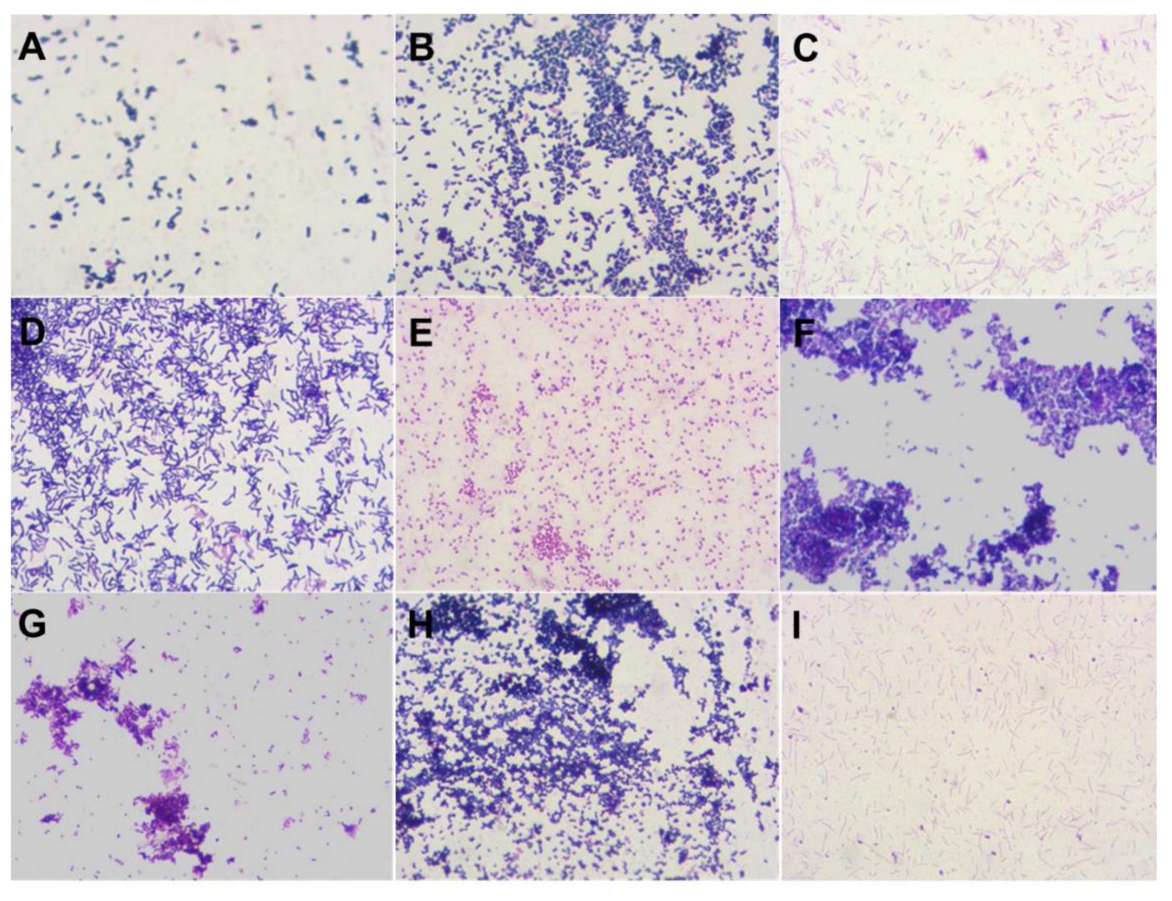
2. Results
3. Discussion
4. Materials and Methods
4.1. Ethics Approval
4.2. Patient Selection Criteria
4.3. Study Groups
4.4. Prostheses Fabrication
4.5. Laboratory Analysis
4.6. Statistical Analysis
5. Conclusions
Supplementary Materials
Author Contributions
Funding
Institutional Review Board Statement
Informed Consent Statement
Data Availability Statement
Conflicts of Interest
References
- Raedel, M.; Priess, H.W.; Bohm, S.; Walter, M.H. Performance of fixed dental prostheses up to 6 years-A massive data analysis. J. Prosthet. Dent. 2021, S0022-3913, 00031–00037. [Google Scholar]
- Sinjari, B.; Murmura, G.; Caputi, S.; Ricci, L.; Varvara, G.; Scarano, A. Use of Oral Chroma™ in the assessment of volatile sulfur compounds in patients with fixed prostheses. Int. J. Immunopathol. Pharmacol. 2013, 26, 691–697. [Google Scholar] [CrossRef]
- Rokaya, D.; Mahat, Y.; Sapkota, B.; Basnyat, S.K.C. Full Coverage Crowns and Resin-Bonded Bridge Combination for Missing Mandibular Anterior Teeth. Kathmandu Univ. Med. J. 2018, 16, 92–94. [Google Scholar]
- Poggio, C.E.; Ercoli, C.; Rispoli, L.; Maiorana, C.; Esposito, M. Metal-free materials for fixed prosthodontic restorations. Cochrane Database Syst. Rev. 2017, 12, CD009606. [Google Scholar] [CrossRef] [PubMed]
- Diaz, P.I.; Chalmers, N.I.; Rickard, A.H.; Kong, C.; Milburn, C.L.; Palmer, R.J., Jr.; Kolenbrander, P.E. Molecular characterization of subject-specific oral microflora during initial colonization of enamel. Appl. Environ. Microbiol. 2006, 72, 2837–2848. [Google Scholar] [CrossRef] [PubMed] [Green Version]
- Pihlstrom, B.L. Periodontal risk assessment, diagnosis and treatment planning. Periodontol. 2000 2001, 25, 37–58. [Google Scholar] [CrossRef]
- Peyyala, R.; Kirakodu, S.S.; Novak, K.F.; Ebersole, J.L. Oral microbial biofilm stimulation of epithelial cell responses. Cytokine 2012, 58, 65–72. [Google Scholar] [CrossRef] [Green Version]
- Heboyan, A.; Syed, A.; Rokaya, D.; Cooper, P.R.; Manrikyan, M.; Markaryan, M. Cytomorphometric Analysis of Inflammation Dynamics in the Periodontium Following the Use of Fixed Dental Prostheses. Molecules 2020, 25, 4650. [Google Scholar] [CrossRef] [PubMed]
- Willis, J.R.; Gabaldón, T. The Human Oral Microbiome in Health and Disease: From Sequences to Ecosystems. Microorganisms 2020, 8, 308. [Google Scholar] [CrossRef] [PubMed] [Green Version]
- Taubman, M.A.; Kawai, T.; Han, X. The new concept of periodontal disease pathogenesis requires new and novel therapeutic strategies. J. Clin. Periodontol. 2007, 34, 367–369. [Google Scholar] [CrossRef] [PubMed]
- Khurshid, Z.; Mali, M.; Naseem, M.; Najeeb, S.; Zafar, M.S. Human Gingival Crevicular Fluids (GCF) Proteomics: An Overview. Dent. J. 2017, 5, 12. [Google Scholar] [CrossRef]
- Bibi, T.; Khurshid, Z.; Rehman, A.; Imran, E.; Srivastava, K.C.; Shrivastava, D. Gingival Crevicular Fluid (GCF): A Diagnostic Tool for the Detection of Periodontal Health and Diseases. Molecules 2021, 26, 1208. [Google Scholar] [CrossRef] [PubMed]
- Khurshid, Z.; Warsi, I.; Moin, S.F.; Latif, M.; Zohaib, S.; Zafar, M.S. Chapter Six—Biochemical analysis of oral fluids for disease detection. Adv. Clin. Chem. 2021, 100, 205–253. [Google Scholar]
- Costantini, E.; Sinjari, B.; Piscopo, F.; Porreca, A.; Reale, M.; Caputi, S.; Murmura, G. Evaluation of Salivary Cytokines and Vitamin D Levels in Periodontopathic Patients. Int. J. Mol. Sci. 2020, 21, 2669. [Google Scholar] [CrossRef] [PubMed] [Green Version]
- Embery, G.; Waddington, R. Gingival crevicular fluid: Biomarkers of periodontal tissue activity. Adv. Dent. Res. 1994, 8, 329–336. [Google Scholar] [CrossRef]
- Ercoli, C.; Tarnow, D.; Poggio, C.E.; Tsigarida, A.; Ferrari, M.; Caton, J.G.; Chochlidakis, K. The Relationships Between Tooth-Supported Fixed Dental Prostheses and Restorations and the Periodontium. J. Prosthodont. 2021, 30, 305–317. [Google Scholar] [CrossRef]
- Pei, J.; Li, F.; Xie, Y.; Liu, J.; Yu, T.; Feng, X. Microbial and metabolomic analysis of gingival crevicular fluid in general chronic periodontitis patients: Lessons for a predictive, preventive, and personalized medical approach. EPMA J. 2020, 11, 197–215. [Google Scholar] [CrossRef] [Green Version]
- Könönen, E.; Gursoy, M.; Gursoy, U.K. Periodontitis: A Multifaceted Disease of Tooth-Supporting Tissues. J. Clin. Med. 2019, 8, 1135. [Google Scholar] [CrossRef] [Green Version]
- Socransky, S.S.; Haffajee, A.D.; Cugini, M.A.; Smith, C.; Kent, R.L., Jr. Microbial complexes in subgingival plaque. J. Clin. Periodontol. 1998, 25, 134–144. [Google Scholar] [CrossRef]
- Dobrzynski, M.; Pajaczkowska, M.; Nowicka, J.; Jaworski, A.; Kosior, P.; Szymonowicz, M.; Kuropka, P.; Rybak, Z.; Bogucki, Z.A.; Filipiak, J.; et al. Study of Surface Structure Changes for Selected Ceramics Used in the CAD/CAM System on the Degree of Microbial Colonization, In Vitro Tests. BioMed Res. Int. 2019, 2019, 9130806. [Google Scholar] [CrossRef] [PubMed] [Green Version]
- Hao, Y.; Huang, X.; Zhou, X.; Li, M.; Ren, B.; Peng, X.; Cheng, L. Influence of Dental Prosthesis and Restorative Materials Interface on Oral Biofilms. Int. J. Mol. Sci. 2018, 19, 3157. [Google Scholar] [CrossRef] [Green Version]
- Said, S.; Suda, W.; Nakagome, S.; Chinen, H.; Oshima, K.; Kim, S.; Kimura, R.; Iraha, A.; Ishida, H.; Fujita, J.; et al. Dysbiosis of salivary microbiota in inflammatory bowel disease and its association with oral immunological biomarkers. DNA Res. 2014, 21, 15–25. [Google Scholar] [CrossRef] [Green Version]
- Xie, H.; Cook, G.S.; Costerton, J.W.; Bruce, G.; Rose, T.M.; Lamont, R.J. Intergeneric communication in dental plaque biofilms. J. Bacteriol. 2000, 182, 7067–7069. [Google Scholar] [CrossRef] [Green Version]
- Periasamy, S.; Kolenbrander, P.E. Mutualistic biofilm communities develop with Porphyromonas gingivalis and initial, early, and late colonizers of enamel. J. Bacteriol. 2009, 191, 6804–6811. [Google Scholar] [CrossRef] [PubMed] [Green Version]
- Elmanfi, S.; Zhou, J.; Sintim, H.O.; Könönen, E.; Gürsoy, M.; Gürsoy, U.K. Regulation of gingival epithelial cytokine response by bacterial cyclic dinucleotides. J. Oral Microbiol. 2018, 11, 1538927. [Google Scholar] [CrossRef] [PubMed] [Green Version]
- Holt, S.C.; Kesavalu, L.; Walker, S.; Genco, C.A. Virulence factors of Porphyromonas gingivalis. Periodontol. 2000 1999, 20, 168–238. [Google Scholar] [CrossRef]
- Benakanakere, M.; Kinane, D.F. Innate cellular responses to the periodontal biofilm. Front. Oral Biol. 2012, 15, 41–55. [Google Scholar]
- Holt, S.C.; Ebersole, J.L. Porphyromonas gingivalis, Treponema denticola, and Tannerella forsythia: The ‘red complex’, a prototype polybacterial pathogenic consortium in periodontitis. Periodontol. 2000 2005, 38, 72–122. [Google Scholar] [CrossRef] [PubMed]
- Heboyan, A.; Manrikyan, M.; Markaryan, M.; Vardanyan, I. Changes in the parameters of gingival crevicular fluid in masticatory function restoration by various prosthodontic constructions. Int. J. Pharm. Res. 2020, 12, 2088–2093. [Google Scholar]
- Heboyan, A.G. Marginal and internal fit of fixed prosthodontic constructions: A literature review. Int. J. Dent. Res. Rev. 2019, 2, 19. [Google Scholar] [CrossRef]
- Shang, L.J.; Wu, Y.; Xu, Y.J. Effect of the CAD/CAM zirconia all-ceramic crown restoration on periodontal tissue. Chin. J. Tissue Eng. Res. 2014, 30, 4804–4809. [Google Scholar]
- Pabst, A.M.; Walter, C.; Grassmann, L.; Weyhrauch, M.; Brüllmann, D.D.; Ziebart, T.; Scheller, H.; Lehmann, K.M. Influence of CAD/CAM all-ceramic materials on cell viability, migration ability and adenylate kinase release of human gingival fibroblasts and oral keratinocytes. Clin. Oral. Investig. 2014, 18, 1111–1118. [Google Scholar] [CrossRef]
- Avetisyan, A.; Markaryan, M.; Rokaya, D.; Tovani-Palone, M.R.; Zafar, M.S.; Khurshid, Z.; Vardanyan, A.; Heboyan, A. Characteristics of Periodontal Tissues in Prosthetic Treatment with Fixed Dental Prostheses. Molecules 2021, 26, 1331. [Google Scholar] [CrossRef] [PubMed]
- Kazmi, S.M.; Iqbal, Z.; Muneer, M.U.; Riaz, S.; Zafar, M.S. Different pontic design for porcelain fused to metal fixed dental prosthesis: Contemporary guidelines and practice by general dental practitioners. Eur. J. Dent. 2018, 12, 375. [Google Scholar] [CrossRef] [PubMed] [Green Version]


| Subject Details | Details |
|---|---|
| Mean age of the subjects | 34 years old (18–50) years old |
| Total Subjects/Prostheses With healthy gums (MC, CC-MC, and CC-Zr) With gingivitis and/or periodontitis | 129 |
| 24 | |
| 105 | |
| 35 35 35 |
| Microorganism | Healthy (CFU/s) | MC (CFU/s) | CC/MC (CFU/s) | CC/Zr (CFU/s) | ||||
|---|---|---|---|---|---|---|---|---|
| Mean | SD | Mean | SD | Mean | SD | Mean | SD | |
| Enterococcus spp. | 6.297 × 10⁴ | 2.037 × 10⁵ | 1.601 × 10⁵ | 3.554 × 10⁵ | 1.057 × 10⁵ | 2.724 × 10⁵ | 2.323 × 10⁵ | 4.261 × 10⁵ |
| Peptostreptococcus spp. | 5.137 × 10⁵ | 2.031 × 10⁶ | 7.576 × 10⁵ | 2.365 × 10⁶ | 9.300 × 10⁵ | 2.742 × 10⁶ | 2.259 × 10⁵ | 3.759 × 10⁵ |
| Neisseria spp. § | 2.597 × 10⁴ | 4.374 × 10⁴ | 5.369 × 10⁴ | 1.725 × 10⁵ | 2.253 × 10⁵ | 3.974 × 10⁵ | 6.241 × 10⁵ | 2.121 × 10⁵ |
| Peptococcus spp. | 2.242 × 10⁵ | 4.077 × 10⁵ | 1.913 × 10⁵ | 3.812 × 10⁵ | 5.861 × 10⁵ | 1.994 × 10⁶ | 2.446 × 10⁵ | 4.205 × 10⁵ |
| Staphylococcus spp. ψ δ | 1.821 × 10⁴ | 3.749 × 10⁴ | 1.968 × 10⁴ | 3.791 × 10⁴ | 6.184 × 10⁴ | 1.994 × 10⁴ | 2.742 × 10⁵ | 4.548 × 10⁵ |
| Beta-hemolytic streptococcus | 1.045 × 10² | 2.787 × 10² | 1.247 × 10⁶ | 3.252 × 10⁶ | 1.621 × 10⁶ | 3.732 × 10⁶ | 1.263 × 10⁴ | 2.861 × 10⁴ |
| Candida albicans | 1.708 × 101 | 3.793 × 101 | 3.655 × 10³ | 1.718 × 10⁴ | 1.292 × 10² | 3.293 × 10³ | 1.595 × 10² | 3.443 × 10² |
| Alpha-haemolytic streptococcus | 1.184 × 10³ | 2.753 × 10³ | 1.368 × 10⁵ | 3.221 × 10⁵ | 1.644 × 10⁶ | 3.726 × 10⁶ | 6.023 × 10⁵ | 2.127 ×10⁶ |
| Lactobacillus spp. | 5.779 × 10⁵ | 2.033 × 10⁶ | 7.173 × 10⁵ | 2.371 × 10⁶ | 4.656 × 10⁵ | 1.996 × 10⁶ | 1.056 × 10⁵ | 2.915 × 10⁵ |
| Corynebacterium spp. § δ | 1.630 × 10⁴ | 3.264 × 10⁴ | 7.884 × 10⁴ | 2.367 × 10⁵ | 2.533 × 10⁵ | 4.294 × 10⁵ | 1.068 × 10⁴ | 2.911 ×10⁴ |
| Fusobacterium spp. | 5.100 × 10³ | 2.041 × 10⁴ | 6.363 × 10⁴ | 2.382 × 10⁵ | 4.164 × 10⁴ | 1.996 × 10⁵ | 5.104 × 10³ | 2.130 × 10⁴ |
| Porphyromonas gingivalis | 1.054 × 10³ | 2.784 × 10³ | 1.352 × 10⁴ | 2.768 × 10⁴ | 6.376 × 10³ | 1.988 × 10⁴ | 6.161 × 10⁵ | 2.123 × 10⁶ |
| Prevotella intermedia | 1.058 × 10³ | 2.783 × 10³ | 6.662 × 10³ | 1.706 × 10⁴ | 3.120 × 10³ | 4.395× 10³ | 5.626 × 10⁵ | 2.127 × 10⁶ |
| Veillonella spp. | 6.520 × 10⁶ | 2.031 × 10⁷ | 1.804 × 10⁸ | 3.482 × 10⁸ | . | . | 4.382 × 10⁷ | 4.798 × 10⁷ |
| Microorganisms | Healthy (CFU/s) | MC (CFU/s) | CC/-MC (CFU/s) | CC/Zr (CFU/s) | ||||
|---|---|---|---|---|---|---|---|---|
| Mean | SD | Mean | SD | Mean | SD | Mean | SD | |
| Enterococcus spp. | 4.262 × 10⁵ | 2.039 × 10³ | 1.358 × 10⁵ | 3.225 × 10⁵ | 2.224 × 10⁴ | 3.986 × 10⁴ | 1.795 × 10⁴ | 3.367 × 10⁴ |
| Peptostreptococcus spp. | 5.212 × 10⁵ | 2.028 × 10⁶ | 1.235 × 10⁶ | 3.248 × 10⁶ | 1.596 × 10⁴ | 3.197 × 10⁴ | 1.075 × 10⁵ | 2.908 × 10⁵ |
| Neisseria spp. | 5.502 × 10⁴ | 2.040 × 10⁵ | 2.413 × 10⁴ | 4.278 × 10⁴ | 9.488 × 10³ | 2.742 × 10⁴ | 1.519 × 10⁴ | 3.465 × 10⁴ |
| Peptococcus spp. | 1.371 × 10⁵ | 3.342 × 10⁵ | 1.870 × 10⁵ | 3.826 × 10⁵ | 2.384 × 10³ | 3.910× 10³ | 1.390 × 10⁵ | 3.502 × 10⁵ |
| Staphylococcus spp. | 4.363 × 10³ | 2.037 × 10⁴ | 1.965 × 10⁴ | 3.792 × 10⁴ | 2.144 × 10³ | 4.021 × 10³ | 1.510 × 10⁴ | 3.469 × 10⁴ |
| Beta-hemolytic streptococcus § η | 9.583 × 101 | 2.804 × 10² | 1.829 × 10⁶ | 3.846 × 10⁶ | 1.424 × 10⁴ | 3.255 × 10⁴ | 1.236 × 10³ | 2.873 × 10³ |
| Candida albicans | 2.083 × 101 | 4.148 × 101 | 6.218 × 10⁵ | 2.385 × 10⁶ | 4.452 × 10² | 2.001 × 10³ | 1.545 × 101 | 3.460 × 101 |
| Alpha-haemolytic streptococcus | 6.421 × 10² | 2.033 × 10³ | 1.335 × 10⁵ | 3.226 × 10⁵ | 4.741 × 10⁵ | 1.994 × 10⁶ | 2.378 × 10⁴ | 4.240 × 10⁴ |
| Lactobacillus spp. | 1.980 × 10⁴ | 3.688 × 10⁴ | 7.319 × 10⁴ | 2.373 × 10⁵ | 4.893 × 10⁴ | 2.001 × 10⁵ | 9.278 × 10⁴ | 2.936 × 10⁵ |
| Corynebacterium spp. | 5.146 × 10⁵ | 2.039 × 10⁶ | 1.272 × 10⁶ | 3.247 × 10⁶ | 8.181 × 10⁵ | 2.763 × 10⁶ | 6.145 × 10³ | 2.124 × 10⁴ |
| Fusobacterium spp. | 9.637 × 10³ | 2.803 × 10⁴ | 1.357 × 10⁴ | 3.225 × 10⁴ | 1.260 × 10³ | 3.300 × 10³ | 2.772 × 10² | 4.534 × 10² |
| Porphyromonas gingivalis | 5.083 × 101 | 2.041 × 10² | 3.425 × 10⁴ | 1.715 × 10⁵ | 2.628 × 10³ | 4.244 × 10³ | 1.524 × 10⁴ | 3.463 × 10⁴ |
| Prevotella intermedia | 2.208 × 101 | 4.096 × 101 | 6.460 × 10³ | 2.374 × 10⁴ | 1.561 × 10³ | 3.209 × 10³ | 1.120 × 10⁴ | 2.898 × 10⁴ |
| Veillonella spp. | 7.825 × 10⁵ | 2.014 × 10⁶ | 4.886 × 10⁷ | 1.722 × 10⁸ | 1.117 × 10⁷ | 2.707 × 10⁷ | 9.860 × 10⁷ | 2.925 × 10⁸ |
| Corynebacteriumanaerobium | 0.000 | 0.000 | 4.153 × 10⁴ | 1.724 × 10⁵ | 5.640 × 10⁴ | 2.001 × 10⁵ | 5.454 × 10³ | 2.132 × 10⁴ |
| Microorganism | Healthy (CFU/s) | MC (CFU/s) | CC/MC (CFU/s) | CC/Zr (CFU/s) | ||||
|---|---|---|---|---|---|---|---|---|
| Mean | SD | Mean | SD | Mean | SD | Mean | SD | |
| Enterococcus spp. | 1.291 ×10² | 3.368 × 10² | 1.651 × 10⁴ | 3.539 × 10⁴ | 1.496 × 10³ | 3.234 × 10³ | 8.422 × 10³ | 2.098 × 10⁴ |
| Peptostreptococcus spp. | 1.072 × 10⁵ | 2.777 × 10⁵ | 9.391 × 10⁵ | 2.861 × 10⁶ | 1.184 × 10³ | 2.692 × 10³ | 2.338 × 10⁵ | 4.257 × 10⁵ |
| Neisseria spp. | 4.725 × 10³ | 2.039 × 10⁴ | 1.037 × 10⁴ | 2.852 × 10⁴ | 9.208 × 10³ | 2.745 × 10⁴ | 2.746 × 10⁴ | 4.546 × 10⁴ |
| Peptococcus spp. δ | 2.133 × 10⁵ | 4.122 × 10⁵ | 4.385 × 10⁴ | 1.711 × 10⁵ | 2.064 × 10³ | 3.562 × 10³ | 2.778 × 10⁵ | 4.531 × 10⁵ |
| Staphylococcus spp. | 8.041 × 101 | 2.016 × 10² | 8.541 × 10³ | 2.356 × 10⁴ | 1.736 × 10² | 3.695 × 10² | 2.327 × 10³ | 4264.268 4.264 × 10³ |
| Beta-hemolytic streptococcus § η ß | 1.708 × 101 | 3.793 × 101 | 2.991 × 10⁴ | 4.595 × 10⁴ | 2.125 × 10³ | 4.031 × 10³ | 5.954 × 10² | 2.129 × 10³ |
| Candida albicans | 5.833 × 101 | 2.041 × 10² | 3.858 × 10³ | 1.722 × 10⁴ | 1.240 × 101 | 3.307 × 101 | 0.500 × 101 | 21.325 2.132 × 101 |
| Alpha-haemolytic streptococcus | 1.420 × 10² | 3.333 × 10² | 4.805 × 10³ | 4.978 × 10³ | 1.728 × 10³ | 3.698 × 10³ | 5.686 × 10³ | 2.126 × 10⁴ |
| Lactobacillus spp. | 6.105 × 10⁴ | 2.033 × 10⁵ | 5.426 × 10³ | 1.715 × 10⁴ | 1.296 × 10³ | 3.291 × 10³ | 9.604 × 10² | 2.933 × 10³ |
| Corynebacterium spp. | 4.692 × 10⁵ | 2.040 × 10⁶ | 4.947 × 10⁴ | 1.721 × 10⁵ | 3.012 × 10³ | 4.463 × 10³ | 1.518 × 10³ | 3.466 × 10³ |
| Fusobacterium spp. | 5.512 × 10³ | 2.040 × 10⁴ | 1.356 × 10⁴ | 3.226 × 10⁴ | 4.880 × 10² | 2.001 × 10³ | 0.454 × 101 | 2.132 × 101 |
| Porphyromonas gingivalis | 8.833 ×101 | 2.815 × 10² | 2.844 × 10² | 4.049 × 10² | 2.252 × 10² | 3.975 × 10² | 5.068 × 10² | 2.130 × 10³ |
| Prevotella intermedia | 1.125 × 101 | 2.771 × 101 | 1.847 × 10² | 3.464 × 10² | 1.904 × 10² | 3.631 × 10² | 6.227 × 101 | 2.121 × 10² |
| Veillonella spp. | 1.071 × 10⁶ | 2.778 × 10⁶ | 1.877 × 10⁷ | 3.826 × 10⁷ | 6.677 × 10⁶ | 1.988 × 10⁷ | 2.020 × 10⁷ | 3.868 × 10⁷ |
| Corynebacteriumanaerobium | 0.000 | 0.000 | 3.620 × 10² | 1.719 × 10³ | 1.248 × 10³ | 3.304 × 10³ | 4.153 × 10² | 1.923 × 10³ |
Publisher’s Note: MDPI stays neutral with regard to jurisdictional claims in published maps and institutional affiliations. |
© 2021 by the authors. Licensee MDPI, Basel, Switzerland. This article is an open access article distributed under the terms and conditions of the Creative Commons Attribution (CC BY) license (https://creativecommons.org/licenses/by/4.0/).
Share and Cite
Heboyan, A.; Manrikyan, M.; Zafar, M.S.; Rokaya, D.; Nushikyan, R.; Vardanyan, I.; Vardanyan, A.; Khurshid, Z. Bacteriological Evaluation of Gingival Crevicular Fluid in Teeth Restored Using Fixed Dental Prostheses: An In Vivo Study. Int. J. Mol. Sci. 2021, 22, 5463. https://doi.org/10.3390/ijms22115463
Heboyan A, Manrikyan M, Zafar MS, Rokaya D, Nushikyan R, Vardanyan I, Vardanyan A, Khurshid Z. Bacteriological Evaluation of Gingival Crevicular Fluid in Teeth Restored Using Fixed Dental Prostheses: An In Vivo Study. International Journal of Molecular Sciences. 2021; 22(11):5463. https://doi.org/10.3390/ijms22115463
Chicago/Turabian StyleHeboyan, Artak, Mikayel Manrikyan, Muhammad Sohail Zafar, Dinesh Rokaya, Ruzan Nushikyan, Izabella Vardanyan, Anna Vardanyan, and Zohaib Khurshid. 2021. "Bacteriological Evaluation of Gingival Crevicular Fluid in Teeth Restored Using Fixed Dental Prostheses: An In Vivo Study" International Journal of Molecular Sciences 22, no. 11: 5463. https://doi.org/10.3390/ijms22115463