Synergistic Anti-Candida Activity of Bengazole A in the Presence of Bengamide A †


1
Department of Chemistry, University of California, San Diego, 9500 Gilman Drive MC0358, La Jolla, CA 92093, USA
2
Skaggs School of Pharmacy and Pharmaceutical Sciences, University of California, San Diego, 9500 Gilman Drive MC0358, La Jolla, CA 92093, USA
*
Author to whom correspondence should be addressed.
†
This paper is dedicated to the memory of Professor Yuzuru Shimizu (1935–2019).
Mar. Drugs 2019, 17(2), 102; https://doi.org/10.3390/md17020102
Submission received: 21 December 2018
/
Revised: 21 January 2019
/
Accepted: 31 January 2019
/
Published: 7 February 2019
(This article belongs to the Special Issue Advances in Marine Alkaloids)
Abstract
:Bengazoles A–G from the marine sponge Jaspis sp. exhibit potent in vitro antifungal activity against Candida spp. and other pathogenic fungi. The mechanism of action (MOA) of bengazole A was explored in Candida albicans under both liquid culture and surface culture on Mueller-Hinton agar. Pronounced dose-dependent synergistic antifungal activity was observed with bengazole A in the presence of bengamide A, which is also a natural product from Jaspis sp. The MOA of bengazole A was further explored by monitoring the sterol composition of C. albicans in the presence of sub-lethal concentrations of bengazole A. The GCMS of solvent extracts prepared from liquid cultures of C. albicans in the presence of clotrimazole―a clinically approved azole antifungal drug that suppresses ergosterol biosynthesis by the inhibition of 14α-demethylase―showed reduced cellular ergosterol content and increased concentrations of lanosterol and 24-methylenedihydrolanosterol (a shunt metabolite of ergosterol biosynthesis). No change in relative sterol composition was observed when C. albicans was cultured with bengazole A. These results eliminate an azole-like MOA for the bengazoles, and suggest that another as-yet unidentified mechanism is operative.
1. Introduction
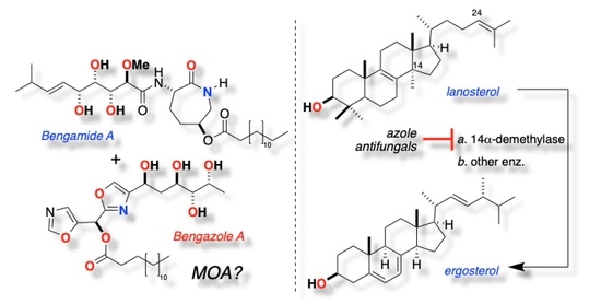
The tropical sponge, Jaspis cf. coriacea, is the original source of two classes of unrelated natural products: bengazoles and bengamides, (Figure 1) first reported by Crews et al. [1]. Bengamides A (1a) [2,3] and B (1b) and related analogs are potent nanomolar inhibitors of cancer cell growth, with selective in vitro activity in the NCI 60 cell line panel [4]. Compound 1a reduced MDA-MB-435 breast carcinoma in an in vivo rat zenograft model [4]. The unique cytotoxicity of bengamides has been attributed to the inhibition of methionine aminopeptidases [5,6]. A synthetic analog of 1a, LAF-389 [7], was advanced to phase I clinical trials before discontinuation of the study due to intolerable toxicity [8].
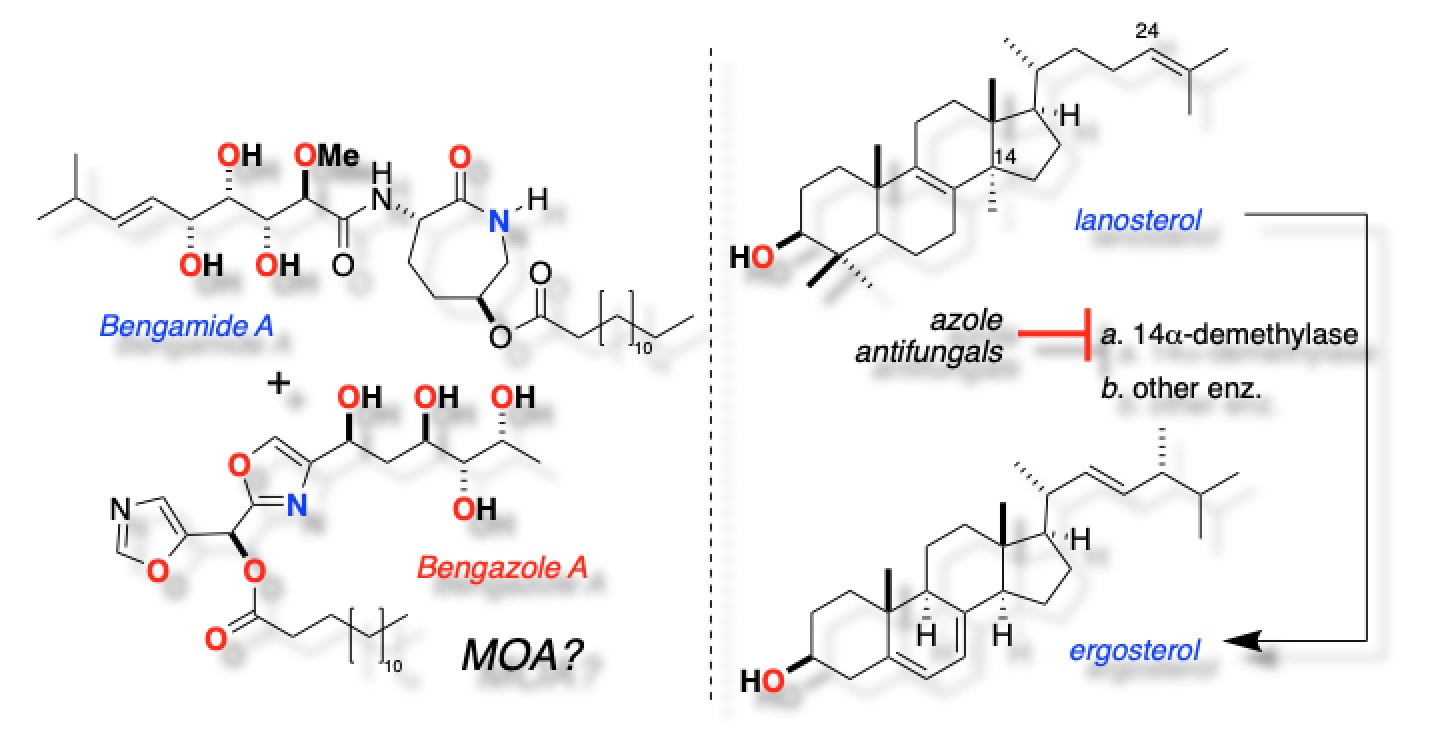
Candida species, including C. albicans, C. krusei, and C. glabrata, are responsible for 8–10% of hospital-acquired (nosocomial) systemic mycoses, and are associated with mortality rates of up to 71%. The rising rate of fatal candidemia is compounded by emergent fluconazole- and voriconazole-resistant non-Candida strains [9]. The bis-oxazole natural products, bengazoles A (2a) and B (2b), also from Jaspis cf. coriacea [10,11], and their homologs C–G (2c–g) from an Australian Jaspis sp. [12], are potent antifungal agents (minimum inhibitory concentration, MIC, ~ 1 µg mL–1 [12]).
The absolute stereochemistry of 1a was determined by measuring the NMR anisotropy of the S- and R-mandelate esters of a related co-isolated lactone [10]. The absolute stereochemistry of 2a was determined through comparisons of the CD spectra of the corresponding tetra p-bromobenzoate esters and synthetic models of defined configuration [12]. The first total syntheses of 1a [13] (which confirmed the absolute configuration) and 2a [14,15] were reported in 1992 and 1999, respectively, followed by several other bengamide syntheses; only two other syntheses of bengazoles have appeared [16,17]. The chemistry and the biology of bengamides and bengazoles were the subject of a recent comprehensive review [18].
A limited structure–activity study suggested that the two oxazole rings are required in 2a–g for potent antifungal activity [19], as is the long fatty acyl chain. The saponification product of bengazoles A–G gave the pentaol 2h [10] which was devoid of activity [12]), but few other substitutions are tolerated [19].
Bengamides―which co-occur with bengazoles in Jaspis sp.―lack antifungal activity against C. albicans yet remarkably, the anti-Candida activity of crude and partially purified Jaspis sp. extracts exceed the activity of purified bengazoles in the disk diffusion assay. For example, pure 2e (MIC = 1 µM against C. albicans [12]) at 0.5 µg disk−1 induced a zone of inhibition of 9–10 mm against C. albicans, but a solvent-partitioned fraction containing a mixture of homologous bengazoles and bengamides created a zone of inhibition far larger (40 mm) at comparable loadings (unpublished results, see Figure S4). This observation suggested synergism in the multi-component mixture that may involve bengazole and bengamide interactions, or more complex activities that impaired fungal cell metabolism, leading to cellular collapse.
The precise molecular mechanism of action (MOA) of the bengazoles has not been determined, but we have shown that the antifungal activity of 2a was suppressed in the presence of exogenous ergosterol (3) in a concentration-dependent manner [20], reminiscent of the MOA of polyene antifungal agents (e.g., amphotericin B [21,22]). The structure of 2a bears some resemblance to clinically approved drugs—the so-called ”azole” antifungal agents (e.g., fluconazole, 4, and clotrimazole, 5, Figure 2). The latter embody two imidazole or pyrazole rings appended to a central carbinol, while 2 displays two 1,3-oxazole rings arrayed around an esterified carbinol.
The structural similarities between antifungal azole drugs and 2 suggested that the two classes of compound might manifest the same MOA: namely, inhibition of lanosterol 14α-demethylase (We credit and thank Professor Yuzuru Shimizu (University of Rhode Island) for suggesting this hypothesis). The proposal is made more plausible by observations of inhibition of the growth of fungi by generic azoles as simple as imidazole itself [23], and the known mode of binding of azoles through coordination of the basic N to the Fe-heme core of 14α-demethylase [24]. Here, we report the results of the investigations of the MOA of bengazole natural products, including the antifungal synergism of bengazole A and bengamide A, and present the disproval of an azole-like suppression of ergosterol biosynthesis for the MOA of the former.
2. Results
Bengazoles are the rare, less-abundant, and less-studied components in extracts of Jaspis sp. Bengazoles are unstable: they are persistent in crude extracts, but upon purification, they undergo spontaneous degradation through autoxidation of the oxazole ring over short timescales. Crews et al. reported the co-isolation of compounds formed from the degradation of 2a; their structures were expected products of photosensitized [4+2] additions of 1O2 to one of the 1,3-oxazole rings, followed by Wasserman-type fragmentation [25,26]. Remarkably, our type-sample extract of Jaspis sp. (90-026) collected from the Great Barrier Reef and stored in MeOH (–20 °C) for 25 years was found to retain antifungal activity. Remarkably, bengazoles, within crude extracts and prior to separation from other components, have much better stability and prolonged ”shelf life”. This useful phenomenon may be possibly attributed to photoprotection by pigments or antioxidant congeneric components in unrefined mixtures. Pure bengazole A (2a, 0.5 µg) gave a zone of inhibition of 9–10 mm [12]. The latter observation suggested the presence of intact bengazoles. In contrast, the bulk of the specimen had been extracted and purified to provide the major compounds, bengazoles A (2a) and B (2b), and minor homologs, C–G (2c–g), all of which subsequently decomposed [12].
2.1. Extraction–Isolation of Bengamides–Bengazoles
A portion of the MeOH supernatant from the type sample was separated by progressive solvent partition, and the CH2Cl2-soluble fraction was further purified by silica gel flash chromatography to yield a fraction containing a mixture of bengazoles (2a–g). Final purification by reversed-phase HPLC gave pure 2, identified by MS and 1H NMR and comparison with literature values [10]. Purified 2a (4 µg), determined with precision by microcryoprobe 1H NMR and quantitation using solvent 13C satellites (QSCS) [27,28], provided sufficient sample for limited quantitative antifungal assays.
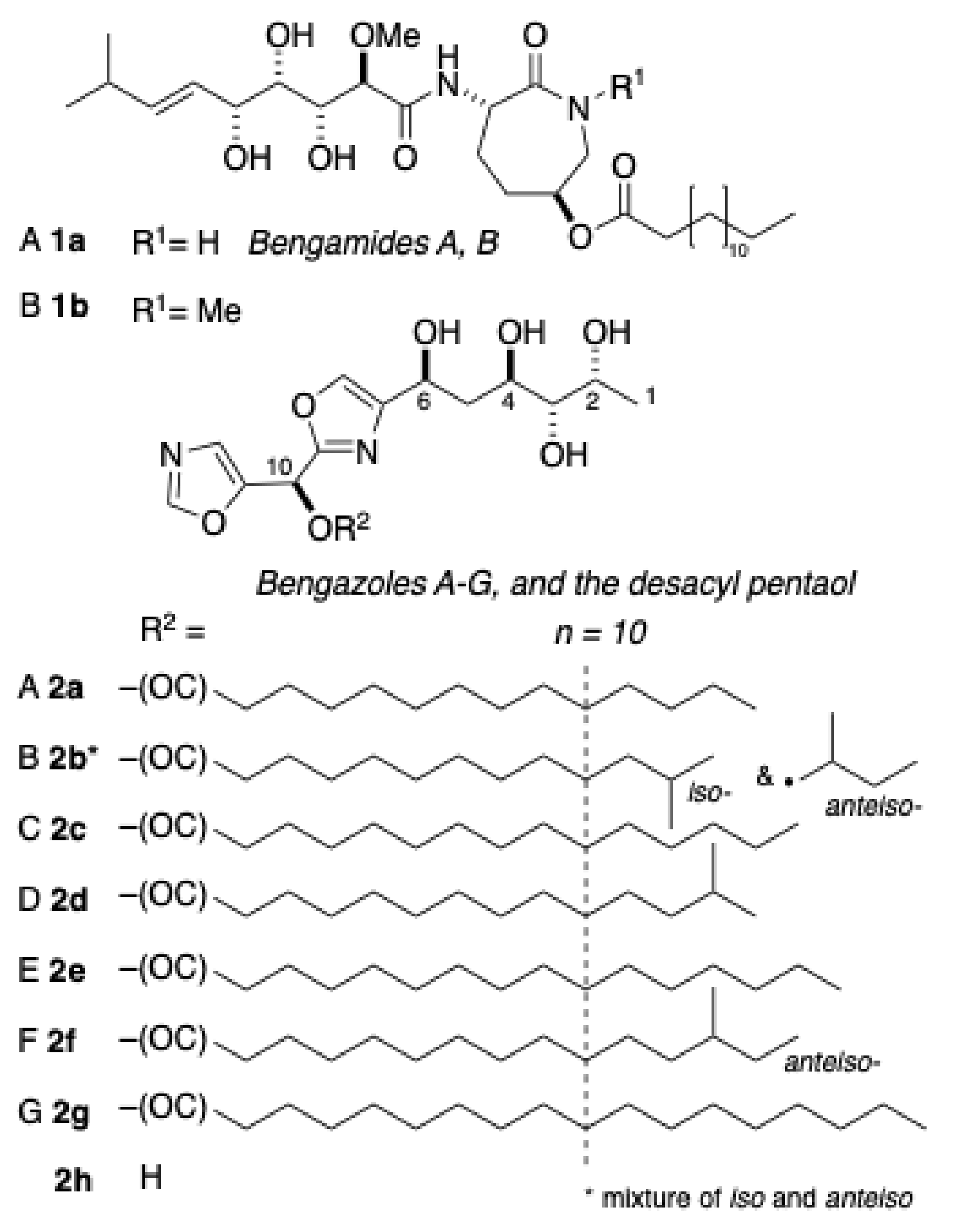
Ergosterol, the major sterol found in yeasts and other fungi, is a critical structural component that maintains the integrity of cellular membranes. Amphotericin B and related polyene antifungal agents exert their action by binding to ergosterol and inducing the formation of membrane pores that are permeable to K+ ions and other small-molecular-weight metabolites [29]. A common target exploited in the design of synthetic antifungal azoles is the inhibition of ergosterol biosynthesis [30], although recent efforts have been aimed at chitin-synthetase inhibitors [31]. Antifungal azoles inhibit the 14α-demethylase, an enzyme that is critical for oxidative remodeling of the common triterpene precursor lanosterol (6) (Figure 2) during ergosterol biosynthesis [32]. Although the structure of 2 is reminiscent of antifungal azoles, the ergosterol-dependent activity of the former suggests that the natural product targets ergosterol-lipid structured membranes by the formation of pores [21,29,30], although the possibility of a dual mode of action for 2 cannot be excluded. In order to test the latter hypothesis, the sterol composition of cultured C. albicans was monitored over time in the presence and absence of drugs that are known disruptors of ergosterol biosynthesis.
2.2. Sterol Composition in C. albicans Co-Cultured with Azoles
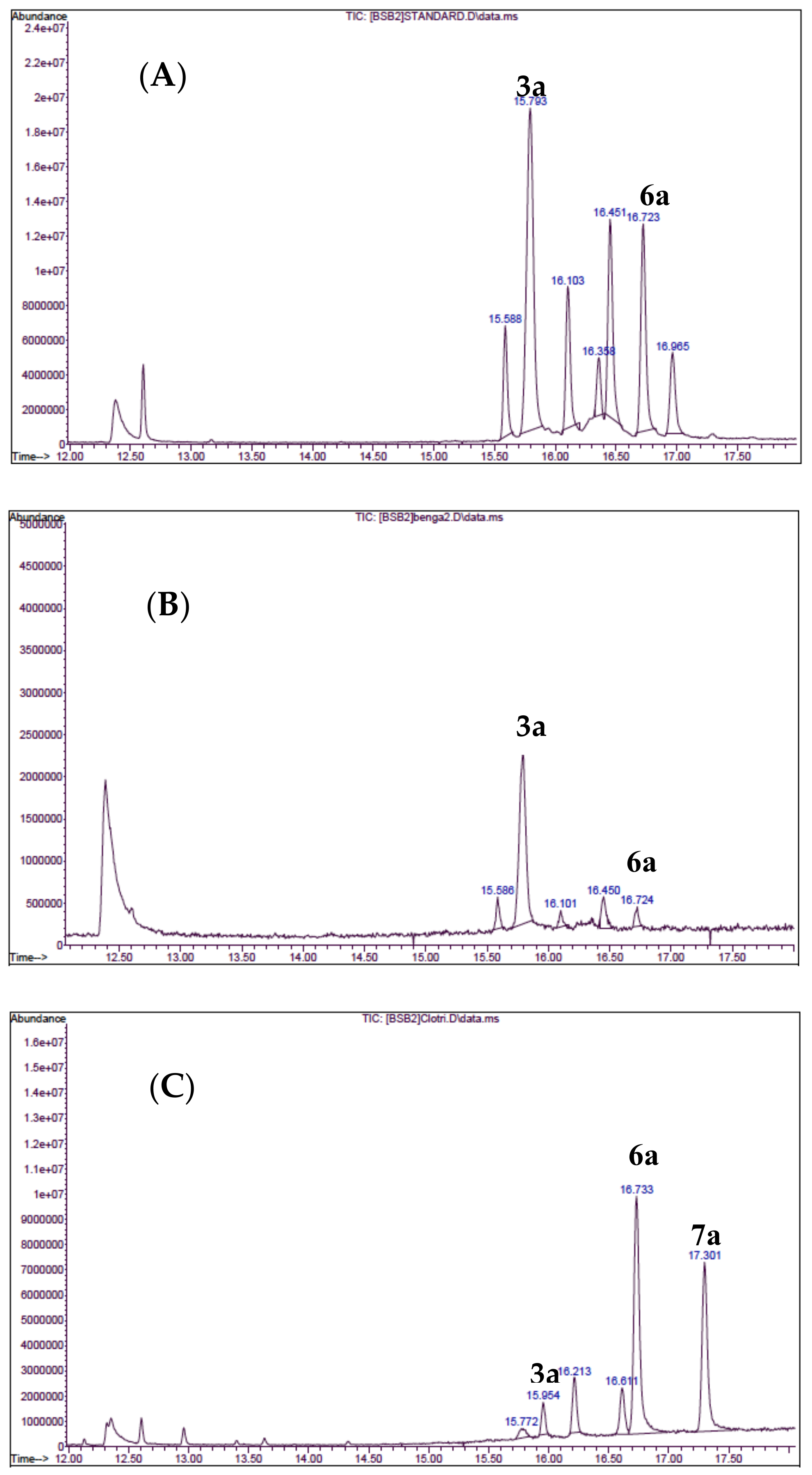
Cultures of C. albicans ATCC 14503 were treated with the serially diluted bengazoles, and incubated overnight at 35 °C. The crude broths were centrifuged and growth inhibition was estimated on the basis of the cellular wet-weight of the pellets compared to the untreated control. Whole pellets were saponified (40% KOH in EtOH-H2O, 2 h, 95 °C), and the non-saponifiable fractions were recovered by extraction with ether, persilylated (N-(trimethylsilyl)imidazole), and the sterol composition was determined by GCMS (Figure 3) of the corresponding O-TMS ethers of ergosterol, lanosterol, and 24-methylenedihydrolanosterol (3a, 6a, and 7a, respectively, see Supporting Information). Growth inhibition was observed in the presence of 2, but there was no change in the concentrations of 3 relative to the control (Figure 3b). To validate the method, azole 5 was tested under the same conditions. A clear decrease in 3 (Figure 3c) was seen along with the appearance of a new peak due to the O-TMS ether 7a of the expected shunt metabolite, 24-methylenedihydrolanosterol (eburicol, 7) [33,34].
2.3. Synergistic Antifungal Activity of Bengazole–Bengamide Mixtures
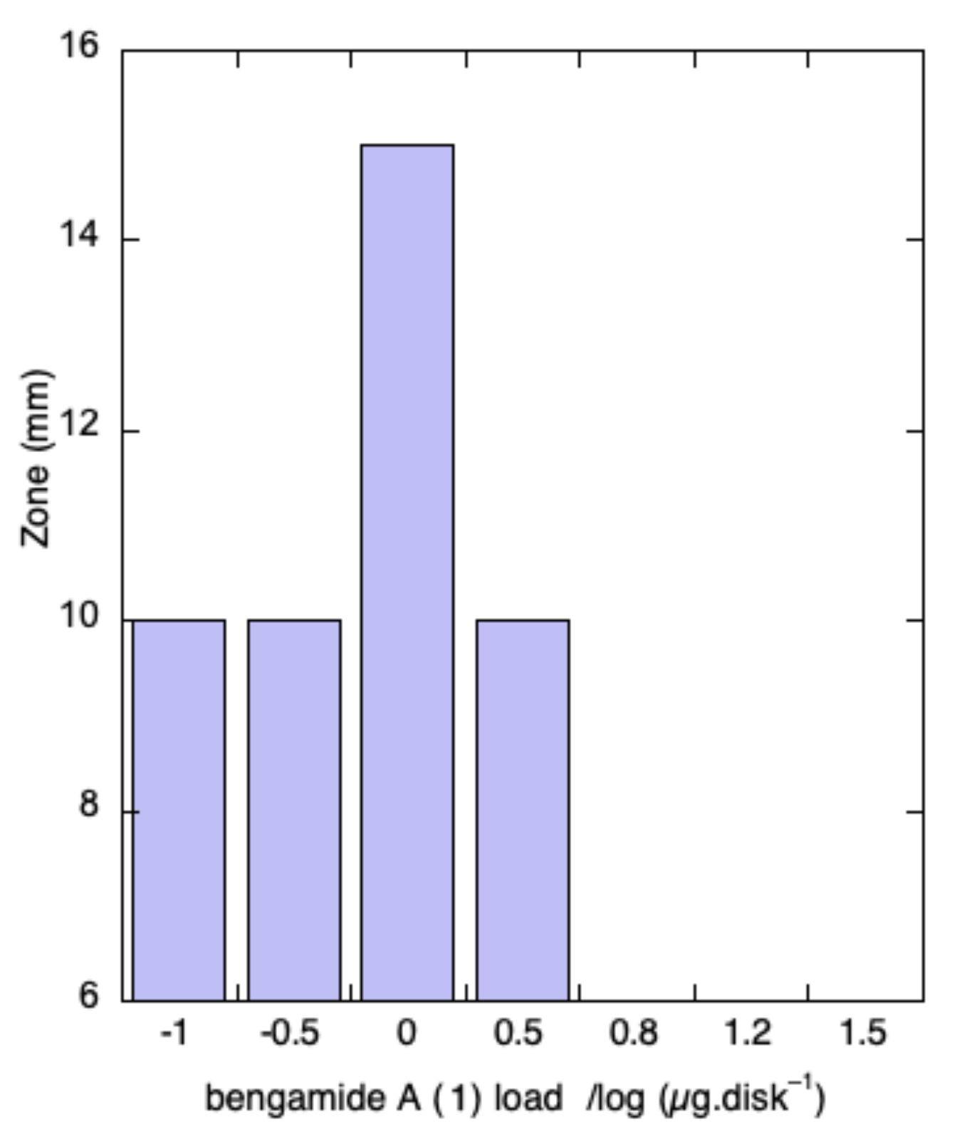
In order to investigate possible antifungal synergism, mixtures of bengazoles A–G (2a–g; hereafter referred to as 2 for brevity), at a constant loading (0.5 µg disk−1), and variable amounts of bengamide A (1a), were combined and tested in a disk diffusion assay (Figure 4). Surprisingly, at 200 µg disk−1 of 1a (400:1 mass ratio of 1a:2), complete inhibition of the antifungal activity of 2 was observed. When the relative concentration of 1a was lowered, an unusual dose-dependent response was observed (Figure 4); 1a inhibited antifungal activity of 2 at 15 µg disk−1 (30:1 ratio), but increased the zone of inhibition (~50%) at 1 µg of 2 (2:1 ratio of 1a to 2).
Further reductions in the ratio of 1a to 2 showed no changes compared to the control. In contrast, the microbroth dilution assay using checkerboard [35] permutations of concentrations of 1a and 2 did not reproduce the synergism observed with disk diffusion assays. As noted earlier, bengazoles are unstable. The time-dependent decomposition of 2 under the more prolonged incubation in the microbroth dilution assay or disk diffusion-related phenomena may account for the different responses under the two assay regimes.
3. Discussion
Analysis of saponified cultures of C. albicans treated with clotrimazole (5) showed the appearance of eburicol (7)—a shunt metabolite sterol biosynthesis as expected—whereas cultures treated with bengazoles (2) did not. This implies that bengazole does not inhibit ergosterol biosynthesis in the same manner as 3 and other azole antifungal drugs which inhibit 14α-demethylase. In the latter case, the relatively higher ratio of lanosterol (6) to ergosterol (3) may suggest a buildup of the former—implying there is an inhibition of biosynthesis at an earlier stage―but absence of an internal standard limited conclusions on a more quantitative basis. We cannot exclude the inhibition of the biosynthesis of 3 by 2 at other steps in sterol biosynthesis (e.g., inhibition of squalene epoxidase [36,37]), however, testing this hypothesis will require an expanded, more quantitative experimental design.
Synergistic activity in antibiotics is not uncommon [38,39], but is less-frequently reported for antifungal drugs [40]. We, and others, have noted synergism in antifungal microbroth dilution assays (MICs) of antifungal cyclic peptides, e.g. lobocyclamides from the cyanobacterium Lyngbya majuscula [41]. Moore and coworkers noted similar behavior in disk diffusion assays with the related peptides, laxaphycins A and B from the cyanobacterium Hormothamnion enteromorphoides (see Refs. [42,43] (interestingly, synergism was also observed for combinations of new laxaphycins in antiproliferative assays of cultured HCT-116 colon cancer cells [44]). Whether the synergistic fungal inhibition observed under microbroth dilution assays can be translated into improved efficacy in animal models of disseminated mycoses is a matter worthy of further study.
It is worth commenting that many studies of naturally occurring marine products with antifungal properties [45], although reports of significant in vitro activity against cultured fungi, have lacked experimental proof that might illuminate details of MOA. These shortfalls may have side-lined potentially new mechanistic insights; yet speculative hypotheses abound. For example, the well-known antifungal activity of long-chain ”two-headed” aminoalkanols (e.g., oceanapiside [46]) found in sponges of the genera Oceanapia, Rhizochalina, Leucetta, and Calyx [47] were thought to exert antifungal activity by mimicking intermediates in the biosynthesis of sphingolipids and related long-chain bases [48], but evidence has mounted to support alternative models of the inhibition of fungal cell growth through actin-binding and the disruption of microfilaments [49]. Clearly, early identification of MOA is an asset in identifying viable leads from ”hits” in antifungal drug discovery campaigns.
Studies of antibiosis against bacteria that follow canonical MOAs are familiar (e.g., penicillins: inhibition of cell-wall biosynthesis; erythromycin: disruption of protein translation at rRNA [21]). In contrast, the characterization of antifungal MOAs has presented inordinately encumbered challenges, complicated by diploid pathogenic organisms that exhibit fewer genetic and metabolic differences from the host organism compared to bacteria. In the future, well-designed in vitro experiments that link read-outs of antifungal phenotypes with specific MOAs will be more desirable in screening-based discovery programs for natural products.
4. Materials and Methods
4.1. General Experimental Procedures
Inverse detected 2D NMR spectra were measured on a ECA (500 MHz) NMR spectrometer (Jeol, Peabody, MA, USA), equipped with a 5 mm 1H-{13C} probe, or an Avance III (600 MHz) NMR spectrometer (Bruker, Billerica, MA, USA), fitted with a 1.7 mm 1H-{13C} microcryoprobe. High-resolution ESITOF analyses were carried out on an Agilent 1200 HPLC coupled to an Agilent 6230 TOFMS (Agilent, Santa Clara, CA, USA), calibrated immediately before measurement against an ESL-L low concentration tuning mix (part number G1969-85000, Agilent Technologies). Low-resolution MS measurements were made on a Thermoelectron Surveyor UHPLC (Thermo Fisher, Waltham, MA, USA) coupled to an MSD single-quadrupole detector. HPLC was performed on an Agilent 1200 HPLC. Other General Experimental details can be found elsewhere [50].
4.2. Extraction and Purification of Bengazole A
An aliquot (5 mL) of the supernatant from Jaspis sp. (type sample 90-20-026), stored in MeOH (10 mL), was extracted with hexanes (5 mL × 2). Concentration of the hexane-soluble layer gave fraction A. The aqueous-MeOH layer was adjusted to 2:3 H2O:MeOH and extracted with CH2Cl2 (7 mL × 2) to yield, after removal of volatiles, fraction B (3.3 mg). Fraction B was separated by silica gel flash chromatography (stepped gradient, 2.5% MeOH increments in CH2Cl2 to 10:90 MeOH:CH2Cl2) followed by a 50:50 MeOH:CH2Cl2 wash, to yield five fractions (monitored by 1H NMR, 500 MHz). Fractions 4 and 5 were combined and purified by reversed-phase HPLC (Phenomenex Luna C18 column, 250 × 4.6 mm, linear gradient; initial conditions 25:75 H2O:CH3CN to 100% CH3CN over 17 min, 1 mL min–1 flow rate, UV 217 nm, and ELSD detection) to yield compound 2a (4 µg, tR = 11.8 min). The amount of 2 was quantitated by the solvent 13C-satellites (QSCS) method [28] using a 1H NMR microcryoprobe (600 MHz, CDCl3).
4.3. Quantification of Ergosterol from C. albicans ATCC 14503
A two-fold dilution series was prepared from Sabouraud-dextrose broth (SabDex, 2 mL) in each of 8 × 25 cm2 flasks. Overnight liquid cultures (10 µL) of C. albicans ATCC 14503 were added and incubated at 35 °C overnight with gentle shaking. The culture broths were centrifuged, washed (2 × PBS), and the wet pellet was weighed to estimated growth inhibition. The control pellet and one with 50% growth inhibition were saponified in 40% KOH solution (8:1 EtOH:water, 500 µL) at 95 °C for 2 hr. The non-saponifiable material was diluted with deionized H2O (2 mL) and vigorously extracted with diethyl ether (2×, vortex 30 s, followed by centrifugation, 5 min). The combined organic layers were dried under a stream of N2 and the residue dissolved in n-hexane (500 µL), then treated with N-(trimethylsilyl)imidazole (10 µL, Pierce). Samples were analyzed by GCMS (1 µL injection, 1.2 mL min–1 He, 70 °C for 2 min, ramp to 270 °C over 24 min, ramp to 300 °C at 35 min, EI detection). Sterols were identified (see Figure 3 and Supporting Information, S1–S3) by comparisons of peak m/z values, including fragmentation patterns, against entries from the NIST MS data library. Extracts of C. albicans liquid cultures, grown in the presence of growth inhibitory concentrations (50% reduction of cellular mass) of bengazole A or clotrimazole, were also analyzed by GCMS and compared with control cultures (see Figure 3).
4.4. Antifungal Disk Diffusion Assay
An overnight liquid culture of C. albicans ATCC 14503 was diluted 100-fold and spread on Mueller-Hinton agar plates. Aliquots of each compound were applied as solutions in MeOH onto sterile paper disks (6.0 mm). After five minutes, the disks were placed on the yeast-coated agar plates, and incubated overnight (~16 h) at 35 °C. Antifungal activity was estimated by the measurement of zones of inhibition (>6.0 mm ± 0.5 mm, Figure 4).
5. Conclusions
We reported the potential synergistic activity of bengamide A (1) and bengazoles. Remarkably, a 25-year old sample of Jaspis sp. collected from the Great Barrier Reef, Australia, retained potent antifungal activity, which guided the separation of 1 from 2 using an agar-based disk diffusion assay. Re-assay of the pure compounds confirmed that 1 was devoid of antifungal activity against Candida albicans, but mixtures of 1 and 2 displayed synergistic activity with optimum enhancement of the zone of inhibition at a ratio of 2:1. Although inhibition of fungal growth was observed in cultures of C. albicans in the presence of 2, no suppression of ergosterol biosynthesis was detected, suggesting a MOA that differs from 14α-demethylase inhibition, common to the azole antifungal drugs. Further investigations into the MOA of potent antifungal bengazoles are in progress.
Supplementary Materials
The following are available online at https://www.mdpi.com/1660-3397/17/2/102/s1. LCMS data of sterol O-TMS ethers and first record of ”synergism”.
Author Contributions
TFM conceived of the project and experimental design. MTJ designed the assays and, together with XW and TC, carried out the experiments. TFM and MTJ wrote the manuscript.
Funding
The 500 MHz NMR spectrometer and the HPLC TOFMS were purchased with funding from the NSF (Chemical Research Instrument Fund, CHE0741968) and the NIH Shared Instrument Grant (S10RR025636) programs, respectively. This research was funded by grants from the NIH (AI107768, AT009783-01).
Acknowledgments
We thank Y. Su (UCSD) for MS data and B. Choudhury (UCSD Glycotechnologies Core) for GCMS data, and P. Crews (University of California, Santa Cruz) for insights on the stability of bengazoles. We are grateful, for suggestions and discussions on antifungal mechanisms, to Yuzuru Shimizu, professor emeritus, University of Rhode Island: accomplished scholar, principled scientist and friend.
Conflicts of Interest
The authors declare no conflict of interest.
References
- Quiñoá, E.; Adamczeski, M.; Crews, P.J. Bengamides, Heterocyclic Anthelmintics from a Jaspidae Marine Sponge. J. Org. Chem. 1986, 51, 4494–4497. [Google Scholar] [CrossRef]
- Adamczeski, M.; Quiñoá, E.; Crews, P.J. Novel Sponge-derived Amino Acids. 11. The Entire Absolute Stereochemistry of the Bengamides. J. Org. Chem. 1990, 55, 240–242. [Google Scholar] [CrossRef]
- Thale, Z.; Kinder, F.R.; Bair, K.W.; Bontempo, J.; Czuchta, A.M.; Versace, R.W.; Phillips, P.E.; Sanders, M.L.; Wattanasin, S.; Crews, P. Bengamides Revisited: New Structures and Antitumor Studies. J. Org. Chem. 2001, 66, 1733–1741. [Google Scholar] [CrossRef] [PubMed]
- Kinder, F.R., Jr.; Versace, R.W.; Bair, K.W.; Bontempo, J.M.; Cesarz, D.; Chen, S.; Crews, P.; Czuchta, A.M.; Jagoe, C.T.; Yin, M.; et al. Synthesis and Antitumor Activity of Ester-Modified Analogues of Bengamide B. J. Med. Chem. 2001, 44, 3692–3699. [Google Scholar] [CrossRef] [PubMed]
- Towbin, H.; Bair, K.W.; DeCaprio, J.A.; Eck, M.J.; Kim, S.; Kinder, F.R.; Morollo, A.; Mueller, D.R.; Schindler, P.; Song, H.K.; et al. Proteomics-based Target Identification: Bengamides as a New Class of Methionine Aminopeptidase Inhibitors. J. Biol. Chem. 2003, 278, 52964–52971. [Google Scholar] [CrossRef]
- Hu, X.; Dang, Y.; Tenney, K.; Crews, P.; Tsai, C.W.; Sixt, K.M.; Cole, P.A.; Liu, J.O. Regulation of c-Src Nonreceptor Tyrosine Kinase Activity by Bengamide A through Inhibition of Methionine Aminopeptidases. Chem. Biol. 2007, 14, 764–774. [Google Scholar] [CrossRef] [Green Version]
- Xu, D.D.; Waykole, L.; Calienni, J.V.; Ciszewski, L.; Lee, G.T.; Liu, W.; Szewczyk, J.; Vargas, K.; Prasad, K.; Repič, O.; et al. An Expedient Synthesis of LAF389, a Bengamide B Analogue. Org. Proc. Res. Dev. 2003, 7, 856–865. [Google Scholar] [CrossRef]
- Dumez, H.; Gall, H.; Capdeville, R.; Dutreix, C.; van Oosterom, A.T.; Giaccone, G. A Phase I and Pharmacokinetic Study of LAF389 Administered to Patients with Advanced Cancer. Anticancer Drugs 2007, 18, 219–225. [Google Scholar] [CrossRef]
- Miceli, M.H.; Díaz, J.A.; Lee, S.A. Emerging Opportunistic Yeast Infections. Lancet Infect. Dis. 2011, 11, 142–151. [Google Scholar] [CrossRef]
- Adamczeski, M.; Quiñoa, E.; Crews, P. Unusual Anthelminthic Oxazoles from a Marine Sponge. J. Am. Chem. Soc. 1988, 110, 1598–1602. [Google Scholar] [CrossRef]
- Rodríguez, J.; Nieto, R.M.; Crews, P. New Structures and Bioactivity Patterns of Bengazole Alkaloids from a Choristid Marine Sponge. J. Nat. Prod. 1993, 56, 2034–2040. [Google Scholar] [CrossRef] [PubMed]
- Searle, P.A.; Richter, R.K.; Molinski, T.F. Bengazoles C−G from the Sponge Jaspis sp. Synthesis of the Side Chain and Determination of Absolute Configuration. J. Org. Chem. 1996, 61, 4073. [Google Scholar] [CrossRef] [PubMed]
- Chida, N.; Tobe, T.; Okada, S.; Ogawa, S. Total Synthesis and Absolute Configuration of Bengamide A. J.C.S. Chem. Comm. 1992, 1064–1066. [Google Scholar] [CrossRef]
- Mulder, R.J.; Shafer, C.M.; Molinski, T.F. First Total Synthesis of Bengazole A. J. Org. Chem. 1999, 64, 4995–4998. [Google Scholar] [CrossRef] [PubMed]
- Shafer, C.M.; Molinski, T.F. Synthesis of the C1-C9 Core of Bengazole A. Harnessing the Ambident Nucleophilicity of 2-Lithiooxazole. Tetrahedron Lett. 1998, 39, 2903–2906. [Google Scholar] [CrossRef]
- Bull, J.A.; Balskus, E.P.; Horan, R.A.; Langner, M.; Ley, S.V. Stereocontrolled Total Synthesis of Bengazole A: A Marine Bisoxazole Natural Product Displaying Potent Antifungal Properties. Angew. Chem. 2006, 13, 6714–6718. [Google Scholar] [CrossRef]
- Chandrasekhar, S.; Sudhakar, A. Total Synthesis of Bengazole A. Org. Lett. 2010, 12, 236–238. [Google Scholar] [CrossRef]
- García-Ruiz, C.; Sarabia, F. Chemistry and Biology of Bengamides and Bengazoles, Bioactive Natural Products from Jaspis Sponges. Mar. Drugs 2014, 12, 1580–1622. [Google Scholar] [CrossRef] [PubMed]
- Mulder, R.J.; Shafer, C.M.; Dalisay, D.S.; Molinski, T.F. Synthesis and Structure–activity Relationships of Bengazole A Analogs. Bioorg. Med. Chem. Lett. 2009, 19, 2928–2930. [Google Scholar] [CrossRef] [PubMed]
- Antonio, J.; Molinski, T.F. Screening of Marine Invertebrates for the Presence of Ergosterol-Sensitive Antifungal Compounds. J. Nat. Prod. 1993, 56, 54–61. [Google Scholar] [CrossRef] [PubMed]
- Goodman Gilman, A.; Rall, T.W.; Nies, A.S.; Taylor, P. Goodman and Gilman’s the Pharmacological Basis of Therapeutics, 8th ed.; Pergamon: New York, NY, USA, 1990. [Google Scholar]
- Molinski, T.F. Developments in Marine Natural Products. Receptor-Specific Bioactive Compounds. J. Nat. Prod. 1993, 56, 1–8. [Google Scholar] [CrossRef] [PubMed]
- Maertens, J.A. History of the Development of Azole Derivatives. Clin. Microb. Infect. 2004, 10, 1–10. [Google Scholar] [CrossRef]
- Sagatova, A.A.; Keniya, M.V.; Wilson, R.K.; Monk, B.C.; Tyndall, J.D.A. Structural Insights into Binding of the Antifungal Drug Fluconazole to Saccharomyces cerevisiae Lanosterol 14α-Demethylase. Antimicrob. Agents Chemother. 2015, 59, 4982–4989. [Google Scholar] [CrossRef] [PubMed]
- Wasserman, H.H.; Vinick, F.J.; Chang, Y.C. Reaction of Oxazoles with Singlet Oxygen. Mechanism of the Rearrangement of Triamides. J. Am. Chem. Soc. 1972, 94, 7180–7182. [Google Scholar] [CrossRef]
- Wasserman, H.H.; Druckrey, E. The Reaction of Oxazoles with Singlet Oxygen. II. A Novel Method for the Preparation of ω-Cyano acids. J. Am. Chem. Soc. 1968, 90, 2440–2441. [Google Scholar]
- Molinski, T.F. Nanomole-scale Natural Products Discovery. Curr. Opin. Drug Discov. Dev. 2009, 12, 197–206. [Google Scholar]
- Dalisay, D.S.; Molinski, T.F. NMR Quantitation of Natural Products at the Nanomole Scale. J. Nat. Prod. 2009, 72, 739–744. [Google Scholar] [CrossRef] [Green Version]
- Bolard, J. How Do the Polyene Macrolide Antibiotics Affect the Cellular Membrane Properties? Biochim. Biophys. Acta 1986, 864, 257–304. [Google Scholar] [CrossRef]
- Kathiravan, M.K.; Salake, A.B.; Chothe, A.S.; Dudhe, P.B.; Watode, R.P.; Mukta, M.S.; Gadhwe, S. The Biology and Chemistry of Antifungal Agents: A Review. Bioorg. Med. Chem. 2012, 20, 5678–5698. [Google Scholar] [CrossRef]
- Chaudhary, P.M.; Tupe, S.G.; Deshpande, M.V. Chitin Synthase Inhibitors as Antifungal Agents. Mini Rev. Med. Chem. 2013, 13, 222–236. [Google Scholar]
- Shalini, K.; Kumar, N.; Drabu, S.; Sharma, P.K. Advances in Synthetic Approach to and Antifungal Activity of Triazoles. Beilstein J. Org. Chem. 2011, 7, 668–677. [Google Scholar] [CrossRef] [PubMed]
- Barton, D.H.R.; Harrison, D.M.; Moss, G.P. 24-Methylenedihydrolanosterol as a Precursor of Steroids and Triterpenoids. Chem. Commun. 1966, 17, 595–596. [Google Scholar] [CrossRef]
- Lahey, F.N.; Strasser, P.H.A. Erburicoic Acid. J. Chem. Soc. 1951, 0, 873–877. [Google Scholar] [CrossRef]
- Orhan, G.; Bayram, A.; Zer, Y.; Balci, I. Synergy Tests by E Test and Checkerboard Methods of Antimicrobial Combinations against Brucella melitensis. J. Clin. Microbiol. 2005, 43, 140–143. [Google Scholar] [CrossRef] [PubMed]
- Rabelo, V.W.-H.; Romeiro, N.C.; Abreu, P.A. Design Strategies of Oxidosqualene Cyclase Inhibitors: Targeting the Sterol Biosynthetic Pathway. J. Steroid Biochem. Mol. Biol. 2017, 171, 305–317. [Google Scholar] [CrossRef] [PubMed]
- Jamison, M.T.; Macho, J.; Molinski, T.F. Structure–activity of Antifungal Compounds Inspired by Aminobisabolenes from the Sponge Halichondria sp. Bioorg. Med. Chem. Lett. 2016, 26, 5244–5246. [Google Scholar] [CrossRef] [PubMed]
- Acar, J. F Antibiotic Synergism and Antagonism. Med. Clin. N. Am. 2000, 84, 1391–1406. [Google Scholar] [CrossRef]
- Wind, C.M.; de Vries, H.J.C.; van Dam, A.P. Determination of In Vitro Synergy for Dual Antimicrobial Therapy Against Resistant Neisseria gonorrhoeae Using Etest and agar dilution. Int. J. Antimicrob. Agents 2015, 45, 305–308. [Google Scholar] [CrossRef]
- Liu, X.; Li, T.; Wang, D.; Yang, Y.; Sun, W.; Liu, J.; Sun, S. Synergistic Antifungal Effect of Fluconazole Combined with Licofelone against Resistant Candida albicans. Front. Microbiol. 2017, 8, 1–13. [Google Scholar] [CrossRef]
- MacMillan, J.B.; Ernst-Russell, M.A.; de Ropp, J.S.; Molinski, T.F. Lobocyclamides A-C, Lipopeptides from a Cryptic Cyanobacterial Mat Containing Lyngbya confervoides. J. Org. Chem. 2002, 67, 8210–8215. [Google Scholar] [CrossRef]
- Frankmölle, W.P.; Knübel, G.; Moore, R.E.; Patterson, G.M.L. Antifungal Cyclic Peptides from the Terrestrial Blue-Green Alga Anabaena laxa. J. Antibiot. 1992, 45, 1458–1466. [Google Scholar] [CrossRef] [PubMed]
- Frankmölle, W.P.; Larsen, L.K.; Caplan, F.R.; Patterson, G.M.L.; Knübel, G.; Levine, I.A.; Moore, R.E. Antifungal Cyclic Peptides from the Terrestrial Blue-Green Alga Anabaena laxa. J. Antibiot. 1992, 45, 1451–1457. [Google Scholar] [CrossRef]
- Cai, W.; Matthew, S.; Chen, Q.-Y.; Paul, V.J.; Luesch, H.P. Discovery of new A- and B-type Laxaphycins with Synergistic Anticancer Activity. Bioorg. Med. Chem. 2018, 26, 2310–2319. [Google Scholar] [CrossRef] [PubMed]
- Molinski, T.F. Antifungal Compounds from Marine Organisms. Curr. Med. Chem. Anti-Infect. Agents 2004, 3, 197–220. [Google Scholar] [CrossRef]
- Nicholas, G.M.; Molinski, T.F. Enantiodivergent Biosynthesis of the Dimeric Sphingolipid Oceanapiside from the Marine Sponge Oceanapia phillipensis. Determination of Remote Stereochemistry. J. Am. Chem. Soc 2000, 122, 4011–4019. [Google Scholar] [CrossRef]
- Pruett, S.T.; Bushnev, A.; Hagedorn, K.; Adiga, M.; Haynes, C.A.; Sullards, M.C.; Liotta, D.C.; Merrill, A.H. Thematic Review Series: Sphingolipids. Biodiversity of Sphingoid Bases (“Sphingosines”) and Related Amino Alcohols. J. Lipid Res. 2008, 49, 1621–1639. [Google Scholar] [CrossRef] [PubMed]
- Nicholas, G.N.; Li, R.; MacMillan, J.B.; Molinski, T.F. Antifungal Activity of Bifunctional Sphingolipids. Intramolecular Synergism Within Long-Chain, α,ω-bis-Aminoalcohols. Bioorg. Med. Chem. Lett. 2002, 12, 2159–2162. [Google Scholar] [CrossRef]
- Dalisay, D.S.; Rogers, E.W.; Molinski, T.F. Oceanapiside Targets the Sphingolipid Pathway of Fluconazole-Resistant Candida glabrata. in preparation.
- Salib, M.N.; Molinski, T.F. Six Trikentrin-like Cyclopentanoindoles from Trikentrion flabbeliforme. Absolute Structural Assignment by NMR and ECD. J. Org. Chem. 2018, 83, 1278–1286. [Google Scholar] [CrossRef]
Figure 1.
Structures of bengamides and bengazoles.

Figure 2.
Oxidative remodeling in the bioconversion of lanosterol (6) to ergosterol (3) (abbreviated). Azole antifungal drugs, fluconazole (4) and clotrimazole (5), block lanosterol 14α-demethylase. Eburicol (7) is a shunt metabolite.
Figure 2.
Oxidative remodeling in the bioconversion of lanosterol (6) to ergosterol (3) (abbreviated). Azole antifungal drugs, fluconazole (4) and clotrimazole (5), block lanosterol 14α-demethylase. Eburicol (7) is a shunt metabolite.

Figure 3.
GCMS traces of O-TMS Sterols from C. albicans ATCC 14503 after co-incubation in the presence and absence of 2 and 5. (A) C. albicans and media only. (B) C. albicans and 2. (C) C. albicans and 5. Key: O-TMS ergosterol (3a), m/z 468, 378, 363, 337; O-TMS lanosterol (6a), m/z 498, 483, 393; O-TMS 24-methylenedihydrolanosterol (O-TMS eburicol, 7a), m/z 512, 497, 407. For complete GCMS spectra, see Supporting Information.

Figure 4.
Antifungal disk diffusion assay a of bengazole b–bengamide mixtures b a. a A lawn of Candida albicans ATCC 14503 was spread onto Muller-Hinton (M-H) agar plates. Bengazoles (2, 0.5 µg disk−1) and bengamide A (1, variable loadings) were applied as solutions to the same 6 mm paper disk. The disks were dried, placed on M-H media agar plates, and incubated overnight at 35 °C. The diameter of the zone of inhibition was recorded (± 0.5 mm); b Under these conditions, bengazoles (2) alone (0.5 µg disk−1) gave a 10 mm zone of inhibition.

© 2019 by the authors. Licensee MDPI, Basel, Switzerland. This article is an open access article distributed under the terms and conditions of the Creative Commons Attribution (CC BY) license (http://creativecommons.org/licenses/by/4.0/).
Share and Cite
MDPI and ACS Style
Jamison, M.T.; Wang, X.; Cheng, T.; Molinski, T.F. Synergistic Anti-Candida Activity of Bengazole A in the Presence of Bengamide A. Mar. Drugs 2019, 17, 102. https://doi.org/10.3390/md17020102
AMA Style
Jamison MT, Wang X, Cheng T, Molinski TF. Synergistic Anti-Candida Activity of Bengazole A in the Presence of Bengamide A. Marine Drugs. 2019; 17(2):102. https://doi.org/10.3390/md17020102
Chicago/Turabian StyleJamison, Matthew T., Xiao Wang, Tina Cheng, and Tadeusz F. Molinski. 2019. "Synergistic Anti-Candida Activity of Bengazole A in the Presence of Bengamide A" Marine Drugs 17, no. 2: 102. https://doi.org/10.3390/md17020102
Note that from the first issue of 2016, this journal uses article numbers instead of page numbers. See further details here.