High Sensitivity Surface Plasmon Resonance Sensor Based on Two-Dimensional MXene and Transition Metal Dichalcogenide: A Theoretical Study


1
SUTD-MIT International Design Center & Science and Math Cluster, Singapore University of Technology and Design (SUTD), 8 Somapah Road, Singapore 487372, Singapore
2
Institute of High Performance Computing, Agency for Science, Technology, and Research (A*STAR), 1 Fusionopolis Way, #16-16 Connexis, Singapore 138632, Singapore
*
Author to whom correspondence should be addressed.
Nanomaterials 2019, 9(2), 165; https://doi.org/10.3390/nano9020165
Submission received: 23 November 2018
/
Revised: 26 January 2019
/
Accepted: 26 January 2019
/
Published: 29 January 2019
(This article belongs to the Special Issue 2D Materials and Van der Waals Heterostructures: Physics and Applications)
Abstract
:MXene, a new class of two-dimensional nanomaterials, have drawn increasing attention as emerging materials for sensing applications. However, MXene-based surface plasmon resonance sensors remain largely unexplored. In this work, we theoretically show that the sensitivity of the surface plasmon resonance sensor can be significantly enhanced by combining two-dimensional MXene and transition metal dichalcogenides. A high sensitivity of /RIU (refractive index unit) with a sensitivity enhancement of 41.43% was achieved in aqueous solutions (refractive index ∼1.33) with the employment of monolayer MXene and five layers of at a 633 nm excitation wavelength. The integration of MXene with a conventional surface plasmon resonance sensor provides a promising approach for bio- and chemical sensing, thus opening up new opportunities for highly sensitive surface plasmon resonance sensors using two-dimensional nanomaterials.
1. Introduction
Optical sensors based on surface plasmon resonance (SPR) has been widely used for biosensing and chemical sensing in the past few decades due to their superior characteristics, such as being highly sensitive, reliable, label-free, and their capacity for real-time detection [1,2,3,4,5]. Various types of SPR sensors [1,2], including prism-coupled SPR sensors, metallic-grating coupled SPR sensors, fiber optic SPR sensors, and waveguide-based SPR sensors, have been designed and demonstrated for sensing applications. The Kretschmann configuration [6] is a typical prism-coupled SPR sensor structure, in which plasmonic metal (e.g., gold) film is deposited onto the base of a prism. A transverse magnetic (TM)-polarized incident light undergoes total internal reflection at the prism/metal film interface and generates an evanescent wave that penetrates through the metal thin film. Thus exciting a surface plasmon at the interface between the metal film and sensing medium (i.e., the outer boundary of metal film). The excitation of the surface plasmons results in a resonant dip in the angular spectrum of the reflected light with a fixed excitation light wavelength. The excitation of the surface plasmon depends on the refractive index (RI) of the sensing medium (or analyte), and a slight change in the analyte RI will produce a variation in the position (i.e., resonance angle) and magnitude of the resonance dip. This variation of resonance angle can be employed for the sensitive detection of RI change [1,2].
To obtain a highly sensitive SPR sensor, various techniques have been proposed and demonstrated [7], such as coating a dielectric material on the metal film [8]. In recent years, graphene, a two-dimensional (2D) nanomaterial, has been proposed and implemented to improve the sensitivities of SPR sensors [9,10,11,12] due to its unusual optical properties [13,14,15,16,17,18,19]. For example, Wu et al. [9] first proposed a graphene-based SPR biosensor consising of a graphene-on-Au structure. This graphene-integrated SPR sensor exhibited enhanced sensitivity, compared to the bare Au-based conventional SPR sensor, and a sensitivity enhancement of 25% was achieved with 10 layers of graphene applied. Besides graphene, SPR sensors with 2D transition metal dichalcogenides (TMDs), including molybdenum disulfide (), molybdenum diselenide (), tungsten disulfide (), and tungsten diselenide (), have been studied [20,21,22,23,24,25]. Ouyang et al. [20] theoretically investigated the sensor performances of TMDs-based SPR sensors with the structure of Au/Si/TMDs under different excitation wavelengths. The highest RI sensitivity of 155.68 (RIU: refractive index unit) was obtained with the 35 nm Au/7 nm Si/monolayer structure at the wavelength of 600 nm. Another study on -integrated SPR sensors has demonstrated that the -based SPR sensor possesses better sensor performance (higher sensitivity and detection accuracy) than that of graphene-based sensors in the near-infrared regime [21].
MXenes [26,27,28], a new class of 2D materials consisting of transition metal carbides, nitrides, and carbonitrides, have attracted increasing attention in recent years due to their exceptional properties, including novel electrochemical properties [29] and extremely high electrical conductivity [30]. Furthermore, MXenes exhibit higly accessible hydrophilic surfaces [31], which is in contrast to graphene and most other 2D materials. Owing to their unique properties, MXenes have demonstrated promise for various applications, such as energy storage [31], water purification [32], chemical catalysts [33], photocatalysts [34], electrocatalysts [35], and photothermal therapy [36]. The MXene is also a promising material for sensing applications [37,38], such as electrochemical sensors [39,40], field effect transistor sensors [41], electrochemiluminescent sensors [42] and gas sensors [43,44]. For example, Kim et al. [44] recently demonstrated a MXene gas sensor by making use of its high metallic conductivity and fully functionalized surface. This MXene sensor exhibited higher sensitivity than that of gas sensors based on conventional semiconducting channel materials. It also possessed an ultra-high signal-to-noise ratio, which was two orders of magnitude greater than those of , black phosphorus, and reduced graphene oxide integrated sensors. Lorencova et al. [45] proposed and demonstrated a -based electrochemical sensor for sensing. A detection limit of 0.7 nM was achieved, which is comparable to the best recorded so far (0.3 nM) [46]. However, few reports on MXene-integrated SPR sensors are available [47]. For example, a recent theoretical investigation on an MXene-based SPR sensor [47] showed that coating layers on Au film could enhance the sensitivity of a conventional Au-based SPR sensor. A RI sensitivity of 160 was achieved with four layers of -coated Au film at a 633 nm excitation wavelength, whereas it was 137 for the -devoid setup.
In this work, we designed a new MXene-based SPR sensor with the combination of MXene and TMDs. The resulting structure exhibited significantly improved sensitivity compared to the 2D materials-devoid setup. A highest RI sensitivity of 198 was achieved for the Au/five-layer-/Au/monolayer MXene structure in aqueous solutions with an excitation wavelength of 633 nm, which was a 41.43% sensitivity enhancement when compared with the conventional bare Au-based SPR sensor. The proposed MXene-TMDs plasmonic platform could offer new opportunities for highly sensitive SPR sensing. In addition, since the traditional prism-based SPR sensors have been successfully commericalized, such as Biacore (GE Healthcare), the proposed 2D nanomaterials-integrated SPR sensor could also stimulate new interest toward the exploration of commercially available high sensitivity SPR sensors.
2. Theoretical Model
The proposed SPR sensor structure is based on a modified Kretschmann configuration, as shown in Figure 1. In the proposed sensor structure, an Au film with the thickness of nm is attached to the base of a BK7 prism. Another thinner Au film ( nm), decorated with TMDs and MXene on each side, is deposited on the previous thick Au film (see Figure 1). The MXene is kept in contact with the sensing medium or analyte. A TM-polarized light from a monochromatic source ( nm) is launched in one side of the BK7 prism and the reflected light is detected from the other side. By scanning the incident angle to obtain an angular spectrum of the reflected light, and monitoring the resonance angle shift, the analyte RI variations can be observed.
The reflectance R of the proposed sensor can be calculated with a generalized N-layer model [48]. The reflectance for the TM-polarized incident light is:
in which , , , and are the four elements of the matrix M given by:
with:
Here,
and
in which is the wavelength of incident TM-polarized light, and is the incident angle. and are the thickness and RI of the kth layer with to , respectively. The first layer () in the sensor structure is the BK7 prism, and the wavelength-dependent RI is given by [49]:
in which the wavelength is given in m. The Nth layer is the analyte, and its RI is defined as (water). The complex RI of Au film is calculated according to the Drude–Lorentz model [50]:
where (= m) and (= m) is the collision wavelength and the plasma wavelength of Au, respectively. Monolayer has a thickness of nm [51], and its refractive index is at the wavelength of 633 nm [52]. For monolayer TMD, the thickness is 0.65 nm, 0.7 nm, 0.8 nm and 0.7 nm for , , and , respectively. And the corresponding complex RI at the wavelength of 633 nm is , , , and , respectively [23,53,54]. In the proposed sensor structure, the layer number of the TMD is , and it is for . The reflectance R depends on the analyte RI , and a variation of analyte RI will result in a change in the reflectance, as well as the resonance angle . Therefore, the sensitivity is defined as:
3. Results and Discussion
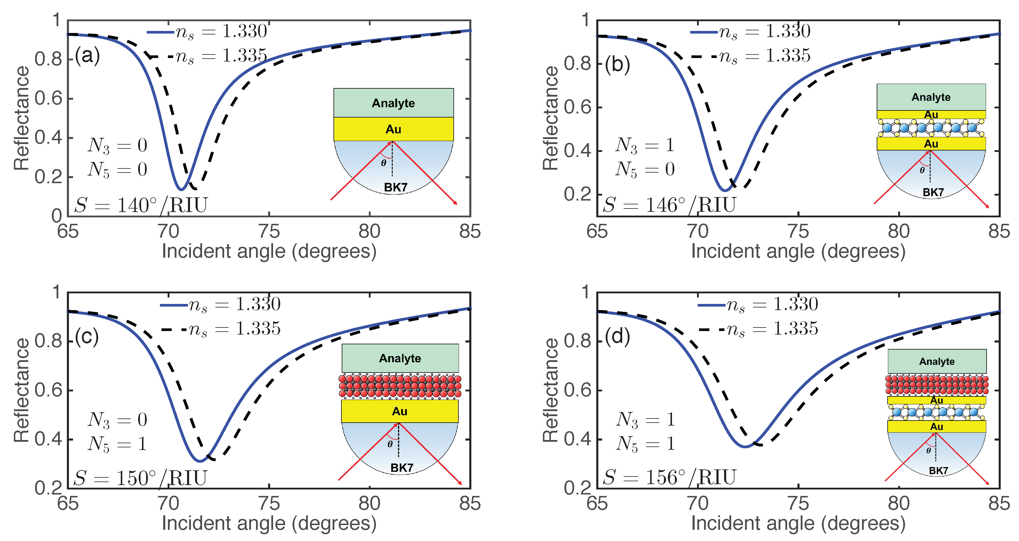
2D material-on-Au has been experimentally obtained in recent years. For example, graphene on Au surface has been experimentally demonstrated using the transfer printing technique [55,56]. The obtained graphene-on-Au structure was experimentally demonstrated for SPR sensing applications [56]. TMDs on the Au surface were also experimentally achieved [57,58,59,60,61,62]. These techniques can be applied for the fabrication of MXene-on-Au structures. Therefore, the proposed SPR sensor based on 2D MXene and TMDs are expected to be achieved easily. In order to illustrate the sensitivity enhancement of the proposed SPR sensor, we calculated the angular spectrum of the reflected light for various sensor structures, as shown in Figure 2, before (solid lines) and after (dashed lines) the RI variation of the sensing medium, assuming a small RI change . For each SPR sensor, the increase of the analyte RI will shift the resonance angle toward a larger value. For example, for the SPR sensor with and (i.e., conventional SPR sensor with 60 nm (=) Au film shown in Figure 2a), the resonance angle is with the ambient RI of 1.330, and increases to with a small analyte RI increment (). Therefore, a sensitivity of was obtained for the bare Au-based SPR sensor. By inserting a monolayer between the two Au films (i.e., and ), an enhanced sensitivity of was achieved (see Figure 2b). To study the sensitivity improvement with reference to the sensitivity of the conventional Au-based SPR sensor, we denoted the sensitivity enhancement as , in which S is the sensitivity of 2D-nanomaterial-integrated SPR sensor. For the SPR sensor shown in Figure 2b, a relatively low sensitivity enhancement of 4.29% was obtained. The sensitivity and sensitivity enhancement were improved to and 7.14%, respectively, with only one layer of (i.e., , , Figure 2c). With the employment of both a MXene and layer ( and ), an enhanced sensitivity of with the sensitivity enhancement of 11.43% was achieved, as shown in Figure 2d. Besides the --based SPR sensor, three other TMDs (, , ) and integrated SPR sensors ( and ) also exhibited enhanced sensitivity (Figures S1–S3 in the Supporting Information). Therefore, the proposed SPR sensor with the simultaneous employment of and TMDs exhibited enhanced sensitivity and offers the potential for highly sensitive sensing applications.
The study above only focuses on monolayer and . Previous investigations on 2D-material-integrated SPR sensors have demonstrated that the sensitivity also depends on the layer number of 2D materials [9,10,11,20,21,22,23,24]. Therefore, it is necessary to study the effect of number of and layers on the sensitivity. First, we investigated the effect of multiple layers of 2D materials on the reflectance for the proposed SPR sensor. The reflectance as a function of the incident angle for the monolayer --based SPR sensor with different numbers of layers is shown in Figure 3a. It was readily apparent that the resonance angle increased with the number of layers due to the increased propagation constant (wavector) of the surface plasmons. In addition, a shallowing and broadening of the reflectance curves was observed when the layers of increased, due to the increased electron energy loss [20,22]. Similar phenomena were found in the reflectance curves for -monolayer -based SPR sensors with different numbers of layers, as shown in Figure 3b. By comparing Figure 3a and Figure 3b, it was found that the increased energy loss caused by the integration of layers was larger than that caused by the additional layers.
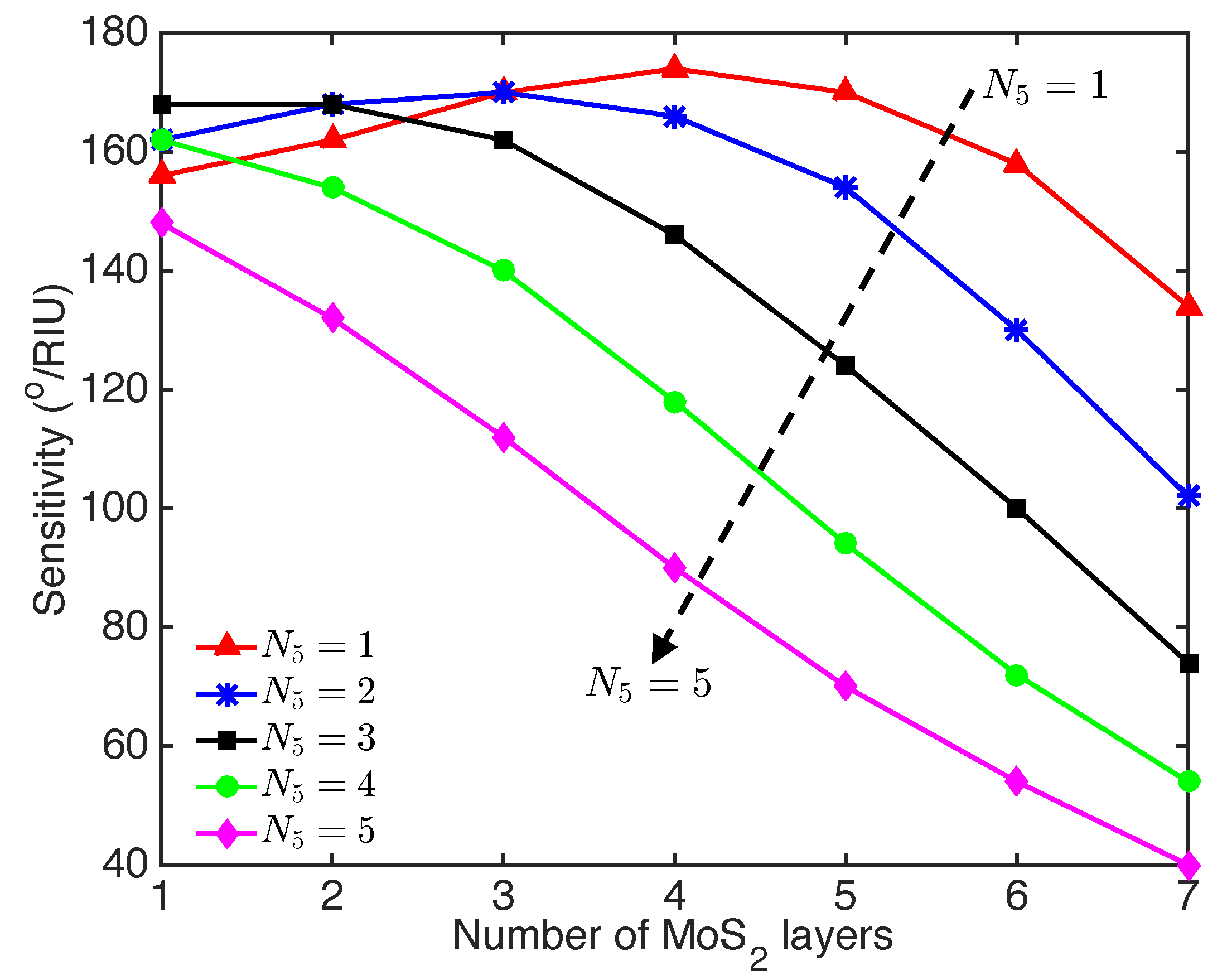
To further improve the sensitivity of proposed SPR sensor, we studied the optimiziation of the sensitivity by varying the layer number of the MXene and TMDs. The sensitivity as a function of the number of layers for the --based SPR sensor with different numbers of layers is shown in Figure 4. The sensitivity first increased and then decreased with the number of layers, when the SPR sensor integrated monolayer and two layers of . However, adding more layers of (e.g., three to five layers) resulted in decreased sensitivity with the number of layers. Due to the relative higher energy loss of the layers, the SPR signal enhancement effect of the layers in the SPR sensor with three to five layers of was overwhelmed by the energy loss with the additional layers. In contrast, with the integration of monolayer , the sensitivity increased with the number of layers from one to four (see Figure 4), where the SPR signal enhancement effect was more significant than the energy loss caused by the layers [22]. The maximum sensitivity of was found for the --based SPR sensor integrated with four-layer and monolayer .
The optimization of various combinations of MXene and TMDs (e.g., -, -, and -) of the SPR sensors are shown in Figures S4–S6 of the Supporting Information. It was found that only monolayer MXene could be used to obtain the maximum sensitivity for the -TMDs-based SPR sensors. The sensitivity and sensitivity enhancement at the optimized number of TMD layers and MXene layers for the proposed SPR sensor structure are summarized in Table 1. The -- and --based SPR sensors possessed sensitivities more than . A maximum sensitivity of was achieved with the sensor structure of Au/ (six layers)/ (one layer)/Au, and a sensitivity enhancement of 41.43% was obtained. The sensitivities achieved with the proposed -TMDs-based SPR sensors at a 633 nm excitation wavelength were significantly higher than that of the conventional Au- (four layer)-based SPR sensor () recently reported by Wu et al. [47]. The combination of TMDs and offers the alternative of sensitivity enhancement for -based SPR sensors.
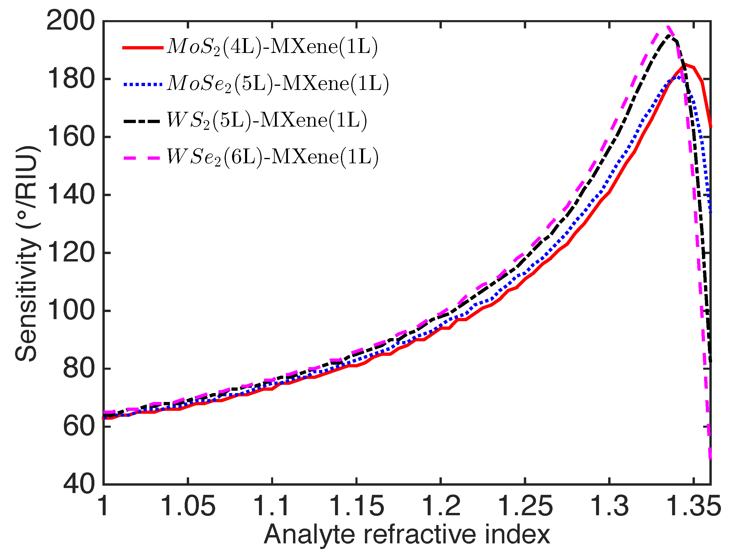
The RI of the surrounding environment was also important to the sensitivity, which determined the appropriate working RI range or working environment (e.g., gas or liquid) of the SPR sensor. The sensitivity for the optimized -TMDs-based SPR sensor was plotted with varying analyte RI in Figure 5. The optimized -TMDs-based SPR sensor possessed a relatively low sensitivity (<90) within the analyte RI range from 1.0 to 1.15. This revealed that the proposed SPR sensor was not appropriate for gas sensing, which typically involves a RI . The sensitivity of the optimized SPR sensor first increased to a maximum and then decreased with the analyte RI in the range of 1.0–1.36. The maximum RI sensitivity was found around the analyte RI of 1.330 (i.e., the RI of water). Therefore, the proposed sensor was more suited for operating in an aqueous medium, particularly for bio- and chemical sensing.
4. Conclusions
A novel SPR sensor based on Au--Au-TMDs is theoretically presented. The MXene-TMDs-integrated SPR sensor possessed enhanced sensitivity as compared to the bare Au film-based SPR sensor. For the aqueous solutions (RI ∼1.33), the RI sensitivities of , , , and for the proposed SPR sensor with monolayer MXene and four-layer , five-layer , five-layer , and six-layer , respectively, were achieved at the 633 nm excitation wavelength. Compared to the conventional Au film SPR sensor, the sensitivity was significantly enhanced by 24.29%, 25.71%, 41.43%, and 37.14%, respectively. The high sensitivities of the proposed MXene-based SPR sensors offer a potential route towards highly sensitive SPR sensors. Although this work was purely based on theoretical calculations, we used realistic material parameters and the results could be readily verified by experimental investigations. Moreover, since the structures of graphene-on-Au and TMDs-on-Au have been experimentally realized in recent years [55,56,57,58,59,60,61,62], it is possible to fabricate the MXene-on-Au structure. Thus the proposed SPR sensor based on 2D MXene and TMDs is experimentally feasible.
Supplementary Materials
The following are available online at https://www.mdpi.com/2079-4991/9/2/165/s1, Figure S1: Reflectance for the --based SPR sensor; Figure S2: Reflectance for the --based SPR sensor; Figure S3: Reflectance for the --based SPR sensor; Figure S4: Variation of sensitivity with number of and layers for the --based SPR sensor; Figure S5: Variation of sensitivity with number of and layers for the --based SPR sensor; Figure S6: Variation of sensitivity with number of and layers for the --based SPR sensor.
Author Contributions
Conceptualization, Y.X.; investigation, Y.X., Y.S.A., L.W. and L.K.A; writing-original draft preparation, Y.X.; writing-review and editing, Y.X., Y.S.A., L.W. and L.K.A.; supervision, L.K.A.; funding acquisition, L.K.A.
Funding
This work was partially supported by the Singapore A*STAR AME IRG (A1783c0011) and USA Air Force Office of Scientific Research (AFOSR) through the Asian Office of Aerospace Research and Development (AOARD) under Grant No. FA2386-17-1-4020.
Conflicts of Interest
The authors declare no conflict of interest.
References
- Homola, J.; Yee, S.S.; Gauglitz, G. Surface plasmon resonance sensors: Review. Sens. Actuators B Chem. 1999, 54, 3–15. [Google Scholar] [CrossRef]
- Homola, J. Surface plasmon resonance sensors for detection of chemical and biological species. Chem. Rev. 2008, 108, 462–493. [Google Scholar] [CrossRef] [PubMed]
- Fan, X.; White, I.M.; Shopova, S.I.; Zhu, H.; Suter, J.D.; Sun, Y. Sensitive optical biosensors for unlabeled targets: A review. Anal. Chim. Acta 2008, 620, 8–26. [Google Scholar] [CrossRef] [PubMed]
- Wijaya, E.; Lenaerts, C.; Maricot, S.; Hastanin, J.; Habraken, S.; Vilcot, J.P.; Boukherroub, R.; Szunerits, S. Surface plasmon resonance-based biosensors: From the development of different SPR structures to novel surface functionalization strategies. Curr. Opin. Solid State Mater. Sci. 2011, 15, 208–224. [Google Scholar] [CrossRef]
- Masson, J.F. Surface plasmon resonance clinical biosensors for medical diagnostics. ACS Sens. 2017, 2, 16–30. [Google Scholar] [CrossRef] [PubMed]
- Kretschmann, E.; Raether, H. Notizen: Radiative decay of non radiative surface plasmons excited by light. Z. Naturforschung A 1968, 23, 2135–2136. [Google Scholar] [CrossRef]
- Shalabney, A.; Abdulhalim, I. Sensitivity-enhancement methods for surface plasmon sensors. Laser Photonics Rev. 2011, 5, 571–606. [Google Scholar] [CrossRef]
- Bhatia, P.; Gupta, B.D. Surface-plasmon-resonance-based fiber-optic refractive index sensor: Sensitivity enhancement. Appl. Opt. 2011, 50, 2032–2036. [Google Scholar] [CrossRef]
- Wu, L.; Chu, H.; Koh, W.; Li, E. Highly sensitive graphene biosensors based on surface plasmon resonance. Opt. Express 2010, 18, 14395–14400. [Google Scholar] [CrossRef] [PubMed]
- Verma, R.; Gupta, B.D.; Jha, R. Sensitivity enhancement of a surface plasmon resonance based biomolecules sensor using graphene and silicon layers. Sens. Actuators B Chem. 2011, 160, 623–631. [Google Scholar] [CrossRef]
- Fu, H.; Zhang, S.; Chen, H.; Weng, J. Graphene enhances the sensitivity of fiber-optic surface plasmon resonance biosensor. IEEE Sens. J. 2015, 15, 5478–5482. [Google Scholar] [CrossRef]
- Wei, W.; Nong, J.; Zhu, Y.; Zhang, G.; Wang, N.; Luo, S.; Chen, N.; Lan, G.; Chuang, C.J.; Huang, Y. Graphene/Au-enhanced plastic clad silica fiber optic surface plasmon resonance sensor. Plasmonics 2018, 13, 483–491. [Google Scholar] [CrossRef]
- Ju, L.; Geng, B.; Horng, J.; Girit, C.; Martin, M.; Hao, Z.; Bechtel, H.A.; Liang, X.; Zettl, A.; Shen, Y.R.; et al. Graphene plasmonics for tunable terahertz metamaterials. Nat. Nanotechnol. 2011, 6, 630–634. [Google Scholar] [CrossRef] [PubMed]
- Rodrigo, D.; Limaj, O.; Janner, D.; Etezadi, D.; De Abajo, F.J.G.; Pruneri, V.; Altug, H. Mid-infrared plasmonic biosensing with graphene. Science 2015, 349, 165–168. [Google Scholar] [CrossRef] [PubMed] [Green Version]
- Huang, S.; Song, C.; Zhang, G.; Yan, H. Graphene plasmonics: Physics and potential applications. Nanophotonics 2016, 6, 1191–1204. [Google Scholar] [CrossRef]
- Ang, Y.S.; Sultan, S.; Zhang, C. Nonlinear optical spectrum of bilayer graphene in the terahertz regime. Appl. Phys. Lett. 2010, 97, 243110. [Google Scholar] [CrossRef] [Green Version]
- Ang, Y.S.; Chen, Q.; Zhang, C. Nonlinear optical response of graphene in terahertz and near-infrared frequency regime. Front. Optoelectron. 2015, 8, 3–26. [Google Scholar] [CrossRef]
- Ooi, K.J.; Tan, D.T. Nonlinear graphene plasmonics. Proc. R. Soc. A: Math. Phys. Eng. Sci. 2017, 473, 20170433. [Google Scholar] [CrossRef] [Green Version]
- Ooi, K.J.; Ang, Y.S.; Cheng, J.L.; Ang, L.K.; Tan, D.T. Electronic scattering of graphene plasmons in the terahertz nonlinear regime. IEEE J. Sel. Top. Quantum Electron. 2017, 23, 1–6. [Google Scholar] [CrossRef]
- Ouyang, Q.; Zeng, S.; Jiang, L.; Hong, L.; Xu, G.; Dinh, X.Q.; Qian, J.; He, S.; Qu, J.; Coquet, P.; et al. Sensitivity enhancement of transition metal dichalcogenides/silicon nanostructure-based surface plasmon resonance biosensor. Sci. Rep. 2016, 6, 28190. [Google Scholar] [CrossRef]
- Xu, Y.; Wu, L.; Ang, L.K. MoS2-based Highly Sensitive Near-infrared Surface Plasmon Resonance Refractive index Sensor. IEEE J. Sel. Top. Quantum Electron. 2019, 25, 4600307. [Google Scholar] [CrossRef]
- Zeng, S.; Hu, S.; Xia, J.; Anderson, T.; Dinh, X.Q.; Meng, X.M.; Coquet, P.; Yong, K.T. Graphene–MoS2 hybrid nanostructures enhanced surface plasmon resonance biosensors. Sens. Actuators B Chem. 2015, 207, 801–810. [Google Scholar] [CrossRef]
- Ouyang, Q.; Zeng, S.; Jiang, L.; Qu, J.; Dinh, X.Q.; Qian, J.; He, S.; Coquet, P.; Yong, K.T. Two-Dimensional Transition Metal Dichalcogenide Enhanced Phase-Sensitive Plasmonic Biosensors: Theoretical Insight. J. Phys. Chem. C 2017, 121, 6282–6289. [Google Scholar] [CrossRef]
- Xu, Y.; Hsieh, C.Y.; Wu, L.; Ang, L.K. Two-dimensional transition metal dichalcogenides mediated long range surface plasmon resonance biosensors. J. Phys. D Appl. Phys. 2019, 52, 065101. [Google Scholar] [CrossRef]
- Lin, Z.; Jiang, L.; Wu, L.; Guo, J.; Dai, X.; Xiang, Y.; Fan, D. Tuning and sensitivity enhancement of surface plasmon resonance biosensor with graphene covered Au-MoS2-Au Films. IEEE Photonics J. 2016, 8, 1–8. [Google Scholar]
- Naguib, M.; Kurtoglu, M.; Presser, V.; Lu, J.; Niu, J.; Heon, M.; Hultman, L.; Gogotsi, Y.; Barsoum, M.W. Two-dimensional nanocrystals produced by exfoliation of Ti3AlC2. Adv. Mater. 2011, 23, 4248–4253. [Google Scholar] [CrossRef] [PubMed]
- Naguib, M.; Mashtalir, O.; Carle, J.; Presser, V.; Lu, J.; Hultman, L.; Gogotsi, Y.; Barsoum, M.W. Two-dimensional transition metal carbides. ACS Nano 2012, 6, 1322–1331. [Google Scholar] [CrossRef]
- Naguib, M.; Mochalin, V.N.; Barsoum, M.W.; Gogotsi, Y. MXenes: A new family of two-dimensional materials. Adv. Mater. 2014, 26, 992–1005. [Google Scholar] [CrossRef] [PubMed]
- Lukatskaya, M.R.; Kota, S.; Lin, Z.; Zhao, M.Q.; Shpigel, N.; Levi, M.D.; Halim, J.; Taberna, P.L.; Barsoum, M.W.; Simon, P.; et al. Ultra-high-rate pseudocapacitive energy storage in two-dimensional transition metal carbides. Nat. Energy 2017, 2, 17105. [Google Scholar] [CrossRef]
- Lipatov, A.; Alhabeb, M.; Lukatskaya, M.R.; Boson, A.; Gogotsi, Y.; Sinitskii, A. Effect of synthesis on quality, electronic properties and environmental stability of individual monolayer Ti3C2 MXene flakes. Adv. Electron. Mater. 2016, 2, 1600255. [Google Scholar] [CrossRef]
- Anasori, B.; Lukatskaya, M.R.; Gogotsi, Y. 2D metal carbides and nitrides (MXenes) for energy storage. Nat. Rev. Mater. 2017, 2, 16098. [Google Scholar] [CrossRef]
- Peng, Q.; Guo, J.; Zhang, Q.; Xiang, J.; Liu, B.; Zhou, A.; Liu, R.; Tian, Y. Unique lead adsorption behavior of activated hydroxyl group in two-dimensional titanium carbide. J. Am. Chem. Soc. 2014, 136, 4113–4116. [Google Scholar] [CrossRef] [PubMed]
- Xie, X.; Xue, Y.; Li, L.; Chen, S.; Nie, Y.; Ding, W.; Wei, Z. Surface Al leached Ti3AlC2 as a substitute for carbon for use as a catalyst support in a harsh corrosive electrochemical system. Nanoscale 2014, 6, 11035–11040. [Google Scholar] [CrossRef] [PubMed]
- Mashtalir, O.; Cook, K.M.; Mochalin, V.; Crowe, M.; Barsoum, M.W.; Gogotsi, Y. Dye adsorption and decomposition on two-dimensional titanium carbide in aqueous media. J. Mater. Chem. A 2014, 2, 14334–14338. [Google Scholar] [CrossRef]
- Seh, Z.W.; Fredrickson, K.D.; Anasori, B.; Kibsgaard, J.; Strickler, A.L.; Lukatskaya, M.R.; Gogotsi, Y.; Jaramillo, T.F.; Vojvodic, A. Two-dimensional molybdenum carbide (MXene) as an efficient electrocatalyst for hydrogen evolution. ACS Energy Lett. 2016, 1, 589–594. [Google Scholar] [CrossRef]
- Xuan, J.; Wang, Z.; Chen, Y.; Liang, D.; Cheng, L.; Yang, X.; Liu, Z.; Ma, R.; Sasaki, T.; Geng, F. Organic-Base-Driven Intercalation and Delamination for the Production of Functionalized Titanium Carbide Nanosheets with Superior Photothermal Therapeutic Performance. Angew. Chem. 2016, 128, 14789–14794. [Google Scholar] [CrossRef]
- Sinha, A.; Dhanjai; Zhao, H.; Huang, Y.; Lu, X.; Chen, J.; Jain, R. MXene: An emerging material for sensing and biosensing. TrAC Trends Anal. Chem. 2018, 105, 424–435. [Google Scholar] [CrossRef]
- Zhu, J.; Ha, E.; Zhao, G.; Zhou, Y.; Huang, D.; Yue, G.; Hu, L.; Sun, N.; Wang, Y.; Lee, L.Y.S.; et al. Recent advance in MXenes: A promising 2D material for catalysis, sensor and chemical adsorption. Coord. Chem. Rev. 2017, 352, 306–327. [Google Scholar] [CrossRef]
- Zhu, X.; Liu, B.; Hou, H.; Huang, Z.; Zeinu, K.M.; Huang, L.; Yuan, X.; Guo, D.; Hu, J.; Yang, J. Alkaline intercalation of Ti3C2 MXene for simultaneous electrochemical detection of Cd(II), Pb(II), Cu(II) and Hg(II). Electrochim. Acta 2017, 248, 46–57. [Google Scholar] [CrossRef]
- Rasheed, P.A.; Pandey, R.P.; Rasool, K.; Mahmoud, K.A. Ultra-sensitive electrocatalytic detection of bromate in drinking water based on Nafion/Ti3C2Tx (MXene) modified glassy carbon electrode. Sens. Actuators B Chem. 2018, 265, 652–659. [Google Scholar] [CrossRef]
- Xu, B.; Zhu, M.; Zhang, W.; Zhen, X.; Pei, Z.; Xue, Q.; Zhi, C.; Shi, P. Ultrathin MXene-Micropattern-Based Field-Effect Transistor for Probing Neural Activity. Adv. Mater. 2016, 28, 3333–3339. [Google Scholar] [CrossRef]
- Fang, Y.; Yang, X.; Chen, T.; Xu, G.; Liu, M.; Liu, J.; Xu, Y. Two-dimensional titanium carbide (MXene)-based solid-state electrochemiluminescent sensor for label-free single-nucleotide mismatch discrimination in human urine. Sens. Actuators B Chem. 2018, 263, 400–407. [Google Scholar] [CrossRef]
- Lee, E.; VahidMohammadi, A.; Prorok, B.C.; Yoon, Y.S.; Beidaghi, M.; Kim, D.J. Room temperature gas sensing of two-dimensional titanium carbide (MXene). ACS Appl. Mater. Interfaces 2017, 9, 37184–37190. [Google Scholar] [CrossRef] [PubMed]
- Kim, S.J.; Koh, H.J.; Ren, C.E.; Kwon, O.; Maleski, K.; Cho, S.Y.; Anasori, B.; Kim, C.K.; Choi, Y.K.; Kim, J.; et al. Metallic Ti3C2Tx MXene Gas Sensors with Ultrahigh Signal-to-Noise Ratio. ACS Nano 2018, 12, 986–993. [Google Scholar] [CrossRef]
- Lorencová, L.; Bertok, T.; Dosekova, E.; Holazová, A.; Paprckova, D.; Vikartovská, A.; Sasinková, V.; Filip, J.; Kasák, P.; Jerigová, M.; et al. Electrochemical performance of Ti3C2Tx MXene in aqueous media: towards ultrasensitive H2O2 sensing. Electrochim. Acta 2017, 235, 471–479. [Google Scholar] [CrossRef]
- Xiao, F.; Zhao, F.; Zhang, Y.; Guo, G.; Zeng, B. Ultrasonic electrodeposition of gold-platinum alloy nanoparticles on ionic liquid-chitosan composite film and their application in fabricating nonenzyme hydrogen peroxide sensors. J. Phys. Chem. C 2008, 113, 849–855. [Google Scholar] [CrossRef]
- Wu, L.; You, Q.; Shan, Y.; Gan, S.; Zhao, Y.; Dai, X.; Xiang, Y. Few-layer Ti3C2Tx MXene: A promising surface plasmon resonance biosensing material to enhance the sensitivity. Sens. Actuators B Chem. 2018, 277, 210–215. [Google Scholar] [CrossRef]
- Yamamoto, M. Surface plasmon resonance (SPR) theory: Tutorial. Rev. Polarogr. 2002, 48, 209–237. [Google Scholar] [CrossRef]
- Polyanskiy, M. Refractive Index Database. Available online: http://refractiveindex.info (accessed on 23 November 2018).
- Maharana, P.K.; Srivastava, T.; Jha, R. On the performance of highly sensitive and accurate graphene-on-aluminum and silicon-based SPR biosensor for visible and near infrared. Plasmonics 2014, 9, 1113–1120. [Google Scholar] [CrossRef]
- Shi, C.; Beidaghi, M.; Naguib, M.; Mashtalir, O.; Gogotsi, Y.; Billinge, S.J. Structure of nanocrystalline Ti3C2 MXene using atomic pair distribution function. Phys. Rev. Lett. 2014, 112, 125501. [Google Scholar] [CrossRef]
- Miranda, A.; Halim, J.; Lorke, A.; Barsoum, M. Rendering Ti3C2Tx (MXene) monolayers visible. Mater. Res. Lett. 2017, 5, 322–328. [Google Scholar] [CrossRef]
- Li, Y.; Chernikov, A.; Zhang, X.; Rigosi, A.; Hill, H.M.; van der Zande, A.M.; Chenet, D.A.; Shih, E.M.; Hone, J.; Heinz, T.F. Measurement of the optical dielectric function of monolayer transition-metal dichalcogenides: MoS2, MoSe2, WS2, and WSe2. Phys. Rev. B 2014, 90, 205422. [Google Scholar] [CrossRef]
- Liu, H.L.; Shen, C.C.; Su, S.H.; Hsu, C.L.; Li, M.Y.; Li, L.J. Optical properties of monolayer transition metal dichalcogenides probed by spectroscopic ellipsometry. Appl. Phys. Lett. 2014, 105, 201905. [Google Scholar] [CrossRef] [Green Version]
- Song, B.; Li, D.; Qi, W.; Elstner, M.; Fan, C.; Fang, H. Graphene on Au(111): A highly conductive material with excellent adsorption properties for high-resolution bio/nanodetection and identification. ChemPhysChem 2010, 11, 585–589. [Google Scholar] [CrossRef] [PubMed]
- Salihoglu, O.; Balci, S.; Kocabas, C. Plasmon-polaritons on graphene-metal surface and their use in biosensors. Appl. Phys. Lett. 2012, 100, 213110. [Google Scholar] [CrossRef] [Green Version]
- Grønborg, S.S.; Ulstrup, S.; Bianchi, M.; Dendzik, M.; Sanders, C.E.; Lauritsen, J.V.; Hofmann, P.; Miwa, J.A. Synthesis of epitaxial single-layer MoS2 on Au(111). Langmuir 2015, 31, 9700–9706. [Google Scholar] [CrossRef] [PubMed]
- Sørensen, S.G.; Füchtbauer, H.G.; Tuxen, A.K.; Walton, A.S.; Lauritsen, J.V. Structure and electronic properties of in situ synthesized single-layer MoS2 on a gold surface. ACS Nano 2014, 8, 6788–6796. [Google Scholar] [CrossRef] [PubMed]
- Dendzik, M.; Michiardi, M.; Sanders, C.; Bianchi, M.; Miwa, J.A.; Grønborg, S.S.; Lauritsen, J.V.; Bruix, A.; Hammer, B.; Hofmann, P. Growth and electronic structure of epitaxial single-layer WS2 on Au(111). Phys. Rev. B 2015, 92, 245442. [Google Scholar] [CrossRef]
- Bruix, A.; Miwa, J.A.; Hauptmann, N.; Wegner, D.; Ulstrup, S.; Grønborg, S.S.; Sanders, C.E.; Dendzik, M.; Čabo, A.G.; Bianchi, M.; et al. Single-layer MoS2 on Au(111): Band gap renormalization and substrate interaction. Phys. Rev. B 2016, 93, 165422. [Google Scholar] [CrossRef]
- Park, S.; Mutz, N.; Schultz, T.; Blumstengel, S.; Han, A.; Aljarb, A.; Li, L.J.; List-Kratochvil, E.J.; Amsalem, P.; Koch, N. Direct determination of monolayer MoS2 and WSe2 exciton binding energies on insulating and metallic substrates. 2D Mater. 2018, 5, 025003. [Google Scholar] [CrossRef]
- Lu, J.; Bao, D.L.; Qian, K.; Zhang, S.; Chen, H.; Lin, X.; Du, S.X.; Gao, H.J. Identifying and Visualizing the Edge Terminations of Single-Layer MoSe2 Island Epitaxially Grown on Au(111). ACS Nano 2017, 11, 1689–1695. [Google Scholar] [CrossRef] [PubMed]
Figure 1.
Schematic illustration of the proposed surface plasmon resonance (SPR) sensor with and 2D transition metal dichalcogenides (TMD) layers.
Figure 1.
Schematic illustration of the proposed surface plasmon resonance (SPR) sensor with and 2D transition metal dichalcogenides (TMD) layers.

Figure 2.
Reflectance as a function of the incident angle before (solid lines) and after (dashed lines) the variation of analyte refractive index (RI) for the --based SPR sensor with (a) , , (i.e., no 2D materials); (b) , , (i.e., monolayer ); (c) , , (i.e., monolayer ), and (d) , , (i.e., monolayer and monolayer ).

Figure 3.
Reflectance as a function of the incident angle for --based SPR sensor with (a) different number of () and monolayer (), and (b) different number of () and monolayer ().
Figure 3.
Reflectance as a function of the incident angle for --based SPR sensor with (a) different number of () and monolayer (), and (b) different number of () and monolayer ().

Figure 4.
Sensitivity as a function of the number of layers for --based SPR sensor with different layers of .
Figure 4.
Sensitivity as a function of the number of layers for --based SPR sensor with different layers of .

Figure 5.
Variation of sensitivity for the optimized -TMD-based SPR sensor with the varying analyte RI.
Figure 5.
Variation of sensitivity for the optimized -TMD-based SPR sensor with the varying analyte RI.


Table 1.
Sensitivity and sensitivity enhancement at the optimized number of TMD layers and layers for the -TMDs-based SPR sensor.
Table 1.
Sensitivity and sensitivity enhancement at the optimized number of TMD layers and layers for the -TMDs-based SPR sensor.
| Type of TMD | Number of TMD Layers N3 | Number of Layers N5 | Sensitivity (°/RIU) | (%) |
|---|---|---|---|---|
| 4 | 1 | 174 | 24.29 | |
| 5 | 1 | 176 | 25.71 | |
| 5 | 1 | 198 | 41.43 | |
| 6 | 1 | 192 | 37.14 |
© 2019 by the authors. Licensee MDPI, Basel, Switzerland. This article is an open access article distributed under the terms and conditions of the Creative Commons Attribution (CC BY) license (http://creativecommons.org/licenses/by/4.0/).
Share and Cite
MDPI and ACS Style
Xu, Y.; Ang, Y.S.; Wu, L.; Ang, L.K. High Sensitivity Surface Plasmon Resonance Sensor Based on Two-Dimensional MXene and Transition Metal Dichalcogenide: A Theoretical Study. Nanomaterials 2019, 9, 165. https://doi.org/10.3390/nano9020165
AMA Style
Xu Y, Ang YS, Wu L, Ang LK. High Sensitivity Surface Plasmon Resonance Sensor Based on Two-Dimensional MXene and Transition Metal Dichalcogenide: A Theoretical Study. Nanomaterials. 2019; 9(2):165. https://doi.org/10.3390/nano9020165
Chicago/Turabian StyleXu, Yi, Yee Sin Ang, Lin Wu, and Lay Kee Ang. 2019. "High Sensitivity Surface Plasmon Resonance Sensor Based on Two-Dimensional MXene and Transition Metal Dichalcogenide: A Theoretical Study" Nanomaterials 9, no. 2: 165. https://doi.org/10.3390/nano9020165
Note that from the first issue of 2016, this journal uses article numbers instead of page numbers. See further details here.