
An Overview of Cellulose Derivatives-Based Dressings for Wound-Healing Management


 , , ,
, , ,
Abstract
:
1. Introduction
2. Wound Classification
- etiology: surgical, traumatic, radiation and malignant wound, chemical or thermal injury, pressure ulcer, diabetic foot ulcer, vascular ulcer, or atypical injury;
- healing time (duration) and nature of the injury restoration process: acute or chronic wounds;
- depth of injury or number of skin layers affected: superficial, partial thickness (deep dermal) or full-thickness wounds;
- complexity: simple, complex, or complicated wounds;
- contamination and postoperative infection risk: clean wound (class I), clean/contaminated wound (class II), contaminated wound (class III) or dirty wound (class IV);
- mode of lesion: abrasion, ulceration, incision, laceration or degloving;
- tissue loss: without tissue loss (surgical wounds) and with tissue loss (burns, traumatic wounds, diabetic foot ulcers, and iatrogenic wounds);
- appearance: necrotic, sloughy, infected, malodorous, granulating, and epithelializing wound;
3. Wound-Healing Process
3.1. Wound-Healing Stages
3.2. Factors Affecting Wound-Healing Process
3.2.1. Local Factors That Affect Wound-Healing Process
3.2.2. Systemic Factors That Affect Wound-Healing Process
4. Wound Dressings: Properties and Classification
4.1. Wound Dressing Properties
4.2. Wound-Dressing Classification
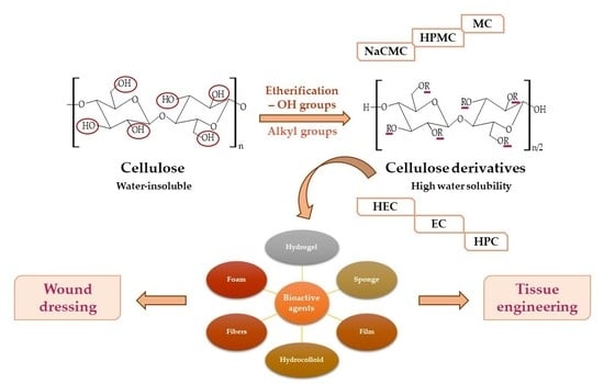
5. Cellulose Derivatives as Wound Dressings
5.1. Cellulose Derivatives Classification
5.2. Sodium Carboxymethylcellulose-Based Wound Dressings
5.3. Hydroxypropylmethylcellulose-Based Wound Dressings
5.4. Methylcellulose-Based Wound Dressings
5.5. Hydroxyethylcellulose-Based Wound Dressings
5.6. Ethylcellulose-Based Wound Dressings
5.7. Hydroxypropylcellulose-Based Wound Dressings
5.8. Combinations of Cellulose Derivatives-Based Wound Dressings
6. Conclusions and Future Perspectives
Author Contributions
Funding
Institutional Review Board Statement
Informed Consent Statement
Data Availability Statement
Conflicts of Interest
References
- Kim, B.S.; Gao, G.; Kim, J.Y.; Cho, D.W. 3D Cell Printing of Perfusable Vascularized Human Skin Equivalent Composed of Epidermis, Dermis, and Hypodermis for Better Structural Recapitulation of Native Skin. Adv. Healthc. Mater. 2019, 8, e1801019. [Google Scholar] [CrossRef] [PubMed]
- Sadeghi, S.; Nourmohammadi, J.; Ghaee, A.; Soleimani, N. Carboxymethyl cellulose-human hair keratin hydrogel with controlled clindamycin release as antibacterial wound dressing. Int. J. Biol. Macromol. 2020, 147, 1239–1247. [Google Scholar] [CrossRef] [PubMed]
- Zimmerman, A.; Bai, L.; Ginty, D.D. The gentle touch receptors of mammalian skin. Science 2014, 346, 950–954. [Google Scholar] [CrossRef] [PubMed] [Green Version]
- Zhao, S.; Li, L.; Wang, H.; Zhang, Y.; Cheng, X.; Zhou, N.; Rahaman, M.N.; Liu, Z.; Huang, W.; Zhang, C. Wound dressings composed of copper-doped borate bioactive glass microfibers stimulate angiogenesis and heal full-thickness skin defects in a rodent model. Biomaterials 2015, 53, 379–391. [Google Scholar] [CrossRef]
- Kilic Bektas, C.; Kimiz, I.; Sendemir, A.; Hasirci, V.; Hasirci, N. A bilayer scaffold prepared from collagen and carboxymethyl cellulose for skin tissue engineering applications. J. Biomater. Sci. Polym. Ed. 2018, 29, 1764–1784. [Google Scholar] [CrossRef]
- Kolarsick, P.A.; Kolarsick, M.A.; Goodwin, C. Anatomy and physiology of the skin. J. Dermatol. Nurses Assoc. 2011, 3, 203–213. [Google Scholar] [CrossRef] [Green Version]
- McLafferty, E.; Hendry, C.; Alistair, F. The integumentary system: Anatomy, physiology and function of skin. Nurs. Stand. 2012, 27, 35–42. [Google Scholar] [CrossRef]
- Joodaki, H.; Panzer, M.B. Skin mechanical properties and modeling: A review. Proc. Inst. Mech. Eng. Part H J. Eng. Med. 2018, 232, 323–343. [Google Scholar] [CrossRef]
- Lai-Cheong, J.E.; McGrath, J.A. Structure and function of skin, hair and nails. Medicine 2021, 49, 337–342. [Google Scholar] [CrossRef]
- Brown, T.M.; Krishnamurthy, K. Histology, Dermis. In StatPearls; StatPearls Publishing LLC.: Treasure Island, FL, USA, 2020. [Google Scholar]
- Hashmi, S.; Marinkovich, M.P. Molecular organization of the basement membrane zone. Clin. Dermatol. 2011, 29, 398–411. [Google Scholar] [CrossRef]
- Zimoch, J.; Zielinska, D.; Michalak-Micka, K.; Rütsche, D.; Böni, R.; Biedermann, T.; Klar, A.S. Bioengineering a prevascularized human tri-layered skin substitute containing a hypodermis. Acta Biomater. 2021, 134, 215–227. [Google Scholar] [CrossRef]
- Wong, R.; Geyer, S.; Weninger, W.; Guimberteau, J.C.; Wong, J.K. The dynamic anatomy and patterning of skin. Exp. Dermatol. 2016, 25, 92–98. [Google Scholar] [CrossRef] [PubMed] [Green Version]
- Bragazzi, N.L.; Sellami, M.; Salem, I.; Conic, R.; Kimak, M.; Pigatto, P.D.M.; Damiani, G. Fasting and Its Impact on Skin Anatomy, Physiology, and Physiopathology: A Comprehensive Review of the Literature. Nutrients 2019, 11, 249. [Google Scholar] [CrossRef] [PubMed] [Green Version]
- Rodrigues, C.; de Assis, A.M.; Moura, D.J.; Halmenschlager, G.; Saffi, J.; Xavier, L.L.; Fernandes Mda, C.; Wink, M.R. New therapy of skin repair combining adipose-derived mesenchymal stem cells with sodium carboxymethylcellulose scaffold in a pre-clinical rat model. PLoS ONE 2014, 9, e96241. [Google Scholar] [CrossRef]
- Okoye, E.; Okolie, T. Development and in vitro characterization of ciprofloxacin loaded polymeric films for wound dressing. Int. J. Health Allied Sci. 2015, 4, 234. [Google Scholar] [CrossRef]
- Varshosaz, J.; Taymouri, S.; Minaiyan, M.; Rastegarnasab, F.; Baradaran, A. Development and in vitro/in vivo evaluation of HPMC/chitosan gel containing simvastatin loaded self-assembled nanomicelles as a potent wound healing agent. Drug Dev. Ind. Pharm. 2018, 44, 276–288. [Google Scholar] [CrossRef]
- Martinengo, L.; Olsson, M.; Bajpai, R.; Soljak, M.; Upton, Z.; Schmidtchen, A.; Car, J.; Jarbrink, K. Prevalence of chronic wounds in the general population: Systematic review and meta-analysis of observational studies. Ann. Epidemiol. 2019, 29, 8–15. [Google Scholar] [CrossRef]
- Sen, C.K. Human Wound and Its Burden: Updated 2020 Compendium of Estimates. Adv. Wound Care 2021, 10, 281–292. [Google Scholar] [CrossRef]
- Walker, J.; Cullen, M.; Chambers, H.; Mitchell, E.; Steers, N.; Khalil, H. Identifying wound prevalence using the Mobile Wound Care program. Int. Wound J. 2014, 11, 319–325. [Google Scholar] [CrossRef]
- Kapp, S.; Miller, C.; Santamaria, N. The quality of life of people who have chronic wounds and who self-treat. J. Clin. Nurs. 2018, 27, 182–192. [Google Scholar] [CrossRef]
- Moreira, M.E.; Markovchick, V.J. Wound Management. Crit. Care Nurs. Clin. N. Am. 2012, 24, 215–237. [Google Scholar] [CrossRef]
- Ghica, M.V.; Kaya, M.G.A.; Dinu-Pirvu, C.E.; Lupuleasa, D.; Udeanu, D.I. Development, Optimization and In Vitro/In Vivo Characterization of Collagen-Dextran Spongious Wound Dressings Loaded with Flufenamic Acid. Molecules 2017, 22, 1552. [Google Scholar] [CrossRef] [PubMed]
- Clark, M. Alginates in Dressings and Wound Management. In Alginates and Their Biomedical Applications; Rehm, B.H.A., Moradali, M.F., Eds.; Springer Series in Biomaterials Science and Engineering; Springer: Berlin/Heidelberg, Germany, 2018; Volume 11, pp. 213–222. [Google Scholar]
- Aramwit, P. Introduction to biomaterials for wound healing. In Wound Healing Biomaterials, Vol. 2: Functional Biomaterials; Agren, M.S., Ed.; Woodhead Publishing Series in Biomaterials; Woodhead Publ. Ltd.: Cambridge, UK, 2016; Volume 115, pp. 3–38. [Google Scholar]
- Lee, D.; Yoo, B. Cellulose derivatives agglomerated in a fluidized bed: Physical, rheological, and structural properties. Int. J. Biol. Macromol. 2021, 181, 232–240. [Google Scholar] [CrossRef]
- Seddiqi, H.; Oliaei, E.; Honarkar, H.; Jin, J.F.; Geonzon, L.C.; Bacabac, R.G.; Klein-Nulend, J. Cellulose and its derivatives: Towards biomedical applications. Cellulose 2021, 28, 1893–1931. [Google Scholar] [CrossRef]
- Jia, B.; Zhou, J.; Zhang, L. Electrospun nano-fiber mats containing cationic cellulose derivatives and poly (vinyl alcohol) with antibacterial activity. Carbohydr. Res. 2011, 346, 1337–1341. [Google Scholar] [CrossRef]
- Oprea, M.; Voicu, S.I. Recent advances in composites based on cellulose derivatives for biomedical applications. Carbohydr. Polym. 2020, 247, 116683. [Google Scholar] [CrossRef] [PubMed]
- Agrawal, P.; Soni, S.; Mittal, G.; Bhatnagar, A. Role of polymeric biomaterials as wound healing agents. Int. J. Low Extrem. Wounds 2014, 13, 180–190. [Google Scholar] [CrossRef]
- Mir, M.; Ali, M.N.; Barakullah, A.; Gulzar, A.; Arshad, M.; Fatima, S.; Asad, M. Synthetic polymeric biomaterials for wound healing: A review. Prog Biomater 2018, 7, 1–21. [Google Scholar] [CrossRef] [PubMed] [Green Version]
- Shende, P.; Gupta, H. Formulation and comparative characterization of nanoparticles of curcumin using natural, synthetic and semi-synthetic polymers for wound healing. Life Sci. 2020, 253, 117588. [Google Scholar] [CrossRef] [PubMed]
- Boateng, J.S.; Matthews, K.H.; Stevens, H.N.; Eccleston, G.M. Wound healing dressings and drug delivery systems: A review. J. Pharm. Sci. 2008, 97, 2892–2923. [Google Scholar] [CrossRef]
- Serena, T.E. A Global Perspective on Wound Care. Adv. Wound Care 2014, 3, 548–552. [Google Scholar] [CrossRef] [Green Version]
- Ambekar, R.S.; Kandasubramanian, B. Advancements in nanofibers for wound dressing: A review. Eur. Polym. J. 2019, 117, 304–336. [Google Scholar] [CrossRef]
- Kordestani, S.S. Chapter 6—Wound Classification. In Atlas of Wound Healing; Kordestani, S.S., Ed.; Elsevier: Amsterdam, The Netherlands, 2019; pp. 49–50. [Google Scholar]
- Kumar, K.S.; Reddy, B.E.J.S.; Processing, I. Wound image analysis classifier for efficient tracking of wound healing status. Signal Image Process. 2014, 5, 15–27. [Google Scholar]
- Wang, M.; Huang, X.; Zheng, H.; Tang, Y.; Zeng, K.; Shao, L.; Li, L. Nanomaterials applied in wound healing: Mechanisms, limitations and perspectives. J. Control. Release 2021, 337, 236–247. [Google Scholar] [CrossRef] [PubMed]
- Low, J.S.; Mak, K.-K.; Zhang, S.; Pichika, M.R.; Marappan, P.; Mohandas, K.; Balijepalli, M.K. In vitro methods used for discovering plant derived products as wound healing agents—An update on the cell types and rationale. Fitoterapia 2021, 154, 105026. [Google Scholar] [CrossRef] [PubMed]
- Nosrati, H.; Khodaei, M.; Alizadeh, Z.; Banitalebi-Dehkordi, M. Cationic, anionic and neutral polysaccharides for skin tissue engineering and wound healing applications. Int. J. Biol. Macromol. 2021, 192, 298–322. [Google Scholar] [CrossRef] [PubMed]
- Skorkowska-Telichowska, K.; Czemplik, M.; Kulma, A.; Szopa, J. The local treatment and available dressings designed for chronic wounds. J. Am. Acad. Dermatol. 2013, 68, e117–e126. [Google Scholar] [CrossRef] [PubMed]
- Thomas, S. Surgical Dressings and Wound Management; Dr Stephen Thomas: Joplin, MI, USA, 2010. [Google Scholar]
- Hachem, R.; Parikh, U.M.; Reitzel, R.; Rosenblatt, J.; Kaul, A.; Vargas-Cruz, N.; Hill, L.; Moore, L.; Meyer, J.; Chaftari, A.M.; et al. Novel antimicrobial ointment for infected wound healing in an in vitro and in vivo porcine model. Wound Repair Regen. 2021, 29, 830–842. [Google Scholar] [CrossRef]
- Albu, M.G.; Ghica, M.V.; Stefanescu, G.A.; Hodorogea, M.; Marin, M.M.; Marin, S.; Danila, E.; Voicu, S.; Simonca, A.G.; Popa, L. Design and Characterization of Collagen-Sodium Carboxymethylcellulose-Lidocaine 3D Composites for Wound Management. Key Eng. Mater. 2016, 695, 309–316. [Google Scholar] [CrossRef]
- Capanema, N.S.V.; Mansur, A.A.P.; de Jesus, A.C.; Carvalho, S.M.; de Oliveira, L.C.; Mansur, H.S. Superabsorbent crosslinked carboxymethyl cellulose-PEG hydrogels for potential wound dressing applications. Int. J. Biol. Macromol. 2018, 106, 1218–1234. [Google Scholar] [CrossRef]
- Sandri, G.; Bonferoni, M.C.; D’Autilia, F.; Rossi, S.; Ferrari, F.; Grisoli, P.; Sorrenti, M.; Catenacci, L.; Del Fante, C.; Perotti, C.; et al. Wound dressings based on silver sulfadiazine solid lipid nanoparticles for tissue repairing. Eur. J. Pharm. Biopharm. 2013, 84, 84–90. [Google Scholar] [CrossRef] [PubMed]
- Cañedo-Dorantes, L.; Cañedo-Ayala, M. Skin Acute Wound Healing: A Comprehensive Review. Int. J. Inflamm. 2019, 2019, 3706315. [Google Scholar] [CrossRef] [PubMed]
- Verma, Y.K.; Verma, R.; Sing, A.K.; Gangenahalli, G. LiCl Containing Thermosensitive Formulation Improves Hemostasis, Wound Healing, and Hair Regrowth. Regen. Eng. Transl. Med. 2020, 17, 362–378. [Google Scholar] [CrossRef]
- Kenet, G.; Barg, A.A.; Nowak-Gottl, U. Hemostasis in the Very Young. Semin. Thromb. Hemost. 2018, 44, 617–623. [Google Scholar] [CrossRef] [PubMed]
- Iacob, A.T.; Dragan, M.; Ionescu, O.M.; Profire, L.; Ficai, A.; Andronescu, E.; Confederat, L.G.; Lupascu, D. An Overview of Biopolymeric Electrospun Nanofibers Based on Polysaccharides for Wound Healing Management. Pharmaceutics 2020, 12, 983. [Google Scholar] [CrossRef]
- Kanikireddy, V.; Varaprasad, K.; Jayaramudu, T.; Karthikeyan, C.; Sadiku, R. Carboxymethyl cellulose-based materials for infection control and wound healing: A review. Int. J. Biol. Macromol. 2020, 164, 963–975. [Google Scholar] [CrossRef]
- Pawar, H.V.; Tetteh, J.; Boateng, J.S. Preparation, optimisation and characterisation of novel wound healing film dressings loaded with streptomycin and diclofenac. Colloid Surf. B Biointerfaces 2013, 102, 102–110. [Google Scholar] [CrossRef]
- Harper, D.; Young, A.; McNaught, C.-E.J.S. The physiology of wound healing. Adv. Ski. Wound Care 2014, 32, 445–450. [Google Scholar] [CrossRef]
- Yang, L.; Liang, F.; Zhang, X.; Jiang, Y.; Duan, F.; Li, L.; Ren, F. Remodeling microenvironment based on MOFs-Hydrogel hybrid system for improving diabetic wound healing. Chem. Eng. J. 2022, 427, 131506. [Google Scholar] [CrossRef]
- Schreml, S.; Szeimies, R.; Prantl, L.; Karrer, S.; Landthaler, M.; Babilas, P.J. Oxygen in acute and chronic wound healing. Br. J. Dermatol. 2010, 163, 257–268. [Google Scholar] [CrossRef]
- El-Ashram, S.; El-Samad, L.M.; Basha, A.A.; El Wakil, A. Naturally-derived targeted therapy for wound healing: Beyond classical strategies. Pharmacol. Res. 2021, 170, 105749. [Google Scholar] [CrossRef] [PubMed]
- Guo, S.; Dipietro, L.A. Factors affecting wound healing. J. Dent. Res. 2010, 89, 219–229. [Google Scholar] [CrossRef]
- Khalil, H.; Cullen, M.; Chambers, H.; Carroll, M.; Walker, J. Elements affecting wound healing time: An evidence based analysis. Wound Repair Regen. 2015, 23, 550–556. [Google Scholar] [CrossRef]
- Desmet, C.M.; Preat, V.; Gallez, B. Nanomedicines and gene therapy for the delivery of growth factors to improve perfusion and oxygenation in wound healing. Adv. Drug Deliv. Rev. 2018, 129, 262–284. [Google Scholar] [CrossRef]
- Gueldner, J.; Zhang, F.; Zechmann, B.; Bruce, E.D. Evaluating a novel oxygenating therapeutic for its potential use in the advancement of wound healing. Toxicol. In Vitro 2017, 43, 62–68. [Google Scholar] [CrossRef] [PubMed]
- Paunica-Panea, G.; Ficai, A.; Marin, M.M.; Marin, S.; Albu, M.G.; Constantin, V.D.; Dinu-Pirvu, C.; Vuluga, Z.; Corobea, M.C.; Ghica, M.V. New Collagen-Dextran-Zinc Oxide Composites for Wound Dressing. J. Nanomater. 2016, 2016, 5805034. [Google Scholar] [CrossRef] [Green Version]
- Powers, J.G.; Higham, C.; Broussard, K.; Phillips, T.J. Wound healing and treating wounds Chronic wound care and management. J. Am. Acad. Dermatol. 2016, 74, 607–625. [Google Scholar] [CrossRef]
- Emmerson, E.; Hardman, M.J. The role of estrogen deficiency in skin ageing and wound healing. Biogerontology 2012, 13, 3–20. [Google Scholar] [CrossRef]
- Gouin, J.P.; Kiecolt-Glaser, J.K. The Impact of Psychological Stress on Wound Healing: Methods and Mechanisms. Crit. Care Nurs. Clin. N. Am. 2012, 24, 201–213. [Google Scholar] [CrossRef] [Green Version]
- Okonkwo, U.A.; DiPietro, L.A. Diabetes and Wound Angiogenesis. Int. J. Mol. Sci. 2017, 18, 1419. [Google Scholar] [CrossRef] [Green Version]
- Goodarzi, P.; Alavi-Moghadam, S.; Sarvari, M.; Beik, A.T.; Falahzadeh, K.; Aghayan, H.; Payab, M.; Larijani, B.; Gilany, K.; Rahim, F.; et al. Adipose Tissue-Derived Stromal Cells for Wound Healing. In Cell Biology and Translational Medicine, Vol. 4: Stem Cells and Cell Based Strategies in Regeneration; Turksen, K., Ed.; Advances in Experimental Medicine and Biology; Springer International Publishing AG: Cham, Switzerland, 2018; Volume 1119, pp. 133–149. [Google Scholar]
- Serra, M.B.; Barroso, W.A.; Silva, N.N.; Silva, S.d.N.; Borges, A.C.R.; Abreu, I.C.; Borges, M.O.d.R. From Inflammation to Current and Alternative Therapies Involved in Wound Healing. Int. J. Inflamm. 2017, 2017, 3406215. [Google Scholar] [CrossRef] [PubMed] [Green Version]
- Beyene, R.T.; Derryberry, S.L.; Barbul, A. The Effect of Comorbidities on Wound Healing. Surg. Clin. N. Am. 2020, 100, 695–705. [Google Scholar] [CrossRef] [PubMed]
- McDaniel, J.C.; Browning, K.K. Smoking, chronic wound healing, and implications for evidence-based practice. J. Wound Ostomy Cont. Nurs. 2014, 41, 415–423. [Google Scholar] [CrossRef] [PubMed] [Green Version]
- Sen, C.K. Human Wounds and Its Burden: An Updated Compendium of Estimates. Adv. Wound Care 2019, 8, 39–48. [Google Scholar] [CrossRef] [PubMed] [Green Version]
- Moura, L.I.; Dias, A.M.; Carvalho, E.; de Sousa, H.C. Recent advances on the development of wound dressings for diabetic foot ulcer treatment—A review. Acta Biomater. 2013, 9, 7093–7114. [Google Scholar] [CrossRef] [Green Version]
- Kus, K.J.B.; Ruiz, E.S. Wound Dressings—A Practical Review. Curr. Dermatol. Rep. 2020, 9, 298–308. [Google Scholar] [CrossRef]
- Mogoşanu, G.D.; Grumezescu, A.M. Natural and synthetic polymers for wounds and burns dressing. Int. J. Pharm. 2014, 463, 127–136. [Google Scholar] [CrossRef]
- Namazi, H.; Rakhshaei, R.; Hamishehkar, H.; Kafil, H.S. Antibiotic loaded carboxymethylcellulose/MCM-41 nanocomposite hydrogel films as potential wound dressing. Int. J. Biol. Macromol. 2016, 85, 327–334. [Google Scholar] [CrossRef]
- Trevisol, T.C.; Fritz, A.R.M.; de Souza, S.M.A.G.U.; Bierhalz, A.C.K.; Valle, J.A.B. Alginate and carboxymethyl cellulose in monolayer and bilayer films as wound dressings: Effect of the polymer ratio. J. Appl. Polym. Sci. 2019, 136, 46941. [Google Scholar] [CrossRef]
- Mishra, S.K.; Mary, D.S.; Kannan, S. Copper incorporated microporous chitosan-polyethylene glycol hydrogels loaded with naproxen for effective drug release and anti-infection wound dressing. Int. J. Biol. Macromol. 2017, 95, 928–937. [Google Scholar] [CrossRef]
- Kamoun, E.A.; Chen, X.; Mohy Eldin, M.S.; Kenawy, E.-R.S. Crosslinked poly(vinyl alcohol) hydrogels for wound dressing applications: A review of remarkably blended polymers. Arab. J. Chem. 2015, 8, 1–14. [Google Scholar] [CrossRef] [Green Version]
- Patil, P.P.; Reagan, M.R.; Bohara, R.A. Silk fibroin and silk-based biomaterial derivatives for ideal wound dressings. Int. J. Biol. Macromol. 2020, 164, 4613–4627. [Google Scholar] [CrossRef] [PubMed]
- Dhivya, S.; Padma, V.V.; Santhini, E. Wound dressings—A review. Biomedicine 2015, 5, 22. [Google Scholar] [CrossRef] [PubMed]
- Marin, S.; Albu Kaya, M.G.; Ghica, M.V.; Dinu-Pirvu, C.; Popa, L.; Udeanu, D.I.; Mihai, G.; Enachescu, M. Collagen-Polyvinyl Alcohol-Indomethacin Biohybrid Matrices as Wound Dressings. Pharmaceutics 2018, 10, 224. [Google Scholar] [CrossRef] [PubMed] [Green Version]
- Akrami-Hasan-Kohal, M.; Tayebi, L.; Ghorbani, M. Curcumin-loaded naturally-based nanofibers as active wound dressing mats: Morphology, drug release, cell proliferation, and cell adhesion studies. New J. Chem. 2020, 44, 10343–10351. [Google Scholar] [CrossRef]
- Gruppuso, M.; Turco, G.; Marsich, E.; Porrelli, D. Polymeric wound dressings, an insight into polysaccharide-based electrospun membranes. Appl. Mater. Today 2021, 24, 101148. [Google Scholar] [CrossRef]
- Farahani, M.; Shafiee, A. Wound Healing: From Passive to Smart Dressings. Adv. Healthc. Mater. 2021, 10, 2100477. [Google Scholar] [CrossRef]
- Sharma, G.; Lee, S.W.; Atanacio, O.; Parvizi, J.; Kim, T.K. In search of the optimal wound dressing material following total hip and knee arthroplasty: A systematic review and meta-analysis. Int. Orthop. 2017, 41, 1295–1305. [Google Scholar] [CrossRef]
- Kong, D.; Zhang, Q.; You, J.; Cheng, Y.; Hong, C.; Chen, Z.; Jiang, T.; Hao, T. Adhesion loss mechanism based on carboxymethyl cellulose-filled hydrocolloid dressings in physiological wounds environment. Carbohydr. Polym. 2020, 235, 115953. [Google Scholar] [CrossRef]
- Yadav, V.; Mittal, A.; Bansal, P.; Singh, S.K. Regulatory approval process for advanced dressings in India: An overview of rules. J. Wound Care 2019, 28, S32–S42. [Google Scholar] [CrossRef]
- Olatunji, O. Classification of Natural Polymers. In Natural Polymers; Olatunji, O., Ed.; Springer: Cham, Switzerland, 2016; pp. 1–17. [Google Scholar]
- Kulkarni Vishakha, S.; Butte Kishor, D.; Rathod Sudha, S. Natural polymers–A comprehensive review. Int. J. Res. Pharm. Biomed. Sci. 2012, 3, 1597–1613. [Google Scholar]
- Alves, T.F.; Morsink, M.; Batain, F.; Chaud, M.V.; Almeida, T.; Fernandes, D.A.; da Silva, C.F.; Souto, E.B.; Severino, P.J.C. Applications of natural, semi-synthetic, and synthetic polymers in cosmetic formulations. Cosmetics 2020, 7, 75. [Google Scholar] [CrossRef]
- Bhatia, S. Natural Polymers vs. Synthetic Polymer. In Natural Polymer Drug Delivery Systems; Springer: Cham, Switzerland, 2016; pp. 95–118. [Google Scholar]
- Maitz, M.F. Applications of synthetic polymers in clinical medicine. Biosurf. Biotribol. 2015, 1, 161–176. [Google Scholar] [CrossRef] [Green Version]
- Gunatillake, P.; Mayadunne, R.; Adhikari, R. Recent developments in biodegradable synthetic polymers. Biotechnol. Annu. Rev. 2006, 12, 301–347. [Google Scholar]
- Tavakoli, S.; Klar, A.S. Advanced Hydrogels as Wound Dressings. Biomolecules 2020, 10, 1169. [Google Scholar] [CrossRef] [PubMed]
- Brumberg, V.; Astrelina, T.; Malivanova, T.; Samoilov, A. Modern Wound Dressings: Hydrogel Dressings. Biomedicines 2021, 9, 1235. [Google Scholar] [CrossRef]
- Sklenar, Z.; Vitkova, Z.; Herdova, P.; Horackova, K.; Simunkova, V. Formulation and release of alaptide from cellulose-based hydrogels. Acta Vet. Brno 2012, 81, 301–306. [Google Scholar] [CrossRef] [Green Version]
- Keogh, S.J.; Nelson, A.; Webster, J.; Jolly, J.; Ullman, A.J.; Chaboyer, W.P. Hydrocolloid dressings for treating pressure ulcers. Cochrane Database Syst. Rev. 2018, 2018, CD010364. [Google Scholar] [CrossRef] [Green Version]
- Khorasani, M.T.; Joorabloo, A.; Moghaddam, A.; Shamsi, H.; MansooriMoghadam, Z. Incorporation of ZnO nanoparticles into heparinised polyvinyl alcohol/chitosan hydrogels for wound dressing application. Int. J. Biol. Macromol. 2018, 114, 1203–1215. [Google Scholar] [CrossRef]
- Ullah, F.; Othman, M.B.; Javed, F.; Ahmad, Z.; Md Akil, H. Classification, processing and application of hydrogels: A review. Mater. Sci. Eng. C Mater. Biol. Appl. 2015, 57, 414–433. [Google Scholar] [CrossRef]
- Ahmed, E.M. Hydrogel: Preparation, characterization, and applications: A review. J. Adv. Res. 2015, 6, 105–121. [Google Scholar] [CrossRef] [PubMed] [Green Version]
- Mahinroosta, M.; Jomeh Farsangi, Z.; Allahverdi, A.; Shakoori, Z. Hydrogels as intelligent materials: A brief review of synthesis, properties and applications. Mater. Today Chem. 2018, 8, 42–55. [Google Scholar] [CrossRef]
- Vowden, K.; Vowden, P. Wound dressings: Principles and practice. Surgery 2017, 35, 489–494. [Google Scholar] [CrossRef]
- Borda, L.J.; Macquhae, F.E.; Kirsner, R.S. Wound Dressings: A Comprehensive Review. Curr. Dermatol. Rep. 2016, 5, 287–297. [Google Scholar] [CrossRef]
- Aljghami, M.E.; Saboor, S.; Amini-Nik, S. Emerging Innovative Wound Dressings. Ann. Biomed. Eng. 2019, 47, 659–675. [Google Scholar] [CrossRef]
- Meuleneire, F. A vapour-permeable film dressing used on superficial wounds. Br. J. Nurs. 2014, 23, S36–S43. [Google Scholar] [CrossRef] [PubMed]
- Samprasit, W.; Chamsai, B.; Settharaksa, S.; Opanasopit, P. Synergistic antibacterial activity of alpha mangostin and resveratrol loaded polymer-based films against bacteria infected wound. J. Drug Deliv. Sci. Technol. 2020, 57, 101629. [Google Scholar] [CrossRef]
- Shi, C.Y.; Wang, C.Y.; Liu, H.; Li, Q.J.; Li, R.H.; Zhang, Y.; Liu, Y.Z.; Shao, Y.; Wang, J.C. Selection of Appropriate Wound Dressing for Various Wounds. Front. Bioeng. Biotechnol. 2020, 8, 182. [Google Scholar] [CrossRef] [Green Version]
- Lundin, J.G.; McGann, C.L.; Daniels, G.C.; Streifel, B.C.; Wynne, J.H. Hemostatic kaolin-polyurethane foam composites for multifunctional wound dressing applications. Mater. Sci. Eng. C Mater. Biol. Appl. 2017, 79, 702–709. [Google Scholar] [CrossRef]
- Dong, R.; Guo, B. Smart wound dressings for wound healing. Nano Today 2021, 41, 101290. [Google Scholar] [CrossRef]
- Powers, J.G.; Morton, L.M.; Phillips, T.J. Dressings for chronic wounds. Dermatol. Ther. 2013, 26, 197–206. [Google Scholar] [CrossRef]
- Dumville, J.C.; Keogh, S.J.; Liu, Z.; Stubbs, N.; Walker, R.M.; Fortnam, M. Alginate dressings for treating pressure ulcers. Cochrane Database Syst. Rev. 2015, 5, CD011277. [Google Scholar] [CrossRef] [Green Version]
- Weller, C.D.; Team, V.; Sussman, G. First-Line Interactive Wound Dressing Update: A Comprehensive Review of the Evidence. Front. Pharmacol. 2020, 11, 155. [Google Scholar] [CrossRef] [Green Version]
- Aderibigbe, B.A.; Buyana, B. Alginate in Wound Dressings. Pharmaceutics 2018, 10, 42. [Google Scholar] [CrossRef] [Green Version]
- Crabbe-Mann, M.; Tsaoulidis, D.; Parhizkar, M.; Edirisinghe, M. Ethyl cellulose, cellulose acetate and carboxymethyl cellulose microstructures prepared using electrohydrodynamics and green solvents. Cellulose 2018, 25, 1687–1703. [Google Scholar] [CrossRef]
- Liu, K.; Du, H.; Zheng, T.; Liu, H.; Zhang, M.; Zhang, R.; Li, H.; Xie, H.; Zhang, X.; Ma, M.; et al. Recent advances in cellulose and its derivatives for oilfield applications. Carbohydr. Polym. 2021, 259, 117740. [Google Scholar] [CrossRef] [PubMed]
- Vlaia, L.; Coneac, G.; Olariu, I.; Vlaia, V.; Lupuleasa, D. Cellulose-Derivatives-Based Hydrogels as Vehicles for Dermal and Transdermal Drug Delivery. In Emerging Concepts in Analysis and Applications of Hydrogels; Majee, S.B., Ed.; IntechOpen: London, UK, 2016; Volume 2, p. 64. [Google Scholar]
- Teixeira, M.A.; Paiva, M.C.; Amorim, M.T.P.; Felgueiras, A.H.P. Electrospun Nanocomposites Containing Cellulose and Its Derivatives Modified with Specialized Biomolecules for an Enhanced Wound Healing. Nanomaterials 2020, 10, 557. [Google Scholar] [CrossRef] [PubMed] [Green Version]
- Heinze, T. Cellulose: Structure and Properties. In Cellulose Chemistry and Properties: Fibers, Nanocelluloses and Advanced Materials; Rojas, O.J., Ed.; Advances in Polymer Science; Springer: Berlin/Heidelberg, Germany, 2016; Volume 271, pp. 1–52. [Google Scholar]
- Miao, J.J.; Pangule, R.C.; Paskaleva, E.E.; Hwang, E.E.; Kane, R.S.; Linhardt, R.J.; Dordick, J.S. Lysostaphin-functionalized cellulose fibers with antistaphylococcal activity for wound healing applications. Biomaterials 2011, 32, 9557–9567. [Google Scholar] [CrossRef]
- Wang, Y.G.; Wang, X.J.; Xie, Y.J.; Zhang, K. Functional nanomaterials through esterification of cellulose: A review of chemistry and application. Cellulose 2018, 25, 3703–3731. [Google Scholar] [CrossRef] [Green Version]
- Dmour, I.; Taha, M.O. Chapter 2—Natural and semisynthetic polymers in pharmaceutical nanotechnology. In Organic Materials as Smart Nanocarriers for Drug Delivery; Grumezescu, A.M., Ed.; William Andrew Publishing: Amsterdam, The Netherlands, 2018; pp. 35–100. [Google Scholar]
- Haldar, D.; Purkait, M.K. Micro and nanocrystalline cellulose derivatives of lignocellulosic biomass: A review on synthesis, applications and advancements. Carbohydr. Polym. 2020, 250, 116937. [Google Scholar] [CrossRef]
- Raucci, M.; Alvarez-Perez, M.; Demitri, C.; Giugliano, D.; De Benedictis, V.; Sannino, A.; Ambrosio, L.J. Effect of citric acid crosslinking cellulose-based hydrogels on osteogenic differentiation. J. Biomed. Mater. Res. Part A 2015, 103, 2045–2056. [Google Scholar] [CrossRef]
- Yu, J.; Wang, C.P.; Wang, J.F.; Chu, F.X. Synthesis and Characterization of Ethyl Cellulose Based Acrylate. In Proceedings of the 4th International Conference on Manufacturing Science and Engineering (ICMSE 2013), Dalian, China, 30–31 March 2013; pp. 124–130. [Google Scholar]
- Fekete, T.; Borsa, J.; Takács, E.; Wojnárovits, L.J.C. Synthesis of cellulose derivative based superabsorbent hydrogels by radiation induced crosslinking. Cellulose 2014, 21, 4157–4165. [Google Scholar] [CrossRef]
- Shaghaleh, H.; Xu, X.; Wang, S.F. Current progress in production of biopolymeric materials based on cellulose, cellulose nanofibers, and cellulose derivatives. RSC Adv. 2018, 8, 825–842. [Google Scholar] [CrossRef] [Green Version]
- Abdelhak, M.J. A Review: Application of Biopolymers in the Pharmaceutical Formulation. J. Adv. Bio-Pharm. Pharmacovigil. 2019, 1, 15–25. [Google Scholar] [CrossRef]
- Goncalves, C.; Favre, C.; Feuardant, P.; Klein, S.; Vaca-Garcia, C.; Cecutti, C.; Thiebaud-Roux, S.; Vedrenne, E. Synthesis of new cellulose ethers using Suzuki-Miyaura reactions. Carbohydr. Polym. 2015, 116, 51–59. [Google Scholar] [CrossRef] [PubMed] [Green Version]
- Kabir, S.M.F.; Sikdar, P.P.; Haque, B.; Bhuiyan, M.A.R.; Ali, A.; Islam, M.N. Cellulose-based hydrogel materials: Chemistry, properties and their prospective applications. Prog. Biomater. 2018, 7, 153–174. [Google Scholar] [CrossRef] [Green Version]
- Gao, C.Z.; Liu, S.; Edgar, K.J. Regioselective chlorination of cellulose esters by methanesulfonyl chloride. Carbohydr. Polym. 2018, 193, 108–118. [Google Scholar] [CrossRef]
- Shokri, J.; Adibki, K. Application of Cellulose and Cellulose Derivatives in Pharmaceutical Industries. In Cellulose—Medical, Pharmaceutical and Electronic Applications; van de Ven, T., Godbout, L., Eds.; IntechOpen: London, UK, 2013. [Google Scholar]
- Karlsson, K.; Schuster, E.; Stading, M.; Rigdahl, M. Foaming behavior of water-soluble cellulose derivatives: Hydroxypropyl methylcellulose and ethyl hydroxyethyl cellulose. Cellulose 2015, 22, 2651–2664. [Google Scholar] [CrossRef]
- Ambrosio, L.; Demitri, C.; Sannino, A. Superabsorbent cellulose-based hydrogels for biomedical applications. In Biomedical Hydrogels; Rimmer, S., Ed.; Woodhead Publishing: Cambridge, UK, 2011; pp. 25–50. [Google Scholar]
- Sannino, A.; Demitri, C.; Madaghiele, M. Biodegradable Cellulose-based Hydrogels: Design and Applications. Materials 2009, 2, 353–373. [Google Scholar] [CrossRef]
- Mao, J.; Li, S.; Huang, J.; Meng, K.; Chen, G.; Lai, Y. Recent Advances of Multifunctional Cellulose-Based Hydrogels. In Cellulose-Based Superabsorbent Hydrogels; Mondal, M., Ed.; Springer: Cham, Switzerland, 2018; pp. 37–64. [Google Scholar]
- Kamel, S.; Khattab, T.A. Recent Advances in Cellulose-Based Biosensors for Medical Diagnosis. Biosensors 2020, 10, 67. [Google Scholar] [CrossRef]
- Burduşel, A.-C.; Stancu, I.; Marin, I.B.M.Ş.; Chelaru, C.; Serafim, A.; Drăguşin, D.; Kaya, M.G.A.; Coară, G. Development and characterization of collagen–carboxymethylcellulose materials for lenses. In Proceedings of the International Conference on Advanced Materials and Systems (ICAMS), Qingdao, China, 26–27 March 2016; pp. 215–220. [Google Scholar]
- Vinklarkova, L.; Masteikova, R.; Vetchy, D.; Dolezel, P.; Bernatoniene, J. Formulation of Novel Layered Sodium Carboxymethylcellulose Film Wound Dressings with Ibuprofen for Alleviating Wound Pain. Biomed. Res. Int. 2015, 2015, 892671. [Google Scholar] [CrossRef] [PubMed] [Green Version]
- Zennifer, A.; Senthilvelan, P.; Sethuraman, S.; Sundaramurthi, D. Key advances of carboxymethyl cellulose in tissue engineering & 3D bioprinting applications. Carbohydr. Polym. 2021, 256, 117561. [Google Scholar] [CrossRef] [PubMed]
- Ramli, N.A.; Wong, T.W. Sodium carboxymethylcellulose scaffolds and their physicochemical effects on partial thickness wound healing. Int. J. Pharm. 2011, 403, 73–82. [Google Scholar] [CrossRef] [PubMed]
- Fan, L.H.; Peng, M.; Zhou, X.Y.; Wu, H.; Hu, J.; Xie, W.G.; Liu, S.H. Modification of carboxymethyl cellulose grafted with collagen peptide and its antioxidant activity. Carbohydr. Polym. 2014, 112, 32–38. [Google Scholar] [CrossRef]
- Rahman, M.S.; Hasan, M.S.; Nitai, A.S.; Nam, S.; Karmakar, A.K.; Ahsan, M.S.; Shiddiky, M.J.A.; Ahmed, M.B. Recent Developments of Carboxymethyl Cellulose. Polymers 2021, 13, 1345. [Google Scholar] [CrossRef] [PubMed]
- Mallakpour, S.; Tukhani, M.; Hussain, C.M. Recent advancements in 3D bioprinting technology of carboxymethyl cellulose-based hydrogels: Utilization in tissue engineering. Adv. Colloid Interface Sci. 2021, 292, 102415. [Google Scholar] [CrossRef]
- Udeanu, D.I.; Kaya, M.G.A.; Ghica, M.V.; Marin, S.; Marin, M.M.; Kaya, D.A.; Popa, L.; Dinu-Pirvu, C. Anti-inflammatory drug-loaded biopolymeric spongious matrices with therapeutic perspectives in burns treatment. Farmacia 2018, 66, 783–790. [Google Scholar] [CrossRef]
- Miraftab, M.; Qiao, Q.; Kennedy, J.F.; Knill, C.J.; Groocock, M.R. Advanced Wound-care Materials: Ultra High Absorbing Fibres made from Alginates Containing Branan Ferulate and Carboxymethyl Cellulose. J. Text. Inst. 2010, 95, 341–348. [Google Scholar] [CrossRef]
- Alavi, M.; Nokhodchi, A. Antimicrobial and Wound Treatment Aspects of Micro- and Nanoformulations of Carboxymethyl, Dialdehyde, and TEMPO-Oxidized Derivatives of Cellulose: Recent Advances. Macromol. Biosci. 2020, 20, 1900362. [Google Scholar] [CrossRef]
- Wong, T.W.; Ramli, N.A. Carboxymethylcellulose film for bacterial wound infection control and healing. Carbohydr. Polym. 2014, 112, 367–375. [Google Scholar] [CrossRef]
- Ali, M.; Khan, N.R.; Basit, H.M.; Mahmood, S. Physico-chemical based mechanistic insight into surfactant modulated sodium Carboxymethylcellulose film for skin tissue regeneration applications. J. Polym. Res. 2019, 27, 20. [Google Scholar] [CrossRef]
- Krizova, H.; Wiener, J. Development of carboxymethyl cellulose/polyphenols gels for textile applications. Autex Res. J. 2013, 13, 33–36. [Google Scholar] [CrossRef] [Green Version]
- Fukuyama, Y.; Maruo, T.; Nishiyama, Y.; Nemoto, Y.; Murayama, K.; Kayanuma, H.; Kawarai, S. Application of a novel carboxymethyl cellulose-based Mohs sol-gel on malignant wounds in three dogs. J. Vet. Med. Sci. 2021, 83, 385–389. [Google Scholar] [CrossRef]
- Lee, S.; Park, Y.H.; Ki, C.S. Fabrication of PEG-carboxymethylcellulose hydrogel by thiol-norbornene photo-click chemistry. Int. J. Biol. Macromol. 2016, 83, 1–8. [Google Scholar] [CrossRef] [PubMed]
- Liu, Y.; Chen, Y.; Zhao, Y.; Tong, Z.R.; Chen, S.S. Superabsorbent Sponge and Membrane Prepared by Polyelectrolyte Complexation of Carboxymethyl Cellulose/Hydroxyethyl Cellulose-Al3+. BioResources 2015, 10, 6479–6495. [Google Scholar] [CrossRef] [Green Version]
- Hu, D.; Qiang, T.; Wang, L. Quaternized chitosan/polyvinyl alcohol/sodium carboxymethylcellulose blend film for potential wound dressing application. Wound Med. 2017, 16, 15–21. [Google Scholar] [CrossRef]
- Nargesi Khoramabadi, H.; Arefian, M.; Hojjati, M.; Tajzad, I.; Mokhtarzade, A.; Mazhar, M.; Jamavari, A. A review of Polyvinyl alcohol/Carboxymethyl cellulose (PVA/CMC) composites for various applications. J. Compos. Compd. 2020, 2, 69–76. [Google Scholar] [CrossRef]
- Lim, S.J.; Lee, J.H.; Piao, M.G.; Lee, M.K.; Oh, D.H.; Hwang, D.H.; Quan, Q.Z.; Yong, C.S.; Choi, H.G. Effect of sodium carboxymethylcellulose and fucidic acid on the gel characterization of polyvinylalcohol-based wound dressing. Arch. Pharm. Res. 2010, 33, 1073–1081. [Google Scholar] [CrossRef]
- Zhang, K.; Wang, Y.N.; Wei, Q.H.; Li, X.P.; Guo, Y.; Zhang, S. Design and Fabrication of Sodium Alginate/Carboxymethyl Cellulose Sodium Blend Hydrogel for Artificial Skin. Gels 2021, 7, 115. [Google Scholar] [CrossRef]
- Shin, J.Y.; Lee, D.Y.; Kim, B.Y.; Yoon, J.I. Effect of polyethylene glycol molecular weight on cell growth behavior of polyvinyl alcohol/carboxymethyl cellulose/polyethylene glycol hydrogel. J. Appl. Polym. Sci. 2020, 137, 49568. [Google Scholar] [CrossRef]
- Fan, L.H.; Zhou, X.Y.; Wu, P.H.; Xie, W.G.; Zheng, H.; Tan, W.; Liu, S.H.; Li, Q.Y. Preparation of carboxymethyl cellulose sulfates and its application as anticoagulant and wound dressing. Int. J. Biol. Macromol. 2014, 66, 245–253. [Google Scholar] [CrossRef] [PubMed]
- Lee, J.Y.; Im, J.N.; Kim, T.H.; Chung, D.J.; Doh, S.J. Structure and Liquid Handling Properties of Water-insoluble Carboxymethyl Cellulose Foam. Fiber. Polym. 2015, 16, 726–734. [Google Scholar] [CrossRef]
- Marchianti, A.C.N.; Sakinah, E.N.; Elfiah, U.; Putri, N.K.S.; Wahyuliswari, D.I.; Maulana, M.; Ulfa, E.U. Gel formulations of Merremia mammosa (Lour.) accelerated wound healing of the wound in diabetic rats. J. Tradit. Complement. Med. 2021, 11, 38–45. [Google Scholar] [CrossRef] [PubMed]
- Park, J.S.; An, S.J.; Jeong, S.I.; Gwon, H.J.; Lim, Y.M.; Nho, Y.C. Chestnut Honey Impregnated Carboxymethyl Cellulose Hydrogel for Diabetic Ulcer Healing. Polymers 2017, 9, 248. [Google Scholar] [CrossRef] [PubMed] [Green Version]
- Ali, N.H.; Amin, M.; Ng, S.F. Sodium carboxymethyl cellulose hydrogels containing reduced graphene oxide (rGO) as a functional antibiofilm wound dressing. J. Biomater. Sci.-Polym. Ed. 2019, 30, 629–645. [Google Scholar] [CrossRef]
- Ng, S.F.; Jumaat, N. Carboxymethyl cellulose wafers containing antimicrobials: A modern drug delivery system for wound infections. Eur. J. Pharm. Sci. 2014, 51, 173–179. [Google Scholar] [CrossRef] [PubMed]
- Javanbakht, S.; Nabi, M.; Shadi, M.; Amini, M.M.; Shaabani, A. Carboxymethyl cellulose/tetracycline@UiO-66 nanocomposite hydrogel films as a potential antibacterial wound dressing. Int. J. Biol. Macromol. 2021, 188, 811–819. [Google Scholar] [CrossRef]
- Rakhshaei, R.; Namazi, H. A potential bioactive wound dressing based on carboxymethyl cellulose/ZnO impregnated MCM-41 nanocomposite hydrogel. Mater. Sci. Eng. C 2017, 73, 456–464. [Google Scholar] [CrossRef]
- Vinklarkova, L.; Masteikova, R.; Foltynova, G.; Muselik, J.; Pavlokova, S.; Bernatoniene, J.; Vetchy, D. Film wound dressing with local anesthetic based on insoluble carboxymethycellulose matrix. J. Appl. Biomed. 2017, 15, 313–320. [Google Scholar] [CrossRef]
- Shi, D.D.; Wang, F.J.; Lan, T.; Zhang, Y.H.; Shao, Z.Q. Convenient fabrication of carboxymethyl cellulose electrospun nanofibers functionalized with silver nanoparticles. Cellulose 2016, 23, 1899–1909. [Google Scholar] [CrossRef]
- Tu, C.; Zhang, R.D.; Yan, C.; Guo, Y.; Cui, L. A pH indicating carboxymethyl cellulose/chitosan sponge for visual monitoring of wound healing. Cellulose 2019, 26, 4541–4552. [Google Scholar] [CrossRef]
- Bang, S.; Ko, Y.G.; Kim, W.I.; Cho, D.; Park, W.H.; Kwon, O.H. Preventing postoperative tissue adhesion using injectable carboxymethyl cellulose-pullulan hydrogels. Int. J. Biol. Macromol. 2017, 105, 886–893. [Google Scholar] [CrossRef]
- Siritientong, T.; Aramwit, P. Characteristics of carboxymethyl cellulose/sericin hydrogels and the influence of molecular weight of carboxymethyl cellulose. Macromol. Res. 2015, 23, 861–866. [Google Scholar] [CrossRef]
- Wang, P.; He, H.W.; Cai, R.; Tao, G.; Yang, M.R.; Zuo, H.; Umar, A.; Wang, Y.J. Cross-linking of dialdehyde carboxymethyl cellulose with silk sericin to reinforce sericin film for potential biomedical application. Carbohydr. Polym. 2019, 212, 403–411. [Google Scholar] [CrossRef]
- Wang, X.; Qi, J.; Zhang, W.; Pu, Y.; Yang, R.; Wang, P.; Liu, S.; Tan, X.; Chi, B. 3D-printed antioxidant antibacterial carboxymethyl cellulose/ε-polylysine hydrogel promoted skin wound repair. Int. J. Biol. Macromol. 2021, 187, 91–104. [Google Scholar] [CrossRef]
- Ninan, N.; Muthiah, M.; Park, I.K.; Kalarikkal, N.; Elain, A.; Wong, T.W.; Thomas, S.; Grohens, Y. Wound healing analysis of pectin/carboxymethyl cellulose/microfibrillated cellulose based composite scaffolds. Mater. Lett. 2014, 132, 34–37. [Google Scholar] [CrossRef]
- Jantrawut, P.; Bunrueangtha, J.; Suerthong, J.; Kantrong, N. Fabrication and Characterization of Low Methoxyl Pectin/Gelatin/Carboxymethyl Cellulose Absorbent Hydrogel Film for Wound Dressing Applications. Materials 2019, 12, 1628. [Google Scholar] [CrossRef] [Green Version]
- Liang, M.; Chen, Z.M.; Wang, F.P.; Liu, L.; Wei, R.N.; Zhang, M. Preparation of self-regulating/anti-adhesive hydrogels and their ability to promote healing in burn wounds. J. Biomed. Mater. Res. Part B 2019, 107, 1471–1482. [Google Scholar] [CrossRef]
- Li, D.F.; Ye, Y.X.; Li, D.R.; Li, X.Y.; Mu, C.D. Biological properties of dialdehyde carboxymethyl cellulose crosslinked gelatin-PEG composite hydrogel fibers for wound dressings. Carbohydr. Polym. 2016, 137, 508–514. [Google Scholar] [CrossRef]
- Doh, S.J.; Lee, J.Y.; Lim, D.Y.; Im, J.N. Manufacturing and analyses of wet-laid nonwoven consisting of carboxymethyl cellulose fibers. Fibers Polym. 2013, 14, 2176–2184. [Google Scholar] [CrossRef]
- Nitipir, C.; Marin, S.; Marin, M.M.; Kaya, M.A.; Ghica, M.V.; Mederle, N. Hybrid Collagen-NaCMC Matrices Loaded with Mefenamic Acid for Wound Healing. Rev. Chim. 2017, 68, 2605–2609. [Google Scholar] [CrossRef]
- Leau, S.A.; Marin, S.; Coara, G.; Albu, L.; Constantinescu, R.R.; Kaya, M.A.; Neacsu, I.A. Study of wound-dressing materials based on collagen, sodium carboxymethylcellulose and silver nanoparticles used for their antibacterial activity in burn injuries. In Proceedings of the 7th International Conference on Advanced Materials and Systems, Bucharest, Romania, 18–20 October 2018; pp. 123–128. [Google Scholar]
- Maver, T.; Kurecic, M.; Pivec, T.; Maver, U.; Gradisnik, L.; Gasparic, P.; Kaker, B.; Bratusa, A.; Hribernik, S.; Kleinschek, K.S. Needleless electrospun carboxymethyl cellulose/polyethylene oxide mats with medicinal plant extracts for advanced wound care applications. Cellulose 2020, 27, 4487–4508. [Google Scholar] [CrossRef]
- Maver, T.; Kurecic, M.; Smrke, D.M.; Kleinschek, K.S.; Maver, U. Electrospun nanofibrous CMC/PEO as a part of an effective pain-relieving wound dressing. J. Sol-Gel Sci. Technol. 2016, 79, 475–486. [Google Scholar] [CrossRef]
- Wang, F.J.; Shi, D.D.; Lan, T.; Zhang, Y.H.; Shao, Z.Q. One-step preparation of carboxymethyl cellulose/polyoxyethylene nanofiber mats containing silver nanoparticles. Integr. Ferroelectr. 2016, 169, 50–57. [Google Scholar] [CrossRef]
- Oliveira, R.N.; Moreira, A.P.D.; Thire, R.; Quilty, B.; Passos, T.M.; Simon, P.; Mancini, M.C.; McGuinness, G.B. Absorbent Polyvinyl Alcohol-Sodium Carboxymethyl Cellulose Hydrogels for Propolis Delivery in Wound Healing Applications. Polym. Eng. Sci. 2017, 57, 1224–1233. [Google Scholar] [CrossRef]
- Joorabloo, A.; Khorasani, M.T.; Adeli, H.; Mansoori-Moghadam, Z.; Moghaddam, A. Fabrication of heparinized nano ZnO/poly(vinylalcohol)/carboxymethyl cellulose bionanocomposite hydrogels using artificial neural network for wound dressing application. J. Ind. Eng. Chem. 2019, 70, 253–263. [Google Scholar] [CrossRef]
- Pagano, C.; Calarco, P.; Di Michele, A.; Ceccarini, M.R.; Beccari, T.; Primavilla, S.; Scuota, S.; Marmottini, F.; Ramella, D.; Ricci, M.; et al. Development of sodium carboxymethyl cellulose based polymeric microparticles for in situ hydrogel wound dressing formation. Int. J. Pharm. 2021, 602, 120606. [Google Scholar] [CrossRef]
- Maver, T.; Hribernik, S.; Mohan, T.; Smrke, D.M.; Maver, U.; Stana-Kleinschek, K. Functional wound dressing materials with highly tunable drug release properties. RSC Adv. 2015, 5, 77873–77884. [Google Scholar] [CrossRef] [Green Version]
- Trevisol, T.C.; Scartazzini, L.; Valerio, A.; de Souza, S.; Bierhalz, A.C.K.; Valle, J.A.B. Diclofenac release from alginate/carboxymethyl cellulose mono and bilayer films for wound dressing applications. Cellulose 2020, 27, 6629–6642. [Google Scholar] [CrossRef]
- Birsan, M.; Bibire, N.; Vieriu, M.; Panainte, A.D.; Cojocaru, I. Influence of Hydroxypropyl Methylcellulose on Flowing and Swelling Parameters in Biomucoadhesive Tablets with Miconazole Nitrate. Rev. Chim. 2017, 68, 2346–2349. [Google Scholar] [CrossRef]
- Zhang, L.; Lu, Y.Q.; Qian, J.Y.; Yue, L.N.; Li, Q.; Xiao, L.X.; Ding, X.L.; Guan, C.R. Microstructures, physical and sustained antioxidant properties of hydroxypropyl methylcellulose based microporous photophobic films. Int. J. Biol. Macromol. 2020, 152, 1002–1009. [Google Scholar] [CrossRef]
- Rao, B.L.; Shivananda, C.S.; Shetty, G.R.; Harish, K.V.; Madhukumar, R.; Sangappa, Y. Influence of UV Irradiation on Hydroxypropyl Methylcellulose Polymer Films. In Proceedings of the 2nd International Conference on Condensed Matter and Applied Physics (ICC), Bikaner, India, 24–25 November 2017. [Google Scholar]
- Jayaramudu, T.; Varaprasad, K.; Pyarasani, R.D.; Reddy, K.K.; Akbari-Fakhrabadi, A.; Carrasco-Sanchez, V.; Amalraj, J. Hydroxypropyl methylcellulose-copper nanoparticle and its nanocomposite hydrogel films for antibacterial application. Carbohydr. Polym. 2021, 254, 117302. [Google Scholar] [CrossRef]
- Akinosho, H.; Hawkins, S.; Wicker, L. Hydroxypropyl methylcellulose substituent analysis and rheological properties. Carbohydr. Polym. 2013, 98, 276–281. [Google Scholar] [CrossRef]
- Tundisi, L.L.; Mostaco, G.B.; Carricondo, P.C.; Petri, D.F.S. Hydroxypropyl methylcellulose: Physicochemical properties and ocular drug delivery formulations. Eur. J. Pharm. Sci. 2021, 159, 105736. [Google Scholar] [CrossRef]
- Kamel, R.; El-batanony, R.; Salama, A. Pioglitazone-loaded three-dimensional composite polymeric scaffolds: A proof of concept study in wounded diabetic rats. Int. J. Pharm. 2019, 570, 118667. [Google Scholar] [CrossRef]
- Wang, T.; Chen, L.M.; Shen, T.T.; Wu, D.Y. Preparation and properties of a novel thermo-sensitive hydrogel based on chitosan/hydroxypropyl methylcellulose/glycerol. Int. J. Biol. Macromol. 2016, 93, 775–782. [Google Scholar] [CrossRef]
- Bonetti, L.; De Nardo, L.; Fare, S. Thermo-Responsive Methylcellulose Hydrogels: From Design to Applications as Smart Biomaterials. Tissue Eng. Part B Rev. 2020, 27, 486–513. [Google Scholar] [CrossRef]
- Siepmann, J.; Peppas, N.A. Modeling of drug release from delivery systems based on hydroxypropyl methylcellulose (HPMC). Adv. Drug Deliv. Rev. 2012, 64, 163–174. [Google Scholar] [CrossRef]
- Kumari, S.; Harjai, K.; Chhibber, S. Topical treatment of Klebsiella pneumoniae B5055 induced burn wound infection in mice using natural products. J. Infect. Dev. Ctries. 2010, 4, 367–377. [Google Scholar] [CrossRef]
- Ghadermazi, R.; Hamdipour, S.; Sadeghi, K.; Ghadermazi, R.; Asl, A.K. Effect of various additives on the properties of the films and coatings derived from hydroxypropyl methylcellulose—A review. Food Sci. Nutr. 2019, 7, 3363–3377. [Google Scholar] [CrossRef] [Green Version]
- Liu, H.W.; Chaw, J.R.; Shih, Y.C.; Huang, C.C. Designed hydrocolloid interpenetrating polymeric networks for clinical applications of novel drug-carrying matrix systems using Tris (6-isocyanatohexyl) isocyanurate and hydroxypropylmethylcellulose. Bio-Med. Mater. Eng. 2014, 24, 2065–2072. [Google Scholar] [CrossRef]
- Song, J.; Feng, H.; Wu, M.; Chen, L.; Xia, W.; Zhang, W. Preparation and characterization of arginine-modified chitosan/hydroxypropyl methylcellose antibacterial film. Int. J. Biol. Macromol. 2020, 145, 750–758. [Google Scholar] [CrossRef]
- Yin, J.; Fang, Y.; Xu, L.; Ahmed, A. High-throughput fabrication of silk fibroin/hydroxypropyl methylcellulose (SF/HPMC) nanofibrous scaffolds for skin tissue engineering. Int. J. Biol. Macromol. 2021, 183, 1210–1221. [Google Scholar] [CrossRef]
- Uslu, I.; Aytimur, A.; Serincay, H. Preparation of PVA/PAA/PEG/PVP Nanofibers with HPMC and Aloe Vera. Curr. Nanosci. 2013, 9, 489–493. [Google Scholar] [CrossRef]
- Zhang, L.; Yue, L.N.; Qian, J.Y.; Ding, X.L. Effect of Curdlan on the Rheological Properties of Hydroxypropyl Methylcellulose. Foods 2021, 10, 34. [Google Scholar] [CrossRef]
- Liu, X.X.; Ji, Z.L.; Peng, W.W.; Chen, M.; Yu, L.; Zhu, F. Chemical mapping analysis of compatibility in gelatin and hydroxypropyl methylcellulose blend films. Food Hydrocoll. 2020, 104, 105734. [Google Scholar] [CrossRef]
- Wang, Y.F.; Yu, L.; Xie, F.W.; Li, S.; Sun, Q.J.; Liu, H.S.; Chen, L. On the investigation of thermal/cooling-gel biphasic systems based on hydroxypropyl methylcellulose and hydroxypropyl starch. Ind. Crop. Prod. 2018, 124, 418–428. [Google Scholar] [CrossRef] [Green Version]
- Ding, C.C.; Zhang, M.; Li, G.Y. Rheological Properties of Collagen/Hydroxypropyl Methylcellulose (COL/HPMC) Blended Solutions. J. Appl. Polym. Sci. 2014, 131, 40042. [Google Scholar] [CrossRef]
- Shao, X.R.; Sun, H.T.; Zhou, R.; Zhao, B.B.; Shi, J.F.; Jiang, R.P.; Dong, Y. Effect of bovine bone collagen and nano-TiO2 on the properties of hydroxypropyl methylcellulose films. Int. J. Biol. Macromol. 2020, 158, 937–944. [Google Scholar] [CrossRef]
- Barros, S.C.; da Silva, A.A.; Costa, D.B.; Costa, C.M.; Lanceros-Mendez, S.; Maciavello, M.N.T.; Ribelles, J.L.G.; Sentanin, F.; Pawlicka, A.; Silva, M.M. Thermal-mechanical behaviour of chitosan-cellulose derivative thermoreversible hydrogel films. Cellulose 2015, 22, 1911–1929. [Google Scholar] [CrossRef] [Green Version]
- Ngwabebhoh, F.A.; Zandraa, O.; Patwa, R.; Saha, N.; Capakova, Z.; Saha, P. Self-crosslinked chitosan/dialdehyde xanthan gum blended hypromellose hydrogel for the controlled delivery of ampicillin, minocycline and rifampicin. Int. J. Biol. Macromol. 2021, 167, 1468–1478. [Google Scholar] [CrossRef]
- da Silva, M.N.; Fonseca, J.D.; Feldhaus, H.K.; Soares, L.S.; Valencia, G.A.; de Campos, C.E.M.; Di Luccio, M.; Monteiro, A.R. Physical and morphological properties of hydroxypropyl methylcellulose films with curcumin polymorphs. Food Hydrocoll. 2019, 97, 105217. [Google Scholar] [CrossRef]
- da Silva, J.B.; Cook, M.T.; Bruschi, M.L. Thermoresponsive systems composed of poloxamer 407 and HPMC or NaCMC: Mechanical, rheological and sol-gel transition analysis. Carbohydr. Polym. 2020, 240, 116268. [Google Scholar] [CrossRef]
- Dong, T.; Mi, R.X.; Wu, M.; Zhong, N.P.; Zhao, X.; Chen, X.; Shao, Z.Z. The regenerated silk fibroin hydrogel with designed architecture bioprinted by its microhydrogel. J. Mat. Chem. B 2019, 7, 4328–4337. [Google Scholar] [CrossRef]
- Tekko, I.A.; Chen, G.Y.; Dominguez-Robles, J.; Thakur, R.R.S.; Hamdan, I.M.N.; Vora, L.; Larraneta, E.; McElnay, J.C.; McCarthy, H.O.; Rooney, M.; et al. Development and characterisation of novel poly (vinyl alcohol)/poly (vinyl pyrrolidone)-based hydrogel-forming microneedle arrays for enhanced and sustained transdermal delivery of methotrexate. Int. J. Pharm. 2020, 586, 119580. [Google Scholar] [CrossRef]
- Chen, Y.C.; Chen, Y.H. Thermo and pH-responsive methylcellulose and hydroxypropyl methylcellulose hydrogels containing K2SO4 for water retention and a controlled-release water-soluble fertilizer. Sci. Total Environ. 2019, 655, 958–967. [Google Scholar] [CrossRef]
- Sun, G.H.; Liang, T.Q.; Tan, W.Y.; Wang, L.J. Rheological behaviors and physical properties of plasticized hydrogel films developed from kappa-carrageenan incorporating hydroxypropyl methylcellulose. Food Hydrocoll. 2018, 85, 61–68. [Google Scholar] [CrossRef]
- Hu, M.; Yang, J.L.; Xu, J.H. Structural and biological investigation of chitosan/hyaluronic acid with silanized-hydroxypropyl methylcellulose as an injectable reinforced interpenetrating network hydrogel for cartilage tissue engineering. Drug Deliv. 2021, 28, 607–619. [Google Scholar] [CrossRef]
- Bagheri, M.; Shokoohinia, Y.; Pourmanouchehri, Z.; Jalilian, F.; Khaledian, S.; Mirzaie, S.; Behbood, L. Formulation and evaluation of the novel herbal antibacterial gel to the treatment of cutaneous burn infections. J. Rep. Pharm. Sci. 2021, 10, 93–100. [Google Scholar] [CrossRef]
- Wu, Y.C.; Wu, G.X.; Huang, H.H.; Kuo, S.M. Liposome-encapsulated farnesol accelerated tissue repair in third-degree burns on a rat model. Burns 2019, 45, 1139–1151. [Google Scholar] [CrossRef]
- Chang, R.Y.K.; Morales, S.; Okamoto, Y.; Chan, H.-K. Topical application of bacteriophages for treatment of wound infections. Transl. Res. 2020, 220, 153–166. [Google Scholar] [CrossRef]
- Saddik, M.S.; Alsharif, F.M.; El-Mokhtar, M.A.; Al-Hakkani, M.F.; El-Mahdy, M.M.; Farghaly, H.S.; Abou-Taleb, H.A. Biosynthesis, Characterization, and Wound-Healing Activity of Phenytoin-Loaded Copper Nanoparticles. AAPS PharmSciTech 2020, 21, 175. [Google Scholar] [CrossRef]
- Zakaria, A.S.; Afifi, S.A.; Elkhodairy, K.A. Newly Developed Topical Cefotaxime Sodium Hydrogels: Antibacterial Activity and In Vivo Evaluation. BioMed Res. Int. 2016, 2016, 6525163. [Google Scholar] [CrossRef] [Green Version]
- Febriyenti; Noor, A.M.; Baie, S.B. Mechanical properties and water vapour permeability of film from haruan (Channa striatus) and fusidic acid spray for wound dressing and wound healing. Pak. J. Pharm. Sci. 2010, 23, 155–159. [Google Scholar]
- Huang, T.W.; Lu, H.T.; Ho, Y.C.; Lu, K.Y.; Wang, P.; Mi, F.L. A smart and active film with tunable drug release and color change abilities for detection and inhibition of bacterial growth. Mater. Sci. Eng. C Mater. Biol. Appl. 2021, 118, 111396. [Google Scholar] [CrossRef]
- Claudio-Rizo, J.A.; Hernandez-Hernandez, N.G.; Cano-Salazar, L.F.; Flores-Guia, T.E.; de la Cruz-Duran, F.N.; Cabrera-Munguia, D.A.; Becerra-Rodriguez, J.J. Novel semi-interpenetrated networks based on collagen-polyurethane-polysaccharides in hydrogel state for biomedical applications. J. Appl. Polym. Sci. 2021, 138, 49739. [Google Scholar] [CrossRef]
- Jiang, Q.; Zhou, W.; Wang, J.; Tang, R.P.; Zhang, D.; Wang, X. Hypromellose succinate-crosslinked chitosan hydrogel films for potential wound dressing. Int. J. Biol. Macromol. 2016, 91, 85–91. [Google Scholar] [CrossRef]
- Chen, C.P.; Hsieh, C.M.; Tsai, T.; Yang, J.C.; Chen, C.T. Optimization and Evaluation of a Chitosan/Hydroxypropyl Methylcellulose Hydrogel Containing Toluidine Blue O for Antimicrobial Photodynamic Inactivation. Int. J. Mol. Sci. 2015, 16, 20859–20872. [Google Scholar] [CrossRef] [Green Version]
- Anuroop, U.P.; Nair, S.S.; Chacko, A.J. Development and evaluation of medicated biodegradable film for wounds and burns. Int. J. Pharm.l Sci. Res. 2019, 10, 5664–5672. [Google Scholar] [CrossRef]
- Pathan, I.B.; Munde, S.J.; Shelke, S.; Ambekar, W.; Setty, C.M. Curcumin loaded fish scale collagen-HPMC nanogel for wound healing application: Ex-vivo and In-vivo evaluation. Int. J. Polym. Mater. Polym. Biomat. 2019, 68, 165–174. [Google Scholar] [CrossRef]
- Qiu, Y.M.; Sun, X.X.; Lin, X.L.; Yi, W.Y.; Jiang, J.Y. An injectable metal nanoparticle containing cellulose derivative-based hydrogels: Evaluation of antibacterial and in vitro-vivo wound healing activity in children with burn injuries. Int. Wound J. 2021, 13, 13664. [Google Scholar] [CrossRef] [PubMed]
- Bianchi, S.E.; Machado, B.E.K.; da Silva, M.G.C.; da Silva, M.M.A.; Dal Bosco, L.; Marques, M.S.; Horn, A.P.; Persich, L.; Geller, F.C.; Argenta, D.; et al. Coumestrol/hydroxypropyl-beta-cyclodextrin association incorporated in hydroxypropyl methylcellulose hydrogel exhibits wound healing effect: In vitro and in vivo study. Eur. J. Pharm. Sci. 2018, 119, 179–188. [Google Scholar] [CrossRef] [PubMed]
- Pinho, E.; Henriques, M.; Soares, G. Cyclodextrin/cellulose hydrogel with gallic acid to prevent wound infection. Cellulose 2014, 21, 4519–4530. [Google Scholar] [CrossRef] [Green Version]
- Uslu, I.; Aytimur, A. Production and characterization of poly(vinyl alcohol)/poly(vinylpyrrolidone) iodine/poly(ethylene glycol) electrospun fibers with (hydroxypropyl)methyl cellulose and aloe vera as promising material for wound dressing. J. Appl. Polym. Sci. 2012, 124, 3520–3524. [Google Scholar] [CrossRef]
- Lakshmi, P.K.; Thangellapalli, N.; Chennuri, A.; Prasanthi, D.; Veeresh, B. Wound healing activity of topical lawsone gel on rat model. Int. J. Pharm. Sci. Res. 2017, 8, 3162–3169. [Google Scholar] [CrossRef]
- Abraham, S.; Harsha, G.G.S.; Desai, K.; Furtado, S.; Srinivasan, B. Nano Calcium Oxide Incorporated Hydrocolloid Dressings for Wound Care. J. Pharm. Innov. 2020, 12. [Google Scholar] [CrossRef]
- Kondaveeti, S.; Damato, T.C.; Carmona-Ribeiro, A.M.; Sierakowski, M.R.; Petri, D.F.S. Sustainable hydroxypropyl methylcellulose/xyloglucan/gentamicin films with antimicrobial properties. Carbohydr. Polym. 2017, 165, 285–293. [Google Scholar] [CrossRef]
- Prabu, D.; Majdalawieh, A.F.; Abu-Yousef, I.A.; Inbasekaran, K.; Balasubramaniam, T.; Nallaperumal, N.; Gunasekar, C.J. Preparation and characterization of gatifloxacin-loaded sodium alginate hydrogel membranes supplemented with hydroxypropyl methylcellulose and hydroxypropyl cellulose polymers for wound dressing. Int. J. Pharm. Investig. 2016, 6, 86–95. [Google Scholar] [CrossRef] [Green Version]
- Arafa, M.G.; El-Kased, R.F.; Elmazar, M.M. Thermoresponsive gels containing gold nanoparticles as smart antibacterial and wound healing agents. Sci. Rep. 2018, 8, 13674. [Google Scholar] [CrossRef]
- Wu, Z.; Hong, Y.L. Combination of the Silver-Ethylene Interaction and 3D Printing to Develop Antibacterial Superporous Hydrogels for Wound Management. ACS Appl. Mater. Interfaces 2019, 11, 33734–33747. [Google Scholar] [CrossRef]
- Grip, J.; Engstad, R.E.; Skjaeveland, I.; Skalko-Basnet, N.; Isaksson, J.; Basnet, P.; Holsaeter, A.M. Beta-glucan-loaded nanofiber dressing improves wound healing in diabetic mice. Eur. J. Pharm. Sci. 2018, 121, 269–280. [Google Scholar] [CrossRef] [PubMed] [Green Version]
- Bilbao-Sainz, C.; Chiou, B.-S.; Valenzuela-Medina, D.; Du, W.-X.; Gregorski, K.S.; Williams, T.G.; Wood, D.F.; Glenn, G.M.; Orts, W.J. Solution blow spun poly(lactic acid)/hydroxypropyl methylcellulose nanofibers with antimicrobial properties. Eur. Polym. J. 2014, 54, 1–10. [Google Scholar] [CrossRef]
- Namuiriyachote, N.; Lipipun, V.; Althhatuattananglzul, Y.; Charoonrut, P.; Ritthidej, G.C. Development of polyurethane foam dressing containing silver and asiaticoside for healing of dermal wound. Asian J. Pharm. Sci. 2019, 14, 63–77. [Google Scholar] [CrossRef] [PubMed]
- Das, S.; De, A.; Das, B.; Mukherjee, B.; Samanta, A. Development of gum odina-gelatin based antimicrobial loaded biodegradable spongy scaffold: A promising wound care tool. J. Appl. Polym. Sci. 2021, 138, 50057. [Google Scholar] [CrossRef]
- Wikene, K.O.; Hegge, A.B.; Bruzell, E.; Tonnesen, H.H. Formulation and characterization of lyophilized curcumin solid dispersions for antimicrobial photodynamic therapy (aPDT): Studies on curcumin and curcuminoids LII. Drug Dev. Ind. Pharm. 2015, 41, 969–977. [Google Scholar] [CrossRef] [Green Version]
- Pinho, E.; Machado, S.; Soares, G. Smart Hydrogel for the pH-Selective Drug Delivery of Antimicrobial Compounds. In Proceedings of the 24th Polymer-Networks-Group Conference/82nd Prague Meeting on Macromolecules, Prague, Czech Republic, 17–21 June 2018. [Google Scholar]
- Oliveira, D.M.L.; Rezende, P.S.; Barbosa, T.C.; Andrade, L.N.; Bani, C.; Tavares, D.S.; da Silva, C.F.; Chaud, M.V.; Padilha, F.; Cano, A.; et al. Double membrane based on lidocaine-coated polymyxin-alginate nanoparticles for wound healing: In vitro characterization and in vivo tissue repair. Int. J. Pharm. 2020, 591, 120001. [Google Scholar] [CrossRef]
- Agubata, C.O.; Okereke, C.; Nzekwe, I.T.; Onoja, R.I.; Obitte, N.C. Development and evaluation of wound healing hydrogels based on a quinolone, hydroxypropyl methylcellulose and biodegradable microfibres. Eur. J. Pharm. Sci. 2016, 89, 1–10. [Google Scholar] [CrossRef]
- Pišlová, M.; Kolářová, K.; Vokatá, B.; Brož, A.; Ulbrich, P.; Bačáková, L.; Kolská, Z.; Švorčík, V. A new way to prepare gold nanoparticles by sputtering—Sterilization, stability and other properties. Mater. Sci. Eng. C 2020, 115, 111087. [Google Scholar] [CrossRef]
- Schuhladen, K.; Mukoo, P.; Liverani, L.; Nescakova, Z.; Boccaccini, A.R. Manuka honey and bioactive glass impart methylcellulose foams with antibacterial effects for wound-healing applications. Biomed. Mater. 2020, 15, 065002. [Google Scholar] [CrossRef] [Green Version]
- Fattahpour, S.; Shamanian, M.; Tavakoli, N.; Fathi, M.; Sadeghi-Aliabadi, H.; Sheykhi, S.R.; Fesharaki, M.; Fattahpour, S. An injectable carboxymethyl chitosan-methylcellulose-pluronic hydrogel for the encapsulation of meloxicam loaded nanoparticles. Int. J. Biol. Macromol. 2020, 151, 220–229. [Google Scholar] [CrossRef]
- Kar, A.K.; Shil, A.; Kar, B.; Dey, S. Formulation development and statistical optimization of zingiberol incorporated sodium alginate-methyl cellulose blend microspheres. Int. J. Biol. Macromol. 2020, 162, 1578–1586. [Google Scholar] [CrossRef] [PubMed]
- Coughlin, M.L.; Liberman, L.; Ertem, S.P.; Edmund, J.; Bates, F.S.; Lodge, T.P. Methyl cellulose solutions and gels: Fibril formation and gelation properties. Prog. Polym. Sci. 2021, 112, 101324. [Google Scholar] [CrossRef]
- Sangfai, T.; Tantishaiyakul, V.; Hirun, N.; Li, L. Microphase Separation and Gelation of Methylcellulose in the Presence of Gallic Acid and NaCl as an In Situ Gel-Forming Drug Delivery System. AAPS PharmSciTech 2017, 18, 605–616. [Google Scholar] [CrossRef] [PubMed]
- El-Naggar, A.W.M.; Senna, M.M.; Mostafa, T.A.; Helal, R.H. Radiation synthesis and drug delivery properties of interpenetrating networks (IPNs) based on poly(vinyl alcohol)/methylcellulose blend hydrogels. Int. J. Biol. Macromol. 2017, 102, 1045–1051. [Google Scholar] [CrossRef] [PubMed]
- Sharma, K.; Bullock, A.; Ralston, D.; MacNeil, S. Development of a one-step approach for the reconstruction of full thickness skin defects using minced split thickness skin grafts and biodegradable synthetic scaffolds as a dermal substitute. Burns 2014, 40, 957–965. [Google Scholar] [CrossRef] [PubMed]
- Synytsya, A.; Grafova, M.; Slepicka, P.; Gedeon, O.; Synytsya, A. Modification of chitosan-methylcellulose composite films with meso-tetrakis(4-sulfonatophenyl)porphyrin. Biomacromolecules 2012, 13, 489–498. [Google Scholar] [CrossRef]
- Abu, N.; Kasim, S.H.; Hisham, S.F.; Shamsudin, S.; Noorsal, K.; Mastor, A. Effect of Methylcellulose on the Hydrophilicity of Chitosan 3D-Porous Scaffold. Adv. Mater. Res. 2016, 1133, 55–59. [Google Scholar] [CrossRef]
- Tan, W.; Zhang, J.; Zhao, X.; Li, Q.; Dong, F.; Guo, Z. Preparation and physicochemical properties of antioxidant chitosan ascorbate/methylcellulose composite films. Int. J. Biol. Macromol. 2020, 146, 53–61. [Google Scholar] [CrossRef]
- Negim, E.S.M.; Nurpeissova, Z.A.; Mangazbayeva, R.A.; Khatib, J.M.; Williams, C.; Mun, G.A. Effect of pH on the physico-mechanical properties and miscibility of methyl cellulose/poly(acrylic acid) blends. Carbohydr. Polym. 2014, 101, 415–422. [Google Scholar] [CrossRef]
- Varshosaz, J.; Sajadi-Javan, Z.S.; Kouhi, M.; Mirian, M. Effect of bassorin (derived from gum tragacanth) and halloysite nanotubes on physicochemical properties and the osteoconductivity of methylcellulose-based injectable hydrogels. Int. J. Biol. Macromol. 2021, 192, 869–882. [Google Scholar] [CrossRef]
- Blinova, A.A.; Blinov, A.V.; Baklanova, O.A.; Yasnaya, M.A.; Baklanov, I.S.; Siddiqui, A.A.; Golik, A.B.; Okolelova, A.I.; Gvozdenko, A.A.; Simonov, A.N.; et al. Study of Wound-Healing Ointment Composition based on Highly Dispersed Zinc Oxide Modified with Nanoscale Silver. Int. J. Pharm. Phytopharm. Res. 2021, 11, 134–142. [Google Scholar] [CrossRef]
- Ghodrati Azadi, H.; Fathi, B.; Kazemi Mehrjerdi, H.; Maleki, M.; Shaterzadeh, H.; Abyazi, M.J. Macroscopic evaluation of wound healing activity of the Persian shallot, Allium hirtifolium in rat. Iran. J. Vet. Sci. Technol. 2011, 3, 31–38. [Google Scholar]
- Volkova, N.; Yukhta, M.; Pavlovich, O.; Goltsev, A. Application of Cryopreserved Fibroblast Culture with Au Nanoparticles to Treat Burns. Nanoscale Res. Lett. 2016, 11, 22. [Google Scholar] [CrossRef] [PubMed] [Green Version]
- Kim, M.H.; Park, H.; Shin, J.Y.; Park, W.H. Effect of vitamin derivatives on gelation rate and gel strength of methylcellulose. Carbohydr. Polym. 2018, 196, 414–421. [Google Scholar] [CrossRef] [PubMed]
- Kim, M.H.; Park, H.; Nam, H.C.; Park, S.R.; Jung, J.Y.; Park, W.H. Injectable methylcellulose hydrogel containing silver oxide nanoparticles for burn wound healing. Carbohydr. Polym. 2018, 181, 579–586. [Google Scholar] [CrossRef] [PubMed]
- Sriwiroch, W.; Chungsamarnyart, N.; Chantakru, S.; Pongket, P.; Saengprapaitip, K.; Pongchairerk, U.J.A.; Resources, N. The effect of Pedilanthus tithymaloides (L.) Poit crude extract on wound healing stimulation in mice. Agric. Nat. Resour. 2010, 44, 1121–1127. [Google Scholar]
- Pislova, M.; Jackova, T.; Fajstavr, D.; Kolarova, K.; Svorcik, V. Release of Silver Nanoparticles from Polysaccharide Films Based on Methylcellulose; Tanger Ltd.: Slezska, Czech Republic, 2019; pp. 517–522. [Google Scholar]
- Jung, H.S.; Kim, H.C.; Park, W.H. Robust methylcellulose hydrogels reinforced with chitin nanocrystals. Carbohydr. Polym. 2019, 213, 311–319. [Google Scholar] [CrossRef]
- Denisova, E.V.; Suprunchuk, V.E.; Dronova, A.A. Biopolymeric materials containing brown algae polysaccharides. Entomol. Appl. Sci. Lett. 2017, 4, 19–23. [Google Scholar] [CrossRef]
- Das, B.; Basu, A.; Maji, S.; Dutta, K.; Dewan, M.; Adhikary, A.; Maiti, T.K.; Chattopadhyay, D. Nanotailored hyaluronic acid modified methylcellulose as an injectable scaffold with enhanced physico-rheological and biological aspects. Carbohydr. Polym. 2020, 237, 116146. [Google Scholar] [CrossRef]
- Nowald, C.; Penk, A.; Chiu, H.Y.; Bein, T.; Huster, D.; Lieleg, O. A Selective Mucin/Methylcellulose Hybrid Gel with Tailored Mechanical Properties. Macromol. Biosci. 2016, 16, 567–579. [Google Scholar] [CrossRef] [Green Version]
- Huang, Y.; Guo, W.; Zhang, J.; Peng, X.; Li, G.; Zhang, L.-M.; Yang, L. Thermosensitive hydrogels based on methylcellulose derivatives for prevention of postoperative adhesion. Cellulose 2019, 27, 1555–1571. [Google Scholar] [CrossRef]
- Laffleur, F.; Strasdat, B. Gelatin-based formulations for dermal application. Eur. Polym. J. 2019, 118, 542–550. [Google Scholar] [CrossRef]
- Yeo, Y.H.; Park, W.H. Dual-crosslinked, self-healing and thermo-responsive methylcellulose/chitosan oligomer copolymer hydrogels. Carbohydr. Polym. 2021, 258, 117705. [Google Scholar] [CrossRef] [PubMed]
- Wang, C.Y.; Liang, C.Y.; Wang, R.; Yao, X.L.; Guo, P.; Yuan, W.Z.; Liu, Y.; Song, Y.; Li, Z.H.; Xie, X.Y. The fabrication of a highly efficient self-healing hydrogel from natural biopolymers loaded with exosomes for the synergistic promotion of severe wound healing. Biomater. Sci. 2020, 8, 313–324. [Google Scholar] [CrossRef]
- Mayol, L.; De Stefano, D.; De Falco, F.; Carnuccio, R.; Maiuri, M.C.; De Rosa, G. Effect of hyaluronic acid on the thermogelation and biocompatibility of its blends with methyl cellulose. Carbohydr. Polym. 2014, 112, 480–485. [Google Scholar] [CrossRef]
- Jiang, J.; Han, X.; Xu, H. Injectable Methylcellulose and Hyaluronic Acid Hydrogel Containing Silver Nanoparticles for Their Effective Anti-Microbial and Wound Healing Activity in Nursing Care for Burn Injuries in Children. J. Polym. Environ. 2021. under review. [Google Scholar] [CrossRef]
- Kim, E.J.; Choi, J.S.; Kim, J.S.; Choi, Y.C.; Cho, Y.W. Injectable and Thermosensitive Soluble Extracellular Matrix and Methylcellulose Hydrogels for Stem Cell Delivery in Skin Wounds. Biomacromolecules 2016, 17, 4–11. [Google Scholar] [CrossRef]
- Nagai, N.; Ogata, F.; Deguchi, S.; Ueno, A.; Kawasaki, N.; Ito, Y.J.B.; Bulletin, P. Combination ointment containing solid tranilast nanoparticles and dissolved sericin is efficacious for treating skin wound-healing deficits and redness in diabetic rats. Biol. Pharm. Bull. 2017, 40, 444–450. [Google Scholar] [CrossRef] [Green Version]
- Park, C.H.; Jeong, L.; Cho, D.; Kwon, O.H.; Park, W.H. Effect of methylcellulose on the formation and drug release behavior of silk fibroin hydrogel. Carbohydr. Polym. 2013, 98, 1179–1185. [Google Scholar] [CrossRef]
- Karavasili, C.; Tsongas, K.; Andreadis, I.I.; Andriotis, E.G.; Papachristou, E.T.; Papi, R.M.; Tzetzis, D.; Fatouros, D.G. Physico-mechanical and finite element analysis evaluation of 3D printable alginate-methylcellulose inks for wound healing applications. Carbohydr. Polym. 2020, 247, 116666. [Google Scholar] [CrossRef]
- Rastin, H.; Ramezanpour, M.; Hassan, K.; Mazinani, A.; Tung, T.T.; Vreugde, S.; Losic, D. 3D bioprinting of a cell-laden antibacterial polysaccharide hydrogel composite. Carbohydr. Polym. 2021, 264, 117989. [Google Scholar] [CrossRef]
- Mishra, R.K.; Ramasamy, K.; Lim, S.M.; Ismail, M.F.; Majeed, A.B. Antimicrobial and in vitro wound healing properties of novel clay based bionanocomposite films. J. Mater. Sci. Mater. Med. 2014, 25, 1925–1939. [Google Scholar] [CrossRef] [PubMed]
- Niziol, M.; Paleczny, J.; Junka, A.; Shavandi, A.; Dawiec-Lisniewska, A.; Podstawczyk, D. 3D Printing of Thermoresponsive Hydrogel Laden with an Antimicrobial Agent towards Wound Healing Applications. Bioengineering 2021, 8, 79. [Google Scholar] [CrossRef] [PubMed]
- Lan, B.Y.; Zhang, L.M.; Yang, L.Q.; Wu, J.F.; Li, N.; Pan, C.L.; Wang, X.Y.; Zeng, L.X.; Yan, L.; Yang, C.; et al. Sustained delivery of MMP-9 siRNA via thermosensitive hydrogel accelerates diabetic wound healing. J. Nanobiotechnol. 2021, 19, 130. [Google Scholar] [CrossRef] [PubMed]
- Gyles, D.A.; Pereira, A.D.; Castro, L.D.; Santa Brigida, A.; Lamarao, M.L.N.; Barbosa, W.L.R.; Silva, J.O.C.; Ribeiro-Costa, R.M. Polyacrylamide-Metilcellulose Hydrogels Containing Aloe barbadensis Extract as Dressing for Treatment of Chronic Cutaneous Skin Lesions. Polymers 2020, 12, 690. [Google Scholar] [CrossRef] [Green Version]
- Schuhladen, K.; Raghu, S.N.V.; Liverani, L.; Nescakova, Z.; Boccaccini, A.R. Production of a novel poly(e-caprolactone)-methylcellulose electrospun wound dressing by incorporating bioactive glass and Manuka honey. J. Biomed. Mater. Res. Part B 2021, 109, 180–192. [Google Scholar] [CrossRef]
- Zhang, H.Y.; Xie, Y.W.; Tang, Y.; Ni, S.H.; Wang, B.; Chen, Z.; Liu, X.J. Development and characterization of thermo-sensitive films containing asiaticoside based on polyvinyl alcohol and Methylcellulose. J. Drug Deliv. Sci. Technol. 2015, 30, 133–145. [Google Scholar] [CrossRef]
- Yadav, B.K.N.; Patel, G.C. Fabrication and characterization of coblended methyl cellulose with polyvinyl alcohol electrospun nanofibers as a carrier for drug delivery system. Polym. Bull. 2021, 20, 1–29. [Google Scholar] [CrossRef]
- Sun, N.; Wang, T.; Yan, X. Self-assembled supermolecular hydrogel based on hydroxyethyl cellulose: Formation, in vitro release and bacteriostasis application. Carbohydr. Polym. 2017, 172, 49–59. [Google Scholar] [CrossRef]
- Lin, P.J.; Liu, L.L.; He, G.H.; Zhang, T.; Yang, M.; Cai, J.Z.; Fan, L.H.; Tao, S.X. Preparation and properties of carboxymethyl chitosan/oxidized hydroxyethyl cellulose hydrogel. Int. J. Biol. Macromol. 2020, 162, 1692–1698. [Google Scholar] [CrossRef]
- Noreen, A.; Zia, K.M.; Tabasum, S.; Khalid, S.; Shareef, R. A review on grafting of hydroxyethylcellulose for versatile applications. Int. J. Biol. Macromol. 2020, 150, 289–303. [Google Scholar] [CrossRef]
- Yamane, L.T.; de Paula, E.; Jorge, M.P.; de Freitas-Blanco, V.S.; Montanari, I.; Figueira, G.M.; Anholeto, L.A.; de Oliveira, P.R.; Rodrigues, R.A. Acmella oleracea and Achyrocline satureioides as Sources of Natural Products in Topical Wound Care. Evid.-Based Complement. Altern. Med. 2016, 2016, 3606820. [Google Scholar] [CrossRef] [PubMed] [Green Version]
- Rosic, R.; Kocbek, P.; Baumgartner, S.; Kristl, J. Electro-spun hydroxyethyl cellulose nanofibers: The relationship between structure and process. J. Drug Deliv. Sci. Technol. 2011, 21, 229–236. [Google Scholar] [CrossRef]
- Raafat, A.I.; Ali, A.E.H. A novel Lawsonia inermis (Henna)/(hydroxyethylcellulose/polyvinylpyrrolidone) wound dressing hydrogel: Radiation synthesis, characterization and biological evaluation. Polym. Bull. 2019, 76, 4069–4086. [Google Scholar] [CrossRef]
- Luo, P.F.; Liu, L.L.; Xu, W.Y.; Fan, L.H.; Nie, M. Preparation and characterization of aminated hyaluronic acid/oxidized hydroxyethyl cellulose hydrogel. Carbohydr. Polym. 2018, 199, 170–177. [Google Scholar] [CrossRef]
- Ma, Y.L.; Zhou, G.; Ding, J.F.; Li, S.L.; Wang, G. Preparation and characterization of an agglomeration-cementing agent for dust suppression in open pit coal mining. Cellulose 2018, 25, 4011–4029. [Google Scholar] [CrossRef]
- Ning, F.; Zhang, J.; Kang, M.X.; Ma, C.P.; Li, H.; Qiu, Z.M. Hydroxyethyl cellulose hydrogel modified with tannic acid as methylene blue adsorbent. J. Appl. Polym. Sci. 2021, 138, 49880. [Google Scholar] [CrossRef]
- Kang, M.; Oderinde, O.; Han, X.; Fu, G.; Zhang, Z. Development of oxidized hydroxyethyl cellulose-based hydrogel enabling unique mechanical, transparent and photochromic properties for contact lenses. Int. J. Biol. Macromol. 2021, 183, 1162–1173. [Google Scholar] [CrossRef]
- Zulkifli, F.H.; Shahitha, F.; Yusuff, M.M.; Hamidon, N.N.; Chahal, S. Cross-Linking Effect on Electrospun Hydroxyethyl Cellulose/Poly(Vinyl Alcohol) Nanofibrous Scaffolds. Procedia Eng. 2013, 53, 689–695. [Google Scholar] [CrossRef] [Green Version]
- El Fawal, G.F.; Abu-Serie, M.M.; Hassan, M.A.; Elnouby, M.S. Hydroxyethyl cellulose hydrogel for wound dressing: Fabrication, characterization and in vitro evaluation. Int. J. Biol. Macromol. 2018, 111, 649–659. [Google Scholar] [CrossRef]
- Taheri, H.; Hietala, M.; Oksman, K. One-step twin-screw extrusion process of cellulose fibers and hydroxyethyl cellulose to produce fibrillated cellulose biocomposite. Cellulose 2020, 27, 8105–8119. [Google Scholar] [CrossRef]
- Fekete, T.; Borsa, J.; Takács, E.; Wojnárovits, L. Synthesis and characterization of superabsorbent hydrogels based on hydroxyethylcellulose and acrylic acid. Carbohydr. Polym. 2017, 166, 300–308. [Google Scholar] [CrossRef] [PubMed]
- Zia, F.; Zia, K.M.; Aftab, W.; Tabasum, S.; Nazli, Z.-i.-H.; Mohammadi, M.; Zuber, M. Synthesis and characterization of graphene nanoplatelets-hydroxyethyl cellulose copolymer-based polyurethane bionanocomposite system. Int. J. Biol. Macromol. 2020, 165, 1889–1899. [Google Scholar] [CrossRef] [PubMed]
- Zia, F.; Zia, K.M.; Nazli, Z.-i.-H.; Tabasum, S.; Khosa, M.K.; Zuber, M. Preparation of hydroxyethyl cellulose/halloysite nanotubes graft polylactic acid-based polyurethane bionanocomposites. Int. J. Biol. Macromol. 2020, 153, 591–599. [Google Scholar] [CrossRef]
- Huang, S.; Shuyi, S.; Gan, H.; Linjun, W.; Lin, C.; Danyuan, X.; Zhou, H.; Lin, X.; Qin, Y. Facile fabrication and characterization of highly stretchable lignin-based hydroxyethyl cellulose self-healing hydrogel. Carbohydr. Polym. 2019, 223, 115080. [Google Scholar] [CrossRef]
- Guo, M.; Yin, X.X.; Wang, P. C-13 NMR Characteristics and Thermal Properties of Hydroxyethyl Cellulose Grafting Poly(caprolactone) Copolymer. Asian J. Chem. 2013, 25, 4501–4505. [Google Scholar] [CrossRef]
- Demina, T.S.; Birdibekova, A.V.; Svidchenko, E.A.; Ivanov, P.L.; Kuryanova, A.S.; Kurkin, T.S.; Khaibullin, Z.I.; Goncharuk, G.P.; Zharikova, T.M.; Bhuniya, S.; et al. Solid-State Synthesis of Water-Soluble Chitosan-g-Hydroxyethyl Cellulose Copolymers. Polymers 2020, 12, 611. [Google Scholar] [CrossRef] [Green Version]
- Balaita, L.; Peptu, C.A.; Postolache, P.; Lisa, G.; Popa, M. Gelatin-hydroxyethyl cellulose magnetic microparticles as drug carriers: Preparation and characterization. J. Optoelectron. Adv. Mater. 2012, 14, 1023–1033. [Google Scholar]
- Jamlang, J.; Marañon, M.T.; Rigor, G.J.; Tumolva, T. Developing a physically cross-linked hydroxyethyl cellulose hydrogel for wound dressing applications. In Materials Science Forum; Trans Tech Publications Ltd.: Kapellweg, Switzerland, 2019; pp. 3–12. [Google Scholar]
- Arafa, A.A.; Nada, A.A.; Ibrahim, A.Y.; Zahran, M.K.; Hakeim, O.A. Greener therapeutic pH-sensing wound dressing based on Curcuma Longa and cellulose hydrogel. Eur. Polym. J. 2021, 159, 110744. [Google Scholar] [CrossRef]
- Wang, C.W.; Niu, H.Y.; Ma, X.Y.; Hong, H.; Yuan, Y.; Liu, C.S. Bioinspired, Injectable, Quaternized Hydroxyethyl Cellulose Composite Hydrogel Coordinated by Mesocellular Silica Foam for Rapid, Noncompressible Hemostasis and Wound Healing. ACS Appl. Mater. Interfaces 2019, 11, 34595–34608. [Google Scholar] [CrossRef]
- Zhang, Z.L.; Yang, R.D. Novel Nanocomposites Based on Hydroxyethyl Cellulose and Graphene Oxide. Fiber Polym. 2017, 18, 334–341. [Google Scholar] [CrossRef]
- Zulkifli, F.H.; Hussain, F.S.J.; Zeyohannes, S.S.; Rasad, M.S.B.A.; Yusuff, M.M. A facile synthesis method of hydroxyethyl cellulose-silver nanoparticle scaffolds for skin tissue engineering applications. Mater. Sci. Eng. C 2017, 79, 151–160. [Google Scholar] [CrossRef] [PubMed] [Green Version]
- Anumary, A.; Thanikaivelan, P.; Ashokkumar, M.; Kumar, R.; Sehgal, P.K.; Chandrasekaran, B. Synthesis and Characterization of Hybrid Biodegradable Films From Bovine Hide Collagen and Cellulose Derivatives for Biomedical Applications. Soft Mater. 2013, 11, 181–194. [Google Scholar] [CrossRef]
- Zulkifli, F.H.; Jahir Hussain, F.S.; Abdull Rasad, M.S.B.; Mohd Yusoff, M. In vitro degradation study of novel HEC/PVA/collagen nanofibrous scaffold for skin tissue engineering applications. Polym. Degrad. Stab. 2014, 110, 473–481. [Google Scholar] [CrossRef] [Green Version]
- Tohamy, K.M.; Soliman, I.E.; Mabrouk, M.; ElShebiney, S.; Beherei, H.H.; Aboelnasr, M.A.; Das, D.B. Novel polysaccharide hybrid scaffold loaded with hydroxyapatite: Fabrication, bioactivity, and in vivo study. Mater. Sci. Eng. C 2018, 93, 1–11. [Google Scholar] [CrossRef] [Green Version]
- Kwon, S.S.; Kong, B.J.; Park, S.N. Physicochemical properties of pH-sensitive hydrogels based on hydroxyethyl cellulose-hyaluronic acid and for applications as transdermal delivery systems for skin lesions. Eur. J. Pharm. Biopharm. 2015, 92, 146–154. [Google Scholar] [CrossRef] [PubMed]
- Li, H.Y.; Zhang, Z.W.; Godakanda, V.U.; Chiu, Y.J.; Angkawinitwong, U.; Patel, K.; Stapleton, P.G.; de Silva, R.M.; de Silva, K.M.N.; Zhu, L.M.; et al. The effect of collection substrate on electrospun ciprofloxacin-loaded poly (vinylpyrrolidone) and ethyl cellulose nanofibers as potential wound dressing materials. Mater. Sci. Eng. C-Mater. Biol. Appl. 2019, 104, 109917. [Google Scholar] [CrossRef]
- Zhang, Y.; Sadgrove, M.P.; Mumper, R.J.; Jay, M. Transdermal Prodrug Delivery for Radionuclide Decorporation: Nonaqueous Gel Formulation Development and In Vitro and In Vivo Assessment. Drug Dev. Res. 2013, 74, 322–331. [Google Scholar] [CrossRef]
- Lin, Y.M.; Li, M.; Xia, J.L.; Ding, H.Y.; Xu, L.N.; Yang, X.H.; Li, S.H. Synthesis of plant oil derived polyols and their effects on the properties of prepared ethyl cellulose composite films. Cellulose 2021, 28, 4211–4222. [Google Scholar] [CrossRef]
- Ghorbani, M.; Ramezani, S.; Rashidi, M.R. Fabrication of honey-loaded ethylcellulose/gum tragacanth nanofibers as an effective antibacterial wound dressing. Colloid Surf. A-Physicochem. Eng. Asp. 2021, 621, 126615. [Google Scholar] [CrossRef]
- Xu, J.Y.; Jia, H.G.; Yang, N.; Wang, Q.J.; Yang, G.X.; Zhang, M.Y.; Xu, S.P.; Zang, Y.; Ma, L.Q.; Jiang, P.F.; et al. High Efficiency Gas Permeability Membranes from Ethyl Cellulose Grafted with Ionic Liquids. Polymers 2019, 11, 1900. [Google Scholar] [CrossRef] [PubMed] [Green Version]
- Mahnaj, T.; Ahmed, S.U.; Plakogiannis, F.M. Characterization of ethyl cellulose polymer. Pharm. Dev. Technol. 2013, 18, 982–989. [Google Scholar] [CrossRef] [PubMed]
- Zhao, X.Y.; Yan, G.H.; Sun, Y.; Tang, X.; Zeng, X.H.; Lin, L.; Lei, T.Z. Preparation of Ethyl Cellulose Composite Film with Down Conversion Luminescence Properties by Doping Perovskite Quantum Dots. ChemistrySelect 2019, 4, 6516–6523. [Google Scholar] [CrossRef]
- Djerafi, R.; Masmoudi, Y.; Crampon, C.; Meniai, A.; Badens, E. Supercritical anti-solvent precipitation of ethyl cellulose. J. Supercrit. Fluids 2015, 105, 92–98. [Google Scholar] [CrossRef]
- Suksaeree, J.; Thuengernthong, A.; Pongpichayasiri, K.; Maneewattanapinyo, P.; Settharaksa, S.; Pichayakorn, W. Formulation and Evaluation of Matrix Type Transdermal Patch Containing Silver Nanoparticles. J. Polym. Environ. 2018, 26, 4369–4375. [Google Scholar] [CrossRef]
- Su, X.C.; Yang, Z.; Tan, K.B.; Chen, J.F.; Huang, J.L.; Li, Q.B. Preparation and characterization of ethyl cellulose film modified with capsaicin. Carbohydr. Polym. 2020, 241, 116259. [Google Scholar] [CrossRef]
- Li, H.Y.; Liu, K.L.; Sang, Q.Q.; Williams, G.R.; Wu, J.Z.; Wang, H.J.; Wu, J.R.; Zhu, L.M. A thermosensitive drug delivery system prepared by blend electrospinning. Colloid Surf. B Biointerfaces 2017, 159, 277–283. [Google Scholar] [CrossRef] [Green Version]
- Girija, B.G.; Sailaja, R.R.N.; Biswas, S.; Deepthi, M.V. Mechanical and Thermal Properties of Eva Blended with Biodegradable Ethyl Cellulose. J. Appl. Polym. Sci. 2010, 116, 1044–1056. [Google Scholar] [CrossRef]
- Chen, J.X.; Wu, D.F.; Pan, K.R. Effects of ethyl cellulose on the crystallization and mechanical properties of poly(beta-hydroxybutyrate). Int. J. Biol. Macromol. 2016, 88, 120–129. [Google Scholar] [CrossRef]
- Li, X.; Jiang, F.T.; Ni, X.W.; Yan, W.L.; Fang, Y.P.; Corke, H.; Xiao, M. Preparation and characterization of konjac glucomannan and ethyl cellulose blend films. Food Hydrocoll. 2015, 44, 229–236. [Google Scholar] [CrossRef]
- Borujeni, S.H.; Mirdamadian, S.Z.; Varshosaz, J.; Taheri, A. Three-dimensional (3D) printed tablets using ethyl cellulose and hydroxypropyl cellulose to achieve zero order sustained release profile. Cellulose 2020, 27, 1573–1589. [Google Scholar] [CrossRef]
- Godakanda, V.U.; Li, H.Y.; Alquezar, L.; Zhao, L.X.; Zhu, L.M.; de Silva, R.; de Silva, K.M.N.; Williams, G.R. Tunable drug release from blend poly(vinyl pyrrolidone)-ethyl cellulose nanofibers. Int. J. Pharm. 2019, 562, 172–179. [Google Scholar] [CrossRef] [PubMed]
- Yang, J.K.; Wang, K.; Yu, D.G.; Yang, Y.Y.; Bligh, S.W.A.; Williams, G.R. Electrospun Janus nanofibers loaded with a drug and inorganic nanoparticles as an effective antibacterial wound dressing. Mater. Sci. Eng. C Mater. Biol. Appl. 2020, 111, 110805. [Google Scholar] [CrossRef] [PubMed]
- Iqbal, H.M.N.; Kyazze, G.; Locke, I.C.; Tron, T.; Keshavarz, T. Poly(3-hydroxybutyrate)-ethyl cellulose based bio-composites with novel characteristics for infection free wound healing application. Int. J. Biol. Macromol. 2015, 81, 552–559. [Google Scholar] [CrossRef] [Green Version]
- Ahmadian, S.; Ghorbani, M.; Mahmoodzadeh, F. Silver sulfadiazine-loaded electrospun ethyl cellulose/polylactic acid/collagen nanofibrous mats with antibacterial properties for wound healing. Int. J. Biol. Macromol. 2020, 162, 1555–1565. [Google Scholar] [CrossRef] [PubMed]
- Qiu, Y.; Wang, Q.Q.; Chen, Y.J.; Xia, S.F.; Huang, W.; Wei, Q.F. A Novel Multilayer Composite Membrane for Wound Healing in Mice Skin Defect Model. Polymers 2020, 12, 573. [Google Scholar] [CrossRef] [Green Version]
- Kapileshwari, G.R.; Barve, A.R.; Kumar, L.; Bhide, P.J.; Joshi, M.; Shirodkar, R.K. Novel drug delivery system of luliconazole—Formulation and characterisation. J. Drug Deliv. Sci. Technol. 2020, 55, 101302. [Google Scholar] [CrossRef]
- Ogawa, A.; Nakayama, S.; Uehara, M.; Mori, Y.; Takahashi, M.; Aiba, T.; Kurosaki, Y. Pharmaceutical properties of a low-substituted hydroxypropyl cellulose (L-HPC) hydrogel as a novel external dressing. Int. J. Pharm. 2014, 477, 546–552. [Google Scholar] [CrossRef]
- Bhatt, N.; Gupta, P.M.; Naithani, S. Hydroxypropyl cellulose from alpha-cellulose isolated from Lantana camara with respect to DS and rheological behavior. Carbohydr. Polym. 2011, 86, 1519–1524. [Google Scholar] [CrossRef]
- Lin, C.M.; Chang, Y.C.; Cheng, L.C.; Liu, C.H.; Chang, S.C.; Hsien, T.Y.; Wang, D.M.; Hsieh, H.J. Preparation of graphene-embedded hydroxypropyl cellulose/chitosan/polyethylene oxide nanofiber membranes as wound dressings with enhanced antibacterial properties. Cellulose 2020, 27, 2651–2667. [Google Scholar] [CrossRef]
- Miao, L.; Hu, J.W.; Lu, M.G.; Tu, Y.Y.; Chen, X.; Li, Y.W.; Lin, S.D.; Li, F.; Hu, S.Y. Alkynyl-functionalization of hydroxypropyl cellulose and thermoresponsive hydrogel thereof prepared with P(NIPAAm-co-HEMAPCL). Carbohydr. Polym. 2016, 137, 433–440. [Google Scholar] [CrossRef]
- Szabo, P.; Kallai-Szabo, B.; Kallai-Szabo, N.; Sebe, I.; Zelko, R. Preparation of hydroxypropyl cellulose microfibers by high-speed rotary spinning and prediction of the fiber-forming properties of hydroxypropyl cellulose gels by texture analysis. Cellulose 2014, 21, 4419–4427. [Google Scholar] [CrossRef]
- Ishii, D.; Ueda, K.; Stroeve, P.; Nakaoki, T.; Hayashi, H. Transport properties of chemically crosslinked hydroxypropyl cellulose in solvated state. Cell Chem. Technol. 2016, 50, 755–760. [Google Scholar]
- Ma, L.; Wang, L.L.; Wu, L.X.; Zhuo, D.X.; Weng, Z.X.; Ren, R.R. Cellulosic nanocomposite membranes from hydroxypropyl cellulose reinforced by cellulose nanocrystals. Cellulose 2014, 21, 4443–4454. [Google Scholar] [CrossRef]
- El-Newehy, M.H.; El-Naggar, M.E.; Alotaiby, S.; El-Hamshary, H.; Moydeen, M.; Al-Deyab, S. Green Electrospining of Hydroxypropyl Cellulose Nanofibres for Drug Delivery Applications. J. Nanosci. Nanotechnol. 2018, 18, 805–814. [Google Scholar] [CrossRef]
- Saraiva, D.V.; Chagas, R.; de Abreu, B.M.; Gouveia, C.N.; Silva, P.E.S.; Godinho, M.H.; Fernandes, S.N. Flexible and Structural Coloured Composite Films from Cellulose Nanocrystals/Hydroxypropyl Cellulose Lyotropic Suspensions. Crystals 2020, 10, 122. [Google Scholar] [CrossRef] [Green Version]
- Bielska, D.; Karewicz, A.; Kaminski, K.; Kielkowicz, I.; Lachowicz, T.; Szczubialka, K.; Nowakowska, M. Self-organized thermo-responsive hydroxypropyl cellulose nanoparticles for curcumin delivery. Eur. Polym. J. 2013, 49, 2485–2494. [Google Scholar] [CrossRef]
- El-Wakil, N.A.; Kassem, N.F.; Hassan, M.L. Hydroxypropyl cellulose/rice straw oxidized cellulose nanocrystals nanocomposites and their use in paper coating. Ind. Crop. Prod. 2016, 93, 186–192. [Google Scholar] [CrossRef]
- Sudo, S. Dielectric Properties of the Free Water in Hydroxypropyl Cellulose. J. Phys. Chem. B 2011, 115, 2–6. [Google Scholar] [CrossRef] [PubMed]
- Veerapur, R.S.; Gudasi, K.B.; Aminabhavi, T.M. Pervaporation dehydration of isopropanol using blend membranes of chitosan and hydroxypropyl cellulose. J. Membr. Sci. 2007, 304, 102–111. [Google Scholar] [CrossRef]
- Onofrei, M.D.; Dobos, A.M.; Stoica, I.; Olaru, N.; Olaru, L.; Ioan, S. Lyotropic Liquid Crystal Phases in Cellulose Acetate Phthalate/Hydroxypropyl Cellulose Blends. J. Polym. Environ. 2014, 22, 99–111. [Google Scholar] [CrossRef]
- Gan, S.C.; Bai, S.H.; Chen, C.; Zou, Y.L.; Sun, Y.J.; Zhao, J.H.; Rong, J.H. Hydroxypropyl cellulose enhanced ionic conductive double-network hydrogels. Int. J. Biol. Macromol. 2021, 181, 418–425. [Google Scholar] [CrossRef] [PubMed]
- Lu, C.W.; Wang, C.P.; Zhang, D.H.; Wang, J.F.; Yong, Q.; Chu, F.X. Ultra-strong hydroxypropyl cellulose/polyvinyl alcohol composite hydrogel by combination of triple-network and mechanical training. Int. J. Biol. Macromol. 2021, 184, 200–208. [Google Scholar] [CrossRef] [PubMed]
- Bjorn, C.; Noppa, L.; Salomonsson, E.N.; Johansson, A.L.; Nilsson, E.; Mahlapuu, M.; Hakansson, J. Efficacy and safety profile of the novel antimicrobial peptide PXL150 in a mouse model of infected burn wounds. Int. J. Antimicrob. Agents 2015, 45, 519–524. [Google Scholar] [CrossRef] [PubMed]
- Wang, Y.W.; Shi, L.J.; Wu, H.P.; Li, Q.Y.; Hu, W.; Zhang, Z.B.; Huang, L.H.; Zhang, J.X.; Chen, D.J.; Deng, S.P.; et al. Graphene Oxide-IPDI-Ag/ZnO@Hydroxypropyl Cellulose Nanocomposite Films for Biological Wound-Dressing Applications. ACS Omega 2019, 4, 15373–15381. [Google Scholar] [CrossRef] [Green Version]
- Wang, J.J.; Wei, J. Interpenetrating network hydrogels with high strength and transparency for potential use as external dressings. Mater. Sci. Eng. C Mater. Biol. Appl. 2017, 80, 460–467. [Google Scholar] [CrossRef]
- Rojewska, A.; Karewicz, A.; Karnas, K.; Wolski, K.; Zajac, M.; Kaminski, K.; Szczubialka, K.; Zapotoczny, S.; Nowakowska, M. Pioglitazone-Loaded Nanostructured Hybrid Material for Skin Ulcer Treatment. Materials 2020, 13, 2050. [Google Scholar] [CrossRef]
- Tolouei, S.; Hasheminia, S.J.; Narimani, M.; Khamesipour, A.; Shatalebi, M.A.; Hejazi, S.H. Leishmanicidal Activity of Films Containing Paromomycin and Gentamicin Sulfate both In Vitro and In Vivo. Iran. J. Parasitol. 2011, 6, 60–65. [Google Scholar]
- Mohebian, Z.; Tajmohammadi, I.; Maroufi, L.Y.; Ramezani, S.; Ghorbani, M. A Novel Aloe Vera-Loaded Ethylcellulose/Hydroxypropyl Methylcellulose Nanofibrous Mat Designed for Wound Healing Application. J. Polym. Environ. 2021, 11. [Google Scholar] [CrossRef]
- Koneru, A.; Dharmalingam, K.; Anandalakshmi, R. Cellulose based nanocomposite hydrogel films consisting of sodium carboxymethylcellulose-grapefruit seed extract nanoparticles for potential wound healing applications. Int. J. Biol. Macromol. 2020, 148, 833–842. [Google Scholar] [CrossRef]
- Dharmalingam, K.; Bordoloi, D.; Kunnumakkara, A.B.; Anandalakshmi, R. Preparation and characterization of cellulose-based nanocomposite hydrogel films containing CuO/Cu2O/Cu with antibacterial activity. J. Appl. Polym. Sci. 2020, 137, 49216. [Google Scholar] [CrossRef]
- Dharmalingam, K.; Anandalakshmi, R. Fabrication, characterization and drug loading efficiency of citric acid crosslinked NaCMC-HPMC hydrogel films for wound healing drug delivery applications. Int. J. Biol. Macromol. 2019, 134, 815–829. [Google Scholar] [CrossRef] [PubMed]
- Dharmalingam, K.; Bordoloi, D.; Kunnumakkara, A.B.; Anandalakshmi, R. Formation and characterization of zinc oxide complexes in composite hydrogel films for potential wound healing applications. Polym. Compos. 2020, 41, 2274–2287. [Google Scholar] [CrossRef]
- Amanat, S.; Taymouri, S.; Varshosaz, J.; Minaiyan, M.; Talebi, A. Carboxymethyl cellulose-based wafer enriched with resveratrol-loaded nanoparticles for enhanced wound healing. Drug Deliv. Transl. Res. 2020, 10, 1241–1254. [Google Scholar] [CrossRef]
- Rezvanian, M.; Tan, C.K.; Ng, S.F. Simvastatin-loaded lyophilized wafers as a potential dressing for chronic wounds. Drug Dev. Ind. Pharm. 2016, 42, 2055–2062. [Google Scholar] [CrossRef] [PubMed]


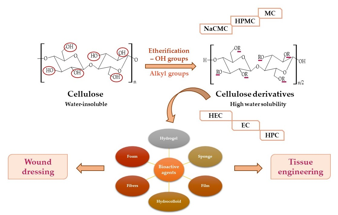
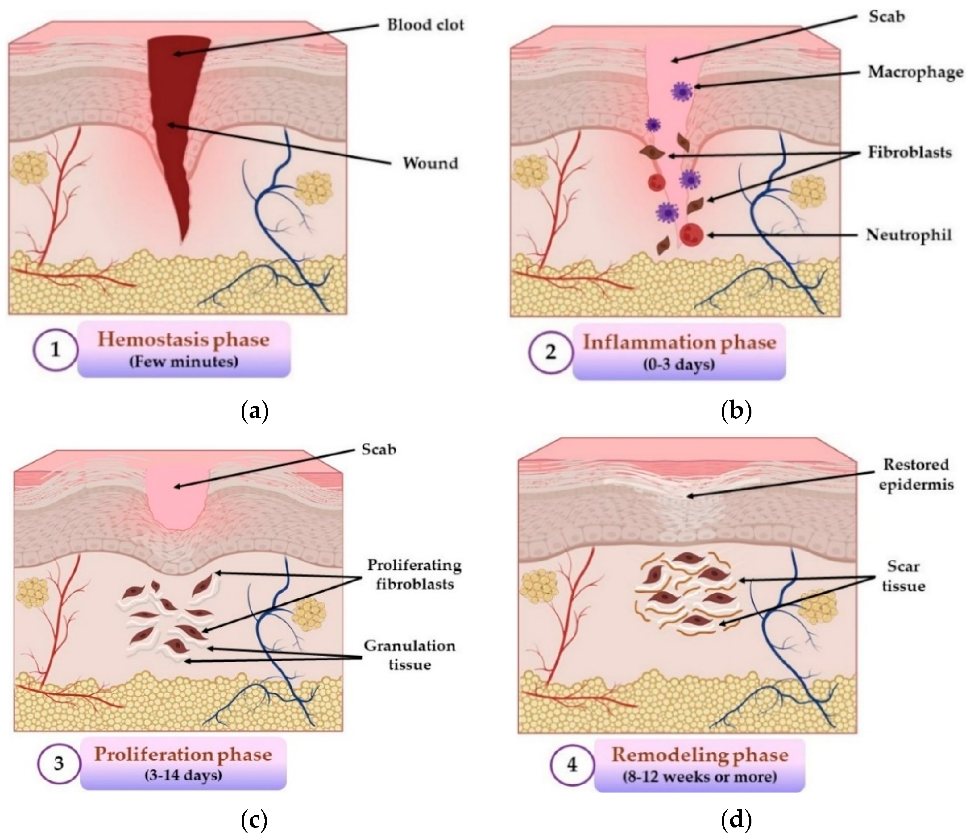
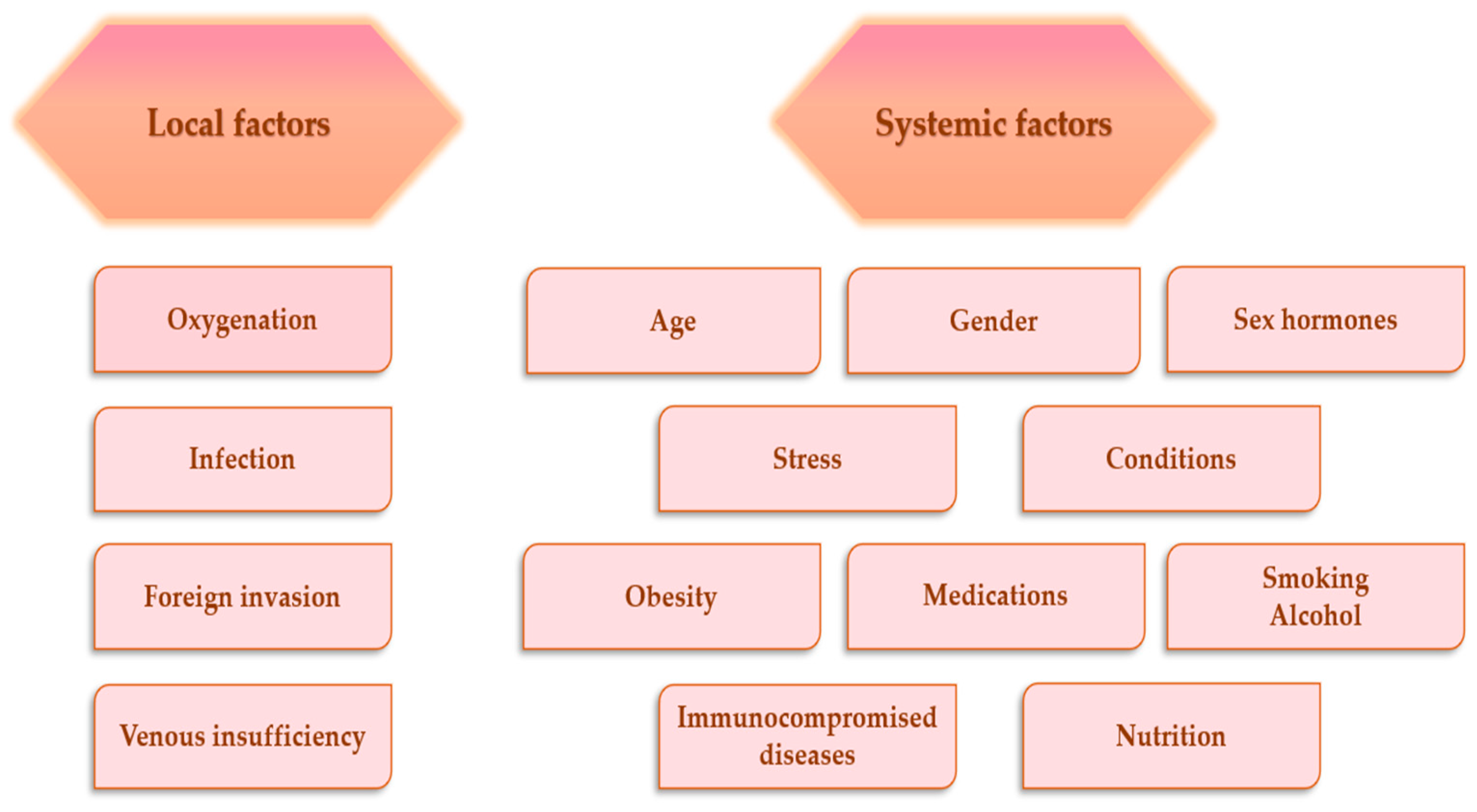
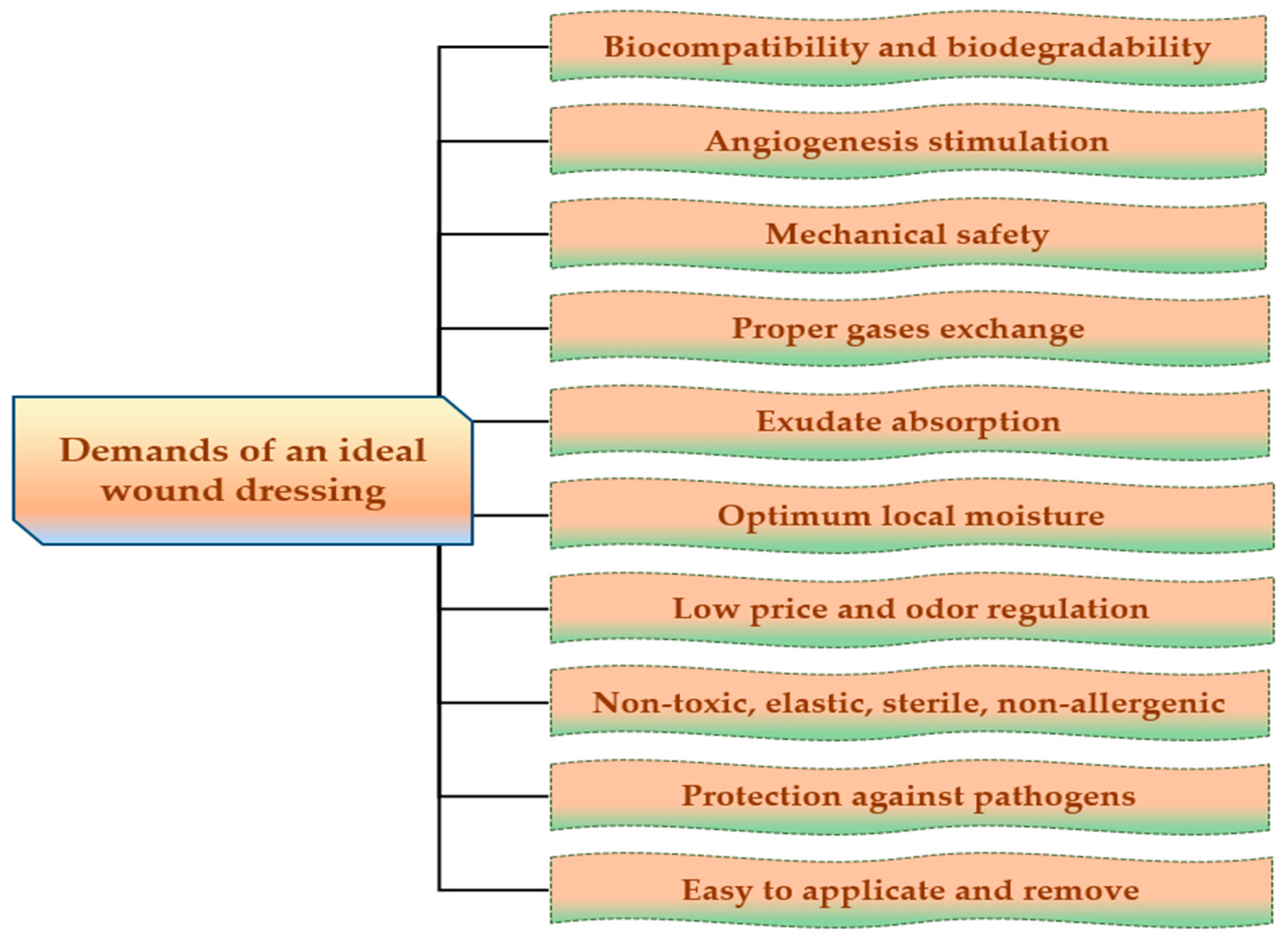
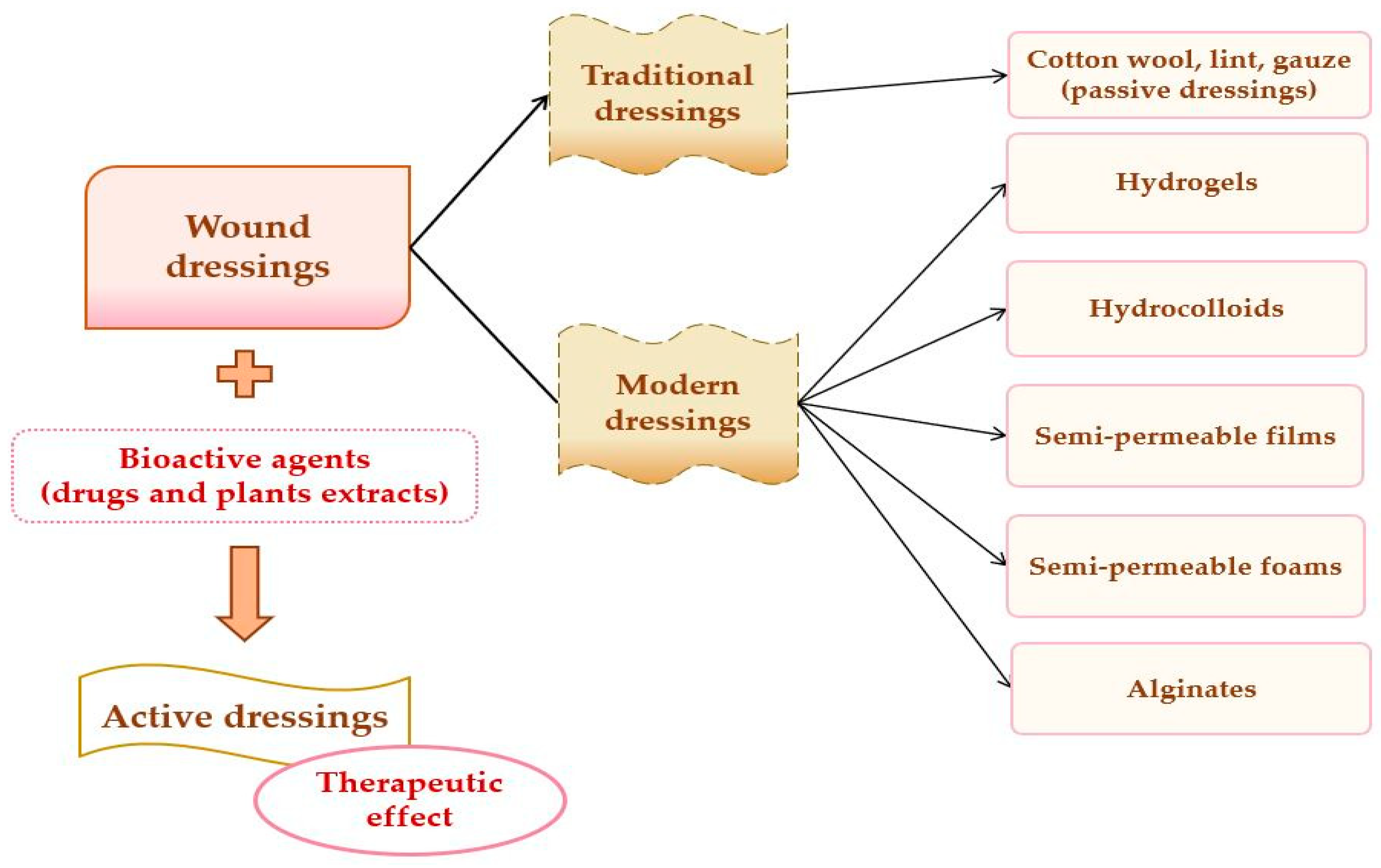
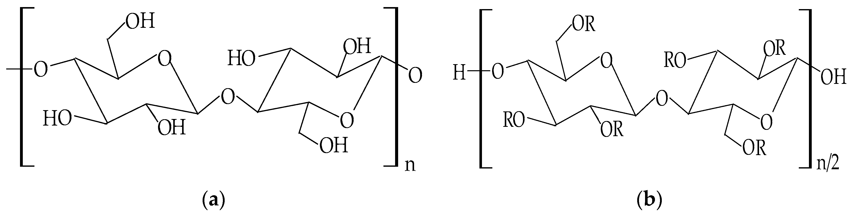
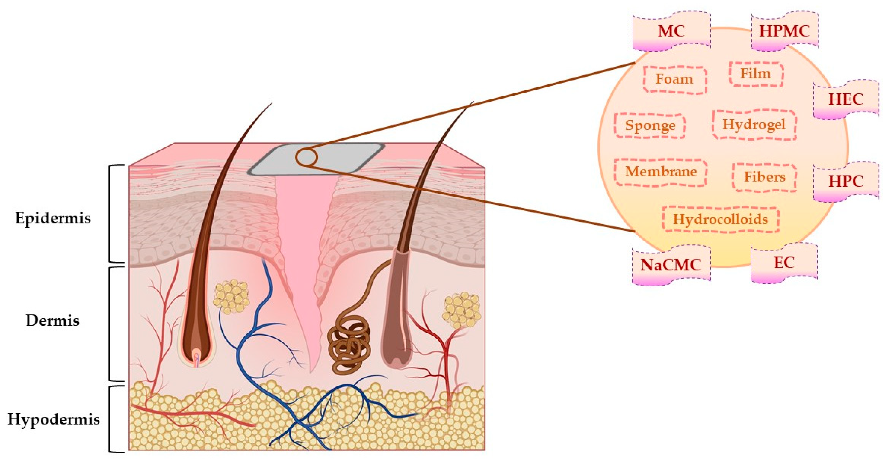
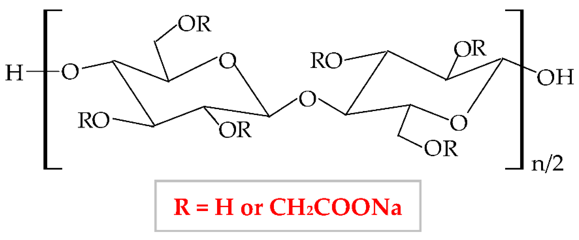
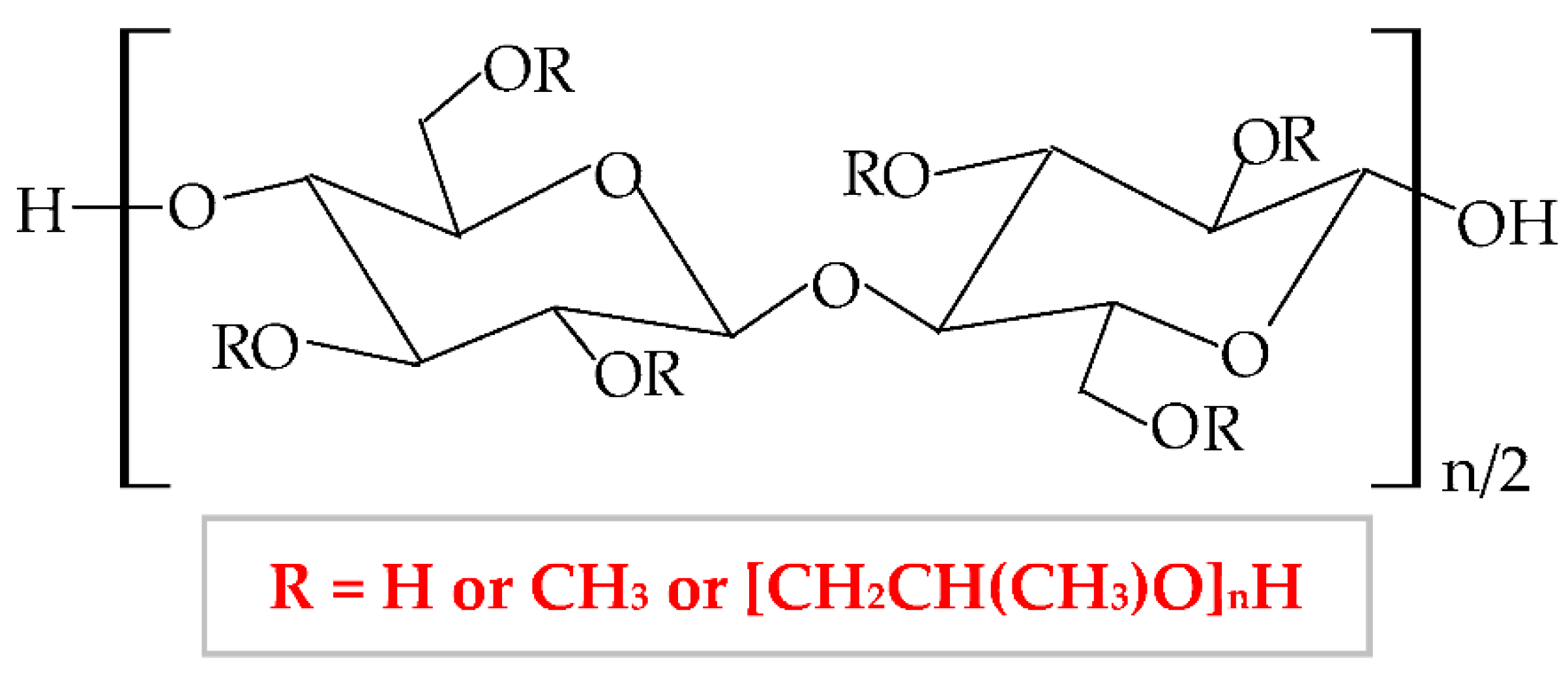
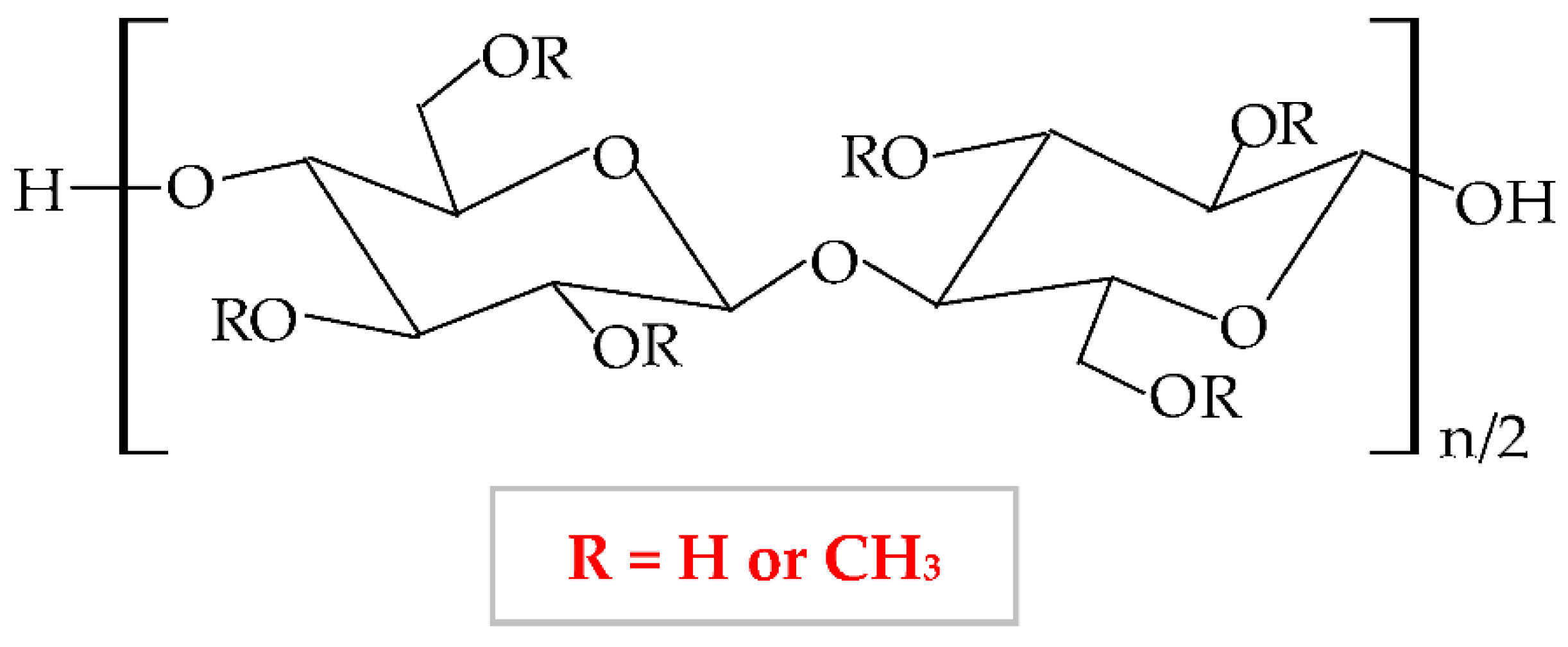
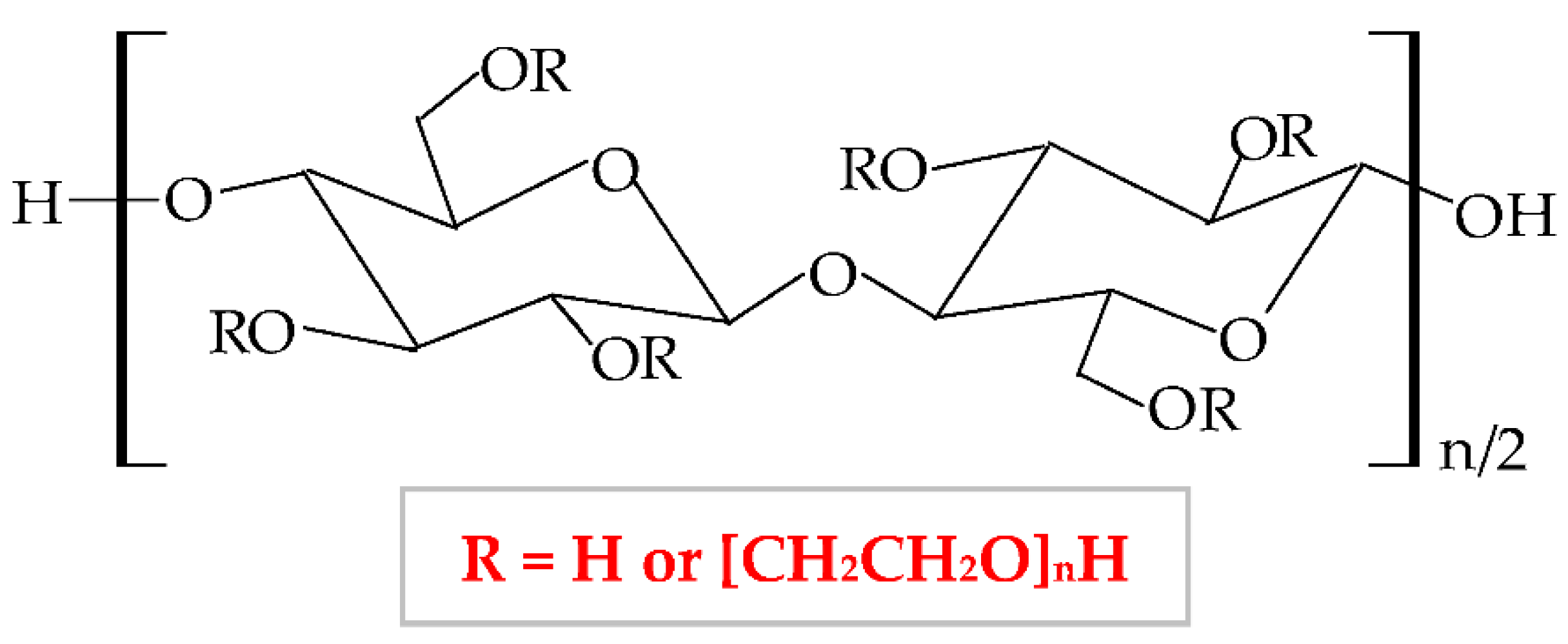
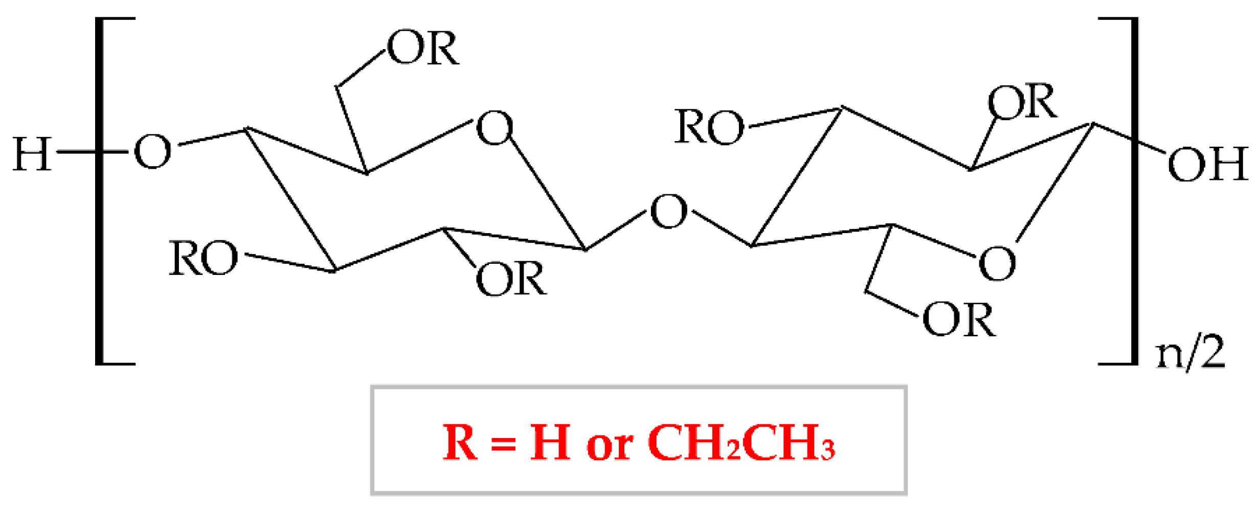
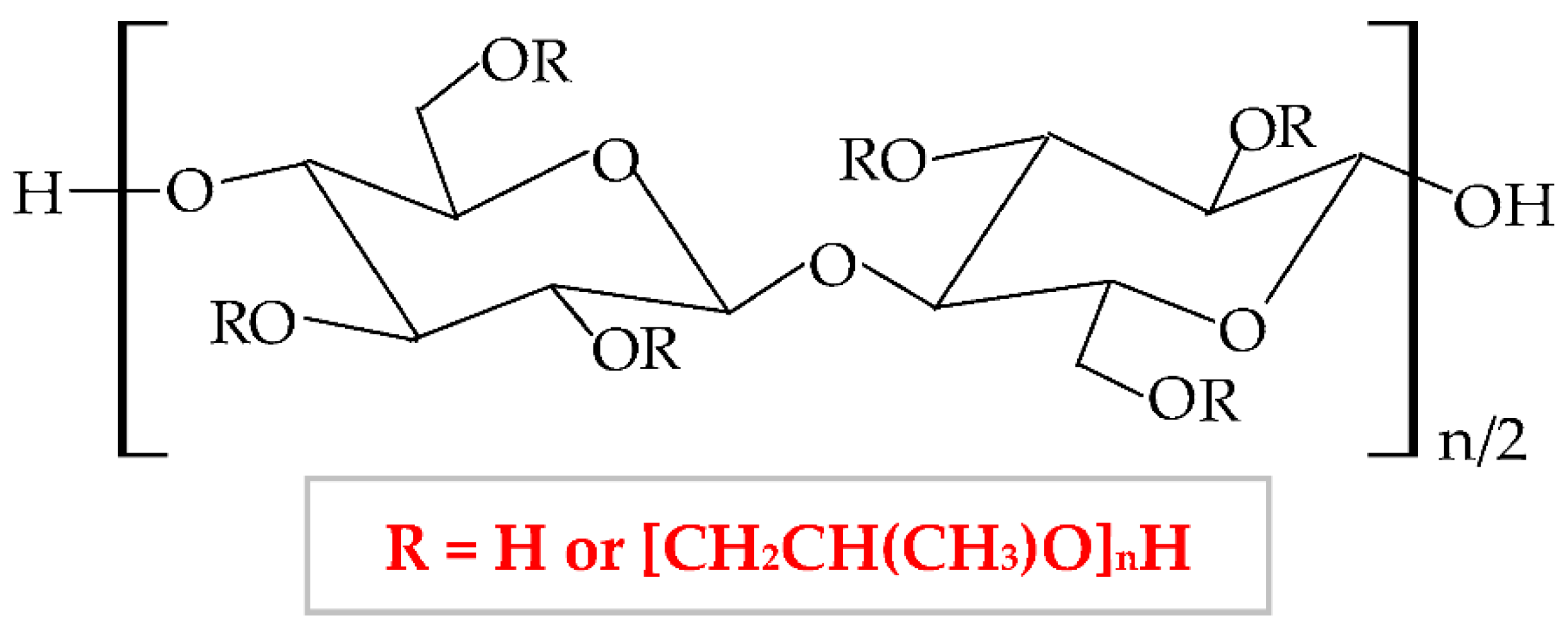
| Cellulose Ethers | R Groups |
|---|---|
| Methylcellulose | H, CH3 |
| Ethylcellulose | H, CH2CH3 |
| Benzylcellulose | H, C6H5CH2 |
| Sodium carboxymethylcellulose | H, CH2COONa |
| Hydroxyethylcellulose | H, [CH2CH2O]nH |
| Hydroxypropylcellulose | H, [CH2CH(CH3)O]nH |
| Hydroxyethylmethylcellulose | H, CH3, [CH2CH2O]nH |
| Hydroxypropylmethylcellulose | H, CH3, [CH2CH(CH3)O]nH |
| Cellulose Esters | R Groups |
|---|---|
| Acetate | H, I |
| Acetate trimelliate | H, I, II |
| Acetate phthalate | I, III |
| Hydroxypropylmthylphthalate | H, CH3, CH2CH(OH)CH3, III, IV |
| Hydroxypropylmthylphthalate acetate succinate | H, CH3, CH2CH(OH)CH3, III, V |
| Biopolymer/-s | Active Pharmaceutical Ingredient (Natural or Synthetic Substances) | Type of Wound Dressing | Main Findings | References |
|---|---|---|---|---|
| NaCMC | - | Gel | Anticoagulant activity at second-degree burn injuries; this action was influenced by concentration, substitution degree, and molecular weight of the biopolymer. | [157] |
| Hydrocolloid | Appropriate management of humidity, maintained self-adhesiveness, and increase of the surface energy. | [85] | ||
| Film | Enhanced tensile strength, absorption capacity, and decreased percent erosion. | [147] | ||
| Foam | Suitable material for wound dressing, because it can maintain optimal local moisture. | [158] | ||
| Merremia mammosa | Gel | Good gel properties, non-toxicity, speeding the wound healing process in diabetic ulcer (increases collagen synthesis and improves angiogenesis). | [159] | |
| Chestnut honey | Hydrogel | Enhanced moisture retention, non-adherence, and excellent antibacterial action to treat diabetic ulcer injuries. | [160] | |
| Reduced graphene oxide | Hydrogel | The inhibition of bacteria biofilm from infected wounds. | [161] | |
| Neomycin trisulphate, sulphacetamide sodium, and silver nitrate | Hydrogel | Proper elasticity, sponginess, homogenous texture, suitable smell, high drugs delivery, and antimicrobial effect. | [162] | |
| Tetracycline–UiO-66 | Hydrogel | Increased mechanical properties, antibiotic delivery, and good antibacterial activity. | [163] | |
| ZnO-MCM-41 and tetracycline | Hydrogel | Improved tensile strength, permeability for gases, and swelling ratio; optimal antibacterial effect. | [164] | |
| Lidocaine hydrochloride | Film | Optimal physicochemical and mechanical properties; high drug release (90%) in the first 15 min. | [165] | |
| AgNPs | Membrane | Good morphology and superior antibacterial activity. | [166] | |
| NaCMC/Chitosan | - | Sponge | High porosity, air permeability, and proper antibacterial effect; in the presence of the cyanine dye, the sponge indicated the pH of the wound site. | [167] |
| NaCMC/Pullulan | - | Hydrogel | An important decrease of the hydrogel adherence at the postoperative lesions. | [168] |
| NaCMC/Sericin | - | Hydrogel | Increased mechanical stability and the strength to hydrolysis and enzymatic degeneration; the molecular weight of CMC influenced the sericin release. | [169] |
| Film | Higher mechanical properties, hydrophilicity, swelling power, stability, compatibility with cells and blood, and stimulation of cell proliferation. | [170] | ||
| NaCMC/ε-Polylysine | - | Hydrogel | Optimal rheological properties, degradability, compression modulus, and high antibacterial activity. | [171] |
| NaCMC/Pectin/Cellulose | - | Film | NaCMC and cellulose enhanced the mechanical properties of pectin; the new composite accelerated the tissue repair through re-epithelialization and collagen deposition. | [172] |
| NaCMC/Gelatin/Pectin | - | Film | Excellent capacity to absorb biological fluid, elongation at break, mechanical properties, and water vapor transmission rate; ideal to cure infected moist injuries. | [173] |
| NaCMC/Chitosan/Sodium alginate | - | Hydrogel | Optimal water vapor permeability, anti-adhesion capacity to treat second-degree burn injuries; reduced the TNF-α and IL-6 levels. | [174] |
| NaCMC/Gelatin/PEG | - | Hydrogel | CMC increased the mechanical properties, enzymatic stability, and sanguineous compatibility; the new hydrogel had 3D and porous structure, absorbed the exudates, and maintained proper moisture. | [175] |
| NaCMC/PEG | - | Hydrogel/Film | Superabsorbent hydrogel with various swelling degrees from 100% to 5000%, appropriate physicochemical and morphological properties to regenerate chronic wounds. | [45] |
| NaCMC/PE/PP | - | Fibers | A serious increase of wet strength; future perspective as a material for the treatment of wounds with mild exudate. | [176] |
| NaCMC/Collagen | Mefenamic acid | Hydrogel | Good ability to swell, optimal morphological structure and excellent release model to treat burns. | [177] |
| AgNPs | Membrane | Suitable antimicrobial action against pathogens from infected burn wounds. | [178] | |
| NaCMC/Gelatin | Ciprofloxacin | Film | In a ratio of 1:1 and 1:2, the film showed good thickness, sorption capacity, blending endurance, and proper antibacterial effect. | [16] |
| NaCMC/Keratin | Clindamycin | Sponge | The keratin enhanced the water stability and the water vapor transmission rate; the sponge impeded the bacterial multiplication. | [2] |
| NaCMC/PEO | Achillea millefolium, Calendula officinalis, Matricaria chamomilla, Echinacea purpurea and Hypericum perforatum | Fibers | Antibacterial and antioxidant activity for acute injuries treatment. | [179] |
| Lidocaine | Nanofibers | Similar characteristics as extracellular matrix; ~50% of lidocaine has been delivered in the first 10 min, relieving the pain quickly. | [180] | |
| AgNPs | Nanofibers | Smooth surface and limited size distribution; excellent antimicrobial effect and photoluminescent act, used as anti-adhesion composite and wound dressing. | [181] | |
| NaCMC/PVA | Propolis | Hydrogel | High swelling power and antimicrobial effect for second-degree burn injuries treatment. | [182] |
| ZnO and heparin | Hydrogel | Favorable mechanical properties, water vapor transmission rate, swelling ratio, and excellent antibacterial effect. | [183] | |
| NaCMC/Soluplus® | 18β-glycyrrhetinic acid (licorice) | Hydrogel | Soluplus® augmented the solubility in water of the bioactive agent; at the lesion site, and in situ hydrogel formed, which had high swelling power and was easy to eliminate by washing. | [184] |
| NaCMC/Sodium alginate/PET/Viscose | Diclofenac sodium | Membrane | Optimal drug release and efficient anti-inflammatory activity. | [185] |
| NaCMC/Sodium alginate | Diclofenac | Film | Homogeneous film with proper water vapor transmission rate; faster alleviation of pain. | [186] |
| Biopolymer/-s | Active Pharmaceutical Ingredient (Natural or Synthetic Substances) | Type of Wound Dressing | Main Findings | References |
|---|---|---|---|---|
| HPMC | Lawsonia inermis (henna) and Matricaria chamomilla | Gel | Adequate stability, extrudability, viscosity, homogeneity, good herbal extract release, and antibacterial effect for burn infection cure. | [217] |
| Liposomal farnesol | Gel | The combination of HPMC:farnesol in a ratio of 1:2 and 2:1 had an excellent effect on tissue regeneration at third-degree burns. | [218] | |
| Bacteriophage | Gel | The gel containing 3% HPMC presented good stability at 37 °C and had an effective antibacterial action against Klebsiella pneumoniae in wound infection treatment. | [219] | |
| CuNPs-licorice and phenytoin | Gel | Effect on acute lesions by the suppression of the inflammatory JAK3 and the synthesis of the procollagen type I. | [220] | |
| Honey and Aloe vera | Hydrogel | At 3% concentration, the hydrogel presented an adequate viscosity to be applied on the burn lesions and showed a proper antibacterial activity on infection with Klebsiella pneumoniae. | [197] | |
| Cefotaxime sodium | Hydrogel | The hydrogel containing 3% HPMC 400 exhibited a high spreadability and released all the drug content after 4 h. | [221] | |
| Haruan/fusidic acid | Film | Films with 1% plasticizers, respectively 2% plasticizers presented a suitable elongation at break and water vapor permeability. | [222] | |
| Epigallocatechin-3-gallate | Film | Enhanced tensile strength and water vapor barrier trait. | [223] | |
| Cooper nanoparticles | Film | Appropriate antibacterial activity. | [190] | |
| HPMC/Silk fibroin | - | Nanofibers | The mixture of HPMC:silk fibroin in a ratio of 7:1 exhibited suitable biocompatibility, mechanical properties, hydrophilicity, and porosity for skin tissue engineering. | [201] |
| HPMC/Collagen/Polyurethane | - | Hydrogel | Increased proteolytic and thermal degradation. | [224] |
| HPMC/Polyacrylate/Tri-isocyanate crosslinked polyurethane | - | Hydrocolloid | The newly designed wound dressing containing 1% cross-linking promoter showed proper humidity (the capacity of the water uptake was 5.1% after 1 h). | [199] |
| HPMC succinate/Chitosan | Gentamycin sulfate | Film | High mechanical properties and antibacterial activity. | [225] |
| HPMC/Chitosan | Simvastatin | Gel | High viscosity and bioadhesive strength. | [17] |
| Toluidine blue O | Hydrogel | High viscosity, hardness, bioadhesion, and bactericidal effect to alleviate the burn wounds caused by light irradiation. | [226] | |
| Pioglitazone hydrochloride | Hydrogel | The formulation containing HPMC E5:chitosan (1:2) showed the best physicochemical properties and the highest drug release. | [193] | |
| Silver sulphadiazine | Film | In a ratio of 1:1, the film showed the best physicochemical properties and the highest drug release; the period of wound healing was of 8 days in comparison with the marketed cream (14 days). | [227] | |
| HPMC K100/Collagen | Curcumin | Nanogel | In vivo studies illustrated a substantial rate of lesion contraction (95.42 ± 12.20%) on the 20th day. | [228] |
| HPMC/Hydroxyapatite | AgNPs | Hydrogel | High porous 3D structure, with excellent mechanical properties and antibacterial activity; after 16 days, wounds healed considerably (94.5 ± 1.4%). | [229] |
| HPMC/Hydroxypropyl-β-cyclodextrin | Coumestrol | Hydrogel | 50% of wounds healed in a shorter time compared to the commercial product, with a suitable tissue re-epithelialization after 12 days. | [230] |
| Gallic acid | Hydrogel | High physicochemical properties and antibacterial effect to prevent wound infection. | [231] | |
| HPMC/PVA/PAA/PVP/PEG | Aloe vera (1%, 6%, 10%) | Nanofibers | The sample with 10 wt% aloe vera showed higher porosity and durability, faster healing of burns, and nonlinear structure. | [200] |
| HPMC/PVA/PVP-I/PEG | Aloe vera (2%, 4%, 6%) | Fibers | Fibers with 6% aloe vera were thinner without any beading, which led to a higher porosity of the fibers. | [232] |
| HPMC K15M/Tara gum | Lawsone | Gel | Suitable homogeneity, uniformity, and drug release; in vivo studies indicated total epithelialization of the excision lesion. | [233] |
| HPMC K15M/Xanthan gum | Nano calcium oxide | Hydrocolloid | Great mechanical strength, superior flexibility, homogeneous thickness, and higher lesion contraction compared to the commercial product. | [234] |
| HPMC/Xyloglucan | Gentamicin sulfate | Film | The best formulation of these polymers was in a ratio of 50:50 and it presented reliable physicochemical properties, good drug release, and favorable antibacterial effect. | [235] |
| HPMC/Sodium alginate | Gatifloxacin | Hydrogel | Advantageous physicochemical properties regarding the tensile strength, the swelling capacity, the elongation, and the drug release. | [236] |
| HPMC/Pluronic® 127 | AuNPs | Gel | Good bioavailability, skin permeation, anti-inflammatory, and antibacterial effect, with an excellent drug release of 98.03% after 6 h. | [237] |
| HPMC/Polyacrylamide | AgNPs | Hydrogel | Superporous hydrogel with high porosity (91.4%), which allows a quicker wound healing, with minimal scar formation. | [238] |
| HPMC/PEO | Beta-glucan | Nanofibers | At the lesion site, the nanofibers produced a hydrogel in situ. | [239] |
| HPMC/Poly(lactic acid) | Tetracycline hydrochloride | Nanofibers | High water sorption rate and antimicrobial activity. | [240] |
| HPMC/Polyurethane | Silver and asiaticoside | Foam | Increased absorption capacity and compressive strength development; adequate antimicrobial activity. | [241] |
| HPMC/Chondroitin sulfate/Sodium hyaluronate | Silver sulphadiazine | Sponge | Proper elasticity, softness, flexibility, and bioadhesive properties, alongside antibacterial activity. | [46] |
| HPMC K100M/Gum Odina/Gelatin | Fluconazole and ofloxacin | Sponge | The formulation with gum Odina-HPMC K100M:gelatin (1:1) showed excellent physicochemical properties and antimicrobial activity to cure chronic wounds. | [242] |
| HPMC/HA/Methyl-β-cyclodextrin | Curcumin | Film | Excellent antimicrobial effect. | [243] |
| HPMC/Hydroxypropyl-β-cyclodextrin/Chitosan | Caffeic acid | Hydrogel | Drug delivery system with superabsorbent capacity, higher swelling property at pH 7, and good antimicrobial effect to prevent wound infection. | [244] |
| HPMC/Chitosan/Sodium alginate | Lidocaine chloride and polymyxin B sulphate | Biomembrane | Proper mechanical properties (elasticity, tension, stiffness) and thickness; in vivo: high antimicrobial effect for tissue regeneration. | [245] |
| HPMC/Polyglycolic acid/Vicryl®/Catgut | Ofloxacin | Hydrogel | Proper physicochemical properties, which ensures a high lesion size contraction after 14 days (95%) and large collagen deposition on the 21st day. | [246] |
| Biopolymer/-s | Active Pharmaceutical Ingredient (Natural or Synthetic Substances) | Type of Wound Dressing | Main Findings | References |
|---|---|---|---|---|
| MC | Zinc oxide and Silver | Ointment | MC 3% ointment showed an elastic behavior and thixotropy, which can be used for injured skin with complex relief. | [260] |
| Allium hirtifolium | Gel | In a ratio of 1:1, the newly developed gel accelerated the open wounds healing through the tissue re-epithelialization. | [261] | |
| Cryopreserved human culture of fibroblasts and AuNPs | Gel | High capacity to heal the third-degree burns because it restored the composition of type I and III collagen on 21st day of the treatment. | [262] | |
| Vitamins (C, B1, and B6) | Hydrogel | Enhanced gelation rate and mechanical strength, with suitable applicability for wound treatment. | [263] | |
| Gallic acid and doxycycline | Hydrogel | At body temperature, the novel formulation formed an in situ gel and released the bioactive agents to heal the deep tissue injuries. | [252] | |
| Neomycin trisulphate, sulphacetamide sodium, and Silver nitrate | Hydrogel | New formulation exhibited elasticity, sponginess, homogenous texture, proper smell, and white color. | [162] | |
| Silver oxide nanoparticles | Hydrogel | Thermo-responsive hydrogel, which led to wound burn regeneration due to its superior antimicrobial effect. | [264] | |
| Pedilanthus tithymaloides | Film | A concentration of 0.5% of the plant extract accelerated the re-epithelialization of the wounded skin. | [265] | |
| AgNPSs | Film | Increased water absorption capacity and contact angle value; the temperature influenced the release of AgNPs. | [266] | |
| Borate bioactive glass and Manuka honey | Foam | The foam exhibited high porosity, better wettability, mechanical properties, and antibacterial effect. | [248] | |
| MC/α-Chitin nanocrystals | - | Hydrogel | The novel nanocomposite presented high mechanical strength and gelation rate, being a promising dressing for tissue engineering. | [267] |
| MC/Fucoidan | - | Film | The newly designed formulation could be a promising dressing for wounds with a smaller production of exudates because it possessed a lower capacity to swell. | [268] |
| MC/Nano hyaluronic acid | - | Hydrogel | The mixture of these polymers led to an in situ hydrogel development, which enhanced tissue regeneration. | [269] |
| MC/Mucin | - | Hydrogel | MC enhanced the mechanical properties of the mucin and formed a thermoresponsive gel, which can be used for different wounds treatment. | [270] |
| MC/2-Methacryloyloxy ethyl phosphorylcholine | - | Hydrogel | It reduced the postoperative adhesion effect by inhibition of collagen proteins. | [271] |
| MC/Gelatin/Gellan gum/PVP | - | Film | The blend of all these polymers led to a novel wound dressing with suitable physicochemical properties to restore minor lesions. | [272] |
| MC/Chitosan | Adenosine and vitamin C | Hydrogel | Thermo-responsive and self-healing ability and a higher release of adenosine. | [273] |
| Exosomes | Hydrogel | Suitable mechanical properties, good gelation time, and excellent self-healing for treatment of severe tissue injuries. | [274] | |
| MC/Hyaluronic acid | - | Gel | It was shown that a low molecular weight of hyaluronic acid increased the biocompatibility and the thermogelation of this new formulated composite. | [275] |
| AgNPs | Hydrogel | Excellent morphological, swelling, and spectral properties, together with the high antibacterial effect to treat burn wounds in children (99.6%). | [276] | |
| MC/sECM | Stem cells | Hydrogel | At the wound site, the hydrogel had a thermosensitive sol-gel transition, which accelerate the wound healing through neovascularization and re-epithelialization. | [277] |
| MC/Sericin | Tranilast | Ointment | The combination of the two polymers and the antiallergic drug exhibited excellent results to augment wound healing and to decrease the redness in diabetic rats. | [278] |
| MC/Silk fibroin | 5-Aminosalicylic acid | Hydrogel | The gelation time of the novel composite has been enhanced by adding MC; thus, this hydrogel presents important uses as a wound dressing and drug release system. | [279] |
| MC/Sodium alginate | Manuka honey, aloe vera, and eucalyptus essential oil | Hydrogel | The formulation presented proper swelling capacity, biocompatibility, and suitable antimicrobial and antibiofilm action. | [280] |
| Gallium (Ga3+) | Hydrogel | It was illustrated that this novel hydrogel is a promising dressing for wounds infection prevention due to its large antibacterial action (99.99%) and cytocompatibility. | [281] | |
| Montmorillonite | Film | A high content of montmorillonite led to higher tensile strength and antibacterial activity of the newly designed film. | [282] | |
| MC/Sodium alginate/Poly(N-isopropylacrylamide) | Octenisept® | Hydrogel | The novel hydrogel had a large viscosity, an expressed shear-thinning nature, high antibacterial action, a homogenous and microporous structure. | [283] |
| MC/Pluronic F-127 | GT/siMMP9 | Hydrogel | It was demonstrated that this new dressing had thermosensitive traits by forming in situ and it reduced the MMP-9 level in diabetic chronic injuries. | [284] |
| MC/Polyacrylamide | Aloe barbadensis | Hydrogel | At a 2% concentration of Aloe barbadensis, the hydrogel showed suitable thermal stability; it also presented an antibacterial effect to cure chronic cutaneous wounds. | [285] |
| MC/Poly(ε-caprolactone) | Bioactive glass and Manuka honey | Fibers | The blend of these two polymers led to higher mechanical properties and wettability. | [286] |
| MC/PVA | Asiaticoside | Film | This newly formulated dressing exhibited significant mechanical properties, flexibility, transparency, and a shorter healing time of skin wounds. | [287] |
| - | Nanofibers | Adequate physicochemical features and antibacterial activity to be used as a drug-delivery system for injuries management. | [288] |
| Biopolymer/-s | Active Pharmaceutical Ingredient (Natural or Synthetic Substances) | Type of Wound Dressing | Main Findings | References |
|---|---|---|---|---|
| HEC | - | Hydrogel | Good antibacterial effect; at a concentration between 12.5% and 15% of HEC, the water absorption rate was the highest. | [309] |
| HEC | Curcuma longa | Hydrogel | Suitable porous network, mechanical and release properties; besides therapeutic effect, the new hydrogel can illustrate the pH of the lesion site. | [310] |
| WO3 | Hydrogel | The hydrogel with 0.04% WO3 showed the highest capacity to heal the injuries due to optimal antibacterial and anti-inflammatory action. | [300] | |
| Mesocellular silica foam | Sponge | Adequate cytocompatibility, antibacterial and hemostatic action to heal full-thickness wounds. | [311] | |
| Graphene oxide | Film | Improved mechanical and thermal properties. | [312] | |
| AgNPs | Membrane | Limited degradation rate, excellent porosity, and water absorption value. | [313] | |
| HEC/Collagen | - | Film | High swelling ratio, mechanical and thermal characteristics, degradation, and adequate biocompatibility. | [314] |
| HEC/Collagen/PVA | - | Nanofibers | Suitable water absorption rate and degradation behavior, a promising material for skin tissue engineering. | [315] |
| HEC/Carboxymethyl chitosan | - | Hydrogel | High biocompatibility, gelation time, water evaporation rate, and swelling power. | [290] |
| HEC/Sodium alginate/Hydroxyapatite | - | Membrane | High porous network, improved mechanical strength, and rigidity gradient. | [316] |
| HEC/Hyaluronic acid | - | Hydrogel | Optimal biocompatibility and blood compatibility, gelation time, water evaporation rate, water retention ability, and swelling power. | [295] |
| Isoliquiritigenin | Hydrogel | pH-sensitive hydrogel with suitable adhesion, rheological properties, and antibacterial efficacy. | [317] |
| Biopolymer/-s | Active Pharmaceutical Ingredient (Natural or Synthetic Substances) | Type of Wound Dressing | Main Findings | References |
|---|---|---|---|---|
| EC/Gum tragacanth | Honey | Nanofibers | The ratio of 85:15 with 20% honey showed the best mechanical, biological, and antibacterial properties to be used as an efficient wound dressing. | [321] |
| EC/PVP | Naproxen | Nanofibers | The ratio of 4:1 had the fastest release of the drug; thus, it can be used as a dressing to reduce the inflammation and the pain in acute wounds. | [333] |
| AgNPs and ciprofloxacin | Nanofibers | Uniform and cylindrical morphology, with over 90% release of ciprofloxacin and a high antibacterial effect. | [334] | |
| EC/Poly(3-hydroxybutyrate) | p-4-hydroxybenzoic acid and ferulic acid | Film | Potent bacteriostatic and bactericidal action to heal wounds infection facilitating the skin restoration. | [335] |
| EC/Polylactic acid/Collagen | Silver sulfadiazine | Nanofibers | Good mechanical properties, proper antimicrobial effect, and enhanced cell proliferation. | [336] |
| EC | Ciprofloxacin | Nanofibers | Cylindrical and homogenous aspect; high antibacterial action. | [318] |
| EC/Zein | Photosensitizer protoporphyrin and vaccarin | Membrane | Adequate flexibility, hygroscopicity, nanonetwork structure, and mechanical properties; in vivo study showed increased angiogenesis of the injured tissue. | [337] |
| EC/PVA | Luliconazole | Nanosponge | Optimal viscosity, spreadability, retention time, permeation rate, and also high antifungal effect against dermatophytes. | [338] |
| Biopolymer/-s | Active Pharmaceutical Ingredient (Natural or Synthetic Substances) | Type of Wound Dressing | Main Findings | References |
|---|---|---|---|---|
| HPC | Peptide PXL150 | Gel | Due to the antimicrobial activity, the gel healed the infection of third-degree burns injuries and surgical wounds. | [355] |
| - | Hydrogel | Suitable water absorption; the hydrogel can maintain an adequate moisture balance at the lesion site. | [339] | |
| Graphene oxide, Isophorone diisocyanate, and Ag/ZnO | Film | Higher mechanical, anti-ultraviolet properties, and antimicrobial activity. | [356] | |
| HPC/Gelatin | Chloramphenicol | Hydrogel | Adequate mechanical strength, water vapor permeability, light transmittance, and excellent antibacterial effect. | [357] |
| HPC/Chitosan/PEO | Graphene | Membrane | Proper mechanical properties, hydrophilicity, water vapor transmission rate, and high antibacterial effect; the membrane can impede the bacterial adhesion. | [341] |
| HPC/Sodium alginate | Gatifloxacin | Hydrogel | Advantageous physicochemical properties regarding the tensile strength, the swelling capacity, the elongation, and the drug release. | [236] |
| Pioglitazone | Hydrogel | By adding HPC, the gelation behavior of the hydrogel increased, with an extended release of the drug up to 5 days; thus, the novel formulation can be used for skin ulcer treatment. | [358] |
| Biopolymer/-s | Active Pharmaceutical Ingredient (Natural or Synthetic Substances) | Type of Wound Dressing | Main Findings | References |
|---|---|---|---|---|
| EC/HPMC | Paromomycin and Gentamicin | Film | Optimum drugs release and inhibition of Leishmania tropica growth. | [359] |
| Aloe vera | Nanofibers | Nanofibers with 10% Aloe vera showed suitable mechanical properties, biocompatibility, bioadhesion, and suitable antibacterial activity. | [360] | |
| NaCMC/HPMC | Grapefruit seed extract | Film | Suitable elongation at break, stability in water, and proper antibacterial action. | [361] |
| CuO | Film | Good biocompatibility and antibacterial effect. | [362] | |
| Tetracycline/Methylene blue | Film | Nanoporous network, increased Tg and elongation at break, sustained drug release for 72 h and high antibacterial effect. | [363] | |
| ZnO NPs | Film | Biocompatibility and optimum antibacterial action. | [364] | |
| NaCMC/HPMC/ CAB | Resveratrol | Membrane | Excellent adhesive capacity, hydration efficiency, and higher porous structure; in vivo studies showed accelerated wound healing. | [365] |
| NaCMC/MC | Simvastatin | Membrane | In a ratio of 2:1, the membrane exhibited appropriate flexibility, viscosity, stability, and sponginess; optimal drug delivery for suppurating injuries. | [366] |
Publisher’s Note: MDPI stays neutral with regard to jurisdictional claims in published maps and institutional affiliations. |
© 2021 by the authors. Licensee MDPI, Basel, Switzerland. This article is an open access article distributed under the terms and conditions of the Creative Commons Attribution (CC BY) license (https://creativecommons.org/licenses/by/4.0/).
Share and Cite
Tudoroiu, E.-E.; Dinu-Pîrvu, C.-E.; Albu Kaya, M.G.; Popa, L.; Anuța, V.; Prisada, R.M.; Ghica, M.V. An Overview of Cellulose Derivatives-Based Dressings for Wound-Healing Management. Pharmaceuticals 2021, 14, 1215. https://doi.org/10.3390/ph14121215
Tudoroiu E-E, Dinu-Pîrvu C-E, Albu Kaya MG, Popa L, Anuța V, Prisada RM, Ghica MV. An Overview of Cellulose Derivatives-Based Dressings for Wound-Healing Management. Pharmaceuticals. 2021; 14(12):1215. https://doi.org/10.3390/ph14121215
Chicago/Turabian StyleTudoroiu, Elena-Emilia, Cristina-Elena Dinu-Pîrvu, Mădălina Georgiana Albu Kaya, Lăcrămioara Popa, Valentina Anuța, Răzvan Mihai Prisada, and Mihaela Violeta Ghica. 2021. "An Overview of Cellulose Derivatives-Based Dressings for Wound-Healing Management" Pharmaceuticals 14, no. 12: 1215. https://doi.org/10.3390/ph14121215