Disordered Mechanical Stress and Tissue Engineering Therapies in Intervertebral Disc Degeneration


Key Laboratory of Biorheological Science and Technology, Ministry of Education, Bioengineering College, Chongqing University, Chongqing 400044, China
*
Authors to whom correspondence should be addressed.
Polymers 2019, 11(7), 1151; https://doi.org/10.3390/polym11071151
Submission received: 31 March 2019
/
Revised: 27 June 2019
/
Accepted: 1 July 2019
/
Published: 5 July 2019
(This article belongs to the Special Issue Polymeric Materials for Tissue Engineering)
Abstract
:Low back pain (LBP), commonly induced by intervertebral disc degeneration, is a lumbar disease with worldwide prevalence. However, the mechanism of degeneration remains unclear. The intervertebral disc is a nonvascular organ consisting of three components: Nucleus pulposus, annulus fibrosus, and endplate cartilages. The disc is structured to support our body motion and endure persistent external mechanical pressure. Thus, there is a close connection between force and intervertebral discs in LBP. It is well established that with aging, disordered mechanical stress profoundly influences the fate of nucleus pulposus and the alignment of collagen fibers in the annulus fibrosus. These support a new understanding that disordered mechanical stress plays an important role in the degeneration of the intervertebral discs. Tissue-engineered regenerative and reparative therapies are being developed for relieving disc degeneration and symptoms of lower back pain. In this paper, we will review the current literature available on the role of disordered mechanical stress in intervertebral disc degeneration, and evaluate the existing tissue engineering treatment strategies of the current therapies.
1. Introduction
Low back pain (LBP) is the most common reason for disability in developed countries accounting for 10.7% of the total population of disabled people [1]. With a three-month prevalence of as high as 40% in the United States, out of which 20%–33% of patients are unable to work, this disease has a major socio-economic impact [2]. Intervertebral disc degeneration (IDD) is a chronic disease that slowly degrades the content of intervertebral disc (IVD) and leads to unstable IVD, which limits the mobility of the spinal cord [3]. It is well established that IDD is the main contributor of LBP [4,5,6,7,8,9,10]. Depending on the stage of degeneration, current clinical strategies are divided into conservative and surgical therapies. However, these strategies are restricted to relieving the pain and symptoms without eliminating the disease itself due to an incomplete understanding of the pathobiology of IDD [11,12,13,14,15].
The main functions of the IVD are to transmit and absorb mechanical stress onto the spine and to maintain a mobile segment that allows flexion, extension, bending, and rotation [3,16]. Anatomically, a normal IVD can be roughly divided into the following three regions: (1) Nucleus pulposus (NP) with a gel-like core comprised of type II collagen and proteoglycans (Figure 1) [17,18]. NP cells are highly hydrated and account for the strength and mobility of the spine [19]. (2) Annulus fibrosus (AF) is a multi-layered fibrous tissue that surrounds the NP [20]. Unlike NP, AF is highly organized, consisting of stacked lamellae predominantly of type I collagen [21]. AF is required for transmitting stress from the NP, and maintaining the integrity of IVD and protecting it from injuries from bending, stretching, and twisting [22,23,24,25,26,27,28]. (3) End plates (EPs) are two hyaline cartilages that are enclosed, at the interfaces, by the superior and inferior vertebral bodies [29,30].
The altered biomechanics in IVD has been widely accepted as an important contributor to IDD because biomechanical loading directly affects IVD cell metabolism [31,32]. Macroscopically, overloading such as frequent bending and twisting [33], fatigue loading [34], and heavy physical work [35] raise a high risk of lumbar disc degeneration. While hypomobility, such as sedentary environment, also increase the intradiscal pressure depending on seated posture [36]. Herein, we define these aberrant altered biomechanics as disordered mechanical stress. NP stems from the axial notochord. In the embryonic stage, NP consists of notochordal cells (NCs), which gradually transform into chondrocyte-like cells (CLCs) post-embryonic and adult states [37]. A number of studies have shown that disordered mechanical stress can result in a loss of NCs, subsequently leading to IDD [38,39,40,41]. The alignment of collagen fibers is regulated by the orientation of cells in the AF, which are susceptible to mechanical stress [42,43,44,45,46,47,48]. However, disordered mechanical stress alters the structure of AF resulting from reorientation of, and that may impair the function of, AF and causes IDD. Besides NP and AF, angiogenesis is another constant feature of IDD [49] and disordered mechanical stress supported angiogenesis by inducing vascular endothelial growth factor (VEGF) [50]. Taken together, disordered mechanical stress may become another reason for IDD besides biochemistry, except for the common concept of biochemistry factor. Because disordered mechanical stress mainly induces the structure pathological changes of NP and AF, further results in the loss of mechanical function of IVD. Strategies for restoring the structures of NP and AF are essential in the clinical trials.
Evidence is accumulating on the use of tissue engineering strategies for treating IDD [51,52,53,54]. Various tissue-engineered scaffolds with feasible substances (e.g., functional cells, growth factors, proteins, peptides, etc.) are being applied in IDD therapy, achieving significant curative effects. In this paper, we will discuss the effect of disordered mechanical stress on NP, AF, and angiogenesis, and the current tissue engineering therapies in IDD. We expect that this review will support a new direction to an understanding of IDD mechanism and therapy application.
2. Disordered Mechanical Stress Leads to NP Degeneration
The NP is derived from the notochord during embryonic gastrulation [38,41]. During the early stages of embryogenesis, cells migrate to the endoderm transiently and form the notochordal plate that subsequently detaches from the ectoderm to form the definitive notochord, a rod-shaped embryonic structure that crosses the sclerotome. When the sclerotome starts to form the vertebral body, the notochord condenses and locates to the center of disc to form the NP [55]. NCs are the main cell type of the NP during the embryonic period. The most significant feature of NCs is that they have giant cytoplasmic vacuoles [39,56]. Studies have revealed that biosynthetic trafficking, and not endocytosis, is necessary for vacuoles formation [51].
In humans, this population of NCs is reported to decrease during the first decade of life and to have disappeared after that period [37,57]. There are, however, reports of a small percentage of cells expressing notochordal markers persisting until adult life [58]. Recent studies found that NCs secrete connective tissue growth factor that promotes the proliferation of CLCs and induces ECM secretion [59] and protects CLCs against apoptosis [60]. However, as IVD experiences long-term mechanical stress, experiments revealed that there is a negative influence of disordered mechanical stress to NCs and vacuoles. Compared with CLCs, NCs are more sensitive to mechanical stress [61]. Thus, it is notable that disordered mechanical stress-induced NCs loss may contribute to the initiation of IDD. However, other animals, such as pigs, rabbits, non-chondrodystrophic dogs, mice, and rats retain the vacuolated morphology of BCs until much later in life (Table 1) [62]. Taking into account that humans commonly lose NCs in adolescence and the cost of experimental animals (such as pigs and dogs), rodents are the favored animal model to investigate the role of NCs in IDD. In a rat tail disc degeneration model, static compression for 20 weeks significantly decreased the number of NCs and increased the expression of markers of apoptosis by day 7. Using the same compression model, researchers showed that the population of NCs a sharply decreased from 70% to less than 10% after loading [61,63]. Using genetic ablation of vacuoles or fragmentation of vacuoles in zebrafish, Ellis et al. generated larvae shorter in the anterior–posterior axis after five days post-fertilization, many of which eventually developed scoliosis of the spine later during development [64].
3. Disordered Mechanical Stress Leads to AF Degeneration
The AF is a fibrous connective tissue where collagen fibers align together in the same direction to form a single lamella, and adjacent lamellae are connected in opposite directions. Depending on the direction of the inner AF and the outer AF, the thickness of the collagen fiber lamellae varies between 200 and 400 μm [65]. In an adult IVD, there are about 25 lamellae circumferentially surrounding the NP [66]. Unlike NCs, cells in the AF are organized in a specific orientation. Type I collagen is the main element of the outer layers of AF, which is gradually replaced by type II collagen in the inner layers [67]. Collagen alignment is the key to the functional role of the AF. AF cells generate tractions that induce the ECM to organize along the axis of the AF cells [42,43,44,45,46,47,48]. The traction forces alter the direction of actin fibers connected to collagen fibers in the ECM [68,69] and any perturbation to actin fibers can disrupt the alignment of collagen fibers affecting the function of the AF [70]. AF cells are sensitive to mechanical stress, and mechanical function is superior to cell traction forces in collagen fibers alignment. Mechanical stress influences the AF cells through several cytoskeletal molecules such as adhesion receptors like integrin receptors, protein tyrosine kinase (PTK), and mechanosensitive channels [71,72,73,74,75,76]. A healthy IVD maintains a balance of anabolism and catabolism. Degradation of collagen fibers by zinc-dependent matrix metalloproteinases (MMPs) results in disordered stress [77]. Disordered mechanical stress induces an unbalanced loading to collagen fibers, while MMPs can degrade the unloaded or less loaded collagen fibers and destroy the architecture of AF [78,79,80]. Mechanistically, MMPs degrade collagen fibers by binding to the cleavage site. Studies have shown that disordered mechanical stress may expose the cleavage binding sites by changing the spatial structure of collagen monomers and three alpha chains [79,80,81]. Taken together, it is possible to imply that disordered mechanical stress may change the structure of IVD at cellular as well as tissue level.
4. Disordered Mechanical Stress Leads to Angiogenesis
Accumulating studies demonstrated that angiogenesis exists in IDD. Disordered mechanical stress facilitates the neovascularization by destroying the physical barriers, including increased lamellar disorganization and fissures [82]. Moreover, one study demonstrated that disordered mechanical stress can directly influence the ingrowth of blood vessels. Human AF cells that experienced cyclic tensile strain showed a nearly 70% increase of gene expression of pleiotrophin, in which the pleiotrophin is regarded as a pathologic alteration of disc tissue and its neovascularization [83].
In summary, there is sufficient evidence to support the concept that disordered mechanical stress can widely influence the structure and function of IVD both at the cellular and tissue level. Disordered mechanical stress induces NCs apoptosis, which promotes the proliferation of CLCs and maintains the normal function of NP [84]; the loss of NCs also means the initiation of the IDD. AF is also sensitive to disordered mechanical stress, which can change the AF structure from normal to pathological morphology, and the ingrowth of blood vessels further impairs the integrity of IVD (Figure 2). There are numerous studies that chronicle the development and application of tissue engineering-based therapies in IDD (Table 2). Although these studies are versatile, they can be divided into two areas as follows: Regenerative therapies and displacement therapies in IVD.
5. Tissue Engineering-Inspired Strategies to Address IDD
5.1. Strategies in IDD Regeneration
Based on the structure of the IVD, treatments can be divided into NP regeneration, AF regeneration, and anti-angiogenesis.
5.1.1. NP Regeneration
The NP is gelatinous, consisting predominantly of type II collagen and proteoglycans, with a high water content that allows it to transmit stress and resist compressive forces when unloaded; the NP absorbs water by ionic interaction, which is ejected out into the intercellular space in response to mechanical stress [73,74,75]. In recent years, bioengineered scaffolds that are similar to the native NP structure and mechanical properties have been gaining attention. Hydrogel, which is one of the numerous biomaterials, has been pushed to the forefront of IVD treatment. Hydrogel is not biocompatible and does not have suitable mechanical properties per se. However, by changing the polymer type and optimizing the fabrication methods, hydrogel could obtain high water content, good biocompatibility, 3D network structure, and suitable biomechanical property, which is similar to the natural IVD [98]. Gan et al. created a hydrogel with dextran and gelation as the first network and poly (ethylene glycol) (PEG) as the second network to form a 3D inter-crossing network. This hydrogel was conducive to NP cell proliferation and ECM deposition in vitro, and longer NP cell retention in rat IVDs, and in the degenerated porcine IVD model, the hydrogel contributed to rehydration and regeneration of the NP [99].
Apart from being cost effective, hydrogels fabricated with high-molecular-weight polymers or native substances also perform well due to better biocompatibility, and sensitivity to the surrounding environment. Chen et al., generated a high molecular weight hyaluronic acid-gelatin-adipic acid dihydrazide (oxi-HAG-ADH) hydrogel with several advantages including: (1) Anti-inflammatory and immunosuppressive activities, necessary for clinical application; (2) low viscosity, easy to inject; (3) similar viscoelastic property as native tissue; (4) furthermore, the hydrogel supported NP cell phenotype, promoted attachment as well as proliferation, and induced the expression of type II collagen, aggrecan, Sox-9, and HIF-1A- key genes of the NP ECM [100]. With a better understanding of IDD mechanism, functions as well as NP-related proteins have been discovered; laminin, one of these functional proteins, has been demonstrated to support the attachment and biosynthesis of NP by interacting directly with NP cells [101,102,103]. Laminins are a major component of the NP ECM and interact directly with NP cells to regulate their function. Setton et al. found that laminin 111 or peptides derived from laminins can be coupled with modified PEG or poly-acrylamide gels to form functional scaffolds. These hydrogels provided a favorable environment for NP cell, proliferation, and more importantly, promoted the expression of specific markers characteristic of immature NP cells [104,105,106].
Besides laminin derived peptides, there are a number of self-assembling peptide hydrogels (SAPH) that have been used, but very few of them were evaluated for rheological behavior, a key mechanical property of NP [107,108,109]. A study adopted the FEFEFDKFK (F: Phenylalanine, E: Glutamic acid, L: Lysine) amino acid chain to form SAPH and reported no significant difference in oscillatory rheology between acellular and bovine cells-seeded on SAPH, while the total number of cells decreased with increase in load over time. Bovine cell-seeded in SAPH was less viscous and more elastic compared with the native NP. Additionally, SAPH could upregulate the expression of NP markers KRT8, KRT18, and FOXF1, and restore the NP phenotype and promote a time-dependent increase in the deposition of type II collagen and aggrecan, two crucial components of the NP ECM [110].
Safe and efficient gene therapy modalities are being widely applied as regenerative strategies in IDD. Recently, Feng et al. reported the use of high plasmid DNA (pDNA) connected to an injectable nanofibrous sponge for NP regeneration. pDNA bound to hyperbranched polymer (HP) (pDNA-HP) was complexed with modified PEG chains and allowed for self-assembly as a HP/pDNA polyplex. These polyplexes were then gathered in PLGA nanospheres and finally loaded onto nanofibrous-spongy microsphere for delivery. When injected into the NP of rat tail IDD model, this polyplex repressed fibrosis and promoted NP regeneration [111].
5.1.2. AF Regeneration
The main body of the AF is composed of type I collagen, making it less hydrated and more fibrous. Transgenic mouse models, a common research tool used by scientists, have contributed to a better understanding of molecular mechanisms and cellular pathways in several human diseases including IDD. Nakamichi et al. reported that the homeobox protein Mohawk (Mkx) is crucial to the development, maintenance, and regeneration of AF. The authors found that Mkx was mainly expressed in the outer AF (OAF), and systemic ablation of Mkx in mice resulted in a deficiency of numerous tendon/ligament-related genes in the OAF, decrease in collagen fibril formation, in parallel with a rapid progression of IVD degeneration. Transplantation of mesenchymal stem cells (MSCs) overexpressing Mkx rescued the phenotype and promoted functional AF regeneration with an increase in collagen fibril formation in Mkx-/-mice [112].
In tissue engineering strategies, natural materials such as collagen, hyaluronic acid (HA), chitosan, alginate, silk fibroin, and chondroitin sulfate (CS) are well established in AF regeneration. Meanwhile, some researchers preferred to design natural biologic materials such as decellularized matrix from AF to promote tissue regeneration and repair [113,114]. Benefitting from their origins, the natural scaffolds are endowed with advantages including low toxicity, similar properties to native tissue, and easy large-scale production. Synthetic polymers are obtained from industrial products and their mechanical and physicochemical properties can be finely adjusted. The most commonly synthetic materials used for AF scaffolds include poly (trimethylene carbonate) (PTMC), poly(lactide-co-glycolide) (PLGA), poly(ε-caprolactone) (PCL), poly(D, L-lactide) (PDLLA), poly(L-lactide) (PLLA), polyurethane, and HA-poly(ethylene glycol) (PEG) [115,116]. These scaffolds can be fabricated and processed on the desired structure characteristics (aligned, angle-ply, hierarchical, bilayer, biphasic, etc.) and mechanical properties of the final engineered tissue. In a significant attempt, Pirvu et al. generated a poly (trimethylene carbonate) (PTMC) scaffold as a carrier for MSCs, which covered with a poly (ester-urethane) (PU) membrane to address AF rupture repair in a bovine IVD. In response to a dynamic load for two weeks, the composite material restored IVD height and protected the NP from herniation. MSCs implanted into the material were able to differentiate into AF cells with increased expression of type V collagen [117]. With electrospinning technique, researchers can induce the stem cells to the AF differentiated with fabricating elasticity tunable scaffolds. Zhu et al. achieved similar results on AF regeneration by growing AF-derived stem cells (AFSCs) on a biodegradable poly (ether carbonate urethane) urea (PECUU) material with comparable elasticity to native AF tissue [118].
5.1.3. Anti-Angiogenesis
For suppressing the ingrowth of vessels, tissue engineering strategies normally focus on rebuilding the construction of IVD. However, accumulating evidence indicates that the degenerate NP cells act as a contributor to vessel in-growth through releasing various factors such as fibroblast growth factor (bFGF), vascular endothelial growth factor (VEGF) [119,120], and platelet-derived growth factor (PDGF) [121], as well as related pro-inflammatory cytokines including IL-1β and TNF-α. Therefore, some anti-angiogenesis hydrogels were developed against the neovascularization in IDD. One research group developed an injectable polyethylene glycol-crosslinked albumin gel (AG) that showed an angiogenic potential in IDD treatment [122]. Cell study demonstrated that endothelial cells could not adhere to the gel surface and endothelial cells showed significant lower viability compared with cells seeded on matrigel. Moreover, the AG significantly inhibited the proliferation, migration, and invasion of endothelial cells. Another research group evaluated the angiogenic potential of gellan gum (GG)-based hydrogels in NP regeneration. Their results indicated that ionic-crosslinked methacrylated GG (iGG-MA), and photo-crosslinked methacrylated GG (phGGMA) hydrogels suppressed the ingrowth of chick endothelial, while GG allowed cells infiltration, after four days of implantation [123]. A similar study used iGG-MA hydrogel containing a VEGF blocker peptidic aptamers sequence (WHLPFKC); results showed that the functional hydrogel not only prevented vessel ingrowth, but also induced their regression at the tissue/iGG–MA interface [124].
6. Strategies in IVD Displacement
Currently, clinical solutions to disc repair are discectomy and arthrodesis. But they are only short-term solutions for recurring low back pain or an IDD. Therefore, implant replacement has been regarded as an advanced treatment strategy to IDD. Taking into account the requirements of the clinic, implants of NP and AF should first have the similar mechanical property of native NP and AF of IVD. Second, implants may promote the survival of residential cells and anti-angiogenesis. Third, minimal/non-invasive strategies to deliver injectable materials without causing further damages to the already degenerating IVD are necessary for clinic. Due to that, numerous studies have measured the mechanical properties of native NP and AF, which is really helpful for the implants fabricating (Table 3). Taking into account that NP is rich in negative charge to maintain its water content and that loss of fixed charge results in reduced hydration and loss of disc height, synthesis of fixed charge glycosaminoglycan analogs based on sulphonate-containing polymers has proven beneficial in IVD displacement. Fixed charge implants injected into degenerated NP showed better mechanical strength when tested in vitro, and in vivo, the implant could maintain tissue hydration closer to the native NP [125]. A fabricated photo-polymerizable poly (ethylene glycol) dimethacrylate nano-fibrillated cellulose composite hydrogel that could gel in situ administered via a customized minimally invasive medical device (Figure 3) restored the function and height of degenerated IVD in bovine disc model compared to other gels, and was mechanically resistant even after half a million loading cycles. It is clear that hydrogels with advanced capabilities display a promising future for NP replacement [126]. Besides these under-developed biomaterials, a few NP implants have been extensively used for IDD treatment. The prosthetic disc nucleus (PDN) is a hydrogel that can absorb up to 80% of its weight in water. PDN has passed FDA guidelines of cytotoxicity and biomechanical tests [127]. PDN can endure up to 50 million cycles with loads ranging from 200 N to 800 N. Aquarelle is made of a semihydrated poly vinyl alcohol (PVA) hydrogel. Animal tests showed that Aquarelle has good biocompatibility and can tolerate up to 40 million cycles but high rates of extrusion were reported, ranging 20%–30% depending on the approach [128].
Instead of simply focusing on the mechanical properties, more clinical studies are required to focus on the reparative effects of these bio-materials. A bi-phasic polyurethane scaffold, consisting of a core material and a flexible electrospun envelope, was delivered into a bovine IVD model under a two-week dynamic loading cycle. Besides displaying sufficient mechanical properties during the dynamic loading, the scaffold down-regulated the expression of catabolic genes and type I collagen and stimulated proteoglycan and type II collagen deposition. Additionally, the scaffold could be customized according to the requirement of individuals [136]. Recent trials on AF displacement selected collagen/collagen-based elements as native materials for AF implantation. An 18-week study found that high-density collagen cross-linked material suppressed IDD progression. Histological examination revealed that the material was fully effective in sealing the gap rapidly and partly in joining the disrupted lamella in the disrupted AF [137]. Another study used porcine-derived pericardium as multi-laminate AF repair patches (AFRPs) where the pericardium was decellularized and assembled to generate AFRPs. When cross-linked with carbodiimide, which is resistant to collagenase, AFRPs protected the integrity and stabilized the balance of enzymatic metabolism of the degenerated IVD by promoting the migration and proliferation of AF cells in a bovine caudal IVD [97]. Sealing the gap in the disrupted AF is the main need in clinical AF treatment strategy; thus, it is necessary to evaluate the existing repair strategies. Long et al. showed that a fibrin-genipin hydrogel showed better performance due to its relatively lower risk of herniation and failure compared with other scaffold-based materials-tested. Moreover, fibrin-genipin hydrogel was the easiest to synthesize, which indicated a promising AF repair therapy [138]. To date, artificial scaffolds are difficult to satisfy the requirements of AF engineering in clinical trials because the AF tissue has a complicated structure and unevenly distributed components. With the development of decellularization technique, many researchers have shifted their attentions to the decellularized tissue ECM. ECM scaffold regulates cell survival, proliferation, and differentiation; moreover, it is an ideal carrier for growth factors and cytokines attaching and delivering in vivo. One research group developed a decellularized porcine AF scaffold by using chemical reagents and biological enzymes to remove pig AF cells. With biological and mechanical tests, results showed that the decellularized porcine AF scaffold maintained the similar structure and components compared to the native AF tissue. The mechanical property showed no significant difference between the scaffold and native AF. Most importantly, rabbit AF cells seeded into the scaffold showed good viability, implying the scaffold possessed favorable biocompatibility [113].
Whole-tissue engineering IVD combines two approaches of NP replacement and AF repair together. The methods for constructing whole IVD can be divided into the following three categories: (1) Cells-seeded scaffolds of NP and AF were prepared separately and assembled together into composite constructs. Nesti et al. used MSCs seeded PLLA electrospinning scaffold and HA gel and assembled them into an engineering IVD [139]. This composite scaffold provided a development of chondrocytic phenotype of the seeded cells. (2) Integrated biphasic NP–AF scaffolds. One research group developed an integrated biphasic NP–AF scaffolds from collagen and GAGs. A collagen–GAG co-precipitate core was comprised as the NP tissue and it is encapsulated in multiple lamellae of photo-chemically crosslinked collagen membranes, which comprise the AF-like lamellae [140]. This scaffold showed similar mechanical properties to native discs, with 82%–89% recovery of heights after mechanical loading, compared with a 99% recovery of native discs. (3) Scaffolds made of decellularized natural IVD. With chemistry and physics methods, Chan et al. made a 70% cell-removing scaffold in bovine IVDs [141]. This acellular scaffold maintains GAG content, the structure of collagen fibers, and biomechanical properties. Moreover, NP cells survive more than seven days after being implanted into the decellularized scaffold.
7. Concluding Remarks
In this review, we investigated the role of disordered mechanical stress in NP, AF, and angiogenesis in IDD, and current trends in tissue engineering therapies in IDD (Figure 4). It is well known that the mechanical environment can affect cellular homeostasis [142]. NP cells with a decline in osmotic pressure exhibit a decreased synthesis of aggrecan and an increased production of MMP-3, which initiate NP degeneration [21,143]. With the degeneration of the NP, increasing shear stress and decreasing swelling pressure will lead to a formation of a fibrous tissue because of the depositing of collagen type I [144]. In AF, the reduction of intradiscal pressure will lead to an inward bulging of AF, which can destroy the structure of laminae, increasing the risk of tears [145]. Thus, disordered mechanical stress plays an important role in IDD. We evaluated only a few of these therapies and concur that there are many that we have not included and are beyond the scope of this review. There were more ideas and strategies, which are beyond the scope of this article. Besides paying attention to the NP/AF repair, some studies have focused on the other factors induced by IDD. For example, pro-inflammatory cytokines such as IL-1and TNF-α, which are secreted during IDD, also promote degeneration of IVD. This study synthesized Epoxyeicosatrienoic acids (EETs) through cytochrome P450 enzymes acted on arachidonic acid. The EFTs showed a potential of being anti-inflammatory and anti-catabolism. Therefore, they were evaluated in IDD, and experiments found that EFTs could promote NP cells against apoptosis and suppress the process of IDD [146]. In clinics, tissue engineering methods are potentially available during the onset of IDD and the end stage of IDD. For example, in NP regeneration, growth factor is only sufficient to the resident cells that still respond to GF treatment. If NP cells no longer respond to the GF, a cell-seed scaffold is possible to support NP regeneration. Advanced degeneration of IVD may have a harsh environment, thus, the genetic modified cell-seeded scaffold may be employed to enhance the synthesis of GFs and allow sustained secretion of anabolic proteins [147].
With the development of tissue engineering, scaffolds that satisfy the requirements of clinical treatment are regarded as the ‘holy grail’ to IDD repair. For NP repair: (1) Implanted biomaterials need to restore the height of IVD and the motion segment stability [148,149]. (2) Implanted biomaterials should have sufficient durability, which means biomaterials can maintain physical support over millions of cycles of loading without generating minimal wear debris that may stimulate an immune response. (3) Implanted biomaterials should have a feasible environment for NP cells surviving and prevent the ingrowth of blood vessels because neovascularization facilitates the infiltration of macrophages into the IVD, triggering inflammation [150]. (4) Injectable materials are more appropriate because of its ability to cause minimal damage to the AF tissue. For AF repair: (1) Engineered AF scaffold that mimics the collagen fiber architecture of native tissue is the first choice in clinical trials. (2) Scaffolds that reproduce the mechanical properties, strength, and oriented microstructure of the native AF tissue are considered to be an ideal method for AF repair [114]. Although tissue engineering therapies are promising and versatile, their application to IDD remains challenging, in part, due to: (1) Lack of in-depth understanding of the molecular mechanisms involved in IDD, including the mechanism that regulates the transformation of NCs to CLCs. Understanding the IDD process will support guidelines for the development of compatible bioengineered materials or cells that could inhibit the loss of NCs in IDD. (2) More advanced materials are required in clinical strategies. Technologies must be developed to fabricate materials that not only can provide mechanical strength to withstand overload, but also provide a sufficient environment to promote cell proliferation and diffusion of functional proteins. (3) Lack of pre-clinical animal models for investigating degeneration and treatment. Currently, the rat tail IDD model is the most commonly used model for IDD due to easy accessibility and low cost. The bovine IVD model, on the other hand, is suitable only for in vitro studies, which can make extrapolation to in vivo difficult. Interestingly, a study reported that sheep may be a suitable model, as the IVD of sheep is an age-dependent degeneration, and due to the morphological similarity to human IVD [151].
Author Contributions
Conceptualization, R.Z., W.L.; writing—review and editing, R.Z., T.X.; funding acquisition, L.Y., T.X.
Funding
This work was supported by grants from National Natural Science Foundation of China (11532004) and Postdoctoral Program for Innovative Talents of Chongqing (CQBX201805). And The APC was funded by Postdoctoral Program for Innovative Talents of Chongqing (CQBX201805).
Conflicts of Interest
The authors indicate no potential conflicts of interest.
References
- Vos, T.; Flaxman, A.D.; Naghavi, M.; Lozano, R.; Michaud, C.; Ezzati, M.; Shibuya, K.; Salomon, J.A.; Abdalla, S.; Aboyans, V.; et al. Years lived with disability (YLDs) for 1160 sequelae of 289 diseases and injuries 1990–2010: A systematic analysis for the Global Burden of Disease Study 2010. Lancet 2012, 380, 2163–2196. [Google Scholar] [CrossRef]
- Guyer, R.D.; Shellock, J.; MacLennan, B.; Hanscom, D.; Knight, R.Q.; McCombe, P.; Jacobs, J.J.; Urban, R.M.; Bradford, D.; Ohnmeiss, D.D. Early failure of metal-on-metal artificial disc prostheses associated with lymphocytic reaction: Diagnosis and treatment experience in four cases. Spine 2011, 36, E492–E497. [Google Scholar] [CrossRef] [PubMed]
- Risbud, M.V.; Shapiro, I.M. Role of cytokines in intervertebral disc degeneration: Pain and disc content. Nat. Rev. Rheumatol. 2014, 10, 44–56. [Google Scholar] [CrossRef] [PubMed]
- Cheung, K.M.; Karppinen, J.; Chan, D.; Ho, D.W.; Song, Y.Q.; Sham, P.; Cheah, K.S.; Leong, J.C.; Luk, K.D. Prevalence and pattern of lumbar magnetic resonance imaging changes in a population study of one thousand forty-three individuals. Spine 2009, 34, 934–940. [Google Scholar] [CrossRef] [PubMed]
- De Schepper, E.I.T.; Damen, J.; van Meurs, J.B.J.; Ginai, A.Z.; Popham, M.; Hofman, A.; Koes, B.W.; Bierma-Zeinstra, S.M. The Association Between Lumbar Disc Degeneration and Low Back Pain. The Influence of Age, Gender, and Individual Radiographic Features. Spine 2010, 35, 531–536. [Google Scholar] [CrossRef] [PubMed]
- Lambeek, L.C.; van Tulder, M.W.; Swinkels, I.C.; Koppes, L.L.; Anema, J.R.; van Mechelen, W. The trend in total cost of back pain in The Netherlands in the period 2002 to 2007. Spine 2011, 36, 1050–1058. [Google Scholar] [CrossRef] [PubMed]
- Luoma, K.; Riihimaki, H.; Luukkonen, R.; Raininko, R.; Viikari-Juntura, E.; Lamminen, A. Low back pain in relation to lumbar disc degeneration. Spine 2000, 25, 487–492. [Google Scholar] [CrossRef] [PubMed]
- Scheele, J.; de Schepper, E.I.T.; van Meurs, J.B.J.; Hofman, A.; Koes, B.W.; Luijsterburg, P.A.J.; Bierma-Zeinstra, S.M.A. Association between spinal morning stiffness and lumbar disc degeneration: The Rotterdam Study. Osteoarthr. Cartil. 2012, 20, 982–987. [Google Scholar] [CrossRef] [PubMed]
- Teraguchi, M.; Yoshimura, N.; Hashizume, H.; Muraki, S.; Yamada, H.; Minamide, A.; Oka, H.; Ishimoto, Y.; Nagata, K.; Kagotani, R.; et al. Prevalence and distribution of intervertebral disc degeneration over the entire spine in a population-based cohort: The Wakayama Spine Study. Osteoarthr. Cartil. 2014, 22, 104–110. [Google Scholar] [CrossRef]
- Wang, Y.; Videman, T.; Battie, M.C. ISSLS prize winner: Lumbar vertebral endplate lesions: Associations with disc degeneration and back pain history. Spine 2012, 37, 1490–1496. [Google Scholar] [CrossRef]
- Huang, Y.C.; Urban, J.P.G.; Luk, K.D.K. OPINION Intervertebral disc regeneration: Do nutrients lead the way? Nat. Rev. Rheumatol. 2014, 10, 561–566. [Google Scholar] [CrossRef] [PubMed]
- Wang, F.; Cai, F.; Shi, R.; Wang, X.H.; Wu, X.T. Aging and age related stresses: A senescence mechanism of intervertebral disc degeneration. Osteoarthr. Cartil. 2016, 24, 398–408. [Google Scholar] [CrossRef] [PubMed]
- Wu, X.; Zhuang, S.; Mao, Z.; Chen, H. Microendoscopic discectomy for lumbar disc herniation: Surgical technique and outcome in 873 consecutive cases. Spine 2006, 31, 2689–2694. [Google Scholar] [CrossRef] [PubMed]
- Wang, K.; Hong, X.; Zhou, B.Y.; Bao, J.P.; Xie, X.H.; Wang, F.; Wu, X.T. Evaluation of transforaminal endoscopic lumbar discectomy in the treatment of lumbar disc herniation. Int. Orthop. 2015, 39, 1599–1604. [Google Scholar] [CrossRef] [PubMed]
- Wang, F.; Shi, R.; Cai, F.; Wang, Y.T.; Wu, X.T. Stem Cell Approaches to Intervertebral Disc Regeneration: Obstacles from the Disc Microenvironment. Stem Cells Dev. 2015, 24, 2479–2495. [Google Scholar] [CrossRef] [PubMed]
- Sakai, D.; Andersson, G.B. Stem cell therapy for intervertebral disc regeneration: Obstacles and solutions. Nat. Rev. Rheumatol. 2015, 11, 243–256. [Google Scholar] [CrossRef] [PubMed]
- Eyre, D.R.; Muir, H. Types I and II collagens in intervertebral disc. Interchanging radial distributions in annulus fibrosus. Biochem. J. 1976, 157, 267–270. [Google Scholar] [CrossRef] [PubMed]
- Pereira, D.R.; Silva-Correia, J.; Oliveira, J.M.; Reis, R.L. Hydrogels in acellular and cellular strategies for intervertebral disc regeneration. J. Tissue Eng. Regen. Med. 2013, 7, 85–98. [Google Scholar] [CrossRef]
- Sowa, G.; Vadala, G.; Studer, R.; Kompel, J.; Iucu, C.; Georgescu, H.; Gilbertson, L.; Kang, J. Characterization of intervertebral disc aging: Longitudinal analysis of a rabbit model by magnetic resonance imaging, histology, and gene expression. Spine 2008, 33, 1821–1828. [Google Scholar] [CrossRef]
- Fratzl, P.; Elbaum, R.; Burgert, I. Cellulose fibrils direct plant organ movements. Faraday Discuss. 2008, 139, 275–282. [Google Scholar] [CrossRef]
- Ishihara, H.; Warensjo, K.; Roberts, S.; Urban, J.P. Proteoglycan synthesis in the intervertebral disk nucleus: The role of extracellular osmolality. Am. J. Physiol. 1997, 272, C1499–C1506. [Google Scholar] [CrossRef] [PubMed]
- Adams, P.; Eyre, D.R.; Muir, H. Biochemical aspects of development and ageing of human lumbar intervertebral discs. Rheumatol. Rehabilit. 1977, 16, 22–29. [Google Scholar] [CrossRef]
- Melrose, J.; Smith, S.M.; Appleyard, R.C.; Little, C.B. Aggrecan, versican and type VI collagen are components of annular translamellar crossbridges in the intervertebral disc. Eur. Spine J. 2008, 17, 314–324. [Google Scholar] [CrossRef] [PubMed]
- Pezowicz, C.A.; Robertson, P.A.; Broom, N.D. The structural basis of interlamellar cohesion in the intervertebral disc wall. J. Anat. 2006, 208, 317–330. [Google Scholar] [CrossRef] [PubMed]
- Schollmeier, G.; Lahr-Eigen, R.; Lewandrowski, K.U. Observations on fiber-forming collagens in the anulus fibrosus. Spine 2000, 25, 2736–2741. [Google Scholar] [CrossRef] [PubMed]
- Schollum, M.L.; Robertson, P.A.; Broom, N.D. ISSLS prize winner: Microstructure and mechanical disruption of the lumbar disc annulus: Part I: A microscopic investigation of the translamellar bridging network. Spine 2008, 33, 2702–2710. [Google Scholar] [CrossRef] [PubMed]
- Yu, J.; Fairbank, J.C.; Roberts, S.; Urban, J.P. The elastic fiber network of the anulus fibrosus of the normal and scoliotic human intervertebral disc. Spine 2005, 30, 1815–1820. [Google Scholar] [CrossRef] [PubMed]
- Yu, J.; Tirlapur, U.; Fairbank, J.; Handford, P.; Roberts, S.; Winlove, C.P.; Cui, Z.; Urban, J. Microfibrils, elastin fibres and collagen fibres in the human intervertebral disc and bovine tail disc. J. Anat. 2007, 210, 460–471. [Google Scholar] [CrossRef]
- Roberts, S.; McCall, I.W.; Menage, J.; Haddaway, M.J.; Eisenstein, S.M. Does the thickness of the vertebral subchondral bone reflect the composition of the intervertebral disc? Eur. Spine J. 1997, 6, 385–389. [Google Scholar] [CrossRef] [Green Version]
- Roberts, S.; Menage, J.; Urban, J.P.G. Biochemical and Structural-Properties of the Cartilage Endplate and Its Relation to the Intervertebral-Disk. Spine 1989, 14, 166–174. [Google Scholar] [CrossRef]
- Iatridis, J.C.; MacLean, J.J.; Roughley, P.J.; Alini, M. Effects of mechanical loading on intervertebral disc metabolism in vivo. J. Bone Jt. Surg. Am. 2006, 88 (Suppl. 2), 41–46. [Google Scholar] [CrossRef]
- Wuertz, K.; Godburn, K.; MacLean, J.J.; Barbir, A.; Donnelly, J.S.; Roughley, P.J.; Alini, M.; Iatridis, J.C. In vivo remodeling of intervertebral discs in response to short- and long-term dynamic compression. J. Orthop. Res. 2009, 27, 1235–1242. [Google Scholar] [CrossRef] [PubMed]
- Farfan, H.F. The torsional injury of the lumbar spine. Spine 1984, 9, 53. [Google Scholar] [CrossRef] [PubMed]
- Adams, M.A.; Hutton, W.C. The effect of fatigue on the lumbar intervertebral disc. J. Bone Jt. Surg. Br. Vol. 1983, 65, 199–203. [Google Scholar] [CrossRef]
- Rauck, R.L.; Gargiulo, C.A.; Ruoff, G.E.; Schnitzer, T.J.; Trapp, R.G. Chronic low back pain: New perspectives and treatment guidelines for primary care: Part II. Manag. Care Interface 1998, 11, 71–75. [Google Scholar] [PubMed]
- Nachemson, A.L. Disc pressure measurements. Spine 1981, 6, 93–97. [Google Scholar] [CrossRef] [PubMed]
- Colombier, P.; Camus, A.; Lescaudron, L.; Clouet, J.; Guicheux, J. Intervertebral disc regeneration: A great challenge for tissue engineers. Trends Biotechnol. 2014, 32, 433–435. [Google Scholar] [CrossRef] [PubMed]
- Chan, W.C.W.; Au, T.Y.K.; Tam, V.; Cheah, K.S.E.; Chan, D. Coming together is a beginning: The making of an intervertebral disc. Birth Defects Res. Part C 2014, 102, 83–100. [Google Scholar] [CrossRef]
- Hunter, C.J.; Matyas, J.R.; Duncan, N.A. Cytomorphology of notochordal and chondrocytic cells from the nucleus pulposus: A species comparison. J. Anat. 2004, 205, 357–362. [Google Scholar] [CrossRef]
- Risbud, M.V.; Schaer, T.P.; Shapiro, I.M. Toward an Understanding of the Role of Notochordal Cells in the Adult Intervertebral Disc: From Discord to Accord. Dev. Dyn. 2010, 239, 2141–2148. [Google Scholar] [CrossRef]
- Rodrigues-Pinto, R.; Richardson, S.M.; Hoyland, J.A. An understanding of intervertebral disc development, maturation and cell phenotype provides clues to direct cell-based tissue regeneration therapies for disc degeneration. Eur. Spine J. 2014, 23, 1803–1814. [Google Scholar] [CrossRef] [PubMed]
- Hirai, J.; Matsuda, T. Venous reconstruction using hybrid vascular tissue composed of vascular cells and collagen: Tissue regeneration process. Cell Transpl. 1996, 5, 93–105. [Google Scholar] [CrossRef]
- Klebe, R.J.; Caldwell, H.; Milam, S. Cells Transmit Spatial Information by Orienting Collagen-Fibers. Matrix 1990, 9, 451–458. [Google Scholar] [CrossRef]
- Barocas, V.H.; Tranquillo, R.T. An anisotropic biphasic theory of tissue-equivalent mechanics: The interplay among cell traction, fibrillar network deformation, fibril alignment, and cell contact guidance. J. Biomech. Eng. 1997, 119, 137–145. [Google Scholar] [CrossRef] [PubMed]
- Dahl, S.L.M.; Vaughn, M.E.; Niklason, L.E. An ultrastructural analysis of collagen in tissue engineered arteries. Ann. Biomed. Eng. 2007, 35, 1749–1755. [Google Scholar] [CrossRef] [PubMed]
- Chiquet, M. Regulation of extracellular matrix gene expression by mechanical stress. Matrix Biol. 1999, 18, 417–426. [Google Scholar] [CrossRef]
- Chiquet, M.; Gelman, L.; Lutz, R.; Maier, S. From mechanotransduction to extracellular matrix gene expression in fibroblasts. Biochim. Biophys. Acta (BBA)-Mol. Cell Res. 2009, 1793, 911–920. [Google Scholar] [CrossRef] [Green Version]
- Van Vlimmeren, M.A.A.; Driessen-Mol, A.; Oomens, C.W.J.; Baaijens, F.P.T. An In Vitro Model System to Quantify Stress Generation, Compaction, and Retraction in Engineered Heart Valve Tissue. Tissue Eng. Part C-Methods 2011, 17, 983–991. [Google Scholar] [CrossRef]
- Ali, R.; Le Maitre, C.L.; Richardson, S.M.; Hoyland, J.A.; Freemont, A.J. Connective tissue growth factor expression in human intervertebral disc: Implications for angiogenesis in intervertebral disc degeneration. Biotech. Histochem. 2008, 83, 239–245. [Google Scholar] [CrossRef]
- Pufe, T.; Lemke, A.; Kurz, B.; Petersen, W.; Tillmann, B.; Grodzinsky, A.J.; Mentlein, R. Mechanical overload induces VEGF in cartilage discs via hypoxia-inducible factor. Am. J. Pathol. 2004, 164, 185–192. [Google Scholar] [CrossRef]
- Urban, J.P.G.; Roberts, S. Degeneration of the intervertebral disc. Arthritis Res. Ther. 2003, 5, 120–130. [Google Scholar] [CrossRef] [PubMed] [Green Version]
- Setton, L.A.; Chen, J. Mechanobiology of the intervertebral disc and relevance to disc degeneration. J. Bone Jt. Surg. Am. 2006, 88A, 52–57. [Google Scholar] [CrossRef]
- Hwang, P.Y.; Chen, J.; Jing, L.; Hoffman, B.D.; Setton, L.A. The Role of Extracellular Matrix Elasticity and Composition in Regulating the Nucleus Pulposus Cell Phenotype in the Intervertebral Disc: A Narrative Review. J. Biomech. Eng. 2014, 136. [Google Scholar] [CrossRef] [PubMed]
- Shamji, M.F.; Setton, L.A.; Jarvis, W.; So, S.; Chen, J.; Jing, L.F.; Bullock, R.; Isaacs, R.E.; Brown, C.; Richardson, W.J. Proinflammatory Cytokine Expression Profile in Degenerated and Herniated Human Intervertebral Disc Tissues. Arthritis Rheum. 2010, 62, 1974–1982. [Google Scholar] [CrossRef] [PubMed]
- Fang, Y.; Chang, H.M.; Cheng, J.C.; Klausen, C.; Leung, P.C.; Yang, X. Transforming growth factor-beta1 increases lysyl oxidase expression by downregulating MIR29A in human granulosa lutein cells. Reproduction 2016, 152, 205–213. [Google Scholar] [CrossRef] [PubMed]
- Hunter, C.J.; Matyas, J.R.; Duncan, N.A. The three-dimensional architecture of the notochordal nucleus pulposus: Novel observations on cell structures in the canine intervertebral disc. J. Anat. 2003, 202, 279–291. [Google Scholar] [CrossRef]
- Hunter, C.J.; Matyas, J.R.; Duncan, N.A. The notochordal cell in the nucleus pulposus: A review in the context of tissue engineering. Tissue Eng. 2003, 9, 667–677. [Google Scholar] [CrossRef]
- Stosiek, P.; Kasper, M.; Karsten, U. Expression of cytokeratin and vimentin in nucleus pulposus cells. Differentiation 1988, 39, 78–81. [Google Scholar] [CrossRef]
- Erwin, W.M.; Ashman, K.; O’Donnel, P.; Inman, R.D. Nucleus pulposus notochord cells secrete connective tissue growth factor and up-regulate proteoglycan expression by intervertebral disc chondrocytes. Arthritis Rheum. 2006, 54, 3859–3867. [Google Scholar] [CrossRef]
- Erwin, W.M.; Islam, D.; Inman, R.D.; Fehlings, M.G.; Tsui, F.W.L. Notochordal cells protect nucleus pulposus cells from degradation and apoptosis: Implications for the mechanisms of intervertebral disc degeneration. Arthritis Res. Ther. 2011, 13. [Google Scholar] [CrossRef]
- Guehring, T.; Nerlich, A.; Kroeber, M.; Richter, W.; Omlor, G.W. Sensitivity of notochordal disc cells to mechanical loading: An experimental animal study. Eur. Spine J. 2010, 19, 113–121. [Google Scholar] [CrossRef]
- Miyazaki, T.; Kobayashi, S.; Takeno, K.; Meir, A.; Urban, J.; Baba, H. A phenotypic comparison of proteoglycan production of intervertebral disc cells isolated from rats, rabbits, and bovine tails; which animal model is most suitable to study tissue engineering and biological repair of human disc disorders? Tissue Eng. Part A 2009, 15, 3835–3846. [Google Scholar] [CrossRef]
- Yurube, T.; Hirata, H.; Kakutani, K.; Maeno, K.; Takada, T.; Zhang, Z.Y.; Takayama, K.; Matsushita, T.; Kuroda, R.; Kurosaka, M.; et al. Notochordal cell disappearance and modes of apoptotic cell death in a rat tail static compression-induced disc degeneration model. Arthritis Rec. Ther. 2014, 16. [Google Scholar] [CrossRef]
- Ellis, K.; Hoffman, B.D.; Bagnat, M. The vacuole within: How cellular organization dictates notochord function. Bioarchitecture 2013, 3, 64–68. [Google Scholar] [CrossRef]
- Inoue, H. Three-dimensional observation of collagen framework of intervertebral discs in rats, dogs and humans. Arch. Histol. Jpn. 1973, 36, 39–56. [Google Scholar] [CrossRef]
- Roberts, S. Disc morphology in health and disease. Biochem. Soc. Trans. 2002, 30, 864–869. [Google Scholar] [CrossRef]
- Souter, W.A.; Taylor, T.K. Sulphated acid mucopolysaccharide metabolism in the rabbit intervertebral disc. J. Bone Jt. Surg. Br. Vol. 1970, 52, 371–384. [Google Scholar] [CrossRef]
- Gealy, C.; Hayes, A.J.; Buckwell, R.; Young, R.D.; Caterson, B.; Quantock, A.J.; Ralphs, J.R. Actin and Type I Collagen Propeptide Distribution in the Developing Chick Cornea. Investig. Ophth. Vis. Sci. 2009, 50, 1653–1658. [Google Scholar] [CrossRef]
- Canty, E.G.; Starborg, T.; Lu, Y.H.; Humphries, S.M.; Holmes, D.F.; Meadows, R.S.; Huffman, A.; O’Toole, E.T.; Kadler, K.E. Actin filaments are required for fibripositor-mediated collagen fibril alignment in tendon. J. Biol. Chem. 2006, 281, 38592–38598. [Google Scholar] [CrossRef]
- Canty, E.G.; Lu, Y.H.; Meadows, R.S.; Shaw, M.K.; Holmes, D.F.; Kadler, K.E. Coalignment of plasma membrane channels and protrusions (fibripositors) specifies the parallelism of tendon. J. Cell Biol. 2004, 165, 553–563. [Google Scholar] [CrossRef] [Green Version]
- Sachs, F.; Morris, C.E. Mechanosensitive ion channels in nonspecialized cells. Rev. Physiol. Biochem. Pharmacol. 1998, 132, 1–77. [Google Scholar] [CrossRef]
- Banes, A.J.; Tsuzaki, M.; Yamamoto, J.; Fischer, T.; Brigman, B.; Brown, T.; Miller, L. Mechanoreception at the cellular level: The detection, interpretation, and diversity of responses to mechanical signals. Biochem. Cell Biol. 1995, 73, 349–365. [Google Scholar] [CrossRef]
- Malek, A.M.; Izumo, S. Mechanism of endothelial cell shape change and cytoskeletal remodeling in response to fluid shear stress. J. Cell Sci. 1996, 109, 713–726. [Google Scholar]
- Geiger, B.; Spatz, J.P.; Bershadsky, A.D. Environmental sensing through focal adhesions. Nat. Rev. Mol. Cell Biol. 2009, 10, 21–33. [Google Scholar] [CrossRef]
- Lock, J.G.; Wehrle-Haller, B.; Stromblad, S. Cell-matrix adhesion complexes: Master control machinery of cell migration. Semin. Cancer Biol. 2008, 18, 65–76. [Google Scholar] [CrossRef]
- Berrier, A.L.; Yamada, K.M. Cell-matrix adhesion. J. Cell. Physiol. 2007, 213, 565–573. [Google Scholar] [CrossRef]
- Flynn, B.P.; Bhole, A.P.; Saeidi, N.; Liles, M.; DiMarzio, C.A.; Ruberti, J.W. Mechanical Strain Stabilizes Reconstituted Collagen Fibrils against Enzymatic Degradation by Mammalian Collagenase Matrix Metalloproteinase 8 (MMP-8). PLoS ONE 2010, 5, e12377. [Google Scholar] [CrossRef]
- Birkedalhansen, H.; Moore, W.G.I.; Bodden, M.K.; Windsor, L.J.; Birkedalhansen, B.; Decarlo, A.; Engler, J.A. Matrix Metalloproteinases-a Review. Crit. Rev. Oral Biol. Med. 1993, 4, 197–250. [Google Scholar] [CrossRef]
- Ruberti, J.W.; Hallab, N.J. Strain-controlled enzymatic cleavage of collagen in loaded matrix. Biochem. Bioph. Res. Commun. 2005, 336, 483–489. [Google Scholar] [CrossRef]
- Bhole, A.P.; Flynn, B.P.; Liles, M.; Saeidi, N.; Dimarzio, C.A.; Ruberti, J.W. Mechanical strain enhances survivability of collagen micronetworks in the presence of collagenase: Implications for load-bearing matrix growth and stability. Philos. Trans. R. Soc. A 2009, 367, 3339–3362. [Google Scholar] [CrossRef]
- Miles, C.A.; Ghelashvili, M. Polymer-in-a-box mechanism for the thermal stabilization of collagen molecules in fibers. Biophys. J. 1999, 76, 3243–3252. [Google Scholar] [CrossRef]
- Mirza, S.K.; White, A.A., 3rd. Anatomy of intervertebral disc and pathophysiology of herniated disc disease. J. Clin. Laser Med. Surg. 1995, 13, 131–142. [Google Scholar] [CrossRef]
- Neidlinger-Wilke, C.; Liedert, A.; Wuertz, K.; Buser, Z.; Rinkler, C.; Kafer, W.; Ignatius, A.; Claes, L.; Roberts, S.; Johnson, W.E. Mechanical stimulation alters pleiotrophin and aggrecan expression by human intervertebral disc cells and influences their capacity to stimulate endothelial migration. Spine 2009, 34, 663–669. [Google Scholar] [CrossRef]
- Kim, J.H.; Deasy, B.M.; Seo, H.Y.; Studer, R.K.; Vo, N.V.; Georgescu, H.I.; Sowa, G.A.; Kang, J.D. Differentiation of Intervertebral Notochordal Cells Through Live Automated Cell Imaging System In Vitro. Spine 2009, 34, 2486–2493. [Google Scholar] [CrossRef]
- Ruan, D.K.; Xin, H.; Zhang, C.; Wang, C.; Xu, C.; Li, C.; He, Q. Experimental intervertebral disc regeneration with tissue-engineered composite in a canine model. Tissue Eng. Part A 2010, 16, 2381–2389. [Google Scholar] [CrossRef]
- Buser, Z.; Kuelling, F.; Liu, J.; Liebenberg, E.; Thorne, K.J.; Coughlin, D.; Lotz, J.C. Biological and biomechanical effects of fibrin injection into porcine intervertebral discs. Spine 2011, 36, E1201–E1209. [Google Scholar] [CrossRef]
- Li, C.Q.; Huang, B.; Luo, G.; Zhang, C.Z.; Zhuang, Y.; Zhou, Y. Construction of collagen II/hyaluronate/chondroitin-6-sulfate tri-copolymer scaffold for nucleus pulposus tissue engineering and preliminary analysis of its physico-chemical properties and biocompatibility. J. Mater. Sci. Mater. Med. 2010, 21, 741–751. [Google Scholar] [CrossRef]
- Endres, M.; Abbushi, A.; Thomale, U.W.; Cabraja, M.; Kroppenstedt, S.N.; Morawietz, L.; Casalis, P.A.; Zenclussen, M.L.; Lemke, A.J.; Horn, P.; et al. Intervertebral disc regeneration after implantation of a cell-free bioresorbable implant in a rabbit disc degeneration model. Biomaterials 2010, 31, 5836–5841. [Google Scholar] [CrossRef]
- Woiciechowsky, C.; Abbushi, A.; Zenclussen, M.L.; Casalis, P.; Kruger, J.P.; Freymann, U.; Endres, M.; Kaps, C. Regeneration of nucleus pulposus tissue in an ovine intervertebral disc degeneration model by cell-free resorbable polymer scaffolds. J. Tissue Eng. Regen. Med 2014, 8, 811–820. [Google Scholar] [CrossRef]
- Hu, J.; Chen, B.; Guo, F.; Du, J.; Gu, P.; Lin, X.; Yang, W.; Zhang, H.; Lu, M.; Huang, Y.; et al. Injectable silk fibroin/polyurethane composite hydrogel for nucleus pulposus replacement. J. Mater. Sci. Mater. Med. 2012, 23, 711–722. [Google Scholar] [CrossRef]
- Revell, P.A.; Damien, E.; Di Silvio, L.; Gurav, N.; Longinotti, C.; Ambrosio, L. Tissue engineered intervertebral disc repair in the pig using injectable polymers. J. Mater. Sci. Mater. Med. 2007, 18, 303–308. [Google Scholar] [CrossRef] [PubMed]
- Martin, J.T.; Milby, A.H.; Chiaro, J.A.; Kim, D.H.; Hebela, N.M.; Smith, L.J.; Elliott, D.M.; Mauck, R.L. Translation of an engineered nanofibrous disc-like angle-ply structure for intervertebral disc replacement in a small animal model. Acta Biomater. 2014, 10, 2473–2481. [Google Scholar] [CrossRef] [PubMed] [Green Version]
- Chik, T.K.; Ma, X.Y.; Choy, T.H.; Li, Y.Y.; Diao, H.J.; Teng, W.K.; Han, S.J.; Cheung, K.M.; Chan, B.P. Photochemically crosslinked collagen annulus plug: A potential solution solving the leakage problem of cell-based therapies for disc degeneration. Acta Biomater. 2013, 9, 8128–8139. [Google Scholar] [CrossRef] [PubMed]
- Pan, Y.; Chu, T.; Dong, S.; Hao, Y.; Ren, X.; Wang, J.; Wang, W.; Li, C.; Zhang, Z.; Zhou, Y. Cells scaffold complex for Intervertebral disc Anulus Fibrosus tissue engineering: In vitro culture and product analysis. Mol. Boil. Rep. 2012, 39, 8581–8594. [Google Scholar] [CrossRef] [PubMed]
- Mizuno, H.; Roy, A.K.; Vacanti, C.A.; Kojima, K.; Ueda, M.; Bonassar, L.J. Tissue-engineered composites of anulus fibrosus and nucleus pulposus for intervertebral disc replacement. Spine 2004, 29, 1290–1297. [Google Scholar] [CrossRef] [PubMed]
- Bowles, R.D.; Gebhard, H.H.; Hartl, R.; Bonassar, L.J. Tissue-engineered intervertebral discs produce new matrix, maintain disc height, and restore biomechanical function to the rodent spine. Proc. Natl. Acad. Sci. USA 2011, 108, 13106–13111. [Google Scholar] [CrossRef] [Green Version]
- Yuan, D.; Chen, Z.; Xiang, X.; Deng, S.; Liu, K.; Xiao, D.; Deng, L.; Feng, G. The establishment and biological assessment of a whole tissue-engineered intervertebral disc with PBST fibers and a chitosan hydrogel in vitro and in vivo. J. Biomed. Mater. Res. Part B Appl. Biomater. 2019. [Google Scholar] [CrossRef]
- Seliktar, D. Designing Cell-Compatible Hydrogels for Biomedical Applications. Science 2012, 336, 1124–1128. [Google Scholar] [CrossRef]
- Gan, Y.B.; Li, P.; Wang, L.Y.; Mo, X.M.; Song, L.; Xu, Y.; Zhao, C.; Ouyang, B.; Tu, B.; Luo, L.; et al. An interpenetrating network-strengthened and toughened hydrogel that supports cell-based nucleus pulposus regeneration. Biomaterials 2017, 136, 12–28. [Google Scholar] [CrossRef]
- Chen, Y.C.; Su, W.Y.; Yang, S.H.; Gefen, A.; Lin, F.H. In situ forming hydrogels composed of oxidized high molecular weight hyaluronic acid and gelatin for nucleus pulposus regeneration. Acta. Biomater. 2013, 9, 5181–5193. [Google Scholar] [CrossRef]
- Gilchrist, C.L.; Chen, J.; Richardson, W.J.; Loeser, R.F.; Setton, L.A. Functional integrin subunits regulating cell-matrix interactions in the intervertebral disc. J. Orthop. Res. 2007, 25, 829–840. [Google Scholar] [CrossRef] [PubMed]
- Chen, J.; Jing, L.F.; Gilchrist, C.L.; Richardson, W.J.; Fitch, R.D.; Setton, L.A. Expression of Laminin Isoforms, Receptors, and Binding Proteins Unique to Nucleus Pulposus Cells of Immature Intervertebral Disc. Connect. Tissue Res. 2009, 50, 294–306. [Google Scholar] [CrossRef] [PubMed]
- Nettles, D.L.; Richardson, W.J.; Setton, L.A. Integrin expression in cells of the intervertebral disc. J. Anat. 2004, 204, 515–520. [Google Scholar] [CrossRef] [PubMed]
- Francisco, A.T.; Mancino, R.J.; Bowles, R.D.; Brunger, J.M.; Tainter, D.M.; Chen, Y.T.; Richardson, W.J.; Guilak, F.; Setton, L.A. Injectable laminin-functionalized hydrogel for nucleus pulposus regeneration. Biomaterials 2013, 34, 7381–7388. [Google Scholar] [CrossRef] [PubMed] [Green Version]
- Bridgen, D.T.; Fearing, B.V.; Jing, L.F.; Sanchez-Adams, J.; Cohan, M.C.; Guilak, F.; Chen, J.; Setton, L.A. Regulation of human nucleus pulposus cells by peptide-coupled substrates. Acta. Biomater. 2017, 55, 100–108. [Google Scholar] [CrossRef] [PubMed]
- Francisco, A.T.; Hwang, P.Y.; Jeong, C.G.; Jing, L.F.; Chen, J.; Setton, L.A. Photocrosslinkable laminin-functionalized polyethylene glycol hydrogel for intervertebral disc regeneration. Acta. Biomater. 2014, 10, 1102–1111. [Google Scholar] [CrossRef] [PubMed]
- Sun, J.H.; Zheng, Q.X.; Wu, Y.C.; Liu, Y.D.; Guo, X.D.; Wu, W.G. Culture of nucleus pulposus cells from intervertebral disc on self-assembling KLD-12 peptide hydrogel scaffold. Mater. Sci. Eng. C 2010, 30, 975–980. [Google Scholar] [CrossRef]
- Wang, B.C.; Wu, Y.C.; Shao, Z.W.; Yang, S.H.; Che, B.; Sun, C.X.; Ma, Z.L.; Zhang, Y.N. Functionalized self-assembling peptide nanofiber hydrogel as a scaffold for rabbit nucleus pulposus cells. J. Biomed. Mater. Res. Part A 2012, 100A, 646–653. [Google Scholar] [CrossRef]
- Tao, H.; Zhang, Y.; Wang, C.F.; Zhang, C.; Wang, X.M.; Wang, D.L.; Bai, X.D.; Wen, T.Y.; Xin, H.K.; Wu, J.H.; et al. Biological evaluation of human degenerated nucleus pulposus cells in functionalized self-assembling peptide nanofiber hydrogel scaffold. Tissue Eng. Part A 2014, 20, 1621–1631. [Google Scholar] [CrossRef]
- Wan, S.; Borland, S.; Richardson, S.M.; Merry, C.L.R.; Saiani, A.; Gough, J.E. Self-assembling peptide hydrogel for intervertebral disc tissue engineering. Acta Biomater. 2016, 46, 29–40. [Google Scholar] [CrossRef]
- Feng, G.J.; Zhang, Z.P.; Dang, M.; Zhang, X.J.; Doleyres, Y.; Song, Y.M.; Chen, D.; Ma, P.X. Injectable nanofibrous spongy microspheres for NR4A1 plasmid DNA transfection to reverse fibrotic degeneration and support disc regeneration. Biomaterials 2017, 131, 86–97. [Google Scholar] [CrossRef] [PubMed] [Green Version]
- Nakamichi, R.; Ito, Y.; Inui, M.; Onizuka, N.; Kayama, T.; Kataoka, K.; Suzuki, H.; Mori, M.; Inagawa, M.; Ichinose, S.; et al. Mohawk promotes the maintenance and regeneration of the outer annulus fibrosus of intervertebral discs. Nat. Commun. 2016, 7. [Google Scholar] [CrossRef] [PubMed]
- Xu, H.; Xu, B.; Yang, Q.; Li, X.; Ma, X.; Xia, Q.; Zhang, Y.; Zhang, C.; Wu, Y.; Zhang, Y. Comparison of decellularization protocols for preparing a decellularized porcine annulus fibrosus scaffold. PLoS ONE 2014, 9, e86723. [Google Scholar] [CrossRef] [PubMed]
- Bowles, R.D.; Setton, L.A. Biomaterials for intervertebral disc regeneration and repair. Biomaterials 2017, 129, 54–67. [Google Scholar] [CrossRef] [PubMed]
- Nerurkar, N.L.; Sen, S.; Baker, B.M.; Elliott, D.M.; Mauck, R.L. Dynamic culture enhances stem cell infiltration and modulates extracellular matrix production on aligned electrospun nanofibrous scaffolds. Acta Biomater. 2011, 7, 485–491. [Google Scholar] [CrossRef] [PubMed] [Green Version]
- Vadala, G.; Mozetic, P.; Rainer, A.; Centola, M.; Loppini, M.; Trombetta, M.; Denaro, V. Bioactive electrospun scaffold for annulus fibrosus repair and regeneration. Eur. Spine J. 2012, 21 (Suppl. 1), S20–S26. [Google Scholar] [CrossRef]
- Pirvu, T.; Blanquer, S.B.G.; Benneker, L.M.; Grijpma, D.W.; Richards, R.G.; Alini, M.; Eglin, D.; Grad, S.; Li, Z. A combined biomaterial and cellular approach for annulus fibrosus rupture repair. Biomaterials 2015, 42, 11–19. [Google Scholar] [CrossRef]
- Zhu, C.H.; Li, J.; Liu, C.; Zhou, P.H.; Yang, H.L.; Li, B. Modulation of the gene expression of annulus fibrosus-derived stem cells using poly(ether carbonate urethane)urea scaffolds of tunable elasticity. Acta Biomater. 2016, 29, 228–238. [Google Scholar] [CrossRef]
- Fujita, N.; Imai, J.I.; Suzuki, T.; Yamada, M.; Ninomiya, K.; Miyamoto, K.; Iwasaki, R.; Morioka, H.; Matsumoto, M.; Chiba, K.; et al. Vascular endothelial growth factor-A is a survival factor for nucleus pulposus cells in the intervertebral disc. Biochem. Bioph. Res. Commun. 2008, 372, 367–372. [Google Scholar] [CrossRef]
- Salo, J.; Kaigle Holm, A.; Indahl, A.; Mackiewicz, Z.; Sukura, A.; Holm, S.; Jamsen, E.; Konttinen, Y.T. Expression of vascular endothelial growth factor receptors coincide with blood vessel in-growth and reactive bone remodelling in experimental intervertebral disc degeneration. Clin. Exp. Rheumatol. 2008, 26, 1018–1026. [Google Scholar]
- Tolonen, J.; Gronblad, M.; Virri, J.; Seitsalo, S.; Rytomaa, T.; Karaharju, E.O. Platelet-derived growth factor and vascular endothelial growth factor expression in disc herniation tissue: And immunohistochemical study. Eur. Spine J. 1997, 6, 63–69. [Google Scholar] [CrossRef] [PubMed]
- Scholz, B.; Kinzelmann, C.; Benz, K.; Mollenhauer, J.; Wurst, H.; Schlosshauer, B. Suppression of adverse angiogenesis in an albumin-based hydrogel for articular cartilage and intervertebral disc regeneration. Eur. Cell. Mater. 2010, 20, 24–36; discussion 36–37. [Google Scholar] [CrossRef] [PubMed]
- Silva-Correia, J.; Miranda-Goncalves, V.; Salgado, A.J.; Sousa, N.; Oliveira, J.M.; Reis, R.M.; Reis, R.L. Angiogenic potential of gellan-gum-based hydrogels for application in nucleus pulposus regeneration: In vivo study. Tissue Eng. Part A 2012, 18, 1203–1212. [Google Scholar] [CrossRef] [PubMed]
- Perugini, V.; Guildford, A.L.; Silva-Correia, J.; Oliveira, J.M.; Meikle, S.T.; Reis, R.L.; Santin, M. Anti-angiogenic potential of VEGF blocker dendron loaded on to gellan gum hydrogels for tissue engineering applications. J. Tissue Eng. Regen. Med. 2018, 12, e669–e678. [Google Scholar] [CrossRef] [PubMed]
- Sivan, S.S.; Roberts, S.; Urban, J.P.G.; Menage, J.; Bramhill, J.; Campbell, D.; Franklin, V.J.; Lydon, F.; Merkher, Y.; Maroudas, A.; et al. Injectable hydrogels with high fixed charge density and swelling. pressure for nucleus pulposus repair: Biomimetic glycosaminoglycan analogues. Acta. Biomater. 2014, 10, 1124–1133. [Google Scholar] [CrossRef] [PubMed]
- Schmocker, A.; Khoushabi, A.; Frauchiger, D.A.; Gantenbein, B.; Schizas, C.; Moser, C.; Bourban, P.E.; Pioletti, D.P. A photopolymerized composite hydrogel and surgical implanting tool for a nucleus pulposus replacement. Biomaterials 2016, 88, 110–119. [Google Scholar] [CrossRef] [PubMed] [Green Version]
- Ray, C.D. The PDN prosthetic disc-nucleus device. Eur. Spine J. 2002, 11 (Suppl. 2), S137–S142. [Google Scholar] [CrossRef]
- Allen, M.J.; Schoonmaker, J.E.; Bauer, T.W.; Williams, P.F.; Higham, P.A.; Yuan, H.A. Preclinical evaluation of a poly (vinyl alcohol) hydrogel implant as a replacement for the nucleus pulposus. Spine 2004, 29, 515–523. [Google Scholar] [CrossRef]
- Lewis, N.T.; Hussain, M.A.; Mao, J.J. Investigation of nano-mechanical properties of annulus fibrosus using atomic force microscopy. Micron 2008, 39, 1008–1019. [Google Scholar] [CrossRef] [Green Version]
- Holzapfel, G.A.; Schulze-Bauer, C.A.; Feigl, G.; Regitnig, P. Single lamellar mechanics of the human lumbar anulus fibrosus. Biomech. Model. Mechanobiol. 2005, 3, 125–140. [Google Scholar] [CrossRef]
- Guerin, H.L.; Elliott, D.M. Ouantifying the contributions of structure to annulus fibrosus mechanical function using a nonlinear, anisotropic, hyperelastic model. J. Orthop. Res. 2007, 25, 508–516. [Google Scholar] [CrossRef] [PubMed]
- O’Connell, G.; Sen, S.; Cortes, D.; Elliott, D. Biaxial Mechanics are Inhomogeneous and Altered with Degeneration in the Human Annulus Fibrosus. In Proceedings of the Transactions of the 56rd Annual Meeting of the Orthopaedic Research Society, New Orleans, LA, USA, 6–9 March 2010. [Google Scholar]
- Johannessen, W.; Elliott, D.M. Effects of degeneration on the biphasic material properties of human nucleus pulposus in confined compression. Spine 2005, 30, E724–E729. [Google Scholar] [CrossRef] [PubMed]
- Iatridis, J.C.; Setton, L.A.; Weidenbaum, M.; Mow, V.C. The viscoelastic behavior of the non-degenerate human lumbar nucleus pulposus in shear. J. Biomech. 1997, 30, 1005–1013. [Google Scholar] [CrossRef]
- Nerurkar, N.L.; Elliott, D.M.; Mauck, R.L. Mechanical design criteria for intervertebral disc tissue engineering. J. Biomech. 2010, 43, 1017–1030. [Google Scholar] [CrossRef] [PubMed] [Green Version]
- Li, Z.; Lang, G.; Chen, X.; Sacks, H.; Mantzur, C.; Tropp, U.; Mader, K.T.; Smallwood, T.C.; Sammon, C.; Richards, R.G.; et al. Polyurethane scaffold with in situ swelling capacity for nucleus pulposus replacement. Biomaterials 2016, 84, 196–209. [Google Scholar] [CrossRef] [PubMed]
- Grunert, P.; Borde, B.H.; Towne, S.B.; Moriguchi, Y.; Hudson, K.D.; Bonassar, L.J.; Hartl, R. Riboflavin crosslinked high-density collagen gel for the repair of annular defects in intervertebral discs: An in vivo study. Acta. Biomater. 2015, 26, 215–224. [Google Scholar] [CrossRef] [PubMed]
- Long, R.G.; Burki, A.; Zysset, P.; Eglin, D.; Grijpma, D.W.; Blanquer, S.B.G.; Hecht, A.C.; Iatridis, J.C. Mechanical restoration and failure analyses of a hydrogel and scaffold composite strategy for annulus fibrosus repair. Acta Biomater. 2016, 30, 116–125. [Google Scholar] [CrossRef] [PubMed]
- Nesti, L.J.; Li, W.J.; Shanti, R.M.; Jiang, Y.J.; Jackson, W.; Freedman, B.A.; Kuklo, T.R.; Giuliani, J.R.; Tuan, R.S. Intervertebral disc tissue engineering using a novel hyaluronic acid-nanofibrous scaffold (HANFS) amalgam. Tissue Eng. Part A 2008, 14, 1527–1537. [Google Scholar] [CrossRef]
- Choy, A.T.; Chan, B.P. A Structurally and Functionally Biomimetic Biphasic Scaffold for Intervertebral Disc Tissue Engineering. PLoS ONE 2015, 10, e0131827. [Google Scholar] [CrossRef]
- Chan, L.K.Y.; Leung, V.Y.L.; Tam, V.; Lu, W.W.; Sze, K.Y.; Cheung, K.M.C. Decellularized bovine intervertebral disc as a natural scaffold for xenogenic cell studies. Acta Biomater. 2013, 9, 5262–5272. [Google Scholar] [CrossRef]
- Vergroesen, P.P.; Kingma, I.; Emanuel, K.S.; Hoogendoorn, R.J.; Welting, T.J.; van Royen, B.J.; van Dieen, J.H.; Smit, T.H. Mechanics and biology in intervertebral disc degeneration: A vicious circle. Osteoarthr. Cartil. 2015, 23, 1057–1070. [Google Scholar] [CrossRef] [PubMed]
- Neidlinger-Wilke, C.; Mietsch, A.; Rinkler, C.; Wilke, H.J.; Ignatius, A.; Urban, J. Interactions of environmental conditions and mechanical loads have influence on matrix turnover by nucleus pulposus cells. J. Orthop. Res. 2012, 30, 112–121. [Google Scholar] [CrossRef] [PubMed]
- Smith, R.L.; Carter, D.R.; Schurman, D.J. Pressure and shear differentially alter human articular chondrocyte metabolism: A review. Clin. Orthop. Relat. Res. 2004, 427, S89–S95. [Google Scholar]
- Hwang, D.; Gabai, A.S.; Yu, M.; Yew, A.G.; Hsieh, A.H. Role of load history in intervertebral disc mechanics and intradiscal pressure generation. Biomech. Model. Mechanobiol. 2012, 11, 95–106. [Google Scholar] [CrossRef] [PubMed]
- Li, J.; Guan, H.F.; Liu, H.Y.; Zhao, L.B.; Li, L.; Zhang, Y.; Tan, P.; Mi, B.G.; Li, F. Epoxyeicosanoids prevent intervertebral disc degeneration in vitro and in vivo. Oncotarget 2017, 8, 3781–3797. [Google Scholar] [CrossRef]
- O’Halloran, D.M.; Pandit, A.S. Tissue-engineering approach to regenerating the intervertebral disc. Tissue Eng. 2007, 13, 1927–1954. [Google Scholar] [CrossRef]
- Mehrkens, A.; Muller, A.M.; Valderrabano, V.; Scharen, S.; Vavken, P. Tissue engineering approaches to degenerative disc disease-a meta-analysis of controlled animal trials. Osteoarthr. Cartil. 2012, 20, 1316–1325. [Google Scholar] [CrossRef]
- Iatridis, J.C.; Nicoll, S.B.; Michalek, A.J.; Walter, B.A.; Gupta, M.S. Role of biomechanics in intervertebral disc degeneration and regenerative therapies: What needs repairing in the disc and what are promising biomaterials for its repair? Spine J. 2013, 13, 243–262. [Google Scholar] [CrossRef]
- Walker, M.H.; Anderson, D.G. Molecular basis of intervertebral disc degeneration. Spine J. 2004, 4, 158S–166S. [Google Scholar] [CrossRef]
- Alini, M.; Eisenstein, S.M.; Ito, K.; Little, C.; Kettler, A.A.; Masuda, K.; Melrose, J.; Ralphs, J.; Stokes, I.; Wilke, H.J. Are animal models useful for studying human disc disorders/degeneration? Eur. Spine J. 2008, 17, 2–19. [Google Scholar] [CrossRef]
Figure 1.
Schematic representations of the adult intervertebral disc (IVD). (A) Midsagittal cross-section showing anatomical regions. (B) Three-dimensional view of the annulus fibrosus (AF) lamellar structure.
Figure 1.
Schematic representations of the adult intervertebral disc (IVD). (A) Midsagittal cross-section showing anatomical regions. (B) Three-dimensional view of the annulus fibrosus (AF) lamellar structure.

Figure 2.
Disordered mechanical stress causes IVD degeneration. There are some mechanical fluctuations in normal IVD. Imbalance in the normal homeostatic mechanics due to mechanical disorder initiates IVD degeneration. Early stages of IVD degeneration is represented by a bulging nucleus pulposus (NP) and discrete AF. Slow but continuing degeneration results in white consolidated fibers in the NP from.

Figure 3.
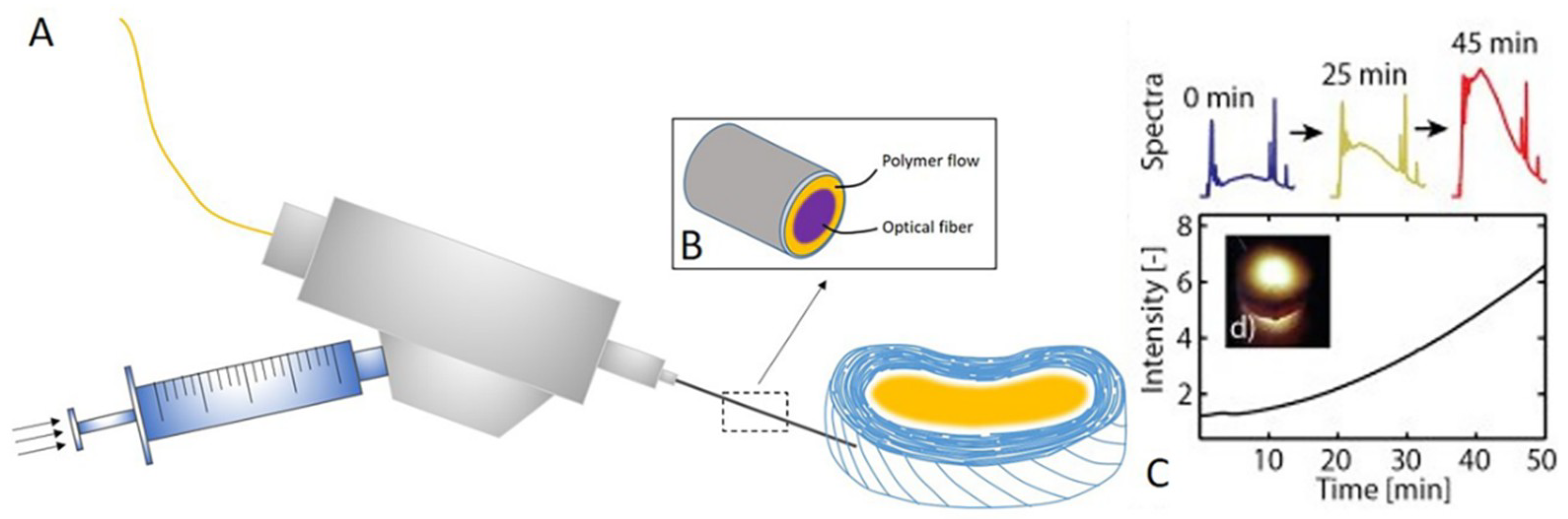
(A) Customized minimally invasive surgical probe used for hydrogel implantation. Liquid hydrogel precursor injection and illumination are performed by a single needle cannula. Different joints ensure high pressurization during the injection. An optical fiber permits the light delivery for photopolymerization. (B) Representation of the distal tip of the instrument, the hydrogel precursor flows between cannula wall, and optical fiber into the IVD. (C) By recording the reflected illumination spectra, signal intensity is calculated. The intensity of the signal correlates with the amount of photopolymerized material and, therefore, provides valuable information on the reaction state of the implant in real time.
Figure 3.
(A) Customized minimally invasive surgical probe used for hydrogel implantation. Liquid hydrogel precursor injection and illumination are performed by a single needle cannula. Different joints ensure high pressurization during the injection. An optical fiber permits the light delivery for photopolymerization. (B) Representation of the distal tip of the instrument, the hydrogel precursor flows between cannula wall, and optical fiber into the IVD. (C) By recording the reflected illumination spectra, signal intensity is calculated. The intensity of the signal correlates with the amount of photopolymerized material and, therefore, provides valuable information on the reaction state of the implant in real time.

Figure 4.
The role of disordered mechanical stress in IDD, and current developments in tissue-engineering therapies.
Figure 4.
The role of disordered mechanical stress in IDD, and current developments in tissue-engineering therapies.


Table 1.
Summary of notochordal cells indifferent species.
| Species | Age of Skeletal Maturity | Age at Loss of Notochordal Cells (According to Literature) |
|---|---|---|
| Dog (c) | 12 months | 12 months |
| Dog (n/c) | 12 months | 60 months |
| Rabbit | 10 months | 6 months |
| Pig | 12 months | Unknown |
| Cat | 24 months | Never |
| Ferret | n/d | Never |
| Sheep | 12 months | Unknown |
| Rat | 2 months | 12 months |
| Mouse | 4 months | n/d |
| Human | 20 years | 6–10 years |
c, chondrodystrophoid (beagles); n/c, non-chondrodystrophoid (mongrels); n/d: no data available. This table was cited from Christopher J. Hunter et al. 2004 [39].
Table 2.
Summary of tissue engineering strategies in intervertebral disc degeneration (IDD) treatment.
Table 2.
Summary of tissue engineering strategies in intervertebral disc degeneration (IDD) treatment.
| Tissue Engineering Strategies in NP Treatment | |||
|---|---|---|---|
| Materials | Test Species | Test Time | Results |
| PLGA | Dog | 8-week | PLGA with cells significantly maintained the height and the stability of disc [85]. |
| Fibrin | Pig | 12-week | Fibrin significantly inhibited the fibrosis and inflammation of NP and enhanced the synthesis of ECM [86]. |
| Collagen II (CII)/hyaluronate (HyA)/chondroitin-6-sulfate (6-CS) | Rabbit | 84-day | The CII/HyA-CS scaffolds have a highly porous structure, high water-binding capacity, and significantly improved mechanical stability. This scaffolds also showed satisfactory biocompatibility [87]. |
| PGA-hyaluronan | Rabbit/Sheep | 12 month/6 month | Enhanced repair tissue formation and MRI intensity [88,89] |
| Silk fibroin (SK) /polyurethane (PU) composite | Pig | NA | SK/PU is an injectable hydrogel with minimally invasive treatment, suitable physical-mechanical properties, and visible CT and T2-weight MRI [90]. |
| Modified hyaluronic acid gels | Pig | 6-week | Both HYAFF® 120 and HYADD 3® treatment supported an NP-like region forming and prevented IVD narrowing, fibrous tissue replacement, and bony end-plates disruption [91]. |
| Tissue engineering strategies in NP treatment in AF treatment | |||
| Electrospun PCL | Rat | 4-week | PCL can mimic the hierarchical organization of the native AF and achieve functional partly with native tissue [92]. |
| Photochemically crosslinked collagen in shape of needle | Rabbit | 1 month | Materials can sustain the physiologically relevant loadings, prevent leakage, and reduce osteophyte formation [93]. |
| Collagen-fibrin gel scaffolds | Rabbit cells in vitro | 4 months | Collagen-fibrin gel significantly delayed the fibrous tissue infiltration. GAG and hydroxyproline content increase over four months [94]. |
| Tissue engineering strategies in the whole IVD | |||
| AF-polyglycolic acid and polylactic acid NP-alginate | Mice | 12-week | The engineered disc maintained the gross morphology and the AF was rich type I collagen but NP contained type II collagen [95]. |
| AF-contracted collagen, NP-alginate | Rat | 6 months | Tissue-engineered IVD maintained disc space height, produced de novo extracellular matrix, and integrated into the spine, yielding an intact motion segment with dynamic mechanical properties similar to that of native IVD [96]. |
| AF- poly (butylene succinate-co-terephthalate) copolyester (PBST), NP-chitosan hydrogel | Rabbit | 4-week | The whole TE-IVD stimulated the natural structure of IVD and retained the height of IVD after four weeks of implant [97]. |
Table 3.
Summary of the mechanical properties of native AF and NP tissue.
| Tissue Scale | Benchmark | Testing Methods | Mechanical Value |
|---|---|---|---|
| AF (Sub-lamella) | E | Nanoindentation | 0.6–1.2 MPa |
| AF (Single Lamella) | E (f = 0°) | Uniaxial tension | 80–120 MPa |
| E (f = 90°) | 0.22 MPa | ||
| AF (Multiple Lamellae) | Eθ (toe/linear) | Uniaxial tension | 2.5/18–45 MPa |
| Axial fixed E (toe/linear) | Biaxial tension | 9.8/27.2 MPa | |
| NP | P swell | Confined compression | 0.138 MPa |
| ǀG *ǀ | Torsional shear | 7.4–19.8 kPa |
E = modulus; θ indicates the loading axes along the disc circumferential direction; toe/linear = toe-region/linear region of stress–strain curve; P swell = swelling pressure. This table was cited from Lewis et al. [129], Holzapfel et al. [130], Guerin and Elliott [131], O’Connell et al. [132], Johannessen and Elliott [133], and Iatridis et al. [134,135].
© 2019 by the authors. Licensee MDPI, Basel, Switzerland. This article is an open access article distributed under the terms and conditions of the Creative Commons Attribution (CC BY) license (http://creativecommons.org/licenses/by/4.0/).
Share and Cite
MDPI and ACS Style
Zhao, R.; Liu, W.; Xia, T.; Yang, L. Disordered Mechanical Stress and Tissue Engineering Therapies in Intervertebral Disc Degeneration. Polymers 2019, 11, 1151. https://doi.org/10.3390/polym11071151
AMA Style
Zhao R, Liu W, Xia T, Yang L. Disordered Mechanical Stress and Tissue Engineering Therapies in Intervertebral Disc Degeneration. Polymers. 2019; 11(7):1151. https://doi.org/10.3390/polym11071151
Chicago/Turabian StyleZhao, Runze, Wanqian Liu, Tingting Xia, and Li Yang. 2019. "Disordered Mechanical Stress and Tissue Engineering Therapies in Intervertebral Disc Degeneration" Polymers 11, no. 7: 1151. https://doi.org/10.3390/polym11071151
Note that from the first issue of 2016, this journal uses article numbers instead of page numbers. See further details here.