
Concurrent Validity and Reliability of Manual Versus Specific Device Transcostal Measurements for Breathing Diaphragm Thickness by Ultrasonography in Lumbopelvic Pain Athletes

 , , ,
, , , 
Abstract
:1. Introduction
2. Methods
2.1. Study Design and Patent Registry
2.2. Sample Size Calculation
2.3. Patients
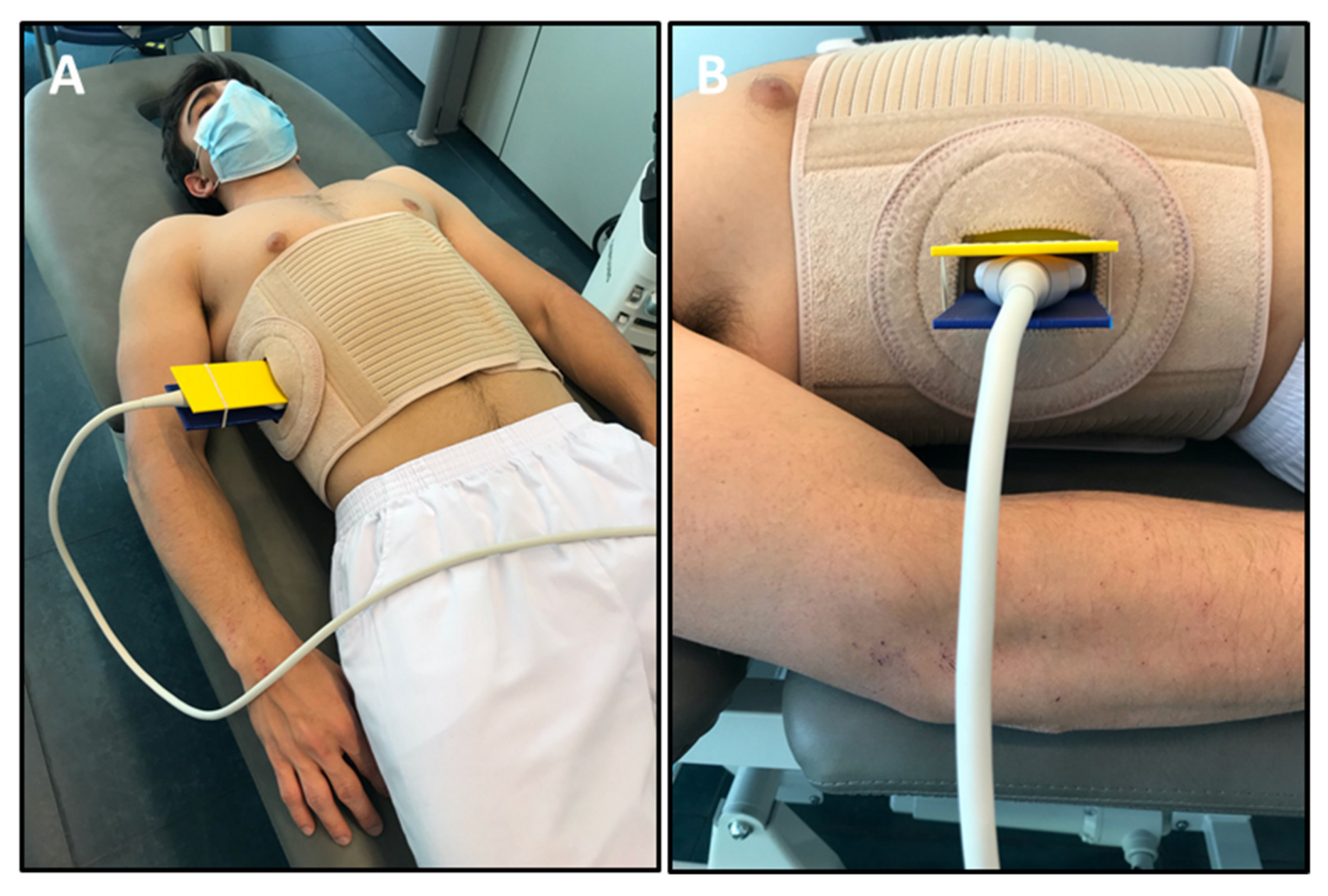
2.4. Procedure
2.5. Descriptive Data
2.6. Clinical Data
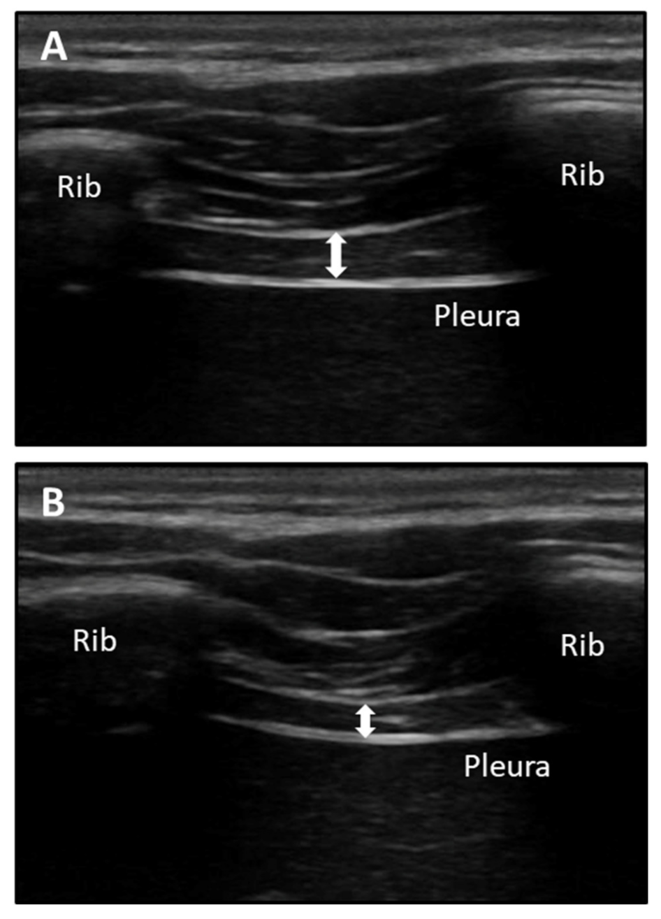
2.7. Ultrasound Measurements
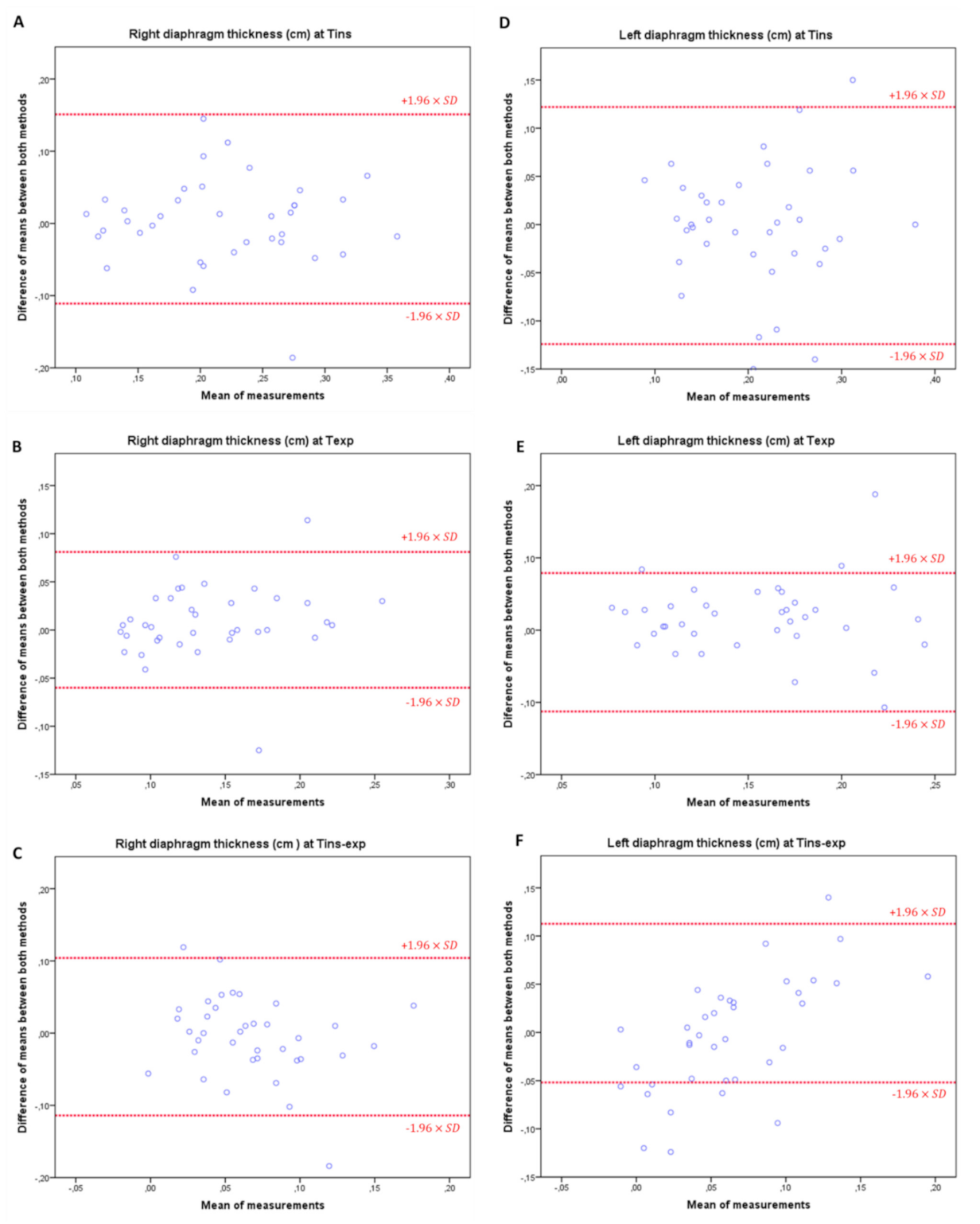
2.8. Statistical Analyses
3. Results
3.1. Descriptive Data
3.2. Clinical Data
3.3. Intra-Rater and Intra-Session Reliability and Concurrent Validity for RUSI Diaphragm Thickness
3.4. Intra-Rater and Inter-Session Reliabilities and Concurrent Validity for RUSI Diaphragm Thickness
3.5. Inter-Rater and Intra-Session Reliabilities and Concurrent Validity for RUSI Diaphragm Thickness
3.6. Inter-Rater and Inter-Session Reliabilities and Concurrent Validity for RUSI Diaphragm Thickness
4. Discussion
4.1. Future Studies
4.2. Limitations
5. Conclusions
Author Contributions
Funding
Institutional Review Board Statement
Informed Consent Statement
Data Availability Statement
Acknowledgments
Conflicts of Interest
References
- Fett, D.; Trompeter, K.; Platen, P. Back pain in elite sports: A cross-sectional study on 1114 athletes. PLoS ONE 2017, 12, e0180130. [Google Scholar] [CrossRef] [PubMed] [Green Version]
- López-López, D.; Vilar-Fernández, J.M.; Calvo-Lobo, C.; Losa-Iglesias, M.E.; Rodriguez-Sanz, D.; Becerro-De-Bengoa-Vallejo, R. Evaluation of depression in subacute low back pain: A case control study. Pain Physician 2017, 20, E499–E505. [Google Scholar] [CrossRef] [PubMed]
- Lobo, C.C.; Fernández, J.M.V.; de Bengoa Vallejo, R.B.; Losa-Iglesias, M.E.; Rodríguez-Sanz, D.; López, P.P.; López, D.L. Relationship of depression in participants with nonspecific acute or subacute low back pain and no-pain by age distribution. J. Pain Res. 2017, 10, 19–135. [Google Scholar] [CrossRef] [Green Version]
- Ferrer-Peña, R.; Calvo-Lobo, C.; Aiguadé, R.; Fernández-Carnero, J. Which Seems to Be Worst? Pain Severity and Quality of Life between Patients with Lateral Hip Pain and Low Back Pain. Pain Res. Manag. 2018, 2018, 9156247. [Google Scholar] [CrossRef] [Green Version]
- Lobo, C.C.; Vilar-Fernández, J.M.; Losa-Iglesias, M.E.; López-López, D.; Rodríguez-Sanz, D.; Palomo-López, P.; Bengoa-Vallejo, R.B. Depression Symptoms Among Older Adults With and Without Subacute Low Back Pain. Rehabil. Nurs. 2019, 44, 47–51. [Google Scholar] [CrossRef]
- Farahbakhsh, F.; Rostami, M.; Noormohammadpour, P.; Mehraki Zade, A.; Hassanmirazaei, B.; Faghih Jouibari, M.; Kordi, R.; Kennedy, D.J. Prevalence of low back pain among athletes: A systematic review. J. Back Musculoskelet. Rehabil. 2018, 31, 901–916. [Google Scholar] [CrossRef]
- Mehra, M.; Hill, K.; Nicholl, D.; Schadrack, J. The burden of chronic low back pain with and without a neuropathic component: A healthcare resource use and cost analysis. J. Med. Econ. 2012, 15, 245–252. [Google Scholar] [CrossRef]
- Juniper, M.; Le, T.K.; Mladsi, D. The epidemiology, economic burden, and pharmacological treatment of chronic low back pain in France, Germany, Italy, Spain and the UK: A literature-based review. Expert Opin. Pharmacother. 2009, 10, 2581–2592. [Google Scholar] [CrossRef]
- Scholtes, S.A.; Van Dillen, L.R. Gender-related differences in prevalence of lumbopelvic region movement impairments in people with low back pain. J. Orthop. Sports Phys. Ther. 2007, 37, 744–752. [Google Scholar] [CrossRef] [Green Version]
- Swain, C.T.V.; Bradshaw, E.J.; Whyte, D.G.; Ekegren, C.L. Life history and point prevalence of low back pain in pre-professional and professional dancers. Phys. Ther. Sport 2017, 25, 34–38. [Google Scholar] [CrossRef]
- Fernández Carnero, S.; Arias Buria, J.; Cuenca Zaldivar, J.; Leal Quiñones, A.; Calvo-Lobo, C.; Martin Saborido, C.; Fernández Carnero, S.; Arias Buria, J.L.; Cuenca Zaldivar, J.N.; Leal Quiñones, A.; et al. Rehabilitative Ultrasound Imaging Evaluation in Physiotherapy: Piloting a Systematic Review. Appl. Sci. 2019, 9, 181. [Google Scholar] [CrossRef] [Green Version]
- Fernández-Carnero, F.; Calvo-Lobo, C.; Garrido-Marin, A.; Arias-Buría, J.L. 2nd Rehabilitative Ultrasound Imaging Symposium in Physiotherapy–Madrid, Spain, 3–5 June 2016. Br. J. Sports Med. 2018, 52, A1–A6. [Google Scholar] [CrossRef]
- Wu, W.T.; Chen, L.R.; Chang, H.C.; Chang, K.V.; Özçakar, L. Quantitative Ultrasonographic Analysis of Changes of the Suprascapular Nerve in the Aging Population With Shoulder Pain. Front. Bioeng. Biotechnol. 2021, 9, 640747. [Google Scholar] [CrossRef] [PubMed]
- Han, D.-S.; Wu, W.-T.; Hsu, P.-C.; Chang, H.-C.; Huang, K.-C.; Chang, K.-V. Sarcopenia Is Associated With Increased Risks of Rotator Cuff Tendon Diseases Among Community-Dwelling Elders: A Cross-Sectional Quantitative Ultrasound Study. Front. Med. 2021, 8, 630009. [Google Scholar] [CrossRef] [PubMed]
- Romero-Morales, C.; Almazán-Polo, J.; Rodríguez-Sanz, D.; Palomo-López, P.; López-López, D.; Vázquez-González, S.; Calvo-Lobo, C. Rehabilitative Ultrasound Imaging Features of the Abdominal Wall Muscles in Elite and Amateur Basketball Players. Appl. Sci. 2018, 8, 809. [Google Scholar] [CrossRef] [Green Version]
- Paris-Alemany, A.; Torres-Palomino, A.; Marino, L.; Calvo-Lobo, C.; Gadea-Mateos, L.; La Touche, R. Comparison of lumbopelvic and dynamic stability between dancers and non-dancers. Phys. Ther. Sport 2018, 33, 33–39. [Google Scholar] [CrossRef]
- Morales, C.R.; Polo, J.A.; Sanz, D.R.; López, D.L.; González, S.V.; Buría, J.L.A.; Lobo, C.C. Ultrasonography features of abdominal perimuscular connective tissue in elite and amateur basketball players: An observational study. Rev. Assoc. Med. Bras. 2018, 64, 936–941. [Google Scholar] [CrossRef] [PubMed]
- Hides, J.A.; Stanton, W.R. Can Motor Control Training Lower the Risk of Injury for Professional Football Players? Med. Sci. Sports Exerc. 2014, 46, 762–768. [Google Scholar] [CrossRef] [Green Version]
- Hides, J.A.; Stanton, W.R.; McMahon, S.; Sims, K.; Richardson, C.A. Effect of stabilization training on multifidus muscle cross-sectional area among young elite cricketers with low back pain. J. Orthop. Sports Phys. Ther. 2008, 38, 101–108. [Google Scholar] [CrossRef]
- Calvo-Lobo, C.; Diez-Vega, I.; Martínez-Pascual, B.; Fernández-Martínez, S.; de la Cueva-Reguera, M.; Garrosa-Martín, G.; Rodríguez-Sanz, D. Tensiomyography, sonoelastography, and mechanosensitivity differences between active, latent, and control low back myofascial trigger points: A cross-sectional study. Medicine 2017, 96, e6287. [Google Scholar] [CrossRef]
- Almazán-Polo, J.; López-López, D.; Romero-Morales, C.; Rodríguez-Sanz, D.; Becerro-de-Bengoa-Vallejo, R.; Losa-Iglesias, M.; Bravo-Aguilar, M.; Calvo-Lobo, C. Quantitative Ultrasound Imaging Differences in Multifidus and Thoracolumbar Fasciae between Athletes with and without Chronic Lumbopelvic Pain: A Case-Control Study. J. Clin. Med. 2020, 9, 2647. [Google Scholar] [CrossRef] [PubMed]
- Whittaker, J.L.; Thompson, J.A.; Teyhen, D.S.; Hodges, P. Rehabilitative ultrasound imaging of pelvic floor muscle function. J. Orthop. Sports Phys. Ther. 2007, 37, 487–498. [Google Scholar] [CrossRef] [PubMed] [Green Version]
- Harper, C.J.; Shahgholi, L.; Cieslak, K.; Hellyer, N.J.; Strommen, J.A.; Boon, A.J. Variability in diaphragm motion during normal breathing, assessed with B-mode ultrasound. J. Orthop. Sports Phys. Ther. 2013, 43, 927–931. [Google Scholar] [CrossRef] [Green Version]
- Vostatek, P.; Novák, D.; Rychnovský, T.; Rychnovská, Š. Diaphragm Postural Function Analysis Using Magnetic Resonance Imaging. PLoS ONE 2013, 8, e56724. [Google Scholar] [CrossRef] [PubMed] [Green Version]
- Janssens, L.; McConnell, A.K.; Pijnenburg, M.; Claeys, K.; Goossens, N.; Lysens, R.; Troosters, T.; Brumagne, S. Inspiratory muscle training affects proprioceptive use and low back pain. Med. Sci. Sports Exerc. 2015, 47, 12–19. [Google Scholar] [CrossRef] [PubMed] [Green Version]
- Kolář, P.; Šulc, J.; Kynčl, M.; Šanda, J.; Čakrt, O.; Andel, R.; Kumagai, K.; Kobesová, A. Postural Function of the Diaphragm in Persons With and Without Chronic Low Back Pain. J. Orthop. Sport. Phys. Ther. 2012, 42, 352–362. [Google Scholar] [CrossRef] [PubMed] [Green Version]
- Finta, R.; Nagy, E.; Bender, T. The effect of diaphragm training on lumbar stabilizer muscles: A new concept for improving segmental stability in the case of low back pain. J. Pain Res. 2018, 11, 3031–3045. [Google Scholar] [CrossRef] [Green Version]
- Chicharro, J.L.; Hoyos, J.; Lucía, A. Effects of endurance training on the isocapnic buffering and hypocapnic hyperventilation phases in professional cyclists. Br. J. Sports Med. 2000, 34, 450–455. [Google Scholar] [CrossRef]
- Hodges, P.W.; Butler, J.E.; McKenzie, D.K.; Gandevia, S.C. Contraction of the human diaphragm during rapid postural adjustments. J. Physiol. 1997, 505 Pt 2, 539–548. [Google Scholar] [CrossRef]
- Calvo-Lobo, C.; Almazán-Polo, J.; Becerro-de-Bengoa-Vallejo, R.; Losa-Iglesias, M.E.; Palomo-López, P.; Rodríguez-Sanz, D.; López-López, D. Ultrasonography comparison of diaphragm thickness and excursion between athletes with and without lumbopelvic pain. Phys. Ther. Sport 2019, 37, 128–137. [Google Scholar] [CrossRef]
- Bunce, S.M.; Hough, A.D.; Moore, A.P. Measurement of abdominal muscle thickness using M-mode ultrasound imaging during functional activities. Man. Ther. 2004, 9, 41–44. [Google Scholar] [CrossRef]
- Dieterich, A.V.; Pickard, C.M.; Strauss, G.R.; Deshon, L.E.; Gibson, W.; McKay, J. Muscle thickness measurements to estimate gluteus medius andminimus activity levels. Man. Ther. 2014, 19, 453–460. [Google Scholar] [CrossRef] [Green Version]
- Bossuyt, P.M.; Reitsma, J.B.; Bruns, D.E.; Gatsonis, C.A.; Glasziou, P.P.; Irwig, L.; Lijmer, J.G.; Moher, D.; Rennie, D.; de Vet, H.C.W.; et al. An updated list of essential items for reporting diagnostic accuracy studies. BMJ 2015, 351, h5527. [Google Scholar] [CrossRef] [Green Version]
- Holt, G.R. Declaration of Helsinki-the world’s document of conscience and responsibility. South. Med. J. 2014, 107, 407. [Google Scholar] [CrossRef]
- Lobo, C.C.; Morales, C.R.; Sanz, D.R.; Corbalan, I.S.; Marin, A.G.; Lopez, D.L. Ultrasonography Comparison of Peroneus Muscle Cross-sectional Area in Subjects With or Without Lateral Ankle Sprains. J. Manip. Physiol. Ther. 2016, 39, 635–644. [Google Scholar] [CrossRef] [Green Version]
- Cohen, J. A Power Primer. Psychol. Bull. 1992, 112, 155–159. [Google Scholar] [CrossRef]
- Whittaker, J.L.; Warner, M.B.; Stokes, M. Comparison of the Sonographic Features of the Abdominal Wall Muscles and Connective Tissues in Individuals With and Without Lumbopelvic Pain. J. Orthop. Sports Phys. Ther. 2013, 43, 11–19. [Google Scholar] [CrossRef] [PubMed]
- Garrow, J.S. Quetelet index as indicator of obesity. Lancet 1986, 1, 1219. [Google Scholar] [CrossRef]
- Gauthier, A.P.; Lariviere, M.; Young, N. Psychometric properties of the IPAQ: A validation study in a sample of northern Franco-Ontarians. J. Phys. Act. Health 2009, 6 (Suppl. 1), S54–S60. [Google Scholar] [CrossRef] [PubMed]
- Boonstra, A.M.; Schiphorst Preuper, H.R.; Reneman, M.F.; Posthumus, J.B.; Stewart, R.E. Reliability and validity of the visual analogue scale for disability in patients with chronic musculoskeletal pain. Int. J. Rehabil. Res. 2008, 31, 165–169. [Google Scholar] [CrossRef] [PubMed] [Green Version]
- Koo, T.K.; Guo, J.; Brown, C.M. Test-retest reliability, repeatability, and sensitivity of an automated deformation-controlled indentation on pressure pain threshold measurement. J. Manip. Physiol. Ther. 2013, 36, 84–90. [Google Scholar] [CrossRef]
- Vilagut, G.; Valderas, J.M.; Ferrer, M.; Garin, O.; López-García, E.; Alonso, J. Interpretación de los cuestionarios de salud SF-36 y SF-12 en España: Componentes físico y mental. Med. Clin. 2008, 130, 726–735. [Google Scholar] [CrossRef] [PubMed] [Green Version]
- Santiago-Nuño, F.; Palomo-López, P.; Becerro-de-Bengoa-Vallejo, R.; Calvo-Lobo, C.; Losa-Iglesias, M.E.; Casado-Hernández, I.; López-López, D. Intra and Inter-rater Reliability between Ultrasound Imaging and Caliper Measures to determine Spring Ligament Dimensions in Cadavers. Sci. Rep. 2019, 9, 14808. [Google Scholar] [CrossRef] [PubMed] [Green Version]
- Chang, P.H.; Chen, Y.J.; Chang, K.V.; Wu, W.T.; Özçakar, L. Ultrasound measurements of superficial and deep masticatory muscles in various postures: Reliability and influencers. Sci. Rep. 2020, 10, 14357. [Google Scholar] [CrossRef]
- Bland, J.M.; Altman, D.G. Statistical methods for assessing agreement between two methods of clinical measurement. Int. J. Nurs. Stud. 2010, 47, 931–936. [Google Scholar] [CrossRef]
- Yeampattanaporn, O.; Mekhora, K.; Jalayondeja, W.; Wongsathikun, J. Immediate effects of breathing re-education on respiratory function and range of motion in chronic neck pain. J. Med. Assoc. Thail. 2014, 97, S55–S59. [Google Scholar]
- Bonetti, F.; Curti, S.; Mattioli, S.; Mugnai, R.; Vanti, C.; Violante, F.S.; Pillastrini, P. Effectiveness of a “Global Postural Reeducation” program for persistent Low Back Pain: A non-randomized controlled trial. BMC Musculoskelet. Disord. 2010, 11, 285. [Google Scholar] [CrossRef] [Green Version]
- Niederer, D.; Mueller, J. Sustainability effects of motor control stabilisation exercises on pain and function in chronic nonspecific low back pain patients: A systematic review with meta-analysis and meta-regression. PLoS ONE 2020, 15, e0227423. [Google Scholar] [CrossRef] [PubMed]
- Fernandez-Rubio, H.; Becerro-de-Bengoa-Vallejo, R.; Rodríguez-Sanz, D.; Calvo-Lobo, C.; Vicente-Campos, D.; Chicharro, J.L. Inspiratory Muscle Training in Patients with Heart Failure. J. Clin. Med. 2020, 9, 1710. [Google Scholar] [CrossRef]
- Fernández-Rubio, H.; Becerro-De-bengoa-vallejo, R.; Rodríguez-Sanz, D.; Calvo-Lobo, C.; Vicente-Campos, D.; Chicharro, J.L. Unraveling the role of respiratory muscle metaboloreceptors under inspiratory training in patients with heart failure. Int. J. Environ. Res. Public Health 2021, 18, 1697. [Google Scholar] [CrossRef]
- Kanehisa, H.; Miyatani, M.; Azuma, K.; Kuno, S.; Fukunaga, T. Influences of age and sex on abdominal muscle and subcutaneous fat thickness. Eur. J. Appl. Physiol. 2004, 91, 534–537. [Google Scholar] [CrossRef] [PubMed]


| RUSI Diaphragm Thickness (cm) | Baseline Mean ± SD (95% CI) | After 1 h Mean ± SD (95% CI) | ICC(1,2) (95% CI) | SEM | MDC | p-Value |
|---|---|---|---|---|---|---|
| Manual Probe Fixation | ||||||
| Tins right diaphragm | 0.21 ± 0.07 (0.19–0.24) | 0.22 ± 0.07 (0.19–0.24) | 0.989 (0.978–0.994) | 0.009 | 0.024 | 0.141 † |
| Texp right diaphragm | 0.13 ± 0.04 (0.11–0.15) | 0.13 ± 0.04 (0.12–0.15) | 0.993 (0.986–0.996) | 0.003 | 0.008 | 0.141 * |
| Tins-exp right diaphragm | 0.08 ± 0.05 (0.06–0.10) | 0.08 ± 0.05 (0.06–0.10) | 0.982 (0.966–0.991) | 0.006 | 0.018 | 0.404 * |
| Tins left diaphragm | 0.20 ± 0.07 (0.18–0.23) | 0.21 ± 0.07 (0.18–0.23) | 0.994 (0.989–0.997) | 0.005 | 0.015 | 0.152 * |
| Texp left diaphragm | 0.15 ± 0.05 (0.13–0.17) | 0.10 ± 0.03 (0.09–0.11) | 0.714 (−0.11–0.92) | 0.021 | 0.059 | <0.001 * |
| Tins-exp left diaphragm | 0.05 ± 0.04 (0.04–0.06) | 0.10 ± 0.04 (0.09–0.12) | 0.982 (0.966–0.991) | 0.005 | 0.014 | <0.001 * |
| Orthosis Device Probe Fixation | ||||||
| Tins right diaphragm | 0.23 ± 0.07 (0.20–0.25) | 0.23 ± 0.07 (0.20–0.26) | 0.991 (0.982–0.995) | 0.006 | 0.018 | 0.327 * |
| Texp right diaphragm | 0.15 ± 0.05 (0.13–0.17) | 0.16 ± 0.05 (0.14–0.18) | 0.947 (0.892–0.973) | 0.002 | 0.007 | 0.067 † |
| Tins-exp right diaphragm | 0.07 ± 0.05 (0.05–0.09) | 0.07 ± 0.05 (0.05–0.09) | 0.935 (0.874–0.966) | 0.012 | 0.035 | 0.283 † |
| Tins left diaphragm | 0.22 ± 0.07 (0.19–0.24) | 0.22 ± 0.07 (0.19–0.24) | 0.993 (0.987–0.997) | 0.005 | 0.015 | 0.823 * |
| Texp left diaphragm | 0.17 ± 0.05 (0.15–0.19) | 0.17 ± 0.05 (0.15–0.19) | 0.996 (0.992–0.998) | 0.0002 | 0.0005 | 0.119 * |
| Tins-exp left diaphragm | 0.04 ± 0.05 (0.03–0.06) | 0.04 ± 0.04 (0.03–0.06) | 0.978 (0.958–0.989) | 0.006 | 0.018 | 0.360 * |
| RUSI Diaphragm Thickness (cm) | Baseline Mean ± SD (95% CI) | After 48 h Mean ± SD (95% CI) | ICC(1,2) (95% CI) | SEM | MDC | p-Value |
|---|---|---|---|---|---|---|
| Manual Probe Fixation | ||||||
| Tins right diaphragm | 0.21 ± 0.07 (0.19–0.24) | 0.22 ± 0.07 (0.19–0.24) | 0.992 (0.985–0.996) | 0.006 | 0.018 | 0.306 † |
| Texp right diaphragm | 0.13 ± 0.04 (0.11–0.15) | 0.13 ± 0.04 (0.12–0.15) | 0.993 (0.985–0.996) | 0.003 | 0.008 | 0.050 * |
| Tins-exp right diaphragm | 0.08 ± 0.05 (0.06–0.10) | 0.08 ± 0.06 (0.06–0.10) | 0.985 (0.971–0.992) | 0.006 | 0.018 | 0.766 * |
| Tins left diaphragm | 0.20 ± 0.07 (0.18–0.23) | 0.20 ± 0.07 (0.18–0.23) | 0.997 (0.995–0.999) | 0.003 | 0.010 | 0.397 * |
| Texp left diaphragm | 0.15 ± 0.05 (0.13–0.17) | 0.15 ± 0.05 (0.13–0.17) | 0.992 (0.985–0.996) | 0.004 | 0.012 | 0.838 * |
| Tins-exp left diaphragm | 0.05 ± 0.04 (0.04–0.06) | 0.05 ± 0.04 (0.03–0.06) | 0.982 (0.965–0.991) | 0.005 | 0.014 | 0.491 * |
| Orthosis Device Probe Fixation | ||||||
| Tins right diaphragm | 0.23 ± 0.07 (0.20–0.25) | 0.23 ± 0.07 (0.20–0.26) | 0.993 (0.986–0.996) | 0.005 | 0.016 | 0.173 * |
| Texp right diaphragm | 0.15 ± 0.05 (0.13–0.17) | 0.16 ± 0.05 (0.14–0.18) | 0.941 (0.881–0.970) | 0.003 | 0.010 | 0.265 † |
| Tins-exp right diaphragm | 0.07 ± 0.05 (0.05–0.09) | 0.07 ± 0.05 (0.05–0.09) | 0.933 (0.870–0.965) | 0.012 | 0.035 | 0.317 † |
| Tins left diaphragm | 0.22 ± 0.07 (0.19–0.24) | 0.22 ± 0.07 (0.19–0.24) | 0.990 (0.981–0.995) | 0.006 | 0.018 | 0.776 * |
| Texp left diaphragm | 0.17 ± 0.05 (0.15–0.19) | 0.17 ± 0.05 (0.15–0.18) | 0.982 (0.965–0.991) | 0.006 | 0.018 | 0.587 * |
| Tins-exp left diaphragm | 0.04 ± 0.05 (0.03–0.06) | 0.04 ± 0.04 (0.03–0.06) | 0.961 (0.925–0.980) | 0.007 | 0.021 | 0.840 * |
| RUSI Diaphragm Thickness (cm) | Examiner 1 Mean ± SD (95% CI) | Examiner 2 Mean ± SD (95% CI) | ICC(2,1) (95% CI) | SEM | MDC | p-Value |
|---|---|---|---|---|---|---|
| Manual Probe Fixation | ||||||
| Tins right diaphragm | 0.21 ± 0.07 (0.19–0.24) | 0.21 ± 0.07 (0.19–0.24) | 0.983 (0.967–0.991) | 0.009 | 0.025 | 0.174 † |
| Texp right diaphragm | 0.13 ± 0.04 (0.11–0.15) | 0.12 ± 0.04 (0.10–0.13) | 0.951 (0.774–0.982) | 0.008 | 0.024 | <0.001 * |
| Tins-exp right diaphragm | 0.08 ± 0.05 (0.06–0.10) | 0.09 ± 0.06 (0.07–0.11) | 0.955 (0.889–0.979) | 0.011 | 0.031 | 0.004 * |
| Tins left diaphragm | 0.20 ± 0.07 (0.18–0.23) | 0.20 ± 0.06 (0.17–0.22) | 0.945 (0.894–0.972) | 0.015 | 0.042 | 0.224 * |
| Texp left diaphragm | 0.15 ± 0.05 (0.13–0.17) | 0.13 ± 0.04 (0.12–0.15) | 0.872 (0.672–0.942) | 0.016 | 0.044 | 0.001 * |
| Tins-exp left diaphragm | 0.05 ± 0.04 (0.04–0.06) | 0.06 ± 0.04 (0.05–0.08) | 0.910 (0.791–0.957) | 0.012 | 0.033 | 0.005 * |
| Orthosis Device Probe Fixation | ||||||
| Tins right diaphragm | 0.23 ± 0.07 (0.20–0.25) | 0.23 ± 0.08 (0.20–0.25) | 0.982 (0.966–0.991) | 0.001 | 0.002 | 0.468 * |
| Texp right diaphragm | 0.15 ± 0.05 (0.13–0.17) | 0.16 ± 0.05 (0.12–0.15) | 0.955 (0.907–0.977) | 0.010 | 0.029 | 0.201 † |
| Tins-exp right diaphragm | 0.07 ± 0.05 (0.05–0.09) | 0.08 ± 0.05 (0.06–0.10) | 0.936 (0.876–0.967) | 0.012 | 0.035 | 0.211 † |
| Tins left diaphragm | 0.22 ± 0.07 (0.19–0.24) | 0.22 ± 0.07 (0.19–0.24) | 0.979 (0.960–0.989) | 0.010 | 0.027 | 0.840 * |
| Texp left diaphragm | 0.17 ± 0.05 (0.15–0.19) | 0.15 ± 0.05 (0.12–0.16) | 0.875 (0.523–0.952) | 0.017 | 0.047 | 0.001 * |
| Tins-exp left diaphragm | 0.04 ± 0.05 (0.03–0.06) | 0.05 ± 0.05 (0.03–0.07) | 0.945 (0.893–0.972) | 0.011 | 0.032 | 0.139 * |
| RUSI Diaphragm Thickness (cm) | Examiner 1 Mean ± SD (95% CI) | Examiner 2 Mean ± SD (95% CI) | ICC(2,1) (95% CI) | SEM | MDC | p-Value |
|---|---|---|---|---|---|---|
| Manual Probe Fixation | ||||||
| Tins right diaphragm | 0.21 ± 0.07 (0.19–0.24) | 0.20 ± 0.07 (0.18–0.23) | 0.965 (0.931–0.982) | 0.013 | 0.036 | 0.010 † |
| Texp right diaphragm | 0.13 ± 0.04 (0.11–0.15) | 0.11 ± 0.04 (0.10–0.13) | 0.865 (0.675–0.938) | 0.014 | 0.040 | 0.002 * |
| Tins-exp right diaphragm | 0.08 ± 0.05 (0.06–0.10) | 0.09 ± 0.06 (0.06–0.11) | 0.953 (0.906–0.976) | 0.014 | 0.039 | 0.047 * |
| Tins left diaphragm | 0.20 ± 0.07 (0.18–0.23) | 0.20 ± 0.06 (0.17–0.22) | 0.936 (0.876–0.967) | 0.016 | 0.045 | 0.117 * |
| Texp left diaphragm | 0.15 ± 0.05 (0.13–0.17) | 0.13 ± 0.05 (0.12–0.15) | 0.784 (0.571–0.890) | 0.023 | 0.064 | 0.025 * |
| Tins-exp left diaphragm | 0.05 ± 0.04 (0.04–0.06) | 0.06 ± 0.03 (0.04–0.07) | 0.828 (0.669–0.911) | 0.014 | 0.040 | 0.172 * |
| Orthosis Device Probe Fixation | ||||||
| Tins right diaphragm | 0.23 ± 0.07 (0.20–0.25) | 0.22 ± 0.07 (0.19–0.24) | 0.861 (0.731–0.928) | 0.026 | 0.072 | 0.141 * |
| Texp right diaphragm | 0.15 ± 0.05 (0.13–0.17) | 0.14 ± 0.05 (0.12–0.16) | 0.927 (0.847–0.964) | 0.013 | 0.037 | 0.410 † |
| Tins-exp right diaphragm | 0.07 ± 0.05 (0.05–0.09) | 0.07 ± 0.06 (0.05–0.09) | 0.852 (0.713–0.924) | 0.021 | 0.058 | 0.717 † |
| Tins left diaphragm | 0.22 ± 0.07 (0.19–0.24) | 0.20 ± 0.07 (0.18–0.23) | 0.920 (0.839–0.959) | 0.054 | 0.027 | 0.051 * |
| Texp left diaphragm | 0.17 ± 0.05 (0.15–0.19) | 0.16 ± 0.05 (0.14–0.18) | 0.877 (0.762–0.937) | 0.017 | 0.048 | 0.106 * |
| Tins-exp left diaphragm | 0.04 ± 0.05 (0.03–0.06) | 0.04 ± 0.05 (0.02–0.06) | 0.884 (0.776–0.940) | 0.017 | 0.047 | 0.687 * |
Publisher’s Note: MDPI stays neutral with regard to jurisdictional claims in published maps and institutional affiliations. |
© 2021 by the authors. Licensee MDPI, Basel, Switzerland. This article is an open access article distributed under the terms and conditions of the Creative Commons Attribution (CC BY) license (https://creativecommons.org/licenses/by/4.0/).
Share and Cite
Marugán-Rubio, D.; Chicharro, J.L.; Becerro-de-Bengoa-Vallejo, R.; Losa-Iglesias, M.E.; Rodríguez-Sanz, D.; Vicente-Campos, D.; Dávila-Sánchez, G.J.; Calvo-Lobo, C. Concurrent Validity and Reliability of Manual Versus Specific Device Transcostal Measurements for Breathing Diaphragm Thickness by Ultrasonography in Lumbopelvic Pain Athletes. Sensors 2021, 21, 4329. https://doi.org/10.3390/s21134329
Marugán-Rubio D, Chicharro JL, Becerro-de-Bengoa-Vallejo R, Losa-Iglesias ME, Rodríguez-Sanz D, Vicente-Campos D, Dávila-Sánchez GJ, Calvo-Lobo C. Concurrent Validity and Reliability of Manual Versus Specific Device Transcostal Measurements for Breathing Diaphragm Thickness by Ultrasonography in Lumbopelvic Pain Athletes. Sensors. 2021; 21(13):4329. https://doi.org/10.3390/s21134329
Chicago/Turabian StyleMarugán-Rubio, Daniel, Jose L. Chicharro, Ricardo Becerro-de-Bengoa-Vallejo, Marta Elena Losa-Iglesias, David Rodríguez-Sanz, Davinia Vicente-Campos, Gabriel J. Dávila-Sánchez, and César Calvo-Lobo. 2021. "Concurrent Validity and Reliability of Manual Versus Specific Device Transcostal Measurements for Breathing Diaphragm Thickness by Ultrasonography in Lumbopelvic Pain Athletes" Sensors 21, no. 13: 4329. https://doi.org/10.3390/s21134329