
Applicability of Smart Tools in Vegetable Disease Diagnostics
by
 , , , ,
Jaroslava Ovesná
1,* ,
,
Michail D. Kaminiaris
2,
Zisis Tsiropoulos
2,
Rosemary Collier
3 ,
,
Alex Kelly
3,
Jonathan De Mey
4 and
Sabien Pollet
4 1
Crop Research Institute, Drnovská 507, 161 06 Prague, Czech Republic
2
Agricultural & Environmental Solutions (AGENSO), Markou Mpotsari 47, 11742 Athens, Greece
3
Warwick Crop Centre, School of Life Sciences, The University of Warwick, Wellesbourne, Warwick CV35 9EF, UK
4
Inagro, Ieperseweg 87, Rumbeke, 8800 Roeselare, Belgium
*
Author to whom correspondence should be addressed.
Agronomy 2023, 13(5), 1211; https://doi.org/10.3390/agronomy13051211
Submission received: 17 March 2023
/
Revised: 17 April 2023
/
Accepted: 18 April 2023
/
Published: 25 April 2023
(This article belongs to the Section Precision and Digital Agriculture)
Abstract
:Various diseases and pests cause serious damage to vegetable crops during the growing season and after harvesting. Growers attempt to minimize losses by protecting their crops, starting with seed and seedling treatments and followed by monitoring their stands. In many cases, synthetic pesticide treatments are applied. Integrated pest management is currently being employed to minimize the impact of pesticides upon human health and the environment. Over the last few years, “smart” approaches have been developed and adopted in practice to predict, detect, and quantify phytopathogen occurrence and contamination. Our review assesses the currently available ready-to-use tools and methodologies that operate via visual estimation, the detection of proteins and DNA/RNA sequences, and the utilization of brand-new innovative approaches, highlighting the availability of solutions that can be used by growers during the process of diagnosing pathogens.
Keywords:
phytopathogen; diagnostic; serology; PCR; LAMP; RPA; barcoding; NGS; CRISPR/Cas; vegetable1. Introduction
As a source of vitamins, minerals, and other nutrients, vegetables constitute an important food component that is produced and traded worldwide. As with other cultivated species, vegetables grow in tandem with beneficial or harmful microbiota that affect the yield and quality of crops. These microbiotas exist in fields, and the global movement of plant material (vegetable seeds, seedlings, and produce) provides opportunities for phytopathogens to cross borders and spread to new areas. It has been shown that the number of phytopathogens in different regions is associated with the volume of imports. In this way, the risk of pathogenic species expansion from one country to another has been increasing [1,2]. To prevent invasions, several control systems have been set up by national authorities, following the recommendations of scientists and international bodies [3]. Growers also try to prevent locally occurring plant pathogens from invading their crops, and they carefully monitor the possible spread of new phytopathogens from neighboring regions [4].To successfully cope with vegetable pathogens, preventive measures should be taken, and consideration should be given to soil management, seed treatment, the cultivar’s natural resistance, and the various monitoring systems that are suited to conventional, biotechnological, and organic farming, forming part of the IPM recommendations [5]. The ability to capture the pathogen upon its first attack as early as possible, as in the early infection stage, allows growers to make appropriate and effective management decisions. It is apparent that tools for disease diagnosis and pathogen identification are greatly needed. Otherwise, it would not be possible to take preventative steps and set up management measures in cases of infection [6].
Several approaches and techniques have been developed to facilitate the diagnosis, monitoring, and study of phytopathogenic agents in plants and [7,8,9] various vegetable species. Some of these techniques have been validated and are used by accredited laboratories for official controls [6,10,11,12]. Others have significant potential for application in “smart” farming [7,9], possibly leading to the implementation of phytopathogen surveillance through intelligent and goal-oriented analysis, planning, and observation [13]. Currently, the importance of smart approaches for disease diagnosis on farms is increasing [14,15]. We aimed to map fully validated procedures employed in official controlsand concurrently tools at various technological readiness level (TRL) that are usually verified in a single laboratory only [16] to determine their future potential as new devices for phytopathogen detection [16].
2. Symptomatic Diagnosis
Symptomatic evaluation was once the sole option for disease identification in field crops. However, the symptoms of individual diseases, especially at the onset of their development, can be ambiguous. Therefore, this approach is based on experience and requires highly skilled specialists [17,18]. The organisms that cause plant diseases are very small (microscale) and can, therefore, only be detected by lay farmers based on the impacts of their attacks. As experts are not always available when needed, several options have been introduced and tested in efforts to improve the available diagnostic approaches. Mainly, these include simple phytopathogen cultivation tests [19], but these tests are time-consuming and dependent on mycological skills and laboratory infrastructure. Moreover, they are not sufficiently sensitive to enable detection in the early stages of infection. Previously, higher-level image analysis of crop diseases was not particularly effective; however, its applicability has been enhanced when combined with smart tools, such as videos or mobile phones linked to knowledge bases (e.g., the system VACDDS [20]). Various computational algorithms and diagnostic models have been tested to improve visual evaluation and further identification [21,22]. The existence of precise databases of sufficiently adequate images has been [23] reported to be the basic prerequisite for enabling such a system to be functional. These precise tools for phytopathogen interception are still expensive and, in the case of vegetables, not sufficiently advanced. Several factors, such as the unclear outlines of lesions, uneven coloring of leaves, and variability in lighting conditions, among others, hinder analysis [24]. The currently available image-based techniques have been reported to be precise under well-defined experimental conditions, but when they are moved to the field, higher precision and reliability are required. Thus, there exists a great demand for tools that can facilitate the precise detection of pathogens for individual crops.
3. Serological Tests
Serological tests are widely used in diagnostics. They work by exploiting the plurality of antibody reactions to pathogens. The enzyme immunoassay (EIA) is employed to detect the presence of a ligand, e.g., proteins in a liquid sample using antibodies directed against the protein to be measured. One modification, the enzyme-linked immunosorbent assay (ELISA) [25], is commonly used as a standard analytical method. ELISA is used not only in human diagnostics but also in the agricultural sector, particularly for the detection of certain plant viruses, fungal phytopathogens, and viral pathogens. Usually, the assay detects proteins (e.g., a viral coat protein) specific to an individual genus or species via polyclonal or monoclonal antibodies [26]. The detection of these proteins is accomplished by complexing antibodies and antigens to produce measurable, usually colored results. The efficiency of the method is often improved by amplifying the primary signal. Although ELISA is the most common means of testing, many other approaches have been described, published, and used in practice.
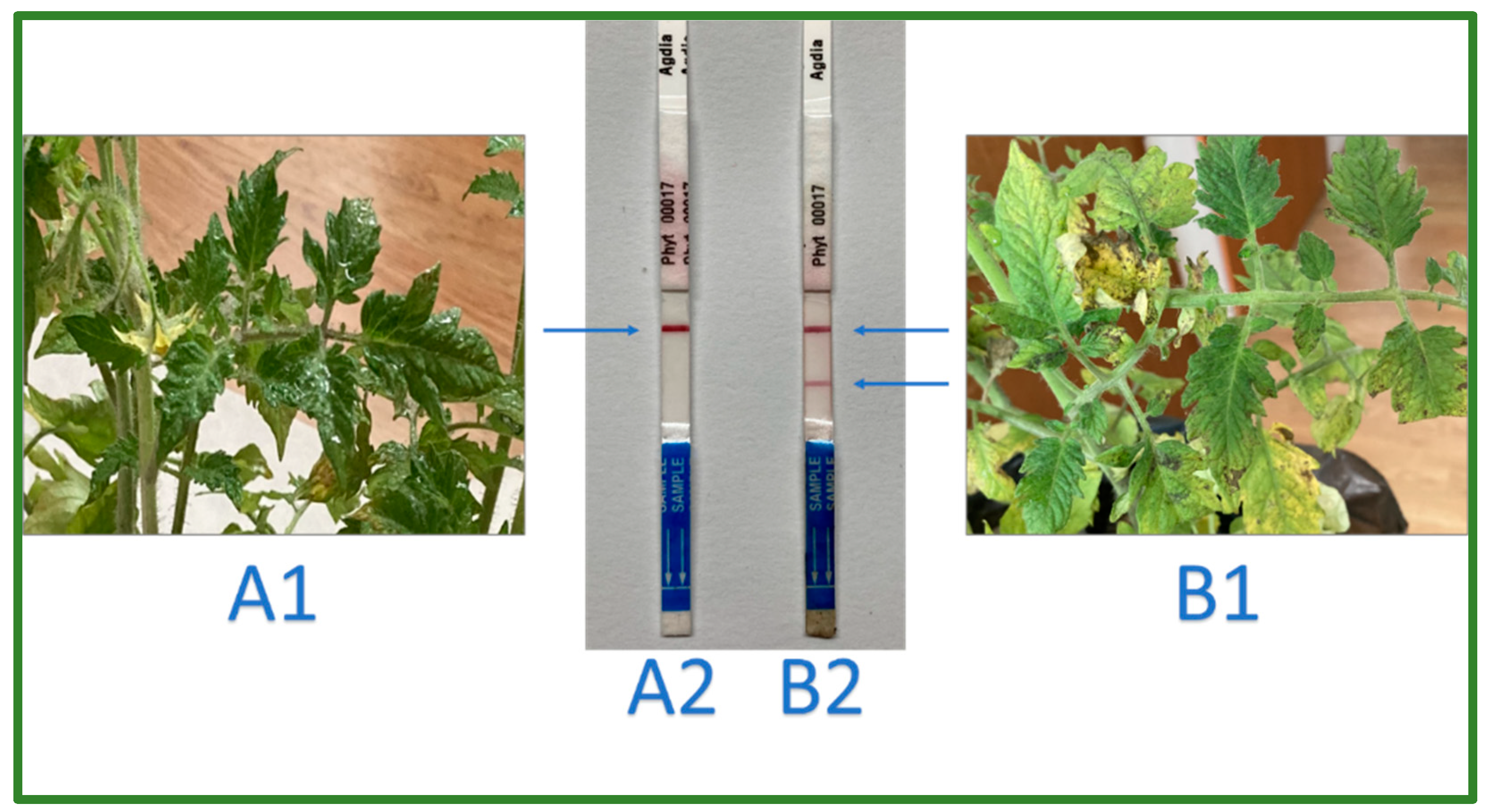
Other immunoassays, exploiting ready-to-use lateral-flow devices (LFDs), are now available on the market, in which specific antibodies are used for detection and color signaling. Such tests are well-known for their use in the monitoring of human diseases, e.g., COVID-19, and they are characterized by their ease of use. The results are easy to interpret; thus, they are commonly used for in-field diagnosis. The advantage of LFDs is that they can be used without the need for sophisticated instruments and equipment. These methods are characterized by their speed; they are considered to be rapid, while also being inexpensive and suited to high-throughput diagnostics (Figure 1). On the other hand, other approaches such as DNA-based methods are more sensitive and amenable to multiplexing. LFDs require a priori knowledge of each pathogen. Furthermore, the test is not suited to non-target screening.
At the same time, a number of DNA/RNA detection approaches have been developed.
4. Polymerase-Chain-Reaction (PCR)-Based Methods for Phytopathogen Detection
Polymerase chain reaction (PCR) is a DNA-based method, as outlined by Mullis and Kaloona [27], that allows for the amplification of selected DNA sequences using a pair of primers, including a short DNA sequence typical of the individual species and individual genotypes surrounding specific DNA stretches. With these primers, along with the enzyme Taq, polymerase-multiplied quantities of target sequences can be visualized. PCR is able to capture even a minute amount of the targeted DNA. Theoretically, even a single copy can be captured, but practically speaking, the limit of detection depends on the matrix, and it can be as low as tens of copies of the target. The procedure requires the isolation of DNA/RNA from the analytical target, and the efficiency of the amplification depends on the quality of the extraction process. The important qualities of PCR, as used in diagnostics, are its high specificity, afforded by knowledge of phytopathogen-specific DNA/RNA sequences, and the possibility of designing primers that target selected stretches of DNA [28].
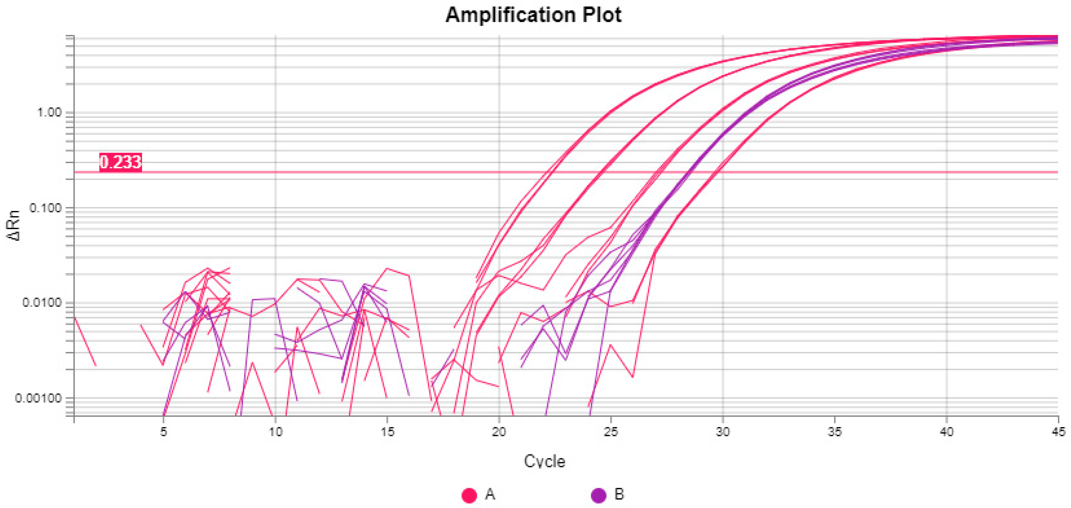
A modification of PCR was introduced a few years ago, when the specificity of PCR was improved as a third-sequence specific primer (probe) was exploited Heid et al. (1996) [29]. In this approach, the probe is fluorescently labelled, and during the synthesis of a new amplicon copy, the fluorescent signal is captured. The approach is composed of real-time PCR (qPCR, if used for target quantification) [30] and digital PCR [31,32]. Real-time PCR may serve as a method for either detection or relative quantification, while digital PCR can be used for the absolute quantification of amplicon copy numbers in the assay. Compared with real-time PCR, digital PCR limits the problems associated with the purity of the extracted DNA through special partitioning. The listed properties of real-time and digital PCR mean that they are well-suited to a wide range of applications in phytopathogen diagnostics [33]. As PCR-based techniques (Figure 2) are capable of detecting even just a few copies of the target, even a small number of individual virus particles or bacterial or fungal cells can be detected [34]. Thus, these techniques may provide information in the early stages of infection, when the symptoms of infections are barely visible but phytopathogens are already present in the tissue. In addition, multiplex assays, e.g., the detection of several pathogenic species in a single reaction, may be used. Multiplexing reduces the cost and time required to determine the pathogen load [35].
With regard to plant pathogen detection/quantification, we found many protocols for vegetable phytopathogen species, but true validation reports, as required by the Minimal Performance Criteria (MIQE) [36], are not available. The in-lab verification followed by the full validation of published and forthcoming methods is a necessity for researchers to rely on their performance after the provision of comparable results across different laboratories [36,37,38,39,40]. To ensure uniform test performance across laboratories, international standards and guidelines have been issued by the International Organization for Standardization (ISO) that are used by accredited testing laboratories [41]. Thus, PCR-based methods can be used in a harmonized manner.
Since the discovery of the PCR principle, the scope of PCR application has expanded due to the combination with other molecular procedures. One PCR modification, immunocapture–reverse transcription–polymerase chain reaction (IC-RT-PCR) [42], has been suggested as a standard method for the highly sensitive analysis of plants infected with a range of RNA viruses [43,44]. In this method, the preliminary purification of virus particles and viral RNA from the plant material may be bypassed. In addition, in contrast to PCR, all of the ingredients and enzymes in the assay, e.g., temperature, ionic strength, and pH, are able to work under the same conditions. The procedure may, thus, be accomplished in a single step with one tube. IC-RT-PCR was found to be more sensitive than ELISA, being able to distinguish between TMV and ToMV [45,46]. Applications of PCR-based methods for vegetable pathogens are summarized in Tables 1–3.
It is apparent that PCR has undergone a number of modifications [47], but the direct field application of PCR-based methods is limited. For this reason, scientists continue to search for other assays that can be carried out under isothermal conditions.
Farmer-friendly SMART approaches call for small, affordable tools, and some PCR types may be a suitable platform for this purpose. ddPCR (digital droplet PCR) may be a suitable candidate. As the reaction occurs in a greater number of small droplets, Chen et al. [48] suggested using a home-made platform to run the reaction and smartphones to detect the results. As reported, the system could capture several copies of the target and be used by non-experts. No direct comparison with ddPCR has been published thus far. The design of specific PCR primers requires some knowledge [49,50], which might still be an obstacle for routine farm applications. However, this is a good example of a DIY (do-it-yourself) application.
5. Isothermal Amplification
The amplification of nucleic acid is a proven procedure but requires experience, lab equipment, and time. Over the past two decades, interest has increased in the possibility of using isothermal amplification systems as an alternative to the most extensive and long-standing method for amplifying nucleic acids, the polymerase chain reaction. The main advantage of isothermal amplification is that it does not require expensive laboratory equipment for thermal cycling. Thus far, several procedures have been developed [51].
5.1. Loop-Mediated Isothermal Amplification (LAMP)
Currently, LAMP (loop-mediated isothermal amplification) of DNA is the most widely used approach implemented under isothermal conditions. It was originally validated in relation to hepatitis B virus (HBV) by Notomi and coworkers [52]. LAMP involves sophisticated primer design and a specific polymerase, and no temperature cycling is needed, thereby relinquishing the need for expensive thermocyclers. Since it was first introduced, this method has become popular, as it is considerably simpler and less demanding than PCR. As the technique is sufficiently robust, LAMP has found application in phytopathogen detection [53,54,55]. It was soon demonstrated that the technique is applicable to the identification of both DNA and RNA matrices in plants [56]. LAMP products can be detected by the naked eye with the use of neutral red dye, which is another advantage of the method. LAMP diagnostics thus allow for immediate analysis and results without the need for specialized and expensive instrumentation. To date, several options for the visualization of the products have been introduced, as well as possibilities for their combination with smartphone applications [57,58]. Becherer et al. [59] summarized the current knowledge regarding the method’s performance parameters (e.g., its robustness, accuracy, precision, reproducibility, and repeatability) under different configurations (e.g., using various types of apparatuses and assays) commonly employed in clinical diagnostics. The authors stated that its diagnostic specificity and sensitivity are well-developed, but they also suggested conducting investigations of its repeatability (intra-assay variance) and reproducibility (inter-assay variance), which were carried out in only 6% of the published cases that they reviewed. This is a serious challenge for researchers developing assays for phytopathogen diagnostics. Moehling et al. [60] highlighted the parameters that should be addressed during assay optimization. Guidelines for validation are offered by the ISO standards [61]. Because of the simplicity of the assay, it can be performed by less-skilled personnel, and this has led to the adoption of the LAMP assay by plant pathologists, opening up possibilities for its use in in-field diagnosis [62]. Currently, several companies offer easy-to-use diagnostic kits, with others in development (see Tables 1–3).
5.2. Isothermal Amplification of Nucleic Acid by Recombinant Polymerase—RPA
Another method among the isothermal amplification procedures is recombinase polymerase amplification (RPA) [63]. Here, specific DNA fragments are isothermally amplified using complementary primers resembling those used in PCR for the template DNA, in addition to a set of three enzymes, polymerase, and a DNA-binding protein to enable amplification, instead of the Taq polymerase used in PCR. The system allows for the amplification of DNA via synthesis to extend the DNA/RNA strand, directly combining both DNA and RNA [64,65]. This technique has attracted great attention because it is insensitive to Taq polymerase inhibitors, and the DNA does not need to be isolated. Previous publications have reported the acceptable limit of detection of the assay [66,67]. Stringent optimization of reaction conditions is highly recommended, as inhibition may occur due to enzyme competition. Piepenburg et al. [64] highlighted the possibility of using a sandwich arrangement as an apparatus-free assay. In addition, when combined with lateral flow dipsticks (LFD), RPA is reported to be just as fast, accurate, and easy to perform as PCR, thus representing a highly promising approach [68,69,70]. It is only a matter of time before this procedure is extended to the detection of various phytopathogens. The simultaneous detection of multiple phytopathogenic species is still a problem to be solved, although Bai et al. [71] suggested using RPA in combination with microfluidics in human diagnostics.
5.3. SMART Modifications
SMART techniques including DIY (do-it-yourself) devices that do not require well-equipped laboratories are in their infancy. Several examples of DIY technologies have been proposed in medical diagnostics. Bektas et al. [72] introduced a streamline technique called ALERT (Accessible LAMP-Enabled Rapid Test) for COVID-19 detection that is easy to carry out. The procedure can be carried out in one tube but requires PCR primers verified a priori. The COVID-19 assay itself is well-validated. It would be helpful to verify the suitability of the protocol for testing vegetable diseases and to use known LAMP primers. DNA/RNA-specific extraction procedures that complement the ALERT approach are yet to be developed, as vegetable tissues differ in their phenolic compounds and starch contents [73,74]. As farmers are usually not experts in PCR diagnostics, ready-to-use tools need to be provided by commercial companies or at least by scientists.
6. CRISPR-Based Techniques
In recent years, new procedures have appeared that can detect and quantify viruses and other phytopathogens in various substrate materials, ranging from human tissues to foodstuffs and environmental components. These are mainly procedures that use not only high-capacity sequencing, but also clustered regularly interspaced short palindromic repeats (CRISPR), and the associated protein, CRISPR/Cas, represents a system that is better-known as a tool used for genome editing [75,76,77] As the system is able to easily recognize specific sequences, its usefulness for the detection of specific sequences is apparent. The specific properties of CRISPR/Cas proteins, known as “molecular scissors”, mean that they may be used to bind specified DNA and RNA sequences, making it possible to employ them for diagnostic purposes [78]. CRISPR/Cas-based techniques target both DNA and RNA; thus, they have the advantages of nucleic-acid-based techniques, meaning that high levels of specificity and sensitivity are retained.
The current gold standard for nucleic acid detection—real-time PCR—requires highly qualified personnel and advanced laboratory equipment. The agro-industry is, however, seeking approaches that allow for high-throughput screening and do not require sophisticated reagents or instrumentation. CRISPR/Cas technology, when merged with isothermal technologies, can be used to establish sensitive and cheap detection assays [79], as has been proven in human diagnostics, e.g., for COVID-19 and several other viruses [80]. A CRISPR/Cas-based assay, similar to an isothermal amplification, can be accomplished in less than 30 min at a single temperature, and it can be combined with commercially available fluorescent dye to enable its visualization. These properties render rapid in-field diagnosis easy, as has been demonstrated with plant RNA viruses [81]. In the literature, a simple assay has been described that can be accomplished within an hour and interpreted using simple equipment that is also suitable, in principle, for the diagnosis of viruses in plants. The rapid deployment of assays for the onsite detection of vegetable pathogens is a highly likely future development, and the first examples of such a technology have already published [82,83].
Other studies have shown that it is possible to efficiently combine lateral flow chemistry and CRISPR diagnostics. Based on this idea, there is a possibility of introducing rapid, reliable, specific, and cheap diagnostic kits in the near future [84,85]. In-field applicable and sensitive tools for the vegetable sector may be also appear in the near future [86]. Depending on the price of the final product, the utilization of such tools may be widespread. However, Selvam et al. [87] noted that more research on specificity and sensitivity in individual cases must be performed. Some authors have highlighted that most CRISPR/Cas-based diagnostics are not suited to quantitative detection. However, recently, Wu et al. [88] reported on a new chip system that has been verified for the diagnosis of human pathogens. The assay uses a combination of LAMP, CRISPR/Cas, and digital PCR. The authors claimed that it has superior sensitivity and inhibition tolerance. Such a procedure might be another candidate for vegetable phytopathogen quantification.
7. Barcoding
DNA barcoding represents a combination of molecular genetics, DNA sequencing, and bioinformatics approaches for the expeditious, detailed, and automatable cataloging of species using short, conserved gene regions [89]. Barcoding is a procedure in which individual DNA sequences can be used to identify an organism unique to the species level, e.g., a unique identifier. This approach is widely used not only in molecular diagnostics but also in schemes designed to trace goods via barcodes that can be used by customers, e.g., in a supermarket, hence the name “barcoding” (https://ibol.org/about/dna-barcoding. accessed on 20 January 2023). These individual DNA sequences possess specific features that are typical of the conserved regions within each species but are highly variable between species. In addition, highly conserved sequences must flank the site to allow for PCR primer design. Several genes have been proven to present such properties, and they are now widely used. Among them, ITSs (ribosomal internal transcribed spacers) are considered to be sufficiently precise. Thus, an ITS was recently designated as the official barcode for fungi [90,91]. Specific sequences, once amplified by PCR, subsequently undergo sequencing. Comparisons of the resulting sequences with the data in existing databases (BOLD, UNITE and NCBI) can then be conducted [92].
Barcoding is used by control laboratories to identify harmful and, in particular, quarantine microorganisms based on the requirements of competent authorities, such as the EPPO, national bodies, or, in the EU, the EU plant health legislation [93].
Currently, the so-called metabarcoding method is employed for the simultaneous identification of multiple targets using multiple genes for barcoding, together with high-throughput sequencing (i.e., next-generation sequencing—NGS) [94]. As observed in nature, multiple phytopathogen infections can occur in a single plant, and their competitiveness, suppression, or synergism may impact on the visible symptoms. The capacity for multiplexing under natural field conditions is therefore highly necessary [95].
8. Overview of Nucleic-Acid-Based Assays for the Detection of Vegetable Pathogens
In order to map the exploitation of the most frequently used principles reported in the literature, we searched the WOS database for publications over the last 5 years using specific strings of keywords covering purpose (detection, identification, diagnostic), in combination with plant species name and disease names. Published protocols were developed and verified only by single laboratories and thus belong to TRL 4 – 7, i.e. not directly applicable in wide practice. Through this approach current trends can be assessed.
8.1. Application of Different Nucleic-Acid-Based Tools for the Detection of Viral Diseases in Vegetables
Viruses are invisible entities that damage humans, animals, or plants, including vegetables. They are the most genetically diverse organisms and are rapidly evolving. In general, viruses are characterized by small genomes that encode only a few proteins. Their variability makes it difficult to control them in plants, and DNA-based methods are often used for their detection, as outlined in Table 1.

Table 1.
List of methods suited to the detection of viral infections of vegetables that have been published in the Web of Science database.
Table 1.
List of methods suited to the detection of viral infections of vegetables that have been published in the Web of Science database.
| Virus | Method | Species | Reference |
|---|---|---|---|
| Alexivirus | RP PCR | Garlic, onion | [99] |
| Begomoviruses | RT PCR | Tomato, pepper | [100] |
| Begomoviruses | LAMP | Chili | [101] |
| BPMV | Lateral flow +RT CPA | Bean | [102] |
| BPMV | RT CFA LF | Bean | [102] |
| CCYV | RT RPA | Cucurbit | [103] |
| CCYV | RT RPA | Cucumber | [103] |
| CCYV | RT RPA | Pea | [103] |
| CGMMV | RTqPCR | Cucumber | [104] |
| CMV | Immunoassay, DAS ELISA | Cucumber, tomato | [105] |
| CMV | DAS ELISA | Tomato, pepper | [105] |
| Multiple | Nested PCR | Multiple | [106] |
| Multiple | RT PCR | Multiple | [107] |
| Multiple | NGS | Multiple | [108] |
| Multiple | PCR, NGS | Multiple | [109] |
| Multiple | PC, RT PCR | Sweet potato | [110] |
| Multiple | RT PCR | Garlic, onion | [111] |
| Multiple | hybridization | Artichoke | [112] |
| Multiple | DAS-ELISA, IC-RTT PCR | Garlic, onion | [11] |
| MYMV | CRISPR-Cas (CCI) | Legumes | [86] |
| OYDV | serological | Garlic, onion | [113] |
| OYDV | RT PCR+DAS ELISA | Garlic, onion | [113] |
| OYDV | RT LAMP | Onion | [114] |
| PLRV, PVY | Isothermal (RT LAMP) | Potato | [115] |
| PLRV, PVY, PVM, PVA, PVX and PVS. | RT PCR (real-time DiRT-PCR) | Potato | [116] |
| PMMoV | IC-RT-PCR (TAS-ELISA | Pepper | [26] |
| Poleovirus | NGS | Garlic, onion, leek | [117] |
| PsTDV | RT PCR | Solanum | [118] |
| PVY | Isothermal RT RPA | Potato | [119] |
| PVY, PLRV, ToTV and ToCV | NGS | Wild potato | [120] |
| RNA virus | NGS | Potato | [121] |
| SPCV | Immunoassay (ELISA) | Potato | [122] |
| SPLCVs | NGS, Sanger | Sweet potato | [95] |
| Sweepotvirus | Lateral flow + RT RPA | Sweet potato | [123] |
| TMV, BWVV2 | LAMP | False starwort | [124] |
| ToBRF | RT PCR | Tomato | [125] |
| ToBRFV | RT PCR | Tomato | [126] |
| ToBRFV | ddPCR | Tomato | [127] |
| ToBRFV | ddPCR | Tomato | [127] |
| ToBRFV | ddPCR | Tomato | [127] |
| ToBRFV | RT PCR | Tomato | [128] |
| ToLCJoV | LAMP | Tomato | [101] |
| ToLCJoV | LAMP | Chili | [101] |
| ToLCJoV | LAMP | Chili | [101] |
| ToLCNDV | qPCR | Tomato | [129] |
| ToMMV, ToBRFV | RT PCRT (duplex) | Tomato | [130] |
| ToMV | Immunoassay | Tomato | [131] |
| ToMV, ToBRFV | CRISPR-Cas | Tomato | [132] |
| ToNStV | RT-LAMP | Tomato | [133] |
| ToYLCV | Lateral flow dipstick RPA | Tomato | [134] |
| TST | RT-PCR | Potato | [135] |
| TSWW | RPA | Tomato | [136] |
| TuYV | LAMP | Brassica | [137] |
| TuYV | LAMP isothermal | Turnip | [137] |
| TYLCSV, TSWV | Raman spectroscopy | Tomato | [138] |
| TYMV | RT PCR | Brassica | [139] |
| WMV | RTqPCR | Watermelon, cucurbits | [140] |
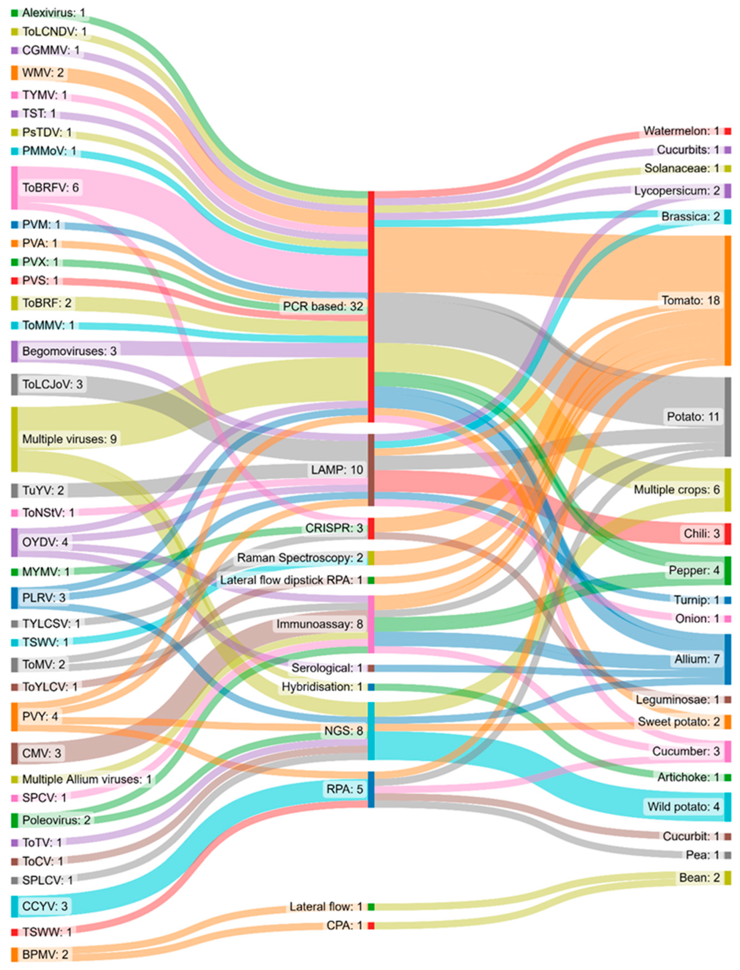
Figure 3 presents viruses, plant species, and detection methods in a Sankey diagram (generated using the SankeyMATIC online tool, available at https://sankeymatic.com/build/ accessed on 21 December 2022) and shows interactions between the virus types, methods of detection, and host species. The figure indicates that PCR-based methods have been exploited most frequently during the past five years. The crops for which the greatest numbers of detection methods have been introduced appear to be tomatoes, brassica vegetables, peppers, and chili.
More specifically, it can be observed that the majority of methods are related to the detection of tomato diseases, e.g., ToBRFV or ToLCJoV, as the worldwide tomato production for the year 2022 was reported to be 37.3 million tones [141].
8.2. Applications of Different Tools for the Detection of Bacterial Diseases in Vegetables
Bacterial diseases can be visually identified, and their quantification in plants can be achieved by means of colony-counting assays. An infection will have already spread by this stage, and it may be difficult to handle, which is why the prevention of bacterial infections requires early detection In addition to methods based on barcoding, which are suited to precise identification, PCR is used, including modifications that enable quantification. Several new approaches are currently being tested, including the CRISPR/Cas methodology, which may be used as a tool for genome editing, but also represents a novel, highly precise detection method [142,143]. The table summarizes the methods that have been used over the past 5 years to develop detection assays for bacterial infections published on the WoS. Table 2 summarizes the currently applicable tools for the detection and quantification of various bacterial species.
Table 2.
List of methods suited to the detection of bacterial infections in vegetables that have been published in the Web of Science database.
Table 2.
List of methods suited to the detection of bacterial infections in vegetables that have been published in the Web of Science database.
| Bacteria | Method | Species | Reference |
|---|---|---|---|
| Bacterial Spot | RPA | Tomato | [144] |
| Clavibacter | PCR | Tomato | [145] |
| Clavibacter | ddPCR | Tomato | [146] |
| Clavibacter | Multiplex qPCR | Tomato | [147] |
| Clavibacter | Multiplex qPCR | Tomato | [148] |
| Clavibacter | Multiplex qPCR | Tomato | [149] |
| Curtobacterium | LAMP | Legumes | [150] |
| Erwinia, Acidovorax | CRISPR/Cas | Vegetable | [151] |
| Multiple | PCR, NGS | Multiple | [109] |
| Multiple | Bar-coding | Multiple | [152] |
| Pectobacterium | LAMP | Radish | [153] |
| Pseudomonas | qPCR | Mung bean | [154] |
| Pectobacterium | LAMP | Celery | [155] |
| Pectobacterium | PCR | Cabbage | [156] |
| Pectobacterium | Multiplex PCR | Potato | [157] |
| Pectobacterium | Multiplex qPCR | Multiple | [158] |
| Phytoplasmas | CRISPR/Cas | Potato | [142] |
| Pseudomonas | LAMP | Tomato | [159] |
| Pseudomonas | LAMP | Pea | [160] |
| Pseudomonas | qPCR | Tomato | [161] |
| Pseudomonas | Multiplex qPCR | Cucumber | [162] |
| Ralstonia | LAMP | Potato | [163] |
| Ralstonia | Multiplex qPCR | Zingiberaceae | [164] |
| Salmonella, Clavibacter | cultivation | Potato | [165] |
| Xanthomonas | LAMP | Beans | [166] |
| Xanthomonas | PCR | Brassica | [167] |
| Yersinia | ddPCR, qPCR | Multiple | [168] |
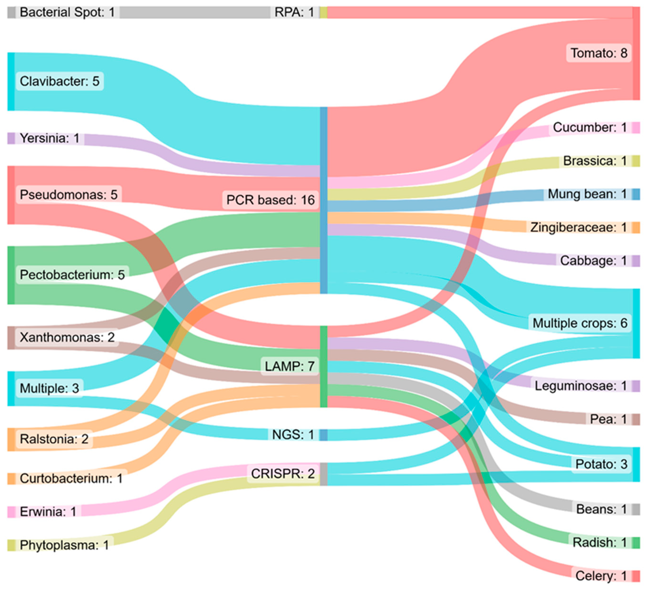
The plant bacteria detection methods, individual bacterial species, and the corresponding crop species listed in Table 2 are also presented in a Sankey chart (see Figure 4) (produced using the SankeyMATIC online tool, available at https://sankeymatic.com/build/ accessed on 21 December 2022), allowing for the extraction of significant findings.
It is apparent that PCR-based tools are still prevalent, but CRISPR/Cas-based methods have also been employed. PCR-based and LAMP techniques are used mainly for tomato crops, and some could be exploited for multiple crops.
The majority of the methods are suited to the detection of Clavibacter, Pseudomonas, Pectobacterium, and Xanthomonas species.
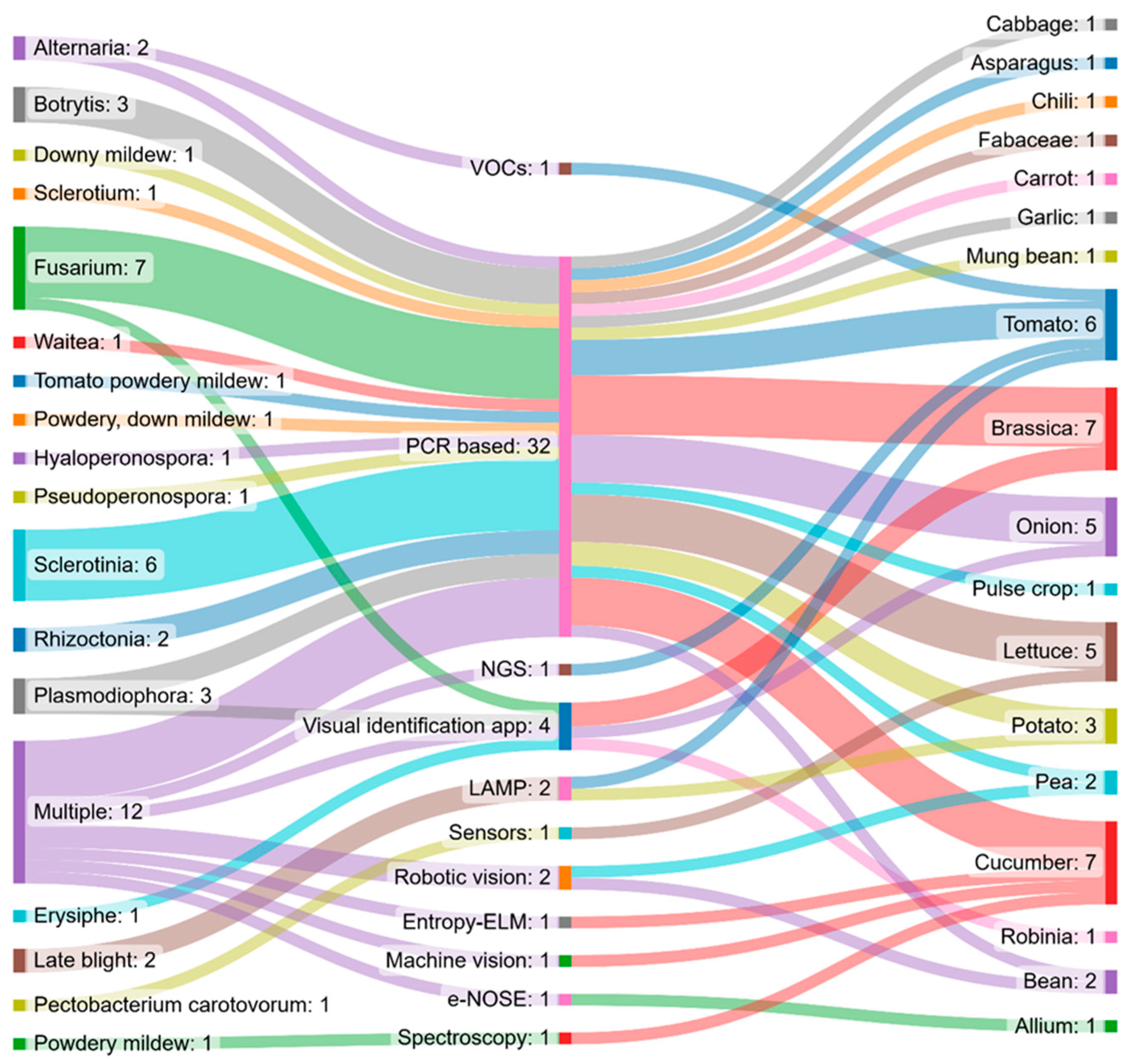
8.3. Applications of Different Tools for the Detection of Fungal Diseases of Vegetables
Fungi represent a major threat to crop production today and will continue to do so in the future [169]. We retrieved data from the WoS database to identify emerging tools for vegetable fungal pathogen diagnostics described over the last five years. From the overview, one can conclude that research studies have focused on the development of PCR-based assays that are specific to individual fungal phytopathogens. Such assays can be used for the detection of early infections when visual symptoms have not yet been revealed, as in the case of other vegetable pathogens. See Table 3.
Table 3.
List of methods suited to the identification of fungal infection in vegetables that have been published in the Web of Science database.
Table 3.
List of methods suited to the identification of fungal infection in vegetables that have been published in the Web of Science database.
| Fungus | Method | Species | Reference |
|---|---|---|---|
| Alternaria | Targeted chem.anal. | Tomato | [170] |
| Atlernaria | Multiplex PCR | Brassica | [171] |
| Botrytis | Species-specific PCR | Onion | [172] |
| Botrytis | PCR | Onion | [172] |
| Botrytis | qPCR | Onion | [173] |
| Downy mildew | RT PCR | Lettuce | [174] |
| Erysiphe Palczewski | Visual + smarphone | Robinia | [175] |
| Foot rot and head rot disease (S.rolfsii) | ITS PCR seq. | Cabbage | [176] |
| Fusarium | ITS barcoding | Potato | [177] |
| Fusarium | Visual/image anal. | Onion | [178] |
| Fusarium | PCR-RFLP | Pea | [179] |
| Fusarium | qPCR | Asparagus | [180] |
| Fusarium | PCR | Tomato | [181] |
| Fusarium | PCR | Onion | [182] |
| Fusarium | ITS barcoding | Chili | [183] |
| Fusarium, Rhizoctonia | Cultivation | Potato | [165] |
| Multiple | NGS | Tomato | [184] |
| Hyaloperonospora | Rt PCR | Cucumber | [185] |
| Late blight | LAMP | Potato, tomato | [186] |
| Multiple | PCR, NGS | Multiple | [109] |
| Multiple | e-NOSE | Garlic | [171] |
| Multiple | Machine vision | Cucumber | [187] |
| Multiple | ITS RNA seq | Brassica | [187] |
| Multiple | Image analysis | Brassica | [188] |
| Multiple | Robotic vision | Bean, pea | [189] |
| Multiple | ITS RNA seq | Bean | [190] |
| Multiple | RT PCR | Fabaceae | [191] |
| Multiple | Machine vision | Cucumber | [185] |
| Multiple | Multiplex PCR | Cucumber | [192] |
| Multiple | ITS barcoding | Tomato | [193] |
| Pectobacterium carotovorum | Sensors | Lettuce | [194] |
| Plasmodiophora | Species-specific PCR | Brassica | [195] |
| Plasmodiophora | PCR, SNPaSHOT | Brassica | [196] |
| Powdery mildew | Spectral data | Cucumber | [197] |
| Powdery, down mildew | Multiplex qPCR | Cucumber | [198] |
| Pseudoperonospora | RT PCR | Cucumber | [199] |
| Rhizoctonia | Multiplex RTPCR | Lettuce | [200] |
| Rhizoctonia | PCR, RTPCR | Pulse crop | [197] |
| Rust | IR spectroscopy | Multiple | [201] |
| Sclerotinia | Spec.specif. PCR | Carrot | [202] |
| Sclerotinia | RTPCR | Potato | [203] |
| Sclerotinia | Spec.spec. PCR | Lettuce | [202] |
| Sclerotinia | Spec.spec. PCR | Lettuce | [204] |
| Sclerotinia | PCR | Garlic | [205] |
| Sclerotinia | ITS RNA seq. | Mung bean | [206] |
| Tomato powdery mildew | RT PCR | Tomato | [207] |
| Waitea circinata | ITS rDNA seq | Brassica sp. | [208] |
In general, the listed PCR-based assays are highly sensitive. Based on the literature reviewed in the current study, the fungi, detection methods, and the corresponding crop species presented in Table 3 are also presented in a Sankey chart shown in Figure 5 (produced using the SankeyMATIC online tool, available at https://sankeymatic.com/build/ accessed on 21 December 2022), allowing for the extraction of significant findings.
It is apparent that most of the published methods refer to the detection of Fusarium and Sclerotinia species. The most widely used detection techniques for fungi are nucleic-acid-based techniques, and the visual identification applications mainly concern tomato, cucumber, and brassica crops.
8.4. Commercially Available IPM Tools/Solutions
Practical applications based on research have been developed, proofs-of-concept have been verified, and several tools have reached the highest level of technological readiness, TRL 9 [209], which means that these tools are available on the market. Descriptions and assessments of these methods are available at https://platform.smartprotect-h2020.eu/en (accessed on 2 January 2023), a database developed by the SmartProtect consortium.
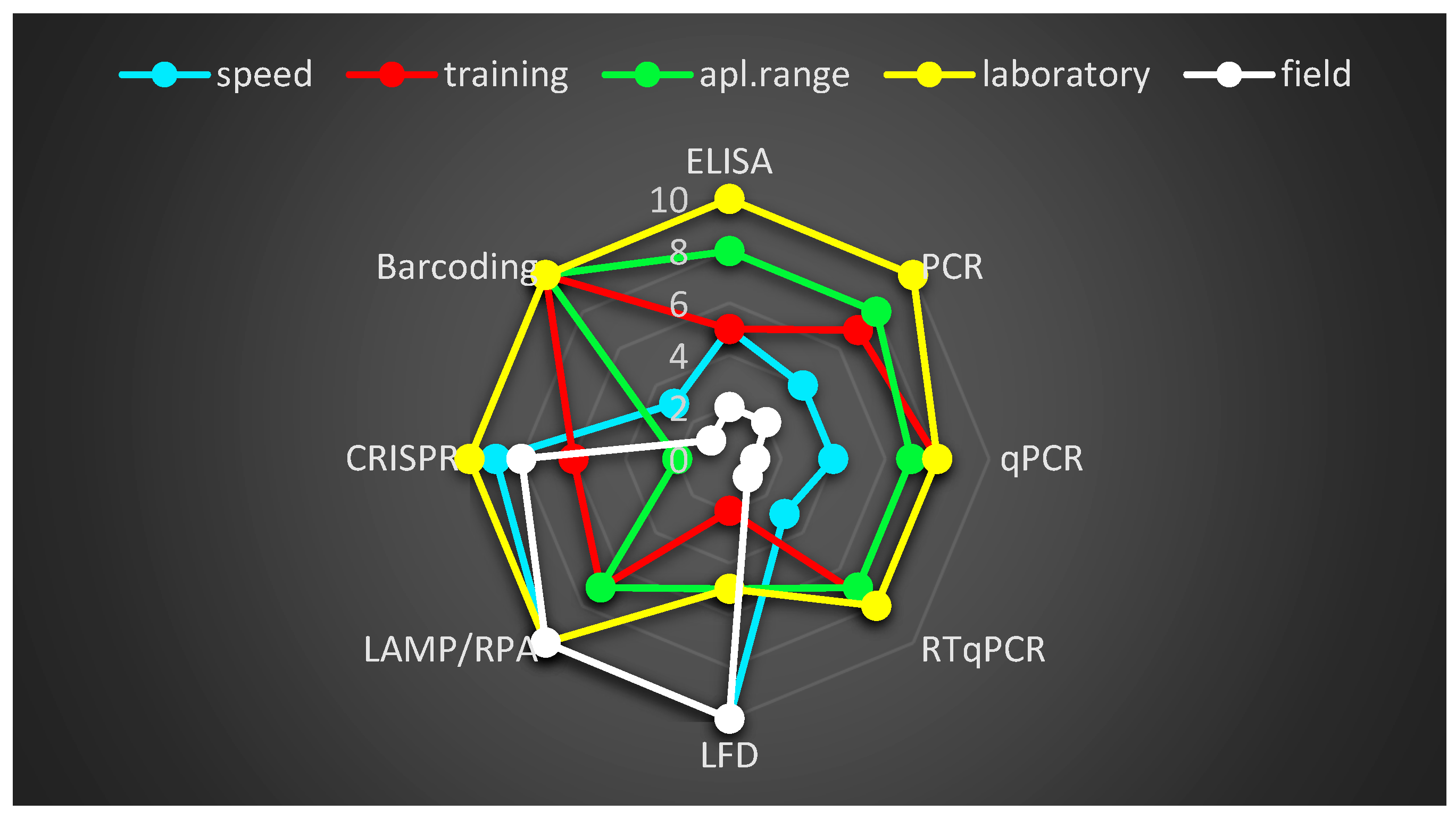
Generally, the attributes of these techniques are assessed according to their applicability, e.g., the range of pathogens they can identify; their requirements for specific equipment; the education and training necessary for the operators; their speed in making determinations; and their availability in individual markets. Figure 6 shows the positive attributes of the principal, most widely used methods.
It is clear that test strips are suitable for quick use on farms and in greenhouses; it follows that their use for accurate determination is even easier in laboratories.
9. Biosensors
Biosensors are among the best approaches for obtaining easy and cheap diagnostics. In principle, biosensors are devices that, in our case, measure biological reactions, giving rise to a signal equivalent to the amount of target. Biosensors rely on a combination of biorecognition molecules and transducers converting one type of variation into another, e.g., an electric signal and a visualization system, making them easy to use [210]. They were first used in on-site diagnostics to identify human diseases [211,212,213]. The current techniques for producing biosensors aim to employ new materials, being manufactured using nanotechnologies and possessing higher biological affinity, to develop new sensors capable of disease surveillance on different scales. The concept has been used for detection of human pathogens on marketed vegetables [214,215,216]. To date, research has mainly focused on human pathogens that contaminate vegetable production, such as Salmonella or Listeria [215,217,218]. Few reports describe the use of biosensors for vegetable phytopathogen detection [215,219,220]. The use of biosensors for the precise detection of vegetable phytopathogens needs to be evaluated in practice. Hopefully, they will find application in the detection and quantification of pathogens that contaminate vegetables in the near future [221].
10. E-Senses
Other approaches do not use species-/pathotype-specific proteins, such as ELISA, or DNA/RNA, such as PCR or sequencing, as a target may be aimed towards metabolites (eNose), visual patterns such as color changes, structural changes (eEyes), or differences in soluble materials (eTongue), resembling the functions of the human senses. Numerous papers have been published describing the basic principles of these assays and showing the current state of the art [222,223,224,225,226,227].
Among the e-Senses, eNose is an approach that allows for the detection of volatiles released by organisms and senses volatiles using miniaturized gas chromatography instruments [228]. In the case of eNose, focus is placed on specific metabolites produced by the pathogen itself and by infected plants.
It is known that volatiles produced by an organism undergo changes related to their physiological state and developmental stage. Hundreds of reports are available on compounds captured upon plant infection [229,230] by various types of chromatography and mass spectrometry [231,232]. Thus, with libraries of volatile mixtures similar to those continuously supplemented for regular metabolomics analysis [233,234] produced upon infection, simple tools might be available for farmers in the future to conduct large-scale analyses of crops, including vegetables. Some authors [235,236] have reported on the practical utilization of eNose for palm tree disease detection. Borowik et al. [237] reported on the successful utilization of eNose for the detection of Phytophtora in Quercus acorn and later, with an improved chamber, Ciboria batschiana [238]. The same group [237] introduced a low-cost detection procedure for phytopathogenic fungi. Although several examples of eNose utilization have been described in the literature, only a few have undertaken the detection of plant pathogens, and applications with vegetables have seldom been carried out. Only one example was found to be commercially available (TRL9; for further details, see the SMARTPROTECT platform database). Additionally, the authors of [239] noted that eNoses may find widespread application for plant diseases diagnostics, as well as other purposes, but more research and testing are required before incorporating this efficient tool into agriculture practice [239]. To spread the techniques used for various vegetable species cultivated across the world and the detection of their pathogens, tremendous effort will be required to combine the technical basis of e-Noses and sensing of specific mixtures of compounds. Further knowledge and verification are needed. Nevertheless, Mohammad-Razdari et al. [240] predicted their potential for farm management. Direct comparisons with conventional laboratory methods will be essential for routine application in practice.
11. Outlook
The timely, correct, and accurate diagnosis of diseases is the fundamental basis for the successful cultivation of agricultural crops, including vegetables. Along with early warning systems, diagnostic tools are essential for disease management and the production of high-quality and high-yielding vegetables. Currently, the diagnosis of plant diseases is based on approaches developed by other sectors, e.g., medicine, and generally, they are first applied to major crops, followed by later extension to vegetables. Commercially, specific procedures such as ELISA, PCR, real-time PCR, and isothermal amplification are available, but these require a priori knowledge of the target molecules used for each pathogen, and they differ in their level of sensitivity (detection limit). Other tests have been described in the scientific literature, but few of these that are applicable to vegetables have been adequately tested in regard to their analytical parameters, in contrast to their use in human medicine. It can be assumed that users will demand multiple assays, enabling the multiplex detection of a number of pathogens simultaneously, in addition to next-generation sequencing (NGS) and barcoding. Thus far, the difficulties lie in the high cost of such methods and in interpreting the results. With decreases in the cost of the equipment and services and the development of simpler interpretation platforms, this type of technology has the potential to be extended to use on vegetables. A second advantageous route lies in the development of simple diagnostics in the field. Currently, isothermal amplification is advantageous due to its ease of use and quick results. However, the use of CRISPR/Cas for diagnostics, in combination with suitable carriers (test strips), appears to be a highly promising method, according to publications from 2021–2022. This is especially true considering the high accuracy of the system and the possibility of signal amplification. Overall, it is clear that the greatest challenge for phytopathogen diagnostics is the production of affordably priced assays that carry the possibility of multiple detections through the analysis of a single sample.
The evaluation of commercially available solutions shows that there is a wealth of lateral flow test solutions covering a wide range of pathogens and crops, while simultaneously forgoing requirements for specialized equipment or training. The ELISA, LAMP, and PCR/qPCR methods are more accurate but require specialized equipment, more time, and trained users to perform the analyses. Thus, lateral flow tests constitute very useful preliminary tests, being performed prior to laboratory analyses, and they can offer an early indication of viral, fungal, and bacterial infections.
Author Contributions
J.O. drafted the outline, conducted the literature search of WoS, drafted the theoretical portion of the review, and conceptualized the study; M.D.K. drafted the section on the practical state of the art and conducted the comparative analysis; Z.T. performed the visualizations; R.C. and A.K. carefully reviewed and edited the text; and J.D.M. and S.P. contributed to project administration. All authors have read and agreed to the published version of the manuscript.
Funding
This research was funded by the European Union project H2020-RUR-1 20119, SmartProtect, Grant agreement ID: 862563, and the Czech Ministry of Agriculture, Grant numbers RO0418 and RO0423.
Institutional Review Board Statement
Not applicable.
Informed Consent Statement
Not applicable.
Data Availability Statement
Further information are available from the corresponding authors upon reasonable request.
Acknowledgments
We would especially like to thank Barbora Šestáková for the excellent technical assistance and formal editing of the text, and Liga Lepse, who contributed to the SmartProtect database.
Conflicts of Interest
The authors declare no conflict of interest.
References
- Eschen, R.; Britton, K.; Brockerhoff, E.; Burgess, T.; Dalley, V.; Epanchin-Niell, R.S.; Gupta, K.; Hardy, G.; Huang, Y.; Kenis, M.; et al. International variation in phytosanitary legislation and regulations governing importation of plants for planting. Environ. Sci. Policy 2015, 51, 228–237. [Google Scholar] [CrossRef]
- Kumar, P.L.; Cuervo, M.; Kreuze, J.F.; Muller, G.; Kulkarni, G.; Kumari, S.G.; Massart, S.; Mezzalama, M.; Alakonya, A.; Muchugi, A.; et al. Phytosanitary Interventions for Safe Global Germplasm Exchange and the Prevention of Transboundary Pest Spread: The Role of CGIAR Germplasm Health Units. Plants 2021, 10, 328. [Google Scholar] [CrossRef]
- World Trade Organization. World Trade Report 2012; World Trade Organization: Geneva, Switzerland, 2012; p. 252. [Google Scholar]
- Ratnadass, A.; Fernandes, P.; Avelino, J.; Habib, R. Plant species diversity for sustainable management of crop pests and diseases in agroecosystems: A review. Agron. Sustain. Dev. 2012, 32, 273–303. [Google Scholar] [CrossRef]
- U.S. Environmental Protection Agency. Introduction to Integrated Pest Management. 2022. Available online: https://www.epa.gov/ipm/introduction-integrated-pest-management (accessed on 3 December 2022).
- Miller, S.A.; Beed, F.D.; Harmon, C.L. Plant Disease Diagnostic Capabilities and Networks. Annu. Rev. Phytopathol. 2009, 47, 15–38. [Google Scholar] [CrossRef] [PubMed]
- Mansotra, R.; Vakhlu, J. Comprehensive account of present techniques for in-field plant disease diagnosis. Arch. Microbiol. 2021, 203, 5309–5320. [Google Scholar] [CrossRef]
- Hariharan, G.; Prasannath, K. Recent Advances in Molecular Diagnostics of Fungal Plant Pathogens: A Mini Review. Front. Cell. Infect. Microbiol. 2021, 10, 493–504. [Google Scholar] [CrossRef]
- Rubio, L.; Galipienso, L.; Ferriol, I. Detection of Plant Viruses and Disease Management: Relevance of Genetic Diversity and Evolution. Front. Plant Sci. 2020, 11, 1092. [Google Scholar] [CrossRef]
- Gooden, J.; Samac, D.; Caffier, D.; Ophel-Keller, K.; Sheppard, J. Method Validation by Ringtesting to Establish International Standards for Seed Testing; a Case Study. In Plant Pathogenic Bacteria; Springer: Berlin/Heidelberg, Germany, 2001; pp. 425–427. [Google Scholar] [CrossRef]
- Aveling, T. Global standards in seed health testing. In Global Perspectives on the Health of Seeds and Plant Propagation Material; Springer: Berlin/Heidelberg, Germany, 2014; pp. 17–28. [Google Scholar]
- European Union Reference Laboratories. 2018. Available online: https://food.ec.europa.eu/horizontal-topics/european-union-reference-laboratories_en (accessed on 9 April 2023).
- Stack, J.; Cardwell, K.; Hammerschmidt, R.; Byrne, J.; Loria, R.; Snover-Clift, K.; Baldwin, W.; Wisler, G.; Beck, H.; Bostock, R.; et al. The National Plant Diagnostic Network. Plant Dis. 2006, 90, 128–136. [Google Scholar] [CrossRef]
- Pavithra, A.; Kalpana, G.; Vigneswaran, T. Deep learning-based automated disease detection and classification model for precision agriculture. Soft Comput. 2023. [Google Scholar] [CrossRef]
- Inoue, Y. Satellite- and drone-based remote sensing of crops and soils for smart farming—A review. Soil Sci. Plant Nutr. 2020, 66, 798–810. [Google Scholar] [CrossRef]
- Raffaini, P.; Manfredi, L. Chapter 15—Project management. In Endorobotics; Manfredi, L., Ed.; Academic Press: Cambridge, MA, USA, 2022; pp. 337–358. [Google Scholar] [CrossRef]
- Martinelli, F.; Scalenghe, R.; Davino, S.; Panno, S.; Scuderi, G.; Ruisi, P.; Villa, P.; Stroppiana, D.; Boschetti, M.; Goulart, L.R.; et al. Advanced methods of plant disease detection. A review. Agron. Sustain. Dev. 2015, 35, 1–25. [Google Scholar] [CrossRef]
- Zherdev, A.V.; Vinogradova, S.V.; Byzova, N.A.; Porotikova, E.V.; Kamionskaya, A.M.; Dzantiev, B.B. Methods for the Diagnosis of Grapevine Viral Infections: A Review. Agriculture 2018, 8, 195. [Google Scholar] [CrossRef]
- Mancini, V.; Murolo, S.; Romanazzi, G. Diagnostic methods for detecting fungal pathogens on vegetable seeds. Plant Pathol. 2016, 65, 691–703. [Google Scholar] [CrossRef]
- Wen, H.; Fu, Z.; Zhang, L.; Li, X.; Zhao, W. Video Assisted Diagnosis System for Cucumber Disease. J. Food Agric. Environ. 2012, 10, 857–860. [Google Scholar]
- Wei, Q.F.; Luo, C.S.; Cao, C.Z.; Guo, Q. The Intelligent Diagnostic System of Vegetable Diseases Based on a Fuzzy Neural Network. Appl. Mech. Mater. 2013, 321–324, 1907–1911. [Google Scholar] [CrossRef]
- Astuti, E.; Saragih, N.E.; Sribina, N.; Ramadhani, R. Dempster-Shafer Method for Diagnose Diseases on Vegetable. In Proceedings of the 6th International Conference on Cyber and IT Service Management (CITSM), Parapat, Indonesia, 7–9 August 2018; pp. 643–646. [Google Scholar]
- Bohnenkamp, D.; Behmann, J.; Paulus, S.; Steiner, U.; Mahlein, A.K. A Hyperspectral Library of Foliar Diseases of Wheat. Phytopathology 2021, 111, 1583–1593. [Google Scholar] [CrossRef]
- Barbedo, J.G.A. A review on the main challenges in automatic plant disease identification based on visible range images. Biosyst. Eng. 2016, 144, 52–60. [Google Scholar] [CrossRef]
- Engvall, E.; Perlmann, P. Enzyme-Linked Immunosorbent Assay, Elisa: III. Quantitation of Specific Antibodies by Enzyme-Labeled Anti-Immunoglobulin in Antigen-Coated Tubes. J. Immunol. 1972, 109, 129–135. [Google Scholar] [CrossRef]
- Phatsaman, T.; Hongprayoon, R.; Wasee, S. Monoclonal antibody-based diagnostic assays for pepper mild mottle virus. J. Plant Pathol. 2020, 102, 327–333. [Google Scholar] [CrossRef]
- Mullis, K.B.; Faloona, F.A. Specific Synthesis of DNA Invitro via a Polymerase-Catalyzed Chain-Reaction. Methods Enzymol. 1987, 155, 335–350. [Google Scholar]
- Real-Time PCR Handbook. 2012. Available online: www.gene-quantification.de/real-time-pcr-handbook-life-technologies-update-flr.pdf (accessed on 8 April 2023).
- Heid, C.A.; Stevens, J.; Livak, K.J.; Williams, P.M. Real time quantitative PCR. Genome Res. 1996, 6, 986–994. [Google Scholar] [CrossRef] [PubMed]
- Stephenson, F.H. Chapter 9—Real-Time PCR. In Calculations for Molecular Biology and Biotechnology, 3rd ed.; Stephenson, F.H., Ed.; Academic Press: Boston, MA, USA, 2016; pp. 215–320. [Google Scholar] [CrossRef]
- Huggett, J.F.; Cowen, S.; Foy, C.A. Considerations for Digital PCR as an Accurate Molecular Diagnostic Tool. Clin. Chem. 2015, 61, 79–88. [Google Scholar] [CrossRef] [PubMed]
- Fialova, E.; Zdenkova, K.; Jablonska, E.; Demnerova, K.; Ovesna, J. Digital polymerase chain reaction: Principle and Applications. Chem. Listy 2019, 113, 545–552. [Google Scholar]
- Schaad, N.W.; Frederick, R.D.; Shaw, J.; Schneider, W.L.; Hickson, R.; Petrillo, M.D.; Luster, D.G. Advances in molecular-based diagnostics in meeting crop biosecurity and phytosanitary issues. Annu. Rev. Phytopathol. 2003, 41, 305–324. [Google Scholar] [CrossRef]
- Kralik, P.; Ricchi, M. A Basic Guide to Real Time PCR in Microbial Diagnostics: Definitions, Parameters, and Everything. Front. Microbiol. 2017, 8, 108. [Google Scholar] [CrossRef] [PubMed]
- Chen, J.; Tang, J.; Liu, J.; Cai, Z.; Bai, X. Development and evaluation of a multiplex PCR for simultaneous detection of five foodborne pathogens. J. Appl. Microbiol. 2012, 112, 823–830. [Google Scholar] [CrossRef]
- Bustin, S.A.; Johnson, G.; Agrawal, S.G. MIQE—Guidelines for developing robust real-time PCR assays. Mycoses 2012, 55, 30. [Google Scholar]
- Pallas, V.; Sanchez-Navarro, J.; Varga, A.; Aparicio, F.; James, D. Multiplex polymerase chain reaction (PCR) and real-time multiplex PCR for the simultaneous detection of plant viruses. Methods Mol. Biol. 2009, 508, 193–208. [Google Scholar] [CrossRef]
- Catara, V.; Cubero, J.; Pothier, J.F.; Bosis, E.; Bragard, C.; Dermic, E.; Holeva, M.C.; Jacques, M.A.; Petter, F.; Pruvost, O.; et al. Trends in Molecular Diagnosis and Diversity Studies for Phytosanitary Regulated Xanthomonas. Microorganisms 2021, 9, 862. [Google Scholar] [CrossRef]
- Rahman, H.U.; Yue, X.; Yu, Q.; Zhang, W.; Zhang, Q.; Li, P. Current PCR-based methods for the detection of mycotoxigenic fungi in complex food and feed matrices. World Mycotoxin J. 2020, 13, 139–150. [Google Scholar] [CrossRef]
- Baker, M. qPCR: Quicker and easier but don’t be sloppy. Nat. Methods 2011, 8, 207–212. [Google Scholar] [CrossRef]
- ISO20395:2019; Biotechnology—Requirements for Evaluating the Performance of Quantification Methods for Nucleic Acid Target Sequences—qPCR and dPCR. ISO: Geneva, Switzerland, 2019; p. 50.
- Chikh-Ali, M.; Karasev, A.V. Immunocapture-Multiplex RT-PCR for the Simultaneous Detection and Identification of Plant Viruses and Their Strains: Study Case, Potato Virus Y (PVY). In Plant Pathology: Techniques and Protocols; Lacomme, C., Ed.; Springer: New York, NY, USA, 2015; pp. 177–186. [Google Scholar] [CrossRef]
- James, D.; Varga, A.; Pallas, V.; Candresse, T. Strategies for simultaneous detection of multiple plant viruses. Can. J. Plant Pathol. 2006, 28, 16–29. [Google Scholar] [CrossRef]
- Jacobi, V.; Bachand, G.D.; Hamelin, R.C.; Castello, J.D. Development of a multiplex immunocapture RT-PCR assay for detection and differentiation of tomato and tobacco mosaic tobamoviruses. J. Virol. Methods 1998, 74, 167–178. [Google Scholar] [CrossRef]
- Kokko, H.I.; Kivineva, M.; Kärenlampi, S.O. Single-Step Immunocapture RT-PCR in the Detection of Raspberry Bushy Dwarf Virus. BioTechniques 1996, 20, 842–846. [Google Scholar] [CrossRef]
- Kundu, J.K. A rapid and effective RNA release procedure for virus detection in woody plants by reverse transcription-polymerase chain reaction. Acta Virol. 2003, 47, 147–151. [Google Scholar]
- Pallas, V.; Sanchez-Navarro, J.A.; James, D. Recent Advances on the Multiplex Molecular Detection of Plant Viruses and Viroids. Front. Microbiol. 2018, 9, 2087. [Google Scholar] [CrossRef] [PubMed]
- Chen, L.; Zhang, C.; Yadav, V.; Wong, A.; Senapati, S.; Chang, H.-C. A home-made pipette droplet microfluidics rapid prototyping and training kit for digital PCR, microorganism/cell encapsulation and controlled microgel synthesis. Sci. Rep. 2023, 13, 184. [Google Scholar] [CrossRef] [PubMed]
- Burpo, F.J. A critical review of PCR primer design algorithms and crosshybridization case study. Biochemistry 2001, 218, 1–12. [Google Scholar]
- Singh, V.K.; Kumar, A. PCR primer design. Mol. Biol. Today 2001, 2, 27–32. [Google Scholar]
- Asadi, R.; Mollasalehi, H. The mechanism and improvements to the isothermal amplification of nucleic acids, at a glance. Anal. Biochem. 2021, 631, 114260. [Google Scholar] [CrossRef]
- Notomi, T.; Okayama, H.; Masubuchi, H.; Yonekawa, T.; Watanabe, K.; Amino, N.; Hase, T. Loop-mediated isothermal amplification of DNA. Nucleic Acids Res. 2000, 28, e63. [Google Scholar] [CrossRef] [PubMed]
- Wong, Y.-P.; Othman, S.; Lau, Y.-L.; Radu, S.; Chee, H.-Y. Loop-mediated isothermal amplification (LAMP): A versatile technique for detection of micro-organisms. J. Appl. Microbiol. 2018, 124, 626–643. [Google Scholar] [CrossRef] [PubMed]
- Panno, S.; Matic, S.; Tiberini, A.; Caruso, A.; Bella, P.; Torta, L.; Stassi, R.; Davino, S. Loop Mediated Isothermal Amplification: Principles and Applications in Plant Virology. Plants 2020, 9, 461. [Google Scholar] [CrossRef] [PubMed]
- Fukuta, S.; Kato, S.; Yoshida, K.; Mizukami, Y.; Ishida, A.; Ueda, J.; Kanbe, M.; Ishimoto, Y. Detection of tomato yellow leaf curl virus by loop-mediated isothermal amplification reaction. J. Virol. Methods 2003, 112, 35–40. [Google Scholar] [CrossRef] [PubMed]
- Fukuta, S.; Iida, T.; Mizukami, Y.; Ishida, A.; Ueda, J.; Kanbe, M.; Ishimoto, Y. Detection of Japanese yam mosaic virus by RT-LAMP. Arch. Virol. 2003, 148, 1713–1720. [Google Scholar] [CrossRef]
- Tanner, N.A.; Zhang, Y.H.; Evans, T.C. Visual detection of isothermal nucleic acid amplification using pH-sensitive dyes. Biotechniques 2015, 58, 59–68. [Google Scholar] [CrossRef]
- Ahuja, A.; Somvanshi, V.S. Diagnosis of plant-parasitic nematodes using loop-mediated isothermal amplification (LAMP): A review. Crop Prot. 2021, 147, 105459. [Google Scholar] [CrossRef]
- Becherer, L.; Borst, N.; Bakheit, M.; Frischmann, S.; Zengerle, R.; von Stetten, F. Loop-mediated isothermal amplification (LAMP)—Review and classification of methods for sequence-specific detection. Anal. Methods 2020, 12, 717–746. [Google Scholar] [CrossRef]
- Moehling, T.J.; Choi, G.; Dugan, L.C.; Salit, M.; Meagher, R.J. LAMP Diagnostics at the Point-of-Care: Emerging Trends and Perspectives for the Developer Community. Expert Rev. Mol. Diagn. 2021, 21, 43–61. [Google Scholar] [CrossRef]
- ISO22942-1:2022; Molecular Biomarker Analysis—Isothermal Polymerase Chain Reaction (isoPCR) Methods—Part 1: General Requirements. ISO: Geneva, Switzerland, 2022.
- Cassedy, A.; Mullins, E.; O’Kennedy, R. Sowing seeds for the future: The need for on-site plant diagnostics. Biotechnol. Adv. 2020, 39, 107358. [Google Scholar] [CrossRef]
- Glais, L.; Jacquot, E. Detection and Characterization of Viral Species/Subspecies Using Isothermal Recombinase Polymerase Amplification (RPA) Assays. Methods Mol. Biol. 2015, 1302, 207–225. [Google Scholar] [CrossRef] [PubMed]
- Piepenburg, O.; Williams, C.; Stemple, D.; Armes, N. DNA Detection Using Recombination Proteins. PLoS Biol. 2006, 4, e204. [Google Scholar] [CrossRef] [PubMed]
- Zhao, Y.; Chen, F.; Li, Q.; Wang, L.; Fan, C. Isothermal Amplification of Nucleic Acids. Chem. Rev. 2015, 115, 12491–12545. [Google Scholar] [CrossRef] [PubMed]
- Tan, M.; Liao, C.; Liang, L.; Yi, X.; Zhou, Z.; Wei, G. Recent advances in recombinase polymerase amplification: Principle, advantages, disadvantages and applications. Front. Cell. Infect. Microbiol. 2022, 12, 1019071. [Google Scholar] [CrossRef] [PubMed]
- Lau, Y.L.; Ismail, I.b.; Mustapa, N.I.b.; Lai, M.Y.; Tuan Soh, T.S.; Haji Hassan, A.; Peariasamy, K.M.; Lee, Y.L.; Abdul Kahar, M.K.B.; Chong, J.; et al. Development of a reverse transcription recombinase polymerase amplification assay for rapid and direct visual detection of Severe Acute Respiratory Syndrome Coronavirus 2 (SARS-CoV-2). PLoS ONE 2021, 16, e0245164. [Google Scholar] [CrossRef]
- Zhang, C.Y.; Yang, Y.C.; Liu, F.G.; Wang, Y.Y.; Chen, G.F. Recombinase polymerase amplification combined with lateral flow dipstick for the rapid detection of Chattonella marina. J. Appl. Phycol. 2022, 34, 1607–1620. [Google Scholar] [CrossRef]
- Roumani, F.; Rodrigues, C.; Barros-Velazquez, J.; Garrido-Maestu, A.; Prado, M. Development of a Panfungal Recombinase Polymerase Amplification (RPA) Method Coupled with Lateral Flow Strips for the Detection of Spoilage Fungi. Food Anal. Methods 2022. [Google Scholar] [CrossRef]
- Feng, Z.Z.; Chu, X.; Han, M.N.; Yu, C.W.; Jiang, Y.S.; Wang, H.; Lu, L.Q.; Xu, D. Rapid visual detection of Micropterus salmoides rhabdovirus using recombinase polymerase amplification combined with lateral flow dipsticks. J. Fish Dis. 2022, 45, 461–469. [Google Scholar] [CrossRef]
- Bai, Y.M.; Ji, J.C.; Ji, F.D.; Wu, S.; Tian, Y.; Jin, B.R.; Li, Z.D. Recombinase polymerase amplification integrated with microfluidics for nucleic acid testing at point of care. Talanta 2022, 240, 123209. [Google Scholar] [CrossRef]
- Bektaş, A.; Covington, M.F.; Aidelberg, G.; Arce, A.; Matute, T.; Núñez, I.; Walsh, J.; Boutboul, D.; Delaugerre, C.; Lindner, A.B.; et al. Accessible LAMP-Enabled Rapid Test (ALERT) for Detecting SARS-CoV-2. Viruses 2021, 13, 742. [Google Scholar] [CrossRef]
- Tamari, F.; Hinkley, C. Extraction of DNA from Plant Tissue: Review and Protocols. In Sample Preparation Techniques for Soil, Plant, and Animal Samples; Springer: Berlin/Heidelberg, Germany, 2016; pp. 245–263. [Google Scholar] [CrossRef]
- Jina, H.; Rajkumar, K.; Pranab Behari, M. The Chemistry Behind Plant DNA Isolation Protocols. Biochem. Anal. Tools—Methods Bio-Mol. Stud. 2020, 8, 131–141. [Google Scholar] [CrossRef]
- Jansen, R.; van Embden, J.D.A.; Gaastra, W.; Schouls, L.M. Identification of genes that are associated with DNA repeats in prokaryotes. Mol. Microbiol. 2002, 43, 1565–1575. [Google Scholar] [CrossRef] [PubMed]
- Cong, L.; Ran, F.A.; Cox, D.; Lin, S.L.; Barretto, R.; Habib, N.; Hsu, P.D.; Wu, X.B.; Jiang, W.Y.; Marraffini, L.A.; et al. Multiplex Genome Engineering Using CRISPR/Cas Systems. Science 2013, 339, 819–823. [Google Scholar] [CrossRef] [PubMed]
- Brooks, C.; Nekrasov, V.; Lippman, Z.B.; Van Eck, J. Efficient Gene Editing in Tomato in the First Generation Using the Clustered Regularly Interspaced Short Palindromic Repeats/CRISPR-Associated9 System. Plant Physiol. 2014, 166, 1292–1297. [Google Scholar] [CrossRef] [PubMed]
- Li, L.X.; Li, S.Y.; Wu, N.; Wu, J.C.; Wang, G.; Zhao, G.P.; Wang, J. HOLMESv2: A CRISPR-Cas12b-Assisted Platform for Nucleic Acid Detection and DNA Methylation Quantitation. ACS Synth. Biol. 2019, 8, 2228–2237. [Google Scholar] [CrossRef]
- Javalkote, V.S.; Kancharla, N.; Bhadra, B.; Shukla, M.; Soni, B.; Sapre, A.; Goodin, M.; Bandyopadhyay, A.; Dasgupta, S. CRISPR-based assays for rapid detection of SARS-CoV-2. Methods 2022, 203, 594–603. [Google Scholar] [CrossRef]
- Kostyusheva, A.; Brezgin, S.; Babin, Y.; Vasilyeva, I.; Glebe, D.; Kostyushev, D.; Chulanov, V. CRISPR-Cas systems for diagnosing infectious diseases. Methods 2022, 203, 431–446. [Google Scholar] [CrossRef]
- Mahas, A.; Hassan, N.; Aman, R.; Marsic, T.; Wang, Q.C.; Ali, Z.; Mahfouz, M.M. LAMP-Coupled CRISPR-Cas12a Module for Rapid and Sensitive Detection of Plant DNA Viruses. Viruses 2021, 13, 466. [Google Scholar] [CrossRef]
- Zhai, S.S.; Yang, Y.; Wu, Y.H.; Li, J.; Li, Y.J.; Wu, G.; Liang, J.A.; Gao, H.F. A visual CRISPR/dCas9-mediated enzyme-linked immunosorbent assay for nucleic acid detection with single-base specificity. Talanta 2023, 257, 124318. [Google Scholar] [CrossRef]
- Liu, F.X.; Cui, J.Q.; Wu, Z.H.; Yao, S.H. Recent progress in nucleic acid detection with CRISPR. Lab Chip 2023, 23, 1467–1492. [Google Scholar] [CrossRef]
- Sharma, S.K.; Gupta, O.P.; Pathaw, N.; Sharma, D.; Maibam, A.; Sharma, P.; Sanasam, J.; Karkute, S.G.; Kumar, S.; Bhattacharjee, B. CRISPR-Cas-Led Revolution in Diagnosis and Management of Emerging Plant Viruses: New Avenues Toward Food and Nutritional Security. Front. Nutr. 2021, 8, 751512. [Google Scholar] [CrossRef] [PubMed]
- Osborn, M.J.; Bhardwaj, A.; Bingea, S.P.; Knipping, F.; Feser, C.J.; Lees, C.J.; Collins, D.P.; Steer, C.J.; Blazar, B.R.; Tolar, J. CRISPR/Cas9-Based Lateral Flow and Fluorescence Diagnostics. Bioengineering 2021, 8, 23. [Google Scholar] [CrossRef] [PubMed]
- Srivastava, A.; Gupta, T.; Srivastava, S.; Dhir, S.; Kumar, P.; Singhal, T.; Rani, A.; Rishi, N. Development of a new Collateral Cleavage-independent CRISPR/Cas12a based easy detection system for plant viruses. J. Virol. Methods 2022, 300, 114432. [Google Scholar] [CrossRef]
- Selvam, K.; Najib, M.A.; Khalid, M.F.; Ozsoz, M.; Aziah, I. CRISPR-Cas Systems-Based Bacterial Detection: A Scoping Review. Diagnostics 2022, 12, 1335. [Google Scholar] [CrossRef]
- Wu, X.L.; Chan, C.; Springs, S.L.; Lee, Y.H.; Lu, T.K.; Yu, H. A warm-start digital CRISPR/Cas-based method for the quantitative detection of nucleic acids. Anal. Chim. Acta 2022, 1196, 339494. [Google Scholar] [CrossRef] [PubMed]
- Ramesh, M.; Sen, A.; Vachher, M.; Nigam, A. Delineating Bacteria Using DNA Barcoding. Mol. Genet. Microbiol. Virol. 2021, 36, S65–S73. [Google Scholar] [CrossRef]
- Schoch, C.; Seifert, K.; Huhndorf, S.M.; Robert, V.; Spouge, J.; Levesque, C.A.; Chen, W.; Crous, P.; Boekhout, T.; Damm, U.; et al. Nuclear ribosomal internal transcribed spacer (ITS) region as a universal DNA barcode marker for Fungi. Proc. Natl. Acad. Sci. USA 2012, 109, 6241–6246. [Google Scholar] [CrossRef]
- Toju, H.; Tanabe, A.S.; Yamamoto, S.; Sato, H. High-Coverage ITS Primers for the DNA-Based Identification of Ascomycetes and Basidiomycetes in Environmental Samples. PLoS ONE 2012, 7, e40863. [Google Scholar] [CrossRef]
- Xu, J. Fungal DNA barcoding. Genome 2016, 59, 913–932. [Google Scholar] [CrossRef]
- Bonants, P.; Groenewald, E.; Rasplus, J.-Y.; Maes, M.; De Vos, P.; Frey, J.; Boonham, N.; Nicolaisen, M.; Bertacini, A.; Robert, V.; et al. QBOL: A new EU project focusing on DNA barcoding of Quarantine organisms. EPPO Bull. 2010, 40, 30–33. [Google Scholar] [CrossRef]
- Choudhary, P.; Singh, B.N.; Chakdar, H.; Saxena, A.K. DNA barcoding of phytopathogens for disease diagnostics and bio-surveillance. World J. Microbiol. Biotechnol. 2021, 37, 54. [Google Scholar] [CrossRef]
- Bachwenkizi, H.S.; Temu, G.E.; Mbanzibwa, D.R.; Lupembe, M.D.; Ngailo, S.; Tairo, F.D.; Massawe, D.P. Recombination and darwinian selection as drivers of genetic diversity and evolution of sweet potato leaf curl viruses in Tanzania. Physiol. Mol. Plant Pathol. 2022, 120, 101853. [Google Scholar] [CrossRef]
- Harjes, J.; Link, A.; Weibulat, T.; Triebel, D.; Rambold, G. FAIR digital objects in environmental and life sciences should comprise workflow operation design data and method information for repeatability of study setups and reproducibility of results. Database 2020, 2020, baaa059. [Google Scholar] [CrossRef]
- PM 7/129 (2) DNA barcoding as an identification tool for a number of regulated pests. EPPO Bull. 2021, 51, 100–143. [CrossRef]
- Dawnay, N.; Ogden, R.; McEwing, R.; Carvalho, G.R.; Thorpe, R.S. Validation of the barcoding gene COI for use in forensic genetic species identification. Forensic Sci. Int. 2007, 173, 1–6. [Google Scholar] [CrossRef] [PubMed]
- Ayed, C.; Hamdi, I.; Najar, A.; Marais, A.; Faure, C.; Candresse, T.; Dridi, B.A.-M. First Report of Garlic virus A, Garlic virus B, and Garlic virus C on Garlic (Allium sativum) in Tunisia. Plant Dis. 2022, 106, 1312. [Google Scholar] [CrossRef]
- Kwak, H.R.; Hong, S.B.; Byun, H.S.; Park, B.; Choi, H.S.; Myint, S.S.; Kyaw, M.M. Incidence and Molecular Identification of Begomoviruses Infecting Tomato and Pepper in Myanmar. Plants 2022, 11, 1031. [Google Scholar] [CrossRef]
- Krishnan, N.; Kumari, S.; Kumar, R.; Pandey, K.K.; Singh, J. Loop-mediated isothermal amplification assay for quicker detection of tomato leaf curl Joydebpur virus infection in chilli. J. Virol. Methods 2022, 302, 114474. [Google Scholar] [CrossRef]
- Yang, Q.Q.; Zhao, X.X.; Wang, D.; Zhang, P.J.; Hu, X.N.; Wei, S.; Liu, J.Y.; Ye, Z.H.; Yu, X.P. A reverse transcription-cross-priming amplification method with lateral flow dipstick assay for the rapid detection of Bean pod mottle virus. Sci. Rep. 2022, 12, 681. [Google Scholar] [CrossRef]
- Zang, L.Y.; Qiao, N.; Sun, X.H.; Zhang, X.P.; Zhao, D.; Li, J.T.; Zhu, X.P. Reverse transcription recombinase polymerase amplification assay for rapid detection of the cucurbit chlorotic yellows virus. J. Virol. Methods 2022, 300, 114388. [Google Scholar] [CrossRef]
- Pitman, T.L.; Vu, S.; Tian, T.; Posis, K.; Falk, B.W. Genome and Phylogenetic Analysis of Cucumber Green Mottle Mosaic Virus Global Isolates and Validation of a Highly Sensitive RT-qPCR Assay. Plant Dis. 2022, 106, 1713–1722. [Google Scholar] [CrossRef] [PubMed]
- Deloko, D.C.T.; Chofong, N.G.; Ali, I.M.; Kachiwouo, I.G.; Songolo, F.O.; Manock, A.R.N.; Kamgaing, M.; Fonkou, T.; Njukeng, A.P. Detection of Cucumber mosaic virus on Solanum lycopersicum L. and Capsicum annuum L. in the Western region of Cameroon. J. Agric. Food Res. 2022, 8, 100294. [Google Scholar] [CrossRef]
- Davis, R.I.; Tsatsia, H. A survey for plant diseases caused by viruses and virus-like pathogens in the Solomon Islands. Australas. Plant Pathol. 2009, 38, 193–201. [Google Scholar] [CrossRef]
- Almeida, J.E.M.; Figueira, A.D.; Duarte, P.D.G.; Lucas, M.A.; Alencar, N.E. Procedure for detecting tobamovirus in tomato and pepper seeds decreases the cost analysis. Bragantia 2018, 77, 590–598. [Google Scholar] [CrossRef]
- Wang, Y.; Zhu, P.; Zhou, Q.; Zhou, X.J.; Guo, Z.Q.; Cheng, L.R.; Zhu, L.Y.; He, X.C.; Zhu, Y.D.; Hu, Y. Detection of disease in Cucurbita maxima Duch. ex Lam. caused by a mixed infection of Zucchini yellow mosaic virus, Watermelon mosaic virus, and Cucumber mosaic virus in Southeast China using a novel small RNA sequencing method. PeerJ 2019, 7, e7930. [Google Scholar] [CrossRef]
- Diseases/Symptoms Diagnosed on Commercial Crop Samples Submitted to the British Columbia Ministry of Agriculture, Food and Fisheries (Bcmaff), Plant Health Laboratory in 2020 Abstracts. Can. J. Plant Pathol. 2021, 43, S10–S182. Available online: https://www.tandfonline.com/doi/full/10.1080/07060661.2021.1932163 (accessed on 8 April 2023).
- Kiemo, F.W.; Salamon, P.; Jewehan, A.; Toth, Z.; Szabo, Z. Detection and elimination of viruses infecting sweet potatoes in Hungary. Plant Pathol. 2022, 71, 1001–1009. [Google Scholar] [CrossRef]
- Valle-Gough, R.E.; Samaniego-Gamez, B.Y.; Cervantes-Diaz, L.; Samaniego-Gamee, S.U.; Garruna-Hernandez, R.; Nunez-Ramirez, F.; Ruiz-Sanchez, E.; Torres-Bojorquee, A.I. Viral complexes in the Allium cepa L.- Frankliniella occidentalis P. interaction by DAS-ELISA in Baja California, Mexico. Rev. Fac. Agron. Univ. Zulia 2021, 38, 585–607. [Google Scholar] [CrossRef]
- Minutillo, S.A.; Spano, R.; Gallitelli, D.; Mascia, T. Simultaneous detection of 10 viruses in globe artichoke by a synthetic oligonucleotide-based DNA polyprobe. Eur. J. Plant Pathol. 2021, 160, 991–997. [Google Scholar] [CrossRef]
- Kumar, R.; Pant, R.P.; Kapoor, S.; Khar, A.; Baranwal, V.K. Development of polyclonal antibodies using bacterially expressed recombinant coat protein for the detection of Onion yellow dwarf virus (OYDV) and identification of virus free onion genotypes. 3 Biotech 2021, 11, 388. [Google Scholar] [CrossRef]
- Tiberini, A.; Tomlinson, J.; Micali, G.; Fontana, A.; Albanese, G.; Tomassoli, L. Development of a reverse transcription-loop-mediated isothermal amplification (LAMP) assay for the rapid detection of onion yellow dwarf virus. J. Virol. Methods 2019, 271, 113680. [Google Scholar] [CrossRef] [PubMed]
- Halabi, M.H.; Oladokun, J.O.; Nath, P.D. Rapid detection of Potato leafroll virus and Potato virus Y by reverse transcription loop-mediated isothermal amplification method in north-east India. J. Virol. Methods 2022, 300, 114363. [Google Scholar] [CrossRef] [PubMed]
- Prinz, M.; Kellermann, A.; Bauch, G.; Hadersdorfer, J.; Stammler, J. Development of the first PVM TaqMan (R) primer set and a one-step real-time multiplex DiRT-PCR for the detection of PLRV, PVY, PVM, PVS, PVA and PVX in potato tuber sap. Eur. J. Plant Pathol. 2022, 162, 807–823. [Google Scholar] [CrossRef]
- Nurulita, S.; Geering, A.D.W.; Crew, K.S.; Harper, S.M.; Thomas, J.E. Detection of two poleroviruses infecting garlic (Allium sativum) in Australia. Australas. Plant Pathol. 2022, 51, 461–465. [Google Scholar] [CrossRef]
- Matsushita, Y.; Usugi, T.; Tsuda, S. Development of a multiplex RT-PCR detection and identification system for Potato spindle tuber viroid and Tomato chlorotic dwarf viroid. Eur. J. Plant Pathol. 2010, 128, 165–170. [Google Scholar] [CrossRef]
- Cassedy, A.; Della Bartola, M.; Parle-McDermott, A.; Mullins, E.; O’Kennedy, R. A one-step reverse transcription recombinase polymerase amplification assay for lateral flow-based visual detection of PVY. Anal. Biochem. 2022, 642. [Google Scholar] [CrossRef]
- Mahlanza, T.; Pierneef, R.E.; Makwarela, L.; Roberts, R.; van der Merwe, M. Metagenomic analysis for detection and discovery of plant viruses in wild Solanum spp. in South Africa. Plant Pathol. 2022, 71, 1633–1644. [Google Scholar] [CrossRef]
- Gallo, Y.; Marín, M.; Gutiérrez, P. Detection of RNA viruses in Solanum quitoense by high-throughput sequencing (HTS) using total and double stranded RNA inputs. Physiol. Mol. Plant Pathol. 2021, 113, 101570. [Google Scholar] [CrossRef]
- Sukal, A.C.; Dennien, S.; Kidanemariam, D.B.; Norkunas, K.; Coleman, E.; Harding, R.M.; James, A.P. Characterisation of Sweet potato collusive virus (SPCV) isolates from sweet potato (Ipomea batatas) in Australia. Australas. Plant Pathol. 2022, 51, 391–397. [Google Scholar] [CrossRef]
- Wang, H.; Yang, X.K.; Tuo, D.C.; Liu, Y.H.; Zhou, P.; Shen, W.T.; Zhu, G.P. Rapid detection of sweepoviruses through lateral flow dipstick-based recombinase polymerase amplification. Acta Virol. 2022, 66, 186–191. [Google Scholar] [CrossRef]
- Liang, S.; Chen, X.R.; Liu, Y.; Feng, W.Z.; Li, C.; Chen, X.S.; Li, Z. Rapid detection of Broad bean wilt virus 2 and Turnip mosaic virus in Pseudostellaria heterophylla by reverse transcription loop-mediated isothermal amplification assay. J. Phytopathol. 2022, 170, 535–545. [Google Scholar] [CrossRef]
- Luigi, M.; Manglli, A.; Tiberini, A.; Bertin, S.; Ferretti, L.; Taglienti, A.; Faggioli, F.; Tomassoli, L. Inter-Laboratory Comparison of RT-PCR-Based Methods for the Detection of Tomato Brown Rugose Fruit Virus on Tomato. Pathogens 2022, 11, 207. [Google Scholar] [CrossRef]
- Nolasco-Garcia, L.I.; Marin-Leon, J.L.; Ruiz-Nieto, J.E.; Hernandez-Ruiz, J. Identification methods for Tomato brown rugose fruit virus (ToBRFV) in Mexico. Agron. Mesoam. 2020, 31, 835–844. [Google Scholar] [CrossRef]
- Vargas-Hernandez, B.Y.; Ramirez-Pool, J.A.; Nunez-Munoz, L.A.; Calderon-Perez, B.; De la Torre-Almaraz, R.; Hinojosa-Moya, J.; Xoconostle-Cazares, B.; Ruiz-Medrano, R. Development of a droplet digital polymerase chain reaction (ddPCR) assay for the detection of Tomato brown rugose fruit virus (ToBRFV) in tomato and pepper seeds. J. Virol. Methods 2022, 302, 114466. [Google Scholar] [CrossRef] [PubMed]
- Fidan, H.; Sarikaya, P.; Yildiz, K.; Topkaya, B.; Erkis, G.; Calis, O. Robust molecular detection of the new Tomato brown rugose fruit virus in infected tomato and pepper plants from Turkey. J. Integr. Agric. 2021, 20, 2170–2179. [Google Scholar] [CrossRef]
- Romero-Masegosa, J.; Martinez, C.; Aguado, E.; Garcia, A.; Cebrian, G.; Iglesias-Moya, J.; Paris, H.S.; Jamilena, M. Response ofCucurbitaspp. to tomato leaf curl New Delhi virus inoculation and identification of a dominant source of resistance inCucurbita moschata. Plant Pathol. 2021, 70, 206–218. [Google Scholar] [CrossRef]
- Tiberini, A.; Manglli, A.; Taglienti, A.; Vucurovic, A.; Brodaric, J.; Ferretti, L.; Luigi, M.; Gentili, A.; Mehle, N. Development and Validation of a One-Step Reverse Transcription Real-Time PCR Assay for Simultaneous Detection and Identification of Tomato Mottle Mosaic Virus and Tomato Brown Rugose Fruit Virus. Plants 2022, 11, 489. [Google Scholar] [CrossRef]
- Mrkvova, M.; Hancinsky, R.; Gresikova, S.; Kanukova, S.; Barilla, J.; Glasa, M.; Hauptvogel, P.; Kraic, J.; Mihalik, D. Evaluation of New Polyclonal Antibody Developed for Serological Diagnostics of Tomato Mosaic Virus. Viruses 2022, 14, 1331. [Google Scholar] [CrossRef]
- Alon, D.M.; Hak, H.; Bornstein, M.; Pines, G.; Spiegelman, Z. Differential Detection of the Tobamoviruses Tomato Mosaic Virus (ToMV) and Tomato Brown Rugose Fruit Virus (ToBRFV) Using CRISPR-Cas12a. Plants 2021, 10, 1256. [Google Scholar] [CrossRef]
- Li, R.G.; Ling, K.S. Development of reverse transcription loop-mediated isothermal amplification assay for rapid detection of an emerging potyvirus: Tomato necrotic stunt virus. J. Virol. Methods 2014, 200, 35–40. [Google Scholar] [CrossRef]
- Zhou, Y.; Zheng, H.Y.; Jiang, D.M.; Liu, M.; Zhang, W.; Yan, J.Y. A rapid detection of tomato yellow leaf curl virus using recombinase polymerase amplification-lateral flow dipstick assay. Lett. Appl. Microbiol. 2022, 74, 640–646. [Google Scholar] [CrossRef] [PubMed]
- Garcia, A.; Higuita, M.; Hoyos, R.; Gallo, Y.; Marin, M.; Gutierrez, P. Indexing of RNA viruses in certified and uncertified potato seed-tubers from Solanum tuberosum cv. Diacol Capiro, and S. phureja cv. Criolla Colombia: A pilot study. Arch. Phytopathol. Plant Prot. 2022, 55, 1082–1101. [Google Scholar] [CrossRef]
- Lee, H.J.; Cho, I.S.; Ju, H.J.; Jeong, R.D. Rapid and visual detection of tomato spotted wilt virus using recombinase polymerase amplification combined with lateral flow strips. Mol. Cell. Probes 2021, 57, 101727. [Google Scholar] [CrossRef] [PubMed]
- Congdon, B.S.; Kehoe, M.A.; Filardo, F.F.; Coutts, B.A. In-field capable loop-mediated isothermal amplification detection of Turnip yellows virus in plants and its principal aphid vector Myzus persicae. J. Virol. Methods 2019, 265, 15–21. [Google Scholar] [CrossRef]
- Mandrile, L.; Rotunno, S.; Miozzi, L.; Vaira, A.M.; Giovannozzi, A.M.; Rossi, A.M.; Noris, E. Nondestructive Raman Spectroscopy as a Tool for Early Detection and Discrimination of the Infection of Tomato Plants by Two Economically Important Viruses. Anal. Chem. 2019, 91, 9025–9031. [Google Scholar] [CrossRef]
- Lee, S.; Rho, J.Y. Development of a Specific Diagnostic System for Detecting Turnip Yellow Mosaic Virus from Chinese Cabbage in Korea. Indian J. Microbiol. 2016, 56, 103–107. [Google Scholar] [CrossRef]
- Rubio, L.; Gimenez, K.; Romero, J.; Font-San-Ambrosio, M.I.; Alfaro-Fernandez, A.; Galipienso, L. Detection and absolute quantitation of watermelon mosaic virus by real-time RT-PCR with a TaqMan probe. J. Virol. Methods 2022, 300, 114416. [Google Scholar] [CrossRef]
- Morning Star. World Processing Tomato Council (WPTC) Forecasts 6.1% Reduction from Initial Production Intentions. 2022 Season Global Tomato Crop Update 2022. Available online: http://www.morningstarco.com/2022-season-global-tomato-crop-update/ (accessed on 8 April 2023).
- Wheatley, M.S.; Wang, Q.; Wei, W.; Bottner-Parker, K.D.; Zhao, Y.; Yang, Y. Cas12a-Based Diagnostics for Potato Purple Top Disease Complex Associated with Infection by ‘Candidatus Phytoplasma trifolii’-Related Strains. Plant Dis. 2022, 106, 2039–2045. [Google Scholar] [CrossRef]
- Yang, H.; Ledesma-Amaro, R.; Gao, H.; Ren, Y.; Deng, R. CRISPR-based biosensors for pathogenic biosafety. Biosens. Bioelectron. 2023, 228, 115189. [Google Scholar] [CrossRef]
- Strayer-Scherer, A.; Jones, J.B.; Paret, M.L. Recombinase Polymerase Amplification Assay for Field Detection of Tomato Bacterial Spot Pathogens. Phytopathology 2019, 109, 690–700. [Google Scholar] [CrossRef]
- Tripathi, R.; Vishunavat, K.; Tewari, R. Morphological and molecular characterization of Clavibacter michiganensis subsp. michiganensis causing bacterial canker in tomatoes. Physiol. Mol. Plant Pathol. 2022, 119, 101833. [Google Scholar] [CrossRef]
- Wang, L.; Tian, Q.; Zhou, P.; Zhao, W.J.; Sun, X.C. Evaluation of Droplet Digital PCR for the Detection of Black Canker Disease in Tomato. Plant Dis. 2022, 106, 395–405. [Google Scholar] [CrossRef] [PubMed]
- Ramachandran, S.; Dobhal, S.; Alvarez, A.M.; Arif, M. Improved multiplex TaqMan qPCR assay with universal internal control offers reliable and accurate detection of Clavibacter michiganensis. J. Appl. Microbiol. 2021, 131, 1405–1416. [Google Scholar] [CrossRef]
- Thapa, S.P.; O’Leary, M.; Jacques, M.A.; Gilbertson, R.L.; Coaker, G. Comparative Genomics to Develop a Specific Multiplex PCR Assay for Detection of Clavibacter michiganensis. Phytopathology 2020, 110, 556–566. [Google Scholar] [CrossRef] [PubMed]
- Penazova, E.; Dvorak, M.; Ragasova, L.; Kiss, T.; Pecenka, J.; Cechova, J.; Eichmeier, A. Multiplex real-time PCR for the detection of Clavibacter michiganensis subsp. michiganensis, Pseudomonas syringae pv. tomato and pathogenic Xanthomonas species on tomato plants. PLoS ONE 2020, 15, e0227559. [Google Scholar] [CrossRef]
- Tegli, S.; Biancalani, C.; Ignatov, A.N.; Osdaghi, E. A Powerful LAMP Weapon against the Threat of the Quarantine Plant Pathogen Curtobacterium flaccumfaciens pv. flaccumfaciens. Microorganisms 2020, 8, 1705. [Google Scholar] [CrossRef]
- Jiao, J.; Yang, M.J.; Zhang, T.F.; Zhang, Y.L.; Yang, M.L.; Li, M.; Liu, C.H.; Song, S.W.; Bai, T.H.; Song, C.H.; et al. A sensitive visual method for onsite detection of quarantine pathogenic bacteria from horticultural crops using an LbCas12a variant system. J. Hazard. Mater. 2022, 426, 128038. [Google Scholar] [CrossRef]
- Hussain, B.; Chen, J.S.; Hsu, B.M.; Chu, I.T.; Koner, S.; Chen, T.H.; Rathod, J.; Chan, M.W.Y. Deciphering Bacterial Community Structure, Functional Prediction and Food Safety Assessment in Fermented Fruits Using Next-Generation 16S rRNA Amplicon Sequencing. Microorganisms 2021, 9, 1574. [Google Scholar] [CrossRef]
- Chandrashekar, B.S.; PrasannaKumar, M.K.; Parivallal, P.B.; Pramesh, D.; Banakar, S.N.; Patil, S.S.; Mahesh, H.B. Host range and virulence diversity of Pectobacterium carotovorum subsp. brasiliense strain RDKLR infecting radish in India, and development of a LAMP-based diagnostics. J. Appl. Microbiol. 2022, 132, 4400–4412. [Google Scholar] [CrossRef]
- Noble, T.J.; Williams, B.; Douglas, C.A.; Giblot-Ducray, D.; Mundree, S.; Young, A.J. Evaluating molecular diagnostic techniques for seed detection of Pseudomonas savastanoi pv. phaseolicola, causal agent of halo blight disease in mungbean (Vigna radiata). Australas. Plant Pathol. 2022, 51, 453–459. [Google Scholar] [CrossRef]
- Shi, Y.X.; Jin, Z.W.; Meng, X.L.; Wang, L.X.; Xie, X.W.; Chai, A.; Li, B.J. Development and Evaluation of a Loop-mediated Isothermal Amplification Assay for the Rapid Detection and Identification of Pectobacterium carotovorum on Celery in the Field. Hortic. Plant J. 2020, 6, 313–320. [Google Scholar] [CrossRef]
- Chen, C.L.; Li, X.Y.; Bo, Z.J.; Du, W.X.; Fu, L.; Tian, Y.; Cui, S.; Shi, Y.X.; Xie, H. Occurrence, Characteristics, and PCR-Based Detection of Pectobacterium polaris Causing Soft Rot of Chinese Cabbage in China. Plant Dis. 2021, 105, 2872–2879. [Google Scholar] [CrossRef] [PubMed]
- Aono, Y.; Nakayama, T.; Ozawa, T.; Ushio, Y.; Yasuoka, S.; Fujimoto, T.; Ohki, T.; Oka, N.; Maoka, T. Simple and sensitive BIO-PCR detection of potato blackleg pathogens from stem, tuber, and soil samples. J. Gen. Plant Pathol. 2021, 87, 209–218. [Google Scholar] [CrossRef]
- Zaczek-Moczydlowska, M.A.; Fleming, C.C.; Young, G.K.; Campbell, K.; O’Hanlon, R. Pectobacterium and Dickeya species detected in vegetables in Northern Ireland. Eur. J. Plant Pathol. 2019, 154, 635–647. [Google Scholar] [CrossRef]
- Chen, Z.D.; Kang, H.J.; Chai, A.L.; Shi, Y.X.; Xie, X.W.; Li, L.; Li, B.J. Development of a loop-mediated isothermal amplification (LAMP) assay for rapid detection of Pseudomonas syringae pv. tomato in planta. Eur. J. Plant Pathol. 2020, 156, 739–750. [Google Scholar] [CrossRef]
- Kant, P.; Fruzangohar, M.; Mann, R.; Rodoni, B.; Hollaway, G.; Rosewarne, G. Development and Application of a Loop-Mediated Isothermal Amplification (LAMP) Assay for the Detection of Pseudomonas syringae Pathovars pisi and syringae. Agriculture 2021, 11, 875. [Google Scholar] [CrossRef]
- Chai, A.L.; Ben, H.Y.; Guo, W.T.; Shi, Y.X.; Xie, X.W.; Li, L.; Li, B.J. Quantification of Viable Cells of Pseudomonas syringae pv. tomato in Tomato Seed Using Propidium Monoazide and a Real-Time PCR Assay. Plant Dis. 2020, 104, 2225–2232. [Google Scholar] [CrossRef]
- Gazdik, F.; Penazova, E.; Cechova, J.; Baranek, M.; Eichmeier, A. Quantitative real-time PCR assay for rapid detection of Pseudomonas amygdali pv. lachrymans in cucumber leaf rinse. J. Plant Dis. Prot. 2019, 126, 517–528. [Google Scholar] [CrossRef]
- Li, H.W.; Zhang, H.; Liu, Z.H.; Lin, Z.J.; Qiu, Y.X.; Tang, H.; Qiu, S.X. Rapid diagnosis ofRalstonia solanacearuminfection sweet potato in China by loop-mediated isothermal amplification. Arch. Microbiol. 2021, 203, 777–785. [Google Scholar] [CrossRef]
- Horita, M.; Sakai, Y.K. Specific detection and quantification ofRalstonia pseudosolanacearumrace 4 strains from Zingiberaceae plant cultivation soil by MPN-PCR. J. Gen. Plant Pathol. 2020, 86, 393–400. [Google Scholar] [CrossRef]
- Zehra, R.; Moin, S.; Enamullah, S.M.; Ehteshamul-Haque, S. Detection and identification of quarantine bacteria and fungi associated with imported and local potato seed tubers. Pak. J. Bot. 2022, 54, 1157–1161. [Google Scholar] [CrossRef] [PubMed]
- de Paiva, B.A.R.; Wendland, A.; Teixeira, N.C.; Ferreira, M. Rapid Detection of Xanthomonas citri pv. fuscans and Xanthomonas phaseoli pv. phaseoli in Common Bean by Loop-Mediated Isothermal Amplification. Plant Dis. 2020, 104, 198–203. [Google Scholar] [CrossRef] [PubMed]
- Afrin, K.S.; Rahim, M.A.; Rubel, M.H.; Park, J.I.; Jung, H.J.; Kim, H.T.; Nou, I.S. Development of PCR-Based Molecular Marker for Detection of Xanthomonas campestris pv. campestris Race 6, the Causative Agent of Black Rot of Brassicas. Plant Pathol. J. 2020, 36, 418–427. [Google Scholar] [CrossRef] [PubMed]
- Cristiano, D.; Peruzy, M.F.; Aponte, M.; Mancusi, A.; Proroga, Y.T.R.; Capuano, F.; Murru, N. Comparison of droplet digital PCR vs real-time PCR for Yersinia enterocolitica detection in vegetables. Int. J. Food Microbiol. 2021, 354, 109321. [Google Scholar] [CrossRef]
- Chaloner, T.M.; Gurr, S.J.; Bebber, D.P. Plant pathogen infection risk tracks global crop yields under climate change. Nat. Clim. Chang. 2021, 11, 710–715. [Google Scholar] [CrossRef]
- Ventura-Aguilar, R.I.; Bautista-Banos, S.; Hernandez-Lopez, M.; Llamas-Lara, A. Detection of Alternaria alternata in tomato juice and fresh fruit by the production of its biomass, respiration, and volatile compounds. Int. J. Food Microbiol. 2021, 342, 109092. [Google Scholar] [CrossRef]
- Kiran, R.; Kumar, P.; Akhtar, J.; Nair, K.; Dubey, S.C. Development of multiplex PCR assay for detection of Alternaria brassicae, A. brassicicola and Xanthomonas campestris pv. campestris in crucifers. Arch. Microbiol. 2022, 204, 224. [Google Scholar] [CrossRef]
- Singh, P.; Ahmad, F.; Bisht, V.; Thakkar, N.; Sajjad, S. Early detection of onion neck rot disease in Manitoba. Can. J. Plant Sci. 2021, 101, 919–932. [Google Scholar] [CrossRef]
- Fujiwara, K.; Inoue, H.; Sonoda, R.; Iwamoto, Y.; Kusaba, M.; Tashiro, N.; Miyasaka, A. Real-Time PCR Detection of the Onion Downy Mildew Pathogen Peronospora destructor From Symptomless Onion Seedlings and Soils. Plant Dis. 2021, 105, 643–649. [Google Scholar] [CrossRef]
- Dhar, N.; Mamo, B.E.; Subbarao, K.V.; Koike, S.T.; Fox, A.; Anchieta, A.; Klosterman, S.J. Measurements of Aerial Spore Load by qPCR Facilitates Lettuce Downy Mildew Risk Advisement. Plant Dis. 2020, 104, 82–93. [Google Scholar] [CrossRef]
- Kluchevych, M.M.; Stoliar, S.H.; Chumak, P.Y.; Retman, S.V.; Strygun, O.O.; Tkalenko, H.M.; Vigera, S.M. Most recent detection of invasive species Erysiphe palczewskii (Jacz.) U Braun et S Takam on Robinia pseudoacacia L. in Ukraine. Mod. Phytomorphology 2020, 14, 85–92. [Google Scholar]
- Tejaswini, G.S.; Mahadevakumar, S.; Sowmya, R.; Deepika, Y.S.; Meghavarshinigowda, B.R.; Nuthan, B.R.; Sharvani, K.A.; Amruthesh, K.N.; Sridhar, K.R. Molecular detection and pathological investigations on southern blight disease caused by Sclerotium rolfsii on cabbage (Brassica oleracea var. capitata): A new record in India. J. Phytopathol. 2022, 170, 363–372. [Google Scholar] [CrossRef]
- Jassim, S.J.; Al-Salami, I.; Al-Shujairi, K.A.; Al-Abedy, A.N. Molecular diagnosis of some isolates of fusarium solani isolated from potato tubers (Solanum tuberosum L.). Int. J. Agric. Stat. Sci. 2021, 17, 171–175. [Google Scholar]
- Mandal, S.; Cramer, C.S. Comparing Visual and Image Analysis Techniques to Quantify Fusarium Basal Rot Severity in Mature Onion Bulbs. Horticulturae 2021, 7, 156. [Google Scholar] [CrossRef]
- Sharma, K.D.; Hemlata; Rathour, R.; Kapila, R.K.; Paul, Y.S. Detection of pea wilt pathogen Fusarium oxysporum f. sp pisi using DNA-based markers. J. Plant Biochem. Biotechnol. 2018, 27, 342–350. [Google Scholar] [CrossRef]
- de la Lastra, E.; Marin-Guirao, J.I.; Lopez-Moreno, F.J.; Soriano, T.; Cara-Garcia, M.; Capote, N. Potential inoculum sources of Fusarium species involved in asparagus decline syndrome and evaluation of soil disinfestation methods by qPCR protocols. Pest Manag. Sci. 2021, 77, 4749–4757. [Google Scholar] [CrossRef]
- Cabral, C.S.; Goncalves, A.M.; Fonseca, M.E.N.; Urben, A.F.; Costa, H.; Lourenco, V.; Boiteux, L.S.; Reis, A. First detection ofFusarium oxysporumf. sp.radicis-lycopersiciacross major tomato-producing regions in Brazil. Phytoparasitica 2020, 48, 545–553. [Google Scholar] [CrossRef]
- Latvala, S.; Haapalainen, M.; Kivijarvi, P.; Suojala-Ahlfors, T.; Iivonen, S.; Hannukkala, A. Sampling and PCR method for detecting pathogenic Fusarium oxysporum strains in onion harvest. Lett. Appl. Microbiol. 2020, 70, 210–220. [Google Scholar] [CrossRef]
- Hami, A.; Rasool, R.S.; Khan, N.A.; Mansoor, S.; Mir, M.A.; Ahmed, N.; Masoodi, K.Z. Morpho-molecular identification and first report of Fusarium equiseti in causing chilli wilt from Kashmir (Northern Himalayas). Sci. Rep. 2021, 11, 3610. [Google Scholar] [CrossRef]
- Campos, M.D.; Felix, M.D.; Patanita, M.; Materatski, P.; Varanda, C. High throughput sequencing unravels tomato-pathogen interactions towards a sustainable plant breeding. Hortic. Res. 2021, 8, 171. [Google Scholar] [CrossRef]
- Hua, S.; Xu, M.J.; Xu, Z.F.; Ye, H.B.; Zhou, C.Q. Multi-feature decision fusion algorithm for disease detection on crop surface based on machine vision. Neural Comput. Appl. 2022, 34, 9471–9484. [Google Scholar] [CrossRef]
- Rosete, Y.A.; To, H.; Evans, M.; White, K.; Saleh, M.; Trueman, C.; Tomecek, J.; Van Dyk, D.; Summerbell, R.C.; Scott, J.A. Assessing the Use of DNA Detection Platforms Combined with Passive Wind-Powered Spore Traps for Early Surveillance of Potato and Tomato Late Blight in Canada. Plant Dis. 2021, 105, 3610–3622. [Google Scholar] [CrossRef] [PubMed]
- Khan, M.A.; Alqahtani, A.; Khan, A.; Alsubai, S.; Binbusayyis, A.; Ch, M.M.I.; Yong, H.S.; Cha, J. Cucumber Leaf Diseases Recognition Using Multi Level Deep Entropy-ELM Feature Selection. Appl. Sci. 2022, 12, 593. [Google Scholar] [CrossRef]
- Trecene, J.K.D. Brassicaceae Leaf Disease Detection using Image Segmentation Technique. In Proceedings of the 19th International Conference on Smart Technologies (IEEE EUROCON), Lviv, Ukraine, 6–8 July 2021; pp. 30–34. [Google Scholar]
- Abed, S.H.; Al-Waisy, A.S.; Mohammed, H.J.; Al-Fahdawi, S. A modern deep learning framework in robot vision for automated bean leaves diseases detection. Int. J. Intell. Robot. Appl. 2021, 5, 235–251. [Google Scholar] [CrossRef]
- Mukuma, C.; Godoy-Lutz, G.; Eskridge, K.; Steadman, J.; Urrea, C.; Muimui, K. Use of culture and molecular methods for identification and characterization of dry bean fungal root rot pathogens in Zambia. Trop. Plant Pathol. 2020, 45, 385–396. [Google Scholar] [CrossRef]
- Orina, A.S.; Gavrilova, O.P.; Gagkaeva, T.Y.; Burkin, A.A.; Kononenko, G.P. The contamination of Fabaceae plants with fungi and mycotoxins. Agric. Food Sci. 2020, 29, 265–275. [Google Scholar] [CrossRef]
- Yu, J.; Zhao, Y.Y.; Ai, G.; Xu, H.; Dou, D.L.; Shen, D.Y. Development of multiplex PCR assay for simultaneous detection of five cucumber pathogens based on comparative genomics. Australas. Plant Pathol. 2019, 48, 369–372. [Google Scholar] [CrossRef]
- Ye, Q.J.; Wang, R.Q.; Ruan, M.Y.; Yao, Z.P.; Cheng, Y.; Wan, H.J.; Li, Z.M.; Yang, Y.J.; Zhou, G.Z. Genetic Diversity and Identification of Wilt and Root Rot Pathogens of Tomato in China. Plant Dis. 2020, 104, 1715–1724. [Google Scholar] [CrossRef]
- Carmo, G.J.D.; Castoldi, R.; Martins, G.D.; Jacinto, A.C.P.; Tebaldi, N.D.; Charlo, H.C.D.; Zampiroli, R. Detection of Lesions in Lettuce Caused by Pectobacterium carotovorum Subsp. carotovorum by Supervised Classification Using Multispectral Images. Can. J. Remote Sens. 2022, 48, 144–157. [Google Scholar] [CrossRef]
- Holtz, M.D.; Hwang, S.F.; Manolii, V.P.; Strelkov, I.S.; Strelkov, S.E. Development of molecular markers to identify distinct populations of Plasmodiophora brassicae. Eur. J. Plant Pathol. 2021, 159, 637–654. [Google Scholar] [CrossRef]
- Tso, H.H.; Galindo-Gonzalez, L.; Locke, T.; Strelkov, S.E. Protocol: rhPCR and SNaPshot assays to distinguish Plasmodiophora brassicae pathotype clusters. Plant Methods 2022, 18, 91. [Google Scholar] [CrossRef] [PubMed]
- Dubey, S.C.; Tripathi, A.; Upadhyay, B.K.; Kumar, A. Development of conventional and real time PCR assay for detection and quantification of Rhizoctonia solani infecting pulse crops. Biologia 2016, 71, 133–138. [Google Scholar] [CrossRef]
- Bandamaravuri, K.B.; Nayak, A.K.; Bandamaravuri, A.S.; Samad, A. Simultaneous detection of downy mildew and powdery mildew pathogens on Cucumis sativus and other cucurbits using duplex-qPCR and HRM analysis. AMB Express 2020, 10, 135. [Google Scholar] [CrossRef] [PubMed]
- Bello, J.C.; Sakalidis, M.L.; Perla, D.E.; Hausbeck, M.K. Detection of Airborne Sporangia of Pseudoperonospora cubensis and P. humuli in Michigan Using Burkard Spore Traps Coupled to Quantitative PCR. Plant Dis. 2021, 105, 1373–1381. [Google Scholar] [CrossRef]
- Wallon, T.; Sauvageau, A.; Van der Heyden, H. Detection and Quantification of Rhizoctonia solani and Rhizoctonia solani AG1-IB Causing the Bottom Rot of Lettuce in Tissues and Soils by Multiplex qPCR. Plants 2021, 10, 57. [Google Scholar] [CrossRef] [PubMed]
- Yang, W.M.; Liu, G.; Liu, Y.; Lin, H.J.; Ou, Q.H.; An, R.; Shi, Y.M. Detection Method for Crop Rust by Fourier Transform Infrared Spectroscopy. Spectrosc. Spectr. Anal. 2019, 39, 435–442. [Google Scholar]
- Leyronas, C.; Troulet, C.; Duffaud, M.; Villeneuve, F.; Benigni, M.; Leignez, S.; Nicot, P.C. First report of Sclerotinia subarctica in France detected with a rapid PCR-based test. Can. J. Plant Pathol. 2018, 40, 248–253. [Google Scholar] [CrossRef]
- Salamone, A.L.; Okubara, P.A. Real-time PCR quantification of Rhizoctonia solani AG-3 from soil samples. J. Microbiol. Methods 2020, 172, 105914. [Google Scholar] [CrossRef]
- Jones, S.J.; Pilkington, S.J.; Gent, D.H.; Hay, F.S.; Pethybridge, S.J. A polymerase chain reaction assay for ascosporic inoculum of Sclerotinia species. N. Z. J. Crop Hortic. Sci. 2015, 43, 233–240. [Google Scholar] [CrossRef]
- Licona-Juarez, K.C.; Acosta-Garcia, G.; Ramirez-Medina, H.; Huanca-Mamani, W.; Guevara-Olvera, L. Rapid and accurate pcr-based and boiling dna isolation methodology for specific detection of sclerotium cepivorum in garlic (Allium sativum) cloves. Interciencia 2019, 44, 71–74. [Google Scholar]
- Sun, S.L.; Sun, F.F.; Deng, D.; Zhu, X.; Duan, C.X.; Zhu, Z.D. First report of southern blight of mung bean caused by Sclerotium rolfsii in China. Crop Prot. 2020, 130, 105055. [Google Scholar] [CrossRef]
- Iwasaki, S.; Okada, N.; Kimura, Y.; Takikawa, Y.; Suzuki, T.; Kakutani, K.; Matsuda, Y.; Bai, Y.L.; Nonomura, T. Simultaneous Detection of Plant- and Fungus-Derived Genes Constitutively Expressed in Single Pseudoidium neolycopersici-Inoculated Type I Trichome Cells of Tomato Leaves via Multiplex RT-PCR and Nested PCR. Agriculture 2022, 12, 254. [Google Scholar] [CrossRef]
- Vojvodic, M.; Tanovic, B.; Mitrovic, P.; Vico, I.; Bulajic, A. Waitea circinata var. zeae Causing Root Rot of Cabbage and Oilseed Rape. Plant Dis. 2021, 105, 787–796. [Google Scholar] [CrossRef] [PubMed]
- White, R.; Marzano, M.; Fesenko, E.; Inman, A.; Jones, G.; Agstner, B.; Mumford, R. Technology development for the early detection of plant pests: A framework for assessing Technology Readiness Levels (TRLs) in environmental science. J. Plant Dis. Prot. 2022, 129, 1249–1261. [Google Scholar] [CrossRef] [PubMed]
- Arpaia, S.; Birch, A.N.E.; Chesson, A.; du Jardin, P.; Gathmann, A.; Gropp, J.; Herman, L.; Hoen-Sorteberg, H.G.; Jones, H.; Kiss, J.; et al. Scientific Opinion on the use of existing environmental surveillance networks to support the post-market environmental monitoring of genetically modified plants EFSA Panel on Genetically Modified Organisms (GMOs). EFSA J. 2014, 12, 3883. [Google Scholar] [CrossRef]
- Farber, C.; Mahnke, M.; Sanchez, L.; Kurouski, D. Advanced spectroscopic techniques for plant disease diagnostics. A review. TrAC Trends Anal. Chem. 2019, 118, 43–49. [Google Scholar] [CrossRef]
- Ali, Q.; Zheng, H.X.; Rao, M.J.; Ali, M.; Hussain, A.; Saleem, M.H.; Nehela, Y.; Sohail, M.A.; Ahmed, A.M.; Kubar, K.A.; et al. Advances, limitations, and prospects of biosensing technology for detecting phytopathogenic bacteria. Chemosphere 2022, 296, 133773. [Google Scholar] [CrossRef]
- Cesewski, E.; Johnson, B.N. Electrochemical biosensors for pathogen detection. Biosens. Bioelectron. 2020, 159, 112214. [Google Scholar] [CrossRef]
- Li, S.; Horikawa, S.; Shen, W.; Cheng, Z.Y.; Chin, B. Direct Detection of Salmonella on Fresh Vegetables Using Multiple Magnetoelastic Biosensors. Proc. IEEE Sens. 2010, 1066–1070. [Google Scholar] [CrossRef]
- El-Moghazy, A.Y.; Wisuthiphaet, N.; Yang, X.; Sun, G.; Nitin, N. Electrochemical biosensor based on genetically engineered bacteriophage T7 for rapid detection of Escherichia coli on fresh produce. Food Control 2022, 135, 108811. [Google Scholar] [CrossRef]
- Man, Y.; Ban, M.; Li, A.; Jin, X.; Du, Y.; Pan, L. A microfluidic colorimetric biosensor for in-field detection of Salmonella in fresh-cut vegetables using thiolated polystyrene microspheres, hose-based microvalve and smartphone imaging APP. Food Chem. 2021, 354, 129578. [Google Scholar] [CrossRef] [PubMed]
- Osman Abdelrazig, A.; Tran, B.; Rijiravanich, P.; Surareungchai, W. A New and high-performance microfluidic analytical device based on Fusion 5 paper for the detection of chili pepper anthracnose pathogen Colletotrichum truncatum. Anal. Methods 2021, 13, 3764–3771. [Google Scholar] [CrossRef] [PubMed]
- Das, S.; Mandal, B.; Ramgopal Rao, V.; Kundu, T. Detection of tomato leaf curl New Delhi virus DNA using U-bent optical fiber-based LSPR probes. Opt. Fiber Technol. 2022, 74, 103108. [Google Scholar] [CrossRef]
- Patel, P. A review on plant disease diagnosis through biosensor. Int. J. Biosens. Bioelectron. 2021, 7, 50–52. [Google Scholar] [CrossRef]
- Li, B.; Wang, H.L.; Xu, J.G.; Qu, W.; Yao, L.; Yao, B.B.; Yan, C.; Chen, W. Filtration assisted pretreatment for rapid enrichment and accurate detection of Salmonella in vegetables. Food Sci. Hum. Wellness 2023, 12, 1167–1173. [Google Scholar] [CrossRef]
- Nassarawa, S.S.; Luo, Z.S.; Lu, Y.T. Conventional and Emerging Techniques for Detection of Foodborne Pathogens in Horticulture Crops: A Leap to Food Safety. Food Bioprocess Technol. 2022, 15, 1248–1267. [Google Scholar] [CrossRef]
- Castano, L.M.; Flatau, A.B. Smart fabric sensors and e-textile technologies: A review. Smart Mater. Struct. 2014, 23. [Google Scholar] [CrossRef]
- Kaisti, M. Detection principles of biological and chemical FET sensors. Biosens. Bioelectron. 2017, 98, 437–448. [Google Scholar] [CrossRef]
- Meng, Z.; Stolz, R.M.; Mendecki, L.; Mirica, K.A. Electrically-Transduced Chemical Sensors Based on Two Dimensional Nanomaterials. Chem. Rev. 2019, 119, 478–598. [Google Scholar] [CrossRef]
- Tan, J.; Xu, J. Applications of electronic nose (e-nose) and electronic tongue (e-tongue) in food quality-related properties determination: A review. Artif. Intell. Agric. 2020, 4, 104–115. [Google Scholar] [CrossRef]
- Pei, Y.Y.; Zhang, X.L.; Hui, Z.Y.; Zhou, J.Y.; Huang, X.; Sun, G.Z.; Huang, W. Ti3C2TX MXene for Sensing Applications: Recent Progress, Design Principles, and Future Perspectives. Acs Nano 2021, 15, 3996–4017. [Google Scholar] [CrossRef] [PubMed]
- Li, J.; Zhu, W.G.; Hu, W.P. Organic Complex Materials and Devices for Near and Shortwave Infrared Photodetection. Prog. Chem. 2023, 35, 119–134. [Google Scholar] [CrossRef]
- Wu, Z.; Wang, H.; Wang, X.; Zheng, H.; Chen, Z.; Meng, C. Development of Electronic Nose for Qualitative and Quantitative Monitoring of Volatile Flammable Liquids. Sensors 2020, 20, 1817. [Google Scholar] [CrossRef] [PubMed]
- Pang, Z.Q.; Chen, J.; Wang, T.H.; Gao, C.S.; Li, Z.M.; Guo, L.T.; Xu, J.P.; Cheng, Y. Linking Plant Secondary Metabolites and Plant Microbiomes: A Review. Front. Plant Sci. 2021, 12, 621276. [Google Scholar] [CrossRef] [PubMed]
- Shahid, M.; Singh, U.B.; Khan, M.S. Metabolomics-Based Mechanistic Insights into Revealing the Adverse Effects of Pesticides on Plants: An Interactive Review. Metabolites 2023, 13, 246. [Google Scholar] [CrossRef]
- Alseekh, S.; Fernie, A.R. Expanding our coverage: Strategies to detect a greater range of metabolites. Curr. Opin. Plant Biol. 2023, 73. [Google Scholar] [CrossRef]
- Patel, M.K.; Pandey, S.; Kumar, M.; Haque, M.I.; Pal, S.; Yadav, N.S. Plants Metabolome Study: Emerging Tools and Techniques. Plants 2021, 10, 2409. [Google Scholar] [CrossRef]
- Neumann, S.; Böcker, S. Computational mass spectrometry for metabolomics: Identification of metabolites and small molecules. Anal. Bioanal. Chem. 2010, 398, 2779–2788. [Google Scholar] [CrossRef]
- Lai, Z.; Tsugawa, H.; Wohlgemuth, G.; Mehta, S.; Mueller, M.; Yuxuan, Z.; Ogiwara, A.; Meissen, J.; Showalter, M.; Takeuchi, K.; et al. Identifying metabolites by integrating metabolome databases with mass spectrometry cheminformatics. Nat. Methods 2018, 15, 53–56. [Google Scholar] [CrossRef]
- Oates, M.J.; Abu-Khalaf, N.; Molina-Cabrera, C.; Ruiz-Canales, A.; Ramos, J.; Bander, B.W. Detection of Lethal Bronzing Disease in Cabbage Palms (Sabal palmetto) Using a Low-Cost Electronic Nose. Biosensors 2020, 10, 188. [Google Scholar] [CrossRef]
- Griffith, M.P.; Meyer, A.; Grinage, A. Global ex situ Conservation of Palms: Living Treasures for Research and Education. Front. For. Glob. Chang. 2021, 4, 711414. [Google Scholar] [CrossRef]
- Borowik, P.; Adamowicz, L.; Tarakowski, R.; Waclawik, P.; Oszako, T.; Slusarski, S.; Tkaczyk, M. Development of a Low-Cost Electronic Nose for Detection of Pathogenic Fungi and Applying It to Fusarium oxysporum and Rhizoctonia solani. Sensors 2021, 21, 5868. [Google Scholar] [CrossRef]
- Borowik, P.; Grzywacz, T.; Tarakowski, R.; Tkaczyk, M.; Slusarski, S.; Dyshko, V.; Oszako, T. Development of a Low-Cost Electronic Nose with an Open Sensor Chamber: Application to Detection of Ciboria batschiana. Sensors 2023, 23, 627. [Google Scholar] [CrossRef]
- Karakaya, D.; Ulucan, O.; Turkan, M. Electronic Nose and Its Applications: A Survey. Int. J. Autom. Comput. 2020, 17, 179–209. [Google Scholar] [CrossRef]
- Mohammad-Razdari, A.; Rousseau, D.; Bakhshipour, A.; Taylor, S.; Poveda, J.; Kiani, H. Recent advances in E-monitoring of plant diseases. Biosens. Bioelectron. 2022, 201, 113953. [Google Scholar] [CrossRef] [PubMed]
Figure 1.
Detection of Phytophthora ssp. in tomato plants infected artificially with P. infestans via lateral-flow strips: A1, healthy plant (negative control); A2, negative strip; B2, positive strip; and B1, infected plant.
Figure 1.
Detection of Phytophthora ssp. in tomato plants infected artificially with P. infestans via lateral-flow strips: A1, healthy plant (negative control); A2, negative strip; B2, positive strip; and B1, infected plant.

Figure 2.
Real-time PCR quantification of Fusarium fungi in plant tissue: A, calibration standards (red); B, infected sample (violet).
Figure 2.
Real-time PCR quantification of Fusarium fungi in plant tissue: A, calibration standards (red); B, infected sample (violet).

Figure 3.
Sankey chart representing the relationships and interconnections between viruses, detection methods, and corresponding crop species.
Figure 3.
Sankey chart representing the relationships and interconnections between viruses, detection methods, and corresponding crop species.

Figure 4.
Sankey chart representing the relationships and interconnections between bacteria, detection methods, and corresponding crop species.
Figure 4.
Sankey chart representing the relationships and interconnections between bacteria, detection methods, and corresponding crop species.

Figure 5.
Sankey chart representing the relations and interconnections between fungi, detection methods, and corresponding crop species.
Figure 5.
Sankey chart representing the relations and interconnections between fungi, detection methods, and corresponding crop species.

Figure 6.
Comparative analysis of different techniques used for plant diagnosis based on their basic properties (speed, demand for trained personnel, application, level of technological readiness (TRL8 or TRL9), availability of the test in laboratories, and possibility of application in the field).
Figure 6.
Comparative analysis of different techniques used for plant diagnosis based on their basic properties (speed, demand for trained personnel, application, level of technological readiness (TRL8 or TRL9), availability of the test in laboratories, and possibility of application in the field).

Disclaimer/Publisher’s Note: The statements, opinions and data contained in all publications are solely those of the individual author(s) and contributor(s) and not of MDPI and/or the editor(s). MDPI and/or the editor(s) disclaim responsibility for any injury to people or property resulting from any ideas, methods, instructions or products referred to in the content. |
© 2023 by the authors. Licensee MDPI, Basel, Switzerland. This article is an open access article distributed under the terms and conditions of the Creative Commons Attribution (CC BY) license (https://creativecommons.org/licenses/by/4.0/).
Share and Cite
MDPI and ACS Style
Ovesná, J.; Kaminiaris, M.D.; Tsiropoulos, Z.; Collier, R.; Kelly, A.; De Mey, J.; Pollet, S. Applicability of Smart Tools in Vegetable Disease Diagnostics. Agronomy 2023, 13, 1211. https://doi.org/10.3390/agronomy13051211
AMA Style
Ovesná J, Kaminiaris MD, Tsiropoulos Z, Collier R, Kelly A, De Mey J, Pollet S. Applicability of Smart Tools in Vegetable Disease Diagnostics. Agronomy. 2023; 13(5):1211. https://doi.org/10.3390/agronomy13051211
Chicago/Turabian StyleOvesná, Jaroslava, Michail D. Kaminiaris, Zisis Tsiropoulos, Rosemary Collier, Alex Kelly, Jonathan De Mey, and Sabien Pollet. 2023. "Applicability of Smart Tools in Vegetable Disease Diagnostics" Agronomy 13, no. 5: 1211. https://doi.org/10.3390/agronomy13051211
Note that from the first issue of 2016, this journal uses article numbers instead of page numbers. See further details here.