Article

Association of Equine Herpesvirus 5 with Mild Respiratory Disease in a Survey of EHV1, -2, -4 and -5 in 407 Australian Horses

1
Centre for Equine Infectious Diseases, Faculty of Veterinary and Agricultural Sciences, The University of Melbourne, Parkville, VIC 3010, Australia
2
Global One Health Initiative, Ohio State University, Columbus, OH 43210, USA
3
UNSW Medicine, University of New South Wales, Sydney, NSW 2052, Australia
*
Author to whom correspondence should be addressed.
Animals 2021, 11(12), 3418; https://doi.org/10.3390/ani11123418
Submission received: 7 November 2021 / Revised: 24 November 2021 / Accepted: 25 November 2021 / Published: 30 November 2021
(This article belongs to the Special Issue Virus Infection in Equine)

Abstract

:

Simple Summary

Infectious respiratory diseases in horses represent a major health and welfare problem. Although equine influenza is well reported as a cause of respiratory disease in most continents, Australia is free of EIV despite an outbreak in two states in 2007. Horses in Victoria were tested to demonstrate proof of freedom from EIV, hence samples were able to be subsequently tested for this study with the knowledge that EIV was not present as a potential cause of any disease. The equine alphaherpesviruses, EHV1 and -4 are well known agents of equine respiratory disease. The gammaherpesviruses EHV2 and -5 on the other hand are often isolated from clinically healthy horses despite a known association in some disease processes. The consequences of infection with these enigmatic viruses remains unknown. The investigation of several hundred horses with and without respiratory disease provided valuable information in terms of association. The salient findings of this study determined that a large proportion of normal horses were positive for the gammaherpesviruses EHV2 and -5 using PCR methods. However, horses shedding EHV5 were more likely to have had signs of respiratory disease. Like EHV2, EHV5 is a gammaherpesvirus commonly found in horses: its significance is unclear, though it is closely related to the Epstein–Barr virus, the agent responsible for glandular fever in humans. These viruses are known to interfere with the immune response and have potentially wide-ranging effects on infected hosts. This study has added to our awareness of these equine herpesviruses and should stimulate further studies to determine exact causation and consequences of infection.

Abstract

Equine herpesviruses (EHVs) are common respiratory pathogens in horses; whilst the alphaherpesviruses are better understood, the clinical importance of the gammaherpesviruses remains undetermined. This study aimed to determine the prevalence of, and any association between, equine respiratory herpesviruses EHV1, -2, -4 and -5 infection in horses with and without clinical signs of respiratory disease. Nasal swabs were collected from 407 horses in Victoria and included clinically normal horses that had been screened for regulatory purposes. Samples were collected from horses during Australia’s equine influenza outbreak in 2007; however, horses in Victoria required testing for proof of freedom from EIV. All horses tested in Victoria were negative for EIV, hence archived swabs were available to screen for other pathogens such as EHVs. Quantitative PCR techniques were used to detect EHVs. Of the 407 horses sampled, 249 (61%) were clinically normal, 120 (29%) presented with clinical signs consistent with mild respiratory disease and 38 (9%) horses had an unknown clinical history. Of the three horses detected shedding EHV1, and the five shedding EHV4, only one was noted to have clinical signs referable to respiratory disease. The proportion of EHV5-infected horses in the diseased group (85/120, 70.8%) was significantly greater than those not showing signs of disease (137/249, 55%). The odds of EHV5-positive horses demonstrating clinical signs of respiratory disease were twice that of EHV5-negative horses (OR 1.98, 95% CI 1.25 to 3.16). No quantitative difference between mean loads of EHV shedding between diseased and non-diseased horses was detected. The clinical significance of respiratory gammaherpesvirus infections in horses remains to be determined; however, this survey adds to the mounting body of evidence associating EHV5 with equine respiratory disease.

1. Introduction

Equine herpesviruses (EHVs) are common respiratory pathogens in equids. These viruses have serious health and welfare outcomes in horses and significant financial consequences worldwide [1,2,3,4]. Both the alphaherpesviruses EHV1 and -4 are transmitted by the respiratory route, although respiratory disease is more commonly attributed to EHV4. The clinical importance of the gammaherpesviruses EHV2 and -5 is less clear [5,6,7]. This lack of clarity may be attributed to the frequent detection of gammaherpesviruses in horses with and without clinical signs of disease, under both experimental and field conditions [8,9,10,11,12,13,14,15,16,17,18]. Although outbreaks of disease caused by alphaherpesviruses are commonly reported in horses, shedding from the respiratory tract is often of short duration, and usually only detected in a minority of the population [4,19,20,21,22,23,24]. Many studies have detected gammaherpesviruses in a large percentage of horses within a population, often with few clinical signs of disease [9,14,15,25,26]. Although the gammaherpesviruses are commonly detected in clinical samples from horses, there are differences in the frequency of detection of these two viruses. The relative prevalence of these viruses varies in different studies, with some studies showing higher detection of EHV2 than EHV5 [12,15,27], and others showing EHV5 as more prevalent [17,25,26,28,29,30]. Several studies since 2007 have reported an association between EHV5 detection and a pulmonary fibrotic condition of horses, equine multi-nodular pulmonary fibrosis (EMPF) [18,31,32,33,34].
Individual horses can be infected with multiple herpesvirus species [29,35,36,37,38,39,40]. It has been hypothesised that infection with equine gammaherpesvirus may result in immunosuppression and, consequently, increased susceptibility to new or reactivated infections. While equine gammaherpesviruses contain many potential immunomodulating genes, [41,42] and gammaherpesvirus-mediated immunosuppression has been demonstrated in other species [43,44], this has not been reported as extensively in horses.
The opportunity to sample diseased and clinically normal horses arose during Australia’s only recorded equine influenza (EI) outbreak in 2007. The outbreak was limited to states north of Victoria, which remained free of EI. Equine respiratory samples were collected in Victoria for EI exclusion. This formed a central part of the outbreak investigation to confirm that equine influenza virus (EIV) had not spread to Victoria and was required for horse movement permits.
The aim of the study was to examine the prevalence of four endemic equine respiratory herpesviruses, and to determine if there was any association between infection and clinical respiratory disease.

2. Materials and Methods

2.1. Study Population

The study population consisted of 407 horses in Victoria with and without clinical signs of respiratory disease during the Australian EI outbreak from August 2007 to January 2008. These horses were sampled for the purposes of EI exclusion if they (i) had clinical signs of respiratory disease, (ii) potentially had contact with infected horses or (iii) required movement clearances. Clinical signs of mild respiratory disease were recorded as one or more of the following signs: coughing, pyrexia (temperature >38.5 °C) and/or nasal discharge [45]. A total of 522 nasal swabs were collected from these horses for exclusion of EIV. Vaccination histories were not recorded. In Australia, EHV1 and -4 (Duvaxyn, EHV-1, 4, Zoetis P/L, Castle Hill, NSW, Australia) and Streptococcus equi sub-species equi (Equivac-S™, Zoetis P/L, Castle Hill, NSW, Australia) vaccines are commonly used [46] while EIV vaccination is not permitted in Australia, unless for export purposes.

2.2. Nasal Swabs

Nasal swabs were collected using swabs with a 15 cm wooden shaft and a cotton tip (Interpath Services, Heidelberg West, VIC, Australia 163KS01) [47]. Swab tips were placed into 5 mL of Brain Heart Infusion (BHI) broth-based viral transport medium (BHI 3.7% w/v in sterile distilled water with penicillin 5000 U/mL, gentamicin 0.1 mg/mL, streptomycin 5 mg/mL and 200 μg/mL fungizone (Sigma Healthcare, Rowville, VIC, Australia)). Following testing for EI, swabs in transport media were stored at −80 °C.

2.3. Nucleic Acid Extraction from Nasal Swabs

Nucleic acid extraction from nasal swabs was performed using an automated system (X-tractor Gene, Qiagen) using the QIAamp® Virus Biorobot 9604 Kit (QIAGEN, Germantown, MD, USA) according to the manufacturer’s recommendations (https://www.qiagen.com (accessed on 20 December 2020)). Virus culture supernatants were included as known positive control samples and were also extracted using this method [48]). Viruses used as positive controls were EHV1.438/77 [49], EHV4.405/76 [49], EHV2.86/67 [50] and EHV5.2-141 [51].

2.4. Quantitative PCR Assays

All quantitative PCR (qPCR) tests were performed in a Stratagene© MxPro Mx3000P real-time PCR machine (Agilent Technologies Inc., Santa Clara, CA, USA), and analysed with the machine’s software with cycle threshold values assigned using the default threshold algorithm. Standard curves were generated from cycle thresholds of samples with known virus concentrations and genome copy numbers.
EHV1 and EHV4 were detected in a multiplex Taqman assay with the primers and probes targeting the EHV1 glycoprotein H gene, and the EHV4 intergenic region between open reading frames 73 and 74 (Appendix A, Table A1). These were used in a 20 µL reaction containing Brilliant qPCR Multiplex master mix (Stratagene, Agilent Technologies Inc., Santa Clara, CA, USA), 200 nM of each forward and reverse primer and probes, 30 nM ROX reference dye and 5 µL of sample DNA. The reaction thermocycling conditions were 95 °C for 10 min, followed by 40 cycles of 95 °C for 30 s and 60 °C for 1 min. Samples with a Ct value of ≤35 were considered positive.
Equine herpesviruses 2 and 5 were detected in two separate qPCR assays using SYTO® 9 (Invitrogen™, Thermo Fisher Scientific, Waltham, MA, USA) as a double stranded DNA-binding dye. Equine herpesvirus 2 primers were designed to the glycoprotein B gene and equine herpesvirus 5 primers were designed to the glycoprotein H gene of EHV5 (Table A1). Each 25µL reaction volume contained 2 µg/mL SYTO9, 0.2 U GoTaq (Promega Corporation. Madison, WI, USA), 300 nM of the appropriate forward and reverse primers (Table A1) and 1.5 mM MgCl2 in the GoTaq reaction buffer as recommended by the manufacturer (Promega Corporation, Madison, WI, USA). Thermocycling of reactions proceeded at 94 °C for 15 min, then 40 cycles of 94 °C for 15 s, 60 °C annealing for 30 s and 72 °C extension for 30 s. The melting curve analysis of each amplicon was analysed after one cycle of 95 °C for 1 min, 55 °C for 30 s and 95 °C for 30 s. Samples were considered positive if the melting temperature of the amplicon was within the range specified below and the cycle threshold was below 35 in the EHV2 [52], and 37 in the EHV5 [41] assays, respectively. The melting temperature of the amplicon was determined using four diverse EHV2 isolates [10,51], and three EHV5 isolates [51] and occurred within the range 79 to 81 °C for EHV2 and 80 to 82 °C for EHV5. Positive control viruses EHV2.86/67 and EHV5.2-141 and nuclease-free water were included for each 96-well extraction and PCR plate.

2.5. Statistical Analysis

Comparisons of two proportions were determined by Fisher’s exact test. A two-sample t-test was used to compare mean quantitative cycles between samples from non-diseased and diseased horses. Any two-sided Student’s t-test with a p value less than 0.05 was considered to be significant. Logistic regression methods were utilised to test for interaction. Statistical analysis was performed using Stata 12.1 Windows software (StataCorp, College Station, TX, USA).

3. Results

3.1. Stratification of Horses in Terms of Respiratory Disease

Of the 407 horses sampled, 249 (61%) were clinically normal, 120 (29%) presented with clinical signs consistent with mild respiratory disease and 38 (9%) horses had an unknown clinical history (Table 1). For instances where multiple samples were taken at a single time point, a horse was reported as infected if viral DNA was detected in any sample collected at that time.

3.2. Equine Herpesvirus Infections

3.2.1. Equine Herpesvirus 1

Equine herpesvirus 1 was detected in three horses (Table 1). The viral loads detected in these samples ranged from 106.45, 107.91 and 109.22 genome copies/mL of nasal swab. None of these horses exhibited any clinical signs of respiratory disease at the time of sampling.

3.2.2. Equine Herpesvirus 4

Five horses were EHV4 positive. Three of these horses were clinically normal when sampled. The highest EHV4 load was 108.45 genome copies/mL nasal swab from a horse of unknown clinical status. There were insufficient data for a meaningful comparison of aphaherpesvirus shedding between diseased and normal horses.

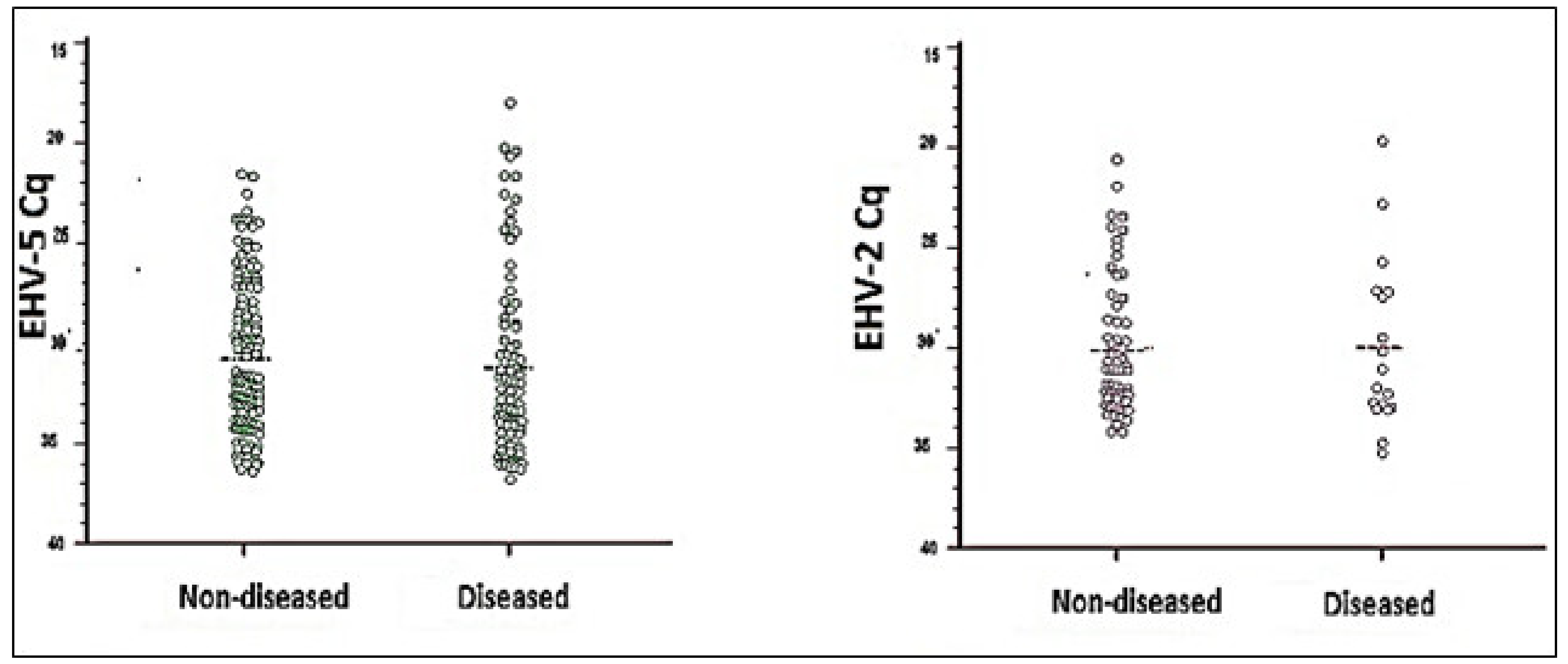
3.3. Equine Gammaherpesvirus Infections, EHV2 and -5

In total, 83 (20.4%) of the 407 horses sampled were EHV2 positive and 249 horses (61.2%) were positive by qPCR for EHV5 (Table 1). There were no differences between the mean viral load of EHV2 or EHV5 detected in diseased and non-diseased horses (Figure 1). There was, however, a statistically significant difference (p = 0.004) between the proportion of horses in the diseased group shedding EHV5 (85/120, 70.8%) compared to the proportion of horses in the non-diseased group that were shedding EHV5 at the time of sampling (137/249, 55%). The odds of respiratory disease in EHV5-positive horses were twice that of EHV5-negative horses (OR 1.98, 95% CI 1.25 to 3.16). The proportion of horses with detectable EHV2 was significantly higher in non-diseased horses (61/249, 24.5%) compared to the diseased group (18/120, 15.0%) (p = 0.042). The odds of EHV2-positive horses also exhibiting clinical signs of disease were approximately half that of EHV2-negative horses (OR 0.54, 95% CI 0.30 to 0.97).

3.4. Concurrent Equine Herpesvirus Infections

Of the 407 horses sampled in this survey, 54 (13.3%) were shedding multiple equine herpesviruses (Table 1). Two of the three horses shedding detectable levels of EHV1 were concurrently shedding EHV4. Three of the five EHV4-positive horses were also shedding EHV5, and a fourth was shedding EHV2. One horse that was clinically normal was shedding EHV2, -4 and -5 concurrently. The horse shedding the highest EHV4 load of 108.45 copies/mL nasal swab was also shedding 107.94 copies/mL of EHV5; however, the disease status of this horse was unknown.
Fifty of the eighty three horses (60.2%) shedding EHV2 were also shedding EHV5; however, there was no greater likelihood of EHV5 detection in these horses compared to those without detectable EHV2 (199/324, 61.4%; p = 0.90). In addition, these co-infected horses were no more likely to exhibit signs of disease (14/46, 30.4%) than those shedding only EHV2 (4/33, 12.1%; p = 0.063) or EHV5 (71/176, 40.3%; p = 0.24). Logistic regression showed no correlation between dual EHV2 and -5 shedding and clinical signs of respiratory disease (p = 0.41). Hence, the association of EHV5 infection and increased likelihood of disease was not modified by the presence or absence of EHV2 infection.

4. Discussion

Equine herpesvirus infections were commonly detected in samples from the respiratory tract, irrespective of clinical disease status at the time of sampling. Approximately 40% of horses were shedding at least one herpesvirus at the time of sampling (Table 1). In total, 67.9% of horses with no obvious clinical disease were shedding detectable levels of at least one herpesvirus. Detection of the alphaherpesviruses in a small proportion of horses (2%, n = 8) contrasted markedly with the high frequency of shedding of the equine gammaherpesviruses (69.3%, n = 282). Although many clinically normal horses were infected, a significantly high proportion of horses with clinical signs of respiratory disease were shedding EHV5. No such association was detected in horses infected with EHV1, -4 and -2.
The increased proportion of horses shedding EHV5 among diseased horses in this study may reflect the contribution of EHV5 to respiratory disease. Alternatively, this shedding may have been reactivated as a consequence of a respiratory disease-associated inflammatory response. The spectrum of clinical disease (or lack of) following gammaherpesvirus infections in horses may be due to a range of factors including virus strain and load, host factors such as age [11,53], and immune responses [54]. Each of these complex factors has been explored in several studies and may help to explain the lack of disease seen in many infected horses. EHV5 is persistently associated with EMPF while it is also regularly detected in both clinically normal and diseased horses [11,13,14,15,16,25,27,31,32,33]. This study showed a significant difference in the proportion of horses shedding EHV5 in the diseased group, such that the odds of disease signs in EHV5-positive horses were twice that of EHV5-negative horses. This difference may be the result of lytic EHV5 infection causing the clinical signs, or that EHV5 is reactivated by infection/inflammation by another agent. B-lymphocytes are a latent reservoir for EHV2 and EHV5, and other sites may exist which have not yet been identified [44,55,56,57]. However, simple reactivation of shedding via B-lymphocytes recruited to these sites does not account for the difference in the clinical associations of EHV5 and EHV2 in this study. Other studies have also shown a protective effect of EHV2 against Rhodococcus equi infection [58]. Whether EHV2 and EHV5 each occupy distinct niches within the respiratory tract, or whether each recruit different types of inflammatory cells that might be protective or immunopathogenic, remains unknown.
The higher incidence of EHV2 in non-diseased horses in this study is consistent with those of previous studies and continues to confound our understanding of the role of this virus, if any, in equine respiratory tract disease. The prevalence of EHV2 infection in large numbers of clinically normal horses has been widely reported [13,14,15,16,26,27]; however, several studies have identified associations between EHV2 infection and mild respiratory disease, particularly in foals [11,13,14,17,59,60].
Quantification of gammaherpesvirus shedding may enable an association to be made between viral load and clinical disease. In humans, an age-range-specific correlation exists between the levels of the gammaherpesvirus Epstein–Barr virus (EBV) in blood, and the presence of clinical disease [61]; however, there is currently little evidence in this or other studies to support an association with acute respiratory disease and gammaherpesvirus load in horses [11,58,62]. Multiple factors are likely to be required for gammaherpesvirus-mediated disease in horses, rather than solely lytic infections. Alternatively, nasal samples may not be the most appropriate samples as predictors for clinical respiratory disease. This is supported by a recent publication linking high viral loads of EHV5 in bronchoalveolar lavage fluid to EMPF [18].
The detection of alphaherpesviruses is reported in a minority of horses within most populations [17,19,20,63,64,65,66]. Five horses without signs of disease were shedding high levels of either alphaherpesvirus EHV1 or -4, consistent with the “cycle of silent herpesvirus shedding” and spread [63,66,67]. The reactivation of latent alphaherpesvirus infection is associated with subclinical viral shedding [20] and can occur following stressful events such as social re-grouping, weaning and long-distance transport [35,65,68,69]. Despite these factors, the levels of detection of equine herpesviruses in this study population were consistent with other studies that have reported ranges of 0–10% for the alphaherpesviruses and 0–100% for the gammaherpesviruses [12,14,16,20,51,63,65,67,70]. The reactivation of latent herpesviruses following a single immunosuppressive event may explain the detection of multiple herpesviruses. This phenomenon has been documented in humans with prolonged sepsis [71]. Shedding of multiple EHVs was detected in 14% (57/407) of horses. Four of the six horses (67%) infected with the alphaherpesviruses EHV1 and -4 were infected by either another alpha- or a gammaherpesvirus(es).
Although Victoria remained free of EIV during Australia’s only recorded EI outbreak, field staff faced logistical challenges and were often time poor. However, samples were successfully collected, and testing was not compromised, ensuring that EIV could be ruled out in all samples analysed. The lack of comprehensive histories and clinical detail for every horse including age, vaccination status and time-course of clinical disease may have limited the analysis of data. The inclusion of the 38 horses of unknown clinical status was made to assist the determination of overall prevalence. A separate analysis of these horses did not show any statistical difference in the proportion of EHV infection in these horses compared with those of known status (diseased and non-diseased).

5. Conclusions

The clinical significance of respiratory gammaherpesvirus infections in horses remains to be determined; however, this survey adds to the mounting body of evidence associating EHV5 with equine respiratory disease. The task of identifying a definitive role of the equine gammaherpesviruses as the cause of respiratory disease on a case-by-case basis remains challenging, since the precise role of both EHV2 and -5 and their relation to clinical disease is likely to be complex and remains to be elucidated for these enigmatic viruses.

Author Contributions

Conceptualisation, J.G., C.H. and C.E.-H.; methodology, C.H., K.D., K.M. and J.G.; validation, C.H., K.D., Z.M., K.M. and J.G.; formal analysis, C.E.-H., C.H., K.D., Z.M. and J.G.; investigation, C.E.-H., C.H., Z.M. and J.G.; writing—C.E.-H., C.H. and J.G.; writing—C.H., K.D., Z.M. and K.M.; supervision, J.G. and C.H.; project administration, J.G. and C.E.-H.; All authors have read and agreed to the published version of the manuscript.

Funding

This research received no external funding.

Institutional Review Board Statement

Ethical review and approval were waived for this study, due to access of archived material collected for surveillance and demonstration of freedom from equine influenza virus.

Informed Consent Statement

Not applicable.

Data Availability Statement

Not applicable.

Acknowledgments

The authors wish to thank the Chief Veterinary Officer’s unit and staff within the (formerly named) Department of Environment and Primary Industries Victoria and Doctors Mark Hawes and Simone Warner for their valued assistance. Thanks to Nino Ficorilli and Cynthia Brown for their valued technical assistance and Garry Anderson who provided skilled statistical advice and assistance.

Conflicts of Interest

The authors declare no conflict of interest.

Appendix A

Table A1. Primers used in this study.
Primer NamePrimer Sequence 5′ to 3′
EHV1.gH.FGCC CGA CAC CTA CAT AAC C
EHV1.gH.RGGC ATA AAA CCA CAC CAA CC
EHV1.gH.ProbeFAM-GCG ACC ACA AAA AGC AAC CC-BHQ1
EHV4.ORF73/74.FGGC AAC CTA CCC GAA GAT G
EHV4.ORF73/74.RCAA CAA CCA CCA GCA ACA A
EHV4.ORF73/74.ProbeCAL Fluor Orange 560-CCC CCA AAC CGC AAA CCA CT-BHQ1
EHV2.gB.1822.FACC CTC AAC CTG ACT GAC AT
EHV2.gB.1953.RTCA AAC ACG TTG GAC AGC CT
EHV5.gH.FTGT GTG CAA TGT TTC TGG GGG
EHV5.gH.RCGC TGC CCA ACA CGT CCC TT

References

  1. Slater, J. Equine herpesviruses. In Equine Infectious Diseases; Sellon, D., Long, M., Eds.; Elsevier: Amsterdam, The Netherlands, 2014; pp. 151–168. [Google Scholar]
  2. Matsumura, T.; Sugiura, T.; Imagawa, H.; Fukunaga, Y.; Kamada, M. Epizootiological Aspects of Type 1 and Type 4 Equine Herpesvirus Infections among Horse Populations. J. Vet. Med. Sci. 1992, 54, 207–211. [Google Scholar] [CrossRef] [PubMed] [Green Version]
  3. Studdert, M. (Ed.) Virus Infections of Vertebrates, Virus Infections of Equines; Elsevier Science Publishers: Amsterdam, The Netherlands, 1996; pp. 281–284. [Google Scholar]
  4. Allen, G.P.; Kydd, J.H.; Slater, J.D.; Smith, K.C. Equid herpesvirus-1 (EHV-1) and -4 (EHV-4) infections. In Infectious Diseases of Livestock; Tustin, J.C.A.R., Ed.; Oxford Press: Cape Town, South Africa, 2004; pp. 829–859. [Google Scholar]
  5. Allen, G.P.; Murray, M. Equid herpesvirus 2 and equid herpesvirus 5 infections. In Infectious Diseases of Livestock; Tustin, J.C.A.R., Ed.; Oxford Press: Cape Town, South Africa, 2004; pp. 860–867. [Google Scholar]
  6. Hartley, C.A.; Dynon, K.J.; Mekuria, Z.H.; El-Hage, C.M.; Holloway, S.A.; Gilkerson, J.R. Equine gammaherpesviruses: Perfect parasites? Vet. Microbiol. 2013, 167, 86–92. [Google Scholar] [CrossRef]
  7. Fortier, G.; van Erck, E.; Pronost, S.; Lekeux, P.; Thiry, E. Equine gammaherpesviruses: Pathogenesis, epidemiology and diagnosis. Vet. J. 2010, 186, 148–156. [Google Scholar] [CrossRef] [PubMed]
  8. Borchers, K.; Lieckfeldt, D.; Ludwig, A.; Fukushi, H.; Allen, G.; Fyumagwa, R.; Hoare, R. Detection of Equid herpesvirus 9 DNA in the trigeminal ganglia of a Burchell’s zebra from the Serengeti ecosystem. J. Vet. Med. Sci. 2008, 70, 1377–1381. [Google Scholar] [CrossRef] [Green Version]
  9. Borchers, K.; Wolfinger, U.; Goltz, M.; Broll, H.; Ludwig, H. Distribution and relevance of equine herpesvirus type 2(EHV-2) infections. Arch. Virol. 1997, 142, 917–928. [Google Scholar] [CrossRef] [PubMed]
  10. Browning, G.F.; Studdert, M.J. Epidemiology of equine herpesvirus 2 (equine cytomegalovirus). J. Clin. Microbiol. 1987, 25, 13–16. [Google Scholar] [CrossRef] [Green Version]
  11. Dunowska, M.; Howe, L.; Hanlon, D.; Stevenson, M. Kinetics of Equid herpesvirus type 2 infections in a group of Thoroughbred foals. Vet. Microbiol. 2011, 152, 176–180. [Google Scholar] [CrossRef]
  12. Dunowska, M.; Wilks, C.R.; Studdert, M.; Meers, J. Equine respiratory viruses in foals in New Zealand. N. Z. Vet. J. 2002, 50, 140–147. [Google Scholar] [CrossRef]
  13. Dunowska, M.; Wilks, C.; Studdert, M.; Meers, J. Viruses associated with outbreaks of equine respiratory disease in New Zealand. N. Z. Vet. J. 2002, 50, 132–139. [Google Scholar] [CrossRef]
  14. Fortier, G.; van Erck, E.; Fortier, C.; Richard, E.; Pottier, D.; Pronost, S.; Miszczak, F.; Thiry, E.; Lekeux, P. Herpesviruses in respiratory liquids of horses: Putative implication in airway inflammation and association with cytological features. Vet. Microbiol. 2009, 139, 34–41. [Google Scholar] [CrossRef] [Green Version]
  15. Torfason, E.G.; Thorsteinsdottir, L.; Torsteinsdóttir, S.; Svansson, V. Study of equid herpesviruses 2 and 5 in Iceland with a type-specific polymerase chain reaction. Res. Vet. Sci. 2008, 85, 605–611. [Google Scholar] [CrossRef]
  16. Bell, S.A.; Balasuriya, U.B.; Gardner, I.A.; Barry, P.A.; Wilson, W.D.; Ferraro, G.L.; MacLachlan, N.J. Temporal detection of equine herpesvirus infections of a cohort of mares and their foals. Vet. Microbiol. 2006, 116, 249–257. [Google Scholar] [CrossRef] [PubMed]
  17. Wang, L.; Raidal, S.L.; Pizzirani, A.; Wilcox, G.E. Detection of respiratory herpesviruses in foals and adult horses determined by nested multiplex PCR. Vet. Microbiol. 2007, 121, 18–28. [Google Scholar] [CrossRef] [PubMed]
  18. Pusterla, N.; Magdesian, K.G.; Mapes, S.M.; Zavodovskaya, R.; Kass, P.H. Assessment of quantitative polymerase chain reaction for equine herpesvirus-5 in blood, nasal secretions and bronchoalveolar lavage fluid for the laboratory diagnosis of equine multinodular pulmonary fibrosis. Equine Vet. J. 2016, 49, 34–38. [Google Scholar] [CrossRef] [PubMed]
  19. Brown, J.A.; Mapes, S.; Ball, B.A.; Hodder, A.D.J.; Liu, I.K.M.; Pusterla, N. Prevalence of equine herpesvirus-1 infection among Thoroughbreds residing on a farm on which the virus was endemic. J. Am. Vet. Med. Assoc. 2007, 231, 577–580. [Google Scholar] [CrossRef] [Green Version]
  20. Pusterla, N.; Mapes, S.; Madigan, J.E.; MacLachlan, N.J.; Ferraro, G.L.; Watson, J.L.; Spier, S.J.; Wilson, W.D. Prevalence of EHV-1 in adult horses transported over long distances. Vet. Rec. 2009, 165, 473–475. [Google Scholar] [CrossRef]
  21. Gilkerson, J.; Jorm, L.R.; Love, D.N.; Lawrence, G.L.; Whalley, J.M. Epidemiological investigation of equid herpesvirus-4 (EHV-4) excretion assessed by nasal swabs taken from thoroughbred foals. Vet. Microbiol. 1994, 39, 275–283. [Google Scholar] [CrossRef]
  22. Gilkerson, J.; Whalley, J.; Drummer, H.; Studdert, M.; Love, D. Epidemiology of EHV-1 and EHV-4 in the mare and foal populations on a Hunter Valley stud farm: Are mares the source of EHV-1 for unweaned foals. Vet. Microbiol. 1999, 68, 27–34. [Google Scholar] [CrossRef]
  23. Gibson, J.S.; Slater, J.D.; Awan, A.R.; Field, H.J. Pathogenesis of equine herpesvirus-1 in specific pathogen-free foals: Primary and secondary infections and reactivation. Arch. Virol. 1992, 123, 351–366. [Google Scholar] [CrossRef]
  24. Yactor, J.; Lunn, K.F.; Morley, P.; Barnett, C.D.; Kohler, A.K.; Kasper, K.S.; Kivi, A.J.; Lunn, D.P. Detection of nasal shedding of EHV-1 and 4 at equine show events and sales by multiplex real-time PCR. In Proceedings of the American Association of Equine Practitioners (AAEP), San Antonio, TX, USA, 6 December 2006; pp. 223–227. [Google Scholar]
  25. Wang, L. An Investigation of the Association between Herpes Viruses and Respiratory Disease; Murdoch University: Perth, WA, Australia, 2003. [Google Scholar]
  26. Back, H.; Ullman, K.; Berndtsson, L.T.; Riihimäki, M.; Penell, J.; Ståhl, K.; Valarcher, J.-F.; Pringle, J. Viral load of equine herpesviruses 2 and 5 in nasal swabs of actively racing Standardbred trotters: Temporal relationship of shedding to clinical findings and poor performance. Vet. Microbiol. 2015, 179, 142–148. [Google Scholar] [CrossRef]
  27. Nordengrahn, A.; Merza, M.; Ros, C.; Lindholm, A.; Hannant, D. Prevalence of equine herpesvirus types 2 and 5 in horse populations by using type-specific PCR assays. Vet. Res. 2002, 33, 251–259. [Google Scholar] [CrossRef] [PubMed] [Green Version]
  28. Diallo, I.S.; Hewitson, G.R.; De Jong, A.; Kelly, M.A.; Wright, D.J.; Corney, B.G.; Rodwell, B.J. Equine herpesvirus infections in yearlings in South-East Queensland. Arch. Virol. 2008, 153, 1643–1649. [Google Scholar] [CrossRef] [Green Version]
  29. McBrearty, K.A.; Murray, A.; Dunowska, M. A survey of respiratory viruses in New Zealand horses. N. Z. Vet. J. 2013, 61, 254–261. [Google Scholar] [CrossRef]
  30. Hue, E.S.; Fortier, G.D.; Fortier, C.I.; Leon, A.M.; Richard, E.A.; Legrand, L.J.; Pronost, S.L. Detection and quantitation of equid gammaherpesviruses (EHV-2, EHV-5) in nasal swabs using an accredited standardised quantitative PCR method. J. Virol. Methods 2014, 198, 18–25. [Google Scholar] [CrossRef] [PubMed]
  31. Back, H.; Kendall, A.; Grandón, R.; Ullman, K.; Treiberg-Berndtsson, L.; Ståhl, K.; Pringle, J. Equine Multinodular Pulmonary Fibrosis in association with asinine herpesvirus type 5 and equine herpesvirus type 5: A case report. Acta Vet. Scand. 2012, 54, 57. [Google Scholar] [CrossRef] [PubMed] [Green Version]
  32. Williams, K.J.; Maes, R.; Del Piero, F.; Lim, A.; Wise, A.; Bolin, D.C.; Caswell, J.; Jackson, C.; Robinson, N.E.; Derksen, F.; et al. Equine Multinodular Pulmonary Fibrosis: A Newly Recognized Herpesvirus-Associated Fibrotic Lung Disease. Vet. Pathol. 2007, 44, 849–862. [Google Scholar] [CrossRef] [Green Version]
  33. Marenzoni, M.L.; Passamonti, F.; Lepri, E.; Cercone, M.; Capomaccio, S.; Cappelli, K.; Felicetti, M.; Coppola, G.; Coletti, M.; Thiry, E. Quantification of Equid herpesvirus 5 DNA in clinical and necropsy specimens collected from a horse with equine multinodular pulmonary fibrosis. J. Vet. Diagn. Investig. 2011, 23, 802–806. [Google Scholar] [CrossRef] [Green Version]
  34. Williams, K.J.; Robinson, N.E.; Lim, A.; Brandenberger, C.; Maes, R.; Behan, A.; Bolin, S.R. Experimental Induction of Pulmonary Fibrosis in Horses with the Gammaherpesvirus Equine Herpesvirus 5. PLoS ONE 2013, 8, e77754. [Google Scholar] [CrossRef] [PubMed] [Green Version]
  35. Edington, N.; Welch, H.; Griffiths, L. The prevalence of latent Equid herpesviruses in the tissues of 40 abattoir horses. Equine Vet. J. 1994, 26, 140–142. [Google Scholar] [CrossRef]
  36. Purewal, A.S.; Smallwood, A.V.; Kaushal, A.; Adegboye, D.; Edington, N. Identification and control of the cis-acting elements of the immediate early gene of equid herpesvirus type 1. J. Gen. Virol. 1992, 73, 513–519. [Google Scholar] [CrossRef]
  37. Welch, H.M.; Bridges, C.G.; Lyon, A.M.; Griffiths, L.; Edington, N. Latent equid herpesviruses 1 and 4: Detection and distinction using the polymerase chain reaction and co-cultivation from lymphoid tissues. J. Gen. Virol. 1992, 73, 261–268. [Google Scholar] [CrossRef] [PubMed]
  38. Marenzoni, M.L.; Bietta, A.; Lepri, E.; Proietti, P.C.; Cordioli, P.; Canelli, E.; Stefanetti, V.; Coletti, M.; Timoney, P.J.; Passamonti, F. Role of equine herpesviruses as co-infecting agents in cases of abortion, placental disease and neonatal foal mortality. Vet. Res. Commun. 2013, 37, 311–317. [Google Scholar] [CrossRef]
  39. Pusterla, N.; Mapes, S.; Wademan, C.; White, A.; Hodzic, E. Investigation of the role of lesser characterised respiratory viruses associated with upper respiratory tract infections in horses. Vet. Rec. 2013, 172, 315. [Google Scholar] [CrossRef] [PubMed]
  40. Dynon, K.; Black, W.; Ficorilli, N.; Hartley, C.; Studdert, M. Detection of viruses in nasal swab samples from horses with acute, febrile, respiratory disease using virus isolation, polymerase chain reaction and serology. Aust. Vet. J. 2007, 85, 46–50. [Google Scholar] [CrossRef] [PubMed]
  41. Mekuria, Z.H. Equine herpesvirus 5; Cellular and molecular biology. In Veterinary Science; University of Melbourne: Melbourne, VIC, Australia, 2015. [Google Scholar]
  42. Telford, E.A.; Watson, M.S.; Aird, H.C.; Perry, J.; Davison, A.J. The DNA Sequence of Equine Herpesvirus 2. J. Mol. Biol. 1995, 249, 520–528. [Google Scholar] [CrossRef] [PubMed]
  43. Silins, S.L.; Sherritt, M.A.; Silleri, J.M.; Cross, S.M.; Elliott, S.L.; Bharadwaj, M.; Le, T.T.T.; Morrison, L.E.; Khanna, R.; Moss, D.J.; et al. Asymptomatic primary Epstein-Barr virus infection occurs in the absence of blood T-cell repertoire perturbations despite high levels of systemic viral load. Blood 2001, 98, 3739–3744. [Google Scholar] [CrossRef] [Green Version]
  44. Sitki-Green, D.L.; Edwards, R.H.; Covington, M.M.; Raab-Traub, N. Biology of Epstein-Barr Virus during Infectious Mononucleosis. J. Infect. Dis. 2004, 189, 483–492. [Google Scholar] [CrossRef] [Green Version]
  45. Anonymous. Information for Victorian Veterinarians. Agriculture Victoria. 2007. Available online: https://agriculture.vic.gov.au/biosecurity/animal-diseases/horse-diseases/equine-influenza (accessed on 20 May 2020).
  46. Goyen, K.A.; Wright, J.D.; Cunneen, A.; Henning, J. Playing with fire—What is influencing horse owners’ decisions to not vaccinate their horses against deadly Hendra virus infection? PLoS ONE 2017, 12, e0180062. [Google Scholar] [CrossRef] [Green Version]
  47. Kirkland, P.; Davis, R.; Wong, D.; Ryan, D.; Hart, K.; Corney, B.; Hewitson, G.; Cooper, K.; Biddle, A.; Eastwood, S.; et al. The first five days: Field and laboratory investigations during the early stages of the equine influenza outbreak in Australia, 2007. Aust. Vet. J. 2011, 89, 6–10. [Google Scholar] [CrossRef]
  48. Dynon, K.; Varrasso, A.; Ficorilli, N.; Holloway, S.; Reubel, G.; Li, F.; Hartley, C.; Studdert, M.; Drummer, H. Identification of equine herpesvirus 3 (equine coital exanthema virus), equine gammaherpesviruses 2 and 5, equine adenoviruses 1 and 2, equine arteritis virus and equine rhinitis A virus by polymerase chain reaction. Aust. Vet. J. 2001, 79, 695–702. [Google Scholar] [CrossRef] [PubMed]
  49. Studdert, M.; Blackney, M. Equine herpesviruses: On the Differentiation of respiratory from foetal strains of type 1. Aust. Vet. J. 1979, 55, 488–492. [Google Scholar] [CrossRef]
  50. Browning, G.F.; Studdert, M.J. Genomic Heterogeneity of Equine Betaherpesviruses. J. Gen. Virol. 1987, 68, 1441–1447. [Google Scholar] [CrossRef]
  51. Turner, A.J.; Studdert, M.J.; Peterson, J.E. 2. Persistence of Equine Herpesviruses in Experimentally Infected Horses and the Experimental Induction of Abortion. Aust. Vet. J. 1970, 46, 90–98. [Google Scholar] [CrossRef] [PubMed]
  52. Dynon, K. Pathogenesis of equine herpes virus type 2 (EHV2) infection and cellular processing of EHV2 glycoprotein. In School of Veterinary Science 2010; University of Melbourne: Melbourne, VIC, Australia, 2010. [Google Scholar]
  53. Brault, S.A.; Bird, B.H.; Balasuriya, U.B.; MacLachlan, N.J. Genetic heterogeneity and variation in viral load during equid herpesvirus-2 infection of foals. Vet. Microbiol. 2011, 147, 253–261. [Google Scholar] [CrossRef]
  54. Brault, S.A.; Blanchard, M.T.; Gardner, I.A.; Stott, J.L.; Pusterla, N.; Mapes, S.M.; Vernaua, W.; De Jongc, K.D.; MacLachlana, N.J. The immune response of foals to natural infection with equid herpesvirus-2 and its association with febrile illness. Vet. Immunol. Immunopathol. 2010, 137, 136–141. [Google Scholar] [CrossRef]
  55. Drummer, H.E.; Reubel, G.H.; Studdert, M.J. Equine gammaherpesvirus 2 (EHV2) is latent in B lymphocytes. Arch. Virol. 1996, 141, 495–504. [Google Scholar] [CrossRef] [PubMed]
  56. Borchers, K.; Wolfinger, U.; Ludwig, H.; Thein, P.; Baxi, S.; Field, H.J.; Slater, J.D. Virological and molecular biological investigations into equine herpes virus type 2 (EHV-2) experimental infections. Virus Res. 1998, 55, 101–106. [Google Scholar] [CrossRef]
  57. Reubel, G.H.; Crabb, B.S.; Studdert, M.J. Diagnosis of equine gammaherpesvirus 2 and 5 infections by polymerase chain reaction. Arch. Virol. 1995, 140, 1049–1060. [Google Scholar] [CrossRef]
  58. Nordengrahn, A.; Rusvai, M.; Merza, M.; Ekstrom, J.; Morein, B.; Belak, S. Equine herpesvirus type 2 (EHV-2) as a predisposing factor for Rhodococcus equi pneumonia in foals: Prevention of the bifactorial disease with EHV-2 immunostimulating complexes. Vet. Microbiol. 1996, 51, 55–68. [Google Scholar] [CrossRef]
  59. Fortier, G.; Richard, E.; Hue, E.; Fortier, C.; Pronost, S.; Pottier, D.; Lemaitre, L.; Lekeux, P.; Borchers, K.; Thiryg, E. Long-lasting airway inflammation associated with equid herpesvirus-2 in experimentally challenged horses. Vet. J. 2013, 197, 492–495. [Google Scholar] [CrossRef] [PubMed]
  60. Murray, M.J.; Eichorn, E.S.; Dubovi, E.J.; Ley, W.B.; Cavey, D.M. Equine herpesvirus type 2: Prevalence and seroepidemiology in foals. Equine Vet. J. 1996, 28, 432–436. [Google Scholar] [CrossRef] [PubMed]
  61. Balfou, H.H.J.; Holman, C.J.; Hokanson, K.M.; Lelonek, M.M.; Giesbrecht, J.E.; White, D.R.; Schmeling, D.O.; Webb, C.-H.; Cavert, W.; Wang, D.H.; et al. A prospective clinical study of Epstein-Barr virus and host interactions during acute infectious mononucleosis. J. Infect. Dis. 2005, 192, 1505–1512. [Google Scholar] [CrossRef] [PubMed]
  62. Brault, S.A.; Maclachlan, N.J. Equid gammaherpesviruses: Persistent bystanders or true pathogens? Vet. J. 2011, 187, 14–15. [Google Scholar] [CrossRef] [PubMed]
  63. Foote, C.E.; Love, D.N.; Gilkerson, J.R.; Whalley, J.M. Detection of EHV-1 and EHV-4 DNA in unweaned Thoroughbred foals from vaccinated mares on a large stud farm. Equine Vet. J. 2004, 36, 341–345. [Google Scholar] [CrossRef] [PubMed]
  64. Pusterla, N.; Leutenegger, C.M.; Wilson, W.D.; Watson, J.L.; Ferraro, G.L.; Madigan, J.E. Equine herpesvirus-4 kinetics in peripheral blood leukocytes and nasopharyngeal secretions in foals using quantitative real-time TaqMan PCR. J. Vet. Diagn. Investig. 2005, 17, 578–581. [Google Scholar] [CrossRef] [PubMed] [Green Version]
  65. Pusterla, N.; Mapes, S.; Wilson, W.D. Use of viral loads in blood and nasopharyngeal secretions for the diagnosis of EHV-1 infection in field cases. Veterinary Record. 2008, 728–729. [Google Scholar] [CrossRef] [PubMed] [Green Version]
  66. Gilkerson, J.R.; Love, D.N.; Whalley, J.M. Epidemiology of equine herpesvirus abortion: Searching for clues to the future. Aust. Vet. J. 1998, 76, 675–676. [Google Scholar] [CrossRef]
  67. Gilkerson, J.R.; Love, D.N.; Whalley, J.M. Incidence of equine herpesvirus 1 infection in thoroughbred weanlings on two stud farms. Aust. Vet. J. 2000, 78, 277–278. [Google Scholar] [CrossRef]
  68. Edington, N.; Bridges, C.G.; Huckle, A. Experimental reactivation of equid herpesvirus 1 (EHV 1) following the administration of corticosteroids. Equine Vet. J. 1985, 17, 369–372. [Google Scholar] [CrossRef]
  69. Slater, J.D.; Borchers, K.; Thackray, A.M.; Field, H.J. The trigeminal ganglion is a location for equine herpesvirus 1 latency and reactivation in the horse. J. Gen. Virol. 1994, 75, 2007–2016. [Google Scholar] [CrossRef]
  70. Borchers, K.; Ebert, M.; Fetsch, A.; Hammond, T.; Sterner-Kock, A. Prevalence of equine herpesvirus type 2 (EHV-2) DNA in ocular swabs and its cell tropism in equine conjunctiva. Vet. Microbiol. 2006, 118, 260–266. [Google Scholar] [CrossRef] [PubMed]
  71. Walton, A.H.; Muenzer, J.T.; Rasche, D.; Boomer, J.S.; Sato, B.; Brownstein, B.H.; Pachot, A.; Brooks, T.L.; Deych, E.; Shannon, W.D. Reactivation of multiple viruses in patients with sepsis. PLoS ONE 2014, 9, e98819. [Google Scholar]
Figure 1. Quantification cycles (Cq) values considered positive for Equine herpesvirus 2 and -5 in nasal swabs of horses with and without clinical signs of disease (diseased and non-diseased). The horizontal line indicates the mean Cq for each group, none of which were statistically different between groups.
Animals 11 03418 g001
Table 1. Clinical status of horses and detection of. Equine Herpesvirus 1, -4, -2 and -5 from nasal swabs.
Virus DetectedRespiratory Disease Signs
Negative Positive Not RecordedTotal
EHV-1 only1001
EHV-2 only294033
EHV-5 only1057022197
EHV-1 and EHV-42002
EHV-2 and EHV-53114449
EHV-4 and EHV-50112
EHV-2, EHV-5 and EHV-41001
No detection803111122
Total24912038407
Publisher’s Note: MDPI stays neutral with regard to jurisdictional claims in published maps and institutional affiliations.

