
LC-ESI/QTOF-MS Profiling of Chicory and Lucerne Polyphenols and Their Antioxidant Activities
by
 , , and
Yasir Iqbal
1 ,
,
Eric N. Ponnampalam
2,
Hafiz A. R. Suleria
1 ,
,
Jeremy J. Cottrell
1 and
and
Frank R. Dunshea
1,3,* 1
School of Agriculture and Food, Faculty of Veterinary and Agricultural Sciences, The University of Melbourne, Parkville, VIC 3010, Australia
2
Animal Production Sciences, Agriculture Victoria Research, Department of Jobs, Precincts and Regions, Bundoora, VIC 3083, Australia
3
Faculty of Biological Sciences, The University of Leeds, Leeds LS2 9JT, UK
*
Author to whom correspondence should be addressed.
Antioxidants 2021, 10(6), 932; https://doi.org/10.3390/antiox10060932
Submission received: 12 April 2021
/
Revised: 4 June 2021
/
Accepted: 7 June 2021
/
Published: 8 June 2021
(This article belongs to the Special Issue Phenolics as Antioxidant Agents)
Abstract
:Chicory and lucerne are used as specialised forages in sheep or dairy production systems in some parts of the world. Recently, these plants are gaining attention as raw materials in the search for natural antioxidants for use in animal feeds, human foods and nutraceutical formulations. The antioxidant potential of these plants is credited to polyphenols, a subgroup of phytochemicals. Therefore, phenolic characterisation is an essential step before their use as ingredients in animal feeds, human food or nutraceutical preparations. In this study, we performed qualitative and quantitative analysis of polyphenols in chicory and lucerne. Profiling of polyphenols from chicory and lucerne was performed by LC-ESI/QTOF-MS with a total of 80 phenolic compounds identified in chicory and lucerne. The quantification of polyphenols was achieved by high performance liquid chromatography, coupled with a photo diode array (HPLC-PDA). Chicoric acid was the major phenolic acid found in chicory, with the highest concentration (1692.33 ± 0.04 µg/g DW) among all the polyphenols quantified in this study. 2-hydroxybenzoic acid was the major phenolic acid found in lucerne, with the highest concentration of 1440.64 ± 0.04 µg/g DW. Total phenolic, flavonoids and total tannin contents were measured, and the antioxidant potential was determined by 2,2-Diphenyl-1-picrylhydrazyl, Ferric Reducing Antioxidant Power, 2,2-Azino-bis-3-ethylbenzothiazoline-6-sulfonic Acid, Hydroxyl (OH−) Radical Scavenging Activity, Chelating Ability of Ferrous Ion (Fe2+) and Reducing Power (RPA) assays. Both chicory (8.04 ± 0.33 mg AAE/g DW) and lucerne (11.29 ± 0.25 mg AAE/g DW) showed high values for Hydroxyl (OH−) Radical Scavenging Activity. The current study allowed us to draw a profile of polyphenols from chicory and lucerne. They provided a molecular fingerprint useful for the application of these plant materials in human foods, animal feeds and pharmaceutical formulations.
1. Introduction
In recent years, attempts to find natural antioxidants for use in animal feeds, human foods and medicines have increased, in order to replace their synthetic counterparts due to their harmful effects to body and consumer choice. Therefore, natural plant materials gain popularity in various industrial sectors like human food, animal feed, cosmetics and pharmaceuticals. Selected plant materials can contribute to nutrition and natural therapeutics for the cure of various ailments, such as inflammation and oxidative stress due to the presence of bioactive compounds like polyphenols. Polyphenols are bioactives formed in plants as defensive compounds against ultraviolet radiation or aggression by pathogens [1]. These phenolic compounds possess various health-promoting properties, for example being antioxidative, anti-inflammatory, antiaging, antibacterial, antidiabetic and anti-mutagenic [2,3,4].
These health-promoting effects of polyphenols are ascribed to their strong antioxidant potential and ability to scavenge free radicals, thereby delaying or avoiding lipid and protein oxidation in plant and animal tissues [5,6,7]. Besides their importance in physiological systems, polyphenols also gain importance as alternatives to synthetic antioxidants and preservatives in food [8]. Therefore, identification and complete profiling of polyphenols from plants can provide valuable information for their optimum use in various industrial sectors, such as livestock production, food production and cosmetics, particularly in pharmaceutical formulations and functional foods [9].
Polyphenols can be extracted from plant materials by various methods using different solvents and extraction techniques. The extraction procedure is of prime importance in the analysis of polyphenols [10]. Hence, the use of an appropriate extraction technique is vital for obtaining accurate results in terms of qualitative and quantitative determination. Various methods are applied to optimise the extraction process. However, a solid-liquid extraction method using different solvents is considered more appropriate for polyphenols [11]. Various techniques can also be used to characterise polyphenols. Recently, high performance liquid chromatography (HPLC), coupled with mass spectrometry (MS), has evolved as a new technique [12] that allows more accurate identification and characterisation of single and complex phenolic compounds and other metabolites. This technique is highly sensitive for identifying small molecules with specific masses in a complex mixture, and identifies compounds based on mass-to-charge ratios [10].
Chicory (Cichorium intybus) and lucerne (Medicago sativa) have strong antioxidant potential. Chicory and lucerne forages are used in dairy production and sheep meat production systems in southern Australia and many parts around the world, due to their high protein, soluble carbohydrates and bioactive compounds. Both forages are perennials with three-to-four years of persistence in the field, with high dry matter production during spring and early summer seasons in Southern Australia, when the weather is warm with adequate water available in the soil. Chicory and lucerne can deliver high forage quality due to their deep tap root systems, resulting in a consistent growth rate with greater assimilation of nutrients (proteins, soluble carbohydrates, vitamins) in the vegetative parts, even with less water availability in the soil. They can also be stored as silage, haylage or hay (lucerne) when produced in excess amounts. These preserved feeds can be used as alternative feeds or supplements during dry seasons to increase the growth and productivity (milk and meat) of farm animals, when the availability of traditional pastures is low. Chicory possesses antioxidant, anti-parasitic, anticancer, antihepatotoxic, antibiotic and anti-inflammatory actions. Lucerne has been used in the treatment of digestive tract ailments and problems of the circulatory and immune systems in traditional (folk) medicine [13,14]. Detoxification and anticarcinogenic properties, particularly in the alimentary tract, have also been attributed to lucerne use [13]. These plants’ health-promoting activities are ascribed to their secondary metabolites like phenolic acids, flavonoids, coumarins and tannins [15,16]. The presence of phenolic acids and flavonoids has been reported in chicory and lucerne [17,18,19]. Based on their high phenolic composition, chicory and lucerne have a high therapeutic potential that has not been fully understood, as there have been few studies characterising the bioactive components of these plants. Therefore, this study was conducted to provide more reliable and accurate information about the phenolic contents of chicory and lucerne for utilisation in foods, feeds and medicinal formulations.
2. Materials and Methods
2.1. Plant Material
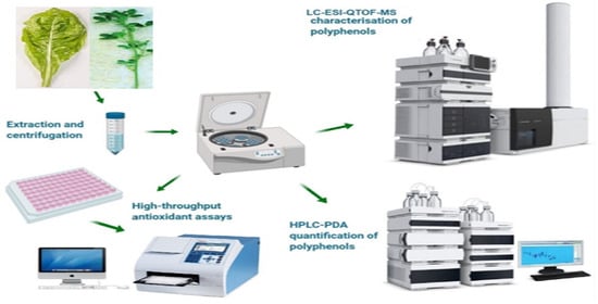
Chicory (Cichorium intybus var. Commander) and lucerne (Medicago sativa var. Sardi 7 series II) were acquired from Hamilton Research Station, Agriculture Victoria Research, Department of Jobs, Precincts and Regions, 915 Mt. Napier Road, Hamilton, VIC 3300, Australia. Vegetative (leaves and stems) parts of chicory and lucerne were collected 2–3 cm above the ground level during spring season 2019 (late October 2019). Chicory and pasture were in the second year of cultivation. During spring, both pastures grow faster and produce greater dry matter yield due to warm weather with adequate rainfall, which is perfect conditions for the assimilation of nutrients and other secondary compounds in the vegetative parts. For both species, samples (vegetative parts) were collected from five locations within a particular paddock located at Hamilton Research Station, Vic, Australia and bulked in a ziplock plastic bag, weighing approximately 1000 g in total. The stage of maturity of both forages during collection time were at pre-bloom. Upon collection, on the same day, samples were brought to the University of Melbourne, Parkville 3030 under refrigerated conditions using an esky with ice. Upon arrival, samples were stored at 4 °C until further processing for grinding and chemical extraction.
2.2. Chemicals and Reagents
The chemicals used were of analytical purity. Methanol, ethanol, sodium acetate, HCl and glacial acetic acid were procured from Thermo Fisher Scientific Inc. (Waltham, MA, USA). H2SO4 (98%) was procured from RCI Labscan (Rongmuang, Thailand). Na2CO3 was purchased from Chem-Supply Pty Ltd (Adelaide, Australia). Folin-Ciocalteu reagent, 2,4,6-tripyridyl-s-triazine (TPTZ), 2,2-diphenyl-1-picrylhydrazyl (DPPH), 2,2′-azino-bis (3-ethylbenzothiazoline-6-sulphonic acid) (ABTS), quercetin, gallic acid, vanillin, L-ascorbic acid, AlCl3.6H2O, FeCl2 Ferrous sulphate, hydrogen peroxide (H2O2), 3-hydroxybenzoic acid, Ferrozine, Potassium ferricyanide, Trichloroacetic acid and FeCl3 were procured from Sigma-Aldrich (Castle Hill, New South Wales, Australia).
2.3. Samples Preparation
Within a week of collection, bulk samples (chicory and lucerne) were crushed using a mortar and pestle to make a paste-like consistency, in order to facilitate the extraction of polyphenol compounds and stored at −20 °C for further analysis.
2.4. Extraction of Phenolics
Samples pastes were extracted with 80% ethanol, and then the sample-ethanol mixtures were homogenised. Homogenised mixtures were incubated in a ZWYR-240 shaker incubator (Labwit, Ashwood, VIC, Australia). Afterwards, centrifugation was performed on a Hettich Rotina 380R centrifuge machine (Tuttlingen, Germany) for 20 min at 5000 rpm (4 °C). The supernatants were collected and filtered through 0.22 µm syringe filter (PTFE membrane) and stored at −20 °C for the characterisation and quantification of polyphenols.
2.5. Antioxidant Assays
Antioxidant activities were determined by previously reported methods [20] using 96-well plates. Absorbance was recorded on Multiskan® Go microplate photometer (Thermo Fisher Scientific, Waltham, MA, USA) and standard curves with R2 ≥ 0.99 were constructed with standard solutions. Results were reported on dry weight basis.
2.5.1. Total Phenolics Content (TPC)
Total phenolic content was determined by the Folin and Ciocalteu’s method [21] with slight modifications using a 96-well plate. Sample (25 µL) was mixed with 25 µL Folin’s Reagent (diluted to 1:3 with water) and allowed to incubate at 25 °C for 5 min. Finally, water (200 µL) and 10% (w/w) Na2CO3 solution (25 µL) were added. This mixture was incubated for 60 min at 25 °C. Absorbance was recorded at 765 nm with a microplate reader. Measurements were made in triplicate, and quantification was done by constructing a standard curve (0–200 µg/mL gallic acid).
2.5.2. Total Flavonoids Content (TFC)
Total flavonoid content for all sample extracts was measured by the AlCl3 colorimetric method [22] with slight modifications using a 96-well plate. 80 µL sample extract was mixed with 80 µL of 2% AlCl3 solution. 120 µL of aqueous solution of sodium acetate (50 g/L) was added. This mixture was incubated for 150 min at 25 °C. Absorbance was taken at 440 nm. Measurements for all samples were made in triplicate, and quantification was done by constructing a standard curve (0–50 µg/mL quercetin).
2.5.3. Total Tannin Contents (TTC)
The total tannin content of samples was measured through a colorimetric method [23] with minor changes. Sample extracts (25 µL) was mixed with 4% vanillin solution (150 µL). 25 µL of H2SO4 solution (32%) was added. This mixture was incubated at 25 °C for 15 min. Absorbance was taken at 500 nm. Measurements were made in triplicate and quantification were done by constructing a standard curve (0–1000 µg/mL catechin solution).
2.5.4. 2,2-Diphenyl-1-picrylhydrazyl (DPPH) Assay
The radical scavenging potential of the samples was estimated by using a previously reported method [24] with slight modifications. 260 µL 0.1 M DPPH solution was mixed with 40 µL sample extract, and allowed to incubate at 25 °C for 30 min. Absorbance was taken at 517 nm. Measurements for all samples were made in triplicate and quantification was done by constructing a standard curve (0–50 µg/mL ascorbic acid).
2.5.5. Ferric Reducing Antioxidant Power (FRAP) Assay
Ferric reducing potential was estimated by a previously used method [24] with modifications in a 96-well plate. 300 mM acetate buffer, 20 mM ferric chloride and 10 mM TPTZ were mixed in 10:1:1 (v/v/v) ratio to prepare FRAP reagent. 280 µL of FRAP reagent was added to 20 µL of sample extract and incubated for 10 min at 37 °C. Absorbance was recorded at 593 nm. Measurements were made in triplicate and quantification was done by constructing a standard curve (0–50 µg/mL ascorbic acid).
2.5.6. 2,2-Azino-bis-3-ethylbenzothiazoline-6-sulfonic Acid (ABTS) Radical Scavenging Assay
The potential of samples to scavenge ABTS radical was determined by ABTS+ radical cation de-colorisation method [24] with slight modifications. 7 mM ABTS solution was mixed with 140 mM K2S2O8 solution, and allowed to incubate in the dark for 16 h for the generation of ABTS+ ions in the solution. The absorbance of this solution was adjusted to 0.70 ± 0.02 by dilution with ethanol. After this, 10 µL of sample extracts were mixed with ABTS+ solution (290 µL), and allowed to incubate at 25 °C for 6 min. Absorbance was taken at 734 nm. Measurements for all samples were made in triplicate, and quantification was done by constructing a standard curve (0–150 µg/mL ascorbic acid).
2.5.7. Hydroxyl (OH−) Radical Scavenging Activity Assay
The hydroxyl (OH−) radical scavenging potential of samples was determined by translating the method of Pavithra and Vadivukkarasi [25] to a 96-well plate method. 50 µL of sample was mixed with 50 µL of 6 mM Ferrous sulphate solution and 50 µL of 6 mM hydrogen peroxide (H2O2) solution in a 96-well plate. The mixture was incubated for 10 min at 25 °C. After incubation, 50 µL of 6 mM 3-hydroxybenzoic acid solution was added. Absorbance was recorded at 510 nm. Measurements for all samples were made in triplicate and quantification was done by constructing a standard curve (0–300 µg/mL ascorbic acid).
2.5.8. Chelating Ability of Ferrous Ion (Fe2+)
The chelating ability of samples was determined by translating the method of Pavithra and Vadivukkarasi [25] to a 96-well plate method. 100 µL of sample was mixed with 80 µL of 2 mM ferrous chloride solution in a 96-well plate. 70 µL of 5 mM Ferrozine solution was added and mixed well. The mixture was allowed to stand for 10 min at 25 °C. Absorbance was taken at 562 nm. Measurements for all samples were made in triplicate, and quantification was done by constructing a standard curve (0–10 µg/mL EDTA solution).
2.5.9. Reducing Power Assay (RPA)
The reducing power of the samples was determined by translating the method of Pavithra and Vadivukkarasi [25] to a 96-well plate method. 10 µL of sample, 25 µL of 0.2 M phosphate buffer (pH 6.6) and 25 µL of Potassium ferricyanide solution (1% w/v) were added sequentially in a 96-well plate, and incubated for 20 min at 25 °C. Following incubation, 25 µL of TCA solution (10% w/v) was added. After this, 85 µL of water and 8.5 µL of Iron (III) chloride solution (0.1% w/v) and incubated for 15 min at 25 °C. Absorbance was recorded at 750 nm. Measurements for all samples were made in triplicate, and quantification was done by constructing a standard curve (0–150 µg/mL ascorbic acid).
2.6. Profiling of Polyphenols by LC-ESI/QTOF-MS
Profiling of polyphenols from samples was achieved by an already published method [20] by using HPLC system (Agilent 1200 series) coupled to Agilent 6520 Accurate-Mass Q-TOF LC/MS (Agilent Technologies, Santa Clara, CA, USA) with an electrospray ionisation source (ESI). Separation of compounds was performed with RP-80A Column (250 mm × 4.6 nm, 4 µm). Mobile phases (A) and (B) were composed of water/acetic acid solution (98:2 v/v) and water/acetonitrile/acetic acid (50:49.5:0.5, v/v/v) respectively and elution program was set as: 0–20 min linear gradient from 90% to 75% eluent A; 20–30 min linear gradient from 75% to 65% eluent A; 30–40 min linear gradient from 65% to 60% eluent A; 40–70 min linear gradient from 60% to 45% eluent A; 70–75 min linear gradient from 45% to 20% eluent A; 75–77 min linear gradient from 20% to 0% eluent A; 77–79 min linear gradient from 0% eluent A; 82–85 min linear gradient from 0% to 90% eluent A. The flow rate was maintained at 0.8 mL/min. Sample injection volume was 6 µL. Peaks were identified in negative and positive ionisation modes. Mass spectra were obtained in the m/z range of 50–1300. Data were analyzed on an Agilent LC/MS/QTOF Mass Hunter Data Acquisition Software (Version B.03.01).
2.7. Quantification of Polyphenols
Quantification of polyphenols was performed by the previously applied method [26] on a HPLC system (Waters Alliance 2690, Chromatograph Separation Module) connected to photodiode array detector (Model 2998, Waters). The column and conditions of analysis were kept same as applied in the LC- MS analysis, with 20µL injection volume of samples. Peak identification was performed at 280 nm, 320 nm and 370 nm. Data analysis was achieved on Empower Software (2010) (Shimadzu Scientific Instruments, Sydney, NSW, Australia). Each polyphenol was quantified by preparing standard curve (R2 ≥ 0.98) with different concentrations of external standards, which include Cinnamic acid (50–250 µg/mL), Gallic acid (62.5–500 µg/mL), Chicoric acid (12.5–200 µg/mL), 2-hydroxybenzoic acid (25–200 µg/mL), m-Coumaric acid (12.5–200 µg/mL), p-hydroxybenzoic acid (3.125–50 µg/mL), Isorhamnetin (12.5–200 µg/mL), Quercetin 3-rhamnoside (50–250 µg/mL) and Epicatechin gallate (31.25–250 µg/mL).
2.8. Statistics Analysis
Results were reported as mean ± standard deviation of the values of three independent analyses. The students’ t-test was performed using Microsoft Excel software (Microsoft Corporation, Redmond, WA, USA) to test statistical significance by comparing the means (significant difference at p ≤ 0.05).
3. Results and Discussion
3.1. Polyphenols Estimation from Chicory and Lucerne Extracts (TPC, TFC and TTC)
Chicory and lucerne are rich in polyphenols. The polyphenol content of chicory and lucerne was determined as TPC, TFC and TTC. The results showed that polyphenolic contents varied considerably in chicory and lucerne (Table 1). Lucerne showed a significantly higher TPC (0.71 ± 0.01 mg GAE/g) and TTC (1.32 ± 0.08 mg CE/g) as compared to chicory, with TPC and TTC values of 0.44 ± 0.04 mg GAE/g and 0.84 ± 0.03 mg CE/g respectively. The TPC of lucerne was determined previously by Zagórska-Dziok et al., [27] in the range of 3.52 mg GAE/g to 73.5 mg GAE/g using different concentrations of water-glycerine extracts of lucerne. Our values for TPC of lucerne were lower than the already reported values. The difference could be explained by the use of different solvents for extraction and the methods applied for determination. No significant difference was observed in total flavonoid content (0.07 ± 0.01 mg QE/g in chicory and 0.07 ± 0.01 mg QE/g in lucerne) in both samples (p ≤ 0.05).
Kaur and coworkers [28] also determined TPC of chicory extracts, and were found in the range of 23.4 to 62.5 mg GAE/100 g dry weight (0.234 to 0.625 mg GAE/g dry weight) using different solvents for extraction. Our results for TPC of chicory (0.44 ± 0.04 mg GAE/g dry weight) is within the range already reported by Kaur and coworkers.
3.2. Antioxidant Activities of Chicory and Lucerne Extracts as Determined by DPPH, FRAP, ABTS, RPA, OH− Radical Scavenging Ability, Chelating Ability of Fe2+
Chicory and lucerne contain phenolic acids and flavonoids that have high antioxidant capacity [29]. The antioxidant activity of chicory and lucerne was determined by DPPH, FRAP, ABTS, OH− radical scavenging ability, chelating ability of Fe2+ and reducing power assays (Table 2). These assays are commonly applied for the determination of antioxidant potential of plant extracts [30]. Lucerne showed significantly higher values (p ≥ 0.05) of ABTS (1.28 ± 0.02 mg AAE/g), OH− radical scavenging ability (11.29 ± 0.25 mg AAE/g), chelating ability of Fe2+ (0.21 ± 0.01 mg EDTAE/g) and reducing power assay (0.59 ± 0.02 mg AAE/g) as compared to chicory that showed 0.27 ± 0.01 mg AAE/g, 8.04 ± 0.33, 0.07 ± 0.01 mg EDTAE/g and 0.34 ± 0.01 mg AAE/g for ABTS, OH− radical scavenging ability, chelating ability of Fe2+ and reducing power assay respectively (Table 2).
No significant difference was observed for DPPH and FRAP values for chicory and lucerne. DPPH and FRAP values of chicory are 0.12 ± 0.01 mg AAE/g and 0.01 ± 0.01 mg AAE/g respectively. Meanwhile, lucerne showed DPPH and FRAP values as 0.13 ± 0.01 mg AAE/g and 0.02 ± 0.01 mg AAE/g, respectively.
3.3. Polyphenols Profile of Chicory and Lucerne
Profiling of polyphenols from chicory and lucerne were performed by verifying m/z value from mass spectra in positive ([M + H]+) and negatiM+He ([M − H]−) ionisation modes and compounds with mass error less than 10 ppm were selected for the verification of m/z for characterisation using the personal compound database library. 80 polyphenols were identified in chicory and lucerne extracts, with 14 phenolic acids, 52 flavonoids, three lignans, one stilbene and 10 other polyphenols (Table 3). Higher diversity of polyphenols was found in lucerne extract with a total of 56 compounds (Table S2—Supplementary materials), as compared to the chicory extract in which 29 polyphenols (Table S1—Supplementary materials) were identified. Flavonoids and phe-nolic acids were the main polyphenol subgroups in both plant extracts. Stilbenes was only identified in the chicory extract (Supplementary materials—Figure S1).
3.3.1. Phenolic Acids
A total of fourteen phenolic acids belonging to three different subclasses (hydroxybenzoic acids, hydroxycinnamic acids and hydroxyphenylpropanoic acids) were tentatively identified in chicory and lucerne extracts. Hydroxycinnamic acids and hydroxybenzoic acids were the dominant subgroups of phenolic acids, with seven and five compounds, respectively. Only two compounds belonging to hydroxyphenylpropanoic acids were tentatively identified.
Hydroxybenzoic Acid Derivatives
Five compounds (Compound 1, 2, 3, 4 & 5) were tentatively identified as hydroxybenzoic acids in both samples. Compounds (1 & 2) were tentatively identified in lucerne extract in positive ionisation mode at m/z 155.0336 and m/z 139.0389 and designated as 2,3-Dihydroxybenzoic acid (C7H6O4) and 2-hydroxybenzoic acid (C7H6O3), respectively. Compound (3) was tentatively identified in chicory extract in positive ionisation mode at m/z 301.0934 and designated as 4-hydroxybenzoic acid 4-O-glucoside (C13H16O8). Compounds (4 & 5) were tentatively identified in the chicory extract in the negative ionisation mode at m/z 331.0652 and 463.0546, and were designated as Gallic acid 4-O-glucoside (C13H16O10) and Ellagic acid glucoside (C20H16O13), respectively.
Hydroxycinnamic Acid Derivatives
Hydroxycinnamic acid were the predominant phenolic acids in chicory and lucerne [31,32]. Compounds (6, 7, 8, 9, 10, 11 & 12) were identified as hydroxycinnamic acid derivatives. Compound (12) was identified in negative ionisation mode in both chicory and lucerne extracts at m/z 473.0764 and m/z 473.0696, respectively, and was designated as chicoric acid (C22H18O12). Chicoric acid has already been reported in methanolic extracts of chicory [17,33]. Compounds (6, 9 & 11) were identified only in the lucerne extract in positive ionisation mode at m/z 165.0548, 399.1288 and 517.1319, and were designated as m-Coumaric acid (C9H8O3), 3-Sinapoylquinic acid (C18H22O10) and 1,5-Dicaffeoylquinic acid (C25H24O12), respectively. m-Coumaric acid was also previously identified in lucerne [34]. Compound (8) was identified in the lucerne extract in negative ionisation mode at m/z 616.1062 and designated as 2-S-Glutathionyl caftaric acid (C23H27N3O15S). However, compounds (7 & 10) were identified in chicory extract in positive ionisation mode at m/z 149.0585 and 355.0999 and were designated as Cinnamic acid (C9H8O2) and 3-Caffeoylquinic acid, respectively (C16H18O9). 3-Caffeoylquinic acid has also previously been identified in chicory [17]. Out of the seven hydroxycinnamic acids identified in this study, three are in the form of quinic acid derivatives. This agrees with a previous finding that hydroxycinnamic acids mainly exist in conjugated form, such as quinic acid [35].
Hydroxyphenylpropanoic Acids
Two hydroxyphenylpropanoic acids (compound 13 & 14) were identified in negative ionisation mode ([M − H]−). Compound (13) was identified in both chicory and lucerne extracts in negative ionisation mode at m/z 357.0847 and 357.0819 respectively and designated as Dihydrocaffeic acid 3-O-glucuronide (C15H18O10). However, compound (14) was only identified in lucerne extract in negative ionisation mode at m/z 275.0218, and designated as Dihydroferulic acid 4-sulfate (C10H12O7S).
3.3.2. Flavonoids and Their Derivatives
Higher diversity of flavonoids derivatives was found among the phenolic compounds identified in chicory and lucerne extracts. A total of 52 flavonoids belonging to seven subgroups were identified in this study.
Anthocyanins Derivatives
Anthocyanins provide protection to arteries and endothelial tissues, inhibit platelet aggregation and reduce the risk of heart diseases [17,36,37,38]. Chicory and lucerne have been reported to contain different anthocyanin derivatives. The anthocyanin derivatives found in chicory are of special interest due to their beneficial effects on visual capacity, brain cognitive function, obesity and cancer prevention [39,40]. Five anthocyanins (compounds 15, 16, 17, 18 & 19) were detected in this study. Out of the five anthocyanins, one compound (18) was putatively identified in negative ionisation mode at m/z 640.145 and 640.1413 in both chicory and lucerne extracts, and was designated as Delphinidin 3-O-feruloyl-glucoside (C31H29O15). Compounds (15 & 19) were putatively identified in negative ionisation mode in lucerne extract at m/z 300.0654 and 740.2187 and designated as Peonidin (C16H13O6) and Pelargonidin 3-O-glucosyl-rutinoside (C33H41O19), respectively. Compound (16) was tentatively identified in chicory extract in negative ionisation mode at m/z 696.1516 and designated as Cyanidin 3-O-(6″-malonyl-3″-glucosyl-glucoside) with molecular formula C30H33O19. Compound (17) was identified at m/z 626.166 in positive ionisation mode in chicory extract, and designated as Petunidin 3-O-(6″-p-coumaroyl-glucoside) with the molecular formula C31H29O14.
Dihydrochalcones
Only one dihydrochalcone compound (compound 20) with molecular formula C21H22O12 was detected in this study in both chicory and lucerne extracts. It was designated as Dihydromyricetin 3-O-rhamnoside. Dihydromyricetin 3-O-rhamnoside was putatively identified in the chicory extract in negative ionisation mode at m/z 465.1041, while it was identified in positive ionisation mode at m/z 467.1162 in the lucerne extract.
Flavanols Derivatives
A total of four flavanols (compounds 21, 22, 23 & 24) were detected in this study. Two flavanols (compounds 21 & 22) were tentatively identified in the lucerne extract in positive ionisation mode. Compound (21) identified at m/z 321.0986 with a formula of C16H16O7 was designated as 4′-O-Methylepigallocatechin, while compound (22) was identified at m/z 473.1049 and designated as 4″-O-Methylepigallocatechin 3-O-gallate (C23H20O11). The other two flavanols (compounds 23 & 24) were tentatively identified in chicory extract in negative ionisation mode. Compound (23) identified at m/z 481.1007 with the formula C21H22O13 was designated as (-)-Epigallocatechin 3′-O-glucuronide, while compound (24) was identified at m/z 479.1214 with the molecular formula C22H24O12, and was designated as 3′-O-Methyl-(-)-epicatechin 7-O-glucuronide.
Flavanones Derivatives
Five flavanones (compounds 25, 26, 27, 28 & 29) were detected in the present study. Of these, four flavanones (compounds 25, 26, 27 & 28) were detected in the lucerne extract, while compound (29) was detected in the chicory extract. Compound (29), with a precursor ion at m/z 477.107 in negative ionisation mode with a molecular formula of C22H22O12 was tentatively identified as Hesperetin 3′-O-glucuronide. Compound (25), with molecular formula C27H32O14, was detected in negative ionisation mode at m/z 579.1719 and designated as Narirutin. Compounds (26, 27 & 28) were detected at m/z 597.1853, 655.1523 and 435.1266, respectively, in positive ionisation mode. Compound (26), with the molecular formula C27H32O15, was designated as Neoeriocitrin, while compounds (27 & 28) with the molecular formula C28H30O18 and C21H22O10 were designated as Hesperetin 3′,7-O-diglucuronide and Naringenin 7-O-glucoside.
Flavones Derivatives
Seven flavones (compounds 30, 31, 32, 33, 34, 35 & 36) were detected. Compound (36) was detected in both the chicory and lucerne extracts. It was detected in chicory extract in negative ionisation mode at m/z 431.0979 and in lucerne extract in positive ionisation mode at m/z 433.1126, and tentatively identified as Apigenin 6-C-glucoside (C21H20O10). Compound (33) was only detected in chicory extract in positive ionisation mode at m/z 463.1231, and was designated as Chrysoeriol 7-O-glucoside (C22H22O11). Compounds (30, 31, 32, 34 & 35) were only detected in the lucerne extract in positive ionisation mode at m/z 639.1192, 449.1073, 447.0925, 549.1236 and 255.0647, respectively. These compounds (30, 31, 32, 34 & 35) with molecular formulae C27H26O18, C21H20O11, C21H18O11, C25H24O14 and C15H10O4 were tentatively identified as Luteolin 7-O-diglucuronide, 6-Hydroxyluteolin 7-O-rhamnoside, Apigenin 7-O-glucuronide, Chrysoeriol 7-O-(6″-malonyl-glucoside) and 7,4′-Dihydroxyflavone.
Flavonols Derivatives
Eleven flavonols (compounds 37, 38, 39, 40, 41, 42, 43, 44, 45, 46 & 47) were detected in samples in this study. Out of the eleven flavonols, seven (compounds 38, 39, 40, 44, 45, 46 & 47) were detected in chicory extract and four (compounds 37, 41, 42 & 43) were detected in lucerne extract. Compounds (37, 41 & 42) were putatively identified in negative ionisation mode in lucerne extract at m/z 625.1404, 801.2084 and 533.0928 and designated as Myricetin 3-O-rutinoside (C27H30O17), Spinacetin 3-O-glucosyl-(1->6)-[apiosyl(1->2)]-glucoside (C34H42O22) and 5,4′-Dihydroxy-3,3′-dimethoxy-6:7-methylenedioxyflavone 4′-O-glucuronide (C24H22O14), respectively. Meanwhile, compound (43) was tentatively identified at m/z 331.0812 in positive ionisation mode in the lucerne extract, with molecular formula C17H14O7, and designated as 3,7-Dimethylquercetin.
Compounds (38, 39, 40 & 45) were putatively identified in positive ionisation mode in chicory extract at m/z 637.1777, 403.1367, 317.0666 and 625.176 having molecular formulae C29H32O16, C21H22O8, C16H12O7 and C28H32O16, respectively. These compounds (38, 39, 40 & 45) were tentatively identified as Kaempferol 3-O-(6″-acetyl-galactoside) 7-O-rhamnoside, 3-Methoxysinensetin, Isorhamnetin and Isorhamnetin 3-O-glucoside 7-O-rhamnoside respectively. Isorhamnetin derivatives have also been previously identified in chicory [17]. Compounds (44, 46 & 47) were detected in negative ionisation mode in chicory extract at m/z 595.1286, 449.0757 and 491.0856 with molecular formulae C26H28O16, C20H18O12 and C22H20O13, and were tentatively identified as Quercetin 3-O-glucosyl-xyloside, Myricetin 3-O-arabinoside and Isorhamnetin 3-O-glucuronide, respectively.
Isoflavonoids Derivatives
A total of nineteen isoflavonoids were detected in the present work. Of these, seventeen compounds (49, 50, 52, 53, 54, 55, 56, 57, 58, 59, 60, 61, 62, 63, 64, 65 & 66) were detected in lucerne extract and two compounds (48 & 51) were detected in chicory extract. Compounds (48 & 51) detected in negative ionisation mode at m/z 487.1232 and 285.0758 with molecular formulae C24H24O11 and C16H14O5 were tentatively identified as 6″-O-Acetylglycitin and Dihydrobiochanin A, respectively.
Compounds (49, 52, 53, 54, 55, 58, 59, 60, 61, 62, 63, 64, 65 & 66) traced in positive ionisation mode in lucerne extract at m/z 289.0704, 417.1195, 431.0968, 623.1238, 491.1189, 519.115, 257.0801, 315.0855, 285.075, 273.0746, 533.1301, 271.0594, 301.0699 and 269.0788 with molecular formulae C15H12O6, C21H20O9, C21H18O10, C27H26O17, C23H22O12, C24H22O13, C15H12O4, C17H14O6, C16H12O5, C15H12O5, C25H24O13, C15H10O5, C16H12O6 and C16H12O4 were tentatively identified as 3′,4′,5,7-Tetrahydroxyisoflavanone, Puerarin, Daidzein 4′-O-glucuronide, Genistein 4′,7-O-diglucuronide, Irisolidone 7-O-glucuronide, 6″-O-Malonylgenistin, 2-Dehydro-O-desmethylangolensin, 2′,7-Dihydroxy-4′,5′-dimethoxyisoflavone, 2′-Hydroxyformononetin, 3′,4′,7-Trihydroxyisoflavanone, 6″-O-Malonylglycitin, 3′-Hydroxydaidzein, 3′-Hydroxymelanettin and Dalbergin, respectively. Meanwhile, compounds (50, 56 & 57) detected in negative ionisation mode in lucerne extract at m/z 285.042, 379.0147 and 501.1035 having molecular formulae C15H10O6, C16H12O9S and C24H22O12 were tentatively identified as 3′-Hydroxygenistein, Tectorigenin 7-sulfate and 6″-O-Malonyldaidzin, respectively.
3.3.3. Lignans and Stilbenes
Three lignans (compounds 67, 68 & 69) and one stilbene (compound 70) were detected in this work. Compound (67) traced in positive ionisation mode at m/z 355.1171 with molecular formula C20H18O6 was designated as Sesamin, and compound (68) detected in negative ionisation mode at m/z 387.1457 was tentatively identified as Trachelogenin (C21H24O7) in the lucerne extract only. On the other hand, compound (69) was only detected in the chicory extract in negative ionisation mode at m/z 415.1389, and tentatively characterised as 1-Acetoxypinoresinol (C22H24O8). The only stilbene (compound 70) was tentatively identified in chicory extract in positive ionisation mode, at m/z 245.0809, with molecular formula C14H12O4 and designated as Piceatannol.
3.3.4. Other Polyphenols
Four categories of other polyphenols were found in samples in the present study. These categories included one alkylphenols (compound 71), one hydroxybenzaldehydes (compound 72), four tyrosols (compounds 73, 74, 75 & 76) and four other polyphenols (compounds 77, 78, 79 & 80). Compounds (71 & 72) detected in lucerne extract in positive ionisation mode at m/z 121.0638 and 123.044 with molecular formulae C8H8O and C7H6O2 were tentatively identified as 4-Vinylphenol and 4-Hydroxybenzaldehyde, respectively. Among tyrosols, compound (73) was detected only in the chicory extract in negative ionisation mode at m/z 377.1238, and with molecular formula C19H22O8 was tentatively identified as Oleuropein-aglycone. On the other hand, the other three tyrosols (compound 74, 75 & 76) were only detected in the lucerne extract. Compound (74) traced in positive ionisation mode at m/z 197.0809 having molecular formula C10H12O4 was tentatively identified as 3,4-DHPEA-AC. Compounds (75 & 76) were traced in negative ionisation mode at m/z 403.1266 and 319.1179 with molecular formulae C17H24O11 and C17H20O6, and were tentatively identified as Oleoside 11-methylester and 3,4-DHPEA-EDA, respectively.
Among other polyphenols, compound (79) was only detected in the chicory extract, while compounds (77, 78 & 80) were only detected in the lucerne extract. Compounds (77 & 78) traced at m/z 237.0408 and 491.1007 in negative ionisation mode with molecular formula C11H10O6 and C26H20O10 were designated as Salvianolic acid D and Salvianolic acid C, respectively. Compound (79) detected in chicory extract at m/z 539.1225 in positive ionisation mode with molecular formula C27H22O12 was tentatively identified as Lithospermic acid, while compound (80) detected at m/z 269.0437 in lucerne extract in positive ionisation mode with the molecular formula C15H8O5 was tentatively identified as Coumestrol.
3.4. Quantification of Polyphenols through HPLC-PDA
HPLC is a commonly applied technique for the quantification of polyphenols from various types of samples. The phenolic acids and flavonoids have medicinal importance, and are well known for their high antioxidant capabilities. These are the main compounds responsible for the high antioxidant potential of plant extracts. Therefore, we quantified four phenolic acids (Cinnamic acid, Chicoric acid, 2-hydroxybenzoic acid and m-Coumaric acid) and one flavonoid (Isorhamnetin) in chicory and lucerne, as these compounds are commonly found polyphenols in chicory and lucerne.
Table 4 shows the data of targeted polyphenolic compounds quantified in chicory and lucerne. Of the nine targeted polyphenols, six compounds belong to the phenolic acids and three are flavonoids. Two compounds (Cinnamic acid and Isorhamnetin) were detected and quantified only in chicory and three compounds (2-hydroxybenzoic acid, m-Coumaric acid and p-hydroxybenzoic acid) quantified in lucerne only. Two phenolic acids (gallic acid and chicoric acid) were detected and quantified in both chicory and lucerne. The concentration of gallic acid was 38.17 ± 0.03 µg/g DW in chicory and 55.74 ± 0.04 µg/g DW in lucerne. Chicoric acid is the major phenolic acid in chicory, with the highest concentration (1692.33 ± 0.04 µg/g DW) among all phenolic compounds quantified in this study. The concentration of chicoric acid in lucerne was lower (1434.36 ± 0.02 µg/g DW) as compared to chicory. p-hydroxybenzoic acid was quantified only in lucerne (11.55 ± 0.02). Cinnamic acid concentration was 115.00 ± 0.01 µg/g DW in chicory. 2-hydroxybenzoic acid and m-coumaric acid concentrations in lucerne were 1440.64 ± 0.04 µg/g DW and 2.64 ± 0.01 µg/g DW, respectively. Out of the three detected flavonoids, isorhamnetin was quantified in only the chicory extract at a concentration of 641.80 ± 0.03 µg/g DW. Meanwhile, quercetin 3-rhamnoside and epicatechin gallate were detected and quantified in both chicory and lucerne. Quercetin 3-rhamnoside concentration in lucerne was much higher (187.74 ± 0.05 µg/g DW) as compared to chicory (5.50 ± 0.04 µg/g DW). The concentration of epicatechin gallate was 29.28 ± 0.02 µg/g DW and 62.77 ± 0.03 µg/g DW in chicory and lucerne, respectively.
3.5. Relationship of Phenolic Contents and Antioxidant Activities
Polyphenols found in chicory and lucerne contribute significantly to their bioactive potential, owing to their strong antioxidant activities. Considering the role of phenolic contents in antioxidant potential, we investigated the TPC, TFC and TTC. Lucerne showed higher values of TPC, TFC and TTC as compared to chicory (Table 1). The DPPH, ABTS, FRAP, OH− Radical Scavenging Ability, Chelating Ability of Fe2+ and RPA were measured to determine the antioxidant potential of chicory and lucerne. Lucerne showed higher values for the all the antioxidant activities corresponding to its high phenolic contents (TPC, TFC and TTC). Therefore, it could be established that the phenolic contents of the plants highly contributed to their antioxidant activities. Results of the study showed that phenolic contents of chicory and lucerne are significant contributors of their antioxidant potential and bioactive properties. However, the antioxidant effects of different phenolic constituents could vary owing to their concentration, synergistic action and antagonistic actions with other chemical moieties present in chicory and lucerne.
4. Conclusions
A major finding of this study was that the lucerne has higher level of phenolic compounds (TPC, TFC and TTC) and greater antioxidant potential (DPPH, FRAP, ABTS, Hydroxyl (OH−) Radical Scavenging Activity, Chelating Ability of Ferrous Ion (Fe2+) and Reducing Power) than chicory. This was supported by the LC-ESI-QTOF/MS analysis, since a higher diversity of polyphenols was observed in the vegetative parts of lucerne (56 compounds) when compared with chicory (29 compounds). Among the polyphenols identified, phenolic acids and flavonoids were the most common polyphenols present in both lucerne and chicory forages. Hence, chicory and lucerne could serve as good sources of antioxidant polyphenols. Moreover, the obtained results could support these plants’ utilisation as ingredients of natural feed additives in animal feeds, functional foods and pharmaceutical formulations. However, further experimental work conducted in animals in vivo is needed to understand the mode of actions of these polyphenols in the body, their inclusion levels in the diet and feeding length that can improve the performance or wellbeing of farm animals and humans.
Supplementary Materials
The following are available online at https://www.mdpi.com/article/10.3390/antiox10060932/s1. Table S1. Phenolic compounds detected and tentatively characterised in chicory by LC-ESI-QTOF/MS in both positive and negative ionisation modes. Table S2. Phenolic compounds detected and tentatively characterised in lucerne by LC-ESI-QTOF/MS in both positive and negative ionisation modes. Figure S1: LC-ESI-QTOF/MS basic peak chromatographs (BPC) for characterisation of phenolic compounds of chicory and lucerne; (a) Base Peak Chromatogram (BPC) of chicory in negative ionisation mode; (b) TIC of chicory in positive ionisation mode; (c) BPC of lucerne in negative ionisation mode; (d) BPC of lucerne in positive ionisation mode; (e) A chromatograph of chicoric acid (Compound 12 Chicory, Table 3), Retention time (RT = 82.603) in the negative mode of ionisation (ESI-/[M-H]-); (f) Mass spectra of chicoric acid showing m/z value 474.0796.
Author Contributions
Conceptualization, methodology, validation and investigation, F.R.D., Y.I., H.A.R.S. and E.N.P.; resources, F.R.D., H.A.R.S. and E.N.P.; writing—original draft preparation, Y.I.; writing—review and editing, F.R.D., J.J.C., E.N.P. and H.A.R.S.; supervision, F.R.D., H.A.R.S. and E.N.P.; and funding acquisition, F.R.D. and H.A.R.S. All authors have read and agreed to the published version of the manuscript.
Funding
This research was funded by the University of Melbourne under the “Faculty Research Initiative Funds” funded by the Faculty of Veterinary and Agricultural Sciences, the University of Melbourne, Australia.
Institutional Review Board Statement
Not applicable.
Informed Consent Statement
Not applicable.
Data Availability Statement
The data presented in this study is available in supplementary material.
Acknowledgments
We would like to thank Nicholas Williamson and Swati Varshney from the Mass Spectrometry and Proteomics Facility, Bio21 Molecular Science and Biotechnology Institute, The University of Melbourne, VIC, Australia for providing access and support for the use of HPLC and LC-QTOF-ESI/MS and data analysis. We would also like to thank the staff of Agriculture Victoria Research at Hamilton Center specially John Byron for their incredible support during sample collection. Moreover, we are thankful to the University of Melbourne, Australia and Higher Education Commission of Pakistan for providing scholarship support.
Conflicts of Interest
The authors declare no conflict of interest.
References
- Gulei, D.; Mehterov, N.; Nabavi, S.M.; Atanasov, A.G.; Berindan-Neagoe, I. Targeting ncRNAs by plant secondary metabolites: The ncRNAs game in the balance towards malignancy inhibition. Biotechnol. Adv. 2018, 36, 1779–1799. [Google Scholar] [CrossRef]
- Del Rio, D.; Rodriguez-Mateos, A.; Spencer, J.P.; Tognolini, M.; Borges, G.; Crozier, A. Dietary (poly) phenolics in human health: Structures, bioavailability, and evidence of protective effects against chronic diseases. Antioxid. Redox Signal. 2013, 18, 1818–1892. [Google Scholar] [CrossRef] [Green Version]
- Amararathna, M.; Johnston, M.R.; Rupasinghe, H. Plant polyphenols as chemopreventive agents for lung cancer. Int. J. Mol. Sci. 2016, 17, 1352. [Google Scholar] [CrossRef] [Green Version]
- Gothai, S.; Ganesan, P.; Park, S.-Y.; Fakurazi, S.; Choi, D.-K.; Arulselvan, P. Natural phyto-bioactive compounds for the treatment of type 2 diabetes: Inflammation as a target. Nutrients 2016, 8, 461. [Google Scholar] [CrossRef] [PubMed]
- Selby-Pham, S.N.; Cottrell, J.J.; Dunshea, F.R.; Ng, K.; Bennett, L.E.; Howell, K.S. Dietary phytochemicals promote health by enhancing antioxidant defence in a pig model. Nutrients 2017, 9, 758. [Google Scholar] [CrossRef] [Green Version]
- Cheng, Y.-C.; Sheen, J.-M.; Hu, W.L.; Hung, Y.-C. Polyphenols and oxidative stress in atherosclerosis-related ischemic heart disease and stroke. Oxidative Med. Cell. Longev. 2017, 2017, 9702820. [Google Scholar] [CrossRef] [PubMed] [Green Version]
- Tufarelli, V.; Casalino, E.; D'Alessandro, A.G.; Laudadio, V. Dietary phenolic compounds: Biochemistry, metabolism and significance in animal and human health. Curr. Drug Metab. 2017, 18, 905–913. [Google Scholar] [CrossRef] [PubMed]
- Qin, Y.-Y.; Yang, J.-Y.; Lu, H.-B.; Wang, S.-S.; Yang, J.; Yang, X.-C.; Chai, M.; Li, L.; Cao, J.-X. Effect of chitosan film incorporated with tea polyphenol on quality and shelf life of pork meat patties. Int. J. Biol. Macromol. 2013, 61, 312–316. [Google Scholar] [CrossRef]
- Sayed-Ahmad, B.; Talou, T.; Saad, Z.; Hijazi, A.; Merah, O. The Apiaceae: Ethnomedicinal family as source for industrial uses. Ind. Crops Prod. 2017, 109, 661–671. [Google Scholar] [CrossRef] [Green Version]
- Aires, A. Phenolics in foods: Extraction, analysis and measurements. In Phenolic Compounds; IntechOpen: London, UK, 2017; pp. 61–88. [Google Scholar]
- Garcia-Salas, P.; Morales-Soto, A.; Segura-Carretero, A.; Fernández-Gutiérrez, A. Phenolic-compound-extraction systems for fruit and vegetable samples. Molecules 2010, 15, 8813–8826. [Google Scholar] [CrossRef]
- Holčapek, M.; Jirásko, R.; Lísa, M. Recent developments in liquid chromatography–mass spectrometry and related techniques. J. Chromatogr. A 2012, 1259, 3–15. [Google Scholar] [CrossRef]
- Iqbal, Y.; Cottrell, J.J.; Suleria, H.A.R.; Dunshea, F.R. Gut Microbiota-Polyphenol Interactions in Chicken: A Review. Animals 2020, 10, 1391. [Google Scholar] [CrossRef] [PubMed]
- Gaweł, E. Chemical composition of lucerne leaf extract (EFL) and its applications as a phytobiotic in human nutrition. Act Sci. Pol. Technol. Aliment. 2012, 11, 303–309. [Google Scholar]
- Hoste, H.; Jackson, F.; Athanasiadou, S.; Thamsborg, S.M.; Hoskin, S.O. The effects of tannin-rich plants on parasitic nematodes in ruminants. Trends. Parasitol. 2006, 22, 253–261. [Google Scholar] [CrossRef]
- Das, S.; Vasudeva, N.; Sharma, S. Cichorium intybus: A concise report on its ethnomedicinal, botanical, and phytopharmacological aspects. Drug Dev. Ther. 2016, 7, 1. [Google Scholar]
- Carazzone, C.; Mascherpa, D.; Gazzani, G.; Papetti, A. Identification of phenolic constituents in red chicory salads (Cichorium intybus) by high-performance liquid chromatography with diode array detection and electrospray ionisation tandem mass spectrometry. Food Chem. 2013, 138, 1062–1071. [Google Scholar] [CrossRef] [PubMed]
- Rafińska, K.; Pomastowski, P.; Wrona, O.; Górecki, R.; Buszewski, B. Medicago sativa as a source of secondary metabolites for agriculture and pharmaceutical industry. Phytochem. Lett. 2017, 20, 520–539. [Google Scholar] [CrossRef]
- Goławska, S.; Łukasik, I.; Kapusta, T.; Janda, B. Analysis of flavonoids content in alfalfa. Ecol. Chem. Eng. A 2010, 17, 261–267. [Google Scholar]
- Gu, C.; Howell, K.; Dunshea, F.R.; Suleria, H.A.R. LC-ESI-QTOF/MS Characterisation of Phenolic Acids and Flavonoids in Polyphenol-Rich Fruits and Vegetables and Their Potential Antioxidant Activities. Antioxidants 2019, 8, 405. [Google Scholar] [CrossRef] [PubMed] [Green Version]
- Severo, J.; Tiecher, A.; Chaves, F.C.; Silva, J.A.; Rombaldi, C.V. Gene transcript accumulation associated with physiological and chemical changes during developmental stages of strawberry cv. Camarosa. Food Chem. 2011, 126, 995–1000. [Google Scholar] [CrossRef] [Green Version]
- Lamien-Meda, A.; Lamien, C.; Compaoré, M.; Meda, R.; Kiendrebeogo, M.; Zeba, B.; Millogo, J.; Nacoulma, O. Polyphenol content and antioxidant activity of fourteen wild edible fruits from Burkina Faso. Molecules 2008, 13, 581–594. [Google Scholar] [CrossRef] [PubMed] [Green Version]
- Rebaya, A.; Belghith, S.I.; Baghdikian, B.; Leddet, V.M.; Mabrouki, F.; Olivier, E.; Cherif, J.; Ayadi, M.T. Total phenolic, total flavonoid, tannin content, and antioxidant capacity of Halimium halimifolium (Cistaceae). J. Appl. Pharm. Sci. 2014, 5, 52–57. [Google Scholar]
- Sogi, D.S.; Siddiq, M.; Greiby, I.; Dolan, K.D. Total phenolics, antioxidant activity, and functional properties of ‘Tommy Atkins’ mango peel and kernel as affected by drying methods. Food Chem. 2013, 141, 2649–2655. [Google Scholar] [CrossRef] [PubMed]
- Pavithra, K.; Vadivukkarasi, S. Evaluation of free radical scavenging activity of various extracts of leaves from Kedrostis foetidissima (Jacq.) Cogn. Food Sci. Hum. Wellness 2015, 4, 42–46. [Google Scholar] [CrossRef] [Green Version]
- Ma, C.; Dunshea, F.R.; Suleria, H.A.R. LC-ESI-QTOF/MS Characterization of Phenolic Compounds in Palm Fruits (Jelly and Fishtail Palm) and Their Potential Antioxidant Activities. Antioxidants 2019, 8, 483. [Google Scholar] [CrossRef] [Green Version]
- Zagórska-Dziok, M.; Ziemlewska, A.; Nizioł-Łukaszewska, Z.; Bujak, T. Antioxidant Activity and Cytotoxicity of Medicago sativa L. Seeds and Herb Extract on Skin Cells. Biores. Open Access 2020, 9, 229–242. [Google Scholar] [CrossRef]
- Kaur, H.P.; Singh, I.; Singh, N. Phytochemical, antioxidant and antibacterial potential of extracts of Cichorium intybus (chicory). Eur. J. Pharm. Med. Res. 2016, 3, 320–326. [Google Scholar]
- Dorta, E.; González, M.; Lobo, M.G.; Sánchez-Moreno, C.; de Ancos, B. Screening of phenolic compounds in by-product extracts from mangoes (Mangifera indica L.) by HPLC-ESI-QTOF-MS and multivariate analysis for use as a food ingredient. Food Res. Int. 2014, 57, 51–60. [Google Scholar] [CrossRef] [Green Version]
- Shahidi, F.; Zhong, Y. Measurement of antioxidant activity. J. Funct. Foods. 2015, 18, 757–781. [Google Scholar] [CrossRef]
- Nwafor, I.C.; Shale, K.; Achilonu, M.C. Chemical Composition and Nutritive Benefits of Chicory (Cichorium intybus) as an Ideal Complementary and/or Alternative Livestock Feed Supplement. Sci. World J. 2017, 2017, 7343928. [Google Scholar] [CrossRef] [Green Version]
- Chiriac, E.R.; Chiţescu, C.L.; Borda, D.; Lupoae, M.; Gird, C.E.; Geană, E.-I.; Blaga, G.-V.; Boscencu, R. Comparison of the Polyphenolic Profile of Medicago sativa L. and Trifolium pratense L. Sprouts in Different Germination Stages Using the UHPLC-Q Exactive Hybrid Quadrupole Orbitrap High-Resolution Mass Spectrometry. Molecules 2020, 25, 2321. [Google Scholar] [CrossRef] [PubMed]
- Drazen, J.M. Inappropriate advertising of dietary supplements. N. Engl. J. Med. 2003, 348, 777–778. [Google Scholar] [CrossRef] [PubMed]
- Chon, S.U.; Kim, J.D. Biological activity and quantification of suspected allelochemicals from alfalfa plant parts. J. Agron. Crop Sci. 2002, 188, 281–285. [Google Scholar] [CrossRef]
- Belščak-Cvitanović, A.; Durgo, K.; Huđek, A.; Bačun-Družina, V.; Komes, D. Overview of polyphenols and their properties. In Polyphenols: Properties, Recovery, and Applications; Elsevier: Amsterdam, The Netherlands, 2018; pp. 3–44. [Google Scholar]
- Tsuda, T.; Horio, F.; Uchida, K.; Aoki, H.; Osawa, T. Dietary cyanidin 3-O-β-D-glucoside-rich purple corn color prevents obesity and ameliorates hyperglycemia in mice. J. Nutr. 2003, 133, 2125–2130. [Google Scholar] [CrossRef]
- Wang, J.; Mazza, G. Effects of anthocyanins and other phenolic compounds on the production of tumor necrosis factor α in LPS/IFN-γ-activated RAW 264.7 macrophages. J. Agric. Food Chem. 2002, 50, 4183–4189. [Google Scholar] [CrossRef]
- Youdim, K.A.; McDonald, J.; Kalt, W.; Joseph, J.A. Potential role of dietary flavonoids in reducing microvascular endothelium vulnerability to oxidative and inflammatory insults. J. Nutr. Biochem. 2002, 13, 282–288. [Google Scholar] [CrossRef]
- Tsuda, T. Dietary anthocyanin-rich plants: Biochemical basis and recent progress in health benefits studies. Mol. Nutr. Food Res. 2012, 56, 159–170. [Google Scholar] [CrossRef] [PubMed]
- Wallace, T.C. Anthocyanins in cardiovascular disease. Adv. Nutr. 2011, 2, 1–7. [Google Scholar] [CrossRef] [Green Version]

Table 1.
Total phenolics content (TPC), total flavonoids content (TFC) and total tannins content (TTC) of chicory and lucerne.
Table 1.
Total phenolics content (TPC), total flavonoids content (TFC) and total tannins content (TTC) of chicory and lucerne.
| Phenolic Content | Chicory | Lucerne |
|---|---|---|
| TPC (mg GAE/g) | 0.44 ± 0.04 a | 0.71 ± 0.01 b |
| TFC (mg QE/g) | 0.07 ± 0.01 a | 0.07 ± 0.01 a |
| TTC (mg CE/g) | 0.84 ± 0.03 a | 1.32 ± 0.08 b |
Results are reported on a dry weight basis; n = three replicates per sample. The terms mg GAE/g, mg QE/g and mg CE/g for milligrams of gallic acid equivalents, milligrams of quercetin equivalents and milligrams of catechin equivalents, respectively. Within a row, significant difference (p ≤ 0.05) is indicated by superscript letters (a,b).
Table 2.
Antioxidant activities of chicory and lucerne extracts.
| Antioxidant Activity | Chicory | Lucerne |
|---|---|---|
| DPPH (mg AAE/g) | 0.12 ± 0.01 a | 0.13 ± 0.01 a |
| ABTS (mg AAE/g) | 0.27 ± 0.01 a | 1.28 ± 0.02 b |
| FRAP (mg AAE/g) | 0.01 ± 0.01 a | 0.02 ± 0.01 a |
| OH− Radical Scavenging Ability (mg AAE/g) | 8.04 ± 0.33 a | 11.29 ± 0.25 b |
| Chelating Ability of Fe2+ (mg EDTAE/g) | 0.07 ± 0.01 a | 0.21 ± 0.01 b |
| RPA (mg AAE/g) | 0.34 ± 0.01 a | 0.59 ± 0.02 b |
Results are reported on a dry weight basis; n = three replicates per sample. The terms mg AAE/g and mg EDTAE/g stand for milligrams of ascorbic acid equivalents and mg of Ethylenediaminetetraacetic acid. a,b Denotes p ≤ 0.05.
Table 3.
Phenolic compounds detected and identified in chicory and lucerne by LC-ESI-QTOF-MS.
| Sr. No. | Proposed Compounds | Molecular Formula | RT (min) | Mode of Ionisation | Molecular Weight | Theoretical (m/z) | Observed (m/z) | Mass Error (ppm) | Samples |
|---|---|---|---|---|---|---|---|---|---|
| Phenolic acids | |||||||||
| Hydroxybenzoic acids | |||||||||
| 1 | 2,3-Dihydroxybenzoic acid | C7H6O4 | 12.217 | [M + H]+ | 154.0266 | 155.0339 | 155.0336 | −1.94 | Lucerne |
| 2 | 2-Hydroxybenzoic acid | C7H6O3 | 13.724 | [M + H]+ | 138.0317 | 139.0390 | 139.0389 | −0.72 | Lucerne |
| 3 | 4-Hydroxybenzoic acid 4-O-glucoside | C13H16O8 | 29.701 | [M + H]+ | 300.0845 | 301.0918 | 301.0934 | 5.31 | Chicory |
| 4 | Gallic acid 4-O-glucoside | C13H16O10 | 45.61 | [M − H]− | 332.0743 | 331.0670 | 331.0652 | −5.44 | Chicory |
| 5 | Ellagic acid glucoside | C20H16O13 | 50.398 | [M − H]− | 464.0591 | 463.0518 | 463.0546 | 6.05 | Chicory |
| Hydroxycinnamic acids | |||||||||
| 6 | m-Coumaric acid | C9H8O3 | 7.811 | [M + H]+ | 164.0473 | 165.0546 | 165.0548 | 1.21 | Lucerne |
| 7 | Cinnamic acid | C9H8O2 | 12.425 | [M + H]+ | 148.0524 | 149.0597 | 149.0585 | −8.05 | Chicory |
| 8 | 2-S-Glutathionyl caftaric acid | C23H27N3O15S | 13.954 | [M − H]− | 617.1163 | 616.1090 | 616.1062 | −4.54 | Lucerne |
| 9 | 3-Sinapoylquinic acid | C18H22O10 | 18.196 | [M + H]+ | 398.1213 | 399.1286 | 399.1288 | 0.50 | Lucerne |
| 10 | 3-Caffeoylquinic acid | C16H18O9 | 24.781 | [M + H]+ | 354.0951 | 355.1024 | 355.0999 | −7.04 | Chicory |
| 11 | 1,5-Dicaffeoylquinic acid | C25H24O12 | 68.797 | [M + H]+ | 516.1268 | 517.1341 | 517.1319 | −4.25 | Lucerne |
| 12 | Chicoric acid | C22H18O12 | 82.603 | [M − H]− | 474.0798 | 473.0725 | 473.0764 | 8.24 | Chicory * & Lucerne |
| Hydroxyphenylpropanoic acids | |||||||||
| 13 | Dihydrocaffeic acid 3-O-glucuronide | C15H18O10 | 28.513 | [M − H]− | 358.09 | 357.0827 | 357.0847 | 5.60 | Chicory * & Lucerne |
| 14 | Dihydroferulic acid 4-sulfate | C10H12O7S | 35.54 | [M − H]− | 276.0304 | 275.0231 | 275.0218 | −4.73 | Lucerne |
| Flavonoids | |||||||||
| Anthocyanins | |||||||||
| 15 | Peonidin | C16H13O6 | 24.126 | [M − H]− | 301.0712 | 300.0639 | 300.0654 | 5.00 | Lucerne |
| 16 | Cyanidin 3-O-(6″-malonyl-3″-glucosyl-glucoside) | C30H33O19 | 28.513 | [M − H]− | 697.1616 | 696.1543 | 696.1516 | −3.88 | Chicory |
| 17 | Petunidin 3-O-(6″-p-coumaroyl-glucoside) | C31H29O14 | 49.428 | [M + H]+ | 625.1557 | 626.1630 | 626.166 | 4.79 | Chicory |
| 18 | Delphinidin 3-O-feruloyl-glucoside | C31H29O15 | 51.955 | [M − H]− | 641.1506 | 640.1433 | 640.145 | 2.66 | Chicory * & Lucerne |
| 19 | Pelargonidin 3-O-glucosyl-rutinoside | C33H41O19 | 79.658 | [M − H]− | 741.2242 | 740.2169 | 740.2187 | 2.43 | Lucerne |
| Dihydrochalcones | |||||||||
| 20 | Dihydromyricetin 3-O-rhamnoside | C21H22O12 | 84.177 | [M + H]+/[M − H]− * | 466.1111 | 465.1038 | 465.1041 | 0.65 | Chicory * & Lucerne |
| Flavanols | |||||||||
| 21 | 4′-O-Methylepigallocatechin | C16H16O7 | 8.142 | [M + H]+ | 320.0896 | 321.0969 | 321.0986 | 5.29 | Lucerne |
| 22 | 4″-O-Methylepigallocatechin 3-O-gallate | C23H20O11 | 19.124 | [M + H]+ | 472.1006 | 473.1079 | 473.1049 | −6.34 | Lucerne |
| 23 | (-)-Epigallocatechin 3′-O-glucuronide | C21H22O13 | 66.318 | [M − H]− | 482.106 | 481.0987 | 481.1007 | 4.16 | Chicory |
| 24 | 3′-O-Methyl-(-)-epicatechin 7-O-glucuronide | C22H24O12 | 75.38 | [M − H]− | 480.1268 | 479.1195 | 479.1214 | 3.97 | Chicory |
| Flavanones | |||||||||
| 25 | Narirutin | C27H32O14 | 12.463 | [M − H]− | 580.1792 | 579.1719 | 579.1719 | 0.00 | Lucerne |
| 26 | Neoeriocitrin | C27H32O15 | 15.645 | [M + H]+ | 596.1741 | 597.1814 | 597.1853 | 6.53 | Lucerne |
| 27 | Hesperetin 3′,7-O-diglucuronide | C28H30O18 | 17.136 | [M + H]+ | 654.1432 | 655.1505 | 655.1523 | 2.75 | Lucerne |
| 28 | Naringenin 7-O-glucoside | C21H22O10 | 60.018 | [M + H]+ | 434.1213 | 435.1286 | 435.1266 | −4.60 | Lucerne |
| 29 | Hesperetin 3′-O-glucuronide | C22H22O12 | 74.519 | [M − H]− | 478.1111 | 477.1038 | 477.107 | 6.71 | Chicory |
| Flavones | |||||||||
| 30 | Luteolin 7-O-diglucuronide | C27H26O18 | 20.714 | [M + H]+ | 638.1119 | 639.1192 | 639.1192 | 0.00 | Lucerne |
| 31 | 6-Hydroxyluteolin 7-O-rhamnoside | C21H20O11 | 40.689 | [M + H]+ | 448.1006 | 449.1079 | 449.1073 | −1.34 | Lucerne |
| 32 | Apigenin 7-O-glucuronide | C21H18O11 | 43.107 | [M + H]+ | 446.0849 | 447.0922 | 447.0925 | 0.67 | Lucerne |
| 33 | Chrysoeriol 7-O-glucoside | C22H22O11 | 46.728 | [M + H]+ | 462.1162 | 463.1235 | 463.1231 | −0.86 | Chicory |
| 34 | Chrysoeriol 7-O-(6″-malonyl-glucoside) | C25H24O14 | 60.201 | [M + H]+ | 548.1166 | 549.1239 | 549.1236 | −0.55 | Lucerne |
| 35 | 7,4′-Dihydroxyflavone | C15H10O4 | 62.139 | [M + H]+ | 254.0579 | 255.0652 | 255.0647 | −1.96 | Lucerne |
| 36 | Apigenin 6-C-glucoside | C21H20O10 | 41.948 | [M + H]+ */[M − H]− | 432.1056 | 433.1129 | 433.1126 | −0.69 | Chicory & Lucerne * |
| Flavonols | |||||||||
| 37 | Myricetin 3-O-rutinoside | C27H30O17 | 8.454 | [M − H]− | 626.1483 | 625.1410 | 625.1404 | −0.96 | Lucerne |
| 38 | Kaempferol 3-O-(6″-acetyl-galactoside) 7-O-rhamnoside | C29H32O16 | 24.682 | [M + H]+ | 636.169 | 637.1763 | 637.1777 | 2.20 | Chicory |
| 39 | 3-Methoxysinensetin | C21H22O8 | 26.703 | [M + H]+ | 402.1315 | 403.1388 | 403.1367 | −5.21 | Chicory |
| 40 | Isorhamnetin | C16H12O7 | 29.717 | [M + H]+ | 316.0583 | 317.0656 | 317.0666 | 3.15 | Chicory |
| 41 | Spinacetin 3-O-glucosyl-(1->6)-[apiosyl(1->2)]-glucoside | C34H42O22 | 32.757 | [M − H]− | 802.2168 | 801.2095 | 801.2084 | −1.37 | Lucerne |
| 42 | 5,4′-Dihydroxy-3,3′-dimethoxy-6:7-methylenedioxyflavone 4′-O-glucuronide | C24H22O14 | 42.498 | [M − H]− | 534.101 | 533.0937 | 533.0928 | −1.69 | Lucerne |
| 43 | 3,7-Dimethylquercetin | C17H14O7 | 44.73 | [M + H]+ | 330.074 | 331.0813 | 331.0812 | −0.30 | Lucerne |
| 44 | Quercetin 3-O-glucosyl-xyloside | C26H28O16 | 47.797 | [M − H]− | 596.1377 | 595.1304 | 595.1286 | −3.02 | Chicory |
| 45 | Isorhamnetin 3-O-glucoside 7-O-rhamnoside | C28H32O16 | 49.593 | [M + H]+ | 624.169 | 625.1763 | 625.176 | −0.48 | Chicory |
| 46 | Myricetin 3-O-arabinoside | C20H18O12 | 50.265 | [M − H]− | 450.0798 | 449.0725 | 449.0757 | 7.13 | Chicory |
| 47 | Isorhamnetin 3-O-glucuronide | C22H20O13 | 55.351 | [M − H]− | 492.0904 | 491.0831 | 491.0856 | 5.09 | Chicory |
| Isoflavonoids | |||||||||
| 48 | 6″-O-Acetylglycitin | C24H24O11 | 7.838 | [M − H]− | 488.1319 | 487.1246 | 487.1232 | −2.87 | Chicory |
| 49 | 3′,4′,5,7-Tetrahydroxyisoflavanone | C15H12O6 | 19.124 | [M + H]+ | 288.0634 | 289.0707 | 289.0704 | −1.04 | Lucerne |
| 50 | 3′-Hydroxygenistein | C15H10O6 | 20.034 | [M − H]− | 286.0477 | 285.0404 | 285.042 | 5.61 | Lucerne |
| 51 | Dihydrobiochanin A | C16H14O5 | 20.594 | [M − H]− | 286.0841 | 285.0768 | 285.0758 | −3.51 | Chicory |
| 52 | Puerarin | C21H20O9 | 24.341 | [M + H]+ | 416.1107 | 417.1180 | 417.1195 | 3.60 | Lucerne |
| 53 | Daidzein 4′-O-glucuronide | C21H18O10 | 24.556 | [M + H]+ | 430.09 | 431.0973 | 431.0968 | −1.16 | Lucerne |
| 54 | Genistein 4′,7-O-diglucuronide | C27H26O17 | 25.186 | [M + H]+ | 622.117 | 623.1243 | 623.1238 | −0.80 | Lucerne |
| 55 | Irisolidone 7-O-glucuronide | C23H22O12 | 26.345 | [M + H]+ | 490.1111 | 491.1184 | 491.1189 | 1.02 | Lucerne |
| 56 | Tectorigenin 7-sulfate | C16H12O9S | 31.332 | [M − H]− | 380.0202 | 379.0129 | 379.0147 | 4.75 | Lucerne |
| 57 | 6″-O-Malonyldaidzin | C24H22O12 | 35.872 | [M − H]− | 502.1111 | 501.1038 | 501.1035 | −0.60 | Lucerne |
| 58 | 6″-O-Malonylgenistin | C24H22O13 | 58.677 | [M + H]+ | 518.106 | 519.1133 | 519.115 | 3.27 | Lucerne |
| 59 | 2-Dehydro-O-desmethylangolensin | C15H12O4 | 61.691 | [M + H]+ | 256.0736 | 257.0809 | 257.0801 | −3.11 | Lucerne |
| 60 | 2′,7-Dihydroxy-4′,5′-dimethoxyisoflavone | C17H14O6 | 64.06 | [M + H]+ | 314.079 | 315.0863 | 315.0855 | −2.54 | Lucerne |
| 61 | 2′-Hydroxyformononetin | C16H12O5 | 67.008 | [M + H]+ | 284.0685 | 285.0758 | 285.075 | −2.81 | Lucerne |
| 62 | 3′,4′,7-Trihydroxyisoflavanone | C15H12O5 | 81.004 | [M + H]+ | 272.0685 | 273.0758 | 273.0746 | −4.39 | Lucerne |
| 63 | 6″-O-Malonylglycitin | C25H24O13 | 82.926 | [M + H]+ | 532.1217 | 533.1290 | 533.1301 | 2.06 | Lucerne |
| 64 | 3′-Hydroxydaidzein | C15H10O5 | 83.472 | [M + H]+ | 270.0528 | 271.0601 | 271.0594 | −2.58 | Lucerne |
| 65 | 3′-Hydroxymelanettin | C16H12O6 | 84.251 | [M + H]+ | 300.0634 | 301.0707 | 301.0699 | −2.66 | Lucerne |
| 66 | Dalbergin | C16H12O4 | 87.066 | [M + H]+ | 268.0736 | 269.0809 | 269.0788 | −7.80 | Lucerne |
| Lignans | |||||||||
| 67 | Sesamin | C20H18O6 | 22.138 | [M + H]+ | 354.1103 | 355.1176 | 355.1171 | −1.41 | Lucerne |
| 68 | Trachelogenin | C21H24O7 | 35.093 | [M − H]− | 388.1522 | 387.1449 | 387.1457 | 2.07 | Lucerne |
| 69 | 1-Acetoxypinoresinol | C22H24O8 | 45.196 | [M − H]− | 416.1471 | 415.1398 | 415.1389 | −2.17 | Chicory |
| Stilbenes | |||||||||
| 70 | Piceatannol | C14H12O4 | 7.44 | [M + H]+ | 244.0736 | 245.0809 | 245.0821 | 4.90 | Chicory |
| Other polyphenols | |||||||||
| Alkylphenols | |||||||||
| 71 | 4-Vinylphenol | C8H8O | 7.894 | [M + H]+ | 120.0575 | 121.0648 | 121.0638 | −8.26 | Lucerne |
| Hydroxybenzaldehydes | |||||||||
| 72 | 4-Hydroxybenzaldehyde | C7H6O2 | 31.761 | [M + H]+ | 122.0368 | 123.0441 | 123.044 | −0.81 | Lucerne |
| Tyrosols | |||||||||
| 73 | Oleuropein-aglycone | C19H22O8 | 7.954 | [M − H]− | 378.1315 | 377.1242 | 377.1238 | −1.06 | Chicory |
| 74 | 3,4-DHPEA-AC | C10H12O4 | 20.796 | [M + H]+ | 196.0736 | 197.0809 | 197.0809 | 0.00 | Lucerne |
| 75 | Oleoside 11-methylester | C17H24O11 | 64.317 | [M − H]− | 404.1319 | 403.1246 | 403.1266 | 4.96 | Lucerne |
| 76 | 3,4-DHPEA-EDA | C17H20O6 | 67.034 | [M − H]− | 320.126 | 319.1187 | 319.1179 | −2.51 | Lucerne |
| Other polyphenols | |||||||||
| 77 | Salvianolic acid D | C11H10O6 | 6.085 | [M − H]− | 238.0477 | 237.0404 | 237.0408 | 1.69 | Lucerne |
| 78 | Salvianolic acid C | C26H20O10 | 21.26 | [M − H]− | 492.1056 | 491.0983 | 491.1007 | 4.89 | Lucerne |
| 79 | Lithospermic acid | C27H22O12 | 24.616 | [M + H]+ | 538.1111 | 539.1184 | 539.1225 | 7.61 | Chicory |
| 80 | Coumestrol | C15H8O5 | 83.986 | [M + H]+ | 268.0372 | 269.0445 | 269.0437 | −2.97 | Lucerne |
* = compound detected in both chicory and lucerne, data presented only with asterisk.
Table 4.
Quantification of targeted polyphenols in chicory and lucerne by HPLC-PDA analysis.
| No. | Compound Name | RT | Chicory (µg/g DW) | Lucerne (µg/g DW) | Polyphenol Class |
|---|---|---|---|---|---|
| 1 | Gallic acid | 5.249 | 38.17 ± 0.03 | 55.74 ± 0.04 | Phenolic acids |
| 2 | Cinnamic acid | 12.871 | 115.00 ± 0.01 | - | Phenolic acids |
| 3 | 2-hydroxybenzoic acid | 16.427 | - | 1440.64 ± 0.04 | Phenolic acids |
| 4 | p-hydroxybenzoic acid | 17.129 | 11.55 ± 0.02 | Phenolic acids | |
| 5 | m-Coumaric acid | 27.598 | - | 2.64 ± 0.01 | Phenolic acids |
| 6 | Isorhamnetin | 29.872 | 641.80 ± 0.03 | - | Flavonoids |
| 7 | Epicatechin gallate | 34.303 | 29.28 ± 0.02 | 62.77 ± 0.03 | Flavonoids |
| 8 | Quercetin 3-rhamnoside | 38.477 | 5.50 ± 0.04 | 187.74 ± 0.05 | Flavonoids |
| 9 | Chicoric acid | 82.402 | 1692.33 ± 0.04 | 1434.36 ± 0.02 | Phenolic acids |
The terms RT and DW stands for retention time and dry weight.
Publisher’s Note: MDPI stays neutral with regard to jurisdictional claims in published maps and institutional affiliations. |
© 2021 by the authors. Licensee MDPI, Basel, Switzerland. This article is an open access article distributed under the terms and conditions of the Creative Commons Attribution (CC BY) license (https://creativecommons.org/licenses/by/4.0/).
Share and Cite
MDPI and ACS Style
Iqbal, Y.; Ponnampalam, E.N.; Suleria, H.A.R.; Cottrell, J.J.; Dunshea, F.R. LC-ESI/QTOF-MS Profiling of Chicory and Lucerne Polyphenols and Their Antioxidant Activities. Antioxidants 2021, 10, 932. https://doi.org/10.3390/antiox10060932
AMA Style
Iqbal Y, Ponnampalam EN, Suleria HAR, Cottrell JJ, Dunshea FR. LC-ESI/QTOF-MS Profiling of Chicory and Lucerne Polyphenols and Their Antioxidant Activities. Antioxidants. 2021; 10(6):932. https://doi.org/10.3390/antiox10060932
Chicago/Turabian StyleIqbal, Yasir, Eric N. Ponnampalam, Hafiz A. R. Suleria, Jeremy J. Cottrell, and Frank R. Dunshea. 2021. "LC-ESI/QTOF-MS Profiling of Chicory and Lucerne Polyphenols and Their Antioxidant Activities" Antioxidants 10, no. 6: 932. https://doi.org/10.3390/antiox10060932
Note that from the first issue of 2016, this journal uses article numbers instead of page numbers. See further details here.