Frequent Microalgae in the Fountains of the Alhambra and Generalife: Identification and Creation of a Culture Collection

 and
and
Abstract
:1. Introduction
1.1. Biodeterioration Caused by Microalgae
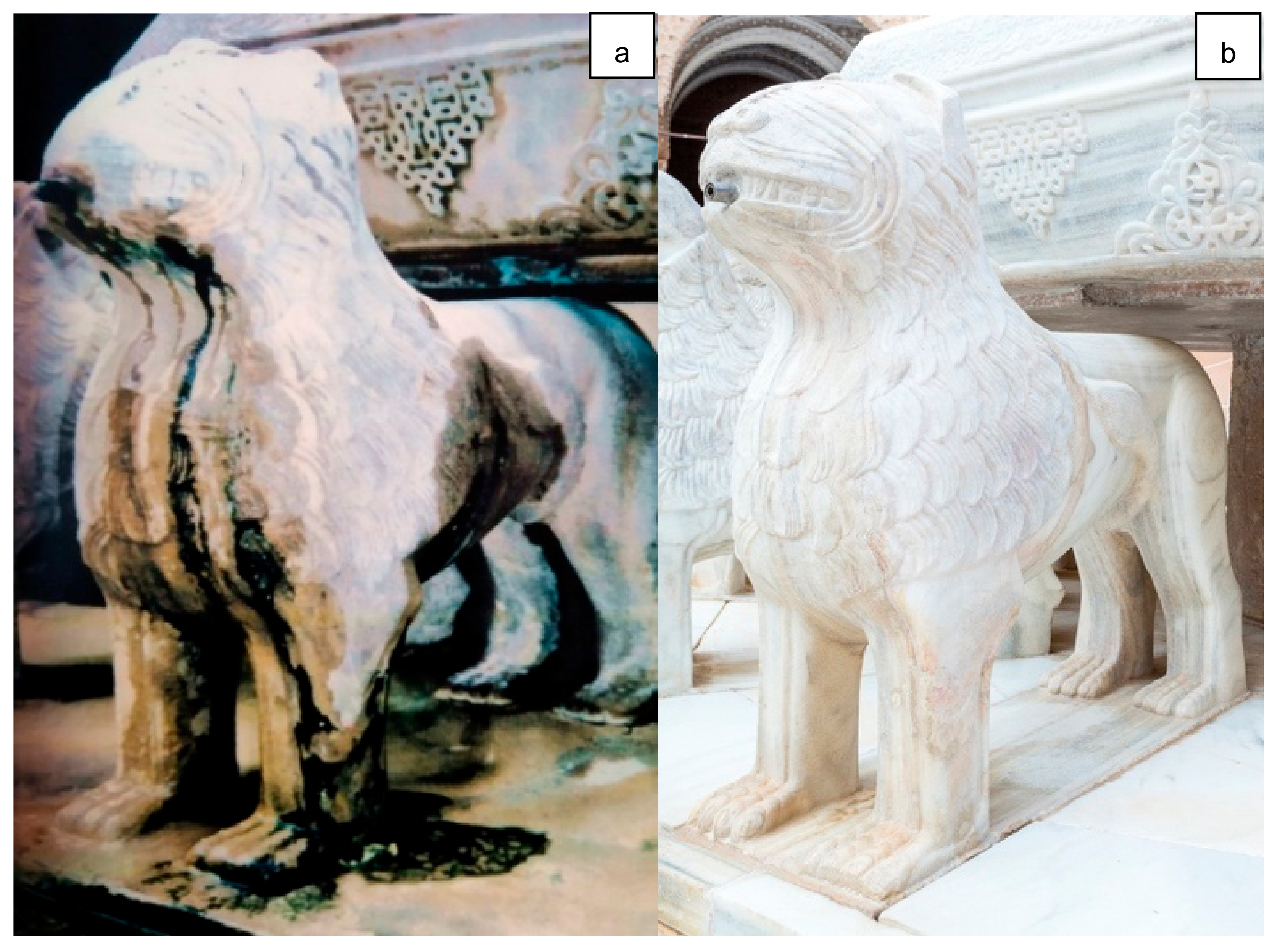
1.2. Microalgae in the Alhambra
1.3. Objectives
- −
- To identify the most common types of microalgae in the fountains of the Alhambra and Generalife today.
- −
- To make unialgal cultures of the species found in the Alhambra in order to begin creating a culture collection of living microalgae available for subsequent studies on new kinds of treatment to keep biodeterioration under control.
- −
- To design a data model and develop a database for storing and retrieving sample details and analytical values from the collection.
2. Materials and Methods
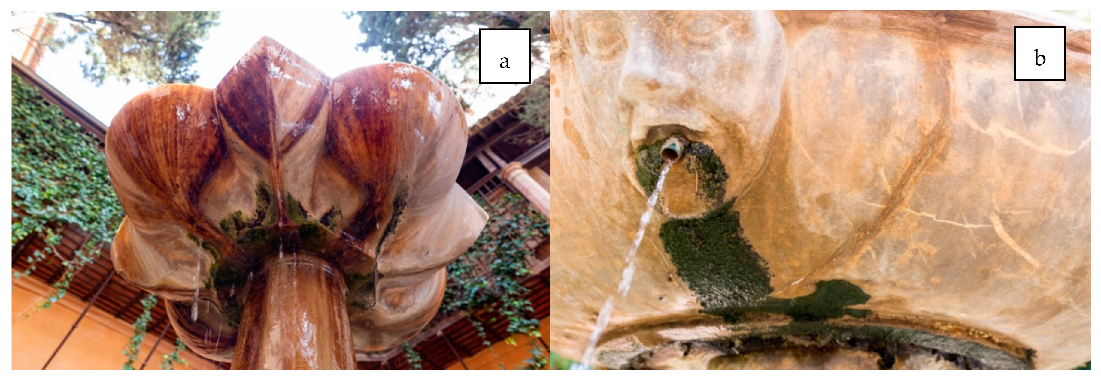
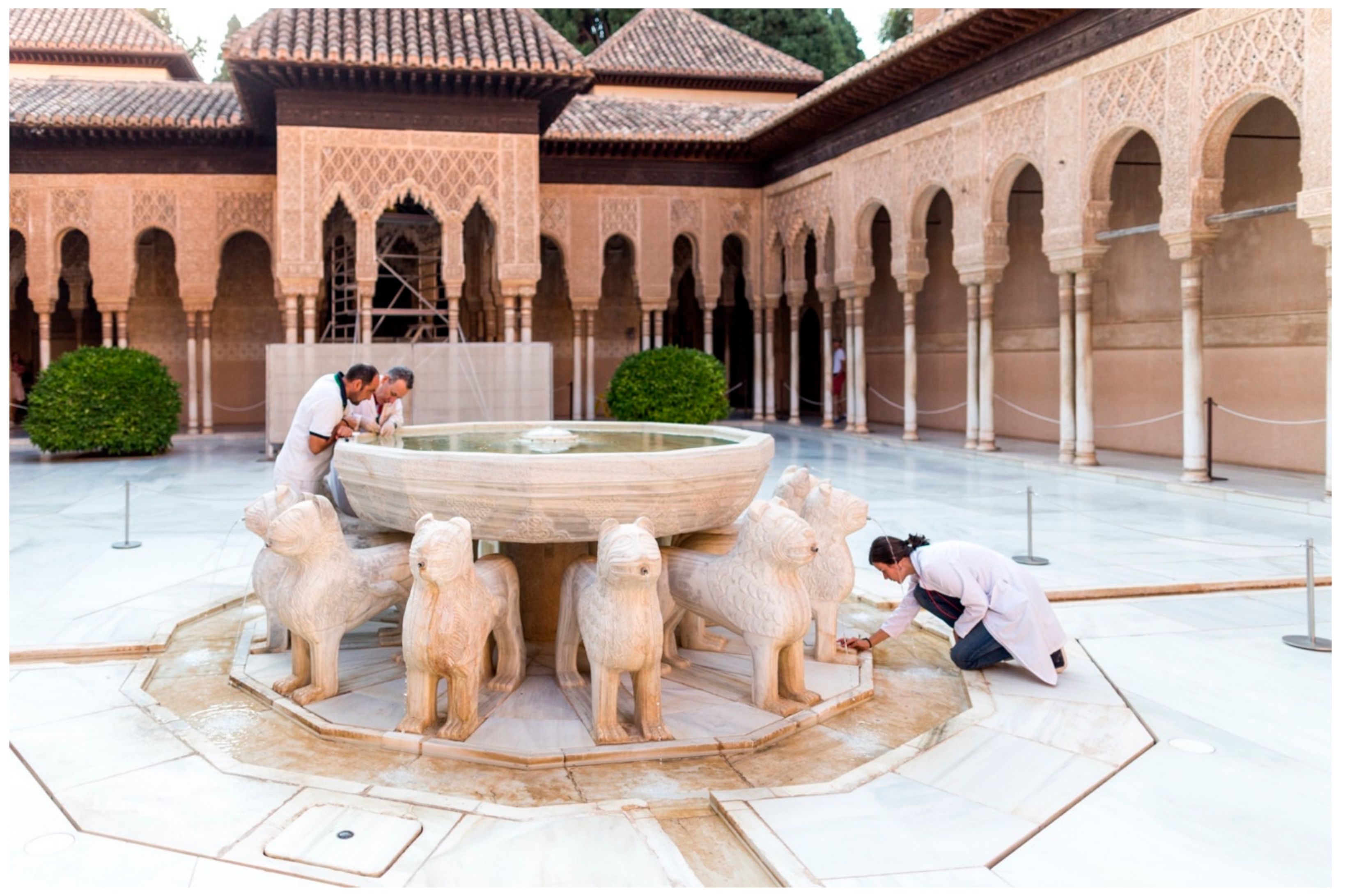
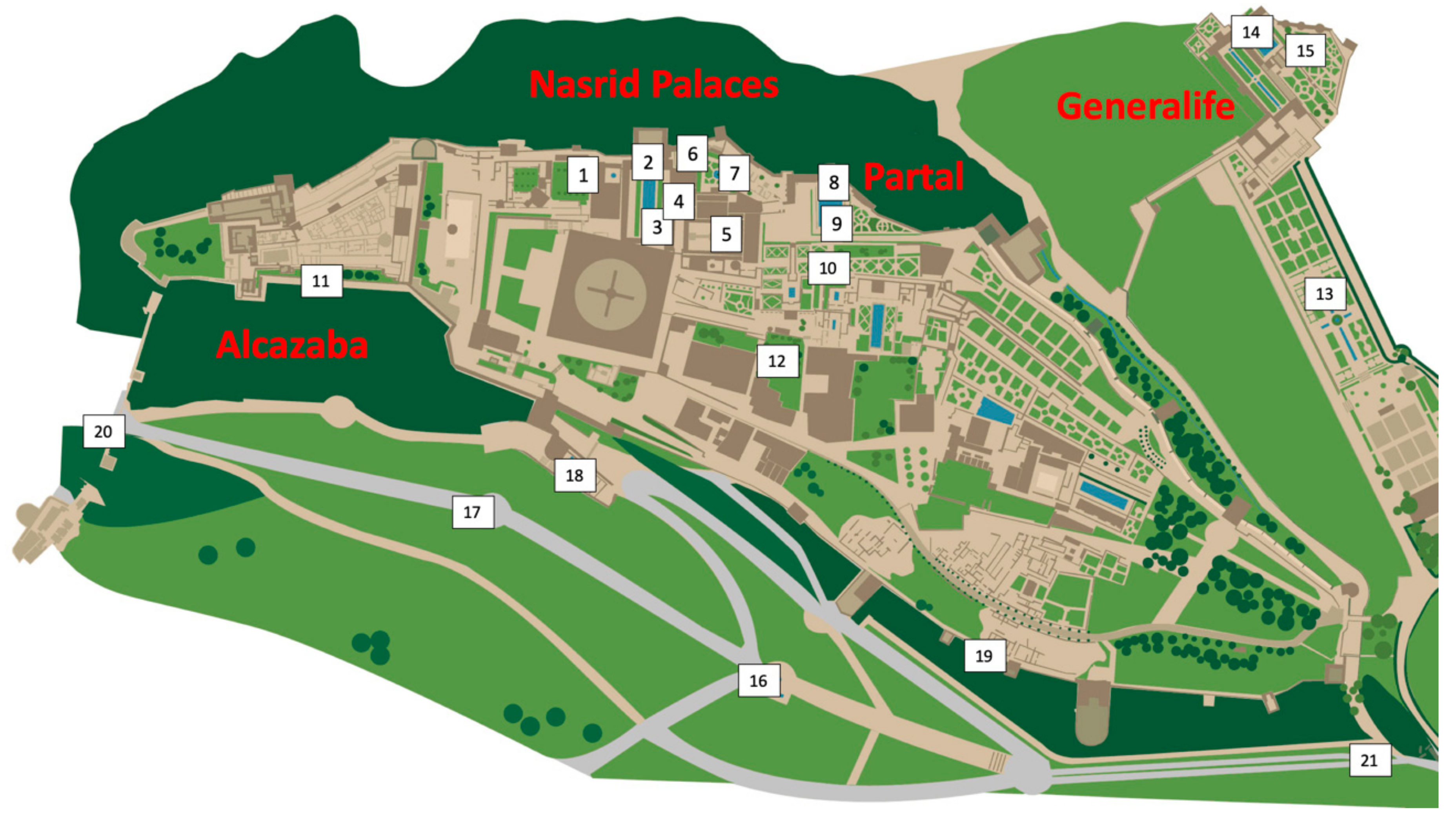
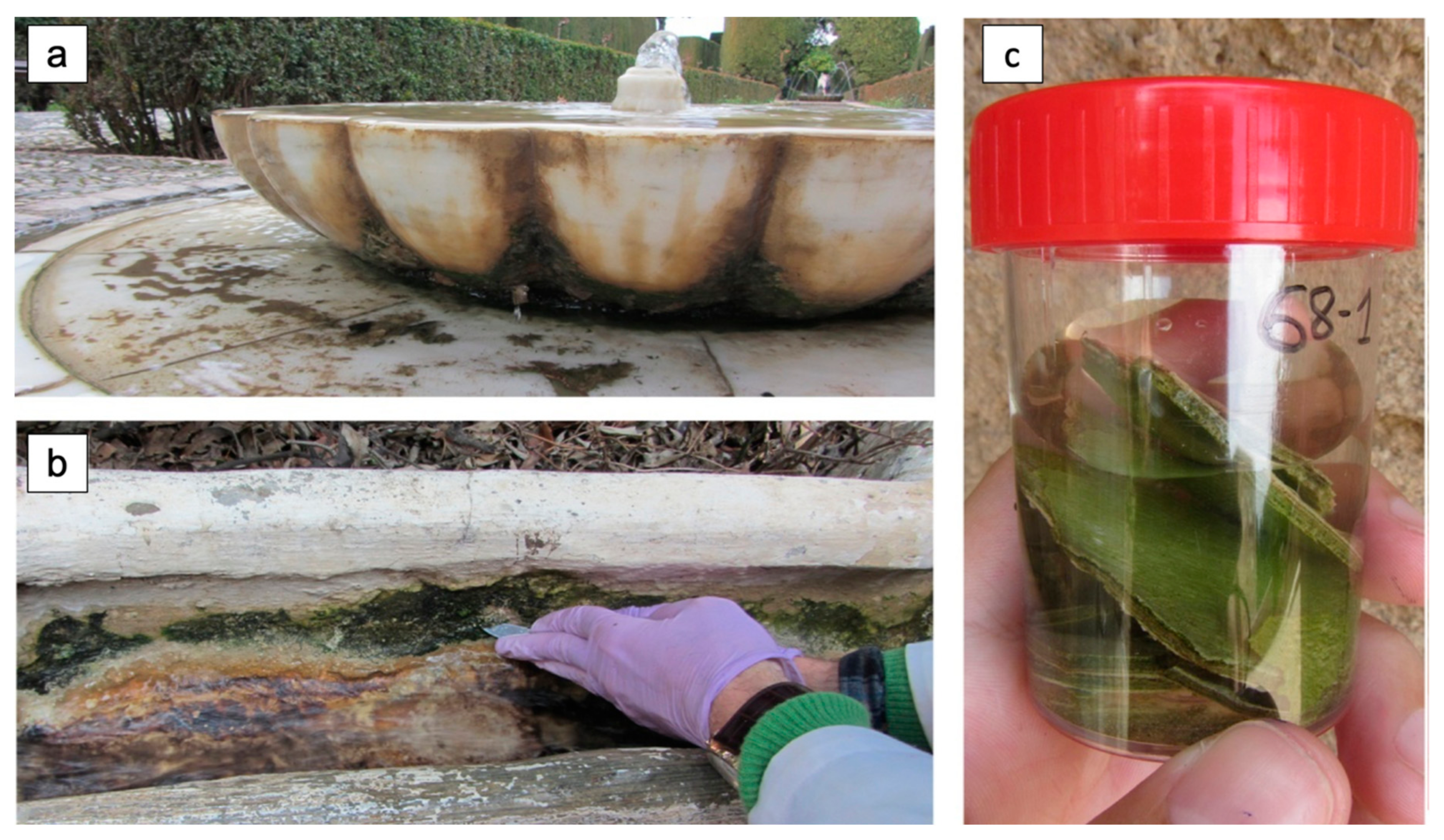
2.1. Sampling
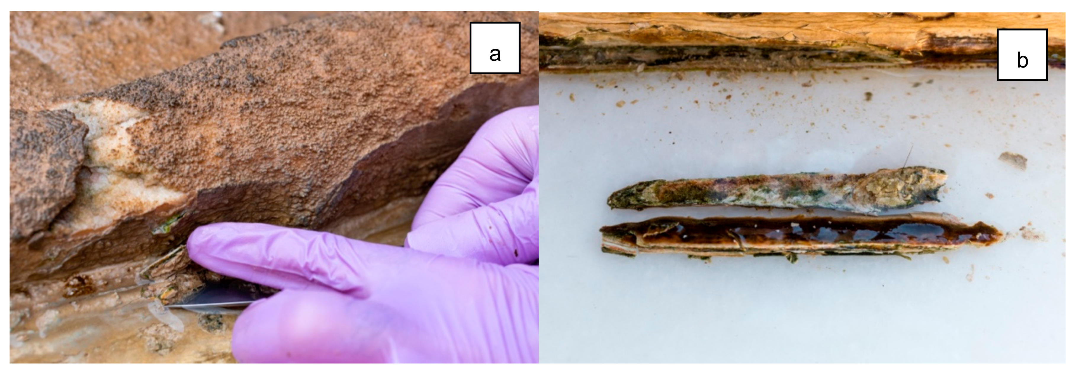
2.2. Procedures
2.3. The Database
3. Results and Discussion
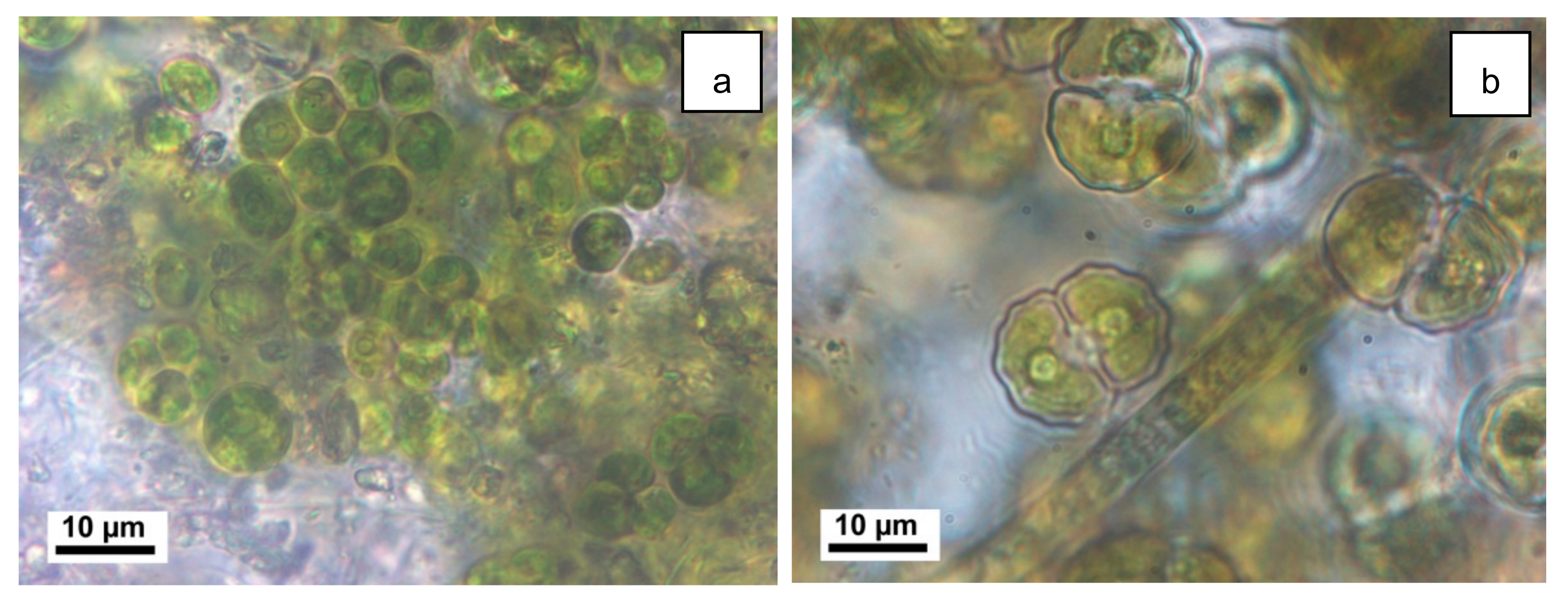
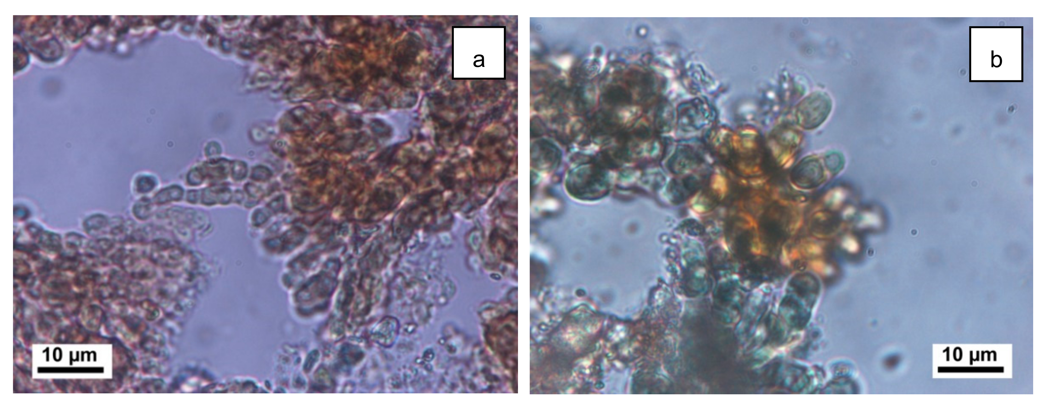
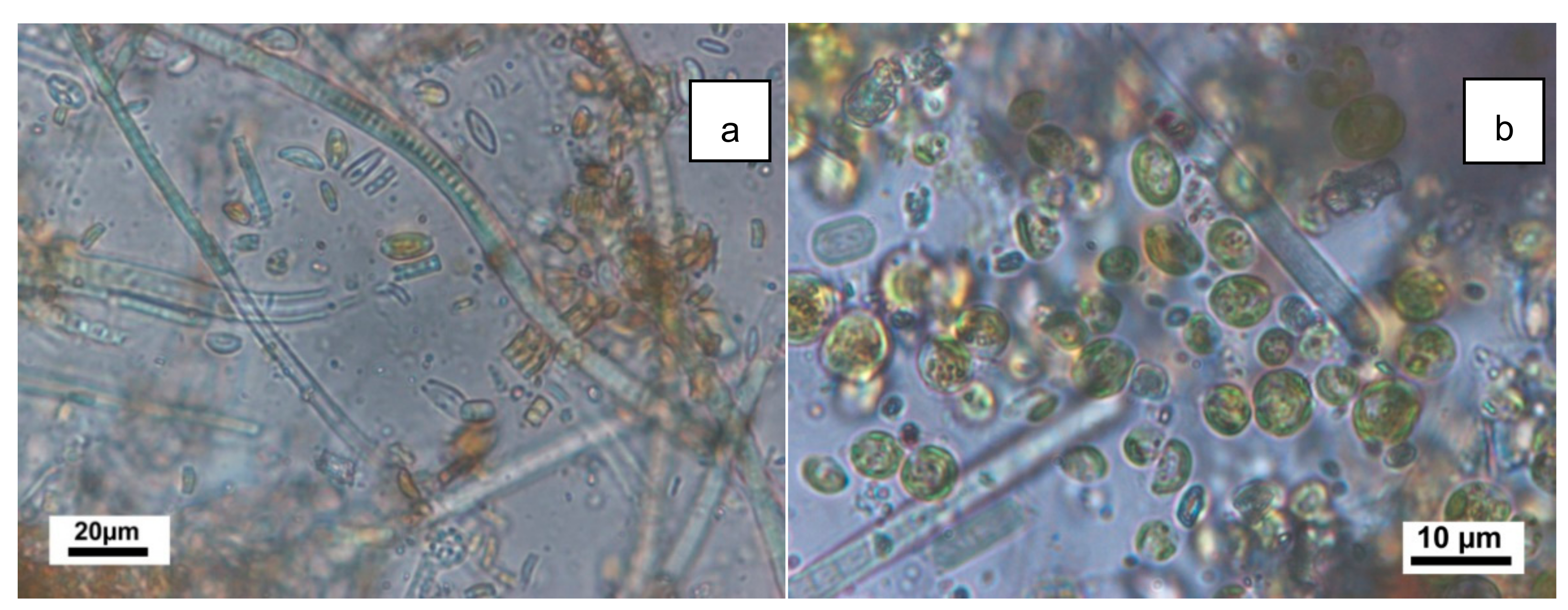
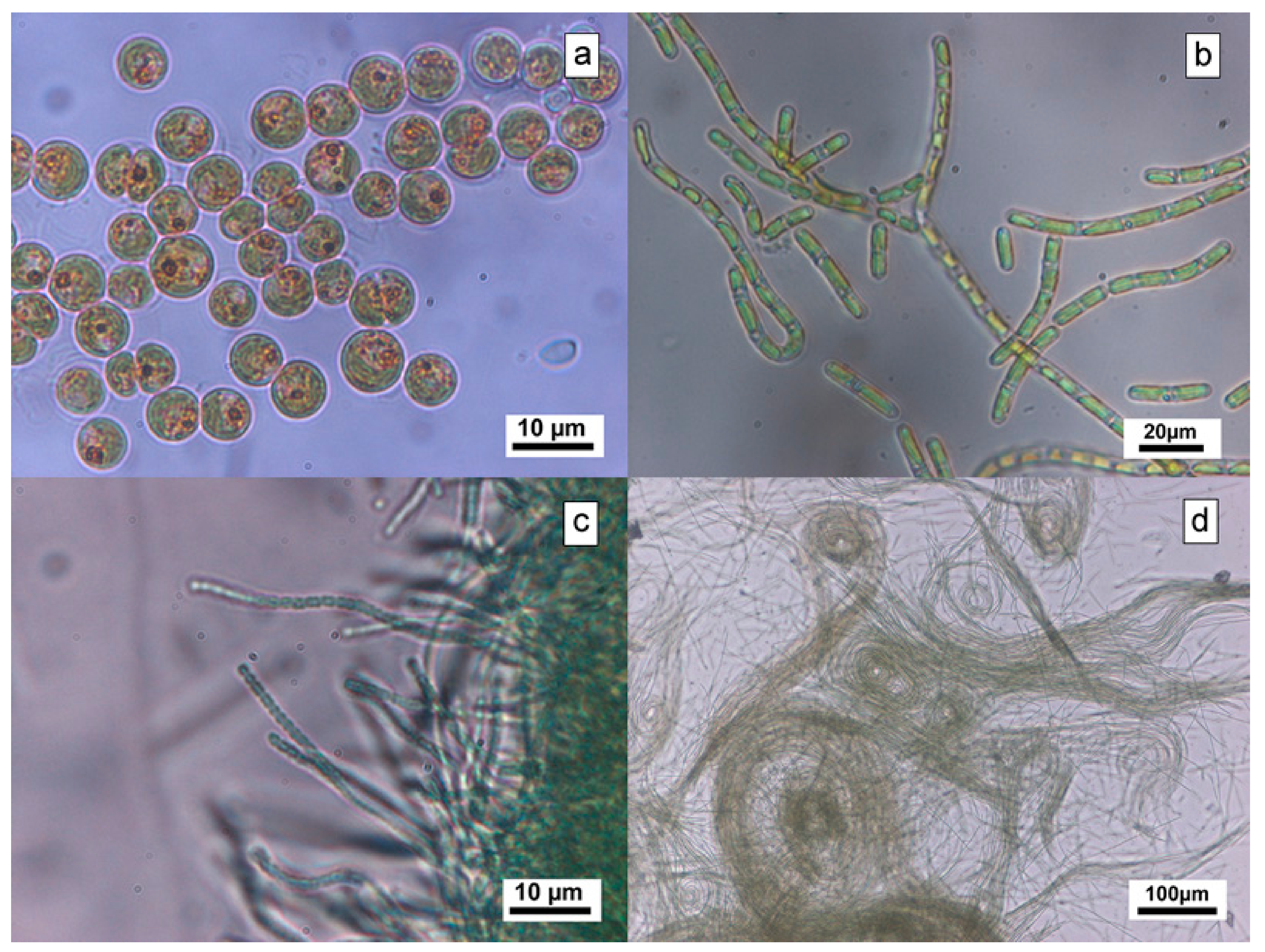
3.1. Microalgae Identification
3.2. Obtaining Unialgal Cultures
4. Conclusions
Author Contributions
Funding
Acknowledgments
Conflicts of Interest
References
- Hueck, H.J. The biodeterioration of materials as a part of hylobiology. Mater. Org. 1965, 1, 5–34. [Google Scholar]
- Anagnostidis, K.; Economou-Amilli, A.; Roussomoustakaki, M. Epilithic and chasmolithic microflora (Cyanophita, Bacillariophyta) from marbles of the Parthenon (Acropolis-Athens, Greece). Nova Hedwigia 1983, 38, 227–287. [Google Scholar]
- Ortega-Calvo, J.J.; Sánchez-Castillo, P.M.; Hernández-Marine, M.; Sáiz-Jiménez, C. Isolation and characterization of epilithic chlorophytes and cyanobacteria from two Spanish cathedrals (Salamanca and Toledo). Nova Hedwigia 1993, 57, 239–253. [Google Scholar]
- Bolivar-Galiano, F.; Peraza-Zurita, Y.; Romero-Noguera, J.; Sanchez-Castillo, P. L’Alhambra a Granada:15 anni di indagini sulla conservazione delle fontane. In L’Acqua, le Pietre i Bronzi. le Fontane Monumentali; Palombi Editori: Roma, Italy, 2010. [Google Scholar]
- Vázquez-Nion, D.; Rodriguez-Castro, J.; López-Rodríguez, M.; Fernández-Silva, I.; Prieto, B. Subaerial biofilms on granitic historic buildings: Microbial diversity and development of phototrophic multi-species cultures. Biofouling 2016, 32, 657–669. [Google Scholar] [CrossRef] [PubMed]
- Ibarra-Gallardo, C.E.; Novelo, E. Algas y cianoprocariontes epilíticos de la Zona Arqueológica de Yaxchilán, Chiapas, México. Rev. Mex. Biodivers. 2018, 89, 590–603. [Google Scholar] [CrossRef]
- Mohammadi, P.; Gholami-Nejad, P.; Asghari-Daryasari, R.; Asgarani, E. The study of microbial communities of rudkhan castle. Geomicrobiol. J. 2019, 37, 119–129. [Google Scholar] [CrossRef]
- Favero-Longo, S.E.; Viles, H. A review of the nature, role and control of lithobionts on stone cultural heritage: Weighing-up and managing biodeterioration and bioprotection. World J. Microbiol. Biotechnol. 2020, 36, 1–18. [Google Scholar] [CrossRef] [PubMed]
- Golubic, I.F.S. The lithobiontic ecological niche, with special reference to microorganisms. J. Sediment. Res. 1981, 51, 475–478. [Google Scholar] [CrossRef]
- Galiano, F.C.B.; Castillo, P.M.S. Biodeterioro del patrimonio artístico por cianobacterias, algas verdes y diatomeas. Rev. PH 1998, 24, 52. [Google Scholar] [CrossRef]
- Hoiczyk, E. Gliding motility in cyanobacteria: Observations and possible explanations. Arch. Microbiol. 2000, 174, 11–17. [Google Scholar] [CrossRef] [PubMed]
- Saiz-Jimenez, C. Biogeochemistry of weathering processes in monuments. Geomicrobiol. J. 1999, 16, 27–37. [Google Scholar] [CrossRef]
- Bolívar-Galiano, F.; Sánchez-Castillo, P.M. Biomineralization processes in the fountains of the Alhambra, Granada, Spain. Int. Biodeterior. Biodegrad. 1997, 40, 205–215. [Google Scholar] [CrossRef]
- Peraza Zurita, Y. Biodeterioro por Microalgas en Fuentes de Mármol. Ph.D. Thesis, University of Granada, Granada, Spain, 2004. [Google Scholar]
- Bolívar-Galiano, F.C. Diagnosis y Tratamiento del Deterioro por Microalgas en los Palacios Nazaríes de la Alhambra. Ph.D. Thesis, University of Granada, Granada, Spain, 1994. [Google Scholar]
- Sánchez Castillo, P.M.; Bolívar Galiano, F.C. Caracterización de comunidades algales epilíticas en fuentes monumentales y su aplicación a la diagnosis del biodeterioro. Limnetica 1997, 13, 31–46. [Google Scholar]
- Sarró, M.I.; García, A.M.; Rivalta, V.M.; Moreno, D.A.; Arroyo, I. Biodeterioration of the lions fountain at the Alhambra Palace, Granada (Spain). Build. Environ. 2006, 41, 1811–1820. [Google Scholar] [CrossRef]
- Cuzman, O.A.; Ventura, S.; Sili, C.; Mascalchi, C.; Turchetti, T.; D’Acqui, L.P.; Tiano, P. Biodiversity of phototrophic biofilms dwelling on monumental fountains. Microb. Ecol. 2010, 60, 81–95. [Google Scholar] [CrossRef] [PubMed]
- Bourrelly, P. Les Algues d’eau Douce. Algues Bleues et Rouges; Éditions N. Boubée and Cie: Paris, France, 1970. [Google Scholar]
- Komárek, J.; Anagnostidis, K. Süßwasserflora von Mitteleuropa, Bd. 19/1: Cyanoprokaryota. 1st Part: Chroococcales; Springer Spektrum: Berlin/Heidelberg, Germany, 1998. [Google Scholar]
- Galiano, F.C.B.; Castillo, P.M.S. Claves de identificación de microalgas frecuentes en monumentos. Rev. PH 1999, 26, 93. [Google Scholar] [CrossRef]
- Komárek, J.; Anagnostidis, K. Süßwasserflora von Mitteleuropa, Bd. 19/2: Cyanoprokaryota. 2nd Part: Oscillatoriales; Elsevier GmbH: Munich, Germany, 2005. [Google Scholar]
- Komárek, J. Süßwasserflora von Mitteleuropa, Bd. 19/3: Cyanoprokaryota. 3nd Part: Heterocytous Genera; Springer Spektrum: Berlin/Heidelberg, Germany, 2013. [Google Scholar]
- Peraza, Y.; Cultrone, G.; Sánchez-Castillo, P.; Bolívar-Galiano, F. Il biodeterioramento delle fontane dei Reales Alcazares di Siviglia e dell’Alhambra di Granada (Spagna). Sci. Tech. Cult. Heritage 2002, 11, 111–118. [Google Scholar]
- Peraza Zurita, Y.; Cultrone, G.; Sánchez Castillo, P.; Sebastián, E.; Bolívar, F.C. Microalgae associated with deteriorated stonework of the fountain of Bibataubín in Granada, Spain. Int. Biodeterior. Biodegrad. 2005, 55, 55–61. [Google Scholar] [CrossRef]
- Gaylarde, C.C.; Gaylarde, P.M. A comparative study of the major microbial biomass of biofilms on exteriors of buildings in Europe and Latin America. Int. Biodeterior. Biodegrad. 2005, 55, 131–139. [Google Scholar] [CrossRef]
- Macedo, M.F.; Miller, A.Z.; Dionisio, A.; Saiz-Jimenez, C. Biodiversity of cyanobacteria and green algae on monuments in the Mediterranean Basin: An overview. Microbiology 2009, 155, 3476–3490. [Google Scholar] [CrossRef] [PubMed] [Green Version]


| Zone | Fountain | No. of Samples | Material | Environment |
|---|---|---|---|---|
| Nasrid Palaces |
| 7 | marble (5), marble and metal (1), mortar (1) | submerged (6), amphibious (1) |
| 15 | marble (10), metal (5) | submerged (9), amphibious (5), aerial (1) | |
| 6 | marble (5), metal (1) | submerged (4), amphibious (2) | |
| 2 | marble (2) | submerged (1), amphibious (1) | |
| 21 | marble (15), mortar (3), metal (3) | submerged (4), amphibious (13), aerial (4) | |
| 11 | marble (11) | submerged (8), amphibious (3) | |
| 10 | marble (10) | submerged (1), amphibious (5), aerial (4) | |
| Partal |
| 6 | marble (5), metal (1) | submerged (4), amphibious (2) |
| 5 | brick (5) | submerged (1), amphibious (2), aerial (2) | |
| Casa del Arquitecto |
| 1 | painted plaster (1) | aerial (1) |
| Alcazaba |
| 3 | marble (3) | amphibious (2), aerial (1) |
| Secano |
| 5 | marble (5) | submerged (3), amphibious (2) |
| Generalife |
| 2 | marble (2) | submerged (1), amphibious (1) |
| 7 | marble (6), brick (1) | submerged (2), amphibious (2), aerial (3) | |
| 4 | glazed tile (2), plaster (2) | submerged (2), amphibious (2) | |
| Walkways |
| 3 | marble (3) | submerged (3) |
| 3 | bronze (1), mortar (2) | submerged (2), amphibious (1) | |
| 3 | marble (3) | submerged (1), amphibious (2) | |
| 3 | marble (3) | amphibious (2), aerial (1) | |
| 2 | marble (2) | aerial (2) | |
| Carmen de Bellavista |
| 1 | marble (1) | amphibious (1) |
| Total samples | 120 | |||
| Fountain | Most Common Genera | ||
|---|---|---|---|
| Green Algae | Cyanobacteria | Diatoms | |
| Bracteacoccus (3-2), Chlorosarcinopsis (2-1) | Navicula (3-1), Nitzschia (2-2) | |
| Chlorosarcinopsis (5-2) | Dichothrix (5-3), Phormidium (6-5), Leptolyngbya (9-6), | |
| Phormidium (4-2), Calothrix (4-1), Chlorogloea (1-1), Pleurocapsa (3-1) | ||
| Leptolyngbya (2-2), Pleurocapsa (2-1) | ||
| Klebsormidium (3-2) | Phormidium (7-6), Leptolyngbya (9-2), Cyanosarcina (5-4) | |
| Chlorosarcina (7-5) | Symploca (1-1), Leptolyngbya (5-4), Chamaesiphon (3-2), Calothrix (2-1) | |
| Chlorococcum (4-2), Chlorosarcinopsis (4-1), Klebsormidium (5-2) | Chamaesiphon (6-3), Phormidium (2-2) | |
| Pleurastrum (2-2) | Symploca (3-2), Phormidium (3-2), Leptolyngbya (2-1) | |
| Bracteacoccus (4-4) | Navicula (2-1) | |
| Klebsormidium (1-1) | ||
| Bracteacoccus (3-2), Choricystis (1-1) | ||
| Bracteacoccus (4-3) | Chamaesiphon (2-1), Cyanosarcina (1-1) | |
| Phormidium (2-2) | ||
| Gongrosira (3-1), Bracteacoccus (3-3) | Phormidium (3-1), Lyngbya (1-1) | Navicula (4-1), Cymbella (3-1) |
| Pseudopleurococcus (1-1), Leptosira (2-1), Chlorosarcina (2-2) | Myxosarcina (1-1), Chlorogloea (1-1) | |
| Bracteacoccus (2-1) | Chlorogloea (1-1), Chamaesiphon (1-1), Phormidium (2-1) | |
| Bracteacoccus (2-2) | Leptolyngbya (1-1) | |
| Bracteacoccus (3-1) | Chamaesiphon (2-1) | |
| Bracteacoccus (2-1) | ||
| Bracteacoccus (1-1) | Chroococcopsis (1-1) | |
| Calothrix (1-1), Phormidium (1-1) | ||
| Fountains | Green Algae | Fountains | Cyanobacteria |
|---|---|---|---|
| 4 | Neochloris sp. | 2 | Dichothrix sp. |
| 14 | Stigeoclonium sp. | 4, 8, 15, 17 | Leptolyngbya spp. |
| 14, 8 | Chlorococcum sp. | 20, 7 | Chroococcopsis sp. |
| 9, 12, 16, 19 | Bracteacoccus sp. | 2, 8 | Pseudophormidium sp. |
| 4, 8, 14 | Scenedesmus spp. | 13 | Pseudanabaena sp. |
| 15 | Pseudopleurococcus sp. | 6, 21 | Calothrix sp. |
| 5, 10 | Klebsormidium sp. | 3 | Pleurocapsa sp. |
| 11, 13 | Choricystis sp. | 3 | Phormidium sp. |
| 3 | Apatococcus sp. | 5 | Cyanosarcina sp. |
| 4 | Gloeocystis sp. | 5 | Schizothrix sp. |
| 5 | Nostoc sp. | ||
| 5 | Chroococcidiopsis sp. | ||
| 4 | Ammatoidea sp. |
© 2020 by the authors. Licensee MDPI, Basel, Switzerland. This article is an open access article distributed under the terms and conditions of the Creative Commons Attribution (CC BY) license (http://creativecommons.org/licenses/by/4.0/).
Share and Cite
Bolívar-Galiano, F.; Abad-Ruiz, C.; Sánchez-Castillo, P.; Toscano, M.; Romero-Noguera, J. Frequent Microalgae in the Fountains of the Alhambra and Generalife: Identification and Creation of a Culture Collection. Appl. Sci. 2020, 10, 6603. https://doi.org/10.3390/app10186603
Bolívar-Galiano F, Abad-Ruiz C, Sánchez-Castillo P, Toscano M, Romero-Noguera J. Frequent Microalgae in the Fountains of the Alhambra and Generalife: Identification and Creation of a Culture Collection. Applied Sciences. 2020; 10(18):6603. https://doi.org/10.3390/app10186603
Chicago/Turabian StyleBolívar-Galiano, Fernando, Clara Abad-Ruiz, Pedro Sánchez-Castillo, Maurizio Toscano, and Julio Romero-Noguera. 2020. "Frequent Microalgae in the Fountains of the Alhambra and Generalife: Identification and Creation of a Culture Collection" Applied Sciences 10, no. 18: 6603. https://doi.org/10.3390/app10186603