
Soft Matter Sample Environments for Time-Resolved Small Angle Neutron Scattering Experiments: A Review


1
Neutron Scattering Division, Oak Ridge National Laboratory, One Bethel Valley Road, Oak Ridge, TN 37831, USA
2
Chemical Sciences Division, Oak Ridge National Laboratory, One Bethel Valley Road, Oak Ridge, TN 37831, USA
*
Authors to whom correspondence should be addressed.
Appl. Sci. 2021, 11(12), 5566; https://doi.org/10.3390/app11125566
Submission received: 13 May 2021
/
Revised: 4 June 2021
/
Accepted: 7 June 2021
/
Published: 16 June 2021
(This article belongs to the Special Issue Recent Advances in Small-Angle Neutron Scattering)
Abstract
:Featured Application
This manuscript has been authored by UT-Battelle, LLC, under contract DE-AC05-00OR22725 with the US Department of Energy (DOE). The US government retains and the publisher, by accepting the article for publication, acknowledges that the US government retains a nonexclusive, paid-up, irrevocable, worldwide license to publish or reproduce the published form of this manuscript, or allow others to do so, for US government purposes.
Abstract
With the promise of new, more powerful neutron sources in the future, the possibilities for time-resolved neutron scattering experiments will improve and are bound to gain in interest. While there is already a large body of work on the accurate control of temperature, pressure, and magnetic fields for static experiments, this field is less well developed for time-resolved experiments on soft condensed matter and biomaterials. We present here an overview of different sample environments and technique combinations that have been developed so far and which might inspire further developments so that one can take full advantage of both the existing facilities as well as the possibilities that future high intensity neutron sources will offer.
1. Introduction
The use of neutron scattering in soft materials research has a long tradition. Neutrons scatter from atomic nuclei and neutron scattering lengths depend unsystematically on the atomic number, and in the case of hydrogen-rich soft materials, the scattering lengths of protium and its isotope, deuterium, are very different. This ability to vary neutron contrast in hydrogen-rich materials with H/D isotopic labels has made neutron scattering an important tool in elucidating some of the basic concepts of polymer dynamics, phase behavior, and molecular conformation, both in solution and in the bulk.
There are a limited number of neutron sources and user facilities, which limits the number of experiments that can be performed relative to those possible using X-ray scattering techniques. Yet neutrons remain vital for materials research, owing to the unique information provided by their unique atomic and isotopic sensitivity, magnetic moment, and highly penetrating nature. An excellent opportunity exists to develop time-resolved experiments and the sample environments needed for these experiments. Related developments have taken place in X-ray synchrotron radiation facilities, in part due to the abundance of beamlines [1,2,3]. Despite the limitations due to the limited number of neutron scattering beamlines, the scope for developments of time-resolved experiments and the required sample environments is much wider than often is perceived.
The impact that neutron scattering can have on soft matter research can be increased when the capabilities of neutrons as applied to static structure determinations can be extended to the time-resolved domain. Apart from the data acquisition systems needed to enable such experiments, it is also required to provide sample environments capable of perturbing samples in a controlled and homogeneous manner.
For soft matter studies, it is not only important to control sample parameters, such as temperature, pressure, pH etc. accurately, but it is also necessary to control the parameter history. For instance, the different thermal histories for a given material can lead to very different morphologies. This is particularly relevant when using complicated sample environments that try to mimic the conditions in which a material is exposed to processing conditions. Here, the combination of temperature, volume, pressure, shear force etc. has to be accurately controlled in order to be able to obtain meaningful insights into the material behavior during the process.
Neutron scattering is rarely the sole tool used to obtain an understanding of samples in out-of-equilibrium conditions and is often used in combination with data sets obtained from other experimental techniques where each technique addresses different length scales or thermodynamic aspects of the problem. If the sample environment is complicated enough and if the time-resolution is so fast that it becomes difficult to combine the data sets from individual experiments at a later stage, it makes sense to combine such experiments in a single multimodal experiment. This adds to the experimental complexity, but the benefit of obtaining complementary data, knowing that the sample is in the same physical state, outweighs the extra required efforts in developing and implementing such sample environments, and represents an excellent growth opportunity for novel neutron scattering instrumentation. Importantly, the availability of multi-modal experiments can increase the productivity and scientific impact of the large-scale neutron scattering facilities in a more cost-effective way than projects aimed solely at increasing neutron flux.
This article provides an overview of what sample environments and experimental techniques have been developed for soft matter research on neutron scattering beamlines with an emphasis, but not exclusively, on time-resolved small angle neutron scattering experiments, where one wants to follow the evolution of structure from an equilibrium state to one that is out of equilibrium, and vice versa.
2. Time Resolution
From a technical point of view, the raw neutron flux that a beamline can produce is an important parameter in defining the time-resolution that is achievable, but it is certainly not the only relevant instrumental parameter. Intrinsic background in the scattering pattern arising from the instrument, scattering contrast and sample chemical composition, sample size, detector efficiency, and associated electronics all play a role. Although the technical differences between experiments on a pulsed neutron source vs. a continuous source must be considered, in some ways they are less relevant than the type of sample environments used to carry out experiments, which can have a profound effect on the quality of the resultant data.
From a practical point of view, the time-scale over which the sample transforms from one physical state to another is the most important. However, equally important is how uniformly the sample can be perturbed. For instance, in a temperature jump experiment, temperature gradients are bound to develop. With the relatively large beam sizes produced by neutron scattering beamlines, one is detecting structural information over a thermal range. The faster the temperature jump, the larger the gradients, and the more complicated the data interpretation becomes.
The data quality per frame in a time-resolved experiment depends on the data collection time, which in turn depends on the progress in time of the process being studied. For different applications, different degrees of statistical accuracy are required. For example, if the goal is to perform a detailed structural analysis, the statistical quality of the data has to be very high. However, if one is satisfied with understanding how the radius of gyration of a particle in solution or the growth of a lamellar peak in a polymer crystallization experiment changes with time, the counting statistics requirements can be lowered and the experiments can be performed in a much more expeditious manner.
With the above points in mind, one can conclude that there is no definitive answer as to how fast a process one can resolve in a neutron scattering experiment. However, one should determine how fast a reasonable quality dataset can be acquired in order to address the problem and then assess whether or not the pertinent experiment is feasible to perform. New ‘event mode’ data acquisition will to some degree allow experimenters to have more leeway to carry out the experiment and afterwards decide on which time-resolution is feasible. ‘Event mode’ data acquisition involves retaining the position and time of each neutron detected, in contrast to traditional histogramming of detector data. Doing so makes it possible to parse a single data set collected into arbitrary time bins after data collection, which is extremely valuable for studies of time-dependent processes. It improves upon fast-frame data collection (i.e., a series of snapshots taken in quick succession) because it is not limited by the performance of the detector read-out, such as might be encountered with detectors used at synchrotrons.
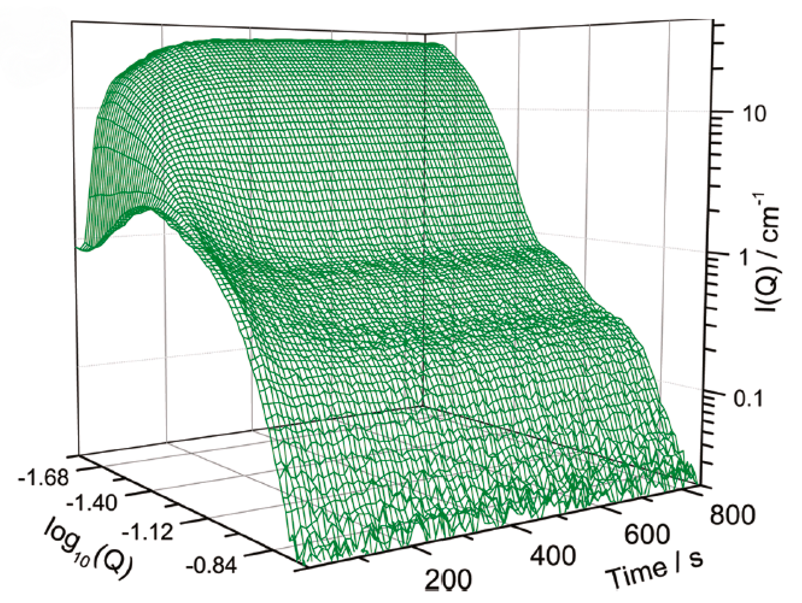
For time-resolved experiments where a trigger is used to initiate a non-reversible process and the data are collected in individual time frames, this capability was available in the early 1990s with time-resolutions of between 2 and 3 min/frame to study late stage spinodal decomposition in polybutadiene-polyisoprene blends, where one of the blocks was deuterated [4]. Similar time resolutions have been reported for polystyrene-polyisoprene diblock-copolymers [5]. Nearly a decade later, a researcher studying micelle-to-vesicle transformations in D2O was surprised about what was feasible and wrote: ‘A measurement time of 60 s already results in astonishingly good statistics, although the total surfactant concentration is less than 1 mg/mL to avoid interparticle interaction effects’ [6]. A decade later, in an experiment on the growth of mesoporous silica nanoparticles, using deuterium contrast variation, a time-resolution of 10 s/frame was reported. See Figure 1 [7].
One may get the impression that every 10 years one gains a factor of 10 in time-resolution, but in reality, it demonstrates how strongly time resolution depends on the system being studied. Even though progress in instrumentation has been considerable, the quality of samples, to a great extent, still determines the achievable time-resolution. For example, in the case of a lysozyme crystallization experiment carried out in 1995, a time resolution of hours/frame was mentioned [8]. Similarly, 10 min/frame was reported for the growth kinetics of lipid-based nano-discs to unilamellar vesicles [9]. When not making full use of selective deuteration, one can still expect 20 min/frame of low statistical data quality in a demixing experiment of incompatible crude oils [10].
Improving the achievable time resolution by increasing the neutron flux is a very expensive process. The European Spallation Source (ESS) being constructed in Lund, Sweden and the proposed Second Target Station (STS) of the Spallation Neutron Source (SNS) at Oak Ridge National Laboratory in the USA will be the next generation of neutron sources offering increased neutron fluxes. However, both will require some years and large budgets until they are completed and ready for experiments. In the meantime, there are experimental methods currently available that significantly improve time-resolved experiments.
For experiments where cycling is an integral part of the study (i.e., oscillatory shear), or the periodic deformation used in simulations of the behavior of rubber tires, one can use a series of repeated short time frames and then merge the data series on-line. In principle, if the sample survives, one can collect for extended periods of time to obtain the required statistics and thus obtain a very high time-resolution. This method has been used for large oscillatory shear measurements (LAOS) on triblock copolymer micelles [11] and the effects of start-up and cessation of flow [12] using a commercial stress-controlled rheometer in the Couette geometry, where the rheometer generates an analog I/O signal that can be used to synchronize with the neutron scattering data acquisition system. In the case of the these strongly scattering systems, 300 cycles were sufficient to achieve 100 ms time-resolved data.
If an experiment that is not intrinsically cyclic can be accurately repeated, then one can follow the same procedure. Such a strategy can work if the sample can be recycled or if the synthesis/preparation is not too costly or time-consuming. Whether this is practically feasible depends on the length of the interval between experiments. In a temperature quench or pressure jump experiment, the dead time would be the required time to heat the sample up again before the next quench or to release/built up the pressure. A recent example of this is a series of pressure jumps that were used to study the phase transition kinetics of smart N-n-propylacrylamide microgels [13]. The jumps (200-40-200 bar) were performed with a frequency of approximately 23 Hz and repeated 5400 times. A time-resolution of 10 ms/frame was obtained (2 Hz frequency) over a period of 1.5 h of experimentation.
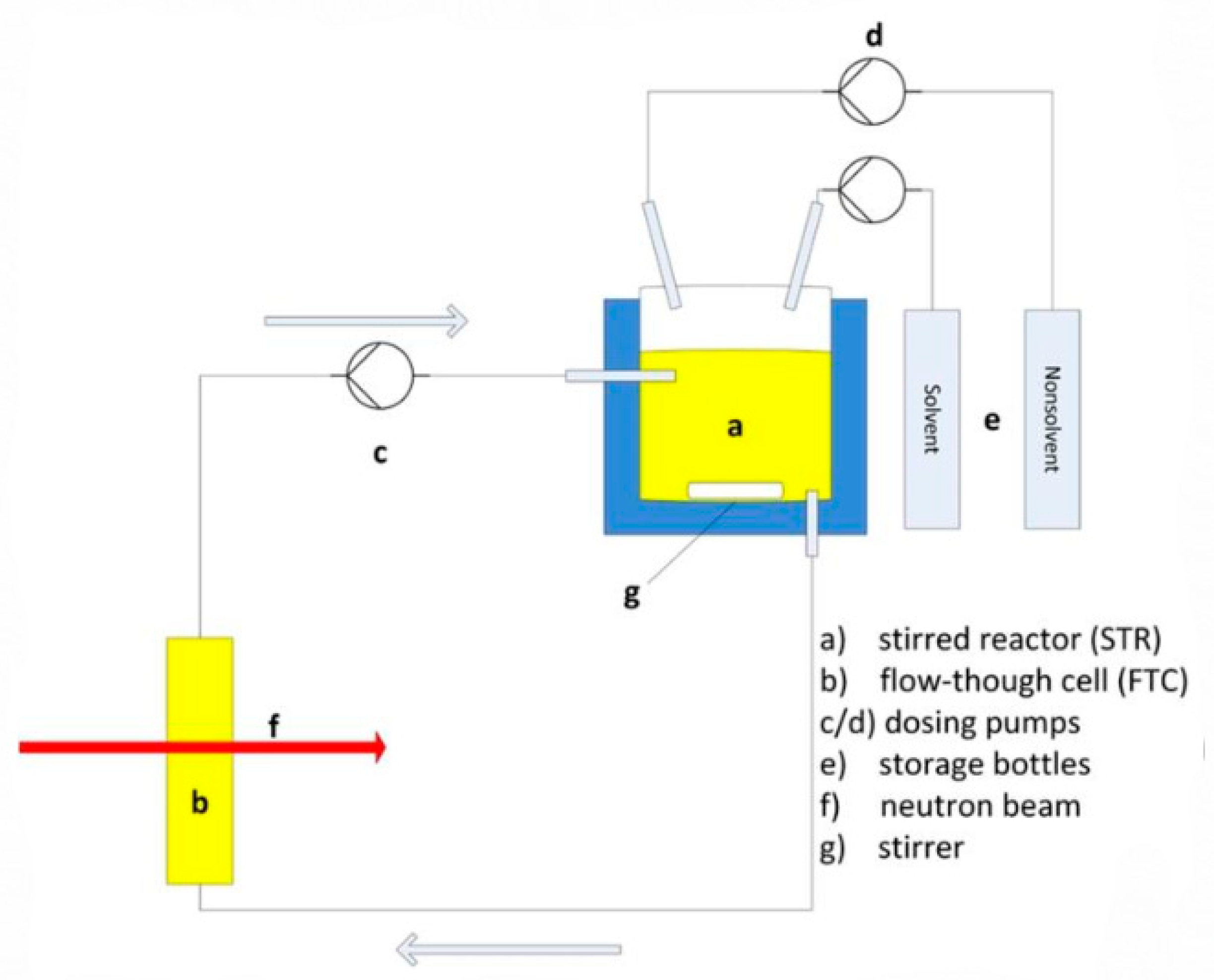
Another approach used to obtain good quality time resolution data of a continuous process is to translate the distance from the point where the perturbation takes place (e.g., mixing, extrusion, temperature change) to the point where the neutron beam interacts with the sample and translating this into time. By varying the time between the initiation point and the beam intercept, one can construct a timeline. An excellent example of this method is the (X-ray) study of polyolefin crystallization using an extruder [14]. Such experiments could easily be used on neutron beamlines as well. This translation method in combination with SANS was applied in studies of phase-separating systems, such as the cellulose nitrate/methyl acetate/isopropanol/deuterium oxide mixture, where the sample environment mimics the relevant conditions for industrial casting evaporative processes. See Figure 2 [15].
Better resolved time data are possible with the TISANE technique [16], and the first condensed matter results using this technique were reported in 2006 [17]. The technique can either be used on a pulsed neutron source or a steady-state source SANS beamline equipped with a timing chopper. Since this technique also requires periodic modulation of the sample, it therefore has a somewhat limited range of applications. Even though the TISANE technique offers great potential for the acquisition of very high-quality time-resolved data, it appears it is available to facility users, and it remains to be utilized to its full potential for time-resolved studies. The TISANE technique can, however, be improved through the use of time-of-flight techniques and event-mode data collection [18]. This can push the boundaries to 0.1 ms/frame but, again, only for experiments where cyclic data collection is feasible, and the samples scatter sufficiently strongly.
We should point out that the above-mentioned examples are not intended to be a comprehensive list of what is feasible with time resolved neutron scattering experiments. Instead, the intention is to show that even though neutron beams are not as intense as X-ray beams, this is not a reason why one should not develop and carry out time-resolved studies. Although beyond the scope of this manuscript, one should also consider a change in mindset regarding time-resolved data. If the purpose of the experiment is to obtain insights into the evolution of the structure, instead of the accurate structural determination, a lower threshold for counting statistics data is acceptable as long as one properly analyzes the data and applies the appropriate error bars to the derived parameters.
3. Sample Environments
A sample environment can be a basic piece of equipment that is used to bring a sample to a steady state in order to measure the material properties and morphology or it can be used to bring the material out of equilibrium and in the experiment follow the structural evolution on the pathway to equilibrium. The latter can be of pure fundamental nature or a process mimicking an industrial process where several parameters like pressure, temperature, shear force, and tensile load are all varied simultaneously.
While many hard-condensed matter neutron scattering experiments use cryogenics, vacuum, and high magnetic fields, soft matter research has its own specialized sample environment requirements. Cryogenic conditions convert soft matter in general to hard matter, which is only in rare circumstances of interest in soft matter research.
Although neutron facilities historically were the first to develop sample environments for non-equilibrium studies, the advances in sample environments at synchrotron sources have in recent times taken place at a quicker pace than those at neutron sources. This does not imply that there is less demand for such developments, but it is more a reflection of the fact that it is more difficult to develop and implement novel sample environments at neutron sources, as a result of the size and intensities of the available neutron beams and the limited number of available instruments. While in the 1990s, X-ray beam sizes on the order of 300 μm were quite common, specialized beamlines currently can produce sub-micron size beams. Neutron beam sizes usually vary between 1 and 10 mm.
Traditionally, one key advantage that neutrons have over X-rays is their penetrating ability through different sample environment materials, making it possible for neutrons to study in a more facile manner materials under extreme conditions, such as elevated pressures, for example. However, the present generation of upgraded high energy storage rings generates high brilliance even in the photon energy range where the penetration is strongly enhanced and can be foreseen to encroach upon this neutron monopoly.
3.1. Temperature
Temperature is one of the basic parameters that can be varied during an experiment. This was already remarked upon by André Guinier in the proceedings of the first small angle scattering conference organized in 1965 [19]. For soft matter, research temperature control is important since the thermal history of a sample can dictate the material’s ultimate properties. The main technical issue for neutron scattering is that neutron beam sizes are relatively large, making it more difficult to minimize sample environment temperature gradients. In the case of sample environments for static experiments, there are good commercial solutions to this problem. However, when there are special requirements, like the need to rotate the sample during a crystallization experiment, where the possibility of sedimentation is a real possibility, temperature control becomes somewhat more complicated, but in the range of between −10 and 80 °C, there are solutions [20,21]. In these cases, temperature control can be achieved using halogen light bulbs as the heating source and an infra-red sensor as thermal sensor, with an accuracy of approximately ±2 °C. Also, accurate control can be achieved by using water baths.
For soft matter, however, the sample’s thermal history can determine its final morphology and it is thus important to accurately control both heating and, especially, cooling rates. As such, a logical development is to combine neutron scattering with differential scanning calorimetry (DSC), a development that was implemented at X-ray facilities in 1995 [22,23]. The engineering difficulties due to the requirement to have access to the sample with larger neutron beams is evident since it took until 2014 before this was implemented on neutron beamlines [24] with an instrument that can operate in the temperature range −150 °C to 500 °C.
The larger beam size also implies that for time-resolved experiments involving temperature jumps, the situation is more complicated due to the low thermal conductivity of the samples. Fast quench rates, combined with the larger sample sizes required for neutron experiments, increases the problem of thermal gradients over the sample so that one does not obtain a realistic correlation between temperature and structure at a given temperature, but over a bandwidth of temperatures. However, compared to X-rays, there is the advantage that metallic window materials can be used which, when connected with the heating/cooling elements, can mitigate the gradient problem to some degree.
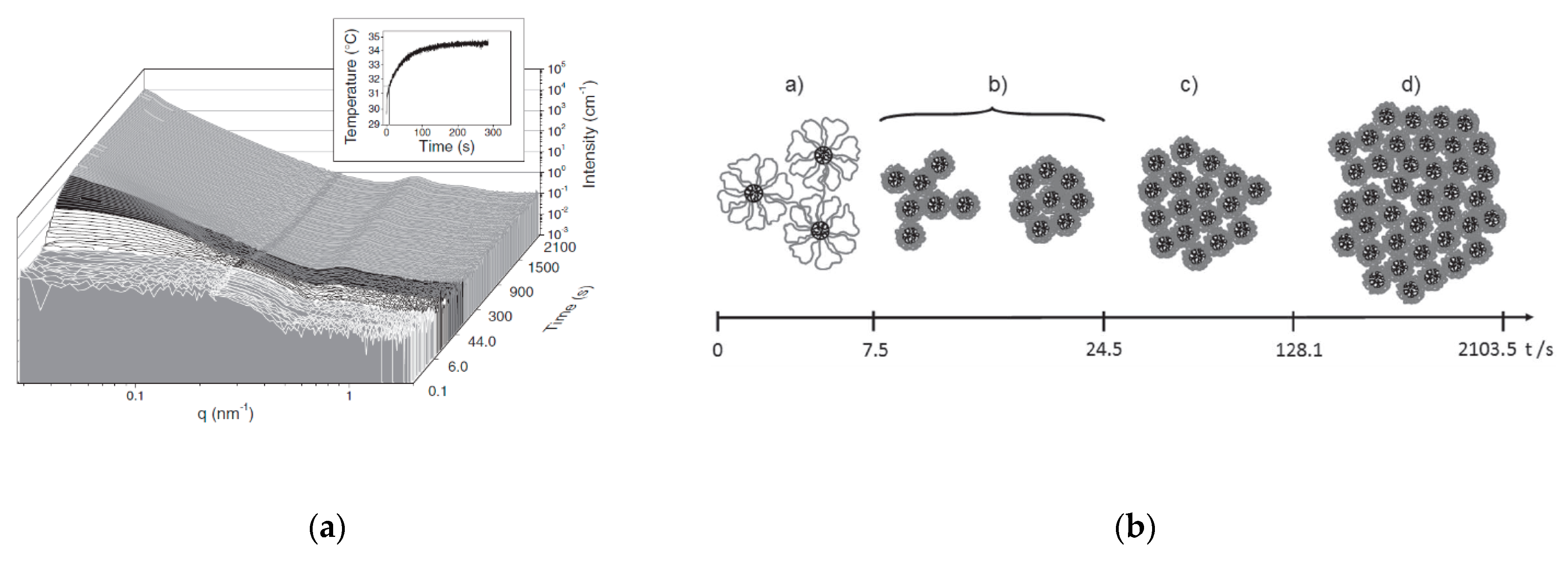
For jumps at higher temperatures, one could contemplate the use of microwave radiation [25], which can provide more uniform heating of the sample. However, for quenches to a lower temperature, material thermal conductivity remains the rate-limiting parameter. For liquids, one can perform temperature jump (T-jump) experiments by injecting the fluid into a pre-heated/cooled cell using, for instance, a syringe pump [26]. A temperature jump device specifically designed to operate on a SANS beamline, using two furnaces between which the sample is shuttled, can operate in the range 150–600 K with heating rates up to 19 K/s and cooling rates 11 K/s [27]. One can also adapt a stop-flow cell to operate as a T-jump device. This was done to understand the kinetics of the collapse, transition, and subsequent cluster formation in a micellar solution of P(S-b-NIPAM-b-S) [28]. See Figure 3. Approximately 100 s were required for a modest temperature jump of 6 °C.
The latter example shows that it is not always necessary to use world record T-jump speeds to obtain the desired information with respect to the scientific question to be answered.
3.2. Pressure
Pressure is the thermodynamic parameter that is relatively easy to change homogeneously. For the larger sample volumes routinely used in neutron scattering, this allows the reduction/elimination of large pressure gradients over the sample, allowing for a better controlled experiment producing more robust results. Most high-pressure experiments use hydrostatic pressure, which is not directional. An example of uni-axial pressure exerted on a sample in combination with a SANS experiment can be found in soil- or geo-mechanics [29]. The pore size distribution in bentonite clay was investigated as a function of pressure whilst simultaneously loading the cell with dry CO2 and water in order to elucidate the role that intercalation of these two components play in the complicated pore-pressure diagram. Pressures up to 10 MPa at ambient temperature were applied.
An example where hydrostatic pressure was applied was in research to determine if CO2 could be stored in porous shales. Here, CO2 was not only the object of study but also the neutron contrast variation medium [30]. Pressures of 25–40 bar were achieved over a temperature range of 20–60 °C. This type of study can be relevant to soft matter where, for example, the porosity of a material must be characterized to understand the impact of processing conditions.
Not only does the static characterization of pores attract attention, but also the combination of hydrostatic and uniaxial pressure is relevant for understanding flow in porous materials [31]. In the fracking of oil-containing shales, water is used as the pressurizing agent. In the case of neutron scattering experiments, this allows one to exploit the difference in scattering length between light and heavy water, in addition to the natural air-matrix scattering length difference. Uniaxial stresses, σax, in combination with the hydrostatic pressure inside the pores, p, allows to generate an effective stress σe = σax-p of 600 Bar.
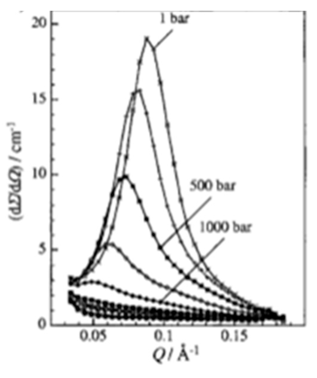
An example of the effects of hydrostatic pressure is surfactant solutions of SDS in D2O-forming microemulsions, as shown in Figure 4. Here, a static pressure was applied and the effect of pressure is the continuous transformation of spherical particles to elongated micelles [32]. The pressure cell used was capable of a maximum pressure of 10 kbar and a maximum temperature of 80 °C.
A detailed design for a temperature-controlled high-pressure cell developed for biological applications, specifically protein denaturation studies, was described by Teixeira et al. [33]. The sample cell was capable of a low temperature of −18 °C with a pressure of 2 kbar. Because of the cell’s large thermal mass, which is required for mechanical strength at these elevated pressures, it required 1 hour for sample equilibration. When working at sub-zero temperatures, one stands the risk of ice crystal formation. Down to about −20 °C, ice formation can be inhibited by anti-freeze agents, but this will alter the biologically relevant environment of the proteins. By using pressure, the freezing point can be lowered, and the use of anti-freeze agents is avoided. There are several other sample cell designs available [34].
Pressure jumps can perturb a sample over the whole volume exposed to the neutron beam without any pressure gradients and can be relatively simply and accurately repeated. This makes it possible to perform such experiments with a reasonably high time resolution using the ‘cyclic’ procedure mentioned in the section on temperature jumps.
An example is the phase transition from swollen chains to polymer mesoglobules of an aqueous solution of poly(N-isopropylacrylamide) with pressure jumps across the coexistence line [35]. This was investigated with kinetic small-angle neutron scattering with a 50 ms time resolution. The pressure jumps were on the order of Δp ≈ 200 bar. The interesting variation here is that the data acquisition time per frame was not constant but starting with a time frame length of 0.05 s that was elongated by a factor of 1.1× for each frame, thus anticipating that events evolved fastest immediately after the pressure jump. Obviously, this has some consequences regarding the statistical quality of the data, but the experiment only had to be repeated five times. This is another area where ‘event mode’ data collection will allow the experimenter more freedom to postmortem decide how to time-slice the data set.
A similar development was used to study the volume phase transition kinetics of N-n-propylacrylamide microgels [13]. A jump sequence of 200 → 40 → 200 bar was performed using a time frame length of 5 ms over a total time period of 350 ms. However, in this case, the experiment had to be repeated 5400 times before satisfactory data sets could be obtained.
It should be noted that pressure jump experiments, as those described above, are not only interesting for fundamental research purposes, but also understanding industrial processes. However, to do so, the apparatus should be capable of jumping both pressure and temperature simultaneously, preferably with the application of a shear force [36,37]. This type of experiment has been implemented for X-ray scattering, but it is probably still beyond the limits of what is feasible for neutron scattering due to beam size limitation and neutron flux.
3.3. Shear and Rheology
The flow of materials is a research area of interest to both the fundamental as well as the applied/industrial research communities, and one that lends itself to neutron scattering experiments. The cell walls of conventional Couette cells commonly used in neutron scattering experiments can be easily penetrated by neutrons and the amount of material required is small, which keeps deuteration costs to a minimum. Couette cells have a long history of commercial development and as such, are a mature technology able to keep a stable flow over the entire time required for the acquisition of data with good signal to noise ratio; one of the first mentions of the use of a Couette cell in a scattering experiment was in 1984 [38].
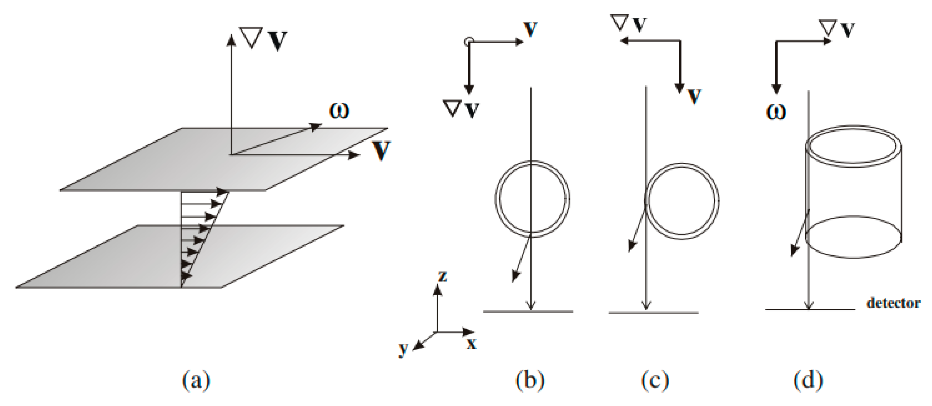
Rheology, however, is a complicated subject in which the observed components of a flow profile depend on the configuration of the cell or in which direction one probes the flow profile within the cell. In Figure 5, the different flow planes are defined and further discussions about the terminology can be found in an extensive review [39]. For the directions where one can probe the desired flow field whilst keeping the Couette cell axis vertical, i.e., the 1–3 and 2–3, radial and tangential directions, respectively, it is feasible to perform quantitative rheological experiments with neutron scattering [40,41]. However, a cell for the 1–2 direction, where one has to align the neutron beam in the gap parallel between the two Couette cylinders, has proven more difficult to design [42]. With the Couette cylinder axis horizontal, the material must be confined, otherwise gravity will force low viscosity materials out of the cell.
For the conventional Couette geometry where the cylinder axis is vertical, one can rely on commercially available rheometers. Since the topic of shear and rheology is rather popular, there have been numerous in-depth reviews on the subject [39]. These reviews include oversights of other available shear geometries that have been used on X-ray and neutron beamlines. Measuring the axis torque allows quantitative measurements of shear stress simultaneously with macroscopic strain gradients imposed by the frequency of rotation and microscopic deformation observed in the SANS data [42]. As mentioned, for most neutron scattering experiments there are commercially available instruments, where the replacement of the commercial Couette cylinders by cylinders made out of neutron transparent materials like quartz and titanium can be straightforward.
The literature contains a wide range of subjects that have been studied using Rheo-SANS methods. They range from investigations into the shear banding mechanism in a fluid comprised of cetyltrimethylammonium bromide wormlike micelles [44], to detailed studies of polymer chain conformations composed of polyethylene-polypropylene blends with Si particles nanocomposites [45], to the shear-induced formation of rod-like entities in metallo-supramolecular gels based on a multitopic cyclam bis-terpyridine [46]. The Couette geometry can also be used to probe material relaxation properties by using an oscillatory flow protocol, which is mentioned in the section on time-resolution.
The 1–3 and 2–3 Couette geometries are now fairly standard for neutron scattering experiments. Progress in instrumentation in the coming years will most likely be incremental and most developments will focus on the analysis of neutron-rheology data sets [47]. Some developments were recently reported on the implementation of the technically more challenging case, where one wants to probe the 1–2 plane [42,48] by directing the neutron beam through the gap between the two cylinders that make up the Couette cell. With a small enough beam, it is possible to probe the different flow profiles as a function of the position within the gap [48]. Such spatial mapping is not commonly associated with neutron scattering since the neutron beams tend to be rather large and can only be reduced in size by sacrificing intensity. For stable flow rheological applications, this is a problem only limited by the available beam time and neutron flux, and not by any technical aspects. From an experimental point of view, it should be remarked that it is often necessary to obtain scattering data along all three shear planes [49]. Unfortunately, this requires the use of two different Couette cells capable of covering the same temperature and shear range. Designs for a cell that can accommodate experiments in all three shear directions are available, but rare [42], and still require substantial reconfiguration when changing between geometries.
In addition to the above-mentioned shear flow geometries, there is the less popular but useful plate-plate geometry [50] used in a range of SANS experimentation, such as studies on the self-assembly of nanofibers composed of fibronectin mimetic peptide-amphiphiles [51].
The design for a cone-plate geometry instrument for use in reflectometry is discussed in the framework of test experiments involving polystyrene/deuterated polystyrene, with the flat bottom plate modified to be transparent to neutrons [52]. A modified commercial instrument, for example, was used for the study of asphaltenes in the reflectometry configuration [53]. However, placing the neutron beam in the gap between the cone and plate, like what has been successfully done with X-rays, requires that very tight neutron beam collimation be used, which impacts data rate and quality. An interesting development is the use of Grazing Incidence SANS or GISANS. The feasibility of this technique in combination with a cone-plate rheometer was demonstrated in experiments on bola-amphiphilic arginine-coated peptide nanotubes [54]. However, data collection took about 5 h per sample, indicating that this method still requires further development.
The more difficult problem of how one goes about studying elongational flow appears to still be out of reach of real time neutron scattering experiments. The methodology of flash freezing a material that has been subjected to elongational/extensional flow off-line and then using the frozen samples to carry out a static measurement was reported in 1990 [55], and the method was still regarded as recently as 2016 as being the best method to study dendritic polymer blends with linear chains [56]. Here, the main issue remains that the time-resolution of the physical process does not match the time-resolution that can be reasonably achieved on SANS beamlines.
Whilst the examples being discussed above fall under the ‘drag flow’ category, there are also configurations that allow ‘pressure flows’ to be studied [57]. Capillary- or Poiseuille-flow and slit-flow are examples of pressure flow. This type of flow is important to understand the fundamental behavior of chain molecules in conditions relevant for industrial processing. For instance, in injection molding, the predominant physical processes are Poiseuille flow and thermal quenches. Not only is flow in an unrestricted path relevant, but studies of the behavior of the material as it flows around obstructions and restrictions are also required. Such information provides firstly, insights into material properties, and secondly, these results can be used to validate computational models as to how entangled polymers behave under elongational flow or when encountering a change in flow geometry. In other words, such physical results provide predictions on how to process materials [58,59]. When run in an ‘open’ configuration, these set-ups require considerable amounts of material, which with deuterated samples tends to be prohibitively expensive. Hence, efforts have gone into recirculating flow cells, which require relatively small amounts (~200 g) of material [60,61,62,63]. This cell is equipped with non-birefringent sapphire windows that are strong enough to resist pressures of up to 10 MPa, and also allow for birefringence measurements to be carried out, thus creating a set-up which can deliver structural and orientational information over a wide range of length scales. See Figure 6.
A recent design based on developments in microfluidics [64], which itself was based upon a larger scale design by G.I. Taylor from 1934, allows for a variety of different flows to be applied within a single sample environment. The Fluidic Four Roll Mill, so named because it uses four rolling cylinders [65], is a device consisting of 4 × 2 orthogonal channels in which flow patterns can be generated by opening/closing a series of valves [66]. See Figure 7.
3.4. Mechanical Deformation
Uniaxial mechanical deformation studies on soft materials are relatively easy to implement and are extensively used by academics and in industrial materials testing laboratories. Several small-scale deformation stages are commercially available, but these are in general, only suited for qualitative tensile data involving local molecular conformations. To obtain quantitative tensile results simultaneously with the neutron scattering data, one has to make sure that the sample is uniform over its entire volume and not only at the point of intercept with the neutron beam. With the exception of ambient or near ambient temperatures, one encounters the issue of how to uniformly heat the sample. This obviously can be achieved by placing the entire deformation load frame in a temperature-controlled chamber, but in doing so, any temperature changes will be slow to equilibrate, thus limiting the technique’s application with neutron scattering instrumentation.
Since polymeric materials exhibit a wide variety of materials properties, the type of required load frame depends on the maximum tensile force and elongation required. Elastic rubber samples require little stress but a high strain, i.e., elongation [67], whilst the reverse is true for High Density Polyethylene (HDPE) [68]. For such samples, the required mechanical performance (i.e., stresses between 10 and 200 N) required of the load frames is easily covered by commercially available equipment from a variety of suppliers.
Early on, mechanical deformation experiments were performed in a quasi-static fashion, i.e., deform-hold-collect data. This deformation protocol works well for some materials where the molecular relaxation times are long. Otherwise one has to revert to continuous stretch protocols where the data collection time can be problematic. This is often the situation when the materials are unclamped and allowed to molecularly relax [69]. Relaxation of isotopic blends of linear polyethylene and ethylene copolymers with butyl and hexyl was studied, but due to the relatively small amount of material in the neutron beam (intrinsic to this type of deformation experiments), data could only be obtained at 30 min time intervals. An extensive discussion on how the anisotropy manifests itself in scattering patterns, due to the molecular and domain orientation induced by applying the deformation, can be found in experiments of elastomeric polypropylene [70].
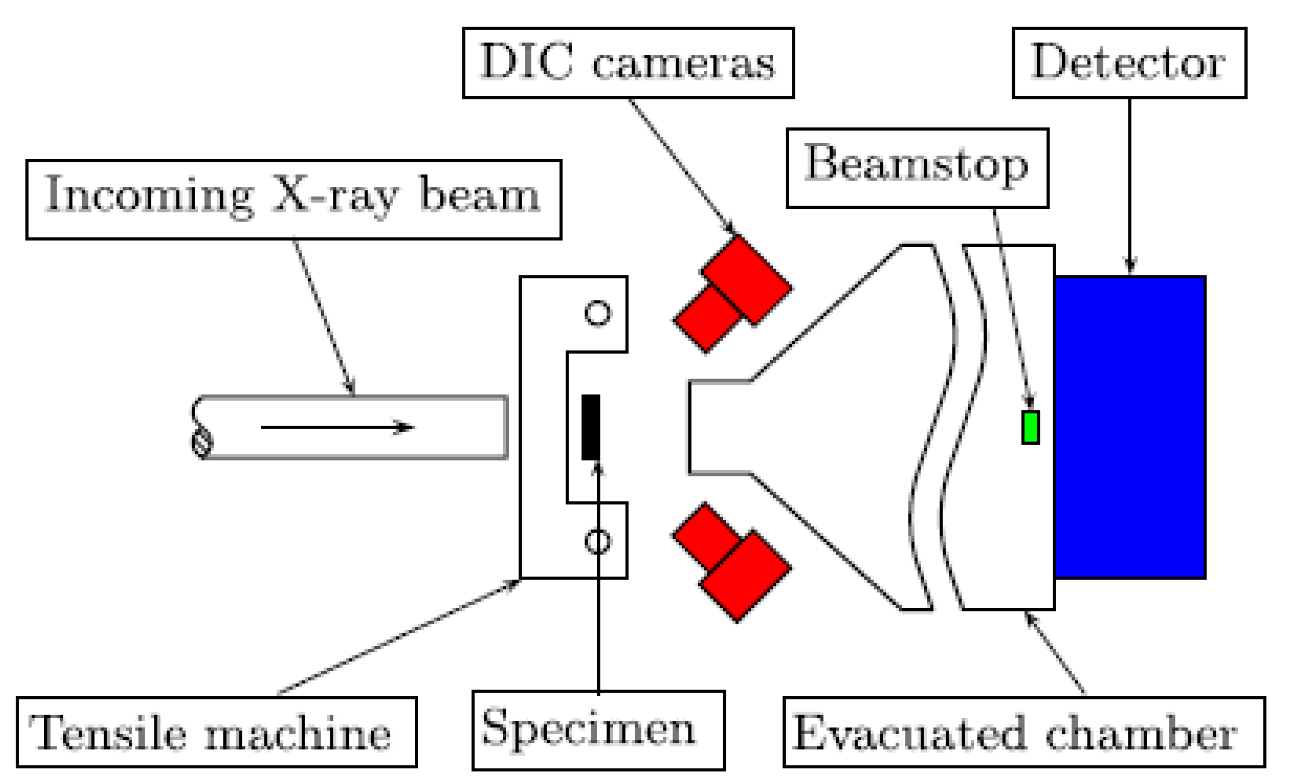
A method to increase the information content of on-line deformation experiments is the use of Digital Image Correlation (DIC) [71]. This optical method is easy to install on a neutron scattering instrument and allows one to probe the homogeneity of the deformation process over the entire sample and also provide macroscopic material parameters. See Figure 8. When combined with neutron scattering experiments, one obtains a more complete picture of the material’s deformation behavior. Simultaneous DIC furthermore supplies additional validation of the reliability of the experimental results since mechanical problems, such as slippage at clamps, will be noticed earlier.
Although homogeneous temperature control on samples that require large elongation is somewhat more complicated to achieve, other parameters like humidity can be relatively easy controlled. This requires the construction of a solid chamber around the moving parts. An example of this is the stress-induced long range ordering in spider silk [72]. Here a combination of finite element modelling and SANS data were used to elucidate the role that crystalline domains play in the ordering of silk when extended. The crystalline domains are less susceptible to water uptake compared to the amorphous domains, and this physical characteristic allows for contrast variation to be used. By placing the tensile load frame in a 100% D2O relative humidity environment, it was possible to selectively deuterate the amorphous parts of the silk.
One of the issues that one might encounter due to beam intensity and the samples becoming thinner when elongated is that the statistical quality of the scattering patterns is rather poor. One might be tempted to use a stop-start technique where the material is not continuously stretched but instead, after a short stretch, a pause is taken to allow data to be collected. Of course, this has the problem that the sample is allowed to relax and the results will not necessarily be the same as with a continuous stretch. The differences between the results obtained with these two approaches when deforming polypropylene are discussed in the literature [73].
3.5. Stop Flow/Chemistry On-Line
To be able to follow structure formation due to chemical reactions requires reaction cells that are resistant to the chemistry being carried out and also capable of attaining the desired temperature and pressure range. In the case of low viscosity solutions, the reaction products can sediment out and therefore change the composition of the sample as ‘seen’ by the probe beam [74]. This can be overcome by the use of a tumbling cell, which rotates the sample solution around the beam axis, preventing sedimentation. These cells are routinely used in all neutron scattering facilities. As an alternative, a cell of sufficient thickness with a mechanical stirrer can also be used. As with any equipment that contains moving parts, this leads to a more complicated temperature control system. One design that contains an array of four tumbling cells, makes use of air cooling/heating, is capable of a limited temperature range of 10–50 °C, and is suitable for slow reaction kinetics without thermal variations [20]. By placing the equipment on a translation stage and triggering the chemical reactions at different times, it is possible to alternate between the samples and map out the kinetics over extended time scales, also making effective use of the allocated beamtime.
For faster experiments, sedimentation is less of a factor and here one can consider the use of conventional stop-flow cells. Due to the required beam size, the sample volume will be rather large, and thus there are constraints on the achievable mixing- and dead-times, but workable solutions do exist. A review on the use of stop flow cells in SAXS and SANS experiments was written by Isabelle Grillo [75], who pioneered such experiments on SANS beamlines. A practical example of such experiments is the formation of microgels due to the precipitation and subsequent polymerization of N-Isopropylacrylamide [76]. Here the early stage of gel formation was the point of interest, which is often the most difficult part since structural changes tend to develop faster than in later stages. A commercially available stop-flow cell was used at a fixed temperature. Data were collected over a period of 20 min with a time framing rate of 5 s/frame. However, in order to obtain sufficient quality data, the experiments were repeated three to four times and the results were averaged. The data yielded the total particle volume and the number density of particles. A similar home-built device was used to report on cationic liposomes complexes with DNA. However, instead of temperature, a pH jump was induced by the stopped flow to trigger the reaction [77].
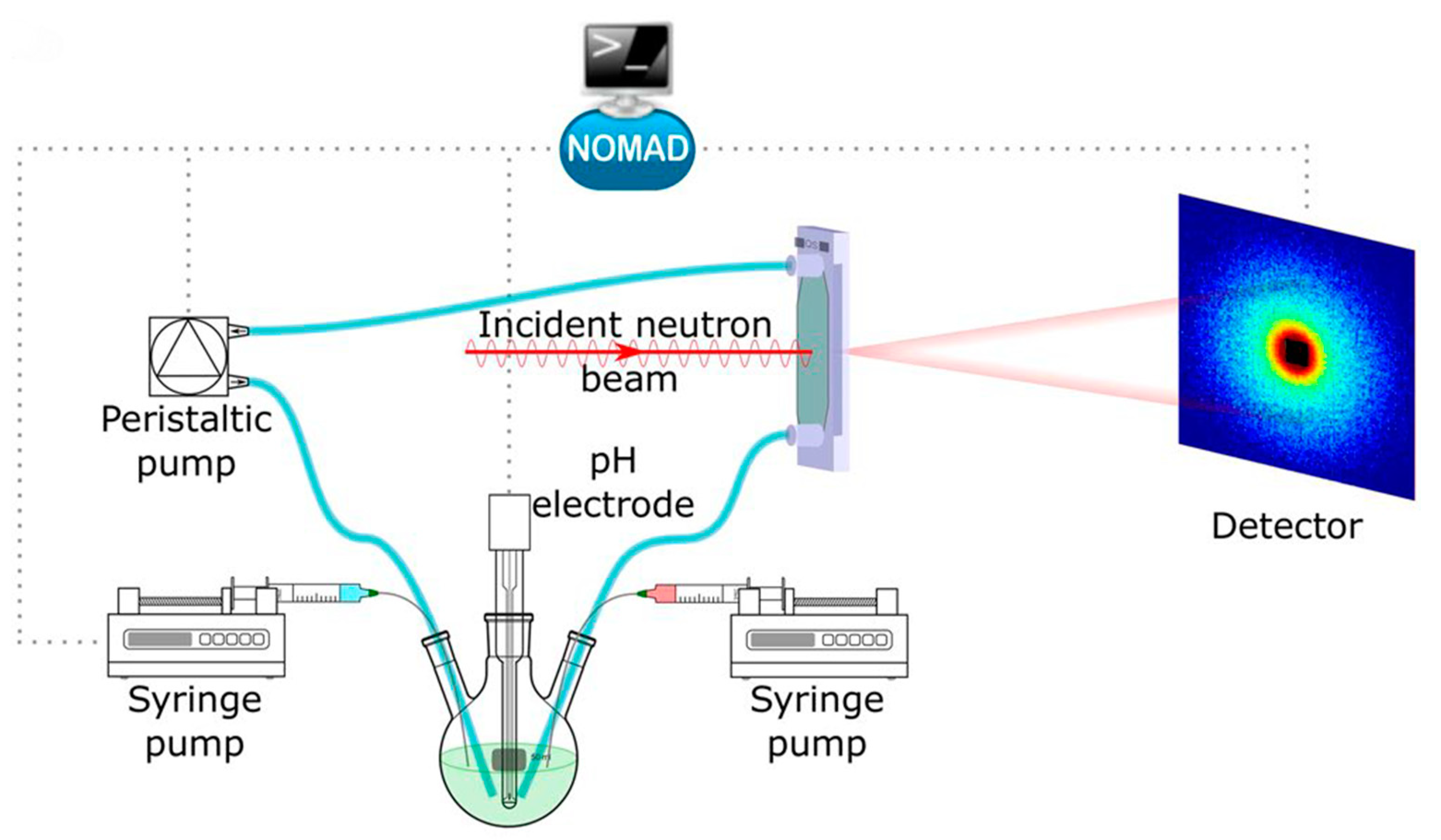
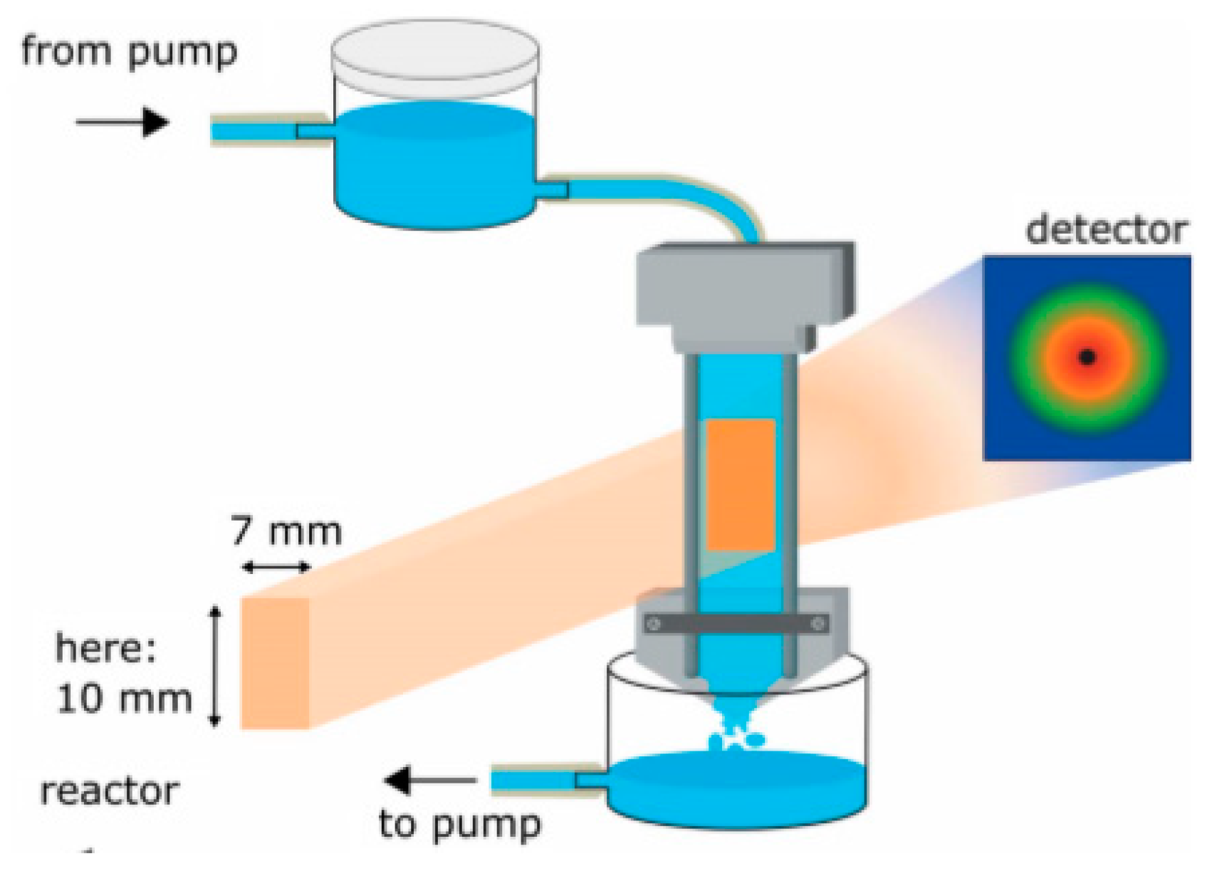
By using continuous flow methods, a larger variety of experiments is feasible even though much larger quantities of materials are required. In some cases, it is even feasible to use a recirculating flow and gradually change the conditions without incurring the penalty of requiring large volumes of sample. This is especially relevant when dealing with deuterated materials. A purposely designed sample environment meeting these requirements was reported. See Figure 9 [78].
For test experiments, aqueous solutions of polyoxyethylene alkylether carboxylic acids, a class of surfactants with strong pH-responsive properties, were used. During the experiments, the pH was changed by titration. As the authors remarked, these time-resolved experiments, down to 1 s/frame, were made feasible in recent years by more efficient detectors and increased neutron flux thanks to improvements in neutron guide technology.
Pressurized CO2 gas can be used as a neutron scattering contrast agent in porous materials like lignite and shale [79], and SANS/USANS experiments are part of the toolset that can be used to determine pore distribution size and morphology for pores that are accessible to the CO2. However, when introduced in its supercritical state (ScCO2), it is a good solvent, primarily for nonpolar low-molecular-weight compounds. For polar higher molecular weight compounds, it is a rather poor solvent. Thanks to the fairly benign conditions at which criticality occurs (7.39 MPa, 31.1 °C), ScCO2 is a fairly attractive option for performing chemical synthesis of surfactants, polymers, and biomaterials [80]. On-line neutron scattering investigations of poly(dimethylsiloxane) polymer-ScCO2 solvent phase diagrams were performed and fundamental applications demonstrated [81]. Time-resolved scattering experiments where the ScCO2 to CO2 transition was used as a foaming agent to create micro and nano foams allowed the kinetics of foam formation to be studied as a function of pressure and pressure modulation [82]. The use of fluorinated surfactants in CO2 microemulsions is widespread but not desired for environmental reasons. In a systematic pressure and contrast variation study, it was shown that partial substitution of ScCO2 by cyclohexane reduced the required amount of fluorinated surfactants considerably [83]. Contrast variation through the use of D2O exchange was crucial in this case and the only way this kind of structural information could be obtained.
Performance of on-line chemical synthesis experiments has so far not been widely explored, although it appears that neutron scattering could play an important role, as the above example has shown. When used for structure forming on-line chemical processing, temperature control and homogenization of the reaction mixture is of the utmost importance. The designs for such cells used in on-line experiments should take this into consideration, as well in allowing for the possibility of siphoning off small amounts of the reaction mixture (for off-line chemical analysis) during the course of the experiments without perturbing the pressure/temperature conditions [84].
The use of on-line size-exclusion chromatography to deliver well-defined and non-aged samples for scattering experiments has become feasible, although here one has to keep in mind the restrictions on the amount of available material. This method requires beamlines that can deliver high intensity and small beam sizes. However, when these conditions are met, the experimental accuracy can be considerably improved. For example, proteins that have a tendency to aggerate in solution can still be studied in their non-aggregated state [85].
3.6. Electromagnetic Fields
Sample environments generating electric fields have not been extensively developed for use at neutron beamlines. AC fields that can be used to (partially) align relatively short rigid polymeric molecules in solution in order to perform fiber diffraction experiments have been used for biological molecules. The main problem with this approach is that the frequencies used should be such that the sample does not heat up and that the fields are high enough that they are not shielded by the salts in the buffer solution [86].
The main problem with using static electric fields is that the voltage required as function of sample thickness is considerable. When using too high a field, one risks electric breakdown damaging the sample. To induce domain alignment in thin films of symmetric diblock-copolymer of polystyrene and poly(methylmethacrylate), (PS-b-PMMA) fields of 40 V/μm were used in a sample cell consisting of an aluminumized Kapton film used as an electrode and a similar electrode that is isolated from the sample by a thin layer of poly(dimethylsiloxane) [87]. The alignment process could be followed by time-resolved SANS experiments. As to be expected, the interplay between the sample thickness and the interfacial interactions between the electrode and the sample plays an important role. For thin samples, where the interfacial interactions are most important, the structure can evolve over periods of hours. On the other hand, for thicker films, complete alignment takes only minutes. Static electric-field-induced deformation of bulk poly(styrene-block-isoprene) lamellar phases, swollen in toluene or tetrahydrofuran, has been studied with SAXS and with SANS. Capacitor cells with 3–5 mm electrode gaps and typically 5 mm beam path length were used to apply up to 60 kV, reaching electric fields up to 12 kV/mm [88]. See Figure 10. Electric breakdown is a common limitation under these conditions, sufficient stability for the duration of experiments can however be reached with careful control of experimental conditions, paying very careful attention to drying polymer and solvent to remove any traces of water.
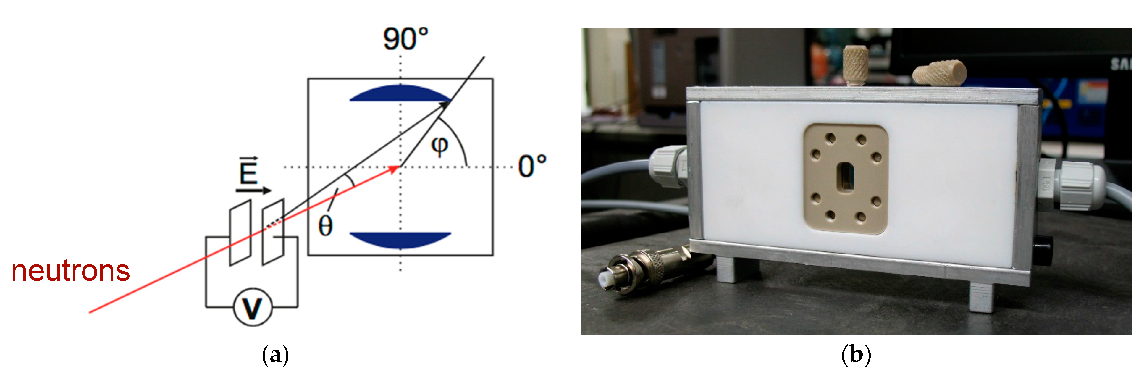
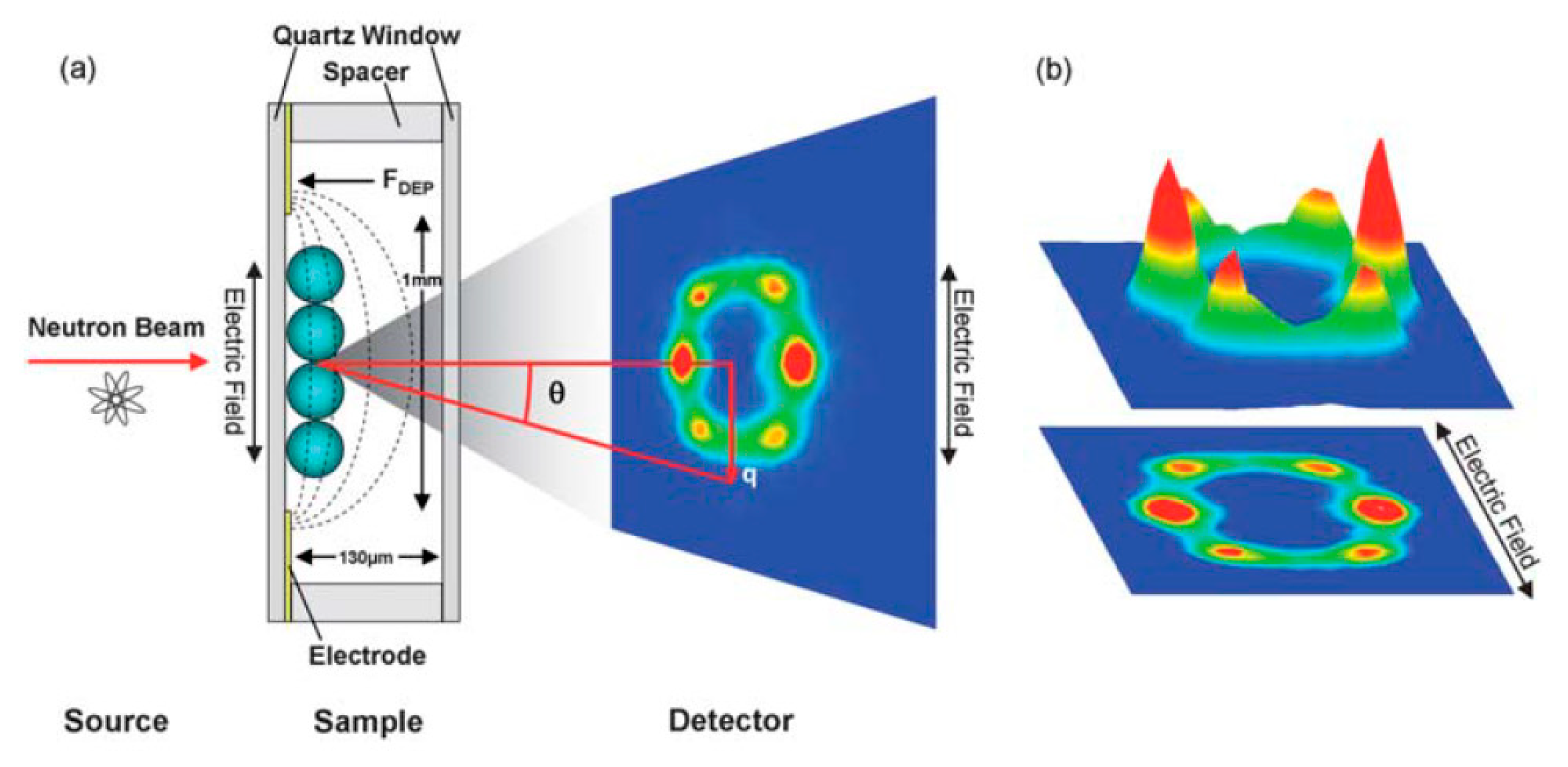
Dielectrophoresis uses the intrinsic polarizability of objects/molecules to be manipulated in a gradient or AC electric field. When this method is applied in combination with SANS, it is possible to follow the development of molecular ordering due to different field strengths and frequencies. A model system, polystyrene particles in H2O (diameter 195 nm and 530 nm), and an applied AC field formed colloidal crystals consistent with the space groups P6mm and C2mm [90]. A schematic overview of the cell is shown in Figure 11. The AC field was applied in transverse direction to the neutron beam and depending on the experiment, a range of 0 < ω < 100 kHz and 0.62 < E<1.4 kV was used with a cell thickness in the neutron beam direction of 130 microns. The cell thickness was limited in this design due to the requirement of exposing the sample to a reasonably homogeneous AC field; a thicker cell would contain field gradients.
Where the formation of colloidal crystals by the application of an AC electric field is maybe somewhat counterintuitive, the formation of oriented bundles of rather stiff fibrous molecules of the conjugated polymer poly(3-hexylthiophene) (P3HT), with a persistence length of several nm, is less surprising [91]. The authors are also describing parallel optical experiments where the data were Fourier transformed to provide an estimate of the degree of orientation.
One of the surprising findings is that on-line strong magnetic fields have so far rarely been used as a sample environment in soft matter-related neutron research. In most neutron scattering laboratories, there is an abundance of resistive and superconducting magnets available for research on magnetic materials. One would have expected that several potential user groups would have taken advantage of this opportunity and used these magnets for soft matter experiments as well. For soft matter, one clearly requires higher sample temperatures than the cryogenic temperatures most commonly used in magnet research and most often a configuration where neutron beam and magnetic field are at 90 degrees, thus requiring a split coil magnet over a simpler solenoid.
In liquid crystal and liquid crystalline polymer research, the study of material response to magnetic fields is common, but often only moderate fields (<2 T) are required. An example of this is the interplay between ordering and microphase separation of liquid-crystal polystyrene-poly(methyl methacrylate) block copolymers bearing a chiral biphenyl ester side group linked to the backbone by a dodecyloxy spacer [92]. The applied magnetic field causes the polymer backbone to partially orient. Experiments on another liquid crystalline block copolymer to map out the temperature-magnetic field phase diagram have also been reported [93]. When doped with lanthanides, lipid membranes can also be oriented [94].
Since the interaction of soft matter often is via diamagnetism, the required fields to be able to observe a relevant alignment interaction are >5 T, i.e., greater than the fields produced by permanent and electromagnets. The requirement to use superconducting magnets is a severe complication. Some static on-line X-ray experiments are published [95,96] and even some dynamic studies, where the sample was rotated inside the magnetic field and observed whilst rotating back [97,98]. For neutron experiments, occasionally an off-line magnet was used to produce aligned samples from fibrous biological macromolecules [99,100] and more recently the use of an on-line 8 T magnet to measure alignment by SANS of bicelles complexed with lanthanides was reported [101].
Transportable higher field on-line superconducting solenoids (17 T) have been used, but so far, no results on soft matter samples have been published [102]. For higher continuous fields, one has to resort to more permanent installations like Bitter magnets, very large superconductors, or hybrids of these two. Pulsed magnetic fields using capacitor banks are often transportable and split coils can reach up to 30 T [103,104,105] and solenoids up to 40 T [106], but the short pulses (msec) and the slow repetition rates (several shots/hour) make these magnets unrealistic options for soft matter research.
3.7. Light
The use of visible or UV light to modify samples on-line has found some applications in chemistry and in biological systems. An example is the use of UV light in a solution with a UV photo-destructible anionic sodium 4-hexylphenylazosulfonate (C6PAS) surfactant in combination with an inert surfactant hexaethylene glycol monododecyl ether (C12E6). Upon exposure to the UV light from a high-pressure Hg lamp, the C6PAS fell apart and released the content of its micelles to react with the inert surfactant. Hg lamps produce a high light intensity so care should be taken in controlling sample temperature [107]. Similar experiments on reversible phase separation systems using narrow bandwidth light sources have also been reported. Here two different UV wave lengths (350 and 450 nm respectively) were used to initiate phase separation and to reverse it [108]. Photorheological studies, where the rheological material properties are influenced by the exposure to UV light, have also been reported, but it should be noted that the UV irradiation was not performed on-line with the SANS experiments [109].
Visible light produced by a ‘white’ halogen lamp was used in SANS experiments to gain insight into the gelation process of the conjugated optoelectronic polymer poly(3-hexylthiophene-2,5-diyl) (P3HT). Here the effect of the exposure to light was found to retard the growth of microstructures [110,111]. Although the authors mention that they monitored the light intensity, no information about differences in growth of microstructures as function of dose rates was provided.
The conformation of biological molecules involved in the photosynthesis process as a function of exposure to light is a research subject that is well-suited for study by SANS. Not only because of the advantages that contrast variation via deuteration can bring, but also due to the fact that one does not have to worry about the interactions of photoelectrons, which invariably are created in X-ray-based experiments. Here, the issue is not so much recognizable radiation damage, but instead, the less recognizable effects of the direct interaction of X-ray photoelectrons possibly causing conformational changes [112]. Using live cells that were, both on- and off-line, exposed to ‘cool white LED light’ with an intensity of ≈ 20 μmol of photons m−2 s−1, the structural organization of membrane systems in cyanobacteria were investigated [113]. An illustrative example of the results is shown in Figure 12. It should also be mentioned that quasi-elastic neutron scattering experiments were performed under similar illumination conditions.
Similar experiments, using SANS and Quasi Elastic Neutron Scattering (QENS), were used to determine the volumetric changes of the light sensitive biological pigment rhodopsin, although the experimental details with respect to the exact illumination conditions are somewhat vague in the manuscript [114].
Microwave radiation can be used for sample heating, but an alternative use is to influence chemical reactions, although the exact way that microwaves influence chemical reactions is not entirely clear. It can change the rate constants, but the reaction pathway may also change. Preliminary neutron scattering experiments with an on-line microwave generator were performed, but these experiments were inconclusive [115] and have not received much follow-up. This may be a missed opportunity since in a later work using X-ray scattering, it was shown that by adding microwave-interactive chemical species it was possible to perform targeted annealing of specific molecular regions [116]. Such a use of microwaves, in combination with the advantages that selective deuteration can impart, may allow one to gain insights in chemical pathways.
3.8. Container-Less Measurements
In some cases, contactless or container-less experiments are carried out to minimize the interaction of the sample with the sample cell or to reduce the background scattering due to window materials. This is particularly important for very high temperature experiments, where the sample–wall interaction can change the chemical composition of the sample [117]. So far, aerodynamic levitation, where a vertical gas stream keeps a droplet of material in the beam, is most commonly used to study materials at high temperature. Powerful lasers are used as a heating source [118]. For soft matter research, aerodynamic levitation appears to be less popular and acoustic levitation is used, instead [119]. A test experiment on the drying of lysozyme solution droplet has been reported [120]. Although this experiment was intentionally aimed at drying and thus increasing the protein concentration, it also highlights one of the method’s shortcomings, namely solvent evaporation. The authors also noted that the strong 22 kHz soundwave, which helps to maintain internal mixing, also has the unwanted side effect of denaturing the protein.
Independent of which levitation method one uses, a drawback is that one can only levitate droplets that are smaller than the average neutron beam, thus reducing the scattering intensity as well as possibly introducing parasitic scatter from the sample-air interface. Even if using a jet of liquid shooting through the beam, one will still be restricted to small sample sizes since it is difficult to create large, stable laminar flows. Another ingenious approach to contact-free measurements that mitigates the problem of size mismatch between sample and neutron beam is to create a free liquid film by flowing a solution between two wires [121]. See Figure 13.
A rather specialized container-less measurement is the formation of soot particles in a combustion process. Here the flame is placed on a vertical translation stage so that the distance between the flame-neutron beam interception point and the flame mixer can be varied. This method has been used at a number of synchrotron and neutron facilities [122]. Just as in similar SAXS experiments, the data quality remains low and suitable caution is required when fitting the data.
Whatever the method used, one should be aware of the fact that container-less measurements do not mean that there is no sample-environment interaction. Oxidation, denaturing of protein, and exchange of deuterated for protiated water have been reported. It is also well-known that elongated molecules can orient themselves around air–water interfaces.
3.9. Ultrasound
Ultrasound used in aqueous solutions, or other solvents, creates cavitation bubbles that cause local agitation, affecting a variety of processes. Depending on the frequency used and physical system under investigation, ultrasound can be destructive. For example, it is used extensively for cleaning objects (20–40 kHz), but it can also help materials to crystallize (20 kHz) [123], or used as a diagnostic tool. Another application of ultrasound at a frequency around 1 MHz is to create a time-dependent mechanical perturbation. This was demonstrated in SANS experiments on sodium dodecyl sulfate (SDS) surfactant micelles in aqueous solution. Here the application of ultrasound allowed for the reversible observation of deformation of micelles as a function of time exposure to ultrasound [91].
A more elaborate ultrasound system that operates in the 1.25 MHz range, uses two spherically focused acoustic transducers and an acoustic cavitation detection system. The latter can be used to decouple changes observed due to cavitation versus changes that occur as a result of the propagation of non-cavitating acoustic waves [124]. In these experiments, a neutron absorbing aperture was used in order to match the dimensions of the neutron beam to those of the acoustic field. Even though the size of acoustic bubbles was within the detection range of the SANS experiments, it was found that due to their short half-lives they did not affect the data analysis to any appreciable extent. Several suggestions for future developments, beyond those in this manuscript, are given by the authors.
3.10. Humidity Control
In a range of fields, the control of humidity is important. For biological materials that have to be studied in their natural state, this is an obvious requirement, but industrial processes and the filling of the pores of porous materials to influence the scattering contrast should also be mentioned.
The simplest method is to place the samples in a closed environment with a reservoir containing a saturated salt solution. However, to change the relative humidity one has to use different salt solutions, which makes this method somewhat cumbersome for on-line experiments [125].
An elaborate system using mass flow controllers with the possibility to mix two different vapors to control the relative humidity between 0 and 90% has been used to study semicrystalline polymers, porous materials, and polyelectrolyte membranes [126]. The advantage of having two independent streams of vapor that can be mixed is that it offers the possibility to vary the H2O/D2O ratio, which in the case of porous materials, allows one to find the matching point for contrast variations very rapidly. The same system can be used to create a vapor pressure of organic solvents.
Developments in commercial humidity generators allow for the construction of rather simplified humidity-controlled cells, which were used in studies of forest products [127] where structural changes as function of moisture were observed. A commercial humidity generator was also used to study novel phases of lipid bilayers that are important for membrane fusion [128].
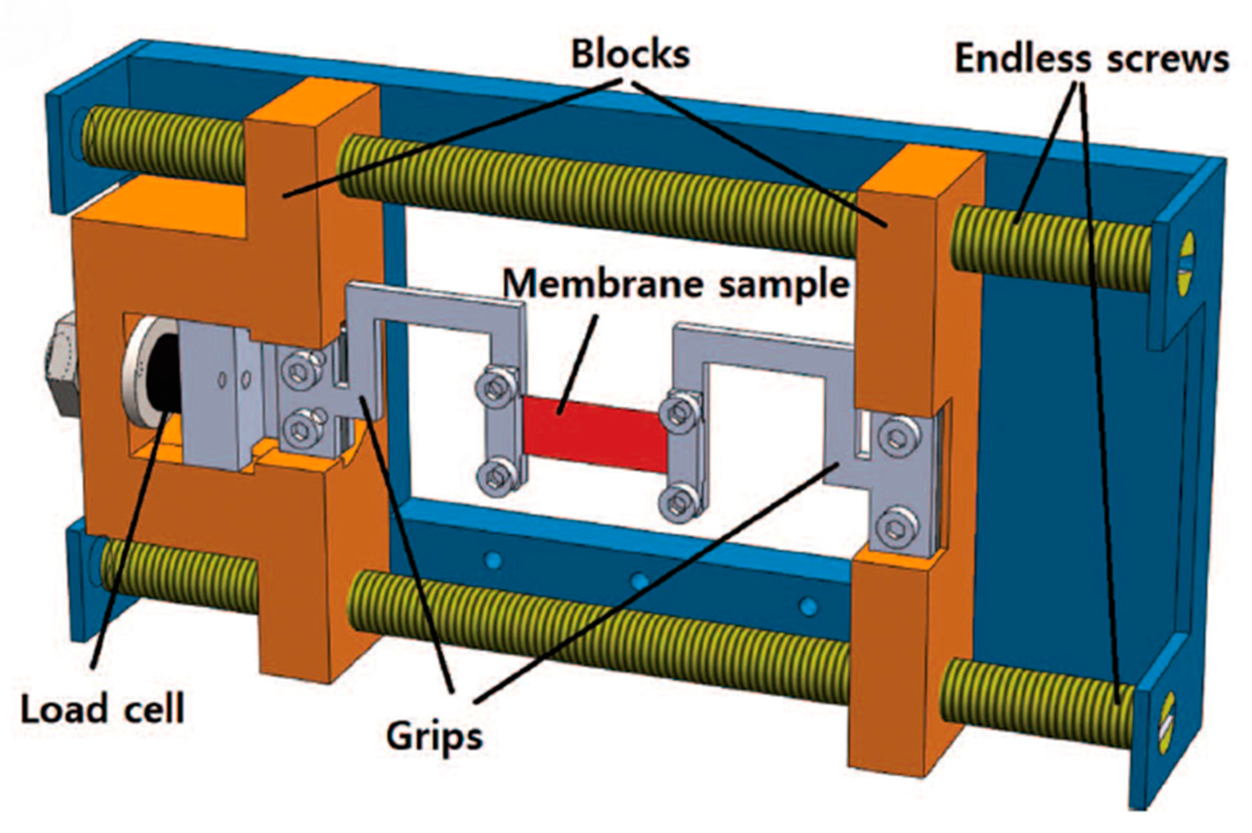
Relative humidity control of membranes in fuel cells during SANS experimentation was reported. However, in order to gain insights into the performance of nafion membranes in fuel cells under normal operating conditions, a total immersion of the membranes in H2O/D2O, whilst the material was uniaxially deformed, was required. To avoid a complicated experimental set-up with multiple mechanical feedthroughs to the liquid reservoir, U-shaped load beams were used, allowing for the material of interest to be immersed in a temperature-controlled D2O reservoir [129]. See Figure 14.
3.11. Devices
Given the high penetration power of neutrons in combination with the possibility of highlighting different parts of a material or the distribution of water via the use of deuteration, one would guess that neutron scattering techniques would be a prime tool for the investigation of devices, like batteries, liquid/gas separating membranes under operando conditions, and soot formation by combustion of hydrocarbons in engines, to name a few. However, this kind of research does not currently have a widespread following. This might be due to a combination of the relatively low neutron fluxes available currently and a certain bias that neutron facilities are somewhat inaccessible to the general user. However, there are examples of the above-mentioned systems studied with neutrons. There also is somewhat of a bias against more engineering-oriented or applied applications that can be found in many beam line access panels. Most of the experiments using simulated devices can be found in energy storage and generation-related materials.
The device that was studied using neutron scattering and imaging techniques was a polymer-electrolyte fuel cell [130]. The interest here is the distribution and transport in the fuel cell and the two techniques, SANS and imaging, were combined in a single experiment where the neutron imaging system could be moved out of the beam during the acquisition of the SANS data. This is particularly interesting since the two techniques might not give the same type of information, but the accessible length scales are complementary, and relevant information over length scales from nano- to milli-meters can be obtained in a single experiment. It can be pointed out that this cell was a realistic model where metal components were illuminated by the neutron beam. Using SAXS experiments, where the electron densities of H2O and polymers are not sufficiently different to generate a contrast that can make aluminum, H2O and polymer all visible, no information about the distribution of water can be obtained. Importantly, the above-mentioned experiment emphasizes the ability to study materials under operando conditions. It is noteworthy that data acquisition time for SANS was 80 s/frame and for imaging 210 s/frame. In a later publication, infrared spectroscopic data were also used to evaluate the physical and chemical events inside the fuel cell [131,132].
Another energy-related application is the study of Li-ion batteries. The concentration of Li-salt and the type of electrode material used has consequences for the operation of the battery and its longevity. By using a half-cell, i.e., a single electrode surrounded by an electrolyte solution, it enables one to follow the events at the solid–electrolyte interface. In the case where one uses an ordered mesoporous carbon electrode, mesoporous events take place in the range where small angle scattering (SAXS/SANS) can yield information. Here again there is a lack of electron density contrast between the Carbon and the Li compared to the pores, which indicates that neutron scattering is better suited to gain insights into the effects of the use of different Li-salts and concentrations during the duty cycle for in-operando cycling/discharging [133]. Similar experiments, but with greater emphasis on the analysis of the scattering results and the role of the scattering contrast in the analysis of the data, were also published [134].
Supercapacitors, hybrid devices with characteristics between a capacitor and a battery, rely on a high internal surface for redox reactions to take place. The most promising materials are porous nitrides of vanadium and molybdenum, which strictly speaking, do not classify as soft matter, but in the context of this review are relevant as they are materials known to have some of the highest storage capacities [135]. By performing the required electrochemistry on-line, it was shown that the smallest pores in these materials allow for the increased adsorption of OH− ions. These experiments would also benefit from on-line X-ray spectroscopy experiments in order to obtain insights about what is driving the charge storage mechanism.
4. In-Situ Technique Combinations
In most research, neutron scattering is only one of the tools in the experimental toolbox required to obtain the knowledge of the material properties that one is investigating. In certain circumstances, it is beneficial if one can combine neutron scattering experiments with complementary on-line auxiliary techniques, as has become a fairly routine approach at synchrotron radiation facilities [136,137]. The advantages of being able to simultaneously collect complementary data sets at the same time, using the same sample, outweigh any disadvantages due to the increased experimental complications regarding synchronization of the data sets and access of different probe beams, which cannot always use the same window material. Conditions in which one can consider the use of a multimodal approach are when dealing with spatially inhomogeneous materials or when the time resolution in an experiment is too fast to be able to stitch the data sets in a reliable way that does not create uncertainties in the time-correlation between the different experiments. In both cases, one should try to interrogate the same sample volume with the different probes.
The use of on-line Differential Scanning Calorimetry (DSC) with X-ray measurements is a good example where the data quality of the DSC signals suffers somewhat but where the synergy of having two simultaneous data sets outweighs the loss of quality [22]. When the strong peaks in a DSC curve can be correlated to the X-ray frame number, one can correlate the real thermodynamic temperature of the sample with the appropriate X-ray structure. Hence, the correlation can be made with an accurate DSC curve obtained with a well calibrated off-line instrument. The design problems for constructing a similar set-up for neutrons are somewhat larger than those for X-rays, since the beam access window needs to be considerably larger. However, this engineering issue can be overcome as was shown in phase separation experiments on partially deuterated alkanes, CnH2n+2:CmH(D)2n+2. A commercial instrument with an operation range from −150 °C to +500 °C was successfully modified to enable neutron beam access. A slight drawback is that it was not feasible to use commercially available DSC pans but instead special pans had to be custom designed and machined [138]. The time resolution that could be achieved in this experiment was 2 min/frame.
The combination of Raman scattering with different neutron scattering techniques has been reported over the years [139,140], including for the protein lysozyme, but these experiments were all carried out at cryogenic temperatures, which is of limited use in soft matter research. However, it was only recently that the cold deformation, i.e., ambient temperature, of low-density polyethylene was followed by both SANS and polarized Raman spectroscopy [141], where the experiments were performed using the same tensile stage but not simultaneously. By combining the different data sets, one can investigate the interplay between chain stretching (SANS) and the transition from amorphous to all trans conformers (Raman). Although these experiments were done separately, with the present generation of portable Raman spectrometers, there is no reason why this could not be carried out simultaneously if the required time-resolution would make this beneficial [142].
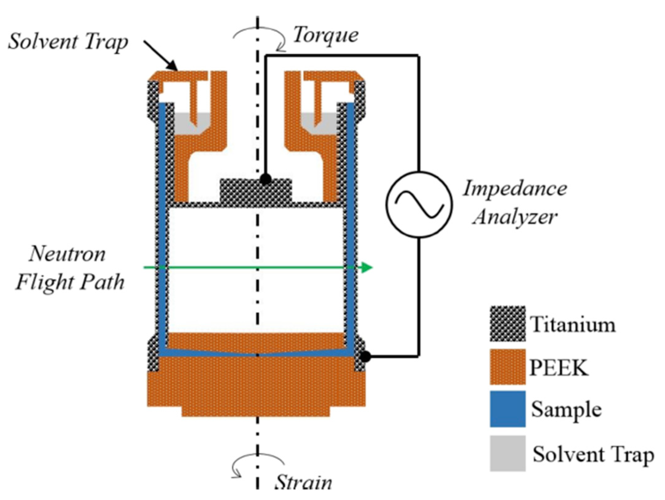
Dielectric spectroscopy can provide information at the molecular level about a variety of soft matter processes. For instance, chemical changes, segmental relaxation in polymers, and phase transitions are among the physical phenomena where dielectric spectroscopy can render complementary information to X-ray [143] and neutron scattering [144]. Such experiments were carried out in combination with a strain controlled Couette shear cell on a SANS instrument [145]. The Couette cylinders were constructed of titanium with thin mylar windows, allowing access to the neutron beam as well as for impedance measurements between the inner and outer cylinders (See Figure 15 for a schematic lay-out of the experiments). Crucial for this type of experiment is the synchronization of the different techniques, which can be readily achieved using ‘event-mode’ data collection [146]. Data were successfully obtained on the self-assembly of conjugated polymer melts and shear sensitive ionic liquids.
In order to investigate how high a molecular weight of polyethylene glycol dimethyl ether (PEGDME) can be incorporated in the crystalline region of syndiotactic polystyrene, one requires both the structural information provided by neutron scattering and vibrational spectroscopy. This was done by combining Fourier Transform Infrared Spectroscopy (FTIR) with SANS [147,148]. Here one encounters the issue that the optimum sample thickness required by both techniques is not the same. This can be partially overcome by using an FTIR sample cell and Attenuated Total Reflection, where the infrared beam makes multiple passes through the sample material [149]. However, a better solution is to place the IR and neutron beams co-parallel to ensure that one is studying the same part of the sample. This can be achieved by coupling the IR beam with an elaborate system of KBr crystals that act as mirrors for the IR beam but are relatively transparent to neutrons. As a compromise, the sample thickness was chosen to be 50 μm, just sufficiently thick for neutron scattering but thin enough for infra-red measurements. The combined data sets showed a distinct difference between the PEGDME conformation in the amorphous and crystalline regions of the polystyrene.
In experiments where the materials exhibit (partial) orientation and are transparent to visible light, birefringence is widely used to determine orientational effects. Neutron and X-ray scattering are also sensitive to orientational order, albeit on different length scales than birefringence. Scattering is mainly sensitive to the alignment of molecules, whilst birefringence is sensitive to bulk material properties [150]. Although such a capability seems to be very useful, it is somewhat surprising that this has not been applied more often [62,150,151].
Even though it is understood that having combined neutron and X-ray scattering data is highly beneficial in understanding a system, these techniques are not combined as often as they should [152,153,154]. In an ideal world, one would be able to use parallel beams of X-rays and neutrons, but an orthogonal combination is technically easier to realize. The achievable time-resolution, i.e., the length per time frame in which useable data can be collected, for the two experiments should be of the same order of magnitude. Until recently, such a combination was not feasible, but developments in X-ray generation and detector technology have allowed the first steps in allowing such a combined scattering instrument to be developed. Specifically, the two probe beams were placed at right angles to one another, making the system less useful for samples that are anisotropic, but it can serve as a proof of principle [155]. The complication that one faces with the development of such an instrument is that one invariably tries to mount an X-ray system on an existing neutron scattering beamline. Hence, one encounters space and shielding issues that make things more complicated (but not impossible).
The combination of neutron scattering with light scattering or birefringence should be more straightforward to achieve and would extend the length scales over which information can be obtained. However, this also seems to be an area waiting to be developed since few publications can be found. A combination of SANS with diffuse wave spectroscopy (DWS) was reported. DWS can be used in high concentration colloid solutions and can be used to obtain information about the ensemble dynamics of particles, while SANS provides the structural information [156].
An example where an industrially relevant testing method, Rapid Visco Analysis (RVA), was combined with SANS experiments was used to determine the nature of the structural changes in starch during pasting. This combined mechanical and thermal treatment of materials plays an important role in process- and quality-control in the food industries. By combining the neutron scattering data with the viscosity, as measured with an adapted RVA system, the abrupt changes in viscosity could be correlated with the changing morphology of the starch and the starch derivative network [157].
5. New Directions
One of the disadvantages of neutron scattering experiments is the size of the neutron beam. Due to the abundance of available photons and the development of optical systems that take advantage of this abundance, X-ray beam sizes on the submicron scale are common. This is not the case with neutrons. A smaller neutron beam is achieved at the expense of available flux, and hence of the attainable time resolution. Although one cannot reasonably expect sub-micron neutron beams, it is still feasible to obtain spatially resolved data where the (inhomogeneous) sample is probed with a small beam of around 100 microns at different positions [48] or to use microfluidic devices [157].
The promise of a higher effective flux that will be made available by the new generation of spallation sources will obviously have an impact on how fast data can be collected. However, this increase in intensity will not bring many orders of magnitude of improvement. Therefore, if one wants to increase the potential time-resolution, one should also look at better ways to perform experiments and at innovative ways of processing the data, and where the emphasis is placed on which parameters one would like to measure instead of trying to obtain the best statistical quality data. Certainly, for non-isotropic scattering samples there are improvements possible in what kind of time-resolution can be achieved.
The combination of neutron scattering, or spectroscopy data and complementary techniques is quite common. Less common, however, are approaches to analyze the data simultaneously, for example, using multidimensional correlation spectroscopy [158,159]. This method has already been applied in X-ray scattering experiments to elucidate events at the early onset of crystallization, where the nascent signals of the new phases are very weak [160,161], and the impact of radiation damage on proteins and the assembly in nanomaterials [162], and can be considered reminiscent of the low statistical data quality one would encounter in fast time-resolved neutron experiments.
So far, we have not mentioned the use of computational modelling, but it is not unreasonable to assume that this can in the near future become a virtual on-line technique combination. Modeling can become an inroad for new approaches to neutron experiments, which are optimized to provide just sufficient data quality to resolve whether the modeling result can be confirmed or disproven. This would require a ‘pipeline’ that feeds from modeling to defining neutron experiments.
An interesting development is the advances in ray-tracing simulations for instrument designs, e.g., McStas [163] or McVine [164]. These programs have expanded to include sample scattering kernels and sample environments [165]. It is now possible to simulate accurate sample geometries, instrument resolution, and counting statistics [166,167] to determine if planned experiments are feasible.
The importance of thin polymer films and grazing incidence studies characterizing the structure and structure formation of materials has increased in recent years [168]. In the case of X-ray studies, complicated on-line experiments, like roll-to-roll printing of polymers, have been reported. Neutron-based grazing incidence studies undoubtedly can play a bigger role in elucidating more complex morphologies. Data collection times, however, are still on the order of hours for a single static measurement [169], although in favorable cases, this can be reduced to minutes and can be further improved when dealing with cyclic processes [18]. It should also be noted that these results were obtained on non-dedicated beamlines. With a specialized design, further improvements could be made.
With the higher flux that one can expect from the new neutron sources that are being developed, obviously the situation for ‘neutron hungry techniques’, like time-resolved, spatially-resolved, and grazing incidence scattering experiments will improve. However, one cannot expect the many decades in improvement, which was the signature of the successive generations of X-ray sources, to be achievable. Still, there is substantial scope for increasing the experimental sophistication in soft matter research using neutron scattering techniques.
6. Conclusions
This article gives an overview of diverse sample environments that can be used in neutron scattering techniques applied in the area of soft condensed matter. We have focused on the small angle neutron scattering technique. Even though we have covered many different areas of sample environment control, such a survey can never be all-encompassing. However, we hope to have demonstrated with this overview that neutron scattering is not only suitable for static structure determination but can be implemented in a greater number of studies than what is being used presently. We suggest that more emphasis should be placed in SANS applications on observing the time-evolution of selected structural parameters during the response of materials to external perturbances. In this way, neutron scattering has the potential to make impactful contributions to the understanding of complex systems, such as soft materials, even when a complete structural analysis is out of reach. Importantly, observing parametrized changes in materials often does not require the highest statistical data quality, and therefore improvements in time resolution can be found by carefully optimizing to the minimum counting statistics level that is necessary, rather than following traditional optimization rules that were developed for obtaining best data from static structures.
Author Contributions
W.B. carried out the literature investigation, all authors contributed equally to the manuscript writing. All authors have read and agreed to the published version of the manuscript.
Funding
This research received no external funding.
Acknowledgments
We would like to acknowledge input from our colleagues Ralf Schweins, Steve King, Paul Butler, Sai Venkatesh Pingali, Frank Bates, Bin Wu and Gary Lynn. A special acknowledgement is due for our sadly missed colleague Isabelle Grillo. T. Plivelic, J. Vermant and R. Winter have made original figures available. The anonymous reviewers have made some valuable suggestions and are acknowledged for their careful reading of the manuscript. W.B.’s contribution is based upon work supported by Oak Ridge National Laboratory, managed by UT-Battelle LLC, for the U.S. Department of Energy. A portion of this research used resources at the High Flux Isotope Reactor and the Spallation Neutron Source, DOE Office of Science User Facilities operated by the Oak Ridge National Laboratory. DOE will provide public access to these results of federally sponsored research in accordance with the DOE Public Access Plan http://energy.gov/downloads/doe-public-access-plan (accessed on 13 May 2021).
Conflicts of Interest
The authors declare no conflict of interest.
References
- Hemberg, O.; Otendal, M.; Hertz, H.M. Liquid-metal-jet anode X-ray tube. Opt. Eng. 2004, 43, 1682–1688. [Google Scholar] [CrossRef]
- Bauters, S.; Tack, P.; Rudloff-Grund, J.H.; Banerjee, D.; Longo, A.; Vekemans, B.; Bras, W.; Brenker, F.E.; van Silfhout, R.; Vincze, L. Polycapillary Optics Based Confocal Micro X-ray Fluorescence and X-ray Absorption Spectroscopy Setup at The European Synchrotron Radiation Facility Collaborative Research Group Dutch-Belgian Beamline. Anal. Chem. 2018, 90, 2389–2394. [Google Scholar] [CrossRef] [PubMed] [Green Version]
- Donath, T.; Brandstetter, S.; Cibik, L.; Commichau, S.; Hofer, P.; Krumrey, M.; Luthi, B.; Marggraf, S.; Muller, P.; Schneebeli, M.; et al. Characterization of the PILATUS photon-counting pixel detector for X-ray energies from 1.75 keV to 60 keV. In Proceedings of the 11th International Conference on Synchrotron Radiation Instrumentation (Sri 2012), Lyon, France, 9–13 July 2012; p. 425. [Google Scholar]
- Jinnai, H.; Hasegawa, H.; Hashimoto, T.; Han, C.C. Time-resolved small-angle neutron-scattering in intermediate-stage and late-stage spinodal decomposition of dpb hpi blends. Macromolecules 1991, 24, 282–289. [Google Scholar] [CrossRef]
- Connell, J.G.; Richards, R.W.; Rennie, A.R. Phase-separation kinetics in concentrated-solutions of linear diblock copolymers of polystyrene and polyisoprene from time-resolved small-angle neutron-scattering. Polymer 1991, 32, 2033–2042. [Google Scholar] [CrossRef]
- Egelhaaf, S.U.; Schurtenberger, P. Micelle-to-vesicle transition: A time-resolved structural study. Phys. Rev. Lett. 1999, 82, 2804–2807. [Google Scholar] [CrossRef]
- Hollamby, M.J.; Borisova, D.; Brown, P.; Eastoe, J.; Grillo, I.; Shchukin, D. Growth of Mesoporous Silica Nanoparticles Monitored by Time-Resolved Small-Angle Neutron Scattering. Langmuir 2012, 28, 4425–4433. [Google Scholar] [CrossRef]
- Niimura, N.; Minezaki, Y.; Ataka, M.; Katsura, T. Aggregation in supersaturated lysozyme solution studied by time-resolved small-angle neutron-scattering. J. Cryst. Growth 1995, 154, 136–144. [Google Scholar] [CrossRef]
- Mahabir, S.; Small, D.; Li, M.; Wan, W.K.; Kucerka, N.; Littrell, K.; Katsaras, J.; Nieh, M.P. Growth kinetics of lipid-based nanodiscs to unilamellar vesicles-A time-resolved small angle neutron scattering (SANS) study. Biochim. Biophys. Acta-Biomembr. 2013, 1828, 1025–1035. [Google Scholar] [CrossRef]
- Mason, T.G.; Lin, M.Y. Time-resolved small angle neutron scattering measurements of asphaltene nanoparticle aggregation kinetics in incompatible crude oil mixtures. J. Chem. Phys. 2003, 119, 565–571. [Google Scholar] [CrossRef] [Green Version]
- Lopez-Barron, C.R.; Wagner, N.J.; Porcar, L. Layering, melting, and recrystallization of a close-packed micellar crystal under steady and large-amplitude oscillatory shear flows. J. Rheol. 2015, 59, 793–820. [Google Scholar] [CrossRef]
- Lopez-Barron, C.R.; Gurnon, A.K.; Eberle, A.P.R.; Porcar, L.; Wagner, N.J. Microstructural evolution of a model, shear-banding micellar solution during shear startup and cessation. Phys. Rev. E 2014, 89, 11. [Google Scholar] [CrossRef]
- Wrede, O.; Reimann, Y.; Lulsdorf, S.; Emmrich, D.; Schneider, K.; Schmid, A.J.; Zauser, D.; Hannappel, Y.; Beyer, A.; Schweins, R.; et al. Volume phase transition kinetics of smart N-n-propylacrylamide microgels studied by time-resolved pressure jump small angle neutron scattering. Sci. Rep. 2018, 8, 13. [Google Scholar] [CrossRef]
- Terrill, N.J.; Fairclough, P.A.; Towns-Andrews, E.; Komanschek, B.U.; Young, R.J.; Ryan, A.J. Density fluctuations: The nucleation event in isotactic polypropylene crystallization. Polymer 1998, 39, 2381–2385. [Google Scholar] [CrossRef]
- Metze, M.; Barbe, S.; Reiche, A.; Kesting, A.; Schweins, R. A Neutron-Transparent Flow-Through Cell (NTFT-Cell) for the SANS investigation of microstructure evolution during industrial evaporative casting. J. Neutron Res. 2017, 19, 177–185. [Google Scholar] [CrossRef]
- Kipping, D.; Gahler, R.; Habicht, K. Small angle neutron scattering at very high time resolution: Principle and simulations of ‘TISANE’. Phys. Lett. A 2008, 372, 1541–1546. [Google Scholar] [CrossRef]
- Wiedenmann, A.; Keiderling, U.; Habicht, K.; Russina, M.; Gahler, R. Dynamics of field-induced ordering in magnetic colloids studied by new time-resolved small-angle neutron-scattering techniques. Phys. Rev. Lett. 2006, 97, 4. [Google Scholar] [CrossRef] [PubMed] [Green Version]
- Adlmann, F.A.; Gutfreund, P.; Ankner, J.F.; Browning, J.F.; Parizzi, A.; Vacaliuc, B.; Halbert, C.E.; Rich, J.P.; Dennison, A.J.C.; Wolff, M.R. Towards neutron scattering experiments with sub-millisecond time resolution. J. Appl. Crystallogr. 2015, 48, 220–226. [Google Scholar] [CrossRef]
- Levelut, A.M.; Guinier, A. X-ray Scattering by Point Defects. In Small Angle X-ray Scattering; Brumberger, H., Ed.; Gordon and Breach: New York, NY, USA, 1967. [Google Scholar]
- Olsson, A.; Hellsing, M.S.; Rennie, A.R. A holder to rotate sample cells to avoid sedimentation in small-angle neutron scattering and ultra small-angle neutron scattering experiments. Meas. Sci. Technol. 2013, 24, 6. [Google Scholar] [CrossRef]
- Leao, J.B.; Murphy, R.P.; Wagner, N.J.; Bleuel, M. Dynamic infrared sample controlled (DISCO) temperature for the tumbler cells for ultra small angle neutron scattering (USANS). J. Neutron Res. 2017, 19, 23–26. [Google Scholar] [CrossRef] [Green Version]
- Bras, W.; Derbyshire, G.E.; Devine, A.; Clark, S.M.; Cooke, J.; Komanschek, B.E.; Ryan, A.J. The Combination of Thermal-Analysis and Time-Resolved X-Ray Techniques—A Powerful Method for Materials Characterization. J. Appl. Crystallogr. 1995, 28, 26–32. [Google Scholar] [CrossRef]
- Russell, T.P.; Koberstein, J.T. Simultaneous Differential Scanning Calorimetry And Small-Angle X-Ray-Scattering. J. Polym. Sci. Part B-Polym. Phys. 1985, 23, 1109–1115. [Google Scholar] [CrossRef]
- Gilbert, B. Finite size effects on the real-space pair distribution function of nanoparticles. J. Appl. Crystallogr. 2008, 41, 554–562. [Google Scholar] [CrossRef]
- Oskolkova, M.Z.; Norrman, E.; Olsson, U. Study of the micelle-to-vesicle transition and smallest possible vesicle size by temperature-jumps. J. Colloid Interface Sci. 2013, 396, 173–177. [Google Scholar] [CrossRef] [PubMed]
- Adelsberger, J.; Grillo, I.; Kulkarni, A.; Sharp, M.; Bivigou-Koumba, A.M.; Laschewsky, A.; Müller-Buschbaum, P.; Papadakis, C.M. Kinetics of aggregation in micellar solutions of thermoresponsive triblock copolymers—Influence of concentration, start and target temperatures. Soft Matter 2013, 9, 1685–1699. [Google Scholar] [CrossRef]
- Pullen, S.A.; Gilbert, E.P.; Olsen, S.R.; Lang, E.A.; Doolan, K.R. An in situ rapid heat-quench cell for small-angle neutron scattering. Meas. Sci. Technol. 2008, 19, 8. [Google Scholar] [CrossRef]
- Adelsberger, J.; Metwalli, E.; Diethert, A.; Grillo, I.; Bivigou-Koumba, A.M.; Laschewsky, A.; Muller-Buschbaum, P.; Papadakis, C.M. Kinetics of Collapse Transition and Cluster Formation in a Thermoresponsive Micellar Solution of P(S-b-NIPAM-b-S) Induced by a Temperature Jump. Macromol. Rapid Commun. 2012, 33, 254–259. [Google Scholar] [CrossRef]
- Dewers, T.A.; Heath, J.E.; Bryan, C.R.; Mang, J.T.; Hjelm, R.P.; Ding, M.; Taylor, M. Oedometric Small-Angle Neutron Scattering: In Situ Observation of Nanopore Structure During Bentonite Consolidation and Swelling in Dry and Hydrous CO2 Environments. Environ. Sci. Technol. 2018, 52, 3758–3768. [Google Scholar] [CrossRef]
- Stefanopoulos, K.L.; Youngs, T.G.A.; Sakurovs, R.; Ruppert, L.F.; Bahadur, J.; Melnichenko, Y.B. Neutron Scattering Measurements of Carbon Dioxide Adsorption in Pores within the Marcellus Shale: Implications for Sequestration. Environ. Sci. Technol. 2017, 51, 6515–6521. [Google Scholar] [CrossRef] [Green Version]
- Hjelm, R.P.; Taylor, M.A.; Frash, L.P.; Hawley, M.E.; Ding, M.; Xu, H.W.; Barker, J.; Olds, D.; Heath, J.; Dewers, T. Flow-through compression cell for small-angle and ultra-small-angle neutron scattering measurements. Rev. Sci. Instrum. 2018, 89, 9. [Google Scholar] [CrossRef]
- Gabke, A.; Kraineva, J.; Kohling, R.; Winter, R. Using pressure in combination with x-ray and neutron scattering techniques for studying the structure, stability and phase behaviour of soft condensed matter and biomolecular systems. J. Phys. Condens. Matter 2005, 17, S3077–S3092. [Google Scholar] [CrossRef]
- Teixeira, S.C.M.; Leao, J.B.; Gagnon, C.; McHugh, M.A. High pressure cell for Bio-SANS studies under sub-zero temperatures or heat denaturing conditions. J. Neutron Res. 2018, 20, 11–21. [Google Scholar] [CrossRef] [Green Version]
- Pingali, S.V.; Smith, M.D.; Liu, S.-H.; Rawal, T.B.; Pu, Y.; Shah, R.; Evans, B.R.; Urban, V.S.; Davison, B.H.; Cai, C.M.; et al. Deconstruction of biomass enabled by local demixing of cosolvents at cellulose and lignin surfaces. Proc. Natl. Acad. Sci. USA 2020, 117, 16776–16781. [Google Scholar] [CrossRef]
- Niebuur, B.-J.; Chiappisi, L.; Zhang, X.; Jung, F.; Schulte, A.; Papadakis, C.M. Formation and Growth of Mesoglobules in Aqueous Poly(N-isopropylacrylamide) Solutions Revealed with Kinetic Small-Angle Neutron Scattering and Fast Pressure Jumps. ACS Macro Lett. 2018, 7, 1155–1160. [Google Scholar] [CrossRef]
- Ryan, A.J.; Bras, W.; Hermida-Merino, D.; Cavallo, D. The interaction between fundamental and industrial research and experimental developments in the field of polymer crystallization. J. Non-Cryst. Solids 2016, 451, 168–178. [Google Scholar] [CrossRef]
- Roozemond, P.C.; Ma, Z.; Cui, K.; Li, L.; Peters, G.W.M. Multimorphological Crystallization of Shish-Kebab Structures in Isotactic Polypropylene: Quantitative Modeling of Parent-Daughter Crystallization Kinetics. Macromolecules 2014, 47, 5152–5162. [Google Scholar] [CrossRef]
- Lindner, P.; Oberthur, R.C. Shear induced deformation of polystyrene coils in dilute-solution from small-angle neutron-scattering. 1. Shear gradient apparatus and 1st experimental results. Colloid Polym. Sci. 1985, 263, 443–453. [Google Scholar] [CrossRef]
- Eberle, A.P.R.; Porcar, L. Flow-SANS and Rheo-SANS applied to soft matter. Curr. Opin. Colloid Interface Sci. 2012, 17, 33–43. [Google Scholar] [CrossRef]
- Huang, G.R.; Wang, Y.Y.; Wu, B.; Wang, Z.; Do, C.; Smith, G.S.; Bras, W.; Porcar, L.; Falus, P.; Chen, W.R. Reconstruction of three-dimensional anisotropic structure from small-angle scattering experiments. Phys. Rev. E 2017, 96, 022612. [Google Scholar] [CrossRef] [PubMed] [Green Version]
- Porcar, L.; Pozzo, D.; Langenbucher, G.; Moyer, J.; Butler, P.D. Rheo-small-angle neutron scattering at the National Institute of Standards and Technology Center for Neutron Research. Rev. Sci. Instrum. 2011, 82, 7. [Google Scholar] [CrossRef] [Green Version]
- Velichko, E.; Tian, B.; Nikolaeva, T.; Koning, J.; van Duynhoven, J.; Bouwman, W.G. A versatile shear cell for investigation of structure of food materials under shear. Colloids Surf. A-Physicochem. Eng. Asp. 2019, 566, 21–28. [Google Scholar] [CrossRef]
- Vermant, J.; Solomon, M.J. Flow-induced structure in colloidal suspensions. J. Phys. Condens. Matter 2005, 17, R187–R216. [Google Scholar] [CrossRef]
- Helgeson, M.E.; Vasquez, P.A.; Kaler, E.W.; Wagner, N.J. Rheology and spatially resolved structure of cetyltrimethylammonium bromide wormlike micelles through the shear banding transition. J. Rheol. 2009, 53, 727–756. [Google Scholar] [CrossRef]
- Nusser, K.; Neueder, S.; Schneider, G.J.; Meyer, M.; Pyckhout-Hintzen, W.; Willner, L.; Radulescu, A.; Richter, D. Conformations of silica− poly (ethylene− propylene) nanocomposites. Macromolecules 2010, 43, 9837–9847. [Google Scholar] [CrossRef]
- Gasnier, A.; Royal, G.; Terech, P. Metallo-Supramolecular Gels Based on a Multitopic Cyclam Bis-Terpyridine Platform. Langmuir 2009, 25, 8751–8762. [Google Scholar] [CrossRef] [PubMed]
- Wang, Z.; Iwashita, T.; Porcar, L.; Wang, Y.Y.; Liu, Y.; Sanchez-Diaz, L.E.; Wu, B.; Huang, G.R.; Egami, T.; Chen, W.R. Local elasticity in nonlinear rheology of interacting colloidal glasses revealed by neutron scattering and rheometry. Phys. Chem. Chem. Phys. 2019, 21, 38–45. [Google Scholar] [CrossRef] [Green Version]
- Liberatore, M.W.; Nettesheim, F.; Wagner, N.J.; Porcar, L. Spatially resolved small-angle neutron scattering in the 1-2 plane: A study of shear-induced phase-separating wormlike micelles. Phys. Rev. E 2006, 73, 020504. [Google Scholar] [CrossRef] [Green Version]
- Kim, J.; Helgeson, M.E. Shear-induced clustering of Brownian colloids in associative polymer networks at moderate Peclet number. Phys. Rev. Fluids 2016, 1, 19. [Google Scholar] [CrossRef]
- Sharma, J.; King, S.M.; Bohlin, L.; Clarke, N. Apparatus for simultaneous rheology and small-angle neutron scattering from high-viscosity polymer melts and blends. Nucl. Instrum. Methods Phys. Res. Sect. A-Accel. Spectrometers Detect. Assoc. Equip. 2010, 620, 437–444. [Google Scholar] [CrossRef] [Green Version]
- Rexeisen, E.L.; Fan, W.; Pangburn, T.O.; Taribagil, R.R.; Bates, F.S.; Lodge, T.P.; Tsapatsis, M.; Kokkoli, E. Self-Assembly of Fibronectin Mimetic Peptide-Amphiphile Nanofibers. Langmuir 2010, 26, 1953–1959. [Google Scholar] [CrossRef] [PubMed] [Green Version]
- Sasa, L.A.; Yearley, E.J.; Welch, C.F.; Taylor, M.A.; Gilbertson, R.D.; Hammeter, C.; Majewski, J.; Hjelm, R.P. The Los Alamos Neutron Science Center neutron rheometer in the cone and plate geometry to examine tethered polymers/polymer melt interfaces via neutron reflectivity. Rev. Sci. Instrum. 2010, 81, 6. [Google Scholar] [CrossRef]
- Corvis, Y.; Barre, L.; Jestin, J.; Gummel, J.; Cousin, F. Asphaltene adsorption mechanism under shear flow probed by in situ neutron reflectivity measurements. Eur. Phys. J. Spec. Top. 2012, 213, 295–302. [Google Scholar] [CrossRef]
- Hamley, I.W.; Burholt, S.; Hutchinson, J.; Castelletto, V.; da Silva, E.R.; Alves, W.; Gutfreund, P.; Porcar, L.; Dattani, R.; Hermida-Merino, D.; et al. Shear Alignment of Bola-Amphiphilic Arginine-Coated Peptide Nanotubes. Biomacromolecules 2017, 18, 141–149. [Google Scholar] [CrossRef] [PubMed] [Green Version]
- Muller, R.; Picot, C.; Zang, Y.H.; Froelich, D. Polymer-chain conformation in the melt during steady elongational flow as measured by sans—Temporary network model. Macromolecules 1990, 23, 2577–2582. [Google Scholar] [CrossRef]
- Ruocco, N.; Auhl, D.; Bailly, C.; Lindner, P.; Pyckhout-Hintzen, W.; Wischnewski, A.; Leal, L.G.; Hadjichristidis, N.; Richter, D. Branch Point Withdrawal in Elongational Startup Flow by Time-Resolved Small Angle Neutron Scattering. Macromolecules 2016, 49, 4330–4339. [Google Scholar] [CrossRef]
- Macosko, C.W.; Mewis, J. Suspension Rheology; Wiley-VCH: New York, NY, USA, 1994. [Google Scholar]
- Camargo, R.; Macosko, C.; Tirrell, M.; Wellinghoff, S. Phase separation studies in RIM polyurethanes catalyst and hard segment crystallinity effects. Polymer 1985, 26, 1145–1154. [Google Scholar] [CrossRef]
- Inkson, N.J.; McLeish, T.C.B.; Harlen, O.G.; Groves, D.J. Predicting low density polyethylene melt rheology in elongational and shear flows with "pom-pom" constitutive equations. J. Rheol. 1999, 43, 873–896. [Google Scholar] [CrossRef]
- Bent, J.F.; Richards, R.W.; Gough, T.D. Recirculation cell for the small-angle neutron scattering investigation of polymer melts in flow. Rev. Sci. Instrum. 2003, 74, 4052–4057. [Google Scholar] [CrossRef] [Green Version]
- Bent, J.; Hutchings, L.R.; Richards, R.W.; Gough, T.; Spares, R.; Coates, P.D.; Grillo, I.; Harlen, O.G.; Read, D.J.; Graham, R.S.; et al. Neutron-mapping polymer flow: Scattering, flow visualization, and molecular theory. Science 2003, 301, 1691–1695. [Google Scholar] [CrossRef] [Green Version]
- Graham, R.S.; Bent, J.; Hutchings, L.R.; Richards, R.W.; Groves, D.J.; Embery, J.; Nicholson, T.M.; McLeish, T.C.B.; Likhtman, A.E.; Harlen, O.G.; et al. Measuring and predicting the dynamics of linear monodisperse entangled polymers in rapid flow through an abrupt contraction. A small angle neutron scattering study. Macromolecules 2006, 39, 2700–2709. [Google Scholar]
- McLeish, T.C.B.; Clarke, N.; de Luca, E.; Hutchings, L.R.; Graham, R.S.; Gough, T.; Grillo, I.; Fernyhough, C.M.; Chambon, P. Neutron flow-mapping: Multiscale modelling opens a new experimental window. Soft Matter 2009, 5, 4426–4432. [Google Scholar] [CrossRef]
- Lee, J.S.; Dylla-Spears, R.; Teclemariam, N.P.; Muller, S.J. Microfluidic four-roll mill for all flow types. Appl. Phys. Lett. 2007, 90, 3. [Google Scholar] [CrossRef]
- Taylor, G.I. The formation of emulsions in definable fields of flow. Proc. R. Soc. A 1934, 146, 501–523. [Google Scholar]
- Corona, P.T.; Ruocco, N.; Weigandt, K.M.; Leal, L.G.; Helgeson, M.E. Probing flow-induced nanostructure of complex fluids in arbitrary 2D flows using a fluidic four-roll mill (FFoRM). Sci. Rep. 2018, 8, 18. [Google Scholar] [CrossRef] [Green Version]
- Straube, E.; Urban, V.; Pyckhouthintzen, W.; Richter, D. Sans investigations of topological constraints and microscopic deformations in rubberelastic networks. Macromolecules 1994, 27, 7681–7688. [Google Scholar] [CrossRef]
- Butler, M.F.; Donald, A.M.; Bras, W.; Mant, G.R.; Derbyshire, G.E.; Ryan, A.J. A real-time simultaneous small-angle and wide-angle X-ray-scattering study of in-situ deformation of isotropic polyethylene. Macromolecules 1995, 28, 6383–6393. [Google Scholar] [CrossRef]
- Coutry, S.; Spells, S.J. Molecular changes on drawing isotopic blends of polyethylene and ethylene copolymers: 1. Static and time-resolved sans studies. Polymer 2003, 44, 1949–1956. [Google Scholar] [CrossRef]
- Wiyatno, W.; Fuller, G.G.; Pople, J.A.; Gast, A.P.; Chen, Z.R.; Waymouth, R.M.; Myers, C.L. Component stress-strain behavior and small-angle neutron scattering investigation of stereoblock elastomeric polypropylene. Macromolecules 2003, 36, 1178–1187. [Google Scholar] [CrossRef]
- Engqvist, J.; Hall, S.A.; Wallin, M.; Ristinmaa, M.; Plivelic, T.S. Multi-scale Measurement of (Amorphous) Polymer Deformation: Simultaneous X-ray Scattering, Digital Image Correlation and In-situ Loading. Exp. Mech. 2014, 54, 1373–1383. [Google Scholar] [CrossRef]
- Wagner, J.A.; Patil, S.P.; Greving, I.; Lammel, M.; Gkagkas, K.; Seydel, T.; Muller, M.; Markert, B.; Grater, F. Stress-induced long-range ordering in spider silk. Sci. Rep. 2017, 7, 6. [Google Scholar] [CrossRef] [Green Version]
- Stribeck, N.; Nochel, U.; Funari, S.S.; Schubert, T. Tensile tests of polypropylene monitored by SAXS. Comparing the stretch-hold technique to the dynamic technique. J. Polym. Sci. Part B-Polym. Phys. 2008, 46, 721–726. [Google Scholar] [CrossRef] [Green Version]
- Dokter, W.H.; Vangarderen, H.F.; Beelen, T.P.M.; Vansanten, R.A.; Bras, W. Homogeneous Versus Heterogeneous Zeolite Nucleation. Angew. Chem. -Int. Ed. Engl. 1995, 34, 73–75. [Google Scholar] [CrossRef]
- Grillo, I. Applications of stopped-flow in SAXS and SANS. Curr. Opin. Colloid Interface Sci. 2009, 14, 402–408. [Google Scholar] [CrossRef]
- Virtanen, O.L.J.; Kather, M.; Meyer-Kirschner, J.; Melle, A.; Radulescu, A.; Viell, J.; Mitsos, A.; Pich, A.; Richtering, W. Direct Monitoring of Microgel Formation during Precipitation Polymerization of N-Isopropylacrylamide Using in Situ SANS. ACS Omega 2019, 4, 3690–3699. [Google Scholar] [CrossRef] [Green Version]
- Uhríková, D.; Teixeira, J.; Hubčík, L.; Búcsi, A.; Kondela, T.; Murugova, T.; Ivankov, O.I. Lipid based drug delivery systems: Kinetics by SANS. J. Phys. Conf. Ser. 2017, 848, 012007. [Google Scholar] [CrossRef] [Green Version]
- Hayward, D.W.; Chiappisi, L.; Prevost, S.; Schweins, R.; Gradzielski, M. A Small-Angle Neutron Scattering Environment for In-Situ Observation of Chemical Processes. Sci. Rep. 2018, 8, 2–11. [Google Scholar] [CrossRef] [PubMed]
- Melnichenko, Y.B.; He, L.L.; Sakurovs, R.; Kholodenko, A.L.; Blach, T.; Mastalerz, M.; Radlinski, A.P.; Cheng, G.; Mildner, D.F.R. Accessibility of pores in coal to methane and carbon dioxide. Fuel 2012, 91, 200–208. [Google Scholar] [CrossRef]
- Woods, H.M.; Silva, M.; Nouvel, C.; Shakesheff, K.M.; Howdle, S.M. Materials processing in supercritical carbon dioxide: Surfactants, polymers and biomaterials. J. Mater. Chem. 2004, 14, 1663–1678. [Google Scholar] [CrossRef]
- Melnichenko, Y.B.; Kiran, E.; Wignall, G.D.; Heath, K.D.; Salaniwal, S.; Cochran, H.D. Stamm, Pressure- and temperature-induced transitions in solutions of poly(dimethylsiloxane) in supercritical carbon dioxide. M. Macromolecules 1999, 32, 5344–5347. [Google Scholar] [CrossRef]
- Muller, A.; Putz, Y.; Oberhoffer, R.; Becker, N.; Strey, R.; Wiedenmann, A.; Sottmann, T. Kinetics of pressure induced structural changes in super- or near-critical CO2-microemulsions. Phys. Chem. Chem. Phys. 2014, 16, 18092–18097. [Google Scholar] [CrossRef] [PubMed]
- Putz, Y.; Grassberger, L.; Lindner, P.; Schweins, R.; Strey, R.; Sottmann, T. Unexpected efficiency boosting in CO2-microemulsions: A cyclohexane depletion zone near the fluorinated surfactants evidenced by a systematic SANS contrast variation study. Phys. Chem. Chem. Phys. 2015, 17, 6122–6134. [Google Scholar] [CrossRef] [PubMed]
- Hermida-Merino, D.; Portale, G.; Fields, P.; Wilson, R.; Bassett, S.P.; Jennings, J.; Dellar, M.; Gommes, C.; Howdle, S.M.; Vrolijk, B.C. A high pressure cell for supercritical CO2 on-line chemical reactions studied with x-ray techniques. Rev. Sci. Instrum. 2014, 85, 093905. [Google Scholar] [CrossRef] [PubMed]
- Jordan, A.; Jacques, M.; Merrick, C.; Devos, J.; Forsyth, V.T.; Porcar, L.; Martel, A. SEC-SANS: Size exclusion chromatography combined in situ with small-angle neutron scattering. J. Appl. Crystallogr. 2016, 49, 2015–2020. [Google Scholar] [CrossRef] [Green Version]
- Koch, M.H.J.; Sayers, Z.; Sicre, P.; Svergun, D. A synchrotron-radiation electric-field X-ray solution scattering study of dna at very-low ionic-strength. Macromolecules 1995, 28, 4904–4907. [Google Scholar] [CrossRef]
- Xu, T.; Zhu, Y.; Gido, S.P.; Russell, T.P. Electric Field Alignment of Symmetric Diblock Copolymer Thin Films. Macromolecules 2004, 37, 2625–2629. [Google Scholar] [CrossRef]
- Pester, C.; Ruppel, M.; Schoberth, H.G.; Schmidt, K.; Liedel, C.; van Rijn, P.; Schindler, K.A.; Hiltl, S.; Czubak, T.; Mays, J.; et al. Piezoelectric Properties of Non-Polar Block Copolymers. Adv. Mater. 2011, 23, 4047–4052. [Google Scholar] [CrossRef]
- Boker, A.; Ruppel, M.; Lindner, P.; Urban, V.S.; Schmidt, K.; Schoberth, H. SANS Study on the Phase Behaviour of Block Copolymers in the Presence of an External Electric Field; Institut Laue-Langevin (ILL) ILL User Club: Grenoble, France, 2009. [Google Scholar]
- McMullan, J.M.; Wagner, N.J. Directed self-assembly of colloidal crystals by dielectrophoretic ordering observed with small angle neutron scattering (SANS). Soft Matter 2010, 6, 5443–5450. [Google Scholar] [CrossRef]
- Li, D.S.; Lee, Y.-T.; Xi, Y.; Pelivanov, I.; O’Donnell, M.; Pozzo, L.D. A small-angle scattering environment for in situ ultrasound studies. Soft Matter 2018, 14, 5283–5293. [Google Scholar] [CrossRef] [PubMed]
- Hamley, I.W.; Castelletto, V.; Lu, Z.B.; Imrie, C.T.; Itoh, T.; Al-Hussein, M. Interplay between Smectic Ordering and Microphase Separation in a Series of Side-Group Liquid-Crystal Block Copolymers. Macromolecules 2004, 37, 4798–4807. [Google Scholar] [CrossRef]
- Hocine, S.; Brulet, A.; Jia, L.; Yang, J.; Di Cicco, A.; Bouteiller, L.; Li, M.H. Structural changes in liquid crystal polymer vesicles induced by temperature variation and magnetic fields. Soft Matter 2011, 7, 2613–2623. [Google Scholar] [CrossRef]
- Nieh, M.P.; Glinka, C.J.; Krueger, S.; Prosser, R.S.; Katsaras, J. SANS study of the structural phases of magnetically alignable lanthanide-doped phospholipid mixtures. Langmuir 2001, 17, 2629–2638. [Google Scholar] [CrossRef]
- van der Beek, D.; Petukhov, A.V.; Davidson, P.; Ferre, J.; Jamet, J.P.; Wensink, H.H.; Vroege, G.J.; Bras, W.; Lekkerkerker, H.N.W. Magnetic-field-induced orientational order in the isotropic phase of hard colloidal platelets. Phys. Rev. E 2006, 73, 10. [Google Scholar] [CrossRef] [PubMed] [Green Version]
- McCulloch, B.; Portale, G.; Bras, W.; Segalman, R.A. Increased Order-Disorder Transition Temperature for a Rod-Coil Block Copolymer in the Presence of a Magnetic Field. Macromolecules 2011, 44, 7503–7507. [Google Scholar] [CrossRef]
- Bras, W.; Emsley, J.W.; Levine, Y.K.; Luckhurst, G.R.; Seddon, J.M.; Timimi, B.A. Field-induced alignment of a smectic-A phase: A time-resolved X-ray diffraction investigation. J. Chem. Phys. 2004, 121, 4397–4413. [Google Scholar] [CrossRef] [PubMed]
- Brimicombe, P.D.; Siemianowski, S.D.; Jaradat, S.; Levine, Y.K.; Thompson, P.; Bras, W.; Gleeson, H.F. Time-resolved x-ray studies of the dynamics of smectic-A layer realignment by magnetic fields. Phys. Rev. E 2009, 79, 031706. [Google Scholar] [CrossRef]
- Torbet, J. Solution behavior of DNA studied with magnetically induced birefringence. Methods Enzym. 1992, 211, 518–532. [Google Scholar]
- Torbet, J.; Dickens, M.J. Orientation of Skeletal-Muscle Actin in Strong Magnetic-Fields. FEBS Lett. 1984, 173, 403–406. [Google Scholar] [CrossRef] [Green Version]
- Liebi, M.; van Rhee, P.G.; Christianen, P.C.M.; Kohlbrecher, J.; Fischer, P.; Walde, P.; Windhab, E.J. Alignment of Bicelles Studied with High-Field Magnetic Birefringence and Small-Angle Neutron Scattering Measurements. Langmuir 2013, 29, 3467–3473. [Google Scholar] [CrossRef] [PubMed]
- Holmes, A.T.; Walsh, G.R.; Blackburn, E.; Forgan, E.M.; Savey-Bennett, M. A 17 T horizontal field cryomagnet with rapid sample change designed for beamline use. Rev. Sci. Instrum. 2012, 83, 023904. [Google Scholar] [CrossRef] [PubMed] [Green Version]
- Linden, P.J.E.M.v.d.; Mathon, O.; Strohm, C.; Sikora, M. Miniature pulsed magnet system for synchrotron x-ray measurements. Rev. Sci. Instrum. 2008, 79, 075104. [Google Scholar] [CrossRef] [PubMed]
- Duc, F.; Fabrèges, X.; Roth, T.; Detlefs, C.; Frings, P.; Nardone, M.; Billette, J.; Lesourd, M.; Zhang, L.; Zitouni, A.; et al. A 31 T split-pair pulsed magnet for single crystal x-ray diffraction at low temperature. Rev. Sci. Instrum. 2014, 85, 053905. [Google Scholar] [CrossRef] [PubMed] [Green Version]
- Frings, P.; Vanacken, J.; Detlefs, C.; Duc, F.; Lorenzo, J.E.; Nardone, M.; Billette, J.; Zitouni, A.; Bras, W.; Rikken, G.L.J.A. Synchrotron x-ray powder diffraction studies in pulsed magnetic fields. Rev. Sci. Instrum. 2006, 77, 063903. [Google Scholar] [CrossRef]
- Duc, F.; Tonon, X.; Billette, J.; Rollet, B.; Knafo, W.; Bourdarot, F.; Béard, J.; Mantegazza, F.; Longuet, B.; Lorenzo, J.E.; et al. 40-Tesla pulsed-field cryomagnet for single crystal neutron diffraction. Rev. Sci. Instrum. 2018, 89, 053905. [Google Scholar] [CrossRef] [PubMed] [Green Version]
- Vesperinas, A.; Eastoe, J.; Wyatt, P.; Grillo, I.; Heenan, R.K.; Richards, J.M.; Bell, G.A. Photoinduced phase separation. J. Am. Chem. Soc. 2006, 128, 1468–1469. [Google Scholar] [CrossRef]
- Tabor, R.F.; Oakley, R.J.; Eastoe, J.; Faul, C.F.J.; Grillo, I.; Heenan, R.K. Reversible light-induced critical separation. Soft Matter 2009, 5, 78–80. [Google Scholar] [CrossRef]
- Oh, H.; Ketner, A.M.; Heymann, R.; Kesselman, E.; Danino, D.; Falvey, D.E.; Raghavan, S.R. A simple route to fluids with photo-switchable viscosities based on a reversible transition between vesicles and wormlike micelles. Soft Matter 2013, 9, 5025–5033. [Google Scholar] [CrossRef]
- Morgan, B.; Dadmun, M.D. The role of incident light intensity, wavelength, and exposure time in the modification of conjugated polymer structure in solution. Eur. Polym. J. 2017, 89, 272–280. [Google Scholar] [CrossRef] [Green Version]
- Morgan, B.; Dadmun, M.D. Illumination of Conjugated Polymer in Solution Alters Its Conformation and Thermodynamics. Macromolecules 2016, 49, 3490–3496. [Google Scholar] [CrossRef]
- Bras, W.; Stanley, H. Unexpected effects in non crystalline materials exposed to X-ray radiation. J. Non-Cryst. Solids 2016, 451, 153–160. [Google Scholar] [CrossRef]
- Liberton, M.; Page, L.E.; O’Dell, W.B.; O’Neill, H.; Mamontov, E.; Urban, V.S.; Pakrasi, H.B. Organization and Flexibility of Cyanobacterial Thylakoid Membranes Examined by Neutron Scattering. J. Biol. Chem. 2013, 288, 3632–3640. [Google Scholar] [CrossRef] [Green Version]
- Perera, S.; Chawla, U.; Shrestha, U.R.; Bhowmik, D.; Struts, A.V.; Qian, S.; Chu, X.Q.; Brown, M.F.J. Small-Angle Neutron Scattering Reveals Energy Landscape for Rhodopsin Photoactivation. Phys. Chem. Lett. 2018, 9, 7064–7071. [Google Scholar] [CrossRef] [PubMed]
- Whittaker, A.G.; Harrison, A.; Oakley, G.S.; Youngson, I.D.; Heenan, R.K.; King, S.M. Preliminary experiments on apparatus for in situ studies of microwave-driven reactions by small angle neutron scattering. Rev. Sci. Instrum. 2001, 72, 173–176. [Google Scholar] [CrossRef]
- Toolan, D.T.W.; Adlington, K.; Isakova, A.; Kalamiotis, A.; Mokarian-Tabari, P.; Dimitrakis, G.; Dodds, C.; Arnold, T.; Terrill, N.J.; Bras, W.; et al. Selective molecular annealing: In situ small angle X-ray scattering study of microwave-assisted annealing of block copolymers. Phys. Chem. Chem. Phys. 2017, 19, 20412–20419. [Google Scholar] [CrossRef] [Green Version]
- Greaves, G.N.; Wilding, M.C.; Fearn, S.; Langstaff, D.; Kargl, F.; Cox, S.; Van, Q.V.; Majerus, O.; Benmore, C.J.; Weber, R.; et al. Detection of first-order liquid/liquid phase transitions in yttrium oxide-aluminum oxide melts. Science 2008, 322, 566–570. [Google Scholar] [CrossRef] [Green Version]
- Hennet, L.; Cristiglio, V.; Kozaily, J.; Pozdnyakova, I.; Fischer, H.E.; Bytchkov, A.; Drewitt, J.W.E.; Leydier, M.; Thiaudiere, D.; Gruner, S.; et al. Aerodynamic levitation and laser heating: Applications at synchrotron and neutron sources. Eur. Phys. J. -Spec. Top. 2011, 196, 151–165. [Google Scholar] [CrossRef]
- Weber, J.K.R.; Rey, C.A.; Neuefeind, J.; Benmore, C.J. Acoustic levitator for structure measurements on low temperature liquid droplets. Rev. Sci. Instrum. 2009, 80, 083904. [Google Scholar] [CrossRef]
- Cristiglio, V.; Grillo, I.; Fomina, M.; Wien, F.; Shalaev, E.; Novikov, A.; Brassamin, S.; Refregiers, M.; Perez, J.; Hennet, L. Combination of acoustic levitation with small angle scattering techniques and synchrotron radiation circular dichroism. Application to the study of protein solutions. Biochim. Biophys. Acta-Gen. Subj. 2017, 1861, 3693–3699. [Google Scholar] [CrossRef] [PubMed]
- Krauss, S.W.; Schweins, R.; Magerl, A.; Zobel, M. Free-film small-angle neutron scattering: A novel container-free in situ sample environment with minimized H/D exchange. J. Appl. Crystallogr. 2019, 52, 284–288. [Google Scholar] [CrossRef] [PubMed] [Green Version]
- Mitchell, J.B.A.; Le Garrec, J.L.; Florescu-Mitchell, A.I.; di Stasio, S. Small-angle neutron scattering study of soot particles in an ethylene-air diffusion flame. Combust. Flame 2006, 145, 80–87. [Google Scholar] [CrossRef]
- Lee, Y.L.; Ristic, R.I.; DeMatos, L.L.; Martin, C.M. Crystallisation Pathways of Polymorphic Triacylglycerols Induced by Mechanical Energy. XIV Int. Conf. Small-Angle Scatt. 2010, 247, 012049. [Google Scholar] [CrossRef]
- Gupta, S.; Bleuel, M.; Schneider, G.J. A new ultrasonic transducer sample cell for in situ small-angle scattering experiments. Rev. Sci. Instrum. 2018, 89, 7. [Google Scholar] [CrossRef] [PubMed] [Green Version]
- Greenspan, L. Humidity fixed-points of binary saturated aqueous-solutions. J. Res. Natl. Bur. Stand. Sect. A-Phys. Chem. 1977, 81, 89–96. [Google Scholar] [CrossRef]
- Kim, M.H.; Glinka, C.J.; Carter, R.N. In situ vapor sorption apparatus for small-angle neutron scattering and its application. Rev. Sci. Instrum. 2005, 76, 10. [Google Scholar] [CrossRef]
- Plaza, N.Z.; Pingali, S.V.; Qian, S.; Heller, W.T.; Jakes, J.E. Informing the improvement of forest products durability using small angle neutron scattering. Cellulose 2016, 23, 1593–1607. [Google Scholar] [CrossRef]
- Qian, S.; Rai, D.K. Grazing-Angle Neutron Diffraction Study of the Water Distribution in Membrane Hemifusion: From the Lamellar to Rhombohedral Phase. J. Phys. Chem. Lett. 2018, 9, 5778–5784. [Google Scholar] [CrossRef] [PubMed]
- Yu, D.J.; An, K.; Gao, C.Y.; Heller, W.T.; Chen, X. A portable hydro-thermo-mechanical loading cell for in situ small angle neutron scattering studies of proton exchange membranes. Rev. Sci. Instrum. 2013, 84, 6. [Google Scholar] [CrossRef]
- Putra, A.; Iwase, H.; Yamaguchi, D.; Koizumi, S.; Maekawa, Y.; Matsubayashi, M.; Hashimoto, T. In-situ observation of dynamic water behavior in polymer electrolyte fuel cell by combined method of Small-Angle Neutron Scattering and Neutron Radiography. J. Phys. Conf. Ser. 2010, 247, 012044. [Google Scholar] [CrossRef]
- Koizumi, S.; Ueda, S.; Ananda, P.; Tsutsumi, Y. Heterogeneous cell performance of polymer electrolyte fuel cell at high current operation: Respiration mode as non-equilibrium phenomenon. Aip Adv. 2019, 9, 9. [Google Scholar] [CrossRef] [Green Version]
- Ueda, S.; Koizumi, S.; Tsutsumi, Y. Initial conditioning of a polymer electrolyte fuel cells: The relationship between microstructure development and cell performance, investigated by small-angle neutron scattering. Results Phys. 2019, 12, 1871–1879. [Google Scholar] [CrossRef]
- Jafta, C.J.; Sun, X.G.; Veith, G.M.; Jensen, G.V.; Mahurin, S.M.; Paranthaman, M.P.; Dai, S.; Bridges, C.A. Probing microstructure and electrolyte concentration dependent cell chemistry via operando small angle neutron scattering. Energy Environ. Sci. 2019, 12, 1866–1877. [Google Scholar] [CrossRef]
- Hattendorff, J.; Seidlmayer, S.; Gasteiger, H.A.; Gilles, R. Li-ion half-cells studied operando during cycling by small-angle neutron scattering. J. Appl. Crystallogr. 2020, 53, 210–221. [Google Scholar] [CrossRef] [Green Version]
- Djire, A.; Pande, P.; Deb, A.; Siegel, J.B.; Ajenifujah, O.T.; He, L.L.; Sleightholme, A.E.; Rasmussen, P.G.; Thompson, L.T. Unveiling the pseudocapacitive charge storage mechanisms of nanostructured vanadium nitrides using in-situ analyses. Nano Energy 2019, 60, 72–81. [Google Scholar] [CrossRef]
- Bras, W.; Ryan, A.J. Sample environments and techniques combined with Small Angle X-ray Scattering. Adv. Colloid Interface Sci. 1998, 75, 1–43. [Google Scholar] [CrossRef]
- Bras, W.; Koizumi, S.; Terrill, N.J. Beyond simple small-angle X-ray scattering: Developments in online complementary techniques and sample environments. IUCrJ 2014, 1, 478–491. [Google Scholar] [CrossRef] [Green Version]
- Pullen, S.A.; Booth, N.; Olsen, S.R.; Day, B.; Franceschini, F.; Mannicke, D.; Gilbert, E.P. Design and implementation of a differential scanning calorimeter for the simultaneous measurement of small angle neutron scattering. Meas. Sci. Technol. 2014, 25, 8. [Google Scholar] [CrossRef]
- Scalambra, F.; Rudic, S.; Romerosa, A. Molecular Insights into Bulk and Porous kappa P-2,N-PTA Metal-Organic Polymers by Simultaneous Raman Spectroscopy and Inelastic Neutron Scattering. Eur. J. Inorg. Chem. 2019, 8, 1155–1161. [Google Scholar] [CrossRef]
- Adams, M.A.; Parker, S.F.; Fernandez-Alonso, F.; Cutler, D.J.; Hodges, C.; King, A. Simultaneous neutron scattering and Raman scattering. Appl. Spectrosc. 2009, 63, 727–732. [Google Scholar] [CrossRef] [PubMed]
- Lopez-Barron, C.R.; Zeng, Y.M.; Schaefer, J.J.; Eberle, A.P.R.; Lodge, T.P.; Bates, F.S. Simultaneous Neutron Scattering and Raman Scattering. Macromolecules 2017, 50, 3627–3636. [Google Scholar]
- Bryant, G.K.; Gleeson, H.F.; Ryan, A.J.; Fairclough, J.P.A.; Bogg, D.; Goossens, J.G.P.; Bras, W. Raman spectroscopy combined with small angle x-ray scattering and wide angle x-ray scattering as a tool for the study of phase transitions in polymers. Rev. Sci. Instrum. 1998, 69, 2114. [Google Scholar] [CrossRef] [Green Version]
- Wurm, A.; Soliman, R.; Goossens, J.G.P.; Bras, W.; Schick, C. Evidence of pre-crystalline-order in super-cooled polymer melts revealed from simultaneous dielectric spectroscopy and SAXS. J. Non-Cryst. Solids 2005, 351, 2773–2779. [Google Scholar] [CrossRef]
- Jimenez-Ruiz, M.; Sanz, A.; Nogales, A.; Ezquerra, T.A. Experimental setup for simultaneous measurements of neutron diffraction and dielectric spectroscopy during crystallization of liquids. Rev. Sci. Instrum. 2005, 76, 043901. [Google Scholar] [CrossRef] [Green Version]
- Richards, J.J.; Wagner, N.J.; Butler, P.D. A strain-controlled RheoSANS instrument for the measurement of the microstructural, electrical, and mechanical properties of soft materials. Rev. Sci. Instrum. 2017, 88, 10. [Google Scholar] [CrossRef] [PubMed]
- Peterson, P.F.; Campbell, S.I.; Reuter, M.A.; Taylor, R.J.; Zikovsky, J. Event-based processing of neutron scattering data. Nucl. Instrum. Methods Phys. Res. Sect. A-Accel. Spectrometers Detect. Assoc. Equip. 2015, 803, 24–28. [Google Scholar] [CrossRef] [Green Version]
- Kaneko, F.; Seto, N.; Sato, S.; Radulescu, A.; Schiavone, M.M.; Allgaier, J.; Ute, K. Development of a Simultaneous SANS/FTIR Measuring System. Chem. Lett. 2015, 44, 497–499. [Google Scholar] [CrossRef] [Green Version]
- Kaneko, F.; Seto, N.; Sato, S.; Radulescu, A.; Schiavone, M.M.; Allgaier, J.; Ute, K. Simultaneous small-angle neutron scattering and Fourier transform infrared spectroscopic measurements on cocrystals of syndiotactic polystyrene with polyethylene glycol dimethyl ethers. J. Appl. Crystallogr. 2016, 49, 1420–1427. [Google Scholar] [CrossRef] [PubMed] [Green Version]
- Bras, W.; Derbyshire, G.E.; Bogg, D.; Cooke, J.; Elwell, M.J.; Komanschek, B.U.; Naylor, S.; Ryan, A.J. Simultaneous Studies of Reaction Kinetics and Structure Development in Polymer Processing. Science 1995, 267, 996–999. [Google Scholar] [CrossRef] [PubMed]
- Hongladarom, K.; Ugaz, V.M.; Cinader, D.K.; Burghardt, W.R.; Quintana, J.P.; Hsiao, B.S.; Dadmun, M.D.; Hamilton, W.A.; Butler, P.D. X-ray scattering, and neutron scattering measurements of molecular orientation in sheared liquid crystal polymer solutions. Macromolecules 1996, 29, 5346–5355. [Google Scholar] [CrossRef]
- Fernandez-Ballester, L.; Gough, T.; Meneau, F.; Bras, W.; Ania, F.; Balta-Calleja, F.J.; Kornfield, J.A. Simultaneous birefringence, small-and wide-angle X-ray scattering to detect precursors and characterize morphology development during flow-induced crystallization of polymers. J. Synchrotron Radiat. 2008, 15, 185–190. [Google Scholar] [CrossRef]
- Lo Celso, F.; Triolo, A.; Triolo, F.; Thiyagarajan, P.; Amenitsch, H.; Steinhart, M.; Kriechbaum, M.; DeSimone, J.M.; Triolo, R. A combined small-angle neutron and X-ray scattering study of block copolymers micellisation in supercritical carbon dioxide. J. Appl. Crystallogr. 2003, 36, 660–663. [Google Scholar] [CrossRef]
- Mao, Y.M.; Liu, K.; Zhan, C.B.; Geng, L.H.; Chu, B.; Hsiao, B.S. Characterization of Nanocellulose Using Small-Angle Neutron, X-ray, and Dynamic Light Scattering Techniques. J. Phys. Chem. B 2017, 121, 1340–1351. [Google Scholar] [CrossRef]
- Liu, D.; Li, X.Y.; Song, H.T.; Wang, P.C.; Chen, J.; Tian, Q.; Sun, L.W.; Chen, L.; Chen, B.; Gong, J.; et al. Hierarchical structure of MWCNT reinforced semicrystalline HDPE composites: A contrast matching study by neutron and X-ray scattering. Eur. Polym. J. 2018, 99, 18–26. [Google Scholar] [CrossRef]
- Metwalli, E.; Götz, K.; Lages, S.; Bär, K.; Zech, T.; Noll, D.M.; Schuldes, I.; Schindler, T.; Prihoda, A.; Lang, H.; et al. A novel experimental approach for nanostructure analysis: Simultaneous small-angle X-ray and neutron scattering. arXiv 2020, arXiv:2003.12585v1. [Google Scholar] [CrossRef]
- Romer, S.; Urban, C.; Lobaskin, V.; Scheffold, F.; Stradner, A.; Kohlbrecher, J.; Schurtenberger, P. Simultaneous light and small-angle neutron scattering on aggregating concentrated colloidal suspensions. J. Appl. Crystallogr. 2003, 36, 1–6. [Google Scholar] [CrossRef]
- Doutch, I.; Bason, M.; Franceschini, F.; James, K.; Clowes, D.; Gilbert, E.P. Structural changes during starch pasting using simultaneous Rapid Visco Analysis and small-angle neutron scattering. Carbohydr. Polym. 2012, 88, 1061–1071. [Google Scholar] [CrossRef]
- Lopez, C.G.; Watanabe, T.; Martel, A.; Porcar, L.; Cabral, J.T. Microfluidic-SANS: Flow processing of complex fluids. Sci. Rep. 2015, 5, 7727. [Google Scholar] [CrossRef] [Green Version]
- Noda, I. Frontiers of Two-Dimensional Correlation Spectroscopy. Part 1. New concepts and noteworthy developments. J. Mol. Struct. 2014, 1069, 3–22. [Google Scholar]
- Noda, I. Frontiers of two-dimensional correlation spectroscopy. Part 2. Perturbation methods, fields of applications, and types of analytical probes. J. Mol. Struct. 2014, 1069, 23–49. [Google Scholar] [CrossRef]
- Smirnova, D.S.; Kornfield, J.A.; Lohse, D.J. Morphology Development in Model Polyethylene via Two-Dimensional Correlation Analysis. Macromolecules 2011, 44, 6836–6848. [Google Scholar] [CrossRef]
- Haas, S.; Plivelic, T.S.; Dicko, C. Combined SAXS/UV-vis/Raman as a Diagnostic and Structure Resolving Tool in Materials and Life Sciences Applications. J. Phys. Chem. B 2014, 118, 2264–2273. [Google Scholar] [CrossRef] [PubMed]
- Willendrup, P.K.; Udby, L.; Knudsen, E.; Farhi, E.; Lefmann, K. Using McStas for modelling complex optics, using simple building bricks. Nucl. Instrum. Methods Phys. Res. Sect. A 2011, 634, S150–S155. [Google Scholar] [CrossRef]
- Lin, J.Y.Y.; Smith, H.L.; Granroth, G.E. MCViNE—An object oriented Monte Carlo neutron ray tracing simulation package. Nucl. Instrum. Methods Phys. Res. Sect. A 2016, 810, 86–96. [Google Scholar] [CrossRef] [Green Version]
- Farhi, E.; Hugouvieux, V.; Johnson, M.; Kob, W. Virtual experiments: Combining realistic neutron scattering instrument and sample simulations. J. Comput. Phys. 2009, 228, 5251–5261. [Google Scholar] [CrossRef]
- Hugouvieux, V.; Farhi, E.; Johnson, M.R. Structure and dynamics of le-G: Neutron scattering experiments and ab initio molecular dynamics simulations. PRB 2007, 75, 104208. [Google Scholar] [CrossRef] [Green Version]
- Willendrup, P.; Filges, U.; Keller, L.; Farhi, E.; Lefmann, K. Validation of a realistic powder sample using data from DMC at PSI. Phys. B Cond. Matt. 2006, 385, 1032. [Google Scholar] [CrossRef]
- Hexemer, A.; Müller-Buschbaum, P. Advanced grazing-incidence techniques for modern soft-matter materials analysis. IUCrJ 2015, 2, 106–125. [Google Scholar] [CrossRef] [PubMed]
- Muller-Buschbaum, P. Grazing incidence small-angle neutron scattering: Challenges and possibilities. Polym. J. 2013, 45, 34–42. [Google Scholar] [CrossRef] [Green Version]
Figure 1.
Time-resolved SANS data from the surfactant templated formation of mesoporous silica nanoparticles. This is one of the fastest neutron scattering experiments reported so far. A time-resolution of 10 s/frame rendered analyzable data (reproduced with permission from [7], American Chemical Society, 2012).

Figure 2.
A flow-through cell used to study the liquid-liquid demixing process, which is the preliminary stage before casting the material to form a porous membrane. The degree of phase separation and the composition of the different phases determines the membrane structure to a large degree. The stirred reaction vessel is connected to the measuring cell, and the reacting mixture can be pumped through the neutron beam (reproduced with permission from [15], AIP Publishing, 2017).

Figure 3.
(a) Left hand panel, SANS curves obtained during a temperature jump on a micellar solution of P(S-b-NIPAM-b-S), from 29 to 35 °C, where the first structural changes were observed 7 s after initiation of the experiment. (b) The right-hand panel shows the structural models determined from the data, where micelles collapse and subsequently aggregate into clusters (reproduced with permission from [28], Wiley, 2012).

Figure 4.
The effects of hydrostatic pressure on a micellar SDS solution as observed in a SANS experiment. At increased pressures, there is a continuous transformation from spherical to elongated micelles as evidenced by the gradual peak shift to lower angle. Figure provided by author [32].
Figure 4.
The effects of hydrostatic pressure on a micellar SDS solution as observed in a SANS experiment. At increased pressures, there is a continuous transformation from spherical to elongated micelles as evidenced by the gradual peak shift to lower angle. Figure provided by author [32].

Figure 5.
The different configurations in which a Couette type shear cell can be used. The different flow fields are indicated with v indicating the flow direction, ∇v the flow gradients, and ω the vorticity direction (a). In neutron scattering experiments, the 1–3 (b) and 2–3 (c) directions can be probed with a cell having a vertical rotation axis. On the other hand, the 1–2 (d) direction requires a cell with a horizontal rotation axis. Figure provided by J. Vermant [43].

Figure 6.
2D scattering patterns from a material encountering an obstruction using a pressure-driven slot flow cell (a,b). The neutron beam is directed at right angles with respect to the flow direction (c). The numbered boxes correspond to the different SANS patterns. The anisotropy is due to the deformation of the backbone of the comb-shaped polymer seed in this experiment (reproduced with permission from [63], ROYAL SOCIETY OF CHEMISTRY, 2009).

Figure 7.
A schematic of the fluidic Four Roll Mill device in which different flows can be generated. The flows can be created by applying a pressure flow in the channels marked with closed dots and opening the valves in the channels marked with the open dots. The neutron beam is directed along the white path between the flow channels with a diameter of 1 mm. The flow channels have dimensions of 2 × 3 mm2 (reproduced with permission from [66], Springer, Nature, 2018).

Figure 8.
An experimental X-ray/neutron scattering set-up incorporating optical digital image correlation. This combination of experiments can increase considerably the information content of experiments and is relatively simple to implement. Figure made available by authors [71].
Figure 8.
An experimental X-ray/neutron scattering set-up incorporating optical digital image correlation. This combination of experiments can increase considerably the information content of experiments and is relatively simple to implement. Figure made available by authors [71].

Figure 9.
Schematic of an experimental set-up that allows structure formation due to chemical reactions to be followed in real time by SANS. The system consists of a reaction vessel where the reactions can be initiated using remotely controlled syringe pumps. A peristaltic pump is used to bring the mixture to the measuring cell. The system has the option to be used in open or closed loop configuration. In a closed loop system, one can, for instance, observe the effects of a gradual change in pH. Here, ‘Nomad’ is the name of the central control computer (reproduced with permission from [78], Springer Nature, 2018).

Figure 10.
Electric field setup used for on-line SANS experiments [89]. Schematic drawing of experimental setup (a). Capacitor dedicated for small-angle neutron scattering experiments of polymer solutions (b). The capacitor consists of gold electrodes and its interior parts are mantled in Teflon.
Figure 10.
Electric field setup used for on-line SANS experiments [89]. Schematic drawing of experimental setup (a). Capacitor dedicated for small-angle neutron scattering experiments of polymer solutions (b). The capacitor consists of gold electrodes and its interior parts are mantled in Teflon.

Figure 11.
Top view of an electrophoresis cell used for on-line SANS experiments (panel (a)). The AC field is applied perpendicular to the beam direction and induces the formation of a colloidal crystal. Panel (b) shows a representative diffraction result. Figures not to scale (reproduced with permission from [90], The Royal Society of Chemistry, 2010).

Figure 12.
SANS data from the membranes of live cyanobacteria cells in both light and dark conditions. WT, CB, CK, and PAL indicate data from different cyanobacteria mutants. The panels marked (A,B) show data obtained under light and dark conditions, respectively. There are subtle differences apparent between the two illumination conditions. The numbers indicate the different peaks visible in the spectrum. For a full explanation, one is referred to the original manuscript. Figure adapted and provided by the authors [113].

Figure 13.
Container-less measuring device. A continuous stream of liquid is spread out by a nozzle between vertical wires. The free-standing film can be approximately matched to the size of a conventional neutron beam. Not shown is the bag filled with inert gas surrounding the system that is used to avoid the sample interacting with air moisture, resulting in an altered H-D ratio [121].

Figure 14.
A load frame with U-shaped load beams that allow the sample to be deformed, whilst being completely immersed in water. The temperature-controlled reservoir containing H2O/D2O can be placed around the sample (reproduced with permission from [129], AIP Publishing, 2013).
Figure 14.
A load frame with U-shaped load beams that allow the sample to be deformed, whilst being completely immersed in water. The temperature-controlled reservoir containing H2O/D2O can be placed around the sample (reproduced with permission from [129], AIP Publishing, 2013).

Figure 15.
Schematic of a Couette rheology cell that allows for the simultaneous collection of rheological, dielectric spectroscopy, and neutron scattering data. The electrical impedance is measured between the Ti cylinders. Temperature control is achieved by using a forced convection oven that surrounds the cell (reproduced with permission from [145], AIP Publishing, 2017).

Publisher’s Note: MDPI stays neutral with regard to jurisdictional claims in published maps and institutional affiliations. |
© 2021 by the authors. Licensee MDPI, Basel, Switzerland. This article is an open access article distributed under the terms and conditions of the Creative Commons Attribution (CC BY) license (https://creativecommons.org/licenses/by/4.0/).
Share and Cite
MDPI and ACS Style
Urban, V.S.; Heller, W.T.; Katsaras, J.; Bras, W. Soft Matter Sample Environments for Time-Resolved Small Angle Neutron Scattering Experiments: A Review. Appl. Sci. 2021, 11, 5566. https://doi.org/10.3390/app11125566
AMA Style
Urban VS, Heller WT, Katsaras J, Bras W. Soft Matter Sample Environments for Time-Resolved Small Angle Neutron Scattering Experiments: A Review. Applied Sciences. 2021; 11(12):5566. https://doi.org/10.3390/app11125566
Chicago/Turabian StyleUrban, Volker S., William T. Heller, John Katsaras, and Wim Bras. 2021. "Soft Matter Sample Environments for Time-Resolved Small Angle Neutron Scattering Experiments: A Review" Applied Sciences 11, no. 12: 5566. https://doi.org/10.3390/app11125566
Note that from the first issue of 2016, this journal uses article numbers instead of page numbers. See further details here.