Review

Nanometer-Resolution Imaging of Living Cells Using Soft X-ray Contact Microscopy

by
Agata Nowak-Stępniowska
1,
Wiktoria Kasprzycka
1,
Paulina Natalia Osuchowska
1,
Elżbieta Anna Trafny
1,
Andrzej Bartnik
2,
Henryk Fiedorowicz
2 and
Przemysław Wachulak
2,*
1
Biomedical Engineering Centre, Institute of Optoelectronics, Military University of Technology, Kaliskiego 2, 00-908 Warsaw, Poland
2
Laser Technology Division, Institute of Optoelectronics, Military University of Technology, Kaliskiego 2, 00-908 Warsaw, Poland
*
Author to whom correspondence should be addressed.
Appl. Sci. 2022, 12(14), 7030; https://doi.org/10.3390/app12147030
Submission received: 7 June 2022 / Revised: 5 July 2022 / Accepted: 9 July 2022 / Published: 12 July 2022
(This article belongs to the Special Issue X-ray Medical and Biological Imaging)

Abstract

:

Featured Application

Soft X-ray contact microscopy (SXCM) is a technique for imaging intact, living cells with nanometer resolution. The development of this technique involved a table-top microscope, providing easy access to this technique for biologists. This review presents the applications, troubleshooting, current progress, and putative directions of SXCM.

Abstract

Soft X-ray microscopy is a powerful technique for imaging cells with nanometer resolution in their native state without chemical fixation, staining, or sectioning. The studies performed in several laboratories have demonstrated the potential of applying this technique for imaging the internal structures of intact cells. However, it is currently used mainly on synchrotrons with restricted access. Moreover, the operation of these instruments and the associated sample-preparation protocols require interdisciplinary and highly specialized personnel, limiting their wide application in practice. This is why soft X-ray microscopy is not commonly used in biological laboratories as an imaging tool. Thus, a laboratory-based and user-friendly soft X-ray contact microscope would facilitate the work of biologists. A compact, desk-top laboratory setup for soft X-ray contact microscopy (SXCM) based on a laser-plasma soft X-ray source, which can be used in any biological laboratory, together with several applications for biological imaging, are described. Moreover, the perspectives of the correlation of SXCM with other super-resolution imaging techniques based on the current literature are discussed.

1. Introduction

Observing live intact cells is an indispensable requisite for progress in biomedical sciences. The visualization of internal cellular structures requires imaging techniques of nanometric spatial resolutions. Only such these resolutions can solve many biological problems and explain the mechanisms biologists encounter in cell biology [1,2]. Many well-established imaging techniques in the major areas of optical microscopy, electron-based microscopy, and scanning-probe microscopy fulfill this requirement [3,4,5,6,7]. One of them is soft X-ray microscopy, which provides a unique set of capabilities between visible light and electron microscopy [8]. Soft X-ray microscopes for cell imaging are typically operated using radiation in the ”water window”, which is the spectral region between the K-shell absorption edges of carbon (284 eV, λ = 4.4 nm) and oxygen (543 eV, λ = 2.3 nm). X-ray radiation in this range can penetrate relatively thick hydrated specimens. The high natural contrast of the images is due to the difference in radiation absorption within the organic material containing carbon and water. It allows the imaging of intact cells with nanometric resolution in their native state without freezing, chemical fixation, chemical staining, or sectioning. Thus, soft X-ray microscopy can analyze the structures of live cells in a hydrated condition and study dynamics in cell morphology [5,9,10,11].
Soft X-ray microscopes, which are currently used for cell imaging, fall into two classes: full-field imaging transmission X-ray microscopes (TXMs) and scanning transmission X-ray microscopes (STXMs). Both microscopes are based on diffraction optics in the form of a Fresnel-zone plate, a diffractive optical element consisting of radially symmetric alternating opaque and transparent rings, called zones [12]. TXMs typically use a Fresnel-zone plate as a diffractive lens to form a magnified X-ray image of a sample on a 2D detector, usually a CCD camera. It is possible to obtain 3D images of biological micro objects using computed tomography methods. However, the biological specimens must be preserved in cryogenic temperatures due to the damaging effect of the absorbed X-rays during multi-projection exposition and image acquisition [13]. In the STXM techniques, a Fresnel-zone plate is used to demagnify an X-ray source to a small, focused spot, through which the sample is scanned. The images are obtained computationally from the transmitted X-ray intensity recorded as a function of the sample position. The spatial resolutions of soft X-ray microscopes, determined by the width of the outermost zone of a Fresnel zone plate, approached 10 nm in some demonstrations [14,15] and are typically in the range of 30–50 nm [16].
In recent years, advances in the technology used to manufacture Fresnel-zone plates and CCD detectors and the growing interest in the use of synchrotron radiation in biological research have resulted in the development of several systems for soft X-ray microscopy and tomography that enable the imaging of cells, which cannot be achieved with other imaging techniques [17,18,19,20].
However, the use of synchrotron radiation is limited to highly specialized experimental centers. Thus, there is a need for a desk-top laboratory-based and user-friendly soft X-ray microscopy system that biologists can operate. This review presents the soft X-ray contact microscopy (SXCM) technique for imaging living cells with nanometer resolution. This technique is well-established and relatively simple. Moreover, adapting the microscope design to users’ requirements makes this technique a helpful instrument in biomedical research, as a complement to standard imaging methods. This study presents the development of this technique, its current applications, troubleshooting, and future development in in comparison other techniques based on the current status of X-ray-based correlative microscopy.

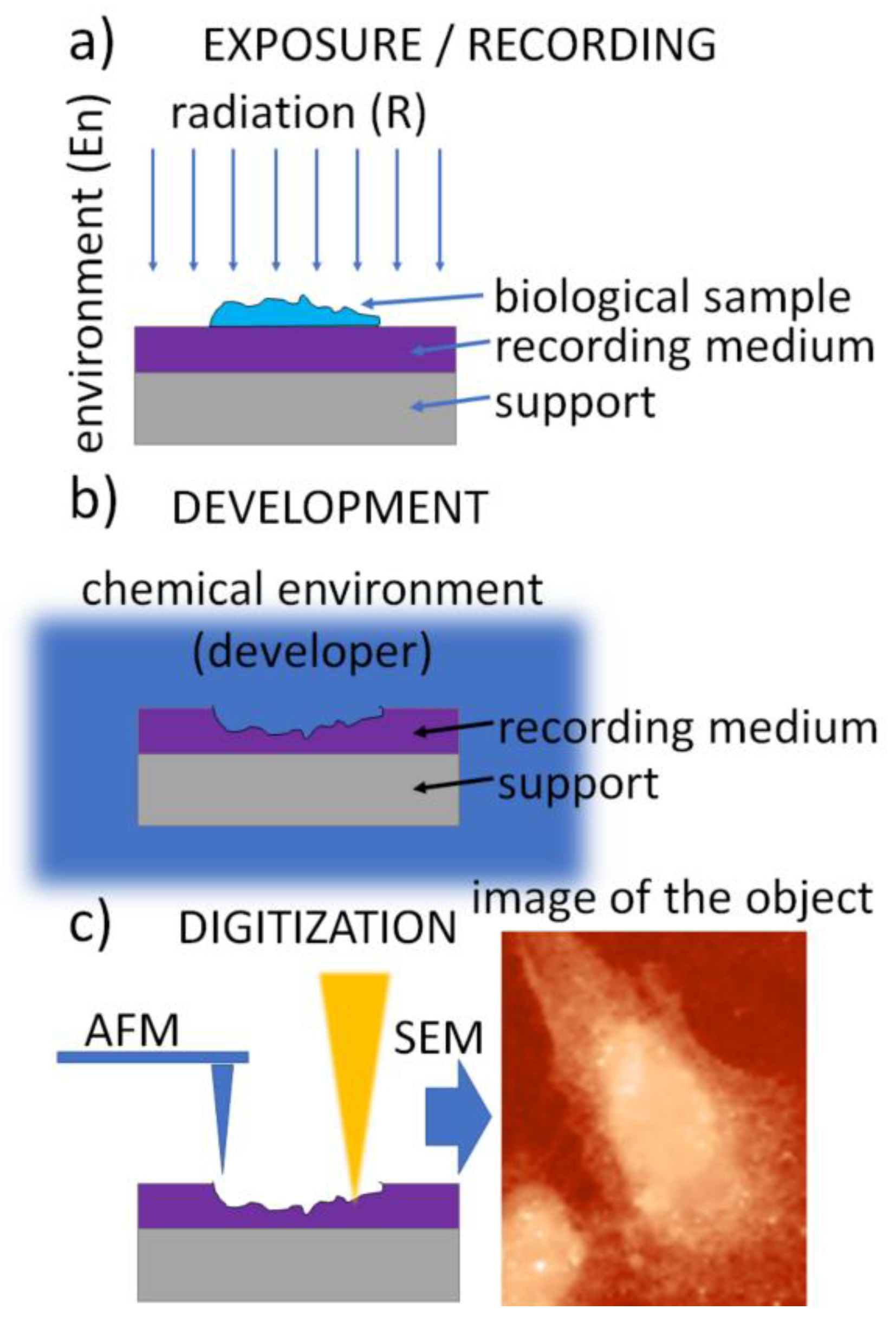
2. Soft X-ray Contact Microscopy

The principle of soft X-ray contact microscopy is well known. It can be considered a variant of X-ray contact microradiography, in which the shadow image of a specimen is obtained by irradiating the sample on the photographic plate with X-rays [21,22]. Soft X-rays in the ”water window” spectral range, produced with a specially designed X-ray tube and fine-grained photographic emulsion, have been used for imaging cellular structures with a sub-micrometer resolution. This spatial resolution corresponds to the resolution of the light microscope system utilized in this experiment to inspect the micro radiograms [23,24]. Resolution in the nanometer range was achieved when a photoresist was used to record the microradiogram instead of a photographic plate and electron microscopy to examine the microradiogram in place of an optical microscope. This SXCM technique, based on X-ray photoresist, was adapted from X-ray lithography techniques and was quickly recognized as a promising microscopy technique for examining biological specimens [25,26]. The unique properties of photons at soft-X-ray wavelength makes it possible to image cells based on the organic composition of unstained subcellular structures. X-ray techniques, including SXCM, enable the study of the remodeling of cell and extracellular matrix (ECM) architecture during normal and disease processes. The investigation of single focal adhesion (FA) in cancer, a crucial evaluation of metastatic potential, is possible thanks to SXCM.
The soft X-ray contact microscopy of biological objects with nanometric resolution was demonstrated for the first time using an X-ray tube and synchrotron operating in the “water window” range [27,28].
The SXCM technique is schematically presented in Figure 1. It uses a three-step procedure. In the first step, called the exposure (Figure 1a), soft X-ray (SXR) radiation emitted from the source is transmitted through the sample to expose a high-resolution recording medium that is “in contact” with the sample. A thin layer of X-ray resist spun on a silicon wafer is usually used as the recording medium. It allows the recording of the local intensity modulation of radiation transmitted through the sample in its structure. The absorbed radiation dose is then converted into the change in the recording-medium morphology, creating an imprint (relief) structure in the recording medium. This conversion is achieved during the development process, a standard chemical procedure (Figure 1b). Finally, the radiation-induced changes in the recording medium can be digitized, providing a digital image (Figure 1c).
To provide superior resolution compared to optical methods, the final step (Figure 1c) is performed using atomic force microscopy (AFM), scanning electron microscopy (SEM), or transmission electron microscopy (TEM); however, fluorescence microscopes (FM) are also used when lithium fluoride crystals are used as the recording medium.

2.1. Radiation Sources

Various types of X-ray sources in the “water window” were used in SXCM, including, X-ray tubes, synchrotrons, and plasma sources. X-ray tubes and synchrotrons were used in the early phase of SXCM research [27,29]. The main disadvantage of using these sources in SXCM was the exposure of the sample for a relatively long time, leading to radiation damage and decreased resolution [30]. It was found that imaging hydrated, living biological specimens required exposure with a sufficient X-ray dose in a very short time to avoid blurring due to radiation-induced and natural movement, as well as the recording of an image of the sample before radiation damage occurred. Therefore, laser-produced plasma sources emitting soft X-ray radiation in the form of nanosecond pulses were proposed [31] and came to dominate the use of SXCM.
A dense and high-temperature (106 K) plasma from the interaction of intense laser pulses with a target material (usually solid) is a bright source of soft X-rays. By choosing suitable target materials and laser parameters, the emitted radiation can be optimized for “water window” X-rays [32]. SXCM experiments were performed using various laser systems, including Nd:glass [33], Nd:YAG [31], iodine, excimer [34], and others, producing sub- and nanosecond pulses with energies varying from less than a Joule to several hundred Joules. The latter made it possible to obtain SXCM microscopy images due to a single-pulse exposure, which made it possible to prevent the radiation damage from limiting the resolution. However, for practical reasons, the most suitable compact laser systems, usually Nd:YAG, which are commercially available produce nanosecond laser pulses with energies up to a few Joules with high repetition.
The use of solid targets is the most popular in SXCM systems. However, they are characterized by the problem of target debris, produced as a result of the laser ablation of the target material, which can cause sample damage when exposed to X-rays. To reduce the debris grazing incidences, X-ray optics in the form of a toroidal mirror was used to focus soft X-rays on a sample [35]. The debris problem was completely solved by applying laser-plasma soft X-ray sources based on a gas puff target in SXCM experiments [36].

2.2. Recording Media

The most common recording medium for SXCM is an X-ray resist in the form of thin (typically a few hundredths of a nanometer) layers of a photosensitive polymer on top of a silicon wafer or a glass slide. Depending on the type of photoresist, the radiation damages the molecular structure of the resist, decreasing the local molecular weight of the polymer chains, and making the resist susceptible to the chemical development procedure (positive photoresist, i.e., PMMA (poly(methyl methacrylate)) [37].
Photoresists must ensure high resolution, preserve the cellular structure, and be suitable for cell imaging, ensuring cytocompatibility. Cells must strongly adhere to the resist. However, the most commonly used photoresist for biological applications is PMMA. Many resists are used in photolithography (e.g., DNQ-Novolac photoresist, epoxy-based polymer, hydrogen silsesquioxane) but are not suitable for cell imaging. Epoxidized Novolac resist (EPR) was also tested in SXCM, but without success [38].
Photoresists are “permanent” media, meaning that once recorded, the image of the object remains there for an extended period and can be digitized long after the exposure if the resist is properly preserved. This also means that the resist does not allow a refresh rate when trying to record changes in a sample. Resists require chemical development procedures but provide the highest spatial resolutions, which are mainly limited by digitization and equipment, such as SEM or AFM.
Another approach is to use lithium fluoride crystals (LiF) as the recording medium. Different centers are produced in LiF crystals when they are exposed to ionizing radiation [39]. Their emission intensity changes locally and is proportional to their density, thus revealing the 2-D X-ray intensity map and the radiographic image of the biological sample, which is in contact with LiF crystal during the exposure to X-rays. The drawback of this recording medium is the need for sufficiently high X-ray fluence values for color formation to obtain high-quality (i.e., high resolution) SXCM images. The advantage is that the LiF does not require a development procedure; however, the digitization is carried out with a fluorescence microscope, which is much more limited by its spatial resolution than SEM or AFM microscopes.

2.3. Exposure Environment

Most of the early SXCM experiments were performed by placing dehydrated specimens directly inside the vacuum chamber of the microscope, in which the soft X-rays propagated from the source to the specimen. With a resist such as PMMA as the recording medium, a relief structure of the sample was obtained and digitized with AFM. Because the primary goal of SXCM is to acquire high-resolution images of living specimens, the environment of the biological sample must be designed to maintain it in a hydrated state during exposure. For observation of living cells, soft X-rays are typically transmitted through silicon nitride membrane with thickness of 100–200 nm, covering the biological specimens cultivated directly onto the top surface of the photoresist. The whole assembly, consisting of the membrane, specimen in the culture medium, and the photoresist, are placed inside the vacuum chamber and sealed, employing specially designed mechanical capsules [40,41] to prevent evaporation and specimen drying.
Another approach is to place the assembly inside the pressurized environment chamber filled with helium [42] separated from the source chamber’s vacuum environment to prevent specimen drying. In this case, no particular effort is required to perform sealing, and the dismounting of the sample is much easier.

2.4. Development Procedure

The development of photoresists, the recording mediums in SXCM, has received considerable attention owing to their use in lithographic techniques. The development procedure is a critical step in SXCM because it affects image contrast and resolution. Irradiation of photoresists with soft X-rays results in bond breakage in the polymer structure, changing the molecular weights of resists locally and enabling these areas to be dissolved away in a chemical bath by solvents, such as methyl isobutyl ketone (MIBK), with the addition of isopropanol (IPA). Generally, each photoresist requires chemical solvents to dilute the irradiated area of the resist. The developer concentration and time exposure during the development process must be optimized experimentally [37]. Usually, a mixture of MIBK and IPA 1:1 (v:v) up to 1:3 (v:v) is used. A 1:1 (v/v) solution [38,41] means that the developer is chemically aggressive. A high concentration of methyl isobutyl ketone in the isopropanol increases the developing sensitivity but might blur some delicate structures in the photoresist. A more chemically gentle mixture of MIBK:IPA 1:3 (v:v) was recommended [42,43], as it provides sufficient sensitivity while preserving the nanometer height in delicate structures and features.
The resist is usually spin-coated on top of a silicon wafer, forming a uniform layer, typically 200–500 nm thick. The thickness is then tested by scratching the layer and measuring the height step using the AFM microscope. Moreover, after the resist is deposited, it must be calibrated for various SXR doses. The dose is changed by adjusting the number of SXR pulses used to expose the resist. For each dose, the depth of the relief is measured under the same developing conditions. It is necessary to optimize the SXR dose delivered to the resist to adjust its response and, finally, the dynamic range of the image contrast [38,41,44,45].

2.5. Digitization

The digitization process converts information stored in the recording medium, that is, the height information (relief pattern) in the photoresist or the density and transmittance of the color centers in the LiF crystal, into a digital image of the imprint, which is a representation of the specimen obtained during the recording phase. Historically, SEM and TEM were the techniques of choice for the digitization process. While SEM [46,47] and TEM [48,49] provided SXCM images with superior spatial resolution, the electron beam acting upon the photoresist sample causes the same effect as the soft X-ray radiation during the exposure/recording phase. This means that the e-beam during SEM/TEM image acquisition damages the photoresist and affects the information stored within it. For this reason, the AFM technique [10,50,51], which uses a sharp tip and van der Waals interactions between the end of the tip and the surface of the recording medium, provides much better height resolution and does not affect the sample in the manner of e-beam. LiF crystals require FM to digitize the information stored in the recording medium [51,52].

2.6. A Note on Spatial Resolution

It is not straightforward to assign the limit on spatial resolution in contact microscopy; however, several factors influence the spatial resolution, as seen in the following equation. This is an approximate relation, assuming all contributions have Gaussian profiles [22].
δ C M = δ s 2 + δ p h r 2 + δ t i p 2
The δ s is a factor related to shadowing (both wavelength-related diffraction and geometrically related to variable sample–resist distances due to non-zero thickness of the sample), δ p h r is a factor related to the spatial resolution of the photoresist, e.g., for HSQ/PMMA combinations below 10 nm [24], PMMA ~10 nm [53], and δ t i p is a factor related to the diameter of the AFM probe used to perform photoresist scanning after the exposure, typically 20 nm in diameter, but can be as sharp as ~2 nm for carbon nanotube probes [26]. Taking into account all the above factors, the spatial resolution in SXCM is routinely much better than 100 nm [28,54], allowing a practical limit of ~10 nm [49,55].
More detailed numerical studies were also performed [43], in which the effects of sample and resist absorption and diffraction were considered, as was the process of isotropic development of the photoresist. The SXCM image resolution heavily depends on the exposure and the sample-to-resist distance. The contrast of small features imprinted in the photoresist depends significantly on the development procedure. If researchers are not careful, the information on the smallest features may be destroyed by excessively aggressive or long development. This is why these issues must be kept in mind during the postprocessing and interpretation of the SXCM images of high-resolution and low-contrast objects, such as biological structures.

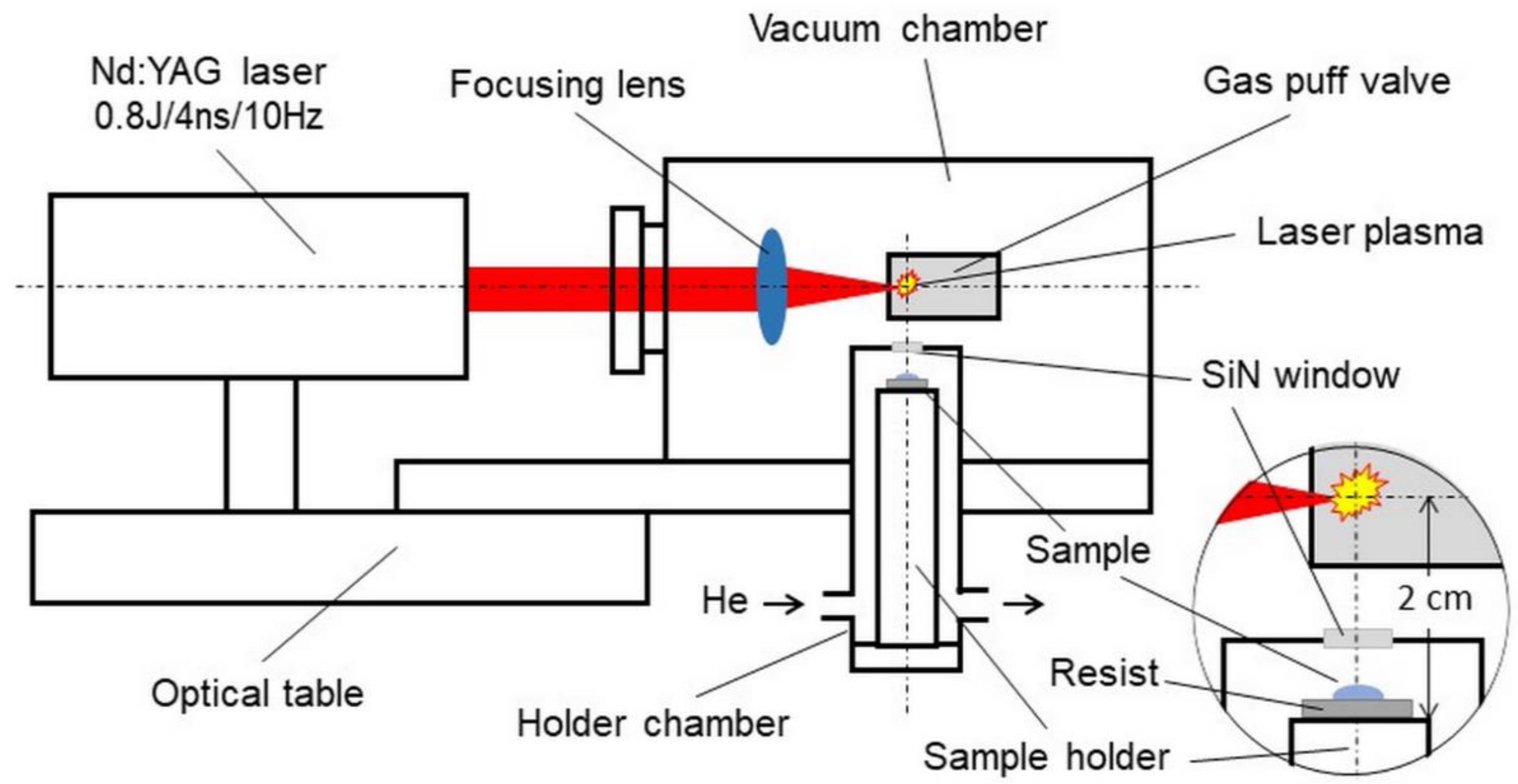
2.7. Desk-Top Laboratory SXCM System

Almost all research on SXCM to date has been carried out using laboratory systems that are not adapted to use by biologists in their laboratory practice. In many articles, the authors emphasize the potential of this simple imaging technique and its usefulness in studying the structures of cells; however, so far, no attempt has been made to commercialize the SXCM microscopy system. The conceptual design of the laboratory microscope was presented only in [56]. Here, we present the compact laboratory system for SXCM microscopy that was developed by our team [42,44], which we believe can meet these expectations.
The schematic of the system is presented in Figure 2. The biological sample is placed on the resist in the form of a 500-nanometer-thick PMMA film spun on a silicon wafer. The plate is attached to a holder mounted inside a helium-filled chamber. The design of the holder and its chamber allows the sample to be positioned at a distance of 15 mm from the source of soft X-rays. The source is a laser plasma resulting from the interaction of laser pulses with a double-stream gas puff target. Laser pulses with a duration of 4 ns and energy up to 0.8 J are generated with a repetition frequency of 10 Hz in a commercially available Nd: YAG laser system (EXPLA). The target is formed by pulsed injection of a small amount of argon gas under high pressure into an annular stream of helium surrounding the argon gas stream using a specially designed electromagnetic valve system. The use of the argon gas puff target allows the production of soft X-ray radiation with high efficiency, comparable to a solid target, without the harmful effect of the target debris. The valve producing the gas puff target is mounted inside the vacuum source chamber in such a way that the target is formed at the focus of the laser beam produced by the aspherical focusing lens. The holder chamber is equipped with a 100-nanometer-thick Si3N4 membrane, which separates the chamber’s helium atmosphere from the vacuum inside the source chamber. This allows the sample to be placed near the source without venting the vacuum chamber. Additionally, a 200-nanometer-thick Ti membrane is used to eliminate visible light from the plasma. The source produces soft X-ray radiation in the “water window” range, with a maximum wavelength of about 3 nm amd energy fluence of about 5 μJ/cm2/pulse, corresponding to the photon fluence of about 103 photons/μm2/pulse. This fluence makes it possible to obtain the SXCM image of a sample as a result of 100–200 laser pulses (10–20 s exposition time). The whole system can be mounted on an optical table with dimensions of approximately 1 × 1 m2. Detailed information on the presented system and the results of preliminary studies on SXCM can be found in the previous publications by our group [9,42].

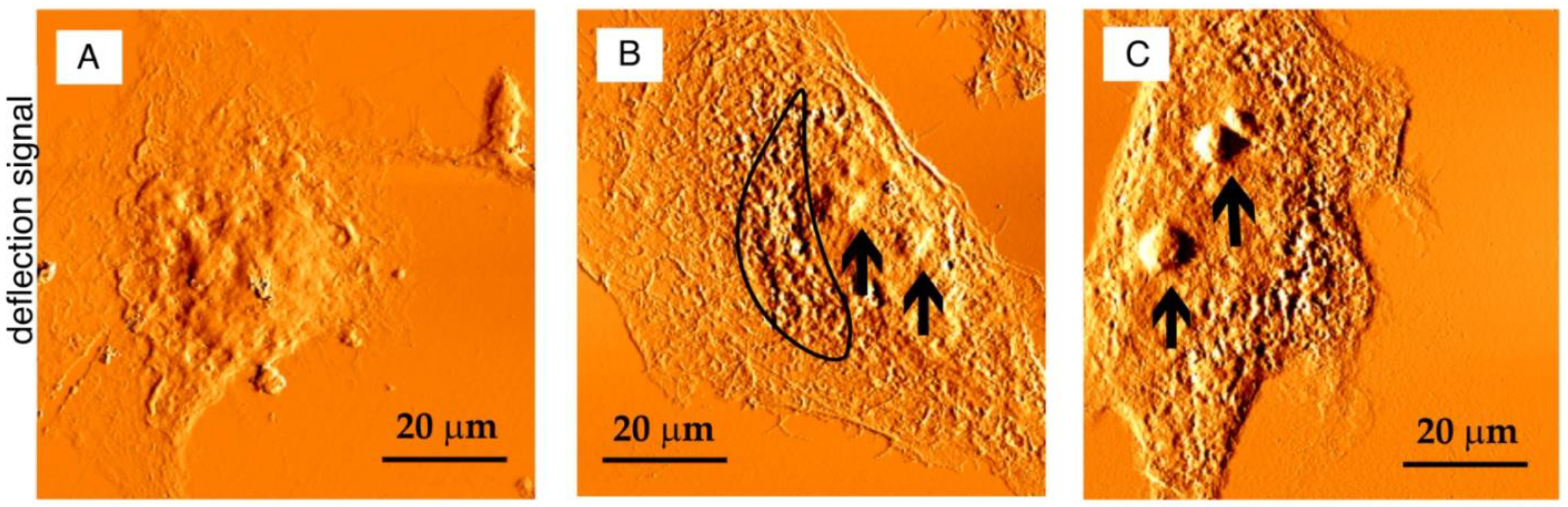
3. Cell Imaging with SXCM

The cell membrane separates the cell’s interior from the external environment. The most significant feature of SXCM is the visualization of the cell’s interior structures located under the membrane without the requirement of a “deroofing” procedure, i.e., a disruption of the plasma membrane. Soft X-ray radiation makes it possible to record and observe the inside of the native cell.
The first image of biological samples (diatoms) using SXCM, demonstrating the possibility o investigating biological specimens with a resolution near 10 nm, was obtained by Spiller 45 years ago [27]. Further works by this group [55,57] have shown that SXCM, a relatively new form of ultrastructural imaging, has a resolution that is better than 10 nm and is uniquely suited to the examination of fragile, unstained biological specimens. Therefore, this technique has been fine-tuned over a long period of time, in order to study subtle biological phenomena, as do other microscopic methods that offer similar efficiency.
The attempt to image mammalian cells, bacteria, and isolated organelles with SXCM was undertaken several times, as presented in Table 1. The mitotic chromosome structure in human lymphocytes was the first problem to be solved with this technique. The first report by Shinohara et al. on the observation of isolated, dried single human chromosome fibers at high resolution appeared in 1990, followed by subsequent team publications [36,58,59]. The “beads-on-a-string” structure with thin filaments (diameters of 7–15 nm) and nucleosomes (diameters of 10–15 nm) in hydrated cells was revealed at a spatial resolution of 10 nm.
The visualization of dry or hydrated human macrophages and Leydig cells demonstrated that SXCM allows the morphologies of actual intact cells to be viewed and encourages further technological development for biomedical research [45,51,60,61,62]. This technique also offered the possibility of monitoring the morphological changes in microorganisms (e.g., upon exposure to antibiotics), as was demonstrated for the Gram-negative bacterium, Pseudomonas aeruginosa, and Candida albicans, a yeast, among others [63,64].
The development and popularization of the atomic force microscope (AFM) allowed its application in imaging samples with high resolution with SXCM, as described above [65]. It was a cheaper device, with less complex sample preparation and better accessibility for biological laboratories than SEM and TEM. However, it should be noted that the spatial resolution of SXCM is determined not only by the AFM resolution but also by a photoresist and the number of X-ray source pulses. Moreover, only single-shot exposure of X-ray radiation can ensure the elimination of the blurring connected with the thermal effect and Brown motion, enhancing the resolution of an image.
As shown in Table 1, the reported spatial resolutions of the imaged samples were in the range of 5–100 nm, but mainly lay within the range of 80–100 nm. PMMA is still the most frequently used photoresist for studying biological objects. EPR was used in 2003 to improve the spatial resolution of submicron structures [38]. The resist could also provide height features as small as 20 nm, and the photoresist response was at least two orders of magnitude “faster” than PMMA. However, EPR did not enter common use in subsequent research.
Nevertheless, from 1980 until 2015, due to the complex experimental procedure, difficulties in the interpretation of the results, the imaging of samples with SEM or TEM (expensive and time-consuming techniques), and the use of synchrotron or advanced laboratory laser systems, only just over a dozen papers presenting original results were published on visualizing biological specimens with SXCM. In 2016, a desktop laboratory system for SXCM with a compact laser-plasma source based on a gas puff target was developed by our group [44]. Since that time, the morphological features of fixed normal human cells (such as bladder HCV29 cells and keratinocytes) and cancer cells (colorectal carcinoma CT26 and urinary bladder carcinoma T24) were revealed [42,54,67]. The SXCM technique was further employed to resolve cancer-metastasis-related issues. First, the optimization of SXCM parameters for imaging cancer-cell structures was performed [44]. It revealed that the relationship between the exposure conditions and the quality of cell imaging with the SCXM was crucial in the efficient visualization of cell structures. A spatial resolution was also influenced by cell thickness and internal complexity. The experimental condition for imprint preparation should be optimized for various biological specimens. A high SXR absorbed energy is essential for visualizing intracellular structures, such as the nucleus. However, the membrane protrusions are visible only at low exposures (Figure 3).
The selection of appropriate exposure parameters allowed the dynamic tracking of adhesion structures, such as focal adhesions (FA), within human triple-negative breast cancer (TNBC) cells [9]. Found in 15–20% of breast cancer patients, TNBC is an aggressive cancer subtype with a poor prognosis because of the lack of targeted therapies. Detailed clinical studies on the adhesion and migration of cancer cells are crucial. These processes involve the actin cytoskeleton and its interactions with the cell membrane and the extracellular matrix (ECM). The actin cytoskeleton is linked to integrin heterodimers and other adhesion proteins that comprise FAs structures. Based on AFM images of FAs, the sizes of both the agglomerates and the native single focal adhesion were calculated (Figure 4). The length of the FA aggregates was 3.11 µm, the size of a single FA’s “dot head” diameter was 0.84 µm, and the length of the “tail” was 1.94 µm. The distribution and size of the adhesive complexes in the SXCM images corresponded to the confocal scanning laser fluorescence (CLSM) microscopy images. The SXCM technique provides a higher-resolution and more detailed view of adhesive complexes’ internal substructures than CLSM. However, the identification of proteins within adhesion complexes can be achieved only via immunostaining and fluorescent microscopy imaging.
Currently developed super-resolution methods, such as stimulated emission depletion (STED) or photoactivated localization microscopy (PALM), which allow the observation of structures such as FAs in living cells, require genetically encoded fluorophores to express the desired proteins. These super-resolution techniques use the GFP-tagging method or modified cell line, obtaining a stably integrated transgene that leads to protein-translation disorders. The inclusion of such a large tag as GFP could potentially affect its stability and ability to bind other proteins by disrupting the native folding pattern of the protein of interest, causing protein destabilization [68,69]. However, to select transferred cells or to induce expression, substances (including antibiotics) are used in the culture of labeled cell lines that may also affect the condition of the cells. It has been proven that long-term exposure to antibiotics, such as Zeocin, which are used during the selection process, leads to mutagenesis in established recombinant cell lines [68,70]. This contrasts with the SXCM imaging of proteins, which does not require staining or interfering with the state of cells, but unfortunately does not allow the identification of individual proteins. Furthermore, the comparison of images obtained with other techniques is difficult.

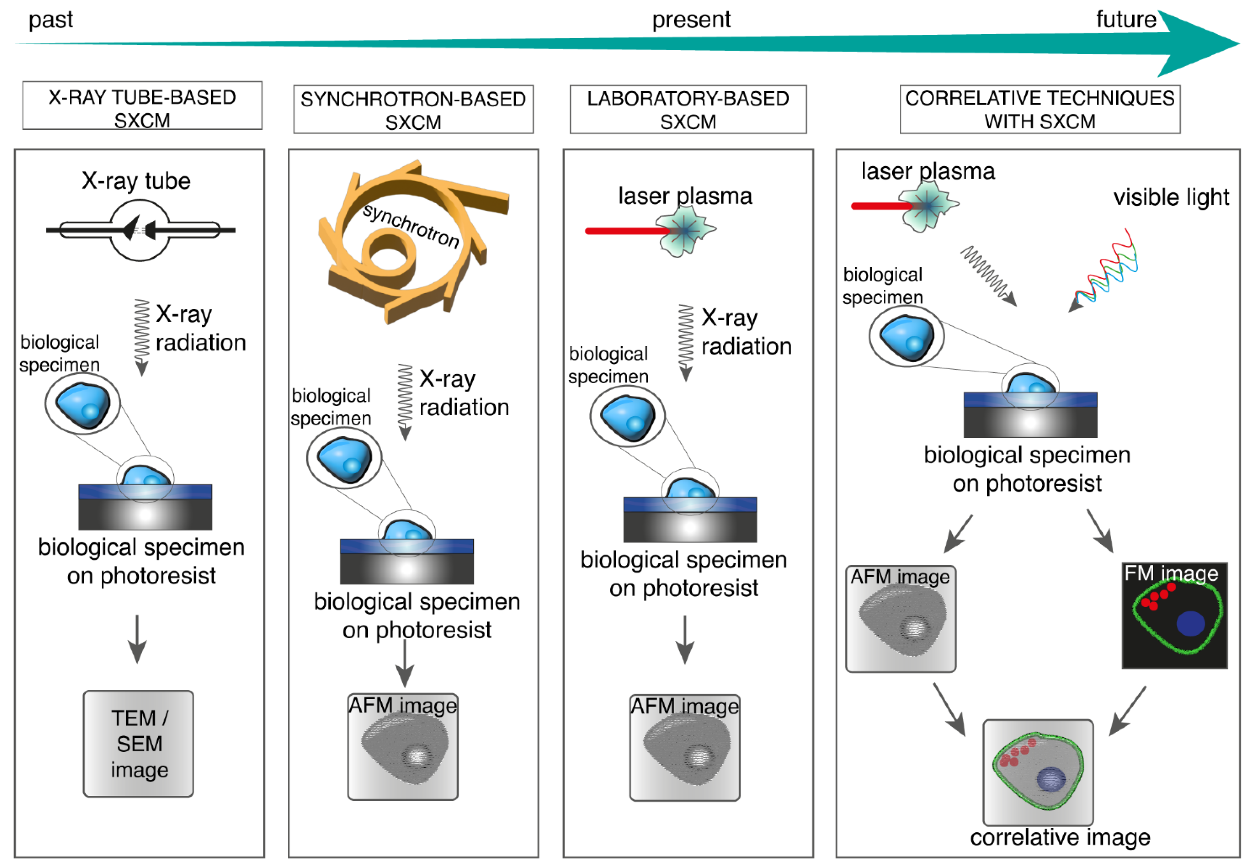
4. SXCM and Correlative Microscopy

Interpreting the results taken with an X-ray microscope often requires the confirmation and identification of cellular structures or proteins. Based on the progress in engineering and advanced microscopy, correlative techniques could solve these issues. Furthermore, the development of various correlative techniques has reduced the resolution of various techniques by as much as an order of magnitude [71,72]. The current status of correlative methods based on X-ray microscopy is presented in Table 2. Soft X-ray cryo-microscopy/tomography provides the three-dimensional structural information with nanometer resolution of biological objects up to 12 μm thick and can be correlated with conventional and super-resolution fluorescence microscopy. So far, (cryo)SXM, (cryo)SXT, and related techniques (such as laser-plasma soft X-ray microscope (LPSXM)) have been correlated with conventional (or cryo-) fluorescence microscopy [73,74,75] and super-resolution fluorescence techniques, such as SIM [76,77]. This has enabled the study of the role of specific proteins in thick cellular objects, such as the nucleus, without serial sectioning.
The proof of principle was presented on mammalian cells and insects [73,74,76,77].
Moreover, X-ray ptychography with XRF and X-ray holography was combined with X-ray scanning diffraction with STED [78,79]. The possibility of imaging Chlamydomonas reinhardtii algae in its native hydrated state by rapid and continuous X-ray scanning was presented in the correlative approach of ptychographic transmission imaging with sub-20-nanometer resolution and X-ray fluorescence imaging with sub-100-nanometer resolution of diffusible and bound ions in cryo-preserved, hydrated cells, without the need to add specific labels [78]. The correlation of holographic X-ray imaging, X-ray scanning diffraction, and STED microscopy for studying labeled and unlabeled structures in a complementary manner was demonstrated [79]. A compressive sensing method combining STXM with XRF for investigating complex biological systems solved the problem of examining large areas at high resolutions, which was very time-consuming and often restricted to pre-selected representative regions [80]. The combination of SXT with SIM gave an excellent resolution of 25–50 nm [77]. SIM is a suitable method for imaging under cryogenic conditions because it provides structural information beyond the system’s diffraction limits, which causes the volumetric resolution to rise as much as 8-fold.
Kado et al. presented, for the first time, a combined SXCM with fluorescent microscopy for the observation and identification of cellular organelles of live hydrated Leydig cells with a nanometric spatial resolution of 90 nm [75]. They applied a high-intensity laser-plasma X-ray source to obtain a single X-ray image. However, the overall system used was extremely large and impossible to apply in biological laboratories. Thus, a compact table-top device for SXCM was presented, with the potential to correlate with fluorescence techniques, as proven by Kado et al.
Most of these correlative techniques used a synchrotron, and the samples were cryo-preserved and fluorescently tagged. In turn, the conducted research was only a proof of concept and demonstrated the potential uses, sample preparation protocols, the spatial resolution reached, and troubleshooting. Mammalian cells, tissues, alga, and insects were imaged [73,74,75,76,77,78,79,80,81].
As previously mentioned, synchrotron-based X-ray techniques are restricted for many reasons. The compactness of the X-ray source offers biologists modern and much more user-friendly methods for investigating native objects (Figure 5). However, it is still difficult to interpret the SXCM results without comparison with other microscopy techniques. This is why there is a requirement for the compact X-ray source to simplify and disseminate methods, such as SXCM, together with the need to develop correlative microscopy.

5. Conclusions and Future Directions

The use of the soft X-ray contact microscopy technique for imaging intact, living cells with nanometer resolution without freezing, chemical fixation, chemical staining, or sectioning s presented. It is a simple method and, as a table-top device, is more accessible for biologists and more suitable for everyday use. Adapting the microscope design to users’ requirements makes this technique a helpful instrument in biomedical research. Because the results are difficult to interpret, the correlation with other standard methods to identify specific proteins of interest is required.
In our opinion, correlative microscopy is an exciting direction of technological development; however, the use of synchrotrons is limited to highly specialized experimental centers. SXCM is a method that allows the observation of intracellular structures in a native state, and the first promising trials to correlate SXCM with fluorescence techniques were performed. Currently developed super-resolution methods, which allow the observation of structures such as FA in living cells, require genetically encoded fluorophores to express the desired proteins. The use of GFP tagging or the modification of the manner in which cell lines are obtained with a stably integrated transgene can lead to protein-translation disorders. Thus, the SXCM technique, as a laboratory system, combined with super-resolution fluorescent techniques, such as STED or PALM, is an interesting imaging option, eliminating the use of expensive, time-consuming X-ray sources, such as synchrotrons, while ensuring the high-resolution imaging of biological specimens. This technique would exploit the potential of SXCM for functional studies of biological samples.

Author Contributions

Conceptualization, A.N.-S., P.N.O., E.A.T., and P.W.; software, W.K., P.W., and H.F.; investigation, A.N.-S., P.N.O., and P.W.; writing—original draft preparation, A.N.-S., P.W., P.N.O., W.K., E.A.T., and H.F.; writing—review and editing, A.N.-S., P.W., E.A.T., and H.F.; visualization, W.K., P.W., and H.F.; supervision, E.A.T. and H.F.; funding acquisition, H.F., A.B., and P.W. All authors have read and agreed to the published version of the manuscript.

Funding

This research was funded by the National Science Centre, Poland, under the Beethoven Program, grant number 2016/23/G/ST2/04319, and the European Union’s Horizon 2020 research and innovation program, Laserlab-Europe V, grant number 871124.

Institutional Review Board Statement

Not applicable.

Informed Consent Statement

Not applicable.

Acknowledgments

We would like to thank the referees for their useful comments.

Conflicts of Interest

The authors declare no conflict of interest.

References

  1. Lidke, D.S.; Lidke, K.A. Advances in high-resolution imaging–techniques for three-dimensional imaging of cellular structures. J. Cell Sci. 2012, 125, 2571–2580. [Google Scholar] [CrossRef] [PubMed] [Green Version]
  2. Sousa, A.A.; Kruhlak, M.J. Introduction: Nanoimaging Techniques in Biology. In Nanoimaging: Methods and Protocols; Sousa, A.A., Kruhlak, M.J., Eds.; Humana Press: Totowa, NJ, USA, 2013; pp. 1–10. [Google Scholar]
  3. Hylton, R.K.; Swulius, M.T. Challenges and triumphs in cryo-electron tomography. iScience 2021, 24, 102959. [Google Scholar] [CrossRef] [PubMed]
  4. Leis, A.; Rockel, B.; Andrees, L.; Baumeister, W. Visualizing cells at the nanoscale. Trends Biochem. Sci. 2009, 34, 60–70. [Google Scholar] [CrossRef]
  5. Pascolo, L.; Sena, G.; Gianoncelli, A.; Cernogoraz, A.; Kourousias, G.; Metscher, B.D.; Romano, F.; Zito, G.; Pacile, S.; Barroso, R.; et al. Hard and soft X-ray imaging to resolve human ovarian cortical structures. J. Synchrotron Radiat. 2019, 26, 1322–1329. [Google Scholar] [CrossRef] [PubMed] [Green Version]
  6. Pedrosa, P.; Baptista, P.V.; Fernandes, A.R.; Guerra, M. Benchtop X-ray fluorescence imaging as a tool to study gold nanoparticle penetration in 3D cancer spheroids. RSC Adv. 2021, 11, 26344–26353. [Google Scholar] [CrossRef]
  7. Penedo, M.; Miyazawa, K.; Okano, N.; Furusho, H.; Ichikawa, T.; Alam, M.S.; Miyata, K.; Nakamura, C.; Fukuma, T. Visualizing intracellular nanostructures of living cells by nanoendoscopy-AFM. Sci. Adv. 2021, 7, eabj4990. [Google Scholar] [CrossRef] [PubMed]
  8. Jacobsen, C. Soft X-ray microscopy. Trends Cell Biol. 1999, 9, 44–47. [Google Scholar] [CrossRef]
  9. Osuchowska, P.N.; Wachulak, P.; Kasprzycka, W.; Nowak-Stępniowska, A.; Wakuła, M.; Bartnik, A.; Fiedorowicz, H.; Trafny, E.A. Adhesion of Triple-Negative Breast Cancer Cells under Fluorescent and Soft X-ray Contact Microscopy. Int. J. Mol. Sci. 2021, 22, 7279. [Google Scholar] [CrossRef] [PubMed]
  10. Yamamoto, Y.; Shinohara, K. Application of X-ray microscopy in analysis of living hydrated cells. Anat. Rec. 2002, 269, 217–223. [Google Scholar] [CrossRef] [PubMed]
  11. Kirz, J.; Jacobsen, C.; Howells, M. Soft X-ray microscopes and their biological applications. Q. Rev. Biophys. 1995, 28, 33–130. [Google Scholar] [CrossRef] [Green Version]
  12. Jacobsen, C.; Howells, M.; Warwick, T. Zone-Plate X-ray Microscopy. In Springer Handbook of Microscopy; Hawkes, P.W., Spence, J.C.H., Eds.; Springer International Publishing: Cham, Switzerland, 2019; pp. 1145–1204. [Google Scholar]
  13. Weiß, D.; Schneider, G.; Niemann, B.; Guttmann, P.; Rudolph, D.; Schmahl, G. Computed tomography of cryogenic biological specimens based on X-ray microscopic images. Ultramicroscopy 2000, 84, 185–197. [Google Scholar] [CrossRef]
  14. Chao, W.; Fischer, P.; Tyliszczak, T.; Rekawa, S.; Anderson, E.; Naulleau, P. Real space soft X-ray imaging at 10 nm spatial resolution. Opt. Express 2012, 20, 9777–9783. [Google Scholar] [CrossRef] [PubMed]
  15. Chao, W.; Harteneck, B.D.; Liddle, J.A.; Anderson, E.H.; Attwood, D.T. Soft X-ray microscopy at a spatial resolution better than 15 nm. Nature 2005, 435, 1210–1213. [Google Scholar] [CrossRef]
  16. Kaulich, B.; Thibault, P.; Gianoncelli, A.; Kiskinova, M. Transmission and emission X-ray microscopy: Operation modes, contrast mechanisms and applications. J. Phys. Condens. Matter 2011, 23, 083002. [Google Scholar] [CrossRef]
  17. Harkiolaki, M.; Darrow, M.C.; Spink, M.C.; Kosior, E.; Dent, K.; Duke, E. Cryo-soft X-ray tomography: Using soft X-rays to explore the ultrastructure of whole cells. Emerg. Top. Life Sci. 2018, 2, 81–92. [Google Scholar] [CrossRef] [Green Version]
  18. Larabell, C.A.; Nugent, K.A. Imaging cellular architecture with X-rays. Curr. Opin. Struct. Biol. 2010, 20, 623–631. [Google Scholar] [CrossRef] [Green Version]
  19. Weinhardt, V.; Chen, J.-H.; Ekman, A.; McDermott, G.; Le Gros, M.A.; Larabell, C. Imaging cell morphology and physiology using X-rays. Biochem. Soc. Trans. 2019, 47, 489–508. [Google Scholar] [CrossRef] [PubMed]
  20. White, K.L.; Singla, J.; Loconte, V.; Chen, J.-H.; Ekman, A.; Sun, L.; Zhang, X.; Francis, J.P.; Li, A.; Lin, W.; et al. Visualizing subcellular rearrangements in intact β cells using soft X-ray tomography. Sci. Adv. 2020, 6, eabc8262. [Google Scholar] [CrossRef]
  21. Cosslett, V.E. Microscopy with X-rays. Nature 1959, 183, 1423–1427. [Google Scholar] [CrossRef]
  22. Wachulak, P.W. Nanometer-Scale and Low-Density Imaging with Extreme Ultraviolet and Soft X-ray Radiation. In 21st Century Nanoscience—A Handbook; CRC Press: Boca Raton, FL, USA, 2020. [Google Scholar]
  23. Greulich, R.C.; Engström, A. A new approach to high resolution microradiography using extremely soft X-rays. Exp. Cell Res. 1956, 10, 251–254. [Google Scholar] [CrossRef]
  24. Rommel, M.; Nilsson, B.; Jedrasik, P.; Bonanni, V.; Dmitriev, A.; Weis, J. Sub-10nm resolution after lift-off using HSQ/PMMA double layer resist. Microelectron. Eng. 2013, 110, 123–125. [Google Scholar] [CrossRef]
  25. Feder, R.; Sayre, D.; Spiller, E.; Topalian, J.; Kirz, J. Specimen replication for electron microscopy using X rays and X-ray resist. J. Appl. Phys. 1976, 47, 1192–1193. [Google Scholar] [CrossRef]
  26. Stevens, R.M. New carbon nanotube AFM probe technology. Mater. Today 2009, 12, 42–45. [Google Scholar] [CrossRef]
  27. Spiller, E.; Feder, R.; Topalian, J.; Eastman, D.; Gudat, W.; Sayre, D. X-ray Microscopy of Biological Objects with Carbon Kα and with Synchrotron Radiation. Science 1976, 191, 1172–1174. [Google Scholar] [CrossRef]
  28. Kado, M.; Kishimoto, M.; Tamotsu, S.; Yasuda, K.; Shinohara, K. In situ observation of cellular organelles with a contact X-ray microscope. J. Phys. Conf. Ser. 2013, 463, 012056. [Google Scholar] [CrossRef]
  29. Feder, R.; Spiller, E.; Topalian, J.; Broers, A.N.; Gudat, W.; Panessa, B.J.; Zadunaisky, Z.A.; Sedat, J. High-Resolution Soft X-ray Microscopy. Science 1977, 197, 259–260. [Google Scholar] [CrossRef]
  30. Ford, T.W.; Stead, A.D.; Cotton, R.A. Soft X-ray contact microscopy of biological materials. Electron Microsc. Rev. 1991, 4, 269–292. [Google Scholar] [CrossRef]
  31. Rosser, R.J.; Feder, R.; Ng, A.; Adams, F.; Celliers, P.; Speer, R.J. Nondestructive single-shot soft X-ray lithography and contact microscopy using a laser-produced plasma source. Appl. Opt. 1987, 26, 4313–4318. [Google Scholar] [CrossRef]
  32. Kado, M.; Richardson, M.; Gaebel, K.; Torres, D.; Rajyaguru, J.; Muszynski, M. Ultrastructural Imaging and Molecular Modeling of Live Bacteria Using Soft X-ray Contact Microscopy with Nanoseconds Laser-Plasma Radiation; SPIE: Bellingham, WA, USA, 1995; Volume 2523. [Google Scholar]
  33. Michette, A.G.; Cheng, P.C.; Easons, R.W.; Feder, R.; O’Neill, F.; Owadano, Y.; Rosser, R.J.; Rumsby, P.; Shaw, M.J. Soft X-ray contact microscopy using laser plasma sources. J. Phys. D Appl. Phys. 1986, 19, 363–372. [Google Scholar] [CrossRef]
  34. Albertano, P.; Reale, L.; Palladino, L.; Reale, A.; Cotton, R.; Bollanti, S.; Di Lazzaro, P.; Flora, F.; Lisi, N.; Nottola, A.; et al. X-ray contact microscopy using an excimer laser plasma source with different target materials and laser pulse durations. J. Microsc. 1997, 187, 96–103. [Google Scholar] [CrossRef]
  35. Rosser, R.J.; Feder, R.; Ng, A.; Adams, F.; Caldarolo, M.; Celliers, P.; Cheng, P.C.; Da Silva, L.; Parfeniuk, D.; Speer, R.J. Biological specimens imaged by soft X-ray contact microscopy using a plasma source produced with a laboratory sized laser. J. Microsc. 1986, 144, RP5–RP6. [Google Scholar] [CrossRef]
  36. Kinjo, Y.; Watanabe, M.; Fiedorowicz, H.; Daido, H.; Yanase, E.; Fujii, S.; Sato, E.; Shinohara, K. Fine structures of human chromosomes observed by X-ray contact microscopy coupled with atomic force microscopy. J. Phys. IV Fr. 2003, 104, 313–316. [Google Scholar] [CrossRef]
  37. Yasin, S.; Hasko, D.G.; Ahmed, H. Comparison of MIBK/IPA and water/IPA as PMMA developers for electron beam nanolithography. Microelectron. Eng. 2002, 61–62, 745–753. [Google Scholar] [CrossRef]
  38. Cefalas, A.C.; Sarantopoulou, E.; Kollia, Z.; Argitis, P.; Tegou, E.; Ford, T.W.; Stead, A.D.; Danson, C.N.; Neely, D.; Kobe, S. Nanostructured imaging of biological specimens in vivo with laser plasma X-ray contact microscopy. Mater. Sci. Eng. C 2003, 23, 105–108. [Google Scholar] [CrossRef]
  39. Bonfigli, F.; Faenov, A.Y.; Flora, F.; Marolo, T.; Montereali, R.M.; Nichelatti, E.; Pikuz, T.A.; Reale, L.; Baldacchini, G. Point defects in lithium fluoride films for micro-radiography, X-ray microscopy and photonic applications. Phys. Status Solidi (a) 2005, 202, 250–255. [Google Scholar] [CrossRef]
  40. Batani, D.; Botto, C.; Moret, M.; Milani, M.; Lucchini, G.; Eidmann, K.; Cotelli, F.; Donin, C.L.L.; Poletti, G.; Ford, T.; et al. The use of high energy laser-plasma sources in soft X-ray contact microscopy of living biological samples. Eur. Phys. J. D 2002, 21, 167–179. [Google Scholar] [CrossRef]
  41. Majima, T. Soft X-ray imaging of living cells in water: Flash contact soft X-ray microscope. TrAC Trends Anal. Chem. 2004, 23, 520–526. [Google Scholar] [CrossRef]
  42. Ayele, M.G.; Wachulak, P.W.; Czwartos, J.; Adjei, D.; Bartnik, A.; Wegrzynski, Ł.; Szczurek, M.; Pina, L.; Fiedorowicz, H. Development and characterization of a laser-plasma soft X-ray source for contact microscopy. Nucl. Instrum. Methods Phys. Res. Sect. B: Beam Interact. Mater. At. 2017, 411, 35–43. [Google Scholar] [CrossRef]
  43. Wang, Y.; Jacobsen. A numerical study of resolution and contrast in soft X-ray contact microscopy. J. Microsc. 1998, 191, 159–169. [Google Scholar] [CrossRef] [Green Version]
  44. Osuchowska, P.N.; Wachulak, P.; Nowak-Stępniowska, A.; Bartnik, A.; Gnanachandran, K.; Lekka, M.; Czwartos, J.; Fiedorowicz, H.; Trafny, E.A. Imaging of Cell Structures Using Optimized Soft X-ray Contact Microscopy. Appl. Sci. 2020, 10, 6895. [Google Scholar] [CrossRef]
  45. Kado, M.; Ishino, M.; Tamotsu, S.; Yasuda, K.; Kishimoto, M.; Nishikino, M.; Kinjo, Y.; Shinohara, K. Observation of Organelles in Leydig Cells by Contact Soft X-ray Microscopy with a Laser Plasma X-ray Source. AIP Conf. Proc. 2011, 1365, 391–394. [Google Scholar] [CrossRef] [Green Version]
  46. Panessa-Warren, B.J. Contact microscopy with synchrotron radiation. Biol Trace Elem Res 1987, 12, 167–183. [Google Scholar] [CrossRef] [PubMed] [Green Version]
  47. Spiller, E.; Feder, R.; Topallan, J. Soft X-rays for biological and industrial pattern replications. J. Phys. Colloq. 1978, 39, C4–C205. [Google Scholar] [CrossRef]
  48. Cheng, P.C.; Shinozaki, D.M.; Tan, K.H. Recent Advances in Contact Imaging of Biological Materials. In X-ray Microscopy: Instrumentation and Biological Applications; Cheng, P.-C., Jan, G.-J., Eds.; Springer: Berlin/Heidelberg, Germany, 1987; pp. 65–104. [Google Scholar]
  49. Feder, R.; Costa Jonathan, L.; Chaudhari, P.; Sayre, D. Improved Detail in Biological Soft X-ray Microscopy: Study of Blood Platelets. Science 1981, 212, 1398–1400. [Google Scholar] [CrossRef] [PubMed]
  50. Kinjo, Y.; Watanabe, M.; Ito, A.; Shinohara, K. X-ray Microscopy and Chromosome Research. In 8th International Conference on X-ray Microscopy; IPAP Conference Series 7; Institute of Pure and Applied Physics: Tokyo, Japan, 2005; pp. 227–229. [Google Scholar]
  51. Reale, L.; Bonfigli, F.; Lai, A.; Flora, F.; Albertano, P.; Di Giorgio, M.L.; Mezi, L.; Montereali, R.M.; Faenov, A.; Pikuz, T.; et al. Contact X-ray microscopy of living cells by using LiF crystal as imaging detector. J. Microsc. 2015, 258, 127–139. [Google Scholar] [CrossRef] [Green Version]
  52. Baldacchini, G.; Bollanti, S.; Bonfigli, F.; Flora, F.; Di Lazzaro, P.; Lai, A.; Marolo, T.; Montereali, R.M.; Murra, D.; Faenov, A.; et al. Soft X-ray submicron imaging detector based on point defects in LiF. Rev. Sci. Instrum. 2005, 76, 113104. [Google Scholar] [CrossRef]
  53. Hu, W.; Sarveswaran, K.; Lieberman, M.; Bernstein, G.H. Sub-10 nm electron beam lithography using cold development of poly(methylmethacrylate). J. Vac. Sci. Technol. B Microelectron. Nanometer Struct. Processing Meas. Phenom. 2004, 22, 1711–1716. [Google Scholar] [CrossRef] [Green Version]
  54. Ayele, M.G.; Czwartos, J.; Adjei, D.; Wachulak, P.; Ahad, I.U.; Bartnik, A.; Wegrzynski, Ł.; Szczurek, M.; Jarocki, R.; Fiedorowicz, H.; et al. Contact Microscopy using a Compact Laser Produced Plasma Soft X-ray Source. Acta Phys. Pol. A 2016, 129, 237–240. [Google Scholar] [CrossRef]
  55. Beese, L.; Feder, R.; Sayre, D. Contact X-ray microscopy. A new technique for imaging cellular fine structure. Biophys. J. 1986, 49, 259–268. [Google Scholar] [CrossRef] [Green Version]
  56. Richardson, M.C.; Shinohara, K.; Tanaka, K.A.; Kinjo, Y.; Ikeda, N.; Kado, M. Pulsed X-ray microscopy of biological specimens with laser plasma sources. In Soft X-ray Microscopy; SPIE: Bellingham, WA, USA, 1993. [Google Scholar]
  57. Feder, R.; Banton, V.; Sayre, D.; Costa, J.; Baldini, M.; Kim, B. Direct Imaging of Live Human Platelets by Flash X-ray Microscopy. Science 1985, 227, 63–64. [Google Scholar] [CrossRef] [PubMed]
  58. Kinjo, Y.; Shinohara, K.; Ito, A.; Nakano, H.; Watanabe, M.; Horiike, Y.; Kikuchi, Y.; Richardson, M.C.; Tanaka, K.A. Direct imaging in a water layer of human chromosome fibres composed of nucleosomes and their higher-order structures by laser-plasma X-ray contact microscopy. J. Microsc. 1994, 176, 63–74. [Google Scholar] [CrossRef] [PubMed]
  59. Kunio, S.; Yasuhito, K.; Martin, C.R.; Atsushi, I.; Noboru, M.; Yasuhiro, H.; Makoto, W.; Keiji, Y.; Kazuo, A.T. Observation of human chromosome fibers in a water layer by laser-plasma X-ray contact microscopy. In Soft X-ray Microscopy; SPIE: Bellingham, WA, USA, 1993. [Google Scholar]
  60. Kado, M.; Daido, H.; Yamamoto, Y.; Shinohara, K.; Richardson, M.C. Development of a laser plasma X-ray microscope to observe live hydrated biological specimens. Laser Phys. Lett. 2006, 3, 205–207. [Google Scholar] [CrossRef]
  61. Kado, M.; Richardson, M.; Rajyaguru, J.M.; Muszynski, M.J.; Friedman, H.; Yamamoto, Y. Direct Ultrastructural Imaging of Macrophages Using a Novel X-ray Contact Microscopy. Proc. Soc. Exp. Biol. Med. 1999, 220, 27–30. [Google Scholar] [CrossRef] [PubMed]
  62. Kishimoto, M.; Kado, M.; Ishino, M.; Tamotsu, S.; Yasuda, K.; Shinohara, K. Development of single shot soft X-ray contact microscopy system for nano-scale dynamics measurement of living biological specimen. AIP Conf. Proc. 2012, 1465, 43–47. [Google Scholar] [CrossRef]
  63. Rajyaguru, J.M.; Kado, M.; Nekula, K.; Richardson, M.C.; Muszynski, M.J. High resolution X-ray micrography of live Candida albicans using laser plasma pulsed point X-ray sources. Microbiology 1997, 143, 733–738. [Google Scholar] [CrossRef] [Green Version]
  64. Rajyaguru, J.M.; Torres, D.S.; Abel, E.; Richardson, M.C.; Muszynski, M.J. Application of X-ray micrography and imaging to study the effect of gentamicin on Pseudomonas aeruginosa. J. Antimicrob. Chemother. 1998, 41, 557–561. [Google Scholar] [CrossRef] [Green Version]
  65. Tomie, T.; Shimizu, H.; Majima, T.; Yamada, M.; Kanayama, T.; Kondo, H.; Yano, M.; Ono, M. Three-Dimensional Readout of Flash X-ray Images of Living Sperm in Water by Atomic-Force Microscopy. Science 1991, 252, 691–693. [Google Scholar] [CrossRef] [Green Version]
  66. Shinohara, K.; Nakano, H.; Kinjo, Y.; Watanabe, M. Fine structure of unstained human chromosome fibres dried with no fixative as observed by X-ray contact microscopy. J. Microsc. 1990, 158, 335–342. [Google Scholar] [CrossRef]
  67. Wachulak, P.; Torrisi, A.; Ayele, M.; Czwartos, J.; Bartnik, A.; Węgrzyński, Ł.; Fok, T.; Parkman, T.; Salačová, Š.; Turňová, J.; et al. Bioimaging Using Full Field and Contact EUV and SXR Microscopes with Nanometer Spatial Resolution. Appl. Sci. 2017, 7. [Google Scholar] [CrossRef] [Green Version]
  68. Lo, C.-A.; Greben, A.W.; Chen, B.E. Generating stable cell lines with quantifiable protein production using CRISPR/Cas9-mediated knock-in. BioTechniques 2017, 62, 165–174. [Google Scholar] [CrossRef] [PubMed] [Green Version]
  69. Sokolovski, M.; Bhattacherjee, A.; Kessler, N.; Levy, Y.; Horovitz, A. Thermodynamic Protein Destabilization by GFP Tagging: A Case of Interdomain Allostery. Biophys. J. 2015, 109, 1157–1162. [Google Scholar] [CrossRef] [PubMed] [Green Version]
  70. Trastoy, M.O.; Defais, M.; Larminat, F. Resistance to the antibiotic Zeocin by stable expression of the Sh ble gene does not fully suppress Zeocin-induced DNA cleavage in human cells. Mutagenesis 2005, 20, 111–114. [Google Scholar] [CrossRef] [PubMed] [Green Version]
  71. Miranda, A.; Gómez-Varela, A.I.; Stylianou, A.; Hirvonen, L.M.; Sánchez, H.; De Beule, P.A.A. How did correlative atomic force microscopy and super-resolution microscopy evolve in the quest for unravelling enigmas in biology? Nanoscale 2021, 13, 2082–2099. [Google Scholar] [CrossRef]
  72. Navikas, V.; Leitao, S.M.; Grussmayer, K.S.; Descloux, A.; Drake, B.; Yserentant, K.; Werther, P.; Herten, D.-P.; Wombacher, R.; Radenovic, A.; et al. Correlative 3D microscopy of single cells using super-resolution and scanning ion-conductance microscopy. Nat. Commun. 2021, 12, 4565. [Google Scholar] [CrossRef]
  73. Duke, E.M.H.; Razi, M.; Weston, A.; Guttmann, P.; Werner, S.; Henzler, K.; Schneider, G.; Tooze, S.A.; Collinson, L.M. Imaging endosomes and autophagosomes in whole mammalian cells using correlative cryo-fluorescence and cryo-soft X-ray microscopy (cryo-CLXM). Ultramicroscopy 2014, 143, 77–87. [Google Scholar] [CrossRef] [Green Version]
  74. Hagen, C.; Guttmann, P.; Klupp, B.; Werner, S.; Rehbein, S.; Mettenleiter, T.C.; Schneider, G.; Grünewald, K. Correlative VIS-fluorescence and soft X-ray cryo-microscopy/tomography of adherent cells. J. Struct. Biol. 2012, 177, 193–201. [Google Scholar] [CrossRef] [Green Version]
  75. Kado, M.; Kishimoto, M.; Tamotsu, S.; Yasuda, K.; Aoyama, M.; Tone, S.; Shinohara, K. Correlative imaging of live biological cells with a soft X-ray microscope and a fluorescence microscope. AIP Conf. Proc. 2016, 1696, 020019. [Google Scholar] [CrossRef] [Green Version]
  76. Kounatidis, I.; Stanifer, M.L.; Phillips, M.A.; Paul-Gilloteaux, P.; Heiligenstein, X.; Wang, H.; Okolo, C.A.; Fish, T.M.; Spink, M.C.; Stuart, D.I.; et al. 3D Correlative Cryo-Structured Illumination Fluorescence and Soft X-ray Microscopy Elucidates Reovirus Intracellular Release Pathway. Cell 2020, 182, 515–530.e17. [Google Scholar] [CrossRef]
  77. Okolo, C.A.; Kounatidis, I.; Groen, J.; Nahas, K.L.; Balint, S.; Fish, T.M.; Koronfel, M.A.; Cortajarena, A.L.; Dobbie, I.M.; Pereiro, E.; et al. Sample preparation strategies for efficient correlation of 3D SIM and soft X-ray tomography data at cryogenic temperatures. Nat. Protoc. 2021, 16, 2851–2885. [Google Scholar] [CrossRef]
  78. Deng, J.; Vine, D.J.; Chen, S.; Jin, Q.; Nashed, Y.S.G.; Peterka, T.; Vogt, S.; Jacobsen, C. X-ray ptychographic and fluorescence microscopy of frozen-hydrated cells using continuous scanning. Sci. Rep. 2017, 7, 445. [Google Scholar] [CrossRef] [PubMed] [Green Version]
  79. Bernhardt, M.; Nicolas, J.D.; Osterhoff, M.; Mittelstädt, H.; Reuss, M.; Harke, B.; Wittmeier, A.; Sprung, M.; Köster, S.; Salditt, T. Correlative microscopy approach for biology using X-ray holography, X-ray scanning diffraction and STED microscopy. Nat. Commun. 2018, 9, 3641. [Google Scholar] [CrossRef] [PubMed]
  80. Kourousias, G.; Billè, F.; Borghes, R.; Pascolo, L.; Gianoncelli, A. Megapixel scanning transmission soft X-ray microscopy imaging coupled with compressive sensing X-ray fluorescence for fast investigation of large biological tissues. Analyst 2021, 146, 5836–5842. [Google Scholar] [CrossRef] [PubMed]
  81. Nicolas, J.-D.; Bernhardt, M.; Krenkel, M.; Richter, C.; Luther, S.; Salditt, T. Combined scanning X-ray diffraction and holographic imaging of cardiomyocytes. J. Appl. Crystallogr. 2017, 50, 612–620. [Google Scholar] [CrossRef]
Figure 1. A flowchart of the contact X-ray microscopy imaging procedure: (a) exposure with radiation penetrating the biological sample and absorbed in the recording medium—photoresist, (b) chemical development of the photoresist converting spatial 2D modulation of the radiation density into the surface morphology, (c) digitization—conversion of the surface morphology into the digital image using high-resolution imaging methods, e.g., AFM or SEM.
Applsci 12 07030 g001
Figure 2. Schematic of the desk-top laboratory system for soft X-ray contact microscopy (SXCM) based on a laser plasma soft X-ray source with a double-stream gas puff target.
Applsci 12 07030 g002
Figure 3. AFM images of cellular imprints of fixed HCC38 breast cancer cells saved in PMMA photoresists by soft X-ray contact microscopy. Images were obtained with SXR exposures of (A) 400, (B) 600, and (C) 800 pulses. The nucleoli of the nuclei are indicated by arrows and the endoplasmic reticulum, vacuoles, and mitochondria are outlined with a frame. Imprints were recorded by AFM working in contact mode (Xe120, Park Systems Suwon, South Korea) with OTR4 cantilevers (0.02 N/m, Bruker, Camarillo, CA, USA). Scan size of 80 × 80 μm2. A deflection signal is presented. Reproduced with permission from Osuchowska et al. [44].
Applsci 12 07030 g003
Figure 4. Analysis of topography of the single FA imprints. (A) Scheme of the FA structure; (B) the AFM image of the FA; (C) the 3D reconstruction; (D) the height distribution of the FA imprints; (E) a side view of the FA reconstruction. AFM images were processed using Gwyddion 2.53 software. Reproduced with permission from Osuchowska et al. [9].
Applsci 12 07030 g004
Figure 5. The anticipated development of SXCM. The development of compact X-ray sources makes SXCM much cheaper, smaller, and easier to use in biological laboratories. The correlation of SXCM with other developing microscopic techniques, such as optical, fluorescence, and super-resolution microscopy, improves the identification of various biological structures and provides additional information on their distribution and functionality.
Applsci 12 07030 g005
Table 1. Major biological application of soft X-ray contact microscopy.
YearX-ray SourceSamplePhotoresistImagingObject of InterestSpatial Resolution (nm)References
1981X-ray tubeDry/fixedPMMATEMHuman blood platelets5(Feder et al., 1981) [49]
1985Discharge plasmaHydratedPMMATEMHuman blood plateletsNS *
1990SynchrotronDry/fixed, isolatedPMMATEMChromosomes from human lymphocytes10(Feder et al., 1985) [57]
1991Laser plasmaHydratedPMMAAFMSea urchin sperm100
1992Laser plasmaHydrated, isolatedPMMATEMChromosomes from human lymphocytes10(Shinohara et al., 1990) [66]
1994Laser plasmaHydrated,
isolated
PMMATEMChromosomes from human lymphocytes10–100
1997SynchrotronHydratedPMMAAFMCandida albicans20–90(Tomie et al., 1991) [65]
1998Laser plasmaHydratedPMMAAFMPseudomonas aeruginosa90
1999Laser plasmaHydratedPMMAAFMMacrophages-(Shinohara et al., 1993) [59]
2003Laser plasmaHydratedEPRSEM, AFMFlagella from green algae100
2003Laser plasmaHydrated,
isolated
PMMAAFMChromosomes from Vicia faba L.10(Kinjo et al., 1994) [58]
2005SynchrotronHydratedPMMAAFMMacrophages10
2011Laser plasmaHydratedPMMAAFMLeydig cells90(Rajyaguru et al., 1997) [63]
2012Laser plasmaHydratedPMMAAFMLeydig cells100
2015Laser plasmaHydratedPMMAAFMMacrophages, Leptolyngbya sp., Chlamydomonas reinhardtii, Chlamydomonas dysosmos 100(Rajyaguru et al., 1998) [64]
2016Laser plasmaDry/fixedPMMAAFMNon-malignant human bladder cells HCV2980
2017Laser plasmaDry/fixedPMMAAFMHuman epidermal keratinocytes HEK80(Kado et al., 1999) [61]
2017Laser plasmaDry/fixedPMMAAFMMurine colorectal carcinoma CT26, human urinary bladder carcinoma T2480
2020Laser plasmaDry/fixedPMMAAFMHuman Poietics™ mesenchymal stem cells, breast cancer cells HCC38, prostate cancer cells DU14580(Cefalas et al., 2003) [38]
2021Laser plasmaHydratedPMMAAFMHuman breast cancer cells HCC3880
2015Laser plasmaHydratedPMMAAFMMacrophages, Leptolyngbya sp., Chlamydomonas reinhardtii, Chlamydomonas dysosmos 100(Kinjo et al., 2003) [36]
2016Laser plasmaDry/fixedPMMAAFMNon-malignant human bladder cells HCV2980
2017Laser plasmaDry/fixedPMMAAFMHuman epidermal keratinocytes HEK80(Kado et al., 2006) [60]
2020Laser plasmaDry/fixedPMMAAFMHuman Poietics™ mesenchymal stem cells, breast cancer cells HCC38, prostate cancer cells DU14580
2021Laser plasmaHydratedPMMAAFMHuman breast cancer cells HCC3880(Kado et al., 2011) [45]
* NS, not specified; PMMA: polymethyl methacrylate; EPR: epoxidized Novolac resist; TEM: transmission electron microscopy; SEM: scanning electron microscopy; AFM: atomic force microscopy.
Table 2. Selected current biological and medical applications of X-ray-related correlative microscopy.
TechniquesX-ray SourceSampleObject of InterestSpatial Resolution
(nm)
References
X-ray TechniqueCorrelated Method
Cryo-SXM/SXTCryo-FL microscopySynchrotron Cryo-preserved;
fluorescently tagged
Porcine epithelial-like embryonic EFN-R kidney cells stably co-expressing pseudorabies viruses pUL31 and pUL34NS *(Hagen et al., 2012) [74]
Cryo-SXM/SXTCryo-FL microscopySynchrotron Cryo-preserved;
fluorescently tagged
Human epidermal keratinocytes HEK29340(Duke et al., 2014) [73]
LPSXMFL microscopyLaser plasmaHydrated;
fluorescently tagged
Leydig cells90(Kado et al., 2016) [75]
X-ray ptychographyXRFSynchrotron Cryo-preservedChlamydomonas reinhardtii85 (Deng et al., 2017) [78]
X-ray holography, X-ray scanning diffractionSTEDSynchrotron Dry/fixed;
cryo-preserved;
fluorescently tagged
Neonatal rat cardiomyocytesnm(Bernhardt et al., 2018) [79]
Cryo-SXTCryo-SIMSynchrotron Cryo-preserved;
fluorescently tagged
Human bone osteosarcoma cells U2OS60–200(Kounatidis et al., 2020) [76]
STXMXRFSynchrotron Paraffin-embeddedHuman lung and ovarian tissues1000 (Kourousias et al., 2021) [80]
Cryo-SXTCryo-SIMSynchrotron Cryo-preserved;
fluorescently tagged
Mouse NIH3T3 embryo fibroblast cells, human bone osteosarcoma cells U2OS, lysosomes in Drosophila melanogaster plasmatocytes25–50(Okolo et al., 2021) [77]
* NS—not specified; LPSXM: laser-plasma soft X-ray microscopy; FL: fluorescence, SIM: super-resolution structured illumination microscopy, SXM: soft X-ray microscopy; SXT: soft X-ray tomography, STED: stimulated emission depletion, XRF: X-ray fluorescence.
Publisher’s Note: MDPI stays neutral with regard to jurisdictional claims in published maps and institutional affiliations.

