
Thermodynamic and Spectroscopic Studies of SDS in Cinnamaldehyde + Ethanol Mixtures: Influences of Temperature and Composition


Abstract
:1. Introduction
2. Experiment
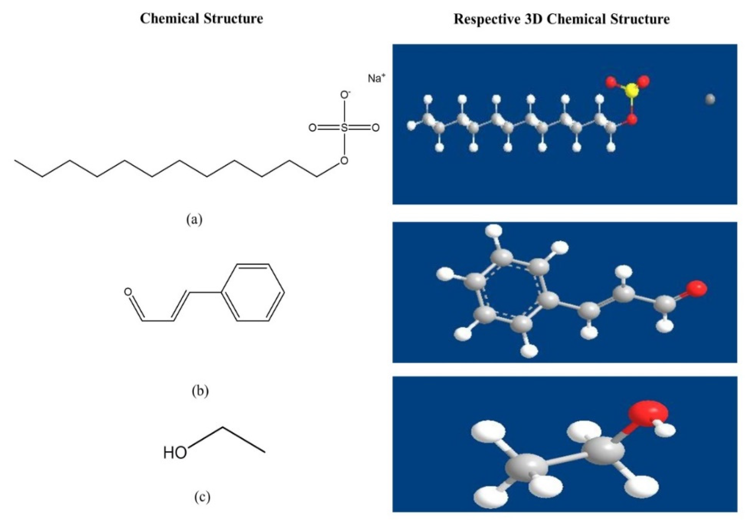
2.1. Materials
2.2. Methods
2.2.1. Stock Solution Preparations
2.2.2. Conductivity Measurements
2.2.3. Fluorescence Measurement
2.2.4. FTIR Measurement
2.3. Calculation of the Thermodynamic Properties of Micellization
3. Results and Discussion
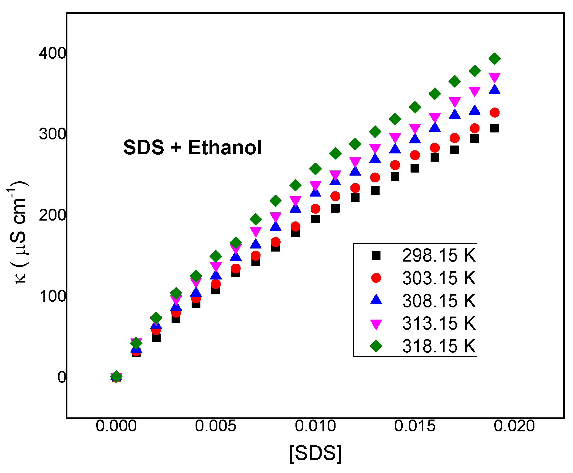
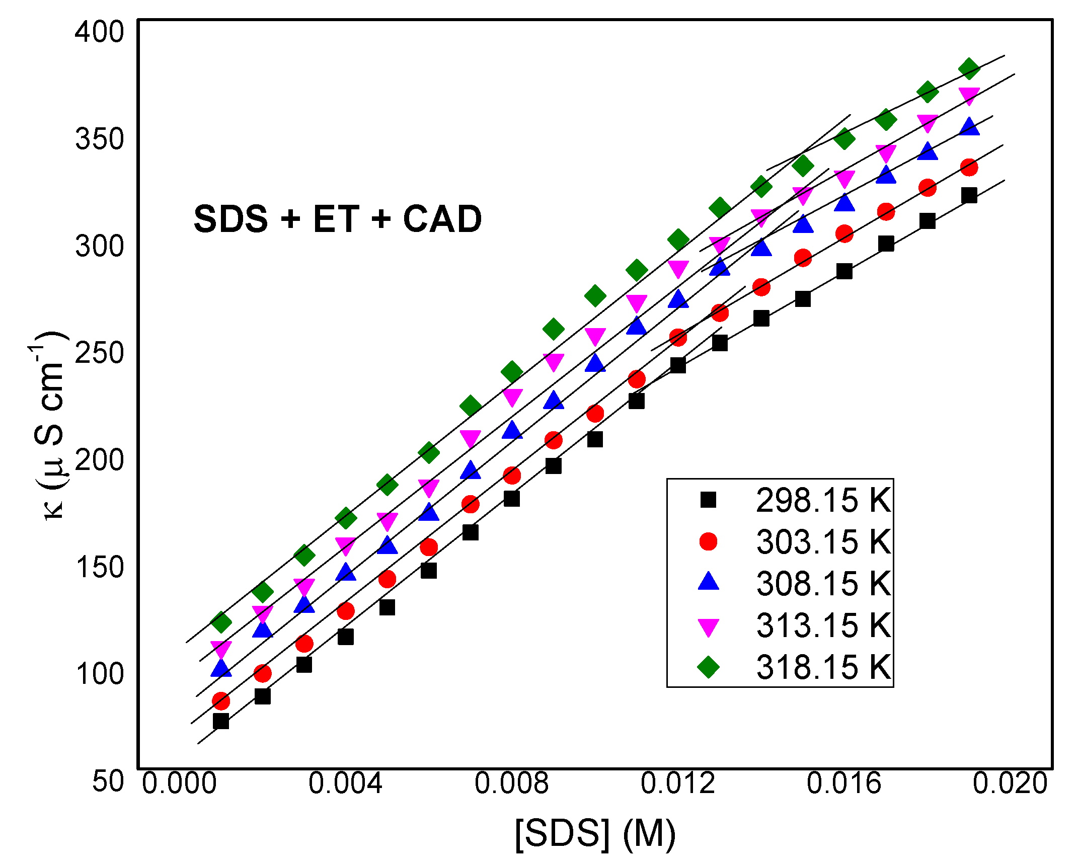
3.1. Conductometric Study
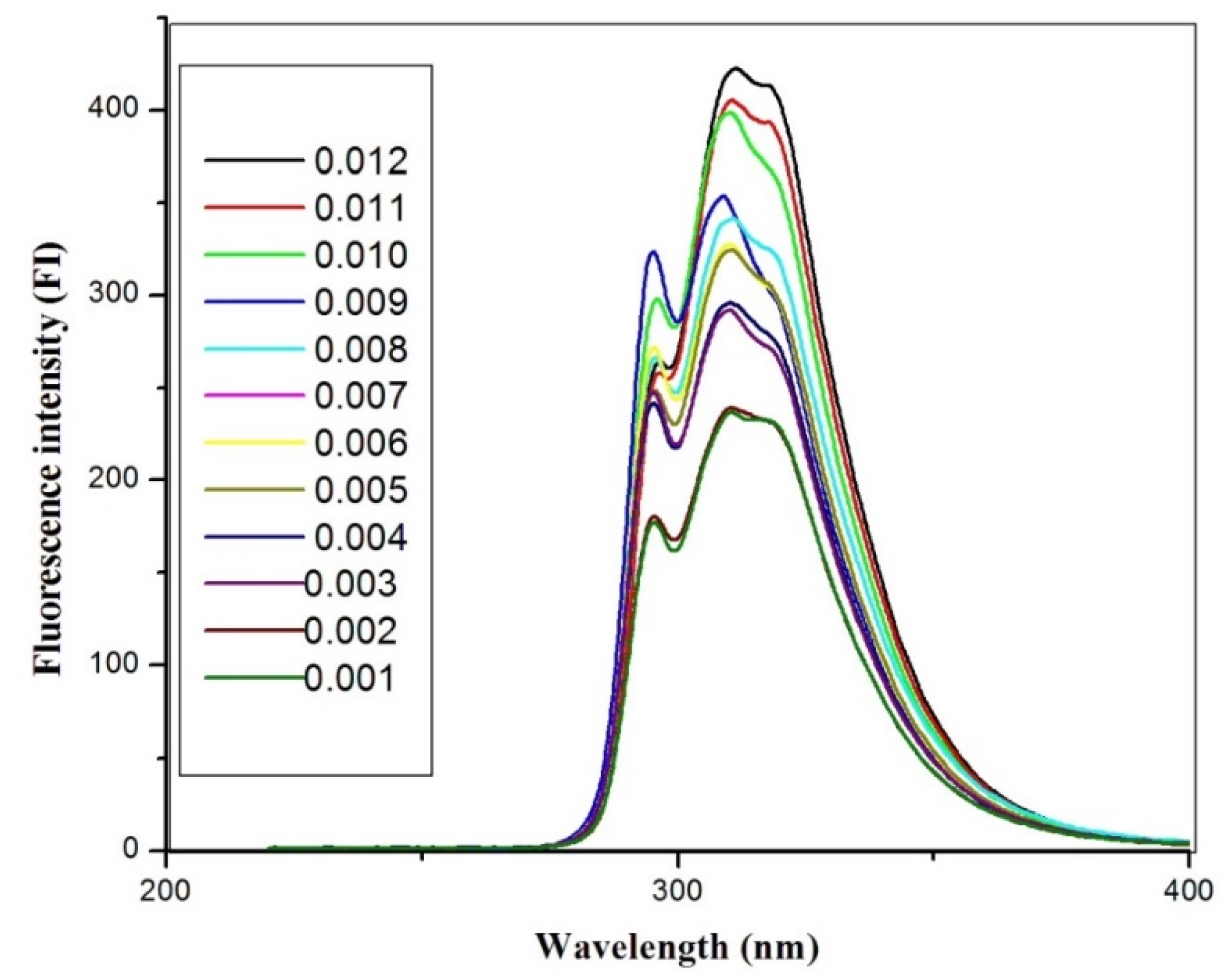
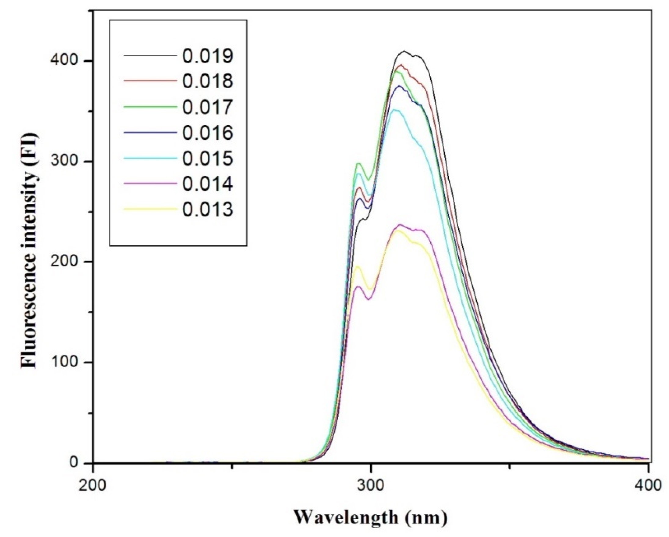
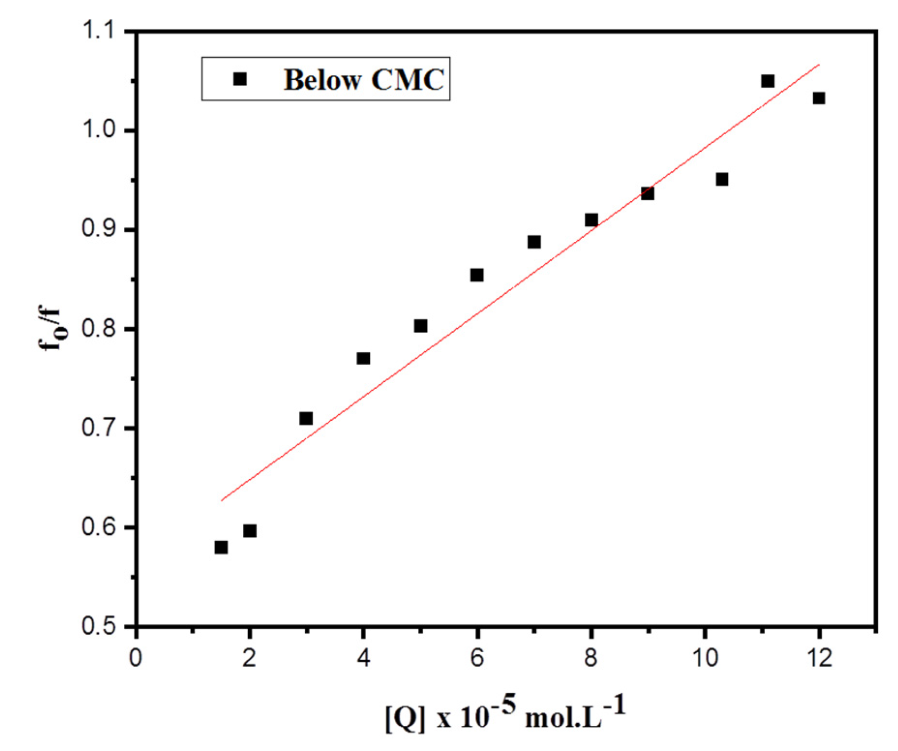
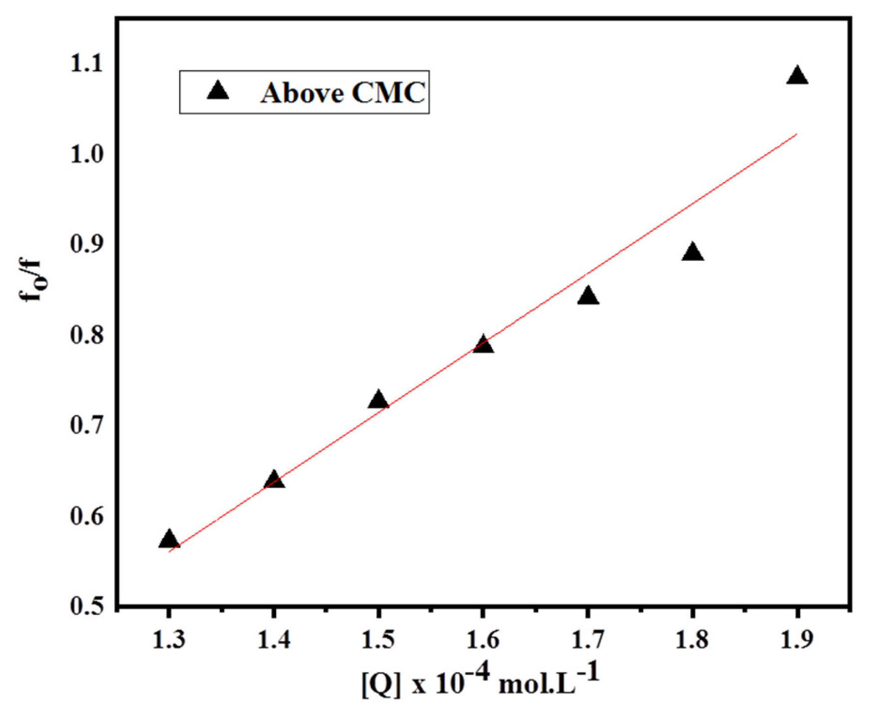
3.2. Fluorescence Spectra Behaviour of Cinnamaldehyde (CAD) in SDS-Ethanol Media
3.3. FTIR Analysis of CAD + SDS + E-OH Media
4. Conclusions
Author Contributions
Funding
Institutional Review Board Statement
Informed Consent Statement
Acknowledgments
Conflicts of Interest
References
- Doyle, A.A.; Stephens, J.C. A review of cinnamaldehyde and its derivatives as antibacterial agents. Fitoterapia 2019, 139, 104405–104454. [Google Scholar] [CrossRef]
- Tan, Y.; Liu, X.; Zhang, L.; Liu, F.; Wang, A.; Zhang, T. Producing of cinnamyl alcohol from cinnamaldehyde over supported gold nanocatalyst. Chin. J. Catal. 2021, 42, 470–481. [Google Scholar] [CrossRef]
- Abbot, V.; Bhardwaj, V.; Sharma, P. Investigation of intermolecular interactions of anionic surfactant SDS and rutin: A physico-chemical approach for pharmaceutical application. J. Mol. Liq. 2021, 337, 116352–116371. [Google Scholar] [CrossRef]
- Sharma, P.; Bhardwaj, V.; Chaudhary, T.; Sharma, I.; Kumar, P.; Chauhan, S. Thermodynamics and micellization of cetyltrimethyl ammonium bromide in the presence of lysozyme. J. Mol. Liq. 2013, 187, 287–293. [Google Scholar] [CrossRef]
- Nabi, A.; Tasneem, S.; Jesudason, C.G.; Lee, V.S.; Md Zain, S.B. Study of interaction between cationic surfactant (CTAB) and paracetamol by electrical conductivity, tensiometric and spectroscopic methods. J. Mol. Liq. 2018, 256, 100–107. [Google Scholar] [CrossRef]
- Roy, S.; Sengupta, P.S.; Guin, P.S. Electrochemical and UV-Vis spectroscopic studies on the interaction of sodium 1,4- dihydroxy-9,10-anthraquinone-2-sulphonate with cetyltrimethylammonium bromide micelles. Chem. Phys. Lett. 2018, 694, 7–13. [Google Scholar] [CrossRef]
- Tanwar, L.K.S.; Banjare, M.K.; Sharma, S.; Ghosh, K.K. Physicochemical studies on the micellization of anionic surfactants in the presence of long alkyl chain ionic liquid. Chem. Phys. Lett. 2021, 769, 138399. [Google Scholar] [CrossRef]
- Autelitano, A.; Minassi, A.; Pagani, A.; Taglialatela-Scafati, O.; Appendino, G. The reaction of cinnamaldehyde and cinnam(o)yl derivatives with thiols. Acta Pharm. Sin. B 2017, 7, 523–526. [Google Scholar] [CrossRef]
- Sawicki, R.; Golus, J.; Przekora, A.; Ludwiczuk, A.; Sieniawska, E.; Ginalska, G. Antimycobacterial Activity of Cinnamaldehyde in a Mycobacterium tuberculosis(H37Ra) Model. Molecules 2018, 23, 2381. [Google Scholar] [CrossRef] [Green Version]
- Tanaka, Y.; Uchi, H.; Furue, M. Antioxidant cinnamaldehyde attenuates UVB-induced photoaging. J. Dermatol. Sci. 2019, 96, 151–158. [Google Scholar] [CrossRef]
- El-Ezz, A.; Maher, A.; Sallam, N.; El-Brairy, A.; Kenawy, S. Trans-cinnamaldehyde Modulates Hippocampal Nrf2 Factor and Inhibits Amyloid Beta Aggregation in LPS-Induced Neuroinflammation Mouse Model. Neurochem. Res. 2018, 43, 2333–2342. [Google Scholar] [CrossRef] [PubMed]
- El-Baroty, G.S.; Abd El-Baky, H.H.; Farag, R.S.; Saleh, M.A. Characterization of antioxidant and antimicrobial compounds of cinnamon and ginger essential oils. Afr. J. Biochem. Res. 2010, 4, 167–174. [Google Scholar]
- Tankam, J.M.; Sawada, Y.; Ito, M. Regular ingestion of cinnamomi cortex pulveratus offers gastroprotective activity in mice. J. Nat. Med. 2013, 67, 289–295. [Google Scholar] [CrossRef] [PubMed] [Green Version]
- Yang, L.; Wu, Q.-Q.; Liu, Y.; Hu, Z.-F.; Bian, Z.-Y.; Tang, Q.-Z. Cinnamaldehyde attenuates pressure overload-induced cardiac hypertrophy. Int. J. Clin. Exp. Pathol. 2015, 8, 14345–14354. [Google Scholar] [PubMed]
- Qi, X.; Zhou, R.; Liu, Y.; Wang, J.; Zhang, W.-N.; Tan, H.-R.; Niu, Y.; Sun, T.; Li, Y.-X.; Yu, J.-Q. Trans-cinnamaldehyde protected PC12 cells against oxygen and glucose deprivation/reperfusion (OGD/R)-induced injury via anti-apoptosis and anti-oxidative stress. Mol. Cell. Biochem. 2016, 421, 67–74. [Google Scholar] [CrossRef]
- Ray, G.B.; Chakraborty, I.; Ghosh, S.; Moulik, S.P.; Palepu, R. Self-Aggregation of Alkyltrimethylammonium Bromides (C10-, C12-, C14-, and C16TAB) and Their Binary Mixtures in Aqueous Medium: A Critical and Comprehensive Assessment of Interfacial Behavior and Bulk Properties with Reference to Two Types of Micelle Formation. Langmuir 2005, 21, 10958–10967. [Google Scholar] [CrossRef]
- Muhammad, M.T.; Khan, M.N. Study of electrolytic effect on the interaction between anionic surfactant and methylene blue using spectrophotometric and conductivity methods. J. Mol. Liq. 2017, 234, 309–314. [Google Scholar] [CrossRef]
- Rosen, M.J.; Kunjappu, J.T. Surfactants and Interfacial Phenomena, 4th ed.; John Wiley & Sons: New York, NY, USA, 2012. [Google Scholar] [CrossRef]
- Ray, G.B.; Ghosh, S.; Moulik, S.P. Physicochemical Studies on the Interfacial and Bulk Behaviors of Sodium N-Dodecanoyl Sarcosinate (SDDS). J. Surfactants Deterg. 2009, 12, 131–143. [Google Scholar] [CrossRef]
- Patist, A.; Oh, S.G.; Leung, R.; Shah, D.O. Kinetics of micellization: Its significance to technological processes. Colloids Surf. A Physicochem. Eng. Asp. 2001, 176, 3–16. [Google Scholar] [CrossRef]
- Shah, S.S.; Jamroz, N.U.; Sharif, Q.M. Micellization parameters and electrostatic interactions in micellar solution of sodium dodecyl sulfate (SDS) at different temperatures. Colloids Surf. A Physicochem. Eng. Asp. 2001, 178, 199–206. [Google Scholar] [CrossRef]
- Bakshi, M.S. Micelle Formation by Sodium Dodecyl Sulfate in Water–Additive Systems. Bull. Chem. Soc. Jpn. 1996, 69, 2723–2729. [Google Scholar] [CrossRef]
- Alhmoud, H.A. The effect of surfactant above and below the critical micelle concentration (CMC) and the mathematical models used to determine the kinetics of drug release from the matrix system. Afr. J. Pharm. Pharmacol. 2016, 10, 88–94. [Google Scholar] [CrossRef]
- Mehta, S.K.; Bhasin, K.K.; Chauhan, R.; Dham, S. Effect of temperature on critical micelle concentration and thermodynamic behavior of dodecyldimethylethyl ammonium bromide and dodecyltrimethylammonium chloride in aqueous media. Colloids Surf. A Physicochem. Eng. Asp. 2005, 255, 153–157. [Google Scholar] [CrossRef]
- Sharma, V.K.; Yadav, O.P.; Singh, J. Physicochemical studies of aqueous sodium dodecylsulphate solutions in pyridine and isomeric picolines. Colloids Surf. A Physicochem. Eng. Asp. 1996, 110, 23–25. [Google Scholar] [CrossRef]
- Sheppard, V.B.; Figueiredo, M.; Canar, J.; Goodman, M.; Caicedo, L.; Kaufman, A.; Norling, G.; Mandelblatt, J. Latina a LatinaSM†: Developing a breast cancer decision support intervention. Psycho-Oncol. J. Psychol. Soc. Behav. Dimens. Cancer 2008, 17, 383–391. [Google Scholar] [CrossRef] [PubMed]
- Mohsen-Nia, M.; Amiri, H.; Jazi, B. Dielectric Constants of Water, Methanol, Ethanol, Butanol and Acetone: Measurement and Computational Study. J. Solut. Chem. 2010, 39, 701–708. [Google Scholar] [CrossRef]
- Usman, M.; Siddiq, M. Surface and micellar properties of Chloroquine Diphosphate and its interactions with surfactants and Human Serum Albumin. J. Chem. Thermodyn. 2013, 58, 359–366. [Google Scholar] [CrossRef]
- Balasubramanian, D.; Srinivas, V.; Gaikar, V.G.; Sharma, M.M. Aggregation behavior of hydrotropic compounds in aqueous solution. J. Phys. Chem. 1989, 93, 3865–3870. [Google Scholar] [CrossRef]
- Vinarov, Z.; Katev, V.; Radeva, D.; Tcholakova, S.; Denkov, N.D. Micellar solubilization of poorly water-soluble drugs: Effect of surfactant and solubilizate molecular structure. Drug Dev. Ind. Pharm. 2018, 44, 677–686. [Google Scholar] [CrossRef]
- Valeur, B. Molecular Fluorescence. Principles and Applications, 2nd ed.; Wiley-VCH Verlag GmbH: New York, NY, USA, 2001; ISBN 978-3-527-32837-6. [Google Scholar]
- Feng, X.Z.; Jin, R.X.; Qu, Y.; He, X.W. Study on the ion effect on the binding interaction between HP and BSA. Chem. J. Chin. Univ. 1996, 17, 866–869. [Google Scholar]
- Salanci, E.; Malík, I.; Šandrik, R.; Pecher, D.; Andriamainty, F. Determination of the critical micelle concentration and thermodynamic parameters of phenylcarbamic acid derivatives using a fluorescence method. Chem. Pap. 2021, 75, 3081–3090. [Google Scholar] [CrossRef]
- Bissantz, C.; Kuhn, B.; Stahl, M. Medicinal Chemist’s Guide to Molecular Interactions. J. Med. Chem. 2010, 53, 5061–5084. [Google Scholar] [CrossRef] [PubMed]
- Sastrohamidjojo, H. Dasar-Dasar Spektroskopi; UGM Press: Yogyakarta, Indonesia, 2018; ISBN 978-979-420-817-5. [Google Scholar]
- Ma, C.-L.; Sun, X.-D. Preparation and characterization of SnO2 nanoparticles with a surfactant-mediated method. Nanotechnology 2002, 13, 565–569. [Google Scholar] [CrossRef]
- Tao, Q.; He, H.; Frost, R.L.; Yuan, P.; Zhu, J. Nanomaterials based upon silylated layered double hydroxides. Appl. Surf. Sci. 2009, 255, 4334–4340. [Google Scholar] [CrossRef]


| T (K) | CMC (M) | (kJ mol−1) | (kJ mol−1) | (J mol−1 K−1) | (J mol−1 K−1) | |
|---|---|---|---|---|---|---|
| SDS + Ethanol | ||||||
| 298.15 | 0.00822 | 0.65 | −29.51 | −13.06 | 0.055 | 16.9051 |
| 303.15 | 0.00881 | 0.64 | −29.88 | −13.56 | 0.054 | 16.3701 |
| 308.15 | 0.00939 | 0.60 | −31.16 | −14.47 | 0.054 | 16.6401 |
| 313.15 | 0.00995 | 0.59 | −31.68 | −15.05 | 0.053 | 16.5969 |
| 318.15 | 0.01073 | 0.56 | −32.50 | −15.83 | 0.052 | 16.5438 |
| SDS + Ethanol + Cinnamaldehyde | ||||||
| 298.15 | 0.0120 | 0.76 | −22.99 | −1.72 | 0.071 | 21.1686 |
| 303.15 | 0.0122 | 0.73 | −23.74 | −2.68 | 0.069 | 20.9173 |
| 308.15 | 0.0125 | 0.71 | −24.49 | −3.74 | 0.067 | 20.6460 |
| 313.15 | 0.0127 | 0.69 | −25.32 | −4.90 | 0.065 | 20.3547 |
| 318.15 | 0.0129 | 0.66 | −26.15 | −6.17 | 0.063 | 20.0434 |
| (Measured at λex 295 nm) | Below CMC | Above CMC |
|---|---|---|
| Ksv (103 M−1) | 4.19 | 7.68 |
| Kq | 4.5 × 10¹⁰ | 3.3 × 10¹⁵ |
| Kb (M−1) | 4.51 × 10² | 3.34 × 10⁷ |
| n | 1.22 | 4.38 |
| ∆G° (104 kJ mol−1) | −1.51 | −4.30 |
Publisher’s Note: MDPI stays neutral with regard to jurisdictional claims in published maps and institutional affiliations. |
© 2022 by the authors. Licensee MDPI, Basel, Switzerland. This article is an open access article distributed under the terms and conditions of the Creative Commons Attribution (CC BY) license (https://creativecommons.org/licenses/by/4.0/).
Share and Cite
Alamier, W.M.; Tasneem, S.; Nabi, A.; Hasan, N.; Nabi, F. Thermodynamic and Spectroscopic Studies of SDS in Cinnamaldehyde + Ethanol Mixtures: Influences of Temperature and Composition. Appl. Sci. 2022, 12, 12020. https://doi.org/10.3390/app122312020
Alamier WM, Tasneem S, Nabi A, Hasan N, Nabi F. Thermodynamic and Spectroscopic Studies of SDS in Cinnamaldehyde + Ethanol Mixtures: Influences of Temperature and Composition. Applied Sciences. 2022; 12(23):12020. https://doi.org/10.3390/app122312020
Chicago/Turabian StyleAlamier, Waleed M., Shadma Tasneem, Arshid Nabi, Nazim Hasan, and Firdosa Nabi. 2022. "Thermodynamic and Spectroscopic Studies of SDS in Cinnamaldehyde + Ethanol Mixtures: Influences of Temperature and Composition" Applied Sciences 12, no. 23: 12020. https://doi.org/10.3390/app122312020