
Targeted Delivery Methods for Anticancer Drugs
by
 , and
, and
Valery V. Veselov
1, 
Alexander E. Nosyrev
1,
László Jicsinszky
2 ,
,
Renad N. Alyautdin
3 and
Giancarlo Cravotto
2,4,*
1
Center of Bioanalytical Investigation and Molecular Design, Sechenov First Moscow State Medical University, 8 Trubetskaya ul, 119991 Moscow, Russia
2
Department of Drug Science and Technology, University of Turin, Via P. Giuria 9, 10125 Turin, Italy
3
Department of Pharmacology, Sechenov First Moscow State Medical University, 119991 Moscow, Russia
4
World-Class Research Center “Digital Biodesign and Personalized Healthcare”, Sechenov First Moscow State Medical University, 8 Trubetskaya ul, 119991 Moscow, Russia
*
Author to whom correspondence should be addressed.
Cancers 2022, 14(3), 622; https://doi.org/10.3390/cancers14030622
Submission received: 30 December 2021
/
Revised: 21 January 2022
/
Accepted: 25 January 2022
/
Published: 26 January 2022
Abstract
:Simple Summary
The current main technological strategies for the delivery of anticancer drugs are discussed herein. This comprehensive review may help researchers design suitable delivery systems.
Abstract
Several drug-delivery systems have been reported on and often successfully applied in cancer therapy. Cell-targeted delivery can reduce the overall toxicity of cytotoxic drugs and increase their effectiveness and selectivity. Besides traditional liposomal and micellar formulations, various nanocarrier systems have recently become the focus of developmental interest. This review discusses the preparation and targeting techniques as well as the properties of several liposome-, micelle-, solid-lipid nanoparticle-, dendrimer-, gold-, and magnetic-nanoparticle-based delivery systems. Approaches for targeted drug delivery and systems for drug release under a range of stimuli are also discussed.
1. Introduction
Over the past 30 years, the number of successful cancer treatments has significantly increased, predominantly driven by our improved understanding of carcinogenesis processes, cell biology, and the tumor microenvironment [1,2]. However, many cancers are still fatal despite the sustained effort being invested in preclinical and clinical research. One of the ways to improve the survival rate of cancer patients is the targeted delivery of anticancer drugs. Advances in biomedical science and biotechnology have led to the discovery and development of effective drug carriers such as liposomes, dendrimers, and gold and magnetic nanoparticles [3,4,5,6]. The principal difference between these new types of formulation and classical ones is their suitability for the potential development of technologies for targeted drug delivery to specific tissues, cells, and even intracellular organelles. The essence of targeted delivery lies in the surface of a drug container (carrier) bearing a modified drug or molecule with a functional group that can be recognized by the target cell receptors. Folic acid modification is a classic example as it is actively taken up by tumor cells [7,8,9]. Antibodies and aptamers are universal molecules that recognize the surface of a target cell [10,11,12]. Thanks to advances in basic biomedical research, the antigenic portraits of cells are becoming more and more detailed, allowing us to distinguish one cell from another based on their surface characteristics. Orally or parenterally administered medicines are distributed throughout the body, with only a small portion reaching the target area. Targeted delivery methods, therefore, make it possible to reduce the dosage of an administered drug and minimize its effect on other cells, which is very important in chemotherapy as drugs are highly toxic. The presence of recognizing or recognizable molecules on the surface of a delivery system allows it to concentrate on the desired area. It is also vital that the delivery system penetrates the cell and that the drug is then delivered to the nucleus, mitochondria, endoplasmic reticulum, and other organelles. In fact, the concept of intracellular drug delivery is under active development. Knowledge of the signaling pathways involving proteins that lead to different cellular structures is essential to achieve efficient intracellular transport [13]. Equally important is the need for more knowledge of the motor proteins of cells, which directionally move loads over long distances inside cells. It is also necessary to understand the mechanisms by which drugs are released from delivery systems, including diffusion, degradation, swelling, and other processes that can control the release of drugs [14,15,16].
2. Types of Containers and Carriers
A drug-delivery system may be considered suitable for clinical practice if it is non-toxic, biocompatible, stable in blood, non-immunogenic, non-thrombogenic, and biodegradable [17].
The enhanced permeability and retention (EPR) effect is the principle behind the passive targeting used in all containers and carriers. This principle and term were proposed in 1986 [18]. Rapid tumor growth is accompanied by neovascularization with wide fenestrations and the suppression of lymphatic drainage [19]. Traditionally, the EPR concept presumes that small molecules enter via diffusion and leave the interstitial space of the tumor, whereas macromolecules (containers, carriers) are no longer able to do so after extravasation [20]. In addition to the traditional explanation, other theories concerning how the pathophysiological characteristics of tumor growth shape the EPR effect have been put forward [21,22]. Thus, the EPR effect has been accepted as a universal principle incorporated into the design of anticancer drug-delivery systems [23]. However, there are currently serious disputes about the effectiveness of the EPR effect when using nanoparticles [24,25]. At the same time, it is important to note that the current understanding of the EPR effect is based on results obtained in animal models, meaning that the results of EPR-effect studies in patients must be collated if delivery systems that fully exploit the EPR effect are to be successfully designed [26].
2.1. Liposomes
Liposomes are spherical vesicles consisting of one or more lipid bilayers. A liposome has a hollow structure that is usually filled with a solvent and can deliver a variety of substances. Its hydrophobic membrane allows it to merge with cell membranes and transport its contents inside cells. Liposomes are most often composed of phospholipids and cholesterol, but may also include other lipids to improve endocytosis and tissue compatibility. Many methods have been developed to produce a range of liposomal compositions [27], and all described liposome fabrication methods combine lipids with the aqueous phase in some way [28,29]. The thin-layer hydration method, also known as the Bangham method, is one of the first and still most commonly used methods for the preparation of liposomes [30]. This method involves lipids being dissolved in the organic phase and removing the organic solvent, usually by evaporation, to form a lipid film. The lipid film is then dispersed in an aqueous medium that contains the drug under vigorous stirring to form the sealed spherical structures; liposomes. The short elimination half-life of liposomes, caused by their opsonization principally in the liver and spleen, is a crucial weak point in their use [31]. The modification of liposome surfaces with various functional ligands, such as polyethylene glycol (PEG) coating, reduces the interaction between the surface and blood components, thus ensuring that the liposomes have a longer residence time in the bloodstream [32]. PEG can be attached to liposome surfaces in a variety of ways:
- Physical adsorption onto the surface of liposomes.
- Covalent attachment using reactive groups on the surface of preformed liposomes.
- Inclusion of a PEG-lipid conjugate in liposome preparations.
The most common method anchors the polymer in the membrane using a cross-linked lipid (e.g., PEG-distearoylphosphatidylethanolamine) [33]. The presence of PEG on liposome surfaces reduces their aggregation [34]. To ensure targeted delivery, PEG is also often covalently bound to proteins (transport, signaling) so that, while the mechanism of action of the proteins does not change, there is a change in protein pharmacokinetics; PEG-asparaginase (used in the treatment of leukemia), PEG-aldesleukin (an antineoplastic agent), PEG-filgrastim (for the treatment of chemotherapy-induced febrile neutropenia), and PEG-epoetin-β (for the treatment of anemia) are commonly used in the treatment of cancer [35,36]. The liposomal delivery of anticancer drugs has been successfully used in cancer therapy for several decades [37].
2.2. Micelles
Micelles are particles of tens of nanometers in size with a hydrophobic core and a hydrophilic surface and are commonly used as carriers of hydrophobic drugs. Like liposomes, they can be delivered directly into the bloodstream through the respiratory tract or skin. In recent years, amphiphilic block copolymers, which spontaneously form micellar structures, have attracted much attention because of their use in the delivery of cytostatic drugs [38,39]. Amphiphilic block copolymers are usually assembled from two or three blocks, with PEG being the most common hydrophilic block in the copolymer structure. Other hydrophilic block-forming polymers include chitosan, polyvinylpyrrolidone, and poly(N-isopropyl acrylamide) [40]. Polymers of various compound classes are used as hydrophobic polymer blocks for micellar core creation: polyethers (poly(propylene oxide)) polyesters (polylactide), polycarboxylic acids (poly(aspartic acid)) and lipids (distearoylphosphatidyl ethanolamine) [40]. Micelles that contain functional groups (-NH2, -COOH) in their core can transfer drugs by chemical modification and not just by physical encapsulation [41], and various cytostatic drug micelles (doxorubicin, paclitaxel) have shown significant results in several in-vitro and in-vivo studies [42]. Paclitaxel encapsulated in micelles has been tested in clinical trials in patients with malignant tumors with a resulting reduction in toxicity and no change in the antitumor activity compared to free paclitaxel [43].
2.3. Solid-Lipid Nanoparticles
Solid-lipid nanoparticles are colloidal nanoparticles stabilized by surfactants and composed of mono-, di- and triglycerides, solid fats, and waxes. They have been developed as an alternative to liposome technologies to increase stability, modulate the release of encapsulated drugs, reduce costs, and simplify manufacturing [44]. Unlike liposomes, which are usually injected into the body intravenously, intraperitoneally, subcutaneously, and orally, solid-lipid nanoparticles can be administered via different routes, via inhalation, intranasally, and intravesically [45], thus ensuring the local targeting of the drug. Recent in-vitro and in-vivo experiments have shown that solid-lipid nanoparticles that contain cytostatic drugs appear to be superior to conventional drug solutions and are comparable to other encapsulated systems in many aspects, such as efficacy, pharmacokinetics, and bioavailability [46]. However, clinical studies have not yet been conducted in this area.
2.4. Gold Nanoparticles
Gold nanoparticles (AuNP) can boast a combination of unique physical and chemical properties relative to other biomedical nanotechnologies and can selectively deliver cytostatic drugs [47,48]. AuNPs offer significant potential for new approaches to cancer treatment as they are easy to produce, have low toxicity, and display antiangiogenic properties [49]. AuNPs are up to 100 nm in size, have a pronounced EPR-effect, and, as a result, preferentially accumulate in tumors.
AuNP-based supports are most often synthesized using colloidal methods; gold salts (e.g., hydrogen tetrachloroaurate (III)) are reduced in the presence of surface stabilizers that prevent the aggregation of the resulting solution [50]. Spherical AuNPs are principally used to create delivery systems because they can be synthesized on a large scale with high monodispersity. The other forms of AuNP include nanorods, nanoshells, and nano cells [51]. AuNPs can undergo surface modification thanks to their covalent and non-covalent bond-forming properties [51]. A stabilizing agent (e.g., citric acid) is responsible for the overall charge of the AuNP surface. The correct choice of a stabilizing agent allows various biomolecules (DNA, antibodies, polypeptides) to be conjugated to the AuNP surface via electrostatic interactions, whereas covalent attachment to AuNPs is usually achieved via the interaction between gold and thiol, amine, and carboxylate functional groups [52]. Unlike liposomes and micelles, the drug is conjugated directly to the AuNP surface using various linkers [52,53,54]. It is worth noting that the overwhelming majority of studies on AuNP-based directional transport are based on spherical AuNPs, and this is, at least in part, because they undergo surface modification and penetrate cells more easily than more complex AuNPs. A drug conjugated to AuNPs has shown increased antitumor potential compared to the free drug in in-vitro and in-vivo studies [47,55,56,57,58].
2.5. Magnetic Nanoparticles
Magnetic targeting is of great interest in the treatment of malign tumors as the technique not only provides targeted drug delivery but also makes it possible to monitor the accumulation of magnetic nanoparticles (MNP) in tumors using magnetic resonance imaging (MRI) [59,60]. MNPs that carry a drug are first accumulated in the target tissue using an external magnetic field, and the drug is then released from the MNPs in a controlled manner [61].
MNPs are magnetic materials with small particle sizes (from 10 to 100 nm), a large specific surface area, magnetic response, and superparamagnetism [62]. This superparamagnetism means that MNPs are in a single-domain state, as they are uniformly magnetized throughout the entire volume [63], and that the orientation of their magnetic moment changes with temperature [63]. Iron oxides, for example magnetite (Fe3O4 or FeO.Fe2O3) and maghemite (γ-Fe2O3), are usually used for MNP production [60,64]. The MNP core, which consists of magnetite, maghemite, or a mixture of the two, is usually obtained via the precipitation of Fe2+ and Fe3+ iron salts from an aqueous solution [65,66]. Moreover, it is possible to regulate the size of the resulting nanoparticles by adding various iron salts (chloride, sulfate, nitrate, etc.) and by changing the ratio of Fe2+ and Fe3+, the pH, and the ionic strength in the solution [62,67]. Reactions are carried out in an inert atmosphere to prevent the oxidation of the formed nanoparticles [68]. The formed MNPs have a hydrophobic surface and are coated with synthetic and natural polymers to reduce nanoparticle agglomeration [60] and further modify the surface to conjugate drugs and biomolecules [69]. The most commonly used polymers are PEG, dextran, polyvinylpyrrolidone, polyaniline, alginate various fatty acids, and chitosan [70,71]. In general, the conjugates of MNPs with various cytostatics show decreased overall toxicity, and the concentration of cytostatic agents is required to achieve a therapeutic effect [72,73,74,75,76,77]. The ability of MNPs to accumulate in tumors has also been confirmed by MRI [59,74,78,79,80].
2.6. Dendrimers
Dendrimers are three-dimensional, monomolecular, highly branched monodisperse macromolecules [81] that usually have rotational symmetry and often take on a spherical shape. In general, dendrimers have a hydrophobic core from which they branch, ending in terminal functional groups responsible for their solubility in water [82]. These dendrimers can retain hydrophobic drugs and increase their concentration in water. Biocompatibility, easy excretion from the body, and a significantly improved EPR effect are the most remarkable advantages of dendrimers. However, dendrimers have one significant drawback; they are cytotoxic for normal cells due to the physiological stability of cationic groups on their surfaces [83]. The problem of dendrimer cytotoxicity is usually solved by modifying their surface using biocompatible polymers, for example, PEG. The PEG-modified dendrimer surface provides the necessary screening of the cationic surface charge, which leads to a biologically safe carrier [84].
Dendrimer synthesis is a rather laborious process. There are two principal approaches to the synthesis of dendrimers; divergent and convergent methods [85]. In the divergent version, a base reagent (a molecule that is protected at its end groups, if necessary) is attached to the original branching center (which has several end groups). The protecting groups are removed, and a 1st generation dendrimer is formed. Subsequently, dendrimers of higher generations are obtained by attaching either the original branching center or the base reagent, followed by deprotection [86]. In the convergent method, the arms of the dendrimer are synthesized first and then connected [86], and this method produces more monodisperse dendrimers than the divergent version. However, the size of dendrimers obtained using the convergent method is limited due to steric hindrance, whereas dendrimers of a wider variety of sizes can be obtained using the divergent method [85,86]. The most widely used dendrimers are currently the commercially available poly(amidoamine) (PAMAM) dendrimers [87,88,89]. Delivery systems based on poly(propylene imine) [90], polylysine [91], carbosilane [92], and phosphorus dendrimers [93] have also been developed. Numerous studies have shown the effectiveness of using different dendrimers for targeted transport in cancer therapy [94,95,96,97,98,99], and several clinical trials using various dendrimers as targeted delivery systems are underway [100].
2.7. Albumin-Based Nanoparticles
Albumin is the most abundant plasma protein in human blood, with a molecular weight of about 67 kDa. Due to its endogenous origin, it is non-toxic, non-immunogenic, biocompatible, and biodegradable [101]. Human serum albumin (HSA) and the cheaper bovine serum albumin (BSA) and ovalbumin (OVA) have been used to create delivery systems [102]. HSA has several ligand binding sites that can be used for transfer via both hydrophobic and electrostatic interactions [103,104], and the presence of a free cysteine residue on its surface means that albumin easily conjugates with a variety of ligands [105,106]. Receptors, such as albondin (Gp60) and secreted protein acidic and rich in cysteine (SPARC), have been shown to overexpress in some cancers [107] and can mediate albumin transcytosis [108], while the Gp30 and Gp18 receptors, the megalin/cubilin complex and the neonatal Fc receptor (FcRn) are also involved in albumin transport [106]. Albumin-based delivery systems can therefore accumulate in tumors via mechanisms beyond the EPR effect. Albumin-based nanoparticles are obtained by various methods, including emulsification, self-assembly of thermal gelation, desolvation, and nanospray drying [109,110]. The patented nanoparticle albumin-bound (NAB) technology, which consists of the evaporation of an emulsion with the creation of cross-links between albumin units, is the best-known preparation method for albumin-based nanoparticles [105,106]. In addition to Abraxane®, which is created with the help of NAB technology and has been successfully used in clinical practice [111,112,113], work is also underway to create a range of albumin-based nanoparticles [104,105,106,107,108,109,110,111,112,113,114,115,116,117,118].
2.8. Porous Materials
Zeolites are hydrated crystalline aluminosilicates consisting of tetrahedral groups, [SiO4]4− and [AlO4]5−, united by common vertices into a three-dimensional framework. The open frame-cavity structure of zeolites has a negative charge, which is compensated for by counterions [119]. Zeolites have a porous structure that can absorb various substances, making zeolites an ideal material for drug-delivery systems [120]. To prevent the untimely release of a drug, either a zeolite with an optimal pore size is selected [121], or its surface is modified with various ligands [122,123]. In general, zeolites are promising carriers for creating systems for the delivery of cytotoxic substances [124,125,126,127].
Mesoporous silica particles (MSP) are another porous material used for drug delivery [128]. Their pore size can be adjusted from 2 to 50 nm, as in the case of zeolites, to tune them for a specific drug [129,130,131]. The surfaces of MSPs are rich in reactive silanol groups, which can be used for conjugation with various substances [132], and MSPs have been developed with several structures. The morphology and size of both the particles themselves and their pores can be controlled via the choice of a synthetic method [133,134]. MSP-based delivery systems have shown high drug-loading capacity, successfully controlled release, and increased antitumor activity [79,135,136,137].
2.9. Carbon Nanoparticles
Carbon has many allotropic modifications, including carbon nanotubes, fullerenes, and nanodiamonds, which have found applications as carriers for drug delivery [138]. Carbon-based quantum dots are also used (see the Section 2.10). Carbon nanoparticles have a high specific surface area and hydrophobicity. Carbon nanotubes and fullerenes have cavities in their structure and can encapsulate active substances [138,139]. However, unlike fullerenes and carbon nanotubes, the surface of nanodiamonds is rougher, which increases adhesion with drugs [140]. Under the action of acidic oxidation, carboxyl groups are formed on the surface of carbon nanoparticles and are used for surface modification, as well as for the covalent attachment of anticancer drugs [141,142,143]. Carbon nanoparticles with the desired properties can be obtained by correctly choosing and adapting the synthesis method [144,145,146].
While carbon nanoparticles are currently widely used for drug delivery, their toxic properties are concentration-dependent [147,148,149]. Attention should therefore be paid to the delivery method when developing carbon-nanoparticle supports. For example, it has been shown that the absorption of fullerene by the respiratory and digestive tracts is low [150]. In addition, as has been demonstrated, inhaled carbon nanotubes can act on the body similarly to asbestos [151].
2.10. Quantum Dots
Quantum dots are inorganic semiconductor nanocrystals and are typically up to 10 nm in size. Quantum dots have fluorescent, optical and electronic properties [152], with cadmium-compound-based and carbon quantum dots being the most widespread [153,154]. In addition to drug delivery, quantum dots can visualize cancer cells due to their unique optical properties, which derive from quantum and other effects [155]. The quantum dots used in biomedicine typically consist of a core and a coating with the core imparting optical properties to the system and the coating performing a protective function, which enables the surface to be functionalized with various ligands and is responsible for water solubility [156]. The quantum-dot core may be composed of cadmium compounds, such as cadmium selenide (CdSe), cadmium sulfide (CdS), and cadmium telluride (CdTe), and these quantum dots have shown notable results as drug carriers [157,158,159]. It is important to mention that these quantum dots are not biodegradable and are not cell and environmentally friendly due to the toxicity of cadmium compounds [160].
Carbon-based quantum dots, which can be classified as either carbon quantum dots or graphene quantum dots, are widely used in various fields of biomedicine [161]. They possess low toxicity, high specific surface area, high photostability and are easily modified [162]. Carbon-based quantum dots are excellent carriers for anticancer drugs due to their biocompatibility, ease of manufacture, and lower environmental impact [163,164,165,166].
2.11. Calcium Phosphate
Calcium phosphate (CaP)-based nanoparticles are crystalline formations of predominantly carbonate apatite capable of transporting a drug both on their surface and within their structure [167,168]. Minerals based on CaPs are the main inorganic components of the bones and teeth of vertebrates and humans [169]. CaP-based nanoparticles have several peculiar properties that make them attractive for delivering anticancer drugs. CaPs are fully biodegradable, release non-toxic calcium and phosphate ions upon degradation, and decompose faster than other inorganic nanoparticles (zeolites, mesoporous silica particles, carbon nanoparticles, and quantum dots) [168,170]. Moreover, CaP-based nanoparticles have pH-sensitive solubility; they are insoluble at the physiological pH of blood plasma (7.4) but quickly dissolve in acidic biological media (pH < 5), for example, in endosomes and lysosomes, where they rapidly release encapsulated substances [170,171,172]. There are currently many approaches for synthesizing CaP-based nanoparticles, and the careful selection of synthesis conditions makes it possible to control the size and morphology of the resulting particles [173,174,175,176]. Although nanoparticles with different morphologies are used for delivery in cancer therapy, including rod shapes [169,177], porous structures [178,179], and core-shell shapes [180,181], spherical nanoparticles are the most commonly used since they are more thermodynamically stable [182,183].
2.12. Oligo- and Polysaccharide-Based Drug-Delivery Systems
2.12.1. Chitosan
Chitosan is a type of amino polysaccharide polymer (see Figure 1) produced via the deacetylation of chitin, and is the second most common biopolymer in nature after cellulose. Chitin is the main component of the exoskeleton of arthropods and many other invertebrates and is also part of the cell walls of fungi [184]. Chitosan has amino functionalities that are useful for biopolymer modification [185]. Its biodegradability, biocompatibility, low immunogenicity, and non-toxicity mean that chitosan is used in delivery systems for various chemotherapeutic drugs [186]. Chitosan and its derivatives, such as carboxymethyl chitosan, sulfated chitosan, sulfated benzaldehyde chitosan, and polypyrrole-chitosan, have been shown to have anticancer activity in and of themselves [187,188,189,190]. This property is assumed to be related to the antioxidant properties of chitosan and its derivatives, which are capable of trapping cancer-causing free radicals [191].
As mentioned earlier, chitosan can be a hydrophilic moiety in an amphiphilic block copolymer [192]. The presence of free amino groups in the chitosan backbone grants it a unique polycationic character that ensures that negatively charged drugs, such as doxorubicin, are properly encapsulated [193]. Some chitosan-based hydrogels that contain a significant amount of water and retain a self-organized three-dimensional structure have been developed and can be used for the encapsulation and delivery of anticancer drugs [194,195]. Various forms of delivery systems, such as microspheres, film capsules, etc., have been obtained using water-insoluble species of chitosan [196], meaning that the properties of chitosan-based delivery systems are easy to modulate. Depending on the preparation method selected, it is possible to regulate the particle size, toxicity, thermal and chemical stability, and release kinetics [197].
2.12.2. Cyclodextrins
Cyclodextrins (CDs) are a family of cyclic oligosaccharides that consist of glucose subunits obtained by enzymatic means from starch [198]. The most commonly used CD types are α-, β- and γ-CDs (Figure 2a), named according to the number of glucose residues they possess [199,200].
CDs have a hydrophilic outer surface and a significantly less hydrophilic cavity. They take the form of a truncated cone with a cylindrical-torus cavity inside (Figure 2b) [201]. CDs can form various complexes with hydrophilic, lipophilic, and amphiphilic substances. CDs, therefore, can often increase the solubility and bioavailability of many anticancer drugs [202], and various cyclodextrin derivatives are widely used to create drug-delivery systems [203].
CDs are often combined with other nanoparticles to create delivery systems [201]. It has been shown that the loading of liposomes with anticancer drugs in combination with cyclodextrin increases their half-life, reduces toxicity, and increases liposome loading [204,205,206,207]. Several CD-based polymers have also been developed and used successfully in drug transport [208].
The cross-linking of CDs results in unique particles, namely CD polymers and nanosponges. Nanosponges are a type of nanoparticle that has a porous structure with a pore size of several nanometers. Due to their unique structure, CD polymers and nanosponges can encapsulate various substances in their pores and act as drug transporters [209]. One of the advantages of natural CD-based delivery systems is the creation of effective oral, mucosal and transdermal drug formulations [210]. Cyclodextrin-based macromolecules can transfer oligonucleotides, siRNAs, or their fragments into the cells. Though the promising results reported by these associations, experimental trials are still in progress [211,212,213,214].
2.12.3. Pectins
Pectins are polysaccharides that are mainly formed from residues of galacturonic acid. Pectins are extracted in different ways from higher plants, mainly from their fruits. Consequently, the structures of pectins can be very diverse, although they can be classified into three types based on their general characteristics: homogalacturonan, rhamnogalacturonan-I, and substituted galacturonans [215]. Pectin and its various modifications have anticancer activities [216,217,218], and the majority of studies on natural and modified pectins, and their delivery systems, have mainly focused on colon cancer [219,220,221]. This is primarily because pectin is not digested in the gastrointestinal tract until it reaches the colon, where it is fermented and breaks down to release encapsulated active ingredients [222,223]. Pectin-based microgranules and microspheres have been developed to encapsulate anticancer drugs and release them directly into the colon [224,225]. Another use of pectins is as a drug carrier in the preparation of various hydrogels [226,227]. As pectins contain carboxyl groups, it is possible to use them to create negatively charged particles that retain drugs thanks to electrostatic interactions [228]. Moreover, pectin has been used to create self-organizing polymer nanoparticles to deliver ursolic acid [229].
3. The Targeting Methods of Delivery Systems
Active targeting is used to increase the concentration of cytostatics in the desired organ or tissues to achieve higher and more selective therapeutic activity. The surface of the container or carrier is modified with various recognizable or recognition molecules, such as monoclonal antibodies or their fragments, aptamers, proteins, peptides, and low molecular weight compounds, to grant active-targeting properties [230].
3.1. Antibodies and Aptamers
Numerous monoclonal antibodies (mAb) have recently been developed against various epitopes of cancer cells and are used as therapeutic agents in and of themselves [231]. mAb-conjugated containers or carriers specifically bind to a target cell (receptor, protein, etc.) in the desired areas and then release the encapsulated drug. The surface modification of the carrier systems with mAbs can either be achieved via non-covalent physical interactions or the formation of covalent bonds [232], with non-covalent bonding being faster than covalent. However, the antigen-binding domains of mAbs are arranged chaotically in non-covalent conjugation, which can lead to disruption in mAb functionality [233]. The most commonly used method for non-covalent conjugation is the streptavidin-biotin method [234]. It consists of the preliminary non-covalent conjugation of the surface of the delivery systems with streptavidin, which has a high affinity for biotin, which, in turn, is covalently linked to the mAbs.
The most commonly used cancer targets for mAbs are:
Conjugation with aptamers is a newer approach to targeted delivery. Aptamers are oligonucleotides (DNA, RNA aptamers) or peptide molecules that specifically bind to specific target molecules and can be considered analogs of monoclonal antibodies. However, they have many advantages over antibodies. Their production is much easier, cheaper, and faster than monoclonal antibodies. They have a much smaller size and, therefore, more easily penetrate tissues and cells, as well as having higher affinity and specificity [248]. The potential for the in-vivo targeting of RNA aptamers in cancer therapy was demonstrated for the first time in 2006 [249]. More than 20 different systems for targeted transport are currently being developed using oligonucleotide aptamers [250].
Peptide aptamers consist of a short (10–20 amino acid), conformationally limited peptide sequence that is inserted into a scaffold protein (most often the bacterial protein thioredoxin A) [251,252]. A unique feature of peptide aptamers is that their variable region has a double limitation as both ends are connected to a framework (protein), unlike oligonucleotide aptamers and antibodies [252]. For this reason, peptide aptamers have limited conformations and require less energy to bind to the target, which, in turn, increases their affinity.
3.2. Proteins and Peptides
The most commonly used protein for targeting delivery systems is transferrin, a serum glycoprotein that transports iron into cells by binding to transferrin receptors on the cell surface [253]. The transferrin receptor is present in malignant tumors at levels that are hundreds of times higher than in normal cells (Figure 3) [254]. Containers and carriers, their surface modified by transferrin molecules, can therefore penetrate cancer cells and accumulate in them [230]. The following proteins and peptides are used to target other receptors that are overexpressed in cancer cells:
Figure 3.
Schematic representation of the penetration of transferrin into a cell via receptor-mediated endocytosis. Original diagram inspired by [263].
Figure 3.
Schematic representation of the penetration of transferrin into a cell via receptor-mediated endocytosis. Original diagram inspired by [263].

3.3. Low Molecular Weight Compounds
Modifying the surfaces of containers and carriers with folic acid is currently a commonly used methodology to ensure the delivery of an encapsulated drug into cancer cells [264]. Folic acid is a low molecular weight compound, a vitamin required by eukaryotic cells for the biosynthesis of purines and pyrimidines [265]. Folate uptake by cells occurs via two mechanisms: via the low-affinity-reduced folate carrier (RFC), found in almost all cells, and via the high-affinity glycosylphosphatidylinositol-linked folate receptor (FOLR), which has limited distribution [266]. FOLR can transport conjugated folate into cells, unlike RFC [266,267], and FOLR is also significantly expressed in different types of human tumors but is minimally expressed in most normal tissues [268]. Consequently, FOLR is a target for the selective delivery of anticancer molecules. A wide range of containers and carriers, such as liposomes, micelles, gold nanoparticles, dendrimers, and magnetic nanoparticles, have been targeted to cancerous tumors using folic acid as the targeting component [269].
Other small molecule compounds are used to target specific cancers. For example, the asialoglycoprotein receptor (ASGPR) is overexpressed on the surfaces of hepatocytes in hepatocellular carcinoma [270]. Studies have shown that modifying the surface of a container or carrier with D-galactose residues or N-acetylgalactosamine effectively targets the delivery system to hepatocytes via ASGPR [271,272]. Surface modification with lactose is also used to target hepatocytes via ASGPR [273,274]. It has been shown that the cells of some cancers, such as brain cancer, colon cancer, melanoma, and breast cancer, overexpress sigma receptors [275,276,277,278]. The conjugation of containers and carriers with anisamide, which has a high affinity for sigma receptors, has been proposed as a means of targeting sigma receptors [279]. More than ten different delivery systems that use the sigma receptor as a target have already been developed [280].
3.4. Small Molecule-Drug Conjugates
Small molecule-drug conjugates (SMDC) are a drug delivery system without using nanocontainers and nanocarriers [281]. Typically, SMDCs consist of an anticancer agent coupled to a targeting ligand via a linker capable of being cleaved under various stimuli (See Section 4 for details) [282]. Besides the binding ability to the cellular target, the spacer also increases the hydrophilicity of the conjugate [281,283]. Antibodies [284,285]) and aptamers [286], peptides [287,288] and low molecular weight compounds [289,290] can act as a targeting ligand. Although SMDCs do not exhibit an EPR effect and, therefore, do not passively accumulate in solid tumors, they nevertheless passively perfuse the cancer mass more thoroughly and faster than nanoparticles [282]. When creating SMDCs, it should be taken into account that they have a short half-life compared to nanoparticles [291]. Various SMDCs have been developed and are being used successfully in cancer therapy [292].
Various small interfering RNA (siRNA) conjugates are also used for cancer gene therapy [293,294]. Chemical modification of siRNAs (at the 2′ position, at the ribose ring, or using nucleotide phosphorothioate) improves their stability, increases cell specificity, and reduces off-target effects [295]. siRNA conjugates show efficient RNA interference both in vitro and in vivo [296].
4. Stimuli-Responsive Drug Release
To be delivered to the desired area of the body, an active substance is either encapsulated in the delivery system or covalently associated with it. There are two main mechanisms of drug release: firstly, as a result of endocytosis or fusion with the cell membrane (in the case of lipid delivery systems), and, secondly, under the influence of stimuli [297]. These stimuli can be internal and thus inherent to the affected area of the body, such as changes in enzyme levels, pH, and temperature; or external, such as a magnetic field, ultrasound, and light [298].
4.1. Enzyme-Sensitive Release
The expression pattern of enzymatic proteins in the tumor may be altered in some types of cancer [299]. There are two main approaches to controlling the release of a drug from delivery systems under the action of enzymes [300]:
- The drug is conjugated to the delivery system with a linker cleaved by an enzyme that is overexpressed in the tumor environment.
- Enzyme cleavage sites are embedded into the envelope of the scaffolds, thereby destroying the envelope near or inside the tumor and releasing the encapsulated drug.
Several materials sensitive to various enzymes have been obtained to date [300]. For example, an octapeptide sensitive to metalloproteinase has been developed and used as a linker [301]. Other enzymes that have been used for drug release include phospholipase [302], α-amylase [303], glucose oxidase [304], and cancer-associated proteases [305].
4.2. pH-Sensitive Release
Due to changes in the metabolic environment, the extracellular pH is usually lower in tumors (≈6.5) than in blood and normal tissues (≈7.4) [306]. The pH level in tumor tissue is not uniform; intracellular pH is similar in tumor and normal tissues, and extracellular pH is more acidic [307]. This difference in pH means that a cellular transmembrane gradient is formed between normal tissue and tumor tissue. Exploiting this gradient allows drugs to be directly delivered into the cytosol of cancer cells, which are weak electrolytes with the corresponding pKa [307]. A weakly acidic drug in protonated form can freely penetrate through a cell membrane, reach a region with a more basic pH, and then become trapped inside the cell, leading to a significant difference in drug concentration between normal and tumor tissues.
There are two main approaches to using pH as a stimulus for drug release. The first approach is to introduce various chemical bonds, which are hydrolyzed and destroyed under conditions of acidic pH, into the delivery system. Most often, bonds are introduced into the delivery system of drugs, as presented in Table 1.
The second approach exploits the ability of different polymers to be protonated/deprotonated at different pH levels. At physiological pH, such polymers remain deprotonated/deionized, but under acidic conditions, the polymers are protonated or change their charge, causing structural transformation or disintegration in the delivery system and the subsequent release of the encapsulated drugs [16]. Conjugation of urocanic acid with various polymers makes it possible to give them pH-dependent properties [308,309].


4.3. Temperature-Sensitive Release
Mild hyperthermia plays a pivotal role in changing the tumor microenvironment by increasing blood-flow velocity, oxygenation, and vascular permeability [319]. It has been shown that most delivery systems, up to 400 nm in diameter, can extravasate from the tumor environment into tumor cells when heated to 42 °C in vitro [320]. Moreover, the inclusion of thermosensitive fragments in a delivery system changes their properties in areas with elevated temperatures, leading to the release of the encapsulated drug. At a specific temperature, lipid carrier systems that contain lysolipids or oligoglycerol undergo a gel-liquid phase transition involving the release of the active substance [321]. Several thermosensitive polymers have been developed with a lower critical solution temperature (LCST) of about 40 °C. Below this temperature, the polymers are soluble in water but become insoluble in water above this temperature. Such polymers are used in anticancer-molecule delivery systems [320]. Table 2 lists some characteristic polymers used for temperature-sensitive release from delivery systems and indicates their LCST [322]. Hyperthermia in the environment of a tumor can also be caused externally, for example, by applying an alternating magnetic field around the tumor, causing magnetic nanoparticles to heat up and creating hyperthermia in the area. Furthermore, in an alternating magnetic field, the magnetic nanoparticles themselves have a strong cytotoxic effect on cancer cells [239,323]. The creation of hyperthermia in the desired area can also be achieved using a laser; photothermal inducing agents can be included in the structure of delivery systems and absorb emitted light and convert it into local heat [324]. The most commonly used photothermal material is gold nanoparticles [325,326].
4.4. Other Stimuli
The redox environment of tumor cells is changed by an increased level of glutathione (GSH) usually 4 times higher than in normal cells [331]. Glutathione regulates the reducing environment of the cell by forming and destroying disulfide bonds via reaction with the excess of reactive oxygen species (ROS) [332,333]. Redox-sensitive delivery systems usually contain disulfide, diselenide, or succinimide-thioether bonds [334,335,336,337]. Under the influence of glutathione, these bonds are reduced and destroyed (Table 3).
Some studies have shown, indicating oncogenic transformation compared to normal cells, that cancer cells constantly generate high levels of intracellular ROS, such as hydrogen peroxide, hydroxyl radical, and superoxide anion [338]. Some ROS-sensitive transport systems have also been developed to exploit this abnormal biochemical change [339,340], and all of the delivery systems that have been developed contain ROS-sensitive linkers. In essence, the linkers are based on the organic compounds of chalcogens (S, Se, Te), such as thioesters [341,342], thioketals [343], diselenides [344], monoselenides [345], and tellurides [346]. Under the action of ROS, the two-stage oxidation of thioesters, monoselenides, and tellurides occurs, first to the oxidation state of +4 and then to +6. Accordingly, delivery systems that contain these groups undergo a phase transition from hydrophobic compounds to more hydrophilic ones [347]. Linkers that contain other ROS-sensitive groups are oxidized with bond cleavage (Table 3). As in the case of pH-sensitive release, delivery systems with ROS-sensitive bonds release their drugs either via a phase transition or the breaking of a chemical bond.
It has been shown that monosulfides and monoselenides are only ROS-sensitive linkers despite disulfides and diselenides being redox- and ROS-sensitive linkers [348,349]. Arylboronic ethers are widely used as the ROS-sensitive linker as, under the action of hydrogen peroxide, arylboronic esters are oxidized to boronic acid and phenol, and the bond in the para-position of the aryl ring is destroyed [350].


5. Conclusions
Over recent decades, tremendous progress has been made in the field of targeted delivery in cancer therapy. Several targeted-delivery drugs have been approved and included in clinical practice. Delivery systems can target different parts of a tumor using specific targeting fragments and avoid the problems associated with multidrug resistance. With detailed studies of the physiological differences between normal and diseased tissues, it is possible to develop target-specific drug delivery systems able to respond to local stimuli. However, some aspects require a more detailed study. In fact, a deeper understanding of the EPR effect, of the interactions between nanoparticles and cells, of tumor targeting, and of the metastatic microenvironment is certainly needed. Moreover, further insights into the biodistribution, pharmacokinetics, toxicity, and role of delivery systems in therapeutic protocols are essential if they are to become part of standard-treatment algorithms. Adverse immunological reactions also require careful consideration when using targeted delivery. Only once studies into these factors are complete will it be possible to unleash the full potential of cytostatic drug-delivery systems in cancer therapy.
Author Contributions
Writing—original draft preparation, V.V.V. and R.N.A.; writing—review and editing, L.J. and G.C.; supervision, A.E.N. and G.C. All authors have read and agreed to the published version of the manuscript.
Funding
This research was supported by the Ministry of Science and Higher Education of the Russian Federation: World-Class Research Center, Sechenov First Moscow State Medical University, and the University of Turin.
Acknowledgments
The Sechenov First Moscow State Medical University and the University of Turin are warmly acknowledged.
Conflicts of Interest
The authors declare no conflict of interest.
References
- Hanahan, D.; Weinberg, R.A. Hallmarks of Cancer: The Next Generation. Cell 2011, 144, 646–674. [Google Scholar] [CrossRef] [PubMed] [Green Version]
- Sonnenschein, C.; Soto, A.M. Over a century of cancer research: Inconvenient truths and promising leads. PLoS Biol. 2020, 18, e3000670. [Google Scholar] [CrossRef] [PubMed] [Green Version]
- Saraf, S.; Jain, A.; Tiwari, A.; Verma, A.; Panda, P.K.; Jain, S.K. Advances in liposomal drug delivery to cancer: An overview. J. Drug Deliv. Sci. Technol. 2020, 56, 101549. [Google Scholar] [CrossRef]
- Mandal, A.K. Dendrimers in targeted drug delivery applications: A review of diseases and cancer. Int. J. Polym. Mater. Polym. Biomater. 2021, 70, 287–297. [Google Scholar] [CrossRef]
- Siddique, S.; Chow, J.C.L. Gold Nanoparticles for Drug Delivery and Cancer Therapy. Appl. Sci. 2020, 10, 3824. [Google Scholar] [CrossRef]
- Farzin, A.; Etesami, S.A.; Quint, J.; Memic, A.; Tamayol, A. Magnetic Nanoparticles in Cancer Therapy and Diagnosis. Adv. Healthc. Mater. 2020, 9, 1901058. [Google Scholar] [CrossRef]
- Park, E.K.; Lee, S.B.; Lee, Y.M. Preparation and characterization of methoxy poly(ethylene glycol)/poly(ε-caprolactone) amphiphilic block copolymeric nanospheres for tumor-specific folate-mediated targeting of anticancer drugs. Biomaterials 2005, 26, 1053–1061. [Google Scholar] [CrossRef]
- Vinothini, K.; Rajendran, N.K.; Ramu, A.; Elumalai, N.; Rajan, M. Folate receptor targeted delivery of paclitaxel to breast cancer cells via folic acid conjugated graphene oxide grafted methyl acrylate nanocarrier. Biomed. Pharmacother. 2019, 110, 906–917. [Google Scholar] [CrossRef]
- Tagde, P.; Kulkarni, G.T.; Mishra, D.K.; Kesharwani, P. Recent advances in folic acid engineered nanocarriers for treatment of breast cancer. J. Drug Deliv. Sci. Technol. 2020, 56, 101613. [Google Scholar] [CrossRef]
- Zhang, Y.; Hong, H.; Cai, W. Tumor-Targeted Drug Delivery with Aptamers. Curr. Med. Chem. 2011, 18, 4185–4194. [Google Scholar] [CrossRef] [Green Version]
- Zununi Vahed, S.; Fathi, N.; Samiei, M.; Maleki Dizaj, S.; Sharifi, S. Targeted cancer drug delivery with aptamer-functionalized polymeric nanoparticles. J. Drug Target. 2019, 27, 292–299. [Google Scholar] [CrossRef] [PubMed]
- Farasat, A.; Rahbarizadeh, F.; Ahmadvand, D.; Ranjbar, S.; Khoshtinat Nikkhoi, S. Effective suppression of tumour cells by oligoclonal HER2-targeted delivery of liposomal doxorubicin. J. Liposome Res. 2019, 29, 53–65. [Google Scholar] [CrossRef] [PubMed]
- Sheeba, C.J.; Marslin, G.; Revina, A.M.; Franklin, G. Signaling pathways influencing tumor microenvironment and their exploitation for targeted drug delivery. Nanotechnol. Rev. 2014, 3, 123–151. [Google Scholar] [CrossRef] [Green Version]
- Alvarez-Lorenzo, C.; Concheiro, A. Chapter 1. From Drug Dosage Forms to Intelligent Drug-delivery Systems: A Change of Paradigm. In Smart Materials for Drug Delivery; Alvarez-Lorenzo, C., Concheiro, A., Eds.; Smart Materials Series; Royal Society of Chemistry: Cambridge, UK, 2013; Volume 29, pp. 1–32. ISBN 978-1-84973-877-4. [Google Scholar]
- Kotzabasaki, M.; Froudakis, G.E. Review of computer simulations on anti-cancer drug delivery in MOFs. Inorg. Chem. Front. 2018, 5, 1255–1272. [Google Scholar] [CrossRef]
- Wang, Z.; Deng, X.; Ding, J.; Zhou, W.; Zheng, X.; Tang, G. Mechanisms of drug release in pH-sensitive micelles for tumour targeted drug delivery system: A review. Int. J. Pharm. 2018, 535, 253–260. [Google Scholar] [CrossRef]
- Hare, J.I.; Lammers, T.; Ashford, M.B.; Puri, S.; Storm, G.; Barry, S.T. Challenges and strategies in anti-cancer nanomedicine development: An industry perspective. Adv. Drug Deliv. Rev. 2017, 108, 25–38. [Google Scholar] [CrossRef] [Green Version]
- Matsumura, Y.; Maeda, H. A New Concept for Macromolecular Therapeutics in Cancer Chemotherapy: Mechanism of Tumoritropic Accumulation of Proteins and the Antitumor Agent Smancs. Cancer Res. 1986, 46, 6387–6392. [Google Scholar]
- Park, J.; Choi, Y.; Chang, H.; Um, W.; Ryu, J.H.; Kwon, I.C. Alliance with EPR Effect: Combined Strategies to Improve the EPR Effect in the Tumor Microenvironment. Theranostics 2019, 9, 8073–8090. [Google Scholar] [CrossRef]
- Huang, D.; Sun, L.; Huang, L.; Chen, Y. Nanodrug Delivery Systems Modulate Tumor Vessels to Increase the Enhanced Permeability and Retention Effect. J. Pers. Med. 2021, 11, 124. [Google Scholar] [CrossRef]
- Fang, J.; Islam, W.; Maeda, H. Exploiting the dynamics of the EPR effect and strategies to improve the therapeutic effects of nanomedicines by using EPR effect enhancers. Adv. Drug Deliv. Rev. 2020, 157, 142–160. [Google Scholar] [CrossRef]
- Wu, J. The Enhanced Permeability and Retention (EPR) Effect: The Significance of the Concept and Methods to Enhance Its Application. J. Pers. Med. 2021, 11, 771. [Google Scholar] [CrossRef] [PubMed]
- Shi, Y.; van der Meel, R.; Chen, X.; Lammers, T. The EPR effect and beyond: Strategies to improve tumor targeting and cancer nanomedicine treatment efficacy. Theranostics 2020, 10, 7921–7924. [Google Scholar] [CrossRef] [PubMed]
- Danhier, F. To exploit the tumor microenvironment: Since the EPR effect fails in the clinic, what is the future of nanomedicine? J. Control. Release 2016, 244, 108–121. [Google Scholar] [CrossRef] [PubMed]
- Liu, X.; Jiang, J.; Meng, H. Transcytosis—An effective targeting strategy that is complementary to “EPR effect” for pancreatic cancer nano drug delivery. Theranostics 2019, 9, 8018–8025. [Google Scholar] [CrossRef]
- Subhan, M.A.; Yalamarty, S.S.K.; Filipczak, N.; Parveen, F.; Torchilin, V.P. Recent Advances in Tumor Targeting via EPR Effect for Cancer Treatment. J. Pers. Med. 2021, 11, 571. [Google Scholar] [CrossRef]
- Maherani, B.; Arab-Tehrany, E.; Mozafari, M.R.; Gaiani, C.; Linder, M. Liposomes: A Review of Manufacturing Techniques and Targeting Strategies. Curr. Nanosci. 2011, 7, 436–452. [Google Scholar] [CrossRef]
- Meure, L.A.; Foster, N.R.; Dehghani, F. Conventional and Dense Gas Techniques for the Production of Liposomes: A Review. AAPS PharmSciTech 2008, 9, 798. [Google Scholar] [CrossRef] [Green Version]
- Torchilin, V.P. Recent advances with liposomes as pharmaceutical carriers. Nat. Rev. Drug Discov. 2005, 4, 145–160. [Google Scholar] [CrossRef]
- Bangham, A.D.; Standish, M.M.; Watkins, J.C. Diffusion of univalent ions across the lamellae of swollen phospholipids. J. Mol. Biol. 1965, 13, 238–252. [Google Scholar] [CrossRef]
- Zahednezhad, F.; Saadat, M.; Valizadeh, H.; Zakeri-Milani, P.; Baradaran, B. Liposome and immune system interplay: Challenges and potentials. J. Control. Release 2019, 305, 194–209. [Google Scholar] [CrossRef]
- Klibanov, A.L.; Maruyama, K.; Torchilin, V.P.; Huang, L. Amphipathic polyethyleneglycols effectively prolong the circulation time of liposomes. FEBS Lett. 1990, 268, 235–237. [Google Scholar] [CrossRef] [Green Version]
- Allen, C.; Dos Santos, N.; Gallagher, R.; Chiu, G.N.C.; Shu, Y.; Li, W.M.; Johnstone, S.A.; Janoff, A.S.; Mayer, L.D.; Webb, M.S.; et al. Controlling the Physical Behavior and Biological Performance of Liposome Formulations Through Use of Surface Grafted Poly(ethylene Glycol). Biosci. Rep. 2002, 22, 225–250. [Google Scholar] [CrossRef] [PubMed]
- Needham, D.; McIntosh, T.J.; Lasic, D.D. Repulsive interactions and mechanical stability of polymer-grafted lipid membranes. Biochim. Biophys. Acta Biomembr. 1992, 1108, 40–48. [Google Scholar] [CrossRef]
- Mahmood, I.; Green, M.D. Pharmacokinetic and Pharmacodynamic Considerations in the Development of Therapeutic Proteins. Clin. Pharmacokinet. 2005, 44, 331–347. [Google Scholar] [CrossRef]
- Macdougall, I.C. CERA (Continuous Erythropoietin Receptor Activator): A new erythropoiesis-stimulating agent for the treatment of anemia. Curr. Hematol. Rep. 2005, 4, 436–440. [Google Scholar]
- Sercombe, L.; Veerati, T.; Moheimani, F.; Wu, S.Y.; Sood, A.K.; Hua, S. Advances and Challenges of Liposome Assisted Drug Delivery. Front. Pharmacol. 2015, 6, 286. [Google Scholar] [CrossRef] [Green Version]
- Torchilin, V.P. Micellar Nanocarriers: Pharmaceutical Perspectives. Pharm. Res. 2006, 24, 1–16. [Google Scholar] [CrossRef]
- Hassankhani Rad, A.; Asiaee, F.; Jafari, S.; Shayanfar, A.; Lavasanifar, A.; Molavi, O. Poly(ethylene glycol)-poly(ε-caprolactone)-based micelles for solubilization and tumor-targeted delivery of silibinin. BioImpacts 2019, 10, 87–95. [Google Scholar] [CrossRef] [Green Version]
- Biswas, S.; Kumari, P.; Lakhani, P.M.; Ghosh, B. Recent advances in polymeric micelles for anti-cancer drug delivery. Eur. J. Pharm. Sci. 2016, 83, 184–202. [Google Scholar] [CrossRef]
- Kwon, G.; Suwa, S.; Yokoyama, M.; Okano, T.; Sakurai, Y.; Kataoka, K. Enhanced tumor accumulation and prolonged circulation times of micelle-forming poly (ethylene oxide-aspartate) block copolymer-adriamycin conjugates. J. Control. Release 1994, 29, 17–23. [Google Scholar] [CrossRef]
- Hanafy, N.; El-Kemary, M.; Leporatti, S. Micelles Structure Development as a Strategy to Improve Smart Cancer Therapy. Cancers 2018, 10, 238. [Google Scholar] [CrossRef] [PubMed] [Green Version]
- Shi, M.; Sun, J.; Zhou, J.; Yu, H.; Yu, S.; Xia, G.; Wang, L.; Teng, Y.; Liu, G.; Yu, C.; et al. Phase I dose escalation and pharmacokinetic study on the nanoparticle formulation of polymeric micellar paclitaxel for injection in patients with advanced solid malignancies. Investig. New Drugs 2018, 36, 269–277. [Google Scholar] [CrossRef] [PubMed]
- Onoue, S.; Yamada, S.; Chan, K. Nanodrugs: Pharmacokinetics and safety. Int. J. Nanomed. 2014, 9, 1025–1037. [Google Scholar] [CrossRef] [PubMed] [Green Version]
- Li, Q.; Cai, T.; Huang, Y.; Xia, X.; Cole, S.; Cai, Y. A Review of the Structure, Preparation, and Application of NLCs, PNPs, and PLNs. Nanomaterials 2017, 7, 122. [Google Scholar] [CrossRef] [PubMed]
- Wong, H.L.; Bendayan, R.; Rauth, A.M.; Li, Y.; Wu, X.Y. Chemotherapy with anticancer drugs encapsulated in solid lipid nanoparticles. Adv. Drug Deliv. Rev. 2007, 59, 491–504. [Google Scholar] [CrossRef] [PubMed]
- Dreaden, E.C.; Mwakwari, S.C.; Sodji, Q.H.; Oyelere, A.K.; El-Sayed, M.A. Tamoxifen−Poly(ethylene glycol)−Thiol Gold Nanoparticle Conjugates: Enhanced Potency and Selective Delivery for Breast Cancer Treatment. Bioconjug. Chem. 2009, 20, 2247–2253. [Google Scholar] [CrossRef] [Green Version]
- Dhar, S.; Daniel, W.L.; Giljohann, D.A.; Mirkin, C.A.; Lippard, S.J. Polyvalent Oligonucleotide Gold Nanoparticle Conjugates as Delivery Vehicles for Platinum(IV) Warheads. J. Am. Chem. Soc. 2009, 131, 14652–14653. [Google Scholar] [CrossRef]
- Mukherjee, P.; Bhattacharya, R.; Wang, P.; Wang, L.; Basu, S.; Nagy, J.A.; Atala, A.; Mukhopadhyay, D.; Soker, S. Antiangiogenic Properties of Gold Nanoparticles. Clin. Cancer Res. 2005, 11, 3530–3534. [Google Scholar] [CrossRef] [Green Version]
- Nel, A.E.; Mädler, L.; Velegol, D.; Xia, T.; Hoek, E.M.V.; Somasundaran, P.; Klaessig, F.; Castranova, V.; Thompson, M. Understanding biophysicochemical interactions at the nano–bio interface. Nat. Mater. 2009, 8, 543–557. [Google Scholar] [CrossRef]
- Khan, A.K.; Rashid, R.; Murtaza, G.; Zahra, A. Gold Nanoparticles: Synthesis and Applications in Drug Delivery. Trop. J. Pharm. Res. 2014, 13, 1169–1177. [Google Scholar] [CrossRef]
- Dreaden, E.C.; Austin, L.A.; Mackey, M.A.; El-Sayed, M.A. Size matters: Gold nanoparticles in targeted cancer drug delivery. Ther. Deliv. 2012, 3, 457–478. [Google Scholar] [CrossRef] [PubMed] [Green Version]
- Manju, S.; Sreenivasan, K. Gold nanoparticles generated and stabilized by water soluble curcumin–polymer conjugate: Blood compatibility evaluation and targeted drug delivery onto cancer cells. J. Colloid Interface Sci. 2012, 368, 144–151. [Google Scholar] [CrossRef] [PubMed]
- Rahme, K.; Guo, J.; Holmes, J.D. Bioconjugated Gold Nanoparticles Enhance siRNA Delivery in Prostate Cancer Cells. In Methods in Molecular Biology; Humana: New York, NY, USA, 2019; Volume 1974, pp. 291–301. [Google Scholar]
- Wang, F.; Wang, Y.-C.; Dou, S.; Xiong, M.-H.; Sun, T.-M.; Wang, J. Doxorubicin-Tethered Responsive Gold Nanoparticles Facilitate Intracellular Drug Delivery for Overcoming Multidrug Resistance in Cancer Cells. ACS Nano 2011, 5, 3679–3692. [Google Scholar] [CrossRef]
- Asadishad, B.; Vossoughi, M.; Alemzadeh, I. Folate-receptor-targeted delivery of doxorubicin using polyethylene glycol-functionalized gold nanoparticles. Ind. Eng. Chem. Res. 2010, 49, 1958–1963. [Google Scholar] [CrossRef]
- Khodashenas, B.; Ardjmand, M.; Rad, A.S.; Esfahani, M.R. Gelatin-coated gold nanoparticles as an effective pH-sensitive methotrexate drug delivery system for breast cancer treatment. Mater. Today Chem. 2021, 20, 100474. [Google Scholar] [CrossRef]
- Yücel, O.; Şengelen, A.; Emik, S.; Önay-Uçar, E.; Arda, N.; Gürdağ, G. Folic acid-modified methotrexate-conjugated gold nanoparticles as nano-sized trojans for drug delivery to folate receptor-positive cancer cells. Nanotechnology 2020, 31, 355101. [Google Scholar] [CrossRef] [PubMed]
- Lim, E.K.; Huh, Y.M.; Yang, J.; Lee, K.; Suh, J.S.; Haam, S. PH-triggered drug-releasing magnetic nanoparticles for cancer therapy guided by molecular imaging by MRI. Adv. Mater. 2011, 23, 2436–2442. [Google Scholar] [CrossRef]
- Gupta, A.K.; Gupta, M. Synthesis and surface engineering of iron oxide nanoparticles for biomedical applications. Biomaterials 2005, 26, 3995–4021. [Google Scholar] [CrossRef]
- Alromi, D.A.; Madani, S.Y.; Seifalian, A. Emerging Application of Magnetic Nanoparticles for Diagnosis and Treatment of Cancer. Polymers 2021, 13, 4146. [Google Scholar] [CrossRef]
- El-Boubbou, K. Magnetic iron oxide nanoparticles as drug carriers: Preparation, conjugation and delivery. Nanomedicine 2018, 13, 929–952. [Google Scholar] [CrossRef]
- Dan, M.; Bae, Y.; Pittman, T.A.; Yokel, R.A. Alternating Magnetic Field-Induced Hyperthermia Increases Iron Oxide Nanoparticle Cell Association/Uptake and Flux in Blood-Brain Barrier Models. Pharm. Res. 2015, 32, 1615–1625. [Google Scholar] [CrossRef] [PubMed] [Green Version]
- Wallyn, J.; Anton, N.; Vandamme, T.F. Synthesis, Principles, and Properties of Magnetite Nanoparticles for In Vivo Imaging Applications—A Review. Pharmaceutics 2019, 11, 601. [Google Scholar] [CrossRef] [PubMed] [Green Version]
- Tourinho, F.; Franck, R.; Massart, R.; Perzynski, R. Synthesis and mangeitc properties of managanese and cobalt ferrite ferrite ferrofluids. In Trends in Colloid and Interface Science III; Steinkopff: Darmstadt, Germany, 2007; pp. 128–134. [Google Scholar]
- Hosu, O.; Tertis, M.; Cristea, C. Implication of Magnetic Nanoparticles in Cancer Detection, Screening and Treatment. Magnetochemistry 2019, 5, 55. [Google Scholar] [CrossRef] [Green Version]
- Manescu (Paltanea), V.; Paltanea, G.; Antoniac, I.; Vasilescu, M. Magnetic Nanoparticles Used in Oncology. Materials 2021, 14, 5948. [Google Scholar] [CrossRef]
- Kim, D.K.; Zhang, Y.; Voit, W.; Rao, K.V.; Muhammed, M. Synthesis and characterization of surfactant-coated superparamagnetic monodispersed iron oxide nanoparticles. J. Magn. Magn. Mater. 2001, 225, 30–36. [Google Scholar] [CrossRef]
- Sun, C.; Du, K.; Fang, C.; Bhattarai, N.; Veiseh, O.; Kievit, F.; Stephen, Z.; Lee, D.; Ellenbogen, R.G.; Ratner, B.; et al. PEG-mediated synthesis of highly dispersive multifunctional superparamagnetic nanoparticles: Their physicochemical properties and function in vivo. ACS Nano 2010, 4, 2402–2410. [Google Scholar] [CrossRef] [Green Version]
- Gupta, A.K.; Wells, S. Surface-Modified Superparamagnetic Nanoparticles for Drug Delivery: Preparation, Characterization, and Cytotoxicity Studies. IEEE Trans. Nanobiosci. 2004, 3, 66–73. [Google Scholar] [CrossRef]
- Sharifianjazi, F.; Irani, M.; Esmaeilkhanian, A.; Bazli, L.; Asl, M.S.; Jang, H.W.; Kim, S.Y.; Ramakrishna, S.; Shokouhimehr, M.; Varma, R.S. Polymer incorporated magnetic nanoparticles: Applications for magnetoresponsive targeted drug delivery. Mater. Sci. Eng. B 2021, 272, 115358. [Google Scholar] [CrossRef]
- Nagesh, P.K.B.; Johnson, N.R.; Boya, V.K.N.; Chowdhury, P.; Othman, S.F.; Khalilzad-Sharghi, V.; Hafeez, B.B.; Ganju, A.; Khan, S.; Behrman, S.W.; et al. PSMA targeted docetaxel-loaded superparamagnetic iron oxide nanoparticles for prostate cancer. Colloids Surf. B Biointerfaces 2016, 144, 8–20. [Google Scholar] [CrossRef] [Green Version]
- Chandra, S.; Noronha, G.; Dietrich, S.; Lang, H.; Bahadur, D. Dendrimer-magnetic nanoparticles as multiple stimuli responsive and enzymatic drug delivery vehicle. J. Magn. Magn. Mater. 2015, 380, 7–12. [Google Scholar] [CrossRef]
- Bhattacharya, D.; Behera, B.; Sahu, S.K.; Ananthakrishnan, R.; Maiti, T.K.; Pramanik, P. Design of dual stimuli responsive polymer modified magnetic nanoparticles for targeted anti-cancer drug delivery and enhanced MR imaging. New J. Chem. 2016, 40, 545–557. [Google Scholar] [CrossRef]
- Roy, E.; Patra, S.; Madhuri, R.; Sharma, P.K. Stimuli-responsive poly(N-isopropyl acrylamide)-co-tyrosine@gadolinium: Iron oxide nanoparticle-based nanotheranostic for cancer diagnosis and treatment. Colloids Surf. B Biointerfaces 2016, 142, 248–258. [Google Scholar] [CrossRef] [PubMed]
- Zohreh, N.; Hosseini, S.H.; Pourjavadi, A. Hydrazine-modified starch coated magnetic nanoparticles as an effective pH-responsive nanocarrier for doxorubicin delivery. J. Ind. Eng. Chem. 2016, 39, 203–209. [Google Scholar] [CrossRef] [Green Version]
- Mangaiyarkarasi, R.; Chinnathambi, S.; Karthikeyan, S.; Aruna, P.; Ganesan, S. Paclitaxel conjugated Fe3O4@LaF3:Ce3+,Tb3+ nanoparticles as bifunctional targeting carriers for Cancer theranostics application. J. Magn. Magn. Mater. 2016, 399, 207–215. [Google Scholar] [CrossRef]
- Chen, H.; Sulejmanovic, D.; Moore, T.; Colvin, D.C.; Qi, B.; Mefford, O.T.; Gore, J.C.; Alexis, F.; Hwu, S.-J.; Anker, J.N. Iron-Loaded Magnetic Nanocapsules for pH-Triggered Drug Release and MRI Imaging. Chem. Mater. 2014, 26, 2105–2112. [Google Scholar] [CrossRef]
- Hovhannisyan, V.; Siposova, K.; Musatov, A.; Chen, S.J. Development of multifunctional nanocomposites for controlled drug delivery and hyperthermia. Sci. Rep. 2021, 11, 5528. [Google Scholar] [CrossRef]
- Hepel, M. Magnetic Nanoparticles for Nanomedicine. Magnetochemistry 2020, 6, 3. [Google Scholar] [CrossRef] [Green Version]
- Gillies, E.R.; Fréchet, J.M.J. Dendrimers and dendritic polymers in drug delivery. Drug Discov. Today 2005, 10, 35–43. [Google Scholar] [CrossRef]
- Gupta, U.; Dwivedi, S.K.D.; Bid, H.K.; Konwar, R.; Jain, N.K. Ligand anchored dendrimers based nanoconstructs for effective targeting to cancer cells. Int. J. Pharm. 2010, 393, 186–197. [Google Scholar] [CrossRef]
- Chan, W.C.W. Nanomedicine 2.0. Acc. Chem. Res. 2017, 50, 627–632. [Google Scholar] [CrossRef]
- She, W.; Pan, D.; Luo, K.; He, B.; Cheng, G.; Zhang, C.; Gu, Z. PEGylated dendrimer-doxorubicin cojugates as pH-sensitive drug delivery systems: Synthesis and in vitro characterization. J. Biomed. Nanotechnol. 2015, 11, 964–978. [Google Scholar] [CrossRef] [PubMed] [Green Version]
- Froehling, P.E. Dendrimers and dyes—A review. Dye Pigment. 2001, 48, 187–195. [Google Scholar] [CrossRef]
- Holister, P.; Vas, C.R.; Harper, T. Dendrimers, Technology White Papers nr.6; Cientifica: London, UK, 2003. [Google Scholar]
- Abedi-Gaballu, F.; Dehghan, G.; Ghaffari, M.; Yekta, R.; Abbaspour-Ravasjani, S.; Baradaran, B.; Ezzati Nazhad Dolatabadi, J.; Hamblin, M.R. PAMAM dendrimers as efficient drug and gene delivery nanosystems for cancer therapy. Appl. Mater. Today 2018, 12, 177–190. [Google Scholar] [CrossRef] [PubMed]
- Ambekar, R.S.; Choudhary, M.; Kandasubramanian, B. Recent advances in dendrimer-based nanoplatform for cancer treatment: A review. Eur. Polym. J. 2020, 126, 109546. [Google Scholar] [CrossRef]
- Majoros, I.J.; Myc, A.; Thomas, T.; Mehta, C.B.; Baker, J.R. PAMAM Dendrimer-Based Multifunctional Conjugate for Cancer Therapy: Synthesis, Characterization, and Functionality. Biomacromolecules 2006, 7, 572–579. [Google Scholar] [CrossRef]
- Singh, V.; Sahebkar, A.; Kesharwani, P. Poly (propylene imine) dendrimer as an emerging polymeric nanocarrier for anticancer drug and gene delivery. Eur. Polym. J. 2021, 158, 110683. [Google Scholar] [CrossRef]
- Gorzkiewicz, M.; Kopeć, O.; Janaszewska, A.; Konopka, M.; Pędziwiatr-Werbicka, E.; Tarasenko, I.I.; Bezrodnyi, V.V.; Neelov, I.M.; Klajnert-Maculewicz, B. Poly(lysine) Dendrimers Form Complexes with siRNA and Provide Its Efficient Uptake by Myeloid Cells: Model Studies for Therapeutic Nucleic Acid Delivery. Int. J. Mol. Sci. 2020, 21, 3138. [Google Scholar] [CrossRef]
- Ambrosio, L.; Argenziano, M.; Cucci, M.A.; Grattarola, M.; de Graaf, I.A.M.; Dianzani, C.; Barrera, G.; Sánchez Nieves, J.; Gomez, R.; Cavalli, R.; et al. Carbosilane Dendrimers Loaded with siRNA Targeting Nrf2 as a Tool to Overcome Cisplatin Chemoresistance in Bladder Cancer Cells. Antioxidants 2020, 9, 993. [Google Scholar] [CrossRef]
- Chen, L.; Li, J.; Fan, Y.; Qiu, J.; Cao, L.; Laurent, R.; Mignani, S.; Caminade, A.-M.; Majoral, J.-P.; Shi, X. Revisiting Cationic Phosphorus Dendrimers as a Nonviral Vector for Optimized Gene Delivery Toward Cancer Therapy Applications. Biomacromolecules 2020, 21, 2502–2511. [Google Scholar] [CrossRef]
- Jain, N.K.; Tare, M.S.; Mishra, V.; Tripathi, P.K. The development, characterization and in vivo anti-ovarian cancer activity of poly(propylene imine) (PPI)-antibody conjugates containing encapsulated paclitaxel. Nanomed. Nanotechnol. Biol. Med. 2015, 11, 207–218. [Google Scholar] [CrossRef]
- Van Dongen, M.A.; Rattan, R.; Silpe, J.; Dougherty, C.; Michmerhuizen, N.L.; Van Winkle, M.; Huang, B.; Choi, S.K.; Sinniah, K.; Orr, B.G.; et al. Poly(amidoamine) Dendrimer-Methotrexate Conjugates: The Mechanism of Interaction with Folate Binding Protein. Mol. Pharm. 2014, 11, 4049–4058. [Google Scholar] [CrossRef] [PubMed] [Green Version]
- Al-Jamal, K.T.; Al-Jamal, W.T.; Wang, J.T.W.; Rubio, N.; Buddle, J.; Gathercole, D.; Zloh, M.; Kostarelos, K. Cationic poly-L-Lysine dendrimer complexes doxorubicin and delays tumor growth in vitro and in vivo. ACS Nano 2013, 7, 1905–1917. [Google Scholar] [CrossRef] [PubMed]
- Poupot, M.; Turrin, C.O.; Caminade, A.M.; Fournié, J.J.; Attal, M.; Poupot, R.; Fruchon, S. Poly(phosphorhydrazone) dendrimers: Yin and yang of monocyte activation for human NK cell amplification applied to immunotherapy against multiple myeloma. Nanomed. Nanotechnol. Biol. Med. 2016, 12, 2321–2330. [Google Scholar] [CrossRef] [PubMed]
- Li, J.; Liang, H.; Liu, J.; Wang, Z. Poly (amidoamine) (PAMAM) dendrimer mediated delivery of drug and pDNA/siRNA for cancer therapy. Int. J. Pharm. 2018, 546, 215–225. [Google Scholar] [CrossRef] [PubMed]
- Dhumal, D.; Lan, W.; Ding, L.; Jiang, Y.; Lyu, Z.; Laurini, E.; Marson, D.; Tintaru, A.; Dusetti, N.; Giorgio, S.; et al. An ionizable supramolecular dendrimer nanosystem for effective siRNA delivery with a favorable safety profile. Nano Res. 2021, 14, 2247–2254. [Google Scholar] [CrossRef]
- Dias, A.P.; da Silva Santos, S.; da Silva, J.V.; Parise-Filho, R.; Igne Ferreira, E.; Seoud, O.E.; Giarolla, J. Dendrimers in the context of nanomedicine. Int. J. Pharm. 2020, 573, 118814. [Google Scholar] [CrossRef]
- Parodi, A.; Miao, J.; Soond, S.M.; Rudzińska, M.; Zamyatnin, A.A. Albumin nanovectors in cancer therapy and imaging. Biomolecules 2019, 9, 218. [Google Scholar] [CrossRef] [Green Version]
- Elzoghby, A.O.; Samy, W.M.; Elgindy, N.A. Albumin-based nanoparticles as potential controlled release drug delivery systems. J. Control. Release 2012, 157, 168–182. [Google Scholar] [CrossRef]
- Krach-Hansen, U.; Chuang, V.T.G.; Otagiri, M. Practical aspects of the ligand-binding and enzymatic properties of human serum albumin. Biol. Pharm. Bull. 2002, 25, 695–704. [Google Scholar] [CrossRef] [Green Version]
- Yang, F.; Zhang, Y.; Liang, H. Interactive Association of Drugs Binding to Human Serum Albumin. Int. J. Mol. Sci. 2014, 15, 3580–3595. [Google Scholar] [CrossRef]
- Kratz, F.; Warnecke, A.; Scheuermann, K.; Stockmar, C.; Rgen Schwab, J.; Lazar, P.; Drü, P.; Esser, N.; Drevs, J.; Rognan, D.; et al. Probing the cysteine-34 position of endogenous serum albumin with thiol-binding doxorubicin derivatives. Improved efficacy of an acid-sensitive doxorubicin. J. Med. Chem. 2002, 45, 5523–5533. [Google Scholar] [CrossRef] [PubMed]
- Larsen, M.T.; Kuhlmann, M.; Hvam, M.L.; Howard, K.A. Albumin-based drug delivery: Harnessing nature to cure disease. Mol. Cell. Ther. 2016, 4, 3. [Google Scholar] [CrossRef] [PubMed] [Green Version]
- An, F.-F.; Zhang, X.-H. Strategies for Preparing Albumin-based Nanoparticles for Multifunctional Bioimaging and Drug Delivery. Theranostics 2017, 7, 3667–3689. [Google Scholar] [CrossRef] [PubMed]
- Van de Sande, L.; Cosyns, S.; Willaert, W.; Ceelen, W. Albumin-based cancer therapeutics for intraperitoneal drug delivery: A review. Drug Deliv. 2020, 27, 40–53. [Google Scholar] [CrossRef]
- Hoogenboezem, E.N.; Duvall, C.L. Harnessing albumin as a carrier for cancer therapies. Adv. Drug Deliv. Rev. 2018, 130, 73–89. [Google Scholar] [CrossRef]
- Solanki, R.; Rostamabadi, H.; Patel, S.; Jafari, S.M. Anticancer nano-delivery systems based on bovine serum albumin nanoparticles: A critical review. Int. J. Biol. Macromol. 2021, 193, 528–540. [Google Scholar] [CrossRef]
- Fu, Q.; Sun, J.; Zhang, W.; Sui, X.; Yan, Z.; He, Z. Nanoparticle Albumin-Bound (NAB) Technology is a Promising Method for Anti-Cancer Drug Delivery. Recent Pat. Anticancer Drug Discov. 2009, 4, 262–272. [Google Scholar] [CrossRef]
- Kianfar, E. Protein nanoparticles in drug delivery: Animal protein, plant proteins and protein cages, albumin nanoparticles. J. Nanobiotechnol. 2021, 19, 159. [Google Scholar] [CrossRef]
- Cortes, J.; Saura, C. Nanoparticle albumin-bound (nabTM)-paclitaxel: Improving efficacy and tolerability by targeted drug delivery in metastatic breast cancer. Eur. J. Cancer Suppl. 2010, 8, 1–10. [Google Scholar] [CrossRef]
- Unger, C.; Häring, B.; Medinger, M.; Drevs, J.; Steinbild, S.; Kratz, F.; Mross, K. Phase I and pharmacokinetic study of the (6-maleimidocaproyl) hydrazone derivative of doxorubicin. Clin. Cancer Res. 2007, 13, 4858–4866. [Google Scholar] [CrossRef] [Green Version]
- Saleh, T.; Soudi, T.; Shojaosadati, S.A. Aptamer functionalized curcumin-loaded human serum albumin (HSA) nanoparticles for targeted delivery to HER-2 positive breast cancer cells. Int. J. Biol. Macromol. 2019, 130, 109–116. [Google Scholar] [CrossRef] [PubMed]
- Prajapati, R.; Garcia-Garrido, E.; Somoza, Á. Albumin-Based Nanoparticles for the Delivery of Doxorubicin in Breast Cancer. Cancers 2021, 13, 3011. [Google Scholar] [CrossRef] [PubMed]
- Mizuta, Y.; Maeda, H.; Ishima, Y.; Minayoshi, Y.; Ichimizu, S.; Kinoshita, R.; Fujita, I.; Kai, T.; Hirata, K.; Nakamura, T.; et al. A Mannosylated, PEGylated Albumin as a Drug Delivery System for the Treatment of Cancer Stroma Cells. Adv. Funct. Mater. 2021, 31, 2104136. [Google Scholar] [CrossRef]
- Wang, D.; Li, H.; Chen, W.; Yang, H.; Liu, Y.; You, B.; Zhang, X. Efficient tumor-targeting delivery of siRNA via folate-receptor mediated biomimetic albumin nanoparticles enhanced by all-trans retinoic acid. Mater. Sci. Eng. C 2021, 119, 111583. [Google Scholar] [CrossRef]
- Kraljević Pavelić, S.; Simović Medica, J.; Gumbarević, D.; Filošević, A.; Pržulj, N.; Pavelić, K. Critical Review on Zeolite Clinoptilolite Safety and Medical Applications in vivo. Front. Pharmacol. 2018, 9, 1350. [Google Scholar] [CrossRef]
- Serati-Nouri, H.; Jafari, A.; Roshangar, L.; Dadashpour, M.; Pilehvar-Soltanahmadi, Y.; Zarghami, N. Biomedical applications of zeolite-based materials: A review. Mater. Sci. Eng. C 2020, 116, 111225. [Google Scholar] [CrossRef]
- Horcajada, P.; Rámila, A.; Pérez-Pariente, J.; Vallet-Regí, M. Influence of pore size of MCM-41 matrices on drug delivery rate. Microporous Mesoporous Mater. 2004, 68, 105–109. [Google Scholar] [CrossRef]
- Vilaça, N.; Bertão, A.R.; Prasetyanto, E.A.; Granja, S.; Costa, M.; Fernandes, R.; Figueiredo, F.; Fonseca, A.M.; De Cola, L.; Baltazar, F.; et al. Surface functionalization of zeolite-based drug delivery systems enhances their antitumoral activity in vivo. Mater. Sci. Eng. C 2021, 120, 111721. [Google Scholar] [CrossRef]
- Hao, J.; Stavljenić Milašin, I.; Batu Eken, Z.; Mravak-Stipetic, M.; Pavelić, K.; Ozer, F. Effects of Zeolite as a Drug Delivery System on Cancer Therapy: A Systematic Review. Molecules 2021, 26, 6196. [Google Scholar] [CrossRef]
- Vilaça, N.; Machado, A.F.; Morais-Santos, F.; Amorim, R.; Patrícia Neto, A.; Logodin, E.; Pereira, M.F.R.; Sardo, M.; Rocha, J.; Parpot, P.; et al. Comparison of different silica microporous structures as drug delivery systems for in vitro models of solid tumors. RSC Adv. 2017, 7, 13104–13111. [Google Scholar] [CrossRef] [Green Version]
- Ahali Abadeh, Z.; Saviano, G.; Ballirano, P.; Santonicola, M.G. Curcumin-loaded zeolite as anticancer drug carrier: Effect of curcumin adsorption on zeolite structure. Pure Appl. Chem. 2020, 92, 461–471. [Google Scholar] [CrossRef] [Green Version]
- Abd-Elsatar, A.G.; Farag, M.M.; Youssef, H.F.; Salih, S.A.; Mounier, M.M.; El-Meliegy, E. Different zeolite systems for colon cancer therapy: Monitoring of ion release, cytotoxicity and drug release behavior. Prog. Biomater. 2019, 8, 101–113. [Google Scholar] [CrossRef] [PubMed] [Green Version]
- Cao, X.-X.; Liu, S.-L.; Lu, J.-S.; Zhang, Z.-W.; Wang, G.; Chen, Q.; Lin, N. Chitosan coated biocompatible zeolitic imidazolate framework ZIF-90 for targeted delivery of anticancer drug methotrexate. J. Solid State Chem. 2021, 300, 122259. [Google Scholar] [CrossRef]
- Iturrioz-Rodríguez, N.; Correa-Duarte, M.A.; Fanarraga, M.L. Controlled drug delivery systems for cancer based on mesoporous silica nanoparticles. Int. J. Nanomed. 2019, 14, 3389–3401. [Google Scholar] [CrossRef] [PubMed] [Green Version]
- Khosraviyan, P.; Shafiee Ardestani, M.; Khoobi, M.; Ostad, S.N.; Dorkoosh, F.A.; Akbari Javar, H.; Amanlou, M. Mesoporous silica nanoparticles functionalized with folic acid/methionine for active targeted delivery of docetaxel. OncoTargets Ther. 2016, 9, 7315–7330. [Google Scholar] [CrossRef] [PubMed] [Green Version]
- Kotcherlakota, R.; Barui, A.K.; Prashar, S.; Fajardo, M.; Briones, D.; Rodríguez-Diéguez, A.; Patra, C.R.; Gómez-Ruiz, S. Curcumin loaded mesoporous silica: An effective drug delivery system for cancer treatment. Biomater. Sci. 2016, 4, 448–459. [Google Scholar] [CrossRef] [PubMed]
- Meng, H.; Wang, M.; Liu, H.; Liu, X.; Situ, A.; Wu, B.; Ji, Z.; Chang, C.H.; Nel, A.E. Use of a lipid-coated mesoporous silica nanoparticle platform for synergistic gmcitabine and pclitaxel dlivery to hman pncreatic cncer in mice. ACS Nano 2015, 9, 3540–3557. [Google Scholar] [CrossRef] [Green Version]
- Yang, Y.; Yu, C. Advances in silica based nanoparticles for targeted cancer therapy. Nanomed. Nanotechnol. Biol. Med. 2016, 12, 317–332. [Google Scholar] [CrossRef]
- Tang, F.; Li, L.; Chen, D. Mesoporous silica nanoparticles: Synthesis, biocompatibility and drug delivery. Adv. Mater. 2012, 24, 1504–1534. [Google Scholar] [CrossRef]
- Wu, S.H.; Lin, H.P. Synthesis of mesoporous silica nanoparticles. Chem. Soc. Rev. 2013, 42, 3862–3875. [Google Scholar] [CrossRef]
- Paredes, K.O.; Díaz-García, D.; García-Almodóvar, V.; Chamizo, L.L.; Marciello, M.; Díaz-Sánchez, M.; Prashar, S.; Gómez-Ruiz, S.; Filice, M. Multifunctional silica-based nanoparticles with controlled release of organotin metallodrug for targeted theranosis of breast cancer. Cancers 2020, 12, 187. [Google Scholar] [CrossRef] [PubMed] [Green Version]
- Chen, M.; Hu, J.; Wang, L.; Li, Y.; Zhu, C.; Chen, C.; Shi, M.; Ju, Z.; Cao, X.; Zhang, Z. Targeted and redox-responsive drug delivery systems based on carbonic anhydrase IX-decorated mesoporous silica nanoparticles for cancer therapy. Sci. Rep. 2020, 10, 14447. [Google Scholar] [CrossRef]
- Shakeran, Z.; Keyhanfar, M.; Varshosaz, J.; Sutherland, D.S. Biodegradable nanocarriers based on chitosan-modified mesoporous silica nanoparticles for delivery of methotrexate for application in breast cancer treatment. Mater. Sci. Eng. C 2021, 118, 111526. [Google Scholar] [CrossRef] [PubMed]
- Lisik, K.; Krokosz, A. Application of Carbon Nanoparticles in Oncology and Regenerative Medicine. Int. J. Mol. Sci. 2021, 22, 8341. [Google Scholar] [CrossRef] [PubMed]
- Zhang, X.; Meng, L.; Lu, Q.; Fei, Z.; Dyson, P.J. Targeted delivery and controlled release of doxorubicin to cancer cells using modified single wall carbon nanotubes. Biomaterials 2009, 30, 6041–6047. [Google Scholar] [CrossRef] [PubMed]
- Reineck, P.; Lau, D.W.M.; Wilson, E.R.; Fox, K.; Field, M.R.; Deeleepojananan, C.; Mochalin, V.N.; Gibson, B.C. Effect of Surface Chemistry on the Fluorescence of Detonation Nanodiamonds. ACS Nano 2017, 11, 10924–10934. [Google Scholar] [CrossRef] [PubMed]
- Heister, E.; Neves, V.; Tîlmaciu, C.; Lipert, K.; Beltrán, V.S.; Coley, H.M.; Silva, S.R.P.; McFadden, J. Triple functionalisation of single-walled carbon nanotubes with doxorubicin, a monoclonal antibody, and a fluorescent marker for targeted cancer therapy. Carbon 2009, 47, 2152–2160. [Google Scholar] [CrossRef] [Green Version]
- Guo, H.; Hu, H.; Yu, X.; Naito, K.; Zhang, Q. Covalent Functionalization of Nanodiamonds with Natural Amino Acids and Ascorbic Acids. J. Nanosci. Nanotechnol. 2019, 19, 7574–7583. [Google Scholar] [CrossRef]
- Raza, K.; Thotakura, N.; Kumar, P.; Joshi, M.; Bhushan, S.; Bhatia, A.; Kumar, V.; Malik, R.; Sharma, G.; Guru, S.K.; et al. C60-fullerenes for delivery of docetaxel to breast cancer cells: A promising approach for enhanced efficacy and better pharmacokinetic profile. Int. J. Pharm. 2015, 495, 551–559. [Google Scholar] [CrossRef]
- Kaur, J.; Gill, G.S.; Jeet, K. Applications of Carbon Nanotubes in Drug Delivery. In Characterization and Biology of Nanomaterials for Drug Delivery; Elsevier: Amsterdam, The Netherlands, 2019; pp. 113–135. ISBN 9780128140321. [Google Scholar]
- Youssef, Z.; Vanderesse, R.; Colombeau, L.; Baros, F.; Roques-Carmes, T.; Frochot, C.; Wahab, H.; Toufaily, J.; Hamieh, T.; Acherar, S.; et al. The application of titanium dioxide, zinc oxide, fullerene, and graphene nanoparticles in photodynamic therapy. Cancer Nanotechnol. 2017, 8, 6. [Google Scholar] [CrossRef]
- Uthappa, U.T.; Arvind, O.R.; Sriram, G.; Losic, D.; Ho-Young-Jung; Kigga, M.; Kurkuri, M.D. Nanodiamonds and their surface modification strategies for drug delivery applications. J. Drug Deliv. Sci. Technol. 2020, 60, 101993. [Google Scholar] [CrossRef]
- Lacerda, L.; Bianco, A.; Prato, M.; Kostarelos, K. Carbon nanotubes as nanomedicines: From toxicology to pharmacology. Adv. Drug Deliv. Rev. 2006, 58, 1460–1470. [Google Scholar] [CrossRef] [PubMed]
- Visalli, G.; Currò, M.; Iannazzo, D.; Pistone, A.; Pruiti Ciarello, M.; Acri, G.; Testagrossa, B.; Bertuccio, M.P.; Squeri, R.; Di Pietro, A. In vitro assessment of neurotoxicity and neuroinflammation of homemade MWCNTs. Environ. Toxicol. Pharmacol. 2017, 56, 121–128. [Google Scholar] [CrossRef] [PubMed]
- Lazzarino, G.; Fresta, C.G.; Chakraborty, A.; Wijesinghe, M.B.; Amorini, A.M.; Lazzarino, G.; Tavazzi, B.; Lunte, S.M.; Caraci, F.; Dhar, P.; et al. Non-toxic engineered carbon nanodiamond concentrations induce oxidative/nitrosative stress, imbalance of energy metabolism, and mitochondrial dysfunction in microglial and alveolar basal epithelial cells article. Cell Death Dis. 2018, 9, 245. [Google Scholar] [CrossRef]
- Naota, M.; Shimada, A.; Morita, T.; Inoue, K.; Takano, H. Translocation pathway of the intratracheally instilled C60 fullerene from the lung into the blood circulation in the mouse: Possible association of diffusion and caveolae-mediated pinocytosis. Toxicol. Pathol. 2009, 37, 456–462. [Google Scholar] [CrossRef] [Green Version]
- Chernova, T.; Murphy, F.A.; Galavotti, S.; Sun, X.M.; Powley, I.R.; Grosso, S.; Schinwald, A.; Zacarias-Cabeza, J.; Dudek, K.M.; Dinsdale, D.; et al. Long-Fiber Carbon Nanotubes Replicate Asbestos-Induced Mesothelioma with Disruption of the Tumor Suppressor Gene Cdkn2a (Ink4a/Arf). Curr. Biol. 2017, 27, 3302–3314.e6. [Google Scholar] [CrossRef] [Green Version]
- Kulkarni, N.S.; Guererro, Y.; Gupta, N.; Muth, A.; Gupta, V. Exploring potential of quantum dots as dual modality for cancer therapy and diagnosis. J. Drug Deliv. Sci. Technol. 2019, 49, 352–364. [Google Scholar] [CrossRef]
- Singh, M.A.; Rana, S.; Singh Sidhu, Y.; Kaur, K.; Kaur, K.; Sandhu, N.K.; Singh, A.; Singh, G.; Narang, R.K. Advances in combination of quantum dots and nanotechnology-based carrier systems against cancer-A critical review. Int. J. Bio-Pharma Res. 2019, 8, 2814–2825. [Google Scholar] [CrossRef]
- Molaei, M.J. Carbon quantum dots and their biomedical and therapeutic applications: A review. RSC Adv. 2019, 9, 6460–6481. [Google Scholar] [CrossRef]
- Khaledian, S.; Nalaini, F.; Mehrbakhsh, M.; Abdoli, M.; Salehi Zahabi, S. Applications of novel quantum dots derived from layered materials in cancer cell imaging. FlatChem 2021, 27, 100246. [Google Scholar] [CrossRef]
- Aladesuyi, O.A.; Oluwafemi, O.S. Synthesis strategies and application of ternary quantum dots—In cancer therapy. Nano-Struct. Nano-Objects 2020, 24, 100568. [Google Scholar] [CrossRef]
- Abdelhamid, H.N.; El-Bery, H.M.; Metwally, A.A.; Elshazly, M.; Hathout, R.M. Synthesis of CdS-modified chitosan quantum dots for the drug delivery of Sesamol. Carbohydr. Polym. 2019, 214, 90–99. [Google Scholar] [CrossRef] [PubMed]
- Kim, J.; Huy, B.T.; Sakthivel, K.; Choi, H.J.; Joo, W.H.; Shin, S.K.; Lee, M.J.; Lee, Y.I. Highly fluorescent CdTe quantum dots with reduced cytotoxicity-A Robust biomarker. Sens. Bio-Sens. Res. 2015, 3, 46–52. [Google Scholar] [CrossRef] [Green Version]
- Yang, J.; Yao, M.H.; Wen, L.; Song, J.T.; Zhang, M.Z.; Zhao, Y.D.; Liu, B. Multifunctional quantum dot-polypeptide hybrid nanogel for targeted imaging and drug delivery. Nanoscale 2014, 6, 11282–11292. [Google Scholar] [CrossRef] [PubMed]
- Couvreur, P.; Grislain, L.; Lenaerts, V.; Brasseur, F.; Guiot, P.; Biernacki, A. Biodegradable Polymeric Nanoparticles as Drug Carrier for Antitumor Agents. In Polymeric Nanoparticles and Microspheres; CRC Press: Boca Raton, FL, USA, 2018; pp. 27–94. [Google Scholar]
- Alaghmandfard, A.; Sedighi, O.; Tabatabaei Rezaei, N.; Abedini, A.A.; Malek Khachatourian, A.; Toprak, M.S.; Seifalian, A. Recent advances in the modification of carbon-based quantum dots for biomedical applications. Mater. Sci. Eng. C 2021, 120, 111756. [Google Scholar] [CrossRef]
- Molaei, M.J. A review on nanostructured carbon quantum dots and their applications in biotechnology, sensors, and chemiluminescence. Talanta 2019, 196, 456–478. [Google Scholar] [CrossRef]
- Wang, X.; Sun, X.; Lao, J.; He, H.; Cheng, T.; Wang, M.; Wang, S.; Huang, F. Multifunctional graphene quantum dots for simultaneous targeted cellular imaging and drug delivery. Colloids Surf. B Biointerfaces 2014, 122, 638–644. [Google Scholar] [CrossRef]
- Ding, H.; Du, F.; Liu, P.; Chen, Z.; Shen, J. DNA-carbon dots function as fluorescent vehicles for drug delivery. ACS Appl. Mater. Interfaces 2015, 7, 6889–6897. [Google Scholar] [CrossRef]
- Han, C.; Zhang, X.; Wang, F.; Yu, Q.; Chen, F.; Shen, D.; Yang, Z.; Wang, T.; Jiang, M.; Deng, T.; et al. Duplex metal co-doped carbon quantum dots-based drug delivery system with intelligent adjustable size as adjuvant for synergistic cancer therapy. Carbon 2021, 183, 789–808. [Google Scholar] [CrossRef]
- Iannazzo, D.; Pistone, A.; Celesti, C.; Triolo, C.; Patané, S.; Giofré, S.V.; Romeo, R.; Ziccarelli, I.; Mancuso, R.; Gabriele, B.; et al. A smart nanovector for cancer targeted drug delivery based on graphene quantum dots. Nanomaterials 2019, 9, 282. [Google Scholar] [CrossRef] [Green Version]
- Khalifehzadeh, R.; Arami, H. Biodegradable calcium phosphate nanoparticles for cancer therapy. Adv. Colloid Interface Sci. 2020, 279, 102157. [Google Scholar] [CrossRef] [PubMed]
- Degli Esposti, L.; Carella, F.; Adamiano, A.; Tampieri, A.; Iafisco, M. Calcium phosphate-based nanosystems for advanced targeted nanomedicine. Drug Dev. Ind. Pharm. 2018, 44, 1223–1238. [Google Scholar] [CrossRef] [PubMed]
- Zhao, J.; Chen, G.; Pang, X.; Zhang, P.; Hou, X.; Chen, P.; Xie, Y.-W.; He, C.-Y.; Wang, Z.; Chen, Z.-Y. Calcium phosphate nanoneedle based gene delivery system for cancer genetic immunotherapy. Biomaterials 2020, 250, 120072. [Google Scholar] [CrossRef] [PubMed]
- Rodríguez-Ruiz, I.; Delgado-López, J.M.; Durán-Olivencia, M.A.; Iafisco, M.; Tampieri, A.; Colangelo, D.; Prat, M.; Gómez-Morales, J. pH-Responsive Delivery of Doxorubicin from Citrate–Apatite Nanocrystals with Tailored Carbonate Content. Langmuir 2013, 29, 8213–8221. [Google Scholar] [CrossRef] [PubMed]
- Tang, J.; Howard, C.B.; Mahler, S.M.; Thurecht, K.J.; Huang, L.; Xu, Z.P. Enhanced delivery of siRNA to triple negative breast cancer cells in vitro and in vivo through functionalizing lipid-coated calcium phosphate nanoparticles with dual target ligands. Nanoscale 2018, 10, 4258–4266. [Google Scholar] [CrossRef]
- Huang, J.-L.; Chen, H.-Z.; Gao, X.-L. Lipid-coated calcium phosphate nanoparticle and beyond: A versatile platform for drug delivery. J. Drug Target. 2018, 26, 398–406. [Google Scholar] [CrossRef]
- Iafisco, M.; Palazzo, B.; Martra, G.; Margiotta, N.; Piccinonna, S.; Natile, G.; Gandin, V.; Marzano, C.; Roveri, N. Nanocrystalline carbonate-apatites: Role of Ca/P ratio on the upload and release of anticancer platinum bisphosphonates. Nanoscale 2012, 4, 206–217. [Google Scholar] [CrossRef]
- Shubhra, Q.T.H.; Oyane, A.; Araki, H.; Nakamura, M.; Tsurushima, H. Calcium phosphate nanoparticles prepared from infusion fluids for stem cell transfection: Process optimization and cytotoxicity analysis. Biomater. Sci. 2017, 5, 972–981. [Google Scholar] [CrossRef]
- Cai, A.; Zhu, Y.; Qi, C. Biodegradable Inorganic Nanostructured Biomaterials for Drug Delivery. Adv. Mater. Interfaces 2020, 7, 2000819. [Google Scholar] [CrossRef]
- Sedelnikova, M.B.; Komarova, E.G.; Sharkeev, Y.P.; Chebodaeva, V.V.; Tolkacheva, T.V.; Kondranova, A.M.; Zakharenko, A.M.; Bakina, O. V Effect of the Porosity, Roughness, Wettability, and Charge of Micro-Arc Coatings on the Efficiency of Doxorubicin Delivery and Suppression of Cancer Cells. Coatings 2020, 10, 664. [Google Scholar] [CrossRef]
- Sun, Y.; Chen, Y.; Ma, X.; Yuan, Y.; Liu, C.; Kohn, J.; Qian, J. Mitochondria-Targeted Hydroxyapatite Nanoparticles for Selective Growth Inhibition of Lung Cancer in Vitro and in Vivo. ACS Appl. Mater. Interfaces 2016, 8, 25680–25690. [Google Scholar] [CrossRef] [PubMed]
- Ansari, L.; Derakhshi, M.; Bagheri, E.; Shahtahmassebi, N.; Malaekeh-Nikouei, B. Folate conjugation improved uptake and targeting of porous hydroxyapatite nanoparticles containing epirubicin to cancer cells. Pharm. Dev. Technol. 2020, 25, 601–609. [Google Scholar] [CrossRef] [PubMed]
- Huang, H.; Du, M.; Chen, J.; Zhong, S.; Wang, J. Preparation and characterization of abalone shells derived biological mesoporous hydroxyapatite microspheres for drug delivery. Mater. Sci. Eng. C 2020, 113, 110969. [Google Scholar] [CrossRef] [PubMed]
- Ram Prasad, S.; Jayakrishnan, A.; Sampath Kumar, T.S. Hydroxyapatite-poly(vinyl alcohol) core-shell nanoparticles for dual delivery of methotrexate and gemcitabine for bone cancer treatment. J. Drug Deliv. Sci. Technol. 2019, 51, 629–638. [Google Scholar] [CrossRef]
- Lee, S.; Miyajima, T.; Sugawara-Narutaki, A.; Kato, K.; Nagata, F. Development of paclitaxel-loaded poly(lactic acid)/hydroxyapatite core–shell nanoparticles as a stimuli-responsive drug delivery system. R. Soc. Open Sci. 2021, 8, 202030. [Google Scholar] [CrossRef]
- Li, S.; Zhang, L.; Zhang, H.; Mu, Z.; Li, L.; Wang, C. Rationally Designed Calcium Phosphate/Small Gold Nanorod Assemblies Using Poly(acrylic acid calcium salt) Nanospheres as Templates for Chemo-photothermal Combined Cancer Therapy. ACS Biomater. Sci. Eng. 2017, 3, 3215–3221. [Google Scholar] [CrossRef]
- Wu, Z.; Chen, J.; Sun, Y.; Zhao, D.; Shen, M.; Huang, L.; Xu, Q.; Duan, Y.; Li, Y. Tumor Microenvironment-Response Calcium Phosphate Hybrid Nanoparticles Enhanced siRNAs Targeting Tumors In Vivo. J. Biomed. Nanotechnol. 2018, 14, 1816–1825. [Google Scholar] [CrossRef]
- Bakshi, P.S.; Selvakumar, D.; Kadirvelu, K.; Kumar, N.S. Chitosan as an environment friendly biomaterial—A review on recent modifications and applications. Int. J. Biol. Macromol. 2020, 150, 1072–1083. [Google Scholar] [CrossRef]
- Negm, N.A.; Hefni, H.H.H.; Abd-Elaal, A.A.A.; Badr, E.A.; Abou Kana, M.T.H. Advancement on modification of chitosan biopolymer and its potential applications. Int. J. Biol. Macromol. 2020, 152, 681–702. [Google Scholar] [CrossRef]
- Shakil, M.S.; Mahmud, K.M.; Sayem, M.; Niloy, M.S.; Halder, S.K.; Hossen, M.S.; Uddin, M.F.; Hasan, M.A. Using Chitosan or Chitosan Derivatives in Cancer Therapy. Polysaccharides 2021, 2, 795–816. [Google Scholar] [CrossRef]
- Sharma, V.K.; Liu, X.; Oyarzún, D.A.; Abdel-Azeem, A.M.; Atanasov, A.G.; Hesham, A.E.-L.; Barik, S.K.; Gupta, V.K.; Singh, B.N. Microbial polysaccharides: An emerging family of natural biomaterials for cancer therapy and diagnostics. In Seminars in Cancer Biology; Academic Press: Cambridge, MA, USA, 2021. [Google Scholar] [CrossRef]
- Jiang, Z.; Han, B.; Li, H.; Yang, Y.; Liu, W. Carboxymethyl chitosan represses tumor angiogenesis in vitro and in vivo. Carbohydr. Polym. 2015, 129, 1–8. [Google Scholar] [CrossRef] [PubMed]
- Jiang, M.; Ouyang, H.; Ruan, P.; Zhao, H.; Pi, Z.; Huang, S.; Yi, P.; Crepin, M. Chitosan derivatives inhibit cell proliferation and induce apoptosis in breast cancer cells. Anticancer Res. 2011, 31, 1321–1328. [Google Scholar] [PubMed]
- Cui, Z.; Ni, N.C.; Wu, J.; Du, G.Q.; He, S.; Yau, T.M.; Weisel, R.D.; Sung, H.W.; Li, R.K. Polypyrrole-chitosan conductive biomaterial synchronizes cardiomyocyte contraction and improves myocardial electrical impulse propagation. Theranostics 2018, 8, 2752–2764. [Google Scholar] [CrossRef] [PubMed]
- Adhikari, H.S.; Yadav, P.N. Anticancer Activity of Chitosan, Chitosan Derivatives, and Their Mechanism of Action. Int. J. Biomater. 2018, 2018, 2952085. [Google Scholar] [CrossRef] [Green Version]
- Almeida, A.; Araújo, M.; Novoa-Carballal, R.; Andrade, F.; Gonçalves, H.; Reis, R.L.; Lúcio, M.; Schwartz, S.; Sarmento, B. Novel amphiphilic chitosan micelles as carriers for hydrophobic anticancer drugs. Mater. Sci. Eng. C 2020, 112, 110920. [Google Scholar] [CrossRef]
- Li, J.; Ying, S.; Ren, H.; Dai, J.; Zhang, L.; Liang, L.; Wang, Q.; Shen, Q.; Shen, J.W. Molecular dynamics study on the encapsulation and release of anti-cancer drug doxorubicin by chitosan. Int. J. Pharm. 2020, 580, 119241. [Google Scholar] [CrossRef]
- Wu, S.W.; Liu, X.; Miller, A.L.; Cheng, Y.S.; Yeh, M.L.; Lu, L. Strengthening injectable thermo-sensitive NIPAAm-g-chitosan hydrogels using chemical cross-linking of disulfide bonds as scaffolds for tissue engineering. Carbohydr. Polym. 2018, 192, 308–316. [Google Scholar] [CrossRef]
- Chen, Y.; Yan, X.; Zhao, J.; Feng, H.; Li, P.; Tong, Z.; Yang, Z.; Li, S.; Yang, J.; Jin, S. Preparation of the chitosan/poly(glutamic acid)/alginate polyelectrolyte complexing hydrogel and study on its drug releasing property. Carbohydr. Polym. 2018, 191, 8–16. [Google Scholar] [CrossRef]
- Rostami, E. Progresses in targeted drug delivery systems using chitosan nanoparticles in cancer therapy: A mini-review. J. Drug Deliv. Sci. Technol. 2020, 58, 101813. [Google Scholar] [CrossRef]
- Zhao, L.-M.; Shi, L.-E.; Zhang, Z.-L.; Chen, J.-M.; Shi, D.-D.; Yang, J.; Tang, Z.-X. Preparation and application of chitosan nanoparticles and nanofibers. Braz. J. Chem. Eng. 2011, 28, 353–362. [Google Scholar] [CrossRef]
- Szejtli, J. Introduction and General Overview of Cyclodextrin Chemistry. Chem. Rev. 1998, 98, 1743–1754. [Google Scholar] [CrossRef]
- Qiu, N.; Li, X.; Liu, J. Application of cyclodextrins in cancer treatment. J. Incl. Phenom. Macrocycl. Chem. 2017, 89, 229–246. [Google Scholar] [CrossRef]
- Szejtli, J.; Osa, T. Volume 3. Cyclodextrins. In Comprehensive Supramolecular Chemistry; Atwood, J.L., Lehn, J.M., Eds.; Pergamon: New York, NY, USA, 1996; ISBN 9780080406107. [Google Scholar]
- Gidwani, B.; Vyas, A. A Comprehensive Review on Cyclodextrin-Based Carriers for Delivery of Chemotherapeutic Cytotoxic Anticancer Drugs. Biomed Res. Int. 2015, 2015, 198268. [Google Scholar] [CrossRef] [PubMed] [Green Version]
- Loftsson, T.; Brewster, M.E. Pharmaceutical applications of cyclodextrins: Basic science and product development. J. Pharm. Pharmacol. 2010, 62, 1607–1621. [Google Scholar] [CrossRef] [PubMed]
- Tian, B.; Hua, S.; Liu, J. Cyclodextrin-based delivery systems for chemotherapeutic anticancer drugs: A review. Carbohydr. Polym. 2020, 232, 115805. [Google Scholar] [CrossRef] [PubMed]
- Arima, H.; Hagiwara, Y.; Hirayama, F.; Uekama, K. Enhancement of antitumor effect of doxorubicin by its complexation with γ-cyclodextrin in pegylated liposomes. J. Drug Target. 2006, 14, 225–232. [Google Scholar] [CrossRef] [PubMed]
- Balogh, L.P. (Ed.) Nanomedicine in Cancer; CRC Press: Boca Raton, FL, USA, 2017; ISBN 9781351627498. [Google Scholar]
- Cui, J.; Li, C.; Wang, C.; Li, Y.; Zhang, L.; Zhang, L.; Xiu, X.; Li, Y.; Wei, N. Development of pegylated liposomal vincristine using novel sulfobutyl ether cyclodextrin gradient: Is improved drug retention sufficient to surpass DSPE-PEG-induced drug leakage? J. Pharm. Sci. 2011, 100, 2835–2848. [Google Scholar] [CrossRef]
- Qiu, N.; Cai, L.; Wang, W.; Wang, G.; Cheng, X.; Xu, Q.; Wen, J.; Liu, J.; Wei, Y.; Chen, L. Barbigerone-in-hydroxypropyl-β-cyclodextrin-liposomal nanoparticle: Preparation, characterization and anti-cancer activities. J. Incl. Phenom. Macrocycl. Chem. 2015, 82, 505–514. [Google Scholar] [CrossRef]
- Heidel, J.D.; Schluep, T. Cyclodextrin-Containing Polymers: Versatile Platforms of Drug Delivery Materials. J. Drug Deliv. 2012, 2012, 262731. [Google Scholar] [CrossRef] [Green Version]
- Swaminathan, S.; Cavalli, R.; Trotta, F. Cyclodextrin-based nanosponges: A versatile platform for cancer nanotherapeutics development. WIREs Nanomed. Nanobiotechnol. 2016, 8, 579–601. [Google Scholar] [CrossRef]
- Ishikawa, M.; Yoshii, H.; Furuta, T. Interaction of modified cyclodextrins with cytochrome P-450. Biosci. Biotechnol. Biochem. 2005, 69, 246–248. [Google Scholar] [CrossRef] [PubMed]
- Gooding, M.; Malhotra, M.; McCarthy, D.J.; Godinho, B.M.D.C.; Cryan, J.F.; Darcy, R.; O’Driscoll, C.M. Synthesis and characterization of rabies virus glycoprotein-tagged amphiphilic cyclodextrins for siRNA delivery in human glioblastoma cells: In vitro analysis. Eur. J. Pharm. Sci. 2015, 71, 80–92. [Google Scholar] [CrossRef] [PubMed]
- O’Mahony, A.M.; O’Neill, M.J.; Godinho, B.M.D.C.; Darcy, R.; Cryan, J.F.; O’Driscoll, C.M. Cyclodextrins for Non-Viral Gene and siRNA Delivery. Pharm. Nanotechnol. 2012, 1, 6–14. [Google Scholar] [CrossRef]
- O’Mahony, A.M.; Doyle, D.; Darcy, R.; Cryan, J.F.; O’Driscoll, C.M. Characterisation of cationic amphiphilic cyclodextrins for neuronal delivery of siRNA: Effect of reversing primary and secondary face modifications. Eur. J. Pharm. Sci. 2012, 47, 896–903. [Google Scholar] [CrossRef] [PubMed] [Green Version]
- Mousazadeh, H.; Pilehvar-Soltanahmadi, Y.; Dadashpour, M.; Zarghami, N. Cyclodextrin based natural nanostructured carbohydrate polymers as effective non-viral siRNA delivery systems for cancer gene therapy. J. Control. Release 2021, 330, 1046–1070. [Google Scholar] [CrossRef]
- Caffall, K.H.; Mohnen, D. The structure, function, and biosynthesis of plant cell wall pectic polysaccharides. Carbohydr. Res. 2009, 344, 1879–1900. [Google Scholar] [CrossRef]
- Bergman, M.; Djaldetti, M.; Salman, H.; Bessler, H. Effect of citrus pectin on malignant cell proliferation. Biomed. Pharmacother. 2010, 64, 44–47. [Google Scholar] [CrossRef]
- Leclere, L.; Van Cutsem, P.; Michiels, C. Anti-cancer activities of pH- or heat-modified pectin. Front. Pharmacol. 2013, 4, 128. [Google Scholar] [CrossRef] [Green Version]
- Hao, M.; Yuan, X.; Cheng, H.; Xue, H.; Zhang, T.; Zhou, Y.; Tai, G. Comparative studies on the anti-tumor activities of high temperature- and pH-modified citrus pectins. Food Funct. 2013, 4, 960–971. [Google Scholar] [CrossRef]
- Khotimchenko, M. Pectin polymers for colon-targeted antitumor drug delivery. Int. J. Biol. Macromol. 2020, 158, 1110–1124. [Google Scholar] [CrossRef]
- Izadi, Z.; Divsalar, A.; Saboury, A.A.; Sawyer, L. β-lactoglobulin–pectin Nanoparticle-based Oral Drug Delivery System for Potential Treatment of Colon Cancer. Chem. Biol. Drug Des. 2016, 88, 209–216. [Google Scholar] [CrossRef] [PubMed]
- Zhang, W.; Mahuta, K.M.; Mikulski, B.A.; Harvestine, J.N.; Crouse, J.Z.; Lee, J.C.; Kaltchev, M.G.; Tritt, C.S. Novel pectin-based carriers for colonic drug delivery. Pharm. Dev. Technol. 2016, 21, 127–130. [Google Scholar] [CrossRef] [PubMed]
- Chambin, O.; Dupuis, G.; Champion, D.; Voilley, A.; Pourcelot, Y. Colon-specific drug delivery: Influence of solution reticulation properties upon pectin beads performance. Int. J. Pharm. 2006, 321, 86–93. [Google Scholar] [CrossRef] [PubMed]
- Wong, T.W.; Colombo, G.; Sonvico, F. Pectin Matrix as Oral Drug Delivery Vehicle for Colon Cancer Treatment. AAPS PharmSciTech 2011, 12, 201–214. [Google Scholar] [CrossRef] [PubMed] [Green Version]
- Elyagoby, A.; Layas, N.; Wong, T.W. Colon-specific delivery of 5-fluorouracil from zinc pectinate pellets through In Situ intracapsular ethylcellulose-pectin plug formation. J. Pharm. Sci. 2013, 102, 604–616. [Google Scholar] [CrossRef]
- Zhang, L.; Cao, F.; Ding, B.; Li, Q.; Xi, Y.; Zhai, G. Eudragit® S100 coated calcium pectinate microspheres of curcumin for colon targeting. J. Microencapsul. 2011, 28, 659–667. [Google Scholar] [CrossRef]
- Lin, L.; Xu, W.; Liang, H.; He, L.; Liu, S.; Li, Y.; Li, B.; Chen, Y. Construction of pH-sensitive lysozyme/pectin nanogel for tumor methotrexate delivery. Colloids Surf. B Biointerfaces 2015, 126, 459–466. [Google Scholar] [CrossRef]
- Zhou, M.; Wang, T.; Hu, Q.; Luo, Y. Low density lipoprotein/pectin complex nanogels as potential oral delivery vehicles for curcumin. Food Hydrocoll. 2016, 57, 20–29. [Google Scholar] [CrossRef] [Green Version]
- Verma, A.K.; Kumar, A. Pharmacokinetics and biodistribution of negatively charged pectin nanoparticles encapsulating paclitaxel. Cancer Nanotechnol. 2013, 4, 99–102. [Google Scholar] [CrossRef] [Green Version]
- Liu, Y.; Kong, T.; Yang, Z.; Zhang, Y.; Lei, J.; Zhao, P. Self-Assembled Folic Acid-Targeted Pectin-Multi-Arm Polyethylene Glycol Nanoparticles for Tumor Intracellular Chemotherapy. ACS Omega 2021, 6, 1223–1234. [Google Scholar] [CrossRef]
- Byrne, J.D.; Betancourt, T.; Brannon-Peppas, L. Active targeting schemes for nanoparticle systems in cancer therapeutics. Adv. Drug Deliv. Rev. 2008, 60, 1615–1626. [Google Scholar] [CrossRef] [PubMed]
- Shukla, A.A.; Thömmes, J. Recent advances in large-scale production of monoclonal antibodies and related proteins. Trends Biotechnol. 2010, 28, 253–261. [Google Scholar] [CrossRef] [PubMed]
- Sapsford, K.E.; Algar, W.R.; Berti, L.; Gemmill, K.B.; Casey, B.J.; Oh, E.; Stewart, M.H.; Medintz, I.L. Functionalizing Nanoparticles with Biological Molecules: Developing Chemistries that Facilitate Nanotechnology. Chem. Rev. 2013, 113, 1904–2074. [Google Scholar] [CrossRef]
- Wiseman, M.E.; Frank, C.W. Antibody adsorption and orientation on hydrophobic surfaces. Langmuir 2012, 28, 1765–1774. [Google Scholar] [CrossRef] [PubMed]
- Le Droumaguet, B.; Nicolas, J.; Brambilla, D.; Mura, S.; Maksimenko, A.; De Kimpe, L.; Salvati, E.; Zona, C.; Airoldi, C.; Canovi, M.; et al. Versatile and efficient targeting using a single nanoparticulate platform: Application to cancer and alzheimer’s disease. ACS Nano 2012, 6, 5866–5879. [Google Scholar] [CrossRef] [PubMed]
- Mamot, C.; Drummond, D.C.; Greiser, U.; Hong, K.; Kirpotin, D.B.; Marks, J.D.; Park, J.W.; Krishnada, A.; Onyuksel, H. Epidermal growth factor receptor (EGFR)-targeted immunoliposomes mediate specific and efficient drug delivery to EGFR- and EGFRvIII-overexpressing tumor cells. Women’s Oncol. Rev. 2004, 4, 101–103. [Google Scholar] [CrossRef]
- Karra, N.; Nassar, T.; Ripin, A.N.; Schwob, O.; Borlak, J.; Benita, S. Antibody conjugated PLGA nanoparticles for targeted delivery of paclitaxel palmitate: Efficacy and biofate in a lung cancer mouse model. Small 2013, 9, 4221–4236. [Google Scholar] [CrossRef]
- Eloy, J.O.; Ruiz, A.; de Lima, F.T.; Petrilli, R.; Raspantini, G.; Nogueira, K.A.B.; Santos, E.; de Oliveira, C.S.; Borges, J.C.; Marchetti, J.M.; et al. EGFR-targeted immunoliposomes efficiently deliver docetaxel to prostate cancer cells. Colloids Surf. B Biointerfaces 2020, 194, 111185. [Google Scholar] [CrossRef]
- Chan, D.P.Y.; Deleavey, G.F.; Owen, S.C.; Damha, M.J.; Shoichet, M.S. Click conjugated polymeric immuno-nanoparticles for targeted siRNA and antisense oligonucleotide delivery. Biomaterials 2013, 34, 8408–8415. [Google Scholar] [CrossRef]
- Kikumori, T.; Kobayashi, T.; Sawaki, M.; Imai, T. Anti-cancer effect of hyperthermia on breast cancer by magnetite nanoparticle-loaded anti-HER2 immunoliposomes. Breast Cancer Res. Treat. 2009, 113, 435–441. [Google Scholar] [CrossRef]
- Vorotnikov, Y.A.; Novikova, E.D.; Solovieva, A.O.; Shanshin, D.V.; Tsygankova, A.R.; Shcherbakov, D.N.; Efremova, O.A.; Shestopalov, M.A. Single-domain antibody C7b for address delivery of nanoparticles to HER2-positive cancers. Nanoscale 2020, 12, 21885–21894. [Google Scholar] [CrossRef] [PubMed]
- Ishida, T.; Kirchmeier, M.J.; Moase, E.H.; Zalipsky, S.; Allen, T.M. Targeted delivery and triggered release of liposomal doxorubicin enhances cytotoxicity against human B lymphoma cells. Biochim. Biophys. Acta Biomembr. 2001, 1515, 144–158. [Google Scholar] [CrossRef] [Green Version]
- Harata, M.; Soda, Y.; Tani, K.; Ooi, J.; Takizawa, T.; Chen, M.; Bai, Y.; Izawa, K.; Kobayashi, S.; Tomonari, A.; et al. CD19-targeting liposomes containing imatinib efficiently kill Philadelphia chromosome-positive acute lymphoblastic leukemia cells. Blood 2004, 104, 1442–1449. [Google Scholar] [CrossRef] [PubMed]
- Pan, H.; Li, S.; Li, M.; Tao, Q.; Jia, J.; Li, W.; Wang, L.; Guo, Z.; Ma, K.; Liu, Y.; et al. Anti-CD19 mAb-conjugated human serum albumin nanoparticles effectively deliver doxorubicin to B-lymphoblastic leukemia cells. Pharmazie 2020, 75, 318–323. [Google Scholar] [CrossRef]
- Li, S.; Goins, B.; Hrycushko, B.A.; Phillips, W.T.; Bao, A. Feasibility of eradication of breast cancer cells remaining in postlumpectomy cavity and draining lymph nodes following intracavitary injection of radioactive immunoliposomes. Mol. Pharm. 2012, 9, 2513–2522. [Google Scholar] [CrossRef] [Green Version]
- Hamaguchi, T.; Matsumura, Y.; Nakanishi, Y.; Muro, K.; Yamada, Y.; Shimada, Y.; Shirao, K.; Niki, H.; Hosokawa, S.; Tagawa, T.; et al. Antitumor effect of MCC-465, pegylated liposomal doxorubicin tagged with newly developed monoclonal antibody GAH, in colorectal cancer xenografts. Cancer Sci. 2004, 95, 608–613. [Google Scholar] [CrossRef]
- Li, F.; Lv, B.; Liu, Y.; Hua, T.; Han, J.; Sun, C.; Xu, L.; Zhang, Z.; Feng, Z.; Cai, Y.; et al. Blocking the CD47-SIRPα axis by delivery of anti-CD47 antibody induces antitumor effects in glioma and glioma stem cells. Oncoimmunology 2018, 7, e1391973. [Google Scholar] [CrossRef] [Green Version]
- Si, Y.; Zhang, Y.; Guan, J.-S.; Ngo, H.G.; Totoro, A.; Singh, A.P.; Chen, K.; Xu, Y.; Yang, E.S.; Zhou, L.; et al. Anti-CD47 Monoclonal Antibody–Drug Conjugate: A Targeted Therapy to Treat Triple-Negative Breast Cancers. Vaccines 2021, 9, 882. [Google Scholar] [CrossRef]
- Jayasena, S.D. Aptamers: An emerging class of molecules that rival antibodies in diagnostics. Clin. Chem. 1999, 45, 1628–1650. [Google Scholar] [CrossRef] [Green Version]
- Farokhzad, O.C.; Cheng, J.; Teply, B.A.; Sherifi, I.; Jon, S.; Kantoff, P.W.; Richie, J.P.; Langer, R. Targeted nanoparticle-aptamer bioconjugates for cancer chemotherapy in vivo. Proc. Natl. Acad. Sci. USA 2006, 103, 6315–6320. [Google Scholar] [CrossRef] [Green Version]
- Zhou, G.; Latchoumanin, O.; Hebbard, L.; Duan, W.; Liddle, C.; George, J.; Qiao, L. Aptamers as targeting ligands and therapeutic molecules for overcoming drug resistance in cancers. Adv. Drug Deliv. Rev. 2018, 134, 107–121. [Google Scholar] [CrossRef] [PubMed]
- Borghouts, C.; Kunz, C.; Groner, B. Peptide aptamers: Recent developments for cancer therapy. Expert Opin. Biol. Ther. 2005, 5, 783–797. [Google Scholar] [CrossRef] [PubMed]
- Baines, I.C.; Colas, P. Peptide aptamers as guides for small-molecule drug discovery. Drug Discov. Today 2006, 11, 334–341. [Google Scholar] [CrossRef] [PubMed]
- Qian, Z.M.; Tang, P.L. Mechanisms of iron uptake by mammalian cells. Biochim. Biophys. Acta Mol. Cell Res. 1995, 1269, 205–214. [Google Scholar] [CrossRef] [Green Version]
- Daniels, T.R.; Bernabeu, E.; Rodríguez, J.A.; Patel, S.; Kozman, M.; Chiappetta, D.A.; Holler, E.; Ljubimova, J.Y.; Helguera, G.; Penichet, M.L. The transferrin receptor and the targeted delivery of therapeutic agents against cancer. Biochim. Biophys. Acta Gen. Subj. 2012, 1820, 291–317. [Google Scholar] [CrossRef] [Green Version]
- Winkler, J.; Martin-Killias, P.; Plückthun, A.; Zangemeister-Wittke, U. EpCAM-targeted delivery of nanocomplexed siRNA to tumor cells with designed ankyrin repeat proteins. Mol. Cancer Ther. 2009, 8, 2674–2683. [Google Scholar] [CrossRef] [Green Version]
- van den Brand, D.; van Lith, S.A.M.; de Jong, J.M.; Gorris, M.A.J.; Palacio-Castañeda, V.; Couwenbergh, S.T.; Goldman, M.R.G.; Ebisch, I.; Massuger, L.F.; Leenders, W.P.J.; et al. EpCAM-Binding DARPins for Targeted Photodynamic Therapy of Ovarian Cancer. Cancers 2020, 12, 1762. [Google Scholar] [CrossRef]
- Yu, D.-H.; Lu, Q.; Xie, J.; Fang, C.; Chen, H.-Z. Peptide-conjugated biodegradable nanoparticles as a carrier to target paclitaxel to tumor neovasculature. Biomaterials 2010, 31, 2278–2292. [Google Scholar] [CrossRef]
- Fu, X.; Lu, Y.; Guo, J.; Liu, H.; Deng, A.; Kuang, C.; Xie, X. K237-modified thermosensitive liposome enhanced the delivery efficiency and cytotoxicity of paclitaxel in vitro. J. Liposome Res. 2019, 29, 86–93. [Google Scholar] [CrossRef]
- Morelli, G.; Accardo, A.; Tesauro, D.; Cicala, C.; Salzano, G.; De Rosa, G.; Morisco, A.; Aloj, L.; Aurilio, M.; Maione, F.; et al. Peptide-modified liposomes for selective targeting of bombesin receptors overexpressed by cancer cells: A potential theranostic agent. Int. J. Nanomed. 2012, 7, 2007. [Google Scholar] [CrossRef] [Green Version]
- Radhakrishnan, R.; Pooja, D.; Kulhari, H.; Gudem, S.; Ravuri, H.G.; Bhargava, S.; Ramakrishna, S. Bombesin conjugated solid lipid nanoparticles for improved delivery of epigallocatechin gallate for breast cancer treatment. Chem. Phys. Lipids 2019, 224, 104770. [Google Scholar] [CrossRef] [PubMed]
- Iwase, Y.; Maitani, Y. Octreotide-Targeted Liposomes Loaded with CPT-11 Enhanced Cytotoxicity for the Treatment of Medullary Thyroid Carcinoma. Mol. Pharm. 2011, 8, 330–337. [Google Scholar] [CrossRef] [PubMed]
- Abdellatif, A.A.H.; Ibrahim, M.A.; Amin, M.A.; Maswadeh, H.; Alwehaibi, M.N.; Al-Harbi, S.N.; Alharbi, Z.A.; Mohammed, H.A.; Mehany, A.B.M.; Saleem, I. Cetuximab Conjugated with Octreotide and Entrapped Calcium Alginate-beads for Targeting Somatostatin Receptors. Sci. Rep. 2020, 10, 4736. [Google Scholar] [CrossRef] [PubMed]
- Daniels, T.R.; Delgado, T.; Rodriguez, J.A.; Helguera, G.; Penichet, M.L. The transferrin receptor part I: Biology and targeting with cytotoxic antibodies for the treatment of cancer. Clin. Immunol. 2006, 121, 144–158. [Google Scholar] [CrossRef] [PubMed]
- Bazak, R.; Houri, M.; El Achy, S.; Kamel, S.; Refaat, T. Cancer active targeting by nanoparticles: A comprehensive review of literature. J. Cancer Res. Clin. Oncol. 2015, 141, 769–784. [Google Scholar] [CrossRef] [Green Version]
- Talekar, M.; Kendall, J.; Denny, W.; Garg, S. Targeting of nanoparticles in cancer. Anti-Cancer Drugs 2011, 22, 949–962. [Google Scholar] [CrossRef]
- Hilgenbrink, A.R.; Low, P.S. Folate Receptor-Mediated Drug Targeting: From Therapeutics to Diagnostics. J. Pharm. Sci. 2005, 94, 2135–2146. [Google Scholar] [CrossRef]
- Matherly, L.H.; Goldman, D.I. Membrane transport of folates. Vitam. Horm. 2003, 66, 403–456. [Google Scholar] [CrossRef]
- Yu, B.; Tai, H.C.; Xue, W.; Lee, L.J.; Lee, R.J. Receptor-targeted nanocarriers for therapeutic delivery to cancer. Mol. Membr. Biol. 2010, 27, 286–298. [Google Scholar] [CrossRef] [Green Version]
- Zwicke, G.L.; Ali Mansoori, G.; Jeffery, C.J. Utilizing the folate receptor for active targeting of cancer nanotherapeutics. Nano Rev. 2012, 3, 18496. [Google Scholar] [CrossRef]
- Lee, R.J.; Lee, R.J.; Zhang, M.; Yung, B.; Li, H.; Zhou, C.; Lee, L. Lactosylated liposomes for targeted delivery of doxorubicin to hepatocellular carcinoma. Int. J. Nanomed. 2012, 7, 5465. [Google Scholar] [CrossRef] [PubMed] [Green Version]
- Seymour, L.W.; Ferry, D.R.; Anderson, D.; Hesslewood, S.; Julyan, P.J.; Poyner, R.; Doran, J.; Young, A.M.; Burtles, S.; Kerr, D.J. Hepatic Drug Targeting: Phase I Evaluation of Polymer-Bound Doxorubicin. J. Clin. Oncol. 2002, 20, 1668–1676. [Google Scholar] [CrossRef] [PubMed]
- Suo, A.; Qian, J.; Yao, Y.; Zhang, W. Galactosylated poly(ethylene glycol)-b-poly (l-lactide-co-β-malic acid) block copolymer micelles for targeted drug delivery: Preparation and in vitro characterization. Int. J. Nanomed. 2010, 5, 1029–1038. [Google Scholar] [CrossRef] [PubMed] [Green Version]
- Wang, Z.; Wu, P.; He, Z.; He, H.; Rong, W.; Li, J.; Zhou, D.; Huang, Y. Mesoporous silica nanoparticles with lactose-mediated targeting effect to deliver platinum(IV) prodrug for liver cancer therapy. J. Mater. Chem. B 2017, 5, 7591–7597. [Google Scholar] [CrossRef]
- Yang, L.; Xiao, H.; Yan, L.; Wang, R.; Huang, Y.; Xie, Z.; Jing, X. Lactose targeting oxaliplatin prodrug loaded micelles for more effective chemotherapy of hepatocellular carcinoma. J. Mater. Chem. B 2014, 2, 2097. [Google Scholar] [CrossRef]
- Thomas, G.E.; Szücs, M.; Mamone, J.Y.; Bem, W.T.; Rush, M.D.; Johnson, F.E.; Coscia, C.J. Sigma and opioid receptors in human brain tumors. Life Sci. 1990, 46, 1279–1286. [Google Scholar] [CrossRef]
- Johnson, F.E.; Thomas, G.E.; Mamone, J.Y.; Homan, S.M.; Levy, B.K.; Johnson, F.E.; Coscia, C.J. Overexpression of σ Receptors in Nonneural Human Tumors. Cancer Res. 1991, 51, 6558–6562. [Google Scholar]
- Vilner, B.J.; John, C.S.; Bowen, W.D. Sigma-1 and sigma-2 receptors are expressed in a wide variety of human and rodent tumor cell lines. Cancer Res. 1995, 55, 408–413. [Google Scholar]
- Crawford, K.W.; Bowen, W.D. Sigma-2 receptor agonists activate a novel apoptotic pathway and potentiate antineoplastic drugs in breast tumor cell lines. Cancer Res. 2002, 62, 313–322. [Google Scholar]
- Nakagawa, O.; Ming, X.; Huang, L.; Juliano, R.L. Targeted Intracellular Delivery of Antisense Oligonucleotides via Conjugation with Small-Molecule Ligands. J. Am. Chem. Soc. 2010, 132, 8848–8849. [Google Scholar] [CrossRef] [Green Version]
- Ramzy, L.; Nasr, M.; Metwally, A.A.; Awad, G.A.S. Cancer nanotheranostics: A review of the role of conjugated ligands for overexpressed receptors. Eur. J. Pharm. Sci. 2017, 104, 273–292. [Google Scholar] [CrossRef] [PubMed]
- Srinivasarao, M.; Galliford, C.V.; Low, P.S. Principles in the design of ligand-targeted cancer therapeutics and imaging agents. Nat. Rev. Drug Discov. 2015, 14, 203–219. [Google Scholar] [CrossRef] [PubMed]
- Zhuang, C.; Guan, X.; Ma, H.; Cong, H.; Zhang, W.; Miao, Z. Small molecule-drug conjugates: A novel strategy for cancer-targeted treatment. Eur. J. Med. Chem. 2019, 163, 883–895. [Google Scholar] [CrossRef] [PubMed]
- Leamon, C.P.; You, F.; Santhapuram, H.K.; Fan, M.; Vlahov, I.R. Properties Influencing the Relative Binding Affinity of Pteroate Derivatives and Drug Conjugates Thereof to the Folate Receptor. Pharm. Res. 2009, 26, 1315–1323. [Google Scholar] [CrossRef] [PubMed]
- Cazzamalli, S.; Dal Corso, A.; Widmayer, F.; Neri, D. Chemically Defined Antibody–and Small Molecule–Drug Conjugates for in Vivo Tumor Targeting Applications: A Comparative Analysis. J. Am. Chem. Soc. 2018, 140, 1617–1621. [Google Scholar] [CrossRef] [Green Version]
- Chari, R.V.J.; Miller, M.L.; Widdison, W.C. Antibody-Drug Conjugates: An Emerging Concept in Cancer Therapy. Angew. Chem. Int. Ed. 2014, 53, 3796–3827. [Google Scholar] [CrossRef]
- Wang, D.; Peng, Y.; Deng, Z.; Tan, Y.; Su, Y.; Kuai, H.; Ai, L.; Huang, Z.; Wang, X.; Zhang, X.; et al. Modularly Engineered Solid-Phase Synthesis of Aptamer-Functionalized Small Molecule Drugs for Targeted Cancer Therapy. Adv. Ther. 2020, 3, 2000074. [Google Scholar] [CrossRef]
- Corti, A.; Pastorino, F.; Curnis, F.; Arap, W.; Ponzoni, M.; Pasqualini, R. Targeted Drug Delivery and Penetration Into Solid Tumors. Med. Res. Rev. 2012, 32, 1078–1091. [Google Scholar] [CrossRef]
- Cazzamalli, S.; Ziffels, B.; Widmayer, F.; Murer, P.; Pellegrini, G.; Pretto, F.; Wulhfard, S.; Neri, D. Enhanced Therapeutic Activity of Non-Internalizing Small-Molecule-Drug Conjugates Targeting Carbonic Anhydrase IX in Combination with Targeted Interleukin-2. Clin. Cancer Res. 2018, 24, 3656–3667. [Google Scholar] [CrossRef] [Green Version]
- Liu, Y.; Dai, Z.; Wang, J.; Tu, Y.; Zhu, L. Folate-targeted pH-sensitive bortezomib conjugates for cancer treatment. Chem. Commun. 2019, 55, 4254–4257. [Google Scholar] [CrossRef]
- Chen, Y.-Y.; Lo, C.-F.; Chiu, T.-Y.; Hsu, C.-Y.; Yeh, T.-K.; Chen, C.-P.; Huang, C.-L.; Huang, C.-Y.; Wang, M.-H.; Huang, Y.-C.; et al. BPRDP056, a novel small molecule drug conjugate specifically targeting phosphatidylserine for cancer therapy. Transl. Oncol. 2021, 14, 100897. [Google Scholar] [CrossRef] [PubMed]
- Lee, S.; Lee, Y.; Kim, H.; Lee, D.Y.; Jon, S. Bilirubin Nanoparticle-Assisted Delivery of a Small Molecule-Drug Conjugate for Targeted Cancer Therapy. Biomacromolecules 2018, 19, 2270–2277. [Google Scholar] [CrossRef]
- Patel, T.K.; Adhikari, N.; Amin, S.A.; Biswas, S.; Jha, T.; Ghosh, B. Small molecule drug conjugates (SMDCs): An emerging strategy for anticancer drug design and discovery. New J. Chem. 2021, 45, 5291–5321. [Google Scholar] [CrossRef]
- Kruspe, S.; Giangrande, P. Aptamer-siRNA Chimeras: Discovery, Progress, and Future Prospects. Biomedicines 2017, 5, 45. [Google Scholar] [CrossRef] [PubMed] [Green Version]
- Turanov, A.A.; Lo, A.; Hassler, M.R.; Makris, A.; Ashar-Patel, A.; Alterman, J.F.; Coles, A.H.; Haraszti, R.A.; Roux, L.; Godinho, B.M.D.C.; et al. RNAi modulation of placental sFLT1 for the treatment of preeclampsia. Nat. Biotechnol. 2018, 36, 1164–1173. [Google Scholar] [CrossRef] [PubMed]
- Watts, J.; Deleavey, G.; Damha, M. Chemically modified siRNA: Tools and applications. Drug Discov. Today 2008, 13, 842–855. [Google Scholar] [CrossRef]
- Dong, Y.; Siegwart, D.J.; Anderson, D.G. Strategies, design, and chemistry in siRNA delivery systems. Adv. Drug Deliv. Rev. 2019, 144, 133–147. [Google Scholar] [CrossRef]
- Ganta, S.; Devalapally, H.; Shahiwala, A.; Amiji, M. A review of stimuli-responsive nanocarriers for drug and gene delivery. J. Control. Release 2008, 126, 187–204. [Google Scholar] [CrossRef]
- Alavizadeh, S.H.; Soltani, F.; Ramezani, M. Recent Advances in Immunoliposome-Based Cancer Therapy. Curr. Pharmacol. Rep. 2016, 2, 129–141. [Google Scholar] [CrossRef] [Green Version]
- Vingerhoeds, M.H.; Haisma, H.J.; van Muijen, M.; van de Rijt, R.B.J.; Crommelin, D.J.A.; Storm, G. A new application for liposomes in cancer therapy. Immunoliposomes bearing enzymes (immuno-enzymosomes) for site-specific activation of prodrugs. FEBS Lett. 1993, 336, 485–490. [Google Scholar] [CrossRef] [Green Version]
- de la Rica, R.; Aili, D.; Stevens, M.M. Enzyme-responsive nanoparticles for drug release and diagnostics. Adv. Drug Deliv. Rev. 2012, 64, 967–978. [Google Scholar] [CrossRef] [PubMed]
- Zhu, L.; Kate, P.; Torchilin, V.P. Matrix Metalloprotease 2-Responsive Multifunctional Liposomal Nanocarrier for Enhanced Tumor Targeting. ACS Nano 2012, 6, 3491–3498. [Google Scholar] [CrossRef] [PubMed]
- Andresen, T.L.; Davidsen, J.; Begtrup, M.; Mouritsen, O.G.; Jørgensen, K. Enzymatic Release of Antitumor Ether Lipids by Specific Phospholipase A 2 Activation of Liposome-Forming Prodrugs. J. Med. Chem. 2004, 47, 1694–1703. [Google Scholar] [CrossRef] [PubMed]
- Ferguson, E.L.; Duncan, R. Dextrin−Phospholipase A 2: Synthesis and Evaluation as a Bioresponsive Anticancer Conjugate. Biomacromolecules 2009, 10, 1358–1364. [Google Scholar] [CrossRef] [PubMed]
- Napoli, A.; Boerakker, M.J.; Tirelli, N.; Nolte, R.J.M.; Sommerdijk, N.A.J.M.; Hubbell, J.A. Letters Glucose-oxidase Based Self-Destructing Polymeric Vesicles. ACS Publ. 2004, 20, 3487–3491. [Google Scholar] [CrossRef]
- Basel, M.T.; Shrestha, T.B.; Troyer, D.L.; Bossmann, S.H. Protease-Sensitive, Polymer-Caged Liposomes: A Method for Making Highly Targeted Liposomes Using Triggered Release. ACS Nano 2011, 5, 2162–2175. [Google Scholar] [CrossRef] [PubMed]
- Vaupel, P.; Kallinowski, F.; Okunieff, P. Blood flow, oxygen and nutrient supply, and metabolic microenvironment of human tumors: A review. Cancer Res. 1989, 49, 6449–6465. [Google Scholar]
- Gerweck, L.E.; Seetharaman, K. Cellular pH gradient in tumor versus normal tissue: Potential exploitation for the treatment of cancer. Cancer Res. 1996, 56, 1194–1198. [Google Scholar]
- Wang, Y.; Liu, Y.; Liu, Y.; Wang, Y.; Wu, J.; Li, R.; Yang, J.; Zhang, N. pH-sensitive pullulan-based nanoparticles for intracellular drug delivery. Polym. Chem. 2014, 5, 423–432. [Google Scholar] [CrossRef]
- Jiang, H.-L.; Xing, L.; Zhou, T.-J.; Gong, J.-H.; He, Y.-J.; Zhuang, W.-R.; Cho, K.; Cho, C.-S. pH-Sensitive Modification of Chitosan as a Gene Carrier among Marine Biomaterials. In Encyclopedia of Marine Biotechnology; Wiley: Hoboken, NJ, USA, 2020; pp. 1249–1282. [Google Scholar]
- Mane, S.R. Advances of hydrazone linker in polymeric drug delivery. J. Crit. Rev. 2019, 6, 1–4. [Google Scholar] [CrossRef]
- Liu, C.G.; Kankala, R.K.; Liao, H.Y.; Chen, A.Z.; Wang, S. Bin Engineered pH-responsive hydrazone-carboxylate complexes-encapsulated 2D matrices for cathepsin-mediated apoptosis in cancer. J. Biomed. Mater. Res. Part A 2019, 107, 1184–1194. [Google Scholar] [CrossRef] [PubMed]
- Liu, B.; Chen, H.; Li, X.; Zhao, C.; Liu, Y.; Zhu, L.; Deng, H.; Li, J.; Li, G.; Guo, F.; et al. PH-responsive flower-like micelles constructed via oxime linkage for anticancer drug delivery. RSC Adv. 2014, 4, 48943–48951. [Google Scholar] [CrossRef]
- Hsu, C.W.; Hsieh, M.H.; Xiao, M.C.; Chou, Y.H.; Wang, T.H.; Chiang, W.H. pH-responsive polymeric micelles self-assembled from benzoic-imine-containing alkyl-modified PEGylated chitosan for delivery of amphiphilic drugs. Int. J. Biol. Macromol. 2020, 163, 1106–1116. [Google Scholar] [CrossRef] [PubMed]
- Hu, X.; Jazani, A.M.; Oh, J.K. Recent advances in development of imine-based acid-degradable polymeric nanoassemblies for intracellular drug delivery. Polymer 2021, 230, 124024. [Google Scholar] [CrossRef]
- Zhou, S.; Fu, S.; Wang, H.; Deng, Y.; Zhou, X.; Sun, W.; Zhai, Y. Acetal-linked polymeric prodrug micelles based on aliphatic polycarbonates for paclitaxel delivery: Preparation, characterization, in vitro release and anti-proliferation effects. J. Biomater. Sci. Polym. Ed. 2020, 31, 2007–2023. [Google Scholar] [CrossRef]
- Zhang, Y.; Lu, Y.; Cao, M.; Chen, P.; Yang, B.; Miao, J.; Xia, R.; Qian, J. Y-shaped copolymers of poly(ethylene glycol)-poly(ε-caprolactone) with ketal bond as the branchpoint for drug delivery. Mater. Sci. Eng. C 2018, 93, 554–564. [Google Scholar] [CrossRef]
- Gao, J.; Qiao, Z.; Liu, S.; Xu, J.; Wang, S.; Yang, X.; Wang, X.; Tang, R. A small molecule nanodrug consisting of pH-sensitive ortho ester–dasatinib conjugate for cancer therapy. Eur. J. Pharm. Biopharm. 2021, 163, 188–197. [Google Scholar] [CrossRef]
- Zhu, S.; Hong, M.; Tang, G.; Qian, L.; Lin, J.; Jiang, Y.; Pei, Y. Partly PEGylated polyamidoamine dendrimer for tumor-selective targeting of doxorubicin: The effects of PEGylation degree and drug conjugation style. Biomaterials 2010, 31, 1360–1371. [Google Scholar] [CrossRef]
- Palazzi, M.; Maluta, S.; Dall’Oglio, S.; Romano, M. The role of hyperthermia in the battle against cancer. Tumori J. 2010, 96, 902–910. [Google Scholar] [CrossRef]
- Meyer, D.; Shin, B.; Kong, G.; Dewhirst, M.W.; Chilkoti, A. Drug targeting using thermally responsive polymers and local hyperthermia. J. Control. Release 2001, 74, 213–224. [Google Scholar] [CrossRef]
- Kono, K. Thermosensitive polymer-modified liposomes. Adv. Drug Deliv. Rev. 2001, 53, 307–319. [Google Scholar] [CrossRef]
- Jeong, B.; Kim, S.W.; Bae, Y.H. Thermosensitive sol–gel reversible hydrogels. Adv. Drug Deliv. Rev. 2012, 64, 154–162. [Google Scholar] [CrossRef]
- Ito, A.; Kuga, Y.; Honda, H.; Kikkawa, H.; Horiuchi, A.; Watanabe, Y.; Kobayashi, T. Magnetite nanoparticle-loaded anti-HER2 immunoliposomes for combination of antibody therapy with hyperthermia. Cancer Lett. 2004, 212, 167–175. [Google Scholar] [CrossRef] [PubMed]
- Karimi, M.; Sahandi Zangabad, P.; Ghasemi, A.; Amiri, M.; Bahrami, M.; Malekzad, H.; Ghahramanzadeh Asl, H.; Mahdieh, Z.; Bozorgomid, M.; Ghasemi, A.; et al. Temperature-Responsive Smart Nanocarriers for Delivery Of Therapeutic Agents: Applications and Recent Advances. ACS Appl. Mater. Interfaces 2016, 8, 21107–21133. [Google Scholar] [CrossRef] [Green Version]
- Zhao, W.; Karp, J.M. Nanoantennas heat up. Nat. Mater. 2009, 8, 453–454. [Google Scholar] [CrossRef]
- Wang, L.; Lin, X.; Wang, J.; Hu, Z.; Ji, Y.; Hou, S.; Zhao, Y.; Wu, X.; Chen, C. Novel Insights into Combating Cancer Chemotherapy Resistance Using a Plasmonic Nanocarrier: Enhancing Drug Sensitiveness and Accumulation Simultaneously with Localized Mild Photothermal Stimulus of Femtosecond Pulsed Laser. Adv. Funct. Mater. 2014, 24, 4229–4239. [Google Scholar] [CrossRef]
- Singh, A.; Vaishagya, K.; Verma, R.K.; Shukla, R. Temperature/pH-Triggered PNIPAM-Based Smart Nanogel System Loaded With Anastrozole Delivery for Application in Cancer Chemotherapy. AAPS PharmSciTech 2019, 20, 213. [Google Scholar] [CrossRef]
- Cheng, C.-C.; Liang, M.-C.; Liao, Z.-S.; Huang, J.-J.; Lee, D.-J. Self-Assembled Supramolecular Nanogels as a Safe and Effective Drug Delivery Vector for Cancer Therapy. Macromol. Biosci. 2017, 17, 1600370. [Google Scholar] [CrossRef]
- Wen, Y.; Oh, J.K. Intracellular delivery cellulose-based bionanogels with dual temperature/pH-response for cancer therapy. Colloids Surf. B Biointerfaces 2015, 133, 246–253. [Google Scholar] [CrossRef]
- Banihashem, S.; Nezhati, M.N.; Panahia, H.A. Synthesis of chitosan-grafted-poly(N-vinylcaprolactam) coated on the thiolated gold nanoparticles surface for controlled release of cisplatin. Carbohydr. Polym. 2020, 227, 115333. [Google Scholar] [CrossRef]
- Meng, F.; Hennink, W.E.; Zhong, Z. Reduction-sensitive polymers and bioconjugates for biomedical applications. Biomaterials 2009, 30, 2180–2198. [Google Scholar] [CrossRef] [PubMed]
- Wu, G.; Fang, Y.-Z.; Yang, S.; Lupton, J.R.; Turner, N.D. Glutathione Metabolism and Its Implications for Health. J. Nutr. 2004, 134, 489–492. [Google Scholar] [CrossRef] [PubMed] [Green Version]
- Aon, M.; Cortassa, S.; O’Rourke, B. Redox-optimized ROS balance: A unifying hypothesis. Biochim. Biophys. Acta Bioenerg. 2010, 1797, 865–877. [Google Scholar] [CrossRef] [PubMed] [Green Version]
- Huo, M.; Yuan, J.; Tao, L.; Wei, Y. Redox-responsive polymers for drug delivery: From molecular design to applications. Polym. Chem. 2014, 5, 1519–1528. [Google Scholar] [CrossRef]
- McCarley, R.L. Redox-Responsive Delivery Systems. Annu. Rev. Anal. Chem. 2012, 5, 391–411. [Google Scholar] [CrossRef]
- Sauraj; Kumar, V.; Kumar, B.; Priyadarshi, R.; Deeba, F.; Kulshreshtha, A.; Kumar, A.; Agrawal, G.; Gopinath, P.; Negi, Y.S. Redox responsive xylan-SS-curcumin prodrug nanoparticles for dual drug delivery in cancer therapy. Mater. Sci. Eng. C 2020, 107, 110356. [Google Scholar] [CrossRef]
- Liang, Y.; Kiick, K.L. Liposome-Cross-Linked Hybrid Hydrogels for Glutathione-Triggered Delivery of Multiple Cargo Molecules. Biomacromolecules 2016, 17, 601–614. [Google Scholar] [CrossRef] [Green Version]
- Mirhadi, E.; Mashreghi, M.; Faal Maleki, M.; Alavizadeh, S.H.; Arabi, L.; Badiee, A.; Jaafari, M.R. Redox-sensitive nanoscale drug delivery systems for cancer treatment. Int. J. Pharm. 2020, 589, 119882. [Google Scholar] [CrossRef]
- Senthil, K.; Aranganathan, S.; Nalini, N. Evidence of oxidative stress in the circulation of ovarian cancer patients. Clin. Chim. Acta 2004, 339, 27–32. [Google Scholar] [CrossRef]
- Saravanakumar, G.; Kim, J.; Kim, W.J. Reactive-Oxygen-Species-Responsive Drug Delivery Systems: Promises and Challenges. Adv. Sci. 2017, 4, 1600124. [Google Scholar] [CrossRef]
- Zhang, Z.; Wang, R.; Huang, X.; Luo, R.; Xue, J.; Gao, J.; Liu, W.; Liu, F.; Feng, F.; Qu, W. Self-Delivered and Self-Monitored Chemo-Photodynamic Nanoparticles with Light-Triggered Synergistic Antitumor Therapies by Downregulation of HIF-1α and Depletion of GSH. ACS Appl. Mater. Interfaces 2020, 12, 5680–5694. [Google Scholar] [CrossRef] [PubMed]
- Cheng, Y.; Jiao, X.; Xu, T.; Wang, W.; Cao, Y.; Wen, Y.; Zhang, X. Free-Blockage Mesoporous Anticancer Nanoparticles Based on ROS-Responsive Wetting Behavior of Nanopores. Small 2017, 13, 1701942. [Google Scholar] [CrossRef] [PubMed]
- Zheng, N.; Xie, D.; Zhang, Z.; Kuang, J.; Zheng, Y.; Wang, Q.; Li, Y. Thioketal-crosslinked: ROS-degradable polycations for enhanced in vitro and in vivo gene delivery with self-diminished cytotoxicity. J. Biomater. Appl. 2019, 34, 326–338. [Google Scholar] [CrossRef] [PubMed]
- Kim, S.G.; Robby, A.I.; Lee, B.C.; Lee, G.; Park, S.Y. Mitochondria-targeted ROS- and GSH-responsive diselenide-crosslinked polymer dots for programmable paclitaxel release. J. Ind. Eng. Chem. 2021, 99, 98–106. [Google Scholar] [CrossRef]
- Wang, Y.; Deng, Y.; Luo, H.; Zhu, A.; Ke, H.; Yang, H.; Chen, H. Light-Responsive Nanoparticles for Highly Efficient Cytoplasmic Delivery of Anticancer Agents. ACS Nano 2017, 11, 12134–12144. [Google Scholar] [CrossRef]
- Liu, C.; Zhang, Y.; Liu, M.; Chen, Z.; Lin, Y.; Li, W.; Cao, F.; Liu, Z.; Ren, J.; Qu, X. A NIR-controlled cage mimicking system for hydrophobic drug mediated cancer therapy. Biomaterials 2017, 139, 151–162. [Google Scholar] [CrossRef]
- Tao, W.; He, Z. ROS-responsive drug delivery systems for biomedical applications. Asian J. Pharm. Sci. 2018, 13, 101–112. [Google Scholar] [CrossRef]
- Ma, N.; Li, Y.; Xu, H.; Wang, Z.; Zhang, X. Dual redox responsive assemblies formed from diselenide block copolymers. J. Am. Chem. Soc. 2010, 132, 442–443. [Google Scholar] [CrossRef]
- Chiang, Y.-T.; Yen, Y.-W.; Lo, C.-L. Reactive oxygen species and glutathione dual redox-responsive micelles for selective cytotoxicity of cancer. Biomaterials 2015, 61, 150–161. [Google Scholar] [CrossRef]
- Luo, C.Q.; Zhou, Y.X.; Zhou, T.J.; Xing, L.; Cui, P.F.; Sun, M.; Jin, L.; Lu, N.; Jiang, H.L. Reactive oxygen species-responsive nanoprodrug with quinone methides-mediated GSH depletion for improved chlorambucil breast cancers therapy. J. Control. Release 2018, 274, 56–68. [Google Scholar] [CrossRef]
Figure 1.
Structural formulas of chitosan and chitin.

Figure 2.
(a) Structural formula of cyclodextrins; (b) 3D structure of cyclodextrins.


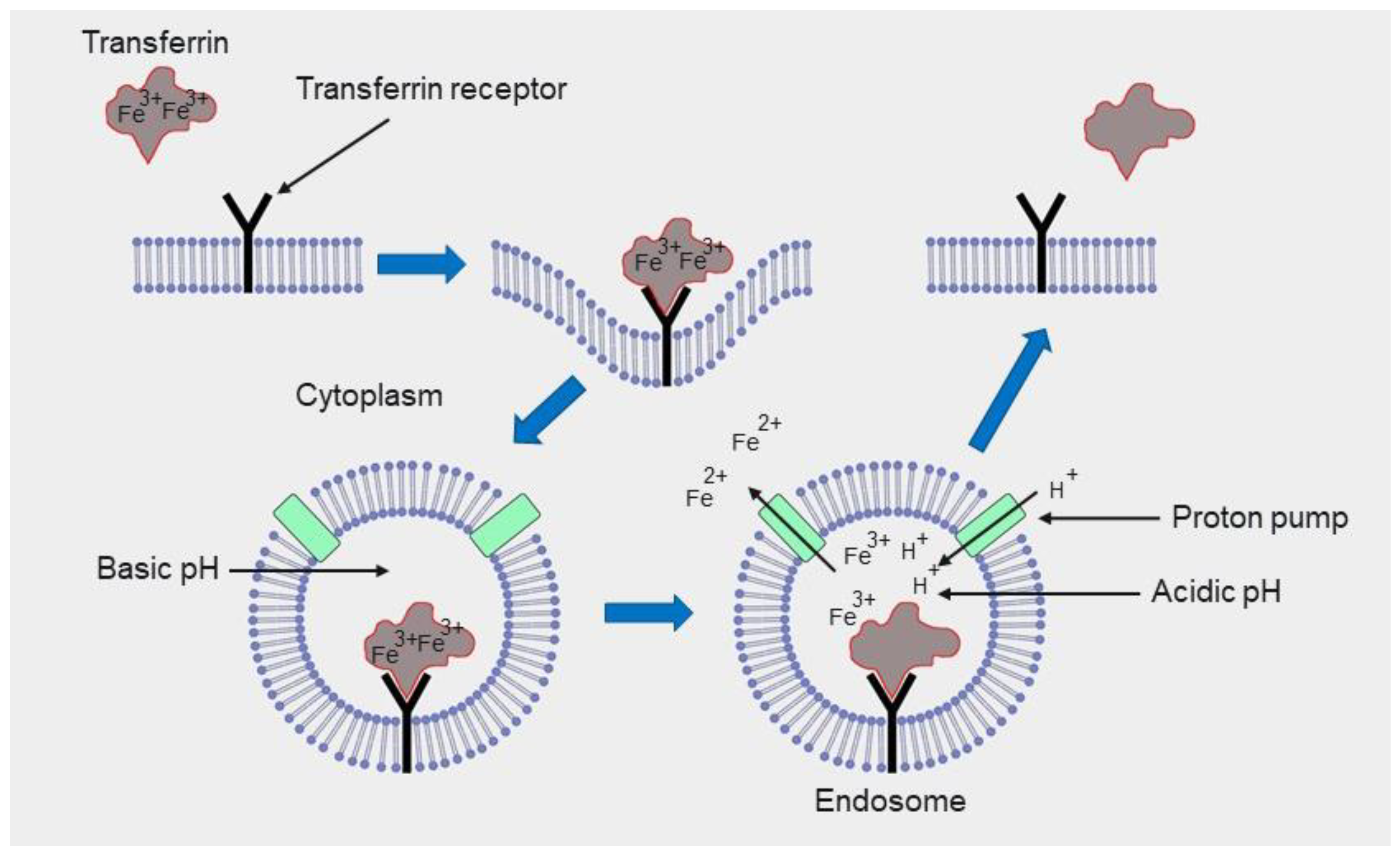
Publisher’s Note: MDPI stays neutral with regard to jurisdictional claims in published maps and institutional affiliations. |
© 2022 by the authors. Licensee MDPI, Basel, Switzerland. This article is an open access article distributed under the terms and conditions of the Creative Commons Attribution (CC BY) license (https://creativecommons.org/licenses/by/4.0/).
Share and Cite
MDPI and ACS Style
Veselov, V.V.; Nosyrev, A.E.; Jicsinszky, L.; Alyautdin, R.N.; Cravotto, G. Targeted Delivery Methods for Anticancer Drugs. Cancers 2022, 14, 622. https://doi.org/10.3390/cancers14030622
AMA Style
Veselov VV, Nosyrev AE, Jicsinszky L, Alyautdin RN, Cravotto G. Targeted Delivery Methods for Anticancer Drugs. Cancers. 2022; 14(3):622. https://doi.org/10.3390/cancers14030622
Chicago/Turabian StyleVeselov, Valery V., Alexander E. Nosyrev, László Jicsinszky, Renad N. Alyautdin, and Giancarlo Cravotto. 2022. "Targeted Delivery Methods for Anticancer Drugs" Cancers 14, no. 3: 622. https://doi.org/10.3390/cancers14030622
Note that from the first issue of 2016, this journal uses article numbers instead of page numbers. See further details here.