
Non-Invasive Biomarkers for Immunotherapy in Patients with Hepatocellular Carcinoma: Current Knowledge and Future Perspectives
 , and
, and
Abstract
:Simple Summary
Abstract
1. Introduction
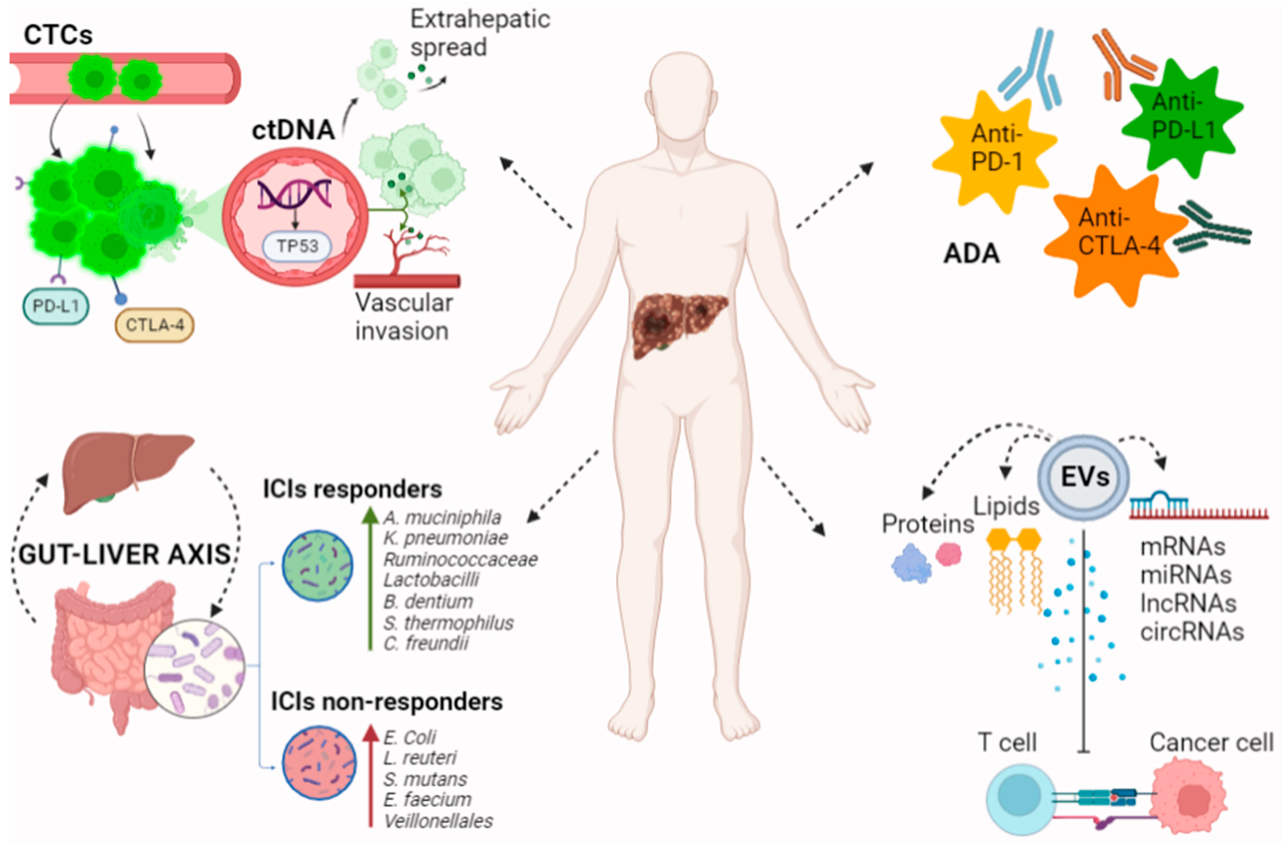
2. Clinical Usefulness of Non-Invasive Biomarkers
3. Traditional Non-Invasive Biomarkers of Response to ICIs
3.1. Cytokines, Immune Checkpoints, and Immune Cells
3.2. Neutrophil to Lymphocyte Ratio, Platelet to Lymphocyte Ratio, Prognostic Nutritional Index, and Their Combined Prognostic Value
3.3. Alpha-Fetoprotein
4. Novel Biomarkers
4.1. Circulating Tumor DNA
4.2. Circulating Tumor Cells
4.3. Extracellular Vesicles
4.4. Antidrug Antibodies
4.5. Gut Microbiome
5. Conclusions and Future Perspectives
Author Contributions
Funding
Acknowledgments
Conflicts of Interest
References
- Cerrito, L.; Santopaolo, F.; Monti, F.; Pompili, M.; Gasbarrini, A.; Ponziani, F.R. Advances in pharmacotherapeutics for hepatocellular carcinoma. Expert Opin. Pharmacother. 2021, 22, 1343–1354. [Google Scholar] [CrossRef] [PubMed]
- Ponziani, F.R.; Bhoori, S.; Germini, A.; Bongini, M.; Flores, M.; Sposito, C.; Facciorusso, A.; Gasbarrini, A.; Mazzaferro, V. Inducing tolerability of adverse events increases sorafenib exposure and optimizes patient’s outcome in advanced hepatocellular carcinoma. Liver Int. 2016, 36, 1033–1042. [Google Scholar] [CrossRef] [PubMed]
- Cerrito, L.; Annicchiarico, B.E.; Iezzi, R.; Gasbarrini, A.; Pompili, M.; Ponziani, F.R. Treatment of hepatocellular carcinoma in patients with portal vein tumor thrombosis: Beyond the known frontiers. World J. Gastroenterol. 2019, 25, 4360–4382. [Google Scholar] [CrossRef]
- Reig, M.; Forner, A.; Rimola, J.; Ferrer-Fàbrega, J.; Burrel, M.; Garcia-Criado, Á.; Kelley, R.K.; Galle, P.R.; Mazzaferro, V.; Salem, R.; et al. BCLC strategy for prognosis prediction and treatment recommendation: The 2022 update. J. Hepatol. 2022, 76, 681–693. [Google Scholar] [CrossRef]
- Buchbinder, E.I.; Desai, A. CTLA-4 and PD-1 Pathways: Similarities, Differences, and Implications of Their Inhibition. Am. J. Clin. Oncol. 2016, 39, 98–106. [Google Scholar] [CrossRef]
- Roche, H.-L. A Phase III, Open-Label, Randomized Study of Atezolizumab in Combination With Bevacizumab Compared with Sorafenib in Patients with Untreated Locally Advanced or Metastatic Hepatocellular Carcinoma. Available online: https://clinicaltrials.gov/ct2/show/NCT03434379 (accessed on 1 August 2022).
- Phase 3 Randomized, Open-Label, Multicenter Study of Tremelimumab (T) and Durvalumab (D) as First-Line Therapy in Patients (Pts) with Unresectable Hepatocellular Carcinoma (uHCC): HIMALAYA. Available online: https://ascopubs.org/doi/abs/10.1200/JCO.2022.40.4_suppl.379 (accessed on 1 August 2022).
- Maestri, M.; Pallozzi, M.; Santopaolo, F.; Cerrito, L.; Pompili, M.; Gasbarrini, A.; Ponziani, F.R. Durvalumab: An investigational agent for unresectable hepatocellular carcinoma. Expert Opin. Investig. Drugs 2021, 31, 347–360. [Google Scholar] [CrossRef] [PubMed]
- Yau, T.; Kang, Y.K.; Kim, T.Y.; El-Khoueiry, A.B.; Santoro, A.; Sangro, B.; Melero, I.; Kudo, M.; Hou, M.M.; Matilla, A.; et al. Efficacy and Safety of Nivolumab Plus Ipilimumab in Patients With Advanced Hepatocellular Carcinoma Previously Treated With Sorafenib: The CheckMate 040 Randomized Clinical Trial. JAMA Oncol. 2020, 6, e204564. [Google Scholar] [CrossRef] [PubMed]
- Zhu, A.X.; Finn, R.S.; Edeline, J.; Cattan, S.; Ogasawara, S.; Palmer, D.; Verslype, C.; Zagonel, V.; Fartoux, L.; Vogel, A.; et al. KEYNOTE-224 investigators. Pembrolizumab in patients with advanced hepatocellular carcinoma previously treated with sorafenib (KEYNOTE-224): A non-randomised, open-label phase 2 trial. Lancet. Oncol. 2018, 19, 940–952. [Google Scholar] [CrossRef]
- Finn, R.S.; Ryoo, B.Y.; Merle, P.; Kudo, M.; Bouattour, M.; Lim, H.Y.; Breder, V.; Edeline, J.; Chao, Y.; Ogasawara, S.; et al. Pembrolizumab As Second-Line Therapy in Patients With Advanced Hepatocellular Carcinoma in KEYNOTE-240: A Randomized, Double-Blind, Phase III Trial. J. Clin. Oncol. 2020, 38, 193–202. [Google Scholar] [CrossRef]
- Giraud, J.; Chalopin, D.; Blanc, J.F.; Saleh, M. Hepatocellular Carcinoma Immune Landscape and the Potential of Immunotherapies. Front. Immunol. 2021, 12, 655697. [Google Scholar] [CrossRef]
- Ponziani, F.R.; Santopaolo, F.; Posa, A.; Pompili, M.; Tanzilli, A.; Maestri, M.; Pallozzi, M.; Ibba, F.; Manfredi, R.; Gasbarrini, A.; et al. SIRT in 2025. Cardiovasc. Interv. Radiol. 2022, 8, 1–12. [Google Scholar] [CrossRef] [PubMed]
- Cabibbo, G.; Aghemo, A.; Lai, Q.; Masarone, M.; Montagnese, S.; Ponziani, F.R.; Italian Association for the Study of the Liver (AISF). Optimizing systemic therapy for advanced hepatocellular carcinoma: The key role of liver function. Dig. Liver Dis. 2022, 54, 452–460. [Google Scholar] [CrossRef] [PubMed]
- Khemlina, G.; Ikeda, S.; Kurzrock, R. The biology of Hepatocellular carcinoma: Implications for genomic and immune therapies. Mol. Cancer. 2017, 16, 149. [Google Scholar] [CrossRef] [PubMed]
- Giannini, E.G.; Aglitti, A.; Borzio, M.; Gambato, M.; Guarino, M.; Iavarone, M.; Lai, Q.; Levi Sandri, G.B.; Melandro, F.; Morisco, F.; et al. Overview of Immune Checkpoint Inhibitors Therapy for Hepatocellular Carcinoma, and The ITA.LI.CA Cohort Derived Estimate of Amenability Rate to Immune Checkpoint Inhibitors in Clinical Practice. Cancers 2019, 11, 1689. [Google Scholar] [CrossRef] [PubMed]
- Robb, M.A.; McInnes, P.M.; Califf, R.M. Biomarkers and Surrogate Endpoints: Developing Common Terminology and Definitions. JAMA 2016, 315, 1107–1108. [Google Scholar] [CrossRef]
- Califf, R.M. Biomarker definitions and their applications. Exp. Biol. Med. 2018, 243, 213–221. [Google Scholar] [CrossRef]
- Febbo, P.G.; Ladanyi, M.; Aldape, K.D.; De Marzo, A.M.; Hammond, M.E.; Hayes, D.F.; Iafrate, A.J.; Kelley, R.K.; Marcucci, G.; Ogino, S.; et al. NCCN Task Force report: Evaluating the clinical utility of tumor markers in oncology. J. Natl. Compr. Cancer Netw. 2011, 9, S1–S32. [Google Scholar] [CrossRef]
- Guan, M.-C.; Ouyang, W.; Wang, M.-D.; Liang, L.; Li, N.; Fu, T.-T.; Shen, F.; Lau, W.-Y.; Xu, Q.-R.; Huang, D.-S.; et al. Biomarkers for hepatocellular carcinoma based on body fluids and feces. World J. Gastrointest. Oncol. 2021, 13, 351–365. [Google Scholar] [CrossRef]
- Trevisan, L.; de Lima, F.; Broszczak, D.; Zhang, X.; Bridle, K.; Crawford, D.; Punyadeera, C. The use of minimally invasive biomarkers for the diagnosis and prognosis of hepatocellular carcinoma. Biochim. Biophys. Acta 2020, 1874, 188451. [Google Scholar]
- Ao, H.; Xin, Z.; Jian, Z. Liquid biopsy to identify biomarkers for immunotherapy in hepatocellular carcinoma. Biomark. Res. 2021, 9, 91. [Google Scholar] [CrossRef]
- Maravelia, P.; Silva, D.N.; Rovesti, G.; Chrobok, M.; Stål, P.; Lu, Y.C.; Pasetto, A. Liquid Biopsy in Hepatocellular Carcinoma: Opportunities and Challenges for Immunotherapy. Cancers 2021, 13, 4334. [Google Scholar] [CrossRef] [PubMed]
- Shimagaki, T.; Yoshio, S.; Kawai, H.; Sakamoto, Y.; Doi, H.; Matsuda, M.; Mori, T.; Osawa, Y.; Fukai, M.; Yoshida, T.; et al. Serum milk fat globule-EGF factor 8 (MFG-E8) as a diagnostic and prognostic biomarker in patients with hepatocellular carcinoma. Sci. Rep. 2019, 9, 15788. [Google Scholar] [CrossRef] [PubMed]
- Rizzo, A.; Brandi, G. Biochemical predictors of response to immune checkpoint inhibitors in unresectable hepatocellular carcinoma. Cancer Treat. Res. Commun. 2021, 27, 100328. [Google Scholar] [CrossRef]
- Myojin, Y.; Kodama, T.; Sakamori, R.; Maesaka, K.; Matsumae, T.; Sawai, Y.; Imai, Y.; Ohkawa, K.; Miyazaki, M.; Tanaka, S.; et al. Interleukin-6 Is a Circulating Prognostic Biomarker for Hepatocellular Carcinoma Patients Treated with Combined Immunotherapy. Cancers 2022, 14, 883. [Google Scholar] [CrossRef] [PubMed]
- Shakiba, E.; Ramezani, M.; Sadeghi, M. Evaluation of serum interleukin-6 levels in hepatocellular carcinoma patients: A systematic review and meta-analysis. Clin. Exp. Hepatol. 2018, 4, 182–190. [Google Scholar] [CrossRef]
- McLoughlin, R.M.; Jenkins, B.J.; Grail, D.; Williams, A.S.; Fielding, C.A.; Parker, C.R.; Ernst, M.; Topley, N.; Jones, S.A. IL-6 trans-signaling via STAT3 directs T cell infiltration in acute inflammation. Proc. Natl. Acad. Sci. USA 2005, 102, 9589–9594. [Google Scholar] [CrossRef] [PubMed]
- Chen, M.F.; Kuan, F.C.; Yen, T.C.; Lu, M.S.; Lin, P.Y.; Chung, Y.H.; Chen, W.C.; Lee, K.D. IL-6-stimulated CD11b+ CD14+ HLA-DR- myeloid-derived suppressor cells, are associated with progression and poor prognosis in squamous cell carcinoma of the esophagus. Oncotarget 2014, 5, 8716–8728. [Google Scholar] [CrossRef] [PubMed]
- Feun, L.G.; Li, Y.; Wu, C.; Wangpaichitr, M.; Jones, P.D.; Richman, S.P.; Madrazo, B.; Kwon, D.; Garcia-Buitrago, M.; Martin, P.; et al. Phase 2 study of pembrolizumab and circulating biomarkers to predict anticancer response in advanced, unresectable hepatocellular carcinoma. Cancer 2019, 125, 3603–3614. [Google Scholar] [CrossRef]
- Patel, S.P.; Kurzrock, R. PD-L1 Expression as a Predictive Biomarker in Cancer Immunotherapy. Mol. Cancer Ther. 2015, 14, 847–856. [Google Scholar] [CrossRef]
- Gibney, G.T.; Weiner, L.M.; Atkins, M.B. Predictive biomarkers for checkpoint inhibitor-based immunotherapy. Lancet Oncol. 2016, 17, e542–e551. [Google Scholar] [CrossRef]
- Hung, Y.P.; Shao, Y.Y.; Hsu, C.; Hsu, C.H.; Lee, J.M.; Yang, M.H.; Chao, Y. The unique characteristic in peripheral immune cells in patients with advanced hepatocellular carcinoma. J. Formos. Med. Assoc. 2021, 120, 1581–1590. [Google Scholar] [CrossRef] [PubMed]
- Oh, S.Y.; Kim, S.; Keam, B.; Kim, T.M.; Kim, D.W.; Heo, D.S. Soluble PD-L1 is a predictive and prognostic biomarker in advanced cancer patients who receive immune checkpoint blockade treatment. Sci. Rep. 2021, 11, 19712. [Google Scholar] [CrossRef] [PubMed]
- Mahoney, K.M.; Ross-Macdonald, P.; Yuan, L.; Song, L.; Veras, E.; Wind-Rotolo, M.; McDermott, D.F.; Hodi, F.S.; Choueiri, T.K.; Freeman, G.J. Soluble PD-L1 as an early marker of progressive disease on nivolumab. J. Immunother. Cancer 2020, 10, e003527. [Google Scholar] [CrossRef] [PubMed]
- Li, X.S.; Li, J.W.; Li, H.; Jiang, T. Prognostic value of programmed cell death ligand 1 (PD-L1) for hepatocellular carcinoma: A meta-analysis. Biosci. Rep. 2020, 40, BSR20200459. [Google Scholar] [CrossRef]
- Mocan, T.; Ilies, M.; Nenu, I.; Craciun, R.; Horhat, A.; Susa, R.; Minciuna, I.; Rusu, I.; Mocan, L.P.; Seicean, A.; et al. Serum levels of soluble programmed death-ligand 1 (sPD-L1): A possible biomarker in predicting post-treatment outcomes in patients with early hepatocellular carcinoma. Int. Immunopharmacol. 2021, 94, 107467. [Google Scholar] [CrossRef]
- Wang, T.; Denman, D.; Bacot, S.M.; Feldman, G.M. Challenges and the Evolving Landscape of Assessing Blood-Based PD-L1 Expression as a Biomarker for Anti-PD-(L)1 Immunotherapy. Biomedicines 2022, 10, 1181. [Google Scholar] [CrossRef]
- Park, L.M.; Lannigan, J.; Jaimes, M.C. OMIP-069: Forty-Color Full Spectrum Flow Cytometry Panel for Deep Immunophenotyping of Major Cell Subsets in Human Peripheral Blood. Citometry A 2020, 97, 1044–1051. [Google Scholar] [CrossRef]
- Agdashian, D.; ElGindi, M.; Xie, C.; Sandhu, M.; Pratt, D.; Kleiner, D.E.; Figg, W.D.; Rytlewski, J.A.; Sanders, C.; Yusko, E.C.; et al. The effect of anti-CTLA4 treatment on peripheral and intra-tumoral T cells in patients with hepatocellular carcinoma. Cancer Immunol. 2019, 68, 599–608. [Google Scholar] [CrossRef]
- Mengos, A.E.; Gastineau, D.A.; Gustafson, M.P. The CD14+HLA-DRlo/neg Monocyte: An Immunosuppressive Phenotype That Restrains Responses to Cancer Immunotherapy. Front. Immunol. 2019, 10, 1147. [Google Scholar] [CrossRef]
- Hung, Y.P.; Shao, Y.Y.; Lee, J.M.; Hsu, C.; Hsu, C.H.; Yang, M.H.; Chao, Y. Potential of circulating immune cells as biomarkers of nivolumab treatment efficacy for advanced hepatocellular carcinoma. J. Chin. Med. Assoc. 2021, 84, 144–150. [Google Scholar] [CrossRef]
- Hong, J.Y.; Cho, H.J.; Sa, J.K.; Liu, X.; Ha, S.Y.; Lee, T.; Kim, H.; Kang, W.; Sinn, D.H.; Gwak, G.-Y.; et al. Hepatocellular carcinoma patients with high circulating cytotoxic T cells and intra-tumoral immune signature benefit from pembrolizumab: Results from a single-arm phase 2 trial. Genome Med. 2022, 14, 1. [Google Scholar] [CrossRef] [PubMed]
- Macek Jilkova, Z.; Aspord, C.; Kurma, K.; Granon, A.; Sengel, C.; Sturm, N.; Marche, P.N.; Decaens, T. Immunologic Features of Patients With Advanced Hepatocellular Carcinoma Before and During Sorafenib or Anti-programmed Death-1/Programmed Death-L1 Treatment. Clin. Transl. Gastroenterol. 2019, 10, e00058. [Google Scholar] [CrossRef] [PubMed]
- He, Y.; Lu, M.; Che, J.; Chu, Q.; Zhang, P.; Chen, Y. Biomarkers and Future Perspectives for Hepatocellular Carcinoma Immunotherapy. Front. Oncol. 2021, 11, 716844. [Google Scholar] [CrossRef] [PubMed]
- Dharmapuri, S.; Özbek, U.; Lin, J.Y.; Sung, M.; Schwartz, M.; Branch, A.D.; Ang, C. Predictive value of neutrophil to lymphocyte ratio and platelet to lymphocyte ratio in advanced hepatocellular carcinoma patients treated with anti-PD-1 therapy. Cancer Med. 2020, 9, 4962–4970. [Google Scholar] [CrossRef] [PubMed]
- Hung, H.C.; Lee, J.C.; Wang, Y.C.; Cheng, C.H.; Wu, T.H.; Lee, C.F.; Wu, T.J.; Chou, H.S.; Chan, K.M.; Lee, W.C. Response Prediction in Immune Checkpoint Inhibitor Immunotherapy for Advanced Hepatocellular Carcinoma. Cancers 2020, 13, 1607. [Google Scholar] [CrossRef]
- Ogihara, K.; Kikuchi, E.; Shigeta, K.; Okabe, T.; Hattori, S.; Yamashita, R.; Yoshimine, S.; Shirotake, S.; Nakazawa, R.; Matsumoto, K.; et al. The pretreatment neutrophil-to-lymphocyte ratio is a novel biomarker for predicting clinical responses to pembrolizumab in platinum-resistant metastatic urothelial carcinoma patients. Urol. Oncol. 2020, 38, 602.e1–602.e10. [Google Scholar] [CrossRef]
- Tada, T.; Kumada, T.; Hiraoka, A.; Hirooka, M.; Kariyama, K.; Tani, J.; Atsukawa, M.; Takaguchi, K.; Itobayashi, E.; Fukunishi, S.; et al. Real-life Practice Experts for HCC (RELPEC) Study Group and the Hepatocellular Carcinoma Experts from 48 clinics in Japan (HCC 48) Group. Neutrophil-lymphocyte ratio predicts early outcomes in patients with unresectable hepatocellular carcinoma treated with atezolizumab plus bevacizumab: A multicenter analysis. Eur. J. Gastroenterol. Hepatol. 2022, 34, 698–706. [Google Scholar]
- Zheng, J.; Cai, J.; Li, H.; Zeng, K.; He, L.; Fu, H.; Zhang, J.; Chen, L.; Yao, J.; Zhang, Y.; et al. Neutrophil to Lymphocyte Ratio and Platelet to Lymphocyte Ratio as Prognostic Predictors for Hepatocellular Carcinoma Patients with Various Treatments: A Meta-Analysis and Systematic Review. Cell. Physiol. Biochem. 2017, 44, 967–981. [Google Scholar] [CrossRef]
- Jiang, Y.; Tu, X.; Zhang, X.; Liao, H.; Han, S.; Jiang, W.; Zheng, Y.; Zhao, P.; Tong, Z.; Fu, Q.; et al. Nutrition and metabolism status alteration in advanced hepatocellular carcinoma patients treated with anti-PD-1 immunotherapy. Support. Care Cancer 2020, 28, 5569–5579. [Google Scholar] [CrossRef]
- Muhammed, A.; Fulgenzi, C.A.M.; Dharmapuri, S.; Pinter, M.; Balcar, L.; Scheiner, B.; Marron, T.U.; Jun, T.; Saeed, A.; Hildebrand, H.; et al. The Systemic Inflammatory Response Identifies Patients with Adverse Clinical Outcome from Immunotherapy in Hepatocellular Carcinoma. Cancers 2021, 14, 186. [Google Scholar] [CrossRef]
- Mei, J.; Sun, X.Q.; Lin, W.P.; Li, S.H.; Lu, L.H.; Zou, J.W.; Wei, W.; Guo, R.P. Comparison of the Prognostic Value of Inflammation-Based Scores in Patients with Hepatocellular Carcinoma After Anti-PD-1 Therapy. J. Inflamm. Res. 2021, 14, 3879–3890. [Google Scholar] [CrossRef] [PubMed]
- Bigot, F.; Castanon, E.; Baldini, C.; Hollebecque, A.; Carmona, A.; Postel-Vinay, S.; Angevin, E.; Armand, J.-P.; Ribrag, V.; Aspeslagh, S.; et al. Prospective validation of a prognostic score for patients in immunotherapy phase I trials: The Gustave Roussy Immune Score (GRIm-Score). Eur. J. Cancer 1990, 84, 212–218. [Google Scholar] [CrossRef] [PubMed]
- Li, Y.; Pan, Y.; Lin, X.; Hou, J.; Hu, Z.; Xu, L.; Zhou, Z.; Zhang, Y.; Chen, M.; Hu, D. Development and Validation of a Prognostic Score for Hepatocellular Carcinoma Patients in Immune Checkpoint Inhibitors Therapies: The Hepatocellular Carcinoma Modified Gustave Roussy Immune Score. Front. Pharmacol. 2022, 12, 819985. [Google Scholar] [CrossRef] [PubMed]
- Zhang, J.; Chen, G.; Zhang, P.; Zhang, J.; Li, X.; Gan, D.; Cao, X.; Han, M.; Du, H.; Ye, Y. The threshold of alpha-fetoprotein (AFP) for the diagnosis of hepatocellular carcinoma: A systematic review and meta-analysis. PLoS ONE 2020, 15, e0228857. [Google Scholar] [CrossRef] [PubMed]
- Mehta, N.; Dodge, J.L.; Roberts, J.P.; Hirose, R.; Yao, F.Y. Alpha-Fetoprotein Decrease from >1000 to <500 ng/mL in Patients with Hepatocellular Carcinoma Leads to Improved Posttransplant Outcomes. Hepatology 2019, 69, 1193–1205. [Google Scholar] [PubMed]
- Lee, P.C.; Chao, Y.; Chen, M.H.; Lan, K.H.; Lee, C.J.; Lee, I.C.; Chen, S.C.; Hou, M.C.; Huang, Y.H. Predictors of Response and Survival in Immune Checkpoint Inhibitor-Treated Unresectable Hepatocellular Carcinoma. Cancers 2020, 12, 182. [Google Scholar] [CrossRef]
- Shao, Y.Y.; Liu, T.H.; Hsu, C.; Lu, L.C.; Shen, Y.C.; Lin, Z.Z.; Cheng, A.L.; Hsu, C.H. Early alpha-foetoprotein response associated with treatment efficacy of immune checkpoint inhibitors for advanced hepatocellular carcinoma. Liver Int. 2019, 39, 2184–2189. [Google Scholar] [CrossRef] [PubMed]
- Hsu, W.F.; Wang, H.W.; Chen, C.K.; Lai, H.C.; Chuang, P.H.; Tsai, M.H.; Su, W.P.; Chen, H.Y.; Chu, C.S.; Chou, J.W.; et al. Alpha-fetoprotein response predicts treatment outcomes in patients with unresectable hepatocellular carcinoma receiving immune checkpoint inhibitors with or without tyrosine kinase inhibitors or locoregional therapies. Am. J. Cancer Res. 2021, 11, 6173–6187. [Google Scholar]
- Teng, W.; Lin, C.C.; Ho, M.M.; Lui, K.W.; Wang, S.F.; Hsu, C.W.; Lin, S.M. Alpha-fetoprotein response at different time-points is associated with efficacy of nivolumab monotherapy for unresectable hepatocellular carcinoma. Am. J. Cancer Res. 2021, 11, 2319–2330. [Google Scholar]
- Semenkova, L.N.; Dudich, E.I.; Dudich, I.V.; Shingarova, L.N.; Korobko, V.G. Alpha-fetoprotein as a TNF resistance factor for the human hepatocarcinoma cell line HepG2. Tumour Biol. 1997, 18, 30–40. [Google Scholar] [CrossRef]
- Li, M.S.; Li, P.F.; He, S.P.; Du, G.G.; Li, G. The promoting molecular mechanism of alpha-fetoprotein on the growth of human hepatoma Bel7402 cell line. World J. Gastroenterol. 2002, 8, 469–475. [Google Scholar] [CrossRef] [PubMed]
- Yamamoto, M.; Tatsumi, T.; Miyagi, T.; Tsunematsu, H.; Aketa, H.; Hosui, A.; Kanto, T.; Hiramatsu, N.; Hayashi, N.; Takehara, T. α-Fetoprotein impairs activation of natural killer cells by inhibiting the function of dendritic cells. Clin. Exp. Immunol. 2011, 165, 211–219. [Google Scholar] [CrossRef] [PubMed]
- Chen, T.; Dai, X.; Dai, J.; Ding, C.; Zhang, Z.; Lin, Z.; Hu, J.; Lu, M.; Wang, Z.; Qi, Y.; et al. AFP promotes HCC progression by suppressing the HuR-mediated Fas/FADD apoptotic pathway. Cell Death Dis. 2020, 11, 822. [Google Scholar] [CrossRef] [PubMed]
- A Phase I Open Label Clinical Trial Evaluating the Safety and Anti-Tumor Activity of Autologous T Cells Expressing Enhanced TCRs Specific for Alpha Fetoprotein (AFPc332T) in HLA-A2 Positive Subjects With Advanced Hepatocellular Carcinoma (HCC) or Other AFP Expressing Tumor. Available online: https://clinicaltrials.gov/ct2/show/NCT03132792 (accessed on 1 August 2022).
- Wu, Y.; Wei, P.; Pengpumkiat, S.; Schumacher, E.A.; Remcho, V.T. Development of a carbon dot (C-Dot)-linked immunosorbent assay for the detection of human α-fetoprotein. Anal. Chem. 2015, 87, 8510–8516. [Google Scholar] [CrossRef]
- European Association for the Study of the Liver. EASL Clinical Practice Guidelines: Management of hepatocellular carcinoma. J. Hepatol. 2018, 69, 182–236. [Google Scholar] [CrossRef]
- Heimbach, J.K.; Kulik, L.M.; Finn, R.S.; Sirlin, C.B.; Abecassis, M.M.; Roberts, L.R.; Zhu, A.X.; Murad, M.H.; Marrero, J.A. AASLD guidelines for the treatment of hepatocellular carcinoma. Hepatology 2018, 67, 358–380. [Google Scholar] [CrossRef] [PubMed]
- Chan, S.L.; Mo, F.; Johnson, P.J.; Siu, D.Y.; Chan, M.H.; Lau, W.Y.; Lai, P.B.; Lam, C.W.; Yeo, W.; Yu, S.C. Performance of serum α-fetoprotein levels in the diagnosis of hepatocellular carcinoma in patients with a hepatic mass. HPB 2014, 16, 366–372. [Google Scholar] [CrossRef] [PubMed]
- Scheiner, B.; Pomej, K.; Kirstein, M.M.; Hucke, F.; Finkelmeier, F.; Waidmann, O.; Himmelsbach, V.; Schulze, K.; von Felden, J.; Fründt, T.W.; et al. Prognosis of patients with hepatocellular carcinoma treated with immunotherapy—Development and validation of the CRAFITY score. J. Hepatol. 2022, 76, 353–363. [Google Scholar] [CrossRef]
- Hatanaka, T.; Kakizaki, S.; Hiraoka, A.; Tada, T.; Hirooka, M.; Kariyama, K.; Tani, J.; Atsukawa, M.; Takaguchi, K.; Itobayashi, E.; et al. Prognostic impact of C-reactive protein and alpha-fetoprotein in immunotherapy score in hepatocellular carcinoma patients treated with atezolizumab plus bevacizumab: A multicenter retrospective study. Hepatol. Int. 2022, 76, 353–363. [Google Scholar] [CrossRef]
- Teng, W.; Lin, C.C.; Su, C.W.; Lin, P.T.; Hsieh, Y.C.; Chen, W.T.; Ho, M.M.; Wang, C.T.; Chai, P.M.; Hsieh, J.C.; et al. Combination of CRAFITY score with Alpha-fetoprotein response predicts a favorable outcome of atezolizumab plus bevacizumab for unresectable hepatocellular carcinoma. Am. J. Cancer Res. 2022, 12, 1899–1911. [Google Scholar] [CrossRef]
- Sun, X.; Mei, J.; Lin, W.; Yang, Z.; Peng, W.; Chen, J.; Zhang, Y.; Xu, L.; Chen, M. Reductions in AFP and PIVKA-II can predict the efficiency of anti-PD-1 immunotherapy in HCC patients. BMC Cancer 2021, 21, 775. [Google Scholar] [CrossRef] [PubMed]
- Bui, T.M.; Wiesolek, H.L.; Sumagin, R. ICAM-1: A master regulator of cellular responses in inflammation, injury resolution, and tumorigenesis. J. Leukoc. Biol. 2020, 108, 787–799. [Google Scholar] [CrossRef] [PubMed]
- Witkowska, A.M.; Borawska, M.H. Soluble intercellular adhesion molecule-1 (sICAM-1): An overview. Eur. Cytokine. Netw. 2004, 15, 91–98. [Google Scholar] [PubMed]
- Cao, W.; Chen, Y.W.; Yuan, J.; Xie, W.; Liu, K.; Qiu, Y.; Wang, X.; Li, X. Potentiality of α-fetoprotein (AFP) and soluble intercellular adhesion molecule-1 (sICAM-1) in prognosis prediction and immunotherapy response for patients with hepatocellular carcinoma. Bioengineered 2021, 12, 9435–9451. [Google Scholar] [CrossRef] [PubMed]
- Ye, L.; Zhang, F.; Li, H.; Yang, L.; Lv, T.; Gu, W.; Song, Y. Circulating Tumor Cells Were Associated with the Number of T Lymphocyte Subsets and NK Cells in Peripheral Blood in Advanced Non-Small-Cell Lung Cancer. Dis. Markers 2017, 2017, 5727815. [Google Scholar] [CrossRef]
- Postel, M.; Roosen, A.; Laurent-Puig, P.; Taly, V.; Wang-Renault, S.F. Droplet-based digital PCR and next generation sequencing for monitoring circulating tumor DNA: A cancer diagnostic perspective. Expert Rev. Mol. Diagn. 2018, 18, 7–17. [Google Scholar] [CrossRef]
- Cullinan, A.; Cantor, C. Sequenom, Inc. Pharmacogenomics 2008, 9, 1211–1215. [Google Scholar] [CrossRef]
- Wang, J.; Huang, A.; Wang, Y.P.; Yin, Y.; Fu, P.Y.; Zhang, X.; Zhou, J. Circulating tumor DNA correlates with microvascular invasion and predicts tumor recurrence of hepatocellular carcinoma. Ann. Transl. Med. 2020, 8, 237. [Google Scholar] [CrossRef]
- Wu, X.; Li, J.; Gassa, A.; Buchner, D.; Alakus, H.; Dong, Q.; Ren, N.; Liu, M.; Odenthal, M.; Stippel, D.; et al. Circulating tumor DNA as an emerging liquid biopsy biomarker for early diagnosis and therapeutic monitoring in hepatocellular carcinoma. Int. J. Biol. Sci. 2020, 16, 1551–1562. [Google Scholar] [CrossRef]
- Von Felden, J.; Craig, A.J.; Garcia-Lezana, T.; Labgaa, I.; Haber, P.K.; D’Avola, D.; Asgharpour, A.; Dieterich, D.; Bonaccorso, A.; Torres-Martin, M.; et al. Mutations in circulating tumor DNA predict primary resistance to systemic therapies in advanced hepatocellular carcinoma. Oncogene 2021, 40, 140–151. [Google Scholar] [CrossRef]
- Oversoe, S.K.; Clement, M.S.; Pedersen, M.H.; Weber, B.; Aagaard, N.K.; Villadsen, G.E.; Grønbæk, H.; Hamilton-Dutoit, S.J.; Sorensen, B.S.; Kelsen, J. TERT promoter mutated circulating tumor DNA as a biomarker for prognosis in hepatocellular carcinoma. Scand. J. Gastroenterol. 2020, 55, 1433–1440. [Google Scholar] [CrossRef] [PubMed]
- Kim, S.S.; Eun, J.W.; Choi, J.H.; Woo, H.G.; Cho, H.J.; Ahn, H.R.; Suh, C.W.; Baek, G.O.; Cho, S.W.; Cheong, J.Y. MLH1 single-nucleotide variant in circulating tumor DNA predicts overall survival of patients with hepatocellular carcinoma. Sci. Rep. 2020, 10, 17862. [Google Scholar] [CrossRef] [PubMed]
- Shen, T.; Li, S.; Wang, J.; Zhang, T.; Zhang, S.; Chen, H.; Xiao, Q.; Ren, W.; Liu, C.; Peng, B.; et al. TP53 R249S mutation detected in circulating tumour DNA is associated with Prognosis of hepatocellular carcinoma patients with or without hepatectomy. Liver Int. 2020, 40, 2834–2847. [Google Scholar] [CrossRef] [PubMed]
- Sun, Y.; Wang, P.; Cheng, J.; Gong, Z.; Huang, A.; Zhou, K.; Hu, B.; Gao, P.; Cao, Y.; Qiu, S.; et al. Postoperative circulating tumor cells: An early predictor of extrahepatic metastases in patients with hepatocellular carcinoma undergoing curative surgical resection. Cancer Cytopathol. 2020, 128, 733–745. [Google Scholar] [CrossRef] [PubMed]
- Alexandrov, L.B.; Nik-Zainal, S.; Wedge, D.C.; Aparicio, S.A.; Behjati, S.; Biankin, A.V.; Bignell, G.R.; Bolli, N.; Borg, A.; Børresen-Dale, A.L.; et al. Signatures of mutational processes in human cancer. Nature 2013, 500, 415–421. [Google Scholar] [CrossRef] [PubMed]
- Goodman, A.M.; Kato, S.; Bazhenova, L.; Patel, S.P.; Frampton, G.M.; Miller, V.; Stephens, P.J.; Daniels, G.A.; Kurzrock, R. Tumor Mutational Burden as an Independent Predictor of Response to Immunotherapy in Diverse Cancers. Mol. Cancer Ther. 2020, 16, 2598–2608. [Google Scholar] [CrossRef]
- Rizvi, N.A.; Cho, B.C.; Reinmuth, N.; Lee, K.H.; Luft, A.; Ahn, M.J.; van den Heuvel, M.M.; Cobo, M.; Vicente, D.; Smolin, A.; et al. MYSTIC Investigators. Durvalumab With or Without Tremelimumab vs Standard Chemotherapy in First-line Treatment of Metastatic Non-Small Cell Lung Cancer: The MYSTIC Phase 3 Randomized Clinical Trial. JAMA Oncol. 2020, 6, 661–674. [Google Scholar] [CrossRef]
- Araujo, D.V.; Wang, A.; Torti, D.; Leon, A.; Marsh, K.; McCarthy, A.; Berman, H.; Spreafico, A.; Hansen, A.R.; Razak, A.-A.; et al. Applications of Circulating Tumor DNA in a Cohort of Phase I Solid Tumor Patients Treated With Immunotherapy. JNCI Cancer Spectr. 2021, 5, pkaa122. [Google Scholar] [CrossRef]
- Zhu, G.; Liu, W.; Tang, Z.; Qu, W.; Fang, Y.; Jiang, X.; Song, S.; Wang, H.; Tao, C.; Zhou, P.; et al. Serial circulating tumor DNA to predict early recurrence in patients with hepatocellular carcinoma: A prospective study. Mol. Oncol. 2022, 16, 549–561. [Google Scholar] [CrossRef]
- Li, Y.; Zheng, Y.; Wu, L.; Li, J.; Ji, J.; Yu, Q.; Dai, W.; Feng, J.; Wu, J.; Guo, C. Current status of ctDNA in precision oncology for hepatocellular carcinoma. J. Exp. Clin. Cancer Res. 2021, 40, 140. [Google Scholar] [CrossRef]
- Ahn, J.C.; Teng, P.C.; Chen, P.J.; Posadas, E.; Tseng, H.R.; Lu, S.C.; Yang, J.D. Detection of Circulating Tumor Cells and Their Implications as a Biomarker for Diagnosis, Prognostication, and Therapeutic Monitoring in Hepatocellular Carcinoma. Hepatology 2021, 73, 422–436. [Google Scholar] [CrossRef] [PubMed]
- Vona, G.; Estepa, L.; Béroud, C.; Damotte, D.; Capron, F.; Nalpas, B.; Mineur, A.; Franco, D.; Lacour, B.; Pol, S.; et al. Impact of cytomorphological detection of circulating tumor cells in patients with liver cancer. Hepatology 2014, 39, 792–797. [Google Scholar] [CrossRef] [PubMed]
- Qi, L.N.; Xiang, B.D.; Wu, F.X.; Ye, J.Z.; Zhong, J.H.; Wang, Y.Y.; Chen, Y.Y.; Chen, Z.S.; Ma, L.; Chen, J.; et al. Circulating Tumor Cells Undergoing EMT Provide a Metric for Diagnosis and Prognosis of Patients with Hepatocellular Carcinoma. Cancer Res. 2018, 78, 4731–4744. [Google Scholar] [CrossRef] [PubMed]
- Sierra-Agudelo, J.; Rodriguez-Trujillo, R.; Samitier, J. Microfluidics for the Isolation and Detection of Circulating Tumor Cells. Adv. Exp. Med. Biol. 2022, 1379, 389–412. [Google Scholar] [PubMed]
- Chen, J.; Cao, S.W.; Cai, Z.; Zheng, L.; Wang, Q. Epithelial-mesenchymal transition phenotypes of circulating tumor cells correlate with the clinical stages and cancer metastasis in hepatocellular carcinoma patients. Cancer Biomark. 2017, 20, 487–498. [Google Scholar] [CrossRef]
- Okegawa, T.; Itaya, N.; Hara, H.; Tambo, M.; Nutahara, K. Circulating tumor cells as a biomarker predictive of sensitivity to docetaxel chemotherapy in patients with castration-resistant prostate cancer. Anticancer. Res. 2014, 34, 6705–6710. [Google Scholar]
- Chen, L.; Bode, A.M.; Dong, Z. Circulating Tumor Cells: Moving Biological Insights into Detection. Theranostics 2017, 7, 2606–2619. [Google Scholar] [CrossRef]
- Yu, J.-J.; Xiao, W.; Dong, S.-L.; Liang, H.-F.; Zhang, Z.-W.; Zhang, B.-X.; Huang, Z.-Y.; Chen, Y.-F.; Zhang, W.-G.; Luo, H.-P.; et al. Effect of surgical liver resection on circulating tumor cells in patients with hepatocellular carcinoma. BMC cancer 2018, 18, 835. [Google Scholar] [CrossRef]
- Ye, X.; Li, G.; Han, C.; Han, Q.; Shang, L.; Su, H.; Han, B.; Gong, Y.; Lu, G.; Peng, T. Circulating tumor cells as a potential biomarker for postoperative clinical outcome in HBV-related hepatocellular carcinoma. Cancer Manag. Res. 2018, 10, 5639–5647. [Google Scholar] [CrossRef]
- Winograd, P.; Hou, S.; Court, C.M.; Lee, Y.-T.; Chen, P.-J.; Zhu, Y.; Sadeghi, S.; Finn, R.S.; Teng, P.-C.; Wang, J.J.; et al. Hepatocellular Carcinoma-Circulating Tumor Cells Expressing PD-L1 Are Prognostic and Potentially Associated With Response to Checkpoint Inhibitors. Hepatol. Commun. 2020, 4, 1527–1540. [Google Scholar] [CrossRef]
- Yue, C.; Jiang, Y.; Li, P.; Wang, Y.; Xue, J.; Li, N.; Li, D.; Wang, R.; Dang, Y.; Hu, Z.; et al. Dynamic change of PD-L1 expression on circulating tumor cells in advanced solid tumor patients undergoing PD-1 blockade therapy. Oncoimmunology 2018, 7, e1438111. [Google Scholar] [CrossRef] [PubMed]
- Winograd, P.; Hou, S.; Court, C.; Sadeghi, S.; Finn, R.; DiPardo, B.; Li, Q.; Kaldas, F.; Busuttil, R.; Tomlinson, J.; et al. Evaluation of hepatocellular carcinoma circulating tumor cells expressing programmed death-ligand 1. HPB 2018, 20, S2–S3. [Google Scholar] [CrossRef]
- Kowalik, A.; Kowalewska, M.; Góźdź, S. Current approaches for avoiding the limitations of circulating tumor cells detection methods-implications for diagnosis and treatment of patients with solid tumors. Transl. Res. 2017, 185, 58–84.e15. [Google Scholar] [CrossRef] [PubMed]
- Bao, Q.; Huang, Q.; Chen, Y.; Wang, Q.; Sang, R.; Wang, L.; Xie, Y.; Chen, W. Tumor-Derived Extracellular Vesicles Regulate Cancer Progression in the Tumor Microenvironment. Front. Mol. Biosci. 2022, 8, 796385. [Google Scholar] [CrossRef] [PubMed]
- Maacha, S.; Bhat, A.A.; Jimenez, L.; Raza, A.; Haris, M.; Uddin, S.; Grivel, J.C. Extracellular vesicles-mediated intercellular communication: Roles in the tumor microenvironment and anti-cancer drug resistance. Mol. Cancer 2019, 18, 55. [Google Scholar] [CrossRef] [PubMed] [Green Version]
- Théry, C.; Witwer, K.W.; Aikawa, E.; Alcaraz, M.J.; Anderson, J.D.; Andriantsitohaina, R.; Antoniou, A.; Arab, T.; Archer, F.; Atkin-Smith, G.K.; et al. Minimal information for studies of extracellular vesicles 2018 (MISEV2018): A position statement of the International Society for Extracellular Vesicles and update of the MISEV2014 guidelines. J. Extracell. Vesicles 2018, 7, 1535750. [Google Scholar] [CrossRef]
- Mashouri, L.; Yousefi, H.; Aref, A.R.; Ahadi, A.M.; Molaei, F.; Alahari, S.K. Exosomes: Composition, biogenesis, and mechanisms in cancer metastasis and drug resistance. Mol. Cancer 2019, 18, 75. [Google Scholar] [CrossRef]
- Zhang, Y.; Bi, J.; Huang, J.; Tang, Y.; Du, S.; Li, P. Exosome: A Review of Its Classification, Isolation Techniques, Storage, Diagnostic and Targeted Therapy Applications. Int. J. Nanomed. 2020, 15, 6917–6934. [Google Scholar] [CrossRef]
- Wortzel, I.; Dror, S.; Kenific, C.M.; Lyden, D. Exosome-Mediated Metastasis: Communication from a Distance. Dev. Cell 2019, 49, 347–360. [Google Scholar] [CrossRef]
- Li, I.; Nabet, B.Y. Exosomes in the tumor microenvironment as mediators of cancer therapy resistance. Mol. Cancer. 2019, 18, 32. [Google Scholar] [CrossRef]
- Li, S.; Chen, L. Exosomes in Pathogenesis, Diagnosis, and Treatment of Hepatocellular Carcinoma. Front. Oncol. 2022, 12, 793432. [Google Scholar] [CrossRef] [PubMed]
- Wang, H.; Yu, L.; Huang, P.; Zhou, Y.; Zheng, W.; Meng, N.; He, R.; Xu, Y.; Keong, T.S.; Cui, Y. Tumor-associated Exosomes Are Involved in Hepatocellular Carcinoma Tumorigenesis, Diagnosis, and Treatment. J. Clin. Transl. Hepatol. 2022, 10, 496–508. [Google Scholar] [CrossRef] [PubMed]
- Xue, D.; Han, J.; Liang, Z.; Jia, L.; Liu, Y.; Tuo, H.; Peng, Y. Current Perspectives on the Unique Roles of Exosomes in Drug Resistance of Hepatocellular Carcinoma. J. Hepatocell. Carcinoma 2022, 9, 99–112. [Google Scholar] [CrossRef] [PubMed]
- Moldogazieva, N.T.; Zavadskiy, S.P.; Terentiev, A.A. Genomic Landscape of Liquid Biopsy for Hepatocellular Carcinoma Personalized Medicine. Cancer Genom. Proteom. 2021, 18, 369–383. [Google Scholar] [CrossRef] [PubMed]
- Abbate, V.; Marcantoni, M.; Giuliante, F.; Vecchio, F.M.; Gatto, I.; Mele, C.; Saviano, A.; Arciuolo, D.; Gaetani, E.; Ferrari, M.C.; et al. HepPar1-Positive Circulating Microparticles Are Increased in Subjects with Hepatocellular Carcinoma and Predict Early Recurrence after Liver Resection. Int. J. Mol. Sci. 2017, 18, 1043. [Google Scholar] [CrossRef] [Green Version]
- Julich-Haertel, H.; Urban, S.K.; Krawczyk, M.; Willms, A.; Jankowski, K.; Patkowski, W.; Kruk, B.; Krasnodębski, M.; Ligocka, J.; Schwab, R.; et al. Cancer-associated circulating large extracellular vesicles in cholangiocarcinoma and hepatocellular carcinoma. J. Hepatol. 2017, 67, 282–292. [Google Scholar] [CrossRef]
- Abels, E.R.; Breakefield, X.O. Introduction to Extracellular Vesicles: Biogenesis, RNA Cargo Selection, Content, Release, and Uptake. Cell Mol. Neurobiol. 2016, 36, 301–312. [Google Scholar] [CrossRef]
- Henderson, M.C.; Azorsa, D.O. The genomic and proteomic content of cancer cell-derived exosomes. Front. Oncol. 2020, 2, 38. [Google Scholar] [CrossRef]
- D’Agnano, I.; Berardi, A.C. Extracellular Vesicles, A Possible Theranostic Platform Strategy for Hepatocellular Carcinoma-An Overview. Cancers 2020, 12, 261. [Google Scholar] [CrossRef]
- Van Niel, G.; D’Angelo, G.; Raposo, G. Shedding light on the cell biology of extracellular vesicles. Nat. Rev. Mol. Cell Biol. 2018, 19, 213–228. [Google Scholar] [CrossRef]
- Ji, J.; Yin, Y.; Ju, H.; Xu, X.; Liu, W.; Fu, Q.; Hu, J.; Zhang, X.; Sun, B. Long non-coding RNA Lnc-Tim3 exacerbates CD8 T cell exhaustion via binding to Tim-3 and inducing nuclear translocation of Bat3 in HCC. Cell Death Dis. 2018, 9, 478. [Google Scholar] [CrossRef] [PubMed]
- Chowdhary, A.; Satagopam, V.; Schneider, R. Long Non-coding RNAs: Mechanisms, Experimental, and Computational Approaches in Identification, Characterization, and Their Biomarker Potential in Cancer. Front. Genet. 2021, 12, 649619. [Google Scholar] [CrossRef] [PubMed]
- Li, L.; Xie, R.; Lu, G. Identification of m6A methyltransferase-related lncRNA signature for predicting immunotherapy and prognosis in patients with hepatocellular carcinoma. Biosci. Rep. 2021, 41, BSR20210760. [Google Scholar] [CrossRef]
- Xu, Q.; Wang, Y.; Huang, W. Identification of immune-related lncRNA signature for predicting immune checkpoint blockade and prognosis in hepatocellular carcinoma. Int. Immunopharmacol. 2021, 92, 107333. [Google Scholar] [CrossRef] [PubMed]
- Zhou, P.; Lu, Y.; Zhang, Y.; Wang, L. Construction of an Immune-Related Six-lncRNA Signature to Predict the Outcomes, Immune Cell Infiltration, and Immunotherapy Response in Patients With Hepatocellular Carcinoma. Front. Oncol. 2021, 11, 661758. [Google Scholar] [CrossRef] [PubMed]
- Tang, P.; Qu, W.; Wang, T.; Liu, M.; Wu, D.; Tan, L.; Zhou, H. Identifying a Hypoxia-Related Long Non-Coding RNAs Signature to Improve the Prediction of Prognosis and Immunotherapy Response in Hepatocellular Carcinoma. Front. Genet. 2021, 12, 785185. [Google Scholar] [CrossRef]
- Zhang, Y.; Zhang, L.; Xu, Y.; Wu, X.; Zhou, Y.; Mo, J. Immune-related long noncoding RNA signature for predicting survival and immune checkpoint blockade in hepatocellular carcinoma. J. Cell. Physiol. 2020, 235, 9304–9316. [Google Scholar] [CrossRef]
- Fan, F.; Chen, K.; Lu, X.; Li, A.; Liu, C.; Wu, B. Dual targeting of PD-L1 and PD-L2 by PCED1B-AS1 via sponging hsa-miR-194-5p induces immunosuppression in hepatocellular carcinoma. Hepatol. Int. 2021, 15, 444–458. [Google Scholar] [CrossRef]
- Peng, L.; Chen, Z.; Chen, Y.; Wang, X.; Tang, N. MIR155HG is a prognostic biomarker and associated with immune infiltration and immune checkpoint molecules expression in multiple cancers. Cancer Med. 2019, 8, 7161–7173. [Google Scholar] [CrossRef]
- Shen, H.; Liu, B.; Xu, J.; Zhang, B.; Wang, Y.; Shi, L.; Cai, X. Circular RNAs: Characteristics, biogenesis, mechanisms and functions in liver cancer. J. Hematol. Oncol. 2021, 14, 134. [Google Scholar] [CrossRef]
- Mi, Z.; Zhongqiang, C.; Caiyun, J.; Yanan, L.; Jianhua, W.; Liang, L. Circular RNA detection methods: A minireview. Talanta 2022, 238, 123066. [Google Scholar] [CrossRef] [PubMed]
- Mok, E.; Wee, E.; Wang, Y.; Trau, M. Comprehensive evaluation of molecular enhancers of the isothermal exponential amplification reaction. Sci. Rep. 2016, 6, 37837. [Google Scholar] [CrossRef] [PubMed]
- Xu, L.; Duan, J.; Chen, J.; Ding, S.; Cheng, W. Recent advances in rolling circle amplification-based biosensing strategies—A review. Anal. Chim. Acta 2021, 1148, 238187. [Google Scholar] [CrossRef] [PubMed]
- Zhang, P.F.; Gao, C.; Huang, X.Y.; Lu, J.C.; Guo, X.J.; Shi, G.M.; Cai, J.B.; Ke, A.W. Cancer cell-derived exosomal circUHRF1 induces natural killer cell exhaustion and may cause resistance to anti-PD1 therapy in hepatocellular carcinoma. Mol. Cancer 2020, 19, 110. [Google Scholar] [CrossRef] [PubMed]
- Huang, X.-Y.; Zhang, P.-F.; Wei, C.-Y.; Peng, R.; Lu, J.-C.; Gao, C.; Cai, J.-B.; Yang, X.; Fan, J.; Ke, A.-W.; et al. Circular RNA circMET drives immunosuppression and anti-PD1 therapy resistance in hepatocellular carcinoma via the miR-30-5p/snail/DPP4 axis. Mol. Cancer 2020, 19, 92. [Google Scholar] [CrossRef] [PubMed]
- Xu, G.; Zhang, P.; Liang, H.; Xu, Y.; Shen, J.; Wang, W.; Li, M.; Huang, J.; Ni, C.; Zhang, X.; et al. Circular RNA hsa_circ_0003288 induces EMT and invasion by regulating hsa_circ_0003288/miR-145/PD-L1 axis in hepatocellular carcinoma. Cancer Cell Int. 2021, 21, 212. [Google Scholar] [CrossRef]
- Huang, G.; Liang, M.; Liu, H.; Huang, J.; Li, P.; Wang, C.; Zhang, Y.; Lin, Y.; Jiang, X. CircRNA hsa_circRNA_104348 promotes hepatocellular carcinoma progression through modulating miR-187-3p/RTKN2 axis and activating Wnt/β-catenin pathway. Cell Death Dis. 2020, 11, 1065. [Google Scholar] [CrossRef]
- Cai, J.; Chen, Z.; Zhang, Y.; Wang, J.; Zhang, Z.; Wu, J.; Mao, J.; Zuo, X. CircRHBDD1 augments metabolic rewiring and restricts immunotherapy efficacy via m6A modification in hepatocellular carcinoma. Mol. Ther. Oncolytics 2022, 24, 755–771. [Google Scholar] [CrossRef]
- Pan, J.H.; Zhou, H.; Zhao, X.X.; Ding, H.; Li, W.; Qin, L.; Pan, Y.L. Role of exosomes and exosomal microRNAs in hepatocellular carcinoma: Potential in diagnosis and antitumour treatments (Review). Int. J. Mol. Med. 2018, 41, 1809–1816. [Google Scholar] [CrossRef]
- Chugh, P.; Tamburro, K.; Dittmer, D.P. Profiling of pre-micro RNAs and microRNAs using quantitative real-time PCR (qPCR) arrays. J. Vis. Exp. 2010, 46, 2210. [Google Scholar] [CrossRef]
- Lu, T.X.; Rothenberg, M.E. MicroRNA. J. Allergy Clin. Immunol. 2018, 141, 1202–1207. [Google Scholar] [CrossRef] [PubMed]
- Sekovanić, A.; Dorotić, A.; Jurasović, J.; Pašalić, D.; Kovačić, J.; Stasenko, S.; Mioč, T.; Piasek, M.; Orct, T. Pre-amplification as a method for improvement of quantitative RT-PCR analysis of circulating miRNAs. Biochem. Med. 2021, 31, 010901. [Google Scholar] [CrossRef] [PubMed]
- Xu, S.; Tao, Z.; Hai, B.; Liang, H.; Shi, Y.; Wang, T.; Song, W.; Chen, Y.; OuYang, J.; Chen, J.; et al. miR-424(322) reverses chemoresistance via T-cell immune response activation by blocking the PD-L1 immune checkpoint. Nat. Commun. 2016, 7, 11406. [Google Scholar] [CrossRef] [PubMed]
- Guyon, N.; Garnier, D.; Briand, J.; Nadaradjane, A.; Bougras-Cartron, G.; Raimbourg, J.; Campone, M.; Heymann, D.; Vallette, F.M.; Frenel, J.-S.; et al. Anti-PD1 therapy induces lymphocyte-derived exosomal miRNA-4315 release inhibiting Bim-mediated apoptosis of tumor cells. Cell Death Dis. 2020, 11, 1048. [Google Scholar] [CrossRef] [PubMed]
- Wei, J.; Nduom, E.K.; Kong, L.Y.; Hashimoto, Y.; Xu, S.; Gabrusiewicz, K.; Ling, X.; Huang, N.; Qiao, W.; Zhou, S.; et al. MiR-138 exerts anti-glioma efficacy by targeting immune checkpoints. Neuro-Oncology 2016, 18, 639–648. [Google Scholar] [CrossRef] [PubMed] [Green Version]
- Wang, Y.; Cao, K. KDM1A Promotes Immunosuppression in Hepatocellular Carcinoma by Regulating PD-L1 through Demethylating MEF2D. J. Immunol. Res. 2021, 2021, 9965099. [Google Scholar] [CrossRef]
- Liu, Z.; Ning, F.; Cai, Y.; Sheng, H.; Zheng, R.; Yin, X.; Lu, Z.; Su, L.; Chen, X.; Zeng, C.; et al. The EGFR-P38 MAPK axis up-regulates PD-L1 through miR-675-5p and down-regulates HLA-ABC via hexokinase-2 in hepatocellular carcinoma cells. Cancer Commun. 2021, 41, 62–78. [Google Scholar] [CrossRef]
- Guangshun, S.; Guoqiang, S.; Xin, C.; Xiangyi, K.; Wubin, Z.; Zhitao, L.; Zhiying, Z.; Hongyong, C.; Chengyu, L.; Yongxiang, X.; et al. Meloxicam Inhibits Hepatocellular Carcinoma Progression and Enhances the Sensitivity of Immunotherapy via the MicroRNA-200/PD-L1 Pathway. J. Oncol. 2022, 2022, 4598573. [Google Scholar] [CrossRef]
- Cortez, M.A.; Ivan, C.; Valdecanas, D.; Wang, X.; Peltier, H.J.; Ye, Y.; Araujo, L.; Carbone, D.P.; Shilo, K.; Giri, D.K.; et al. PDL1 Regulation by p53 via miR-34. J. Natl. Cancer Inst. 2015, 108, djv303. [Google Scholar] [CrossRef]
- Yan, K.; Fu, Y.; Zhu, N.; Wang, Z.; Hong, J.L.; Li, Y.; Li, W.J.; Zhang, H.B.; Song, J.H. Repression of lncRNA NEAT1 enhances the antitumor activity of CD8+T cells against hepatocellular carcinoma via regulating miR-155/Tim-3. Int. J. Biochem. Cell Biol. 2019, 110, 1–8. [Google Scholar] [CrossRef]
- Ludwig, N.; Whiteside, T.L.; Reichert, T.E. Challenges in Exosome Isolation and Analysis in Health and Disease. Int. J. Mol. Sci. 2019, 20, 4684. [Google Scholar] [CrossRef] [PubMed]
- US Food and Drug Administration (FDA). 2019: Immunogenicity Testing of Therapeutic Protein Products—Developing and Validating Assays for Anti-Drug Antibody Detection. Available online: https://www.fda.gov/regulatory-information/search-fda-guidance-documents/immunogenicity-testing-therapeutic-protein-products-developing-and-validating-assays-anti-drug. (accessed on 1 August 2022).
- Van Brummelen, E.M.; Ros, W.; Wolbink, G.; Beijnen, J.H.; Schellens, J.H. Antidrug Antibody Formation in Oncology: Clinical Relevance and Challenges. Oncoligist 2016, 21, 1260–1268. [Google Scholar] [CrossRef] [PubMed]
- FDA Grants Accelerated Approval to Nivolumab and Ipilimumab Combination for Hepatocellular Carcinoma. Available online: https://www.fda.gov/drugs/resources-information-approved-drugs/fda-grants-accelerated-approval-nivolumab-and-ipilimumab-combination-hepatocellular-carcinoma (accessed on 1 August 2022).
- Food and Drug Administration. Opdivo Prescribing Information. Available online: https://www.accessdata.fda.gov/drugsatfda_docs/label/2019/125554s070lbl.pdf (accessed on 1 August 2022).
- Food and Drug Administration. Keytruda Prescribing Information. Available online: https://www.accessdata.fda.gov/drugsatfda_docs/label/2022/125514s110lbl.pdf (accessed on 1 August 2022).
- Food and Drug Administration. Yervoy Prescribing Information. Available online: https://www.accessdata.fda.gov/drugsatfda_docs/label/2015/125377s073lbl.pdf (accessed on 1 August 2022).
- Food and Drug Administration. Imfinzi Prescribing Information. Available online: https://www.accessdata.fda.gov/drugsatfda_docs/label/2020/761069s018lbl.pdf (accessed on 1 August 2022).
- Enrico, D.; Paci, A.; Chaput, N.; Karamouza, E.; Besse, B. Antidrug Antibodies Against Immune Checkpoint Blockers: Impairment of Drug Efficacy or Indication of Immune Activation? Clin. Cancer Res. 2020, 26, 787–792. [Google Scholar] [CrossRef] [PubMed]
- Wu, B.; Sternheim, N.; Agarwal, P.; Suchomel, J.; Vadhavkar, S.; Bruno, R.; Ballinger, M.; Bernaards, C.A.; Chan, P.; Ruppel, J.; et al. Evaluation of atezolizumab immunogenicity: Clinical pharmacology (part 1). Clin. Transl. Sci. 2022, 15, 130–140. [Google Scholar] [CrossRef] [PubMed]
- Peters, S.; Galle, P.R.; Bernaards, C.A.; Ballinger, M.; Bruno, R.; Quarmby, V.; Ruppel, J.; Vilimovskij, A.; Wu, B.; Sternheim, N.; et al. Evaluation of atezolizumab immunogenicity: Efficacy and safety (Part 2). Clin. Transl. Sci. 2022, 15, 141–157. [Google Scholar] [CrossRef]
- Agrawal, S.; Statkevich, P.; Bajaj, G.; Feng, Y.; Saeger, S.; Desai, D.D.; Park, J.S.; Waxman, I.M.; Roy, A.; Gupta, M. Evaluation of Immunogenicity of Nivolumab Monotherapy and Its Clinical Relevance in Patients With Metastatic Solid Tumors. J. Clin. Pharmacol. 2017, 57, 394–400. [Google Scholar] [CrossRef] [PubMed]
- Albillos, A.; de Gottardi, A.; Rescigno, M. The gut-liver axis in liver disease: Pathophysiological basis for therapy. J. Hepatol. 2020, 72, 558–577. [Google Scholar] [CrossRef] [PubMed]
- Tripathi, A.; Debelius, J.; Brenner, D.A.; Karin, M.; Loomba, R.; Schnabl, B.; Knight, R. The gut-liver axis and the intersection with the microbiome. Nature reviews. Gastroenterol. Hepatol. 2018, 15, 397–411. [Google Scholar]
- Rakoff-Nahoum, S.; Paglino, J.; Eslami-Varzaneh, F.; Edberg, S.; Medzhitov, R. Recognition of commensal microflora by toll-like receptors is required for intestinal homeostasis. Cell 2004, 118, 229–241. [Google Scholar] [CrossRef]
- Mazmanian, S.K.; Round, J.L.; Kasper, D.L. A microbial symbiosis factor prevents intestinal inflammatory disease. Nature 2008, 453, 620–625. [Google Scholar] [CrossRef]
- Singh, N.; Gurav, A.; Sivaprakasam, S.; Brady, E.; Padia, R.; Shi, H.; Thangaraju, M.; Prasad, P.D.; Manicassamy, S.; Munn, D.H.; et al. Activation of Gpr109a, receptor for niacin and the commensal metabolite butyrate, suppresses colonic inflammation and carcinogenesis. Immunity 2014, 40, 128–139. [Google Scholar] [CrossRef] [PubMed]
- Kim, M.; Qie, Y.; Park, J.; Kim, C.H. Gut Microbial Metabolites Fuel Host Antibody Responses. Cell Host Microbe 2016, 20, 202–214. [Google Scholar] [CrossRef] [PubMed]
- Hao, H.; Cao, L.; Jiang, C.; Che, Y.; Zhang, S.; Takahashi, S.; Wang, G.; Gonzalez, F.J. Farnesoid X Receptor Regulation of the NLRP3 Inflammasome Underlies Cholestasis-Associated Sepsis. Cell Metab. 2017, 25, 856–867.e5. [Google Scholar] [CrossRef] [PubMed]
- Wang, Y.D.; Chen, W.D.; Wang, M.; Yu, D.; Forman, B.M.; Huang, W. Farnesoid X receptor antagonizes nuclear factor kappaB in hepatic inflammatory response. Hepatology 2008, 48, 1632–1643. [Google Scholar] [CrossRef] [PubMed]
- Wang, Y.D.; Chen, W.D.; Yu, D.; Forman, B.M.; Huang, W. The G-protein-coupled bile acid receptor, Gpbar1 (TGR5), negatively regulates hepatic inflammatory response through antagonizing nuclear factor κ light-chain enhancer of activated B cells (NF-κB) in mice. Hepatology 2011, 54, 1421–1432. [Google Scholar] [CrossRef] [PubMed]
- Fuchs, C.D.; Trauner, M. Role of bile acids and their receptors in gastrointestinal and hepatic pathophysiology. Nat. Rev. Gastroenterol. Hepatol. 2022, 19, 432–450. [Google Scholar] [CrossRef]
- Bajaj, J.S.; Heuman, D.M.; Hylemon, P.B.; Sanyal, A.J.; White, M.B.; Monteith, P.; Noble, N.A.; Unser, A.B.; Daita, K.; Fisher, A.R.; et al. Altered profile of human gut microbiome is associated with cirrhosis and its complications. J. Hepatol. 2014, 60, 940–947. [Google Scholar] [CrossRef] [PubMed]
- Chen, Y.; Yang, F.; Lu, H.; Wang, B.; Chen, Y.; Lei, D.; Wang, Y.; Zhu, B.; Li, L. Characterization of fecal microbial communities in patients with liver cirrhosis. Hepatology 2011, 54, 562–572. [Google Scholar] [CrossRef]
- Yu, L.X.; Schwabe, R.F. The gut microbiome and liver cancer: Mechanisms and clinical translation. Nat. Rev. Gastroenterol. Hepatol. 2017, 14, 527–539. [Google Scholar] [CrossRef]
- Tsiaoussis, G.I.; Assimakopoulos, S.F.; Tsamandas, A.C.; Triantos, C.K.; Thomopoulos, K.C. Intestinal barrier dysfunction in cirrhosis: Current concepts in pathophysiology and clinical implications. World J. Hepatol. 2017, 7, 2058–2068. [Google Scholar] [CrossRef]
- Wies, R.; Lawson, M.; Geuking, M. Pathological bacterial translocation in liver cirrhosis. J. Hepatol. 2014, 60, 197–209. [Google Scholar] [CrossRef] [PubMed]
- Zhu, L.; Baker, S.S.; Gill, C.; Liu, W.; Alkhouri, R.; Baker, R.D.; Gill, S.R. Characterization of gut microbiomes in nonalcoholic steatohepatitis (NASH) patients: A connection between endogenous alcohol and NASH. Hepatology 2013, 57, 601–609. [Google Scholar] [CrossRef] [PubMed]
- Jiang, X.; Zheng, J.; Zhang, S.; Wang, B.; Wu, C.; Guo, X. Advances in the Involvement of Gut Microbiota in Pathophysiology of NAFLD. Front. Med. 2020, 7, 361. [Google Scholar] [CrossRef]
- Albillos, A.; Lario, M.; Álvarez-Mon, M. Cirrhosis-associated immune dysfunction: Distinctive features and clinical relevance. J. Hepatol. 2014, 61, 1385–1396. [Google Scholar] [CrossRef]
- Yu, L.X.; Ling, Y.; Wang, H.Y. Role of nonresolving inflammation in hepatocellular carcinoma development and progression. NPJ Precis Oncol. 2018, 2, 6. [Google Scholar] [CrossRef] [PubMed]
- Ponziani, F.R.; Nicoletti, A.; Gasbarrini, A.; Pompili, M. Diagnostic and therapeutic potential of the gut microbiota in patients with early hepatocellular carcinoma. Ther. Adv. Med. Oncol. 2019, 11, 1758835919848184. [Google Scholar] [CrossRef]
- Shahi, S.K.; Zarei, K.; Guseva, N.V.; Mangalam, A.K. Microbiota Analysis Using Two-step PCR and Next-generation 16S rRNA Gene Sequencing. J. Vis. Exp. 2019, 152. [Google Scholar] [CrossRef]
- Petti, C.A. Detection and identification of microorganisms by gene amplification and sequencing. Clin. Infect. Dis. 2007, 44, 1108–1114. [Google Scholar]
- Durazzi, F.; Sala, C.; Castellani, G.; Manfreda, G.; Remondini, D.; De Cesare, A. Comparison between 16S rRNA and shotgun sequencing data for the taxonomic characterization of the gut microbiota. Sci. Rep. 2021, 11, 3030. [Google Scholar] [CrossRef]
- Iida, N.; Dzutsev, A.; Stewart, C.A.; Smith, L.; Bouladoux, N.; Weingarten, R.A.; Molina, D.A.; Salcedo, R.; Back, T.; Cramer, S.; et al. Commensal bacteria control cancer response to therapy by modulating the tumor microenvironment. Science 2013, 342, 967–970. [Google Scholar] [CrossRef]
- Routy, B.; le Chatelier, E.; DeRosa, L.; Duong, C.P.M.; Alou, M.T.; Daillère, R.; Fluckiger, A.; Messaoudene, M.; Rauber, C.; Roberti, M.P.; et al. Gut microbiome influences efficacy of PD-1-based immunotherapy against epithelial tumors. Science 2018, 359, 91–97. [Google Scholar] [CrossRef] [PubMed]
- Gopalakrishnan, V.; Spencer, C.N.; Nezi, L.; Reuben, A.; Andrews, M.C.; Karpinets, T.V.; Prieto, P.A.; Vicente, D.; Hoffman, K.; Wei, S.C.; et al. Gut microbiome modulates response to anti-PD-1 immunotherapy in melanoma patients. Science 2018, 359, 97–103. [Google Scholar] [CrossRef] [PubMed]
- Baruch, E.N.; Youngster, I.; Ben-Betzalel, G.; Ortenberg, R.; Lahat, A.; Katz, L.; Adler, K.; Dick-Necula, D.; Raskin, S.; Bloch, N.; et al. Fecal microbiota transplant promotes response in immunotherapy-refractory melanoma patients. Science 2021, 371, 602–609. [Google Scholar] [CrossRef] [PubMed]
- Zheng, Y.; Wang, T.; Tu, X.; Huang, Y.; Zhang, H.; Tan, D.; Jiang, W.; Cai, S.; Zhao, P.; Song, R.; et al. Gut microbiome affects the response to anti-PD-1 immunotherapy in patients with hepatocellular carcinoma. J. Immunother. Cancer 2019, 7, 193. [Google Scholar] [CrossRef]
- Chung, M.-W.; Kim, M.-J.; Won, E.J.; Lee, Y.J.; Yun, Y.-W.; Cho, S.B.; Joo, Y.-E.; Hwang, J.-E.; Bae, W.K.; Chung, I.-J.; et al. Gut microbiome composition can predict the response to nivolumab in advanced hepatocellular carcinoma patients. World J. Gastroenterol. 2021, 27, 7340–7349. [Google Scholar] [CrossRef] [PubMed]
- Mao, J.; Wang, D.; Long, J.; Yang, X.; Lin, J.; Song, Y.; Xie, F.; Xun, Z.; Wang, Y.; Wang, Y.; et al. Gut microbiome is associated with the clinical response to anti-PD-1 based immunotherapy in hepatobiliary cancers. J. Immunother. Cancer 2021, 9, e003334. [Google Scholar] [CrossRef]
- Li, L.; Ye, J. Characterization of gut microbiota in patients with primary hepatocellular carcinoma received immune checkpoint inhibitors: A Chinese population-based study. Medicine 2020, 2020, e21788. [Google Scholar] [CrossRef]
- Ponziani, F.R.; De Luca, A.; Picca, A.; Marzetti, E.; Petito, V.; Del Chierico, F.; Reddel, S.; Sterbini, F.P.; Sanguinetti, M.; Putignani, L.; et al. Gut Dysbiosis and Fecal Calprotectin Predict Response to Immune Checkpoint Inhibitors in Patients With Hepatocellular Carcinoma. Hepatol. Commun. 2022, 6, 1492–1501. [Google Scholar] [CrossRef]
- Nomura, M.; Nagatomo, R.; Doi, K.; Shimizu, J.; Baba, K.; Saito, T.; Matsumoto, S.; Inoue, K.; Muto, M. Association of Short-Chain Fatty Acids in the Gut Microbiome With Clinical Response to Treatment With Nivolumab or Pembrolizumab in Patients With Solid Cancer Tumors. JAMA Netw. Open 2020, 3, e202895. [Google Scholar] [CrossRef]
- Li, M.; van Esch, B.; Henricks, P.; Folkerts, G.; Garssen, J. The Anti-inflammatory Effects of Short Chain Fatty Acids on Lipopolysaccharide- or Tumor Necrosis Factor α-Stimulated Endothelial Cells via Activation of GPR41/43 and Inhibition of HDACs. Front. Pharmacol. 2018, 9, 533. [Google Scholar] [CrossRef]
- Licciardi, P.V.; Ververis, K.; Karagiannis, T.C. Histone deacetylase inhibition and dietary short-chain Fatty acids. ISRN Allergy 2011, 2011, 869647. [Google Scholar] [CrossRef] [PubMed]
- Woods, D.M.; Sodré, A.L.; Villagra, A.; Sarnaik, A.; Sotomayor, E.M.; Weber, J. HDAC Inhibition Upregulates PD-1 Ligands in Melanoma and Augments Immunotherapy with PD-1 Blockade. Cancer Immunol. Res. 2015, 3, 1375–1385. [Google Scholar] [CrossRef] [PubMed]
- Coutzac, C.; Jouniaux, J.M.; Paci, A.; Schmidt, J.; Mallardo, D.; Seck, A.; Asvatourian, V.; Cassard, L.; Saulnier, P.; Lacroix, L.; et al. Systemic short chain fatty acids limit antitumor effect of CTLA-4 blockade in hosts with cancer. Nat. Commun. 2020, 11, 2168. [Google Scholar] [CrossRef] [PubMed]
- Behary, J.; Amorim, N.; Jiang, X.T.; Raposo, A.; Gong, L.; McGovern, E.; Ibrahim, R.; Chu, F.; Stephens, C.; Jebeili, H.; et al. Gut microbiota impact on the peripheral immune response in non-alcoholic fatty liver disease related hepatocellular carcinoma. Nat. Commun. 2021, 12, 187. [Google Scholar] [CrossRef] [PubMed]
- Pfister, D.; Núñez, N.G.; Pinyol, R.; Govaere, O.; Pinter, M.; Szydlowska, M.; Gupta, R.; Qiu, M.; Deczkowska, A.; Weiner, A.; et al. NASH limits anti-tumour surveillance in immunotherapy-treated HCC. Nature 2021, 592, 450–456. [Google Scholar] [CrossRef] [PubMed]
- CheckMate 459: A Randomized, Multi-Center Phase III Study of Nivolumab (NIVO) vs Sorafenib (SOR) as First-Line (1L) Treatment in Patients (pts) with Advanced Hepatocellular Carcinoma (aHCC). Available online: https://www.sciencedirect.com/science/article/pii/S0923753419603893 (accessed on 1 August 2022).
- Derosa, L.; Hellmann, M.D.; Spaziano, M.; Halpenny, D.; Fidelle, M.; Rizvi, H.; Long, N.; Plodkowski, A.J.; Arbour, K.C.; Chaft, J.E.; et al. Negative association of antibiotics on clinical activity of immune checkpoint inhibitors in patients with advanced renal cell and non-small-cell lung cancer. Ann. Oncol. 2018, 29, 1437–1444. [Google Scholar] [CrossRef]
- Tinsley, N.; Zhou, C.; Tan, G.; Rack, S.; Lorigan, P.; Blackhall, F.; Krebs, M.; Carter, L.; Thistlethwaite, F.; Graham, D.; et al. Cumulative Antibiotic Use Significantly Decreases Efficacy of Checkpoint Inhibitors in Patients with Advanced Cancer. Oncologist 2020, 25, 55–63. [Google Scholar] [CrossRef] [Green Version]
- Hakozaki, T.; Okuma, Y.; Omori, M.; Hosomi, Y. Impact of prior antibiotic use on the efficacy of nivolumab for non-small cell lung cancer. Oncol. Lett. 2019, 17, 2946–2952. [Google Scholar] [CrossRef]
- Pinato, D.J.; Howlett, S.; Ottaviani, D.; Urus, H.; Patel, A.; Mineo, T.; Brock, C.; Power, D.; Hatcher, O.; Falconer, A.; et al. Association of Prior Antibiotic Treatment With Survival and Response to Immune Checkpoint Inhibitor Therapy in Patients With Cancer. JAMA Oncol. 2019, 5, 1774–1778. [Google Scholar] [CrossRef]
- Ma, C.; Han, M.; Heinrich, B.; Fu, Q.; Zhang, Q.; Sandhu, M.; Agdashian, D.; Terabe, M.; Berzofsky, J.A.; Fako, V.; et al. Gut microbiome-mediated bile acid metabolism regulates liver cancer via NKT cells. Science 2018, 360, eaan5931. [Google Scholar] [CrossRef]
- Dwiyanto, J.; Hussain, M.H.; Reidpath, D.; Ong, K.S.; Qasim, A.; Lee, S.; Lee, S.M.; Foo, S.C.; Chong, C.W.; Rahman, S. Ethnicity influences the gut microbiota of individuals sharing a geographical location: A cross-sectional study from a middle-income country. Sci. Rep. 2021, 11, 2618. [Google Scholar] [CrossRef] [PubMed]
- Redondo-Useros, N.; Nova, E.; González-Zancada, N.; Díaz, L.E.; Gómez-Martínez, S.; Marcos, A. Microbiota and Lifestyle: A Special Focus on Diet. Nutrients 2020, 12, 1776. [Google Scholar] [CrossRef] [PubMed]
- Weersma, R.K.; Zhernakova, A.; Fu, J. Interaction between drugs and the gut microbiome. Gut 2020, 69, 1510–1519. [Google Scholar] [CrossRef] [PubMed]
- Mariam, A.; Miller-Atkins, G.; Moro, A.; Rodarte, A.I.; Siddiqi, S.; Acevedo-Moreno, L.A.; Brown, J.M.; Allende, D.S.; Aucejo, F.; Rotroff, D.M. Salivary miRNAs as non-invasive biomarkers of hepatocellular carcinoma: A pilot study. PeerJ 2022, 10, e12715. [Google Scholar] [CrossRef] [PubMed]
- Hershberger, C.E.; Rodarte, A.I.; Siddiqi, S.; Moro, A.; Acevedo-Moreno, L.A.; Brown, J.M.; Allende, D.S.; Aucejo, F.; Rotroff, D.M. Salivary Metabolites are Promising Non-Invasive Biomarkers of Hepatocellular Carcinoma and Chronic Liver Disease. Liver Cancer Int. 2021, 2, 33–44. [Google Scholar] [CrossRef]
- Xie, F.; Bai, Y.; Yang, X.; Long, J.; Mao, J.; Lin, J.; Wang, D.; Song, Y.; Xun, Z.; Huang, H.; et al. Comprehensive analysis of tumour mutation burden and the immune microenvironment in hepatocellular carcinoma. Int. Immunopharmacol. 2020, 89, 107135. [Google Scholar] [CrossRef]
- Kather, J.N.; Calderaro, J. Development of AI-based pathology biomarkers in gastrointestinal and liver cancer. Nat. Rev. Gastroenterol. Hepatol. 2020, 17, 591–592. [Google Scholar] [CrossRef]
- Zeng, Q.; Klein, C.; Caruso, S.; Maille, P.; Laleh, N.G.; Sommacale, D.; Laurent, A.; Amaddeo, G.; Gentien, D.; Rapinat, A.; et al. Artificial intelligence predicts immune and inflammatory gene signatures directly from hepatocellular carcinoma histology. J. Hepatol. 2022, 77, 116–127. [Google Scholar] [CrossRef]
- Tian, Y.; Komolafe, T.E.; Zheng, J.; Zhou, G.; Chen, T.; Zhou, B.; Yang, X. Assessing PD-L1 Expression Level via Preoperative MRI in HCC Based on Integrating Deep Learning and Radiomics Features. Diagnostics 2021, 11, 1875. [Google Scholar] [CrossRef]
- Chen, S.; Feng, S.; Wei, J.; Liu, F.; Li, B.; Li, X.; Hou, Y.; Gu, D.; Tang, M.; Xiao, H.; et al. Pretreatment prediction of immunoscore in hepatocellular cancer: A radiomics-based clinical model based on Gd-EOB-DTPA-enhanced MRI imaging. Eur. Radiol. 2019, 29, 4177–4187. [Google Scholar] [CrossRef]
- Liao, H.; Zhang, Z.; Chen, J.; Liao, M.; Xu, L.; Wu, Z.; Yuan, K.; Song, B.; Zeng, Y. Preoperative Radiomic Approach to Evaluate Tumor-Infiltrating CD8+ T Cells in Hepatocellular Carcinoma Patients Using Contrast-Enhanced Computed Tomography. Ann. Surg. Oncol. 2019, 26, 4537–4547. [Google Scholar] [CrossRef] [PubMed]
- Hectors, S.J.; Lewis, S.; Besa, C.; King, M.J.; Said, D.; Putra, J.; Ward, S.; Higashi, T.; Thung, S.; Yao, S.; et al. MRI radiomics features predict immuno-oncological characteristics of hepatocellular carcinoma. Eur. Radiol. 2020, 30, 3759–3769. [Google Scholar] [CrossRef] [PubMed]
- Di Tommaso, N.; Gasbarrini, A.; Ponziani, F.R. Intestinal Barrier in Human Health and Disease. Int. J. Environ. Res. Public Health 2021, 18, 12836. [Google Scholar] [CrossRef] [PubMed]


| Study | Biomarkers | Study Design | Patients | Treatment | Outcomes | Results |
|---|---|---|---|---|---|---|
| Myojin Y. et al. [26] | IL-6, IFN-alpha | Prospective | 64, HCC | Atezolizumab plus Bevacizumab as first or second line therapy | PFS, OS | -Higher IL-6 and IFN-alpha levels associated with poor PFS and OS -Higher IL-6 levels were more frequently related to female sex; higher levels of AFP, AST, and DCP; and poor liver function |
| Feun L.G. et al. [30] | TGF-beta, IFN-gamma, IL-10 | Phase II prospective | 28, HCC | Pembrolizumab for 60–90 days at dosage of 200 mg intravenously every 3 weeks | -Primary: DCR Maintained for at least 8 weeks -Secondary: PFS, OS, ORR, duration of response, toxicity profile | -Higher TGF-beta serum levels in non-responders -TGF-beta serum levels > 200 pg/mL associated with poor response -IFN-gamma and IL-10 correlated with PD1/PD-L1 serum levels |
| Mocan T. et al. [37] | sPD-L1 | Prospective | 121, HCC | Anti-PD-1/anti-PD-L1 drugs | -Association between sPD-L1 levels, OS, and DFS | -The best cut-off value of sPD-L1 for both DFS and OS was 96 pg/mL -Patients with high sPD-L1 (>96 pg/mL) had shorter DFS and OS (HR 5.42, 95% CI 2.28–12.91, p < 0.001, and HR 9.67, 95% CI 4.33–21.59, p < 0.001) -High sPD-L1 level independently associated with mortality |
| Hong J.Y. et al. [43] | CD8+ cells | Prospective | 60, HCC | Pembrolizumab or Nivolumab | ORR, PFS, OS | -Partial response or stable disease associated with immunological shift (increase in cytotoxic CD8+ T cells) -Elevation of CD8+ T cells after 4 weeks correlated with response |
| Macek Jilkova Z. et al. [44] | CD4+ PD-L1+ cells and T regulatory cells | Prospective | 32, HCC | Tremelimumab | ORR | -Baseline CD4+ PD-L1+ cells positively correlated with response to anti-CTLA4 therapy -Higher level of Tregs correlated with poor outcome |
| Hung Y.-P. et al. [47] | PMBC | Prospective | 16, HCC | Nivolumab | Immune cells changes after immunotherapy | -Percentage of total αβ T cells or CD4 T cells did not significantly change after treatment with nivolumab, and was not related to outcomes -CD8 T cells significantly increased after 4 weeks (p = 0.016) |
| Tada T. et al. [49] | NLR | Metanalysis | 249, HCC | Atezolizumab plus Bevacizumab | PFS | -Baseline level of NLR (cut-off 3.21 pg/mL) independent predictor of PFS |
| Muhammed A. et al. [52] | NLR, PLR and PNI | Retrospective | 362, HCC | Nivolumab 60.2% Pembrolizumab 45 12.4% Ipilimumab 0.3% Ipilimumab/Nivolumab 3.6% Atezolizumab 3% Durvalumab 2.2% PD-1, CTLA-4 combination 3.9% PD-1, TKI combination 6.6% | PFS, OS | -NLR ≥ 5 and PLR ≥ 300 negatively correlated with prognosis and survival -NLR ≥ 5 and PLR ≥ 300 independent prognostic factors for OS (HR 1.73, 95% CI 1.23–2.42, p = 0.002 and HR 1.60, 95% CI 1.6–2.40, p = 0.020, respectively) -PLR only independent predictor of PFS |
| Mei J. et al. [53] | PNI | Prospective | 442, HCC | Nivolumab 6.6%, Pembrolizumab 7.7%, Toripalimab 62.7%, Sintilimab 21.3% Camrelizumab 6.3% | PLR, NLR, CRP, CAR, PNI | -PNI score prognostic indicator for OS |
| Yongjiang Li. et al. [55] | HCC-GRIm-Score | Retrospective | 261, HCC (161 internal cohort; 80 validation cohort) | ICIs | PFS, OS | -HCC GRIm-score from 0 to 2 points correlated with better OS |
| Shao Y.-Y. et al. [59] | AFP | Retrospective | 60 Patients of several studies with advanced HCC receiving ICIs | ICIs | PFS, OS | -Reduction in AFP correlated with better OS -Early AFP response independent predictor for OS (HR = 0.089, 95% [CI] = 0.018–0.441; p = 0.003 and PFS HR = 0.128, 95% CI = 0.041–0.399; p < 0.001) |
| Hsu W.-F. et al. [60] | AFP | Retrospective | 95 HCC | ICIs alone or in combination with TKIs | ORR, PFS, OS | -AFP decline > 15% in the serum within the initial 3 months of ICI therapy predictor of disease control -AFP independent predictor of OS and PFS |
| Teng W. et al. [61] | AFP | retrospective | 90, HCC | Nivolumab | ORR, PSF, OS | -Patients divided into four classes: class I rapid AFP decrease of ≥ 50% of baseline at week 4; class II AFP changes within ± 50% of baseline at week 4 that later decreased to ≥ 10% of baseline at week 12; class III AFP changes within ± 50% of baseline at week 4 without decreasing to ≥ 10% of baseline at week 12; class IV rapid AFP increase of ≥ 50% of baseline at week 4 -ORR was 47.4%, 36.0%, 7.7%, and 5.0% in class I–IV patients, respectively -Class I and class II had better ORR, PFS, and OS -AFP independent predictor for OS and PFS |
| Sun X. et al. [74] | AAP (AFP and PIVKA-II combined score) | Retrospective | 235 HCC | ICIs | ORR, PFS, OS | -Reduction (>50% from baseline levels at 6 weeks of treatment) in AFP and PIVKA-II correlated with ORR, OS, and PFS -AAP score ≥ 2 points associated with better PFS and OS |
| Hatanaka T. et al. [72] | CRAFITY (AFP and CRP) | Retrospective cohort | 297 HCC | Atezolizumab Bevacizumab | Radiological response, OS | -Lower scores (0–1 points) associated with better response and OS |
| Teng W. et al. [73] | CAR (CRAFITY plus AFP decline after 6 weeks of ICIs) | Retrospective | 89 HCC | Atezolizumab 1200 mg and Bevacizumab 5–7.5 mg/kg intravenously every 3 weeks | ORR, PFS, OS | -Lower CRAFITY score and higher AFP decline associated with better survival |
| Cao W. et al. [77] | AFP and s-ICAM | Prospective | 87, HCC | ICIs | PFS, OS | -AFP ≤ 20 μg/L or sICAM-1 ≤ 1000 μg/L before surgery or recovered to normal after surgery associated with reduced tumor recurrence rate and better OS -Synchronously elevated levels of AFP and s-ICAM-1 showed the lowest PFS and OS |
| Study | Biomarker | Study Design | Patients | Treatment | Methods | Endpoints | Results |
|---|---|---|---|---|---|---|---|
| von Felden J. et al. [83] | ctDNA | prospective | 121 | None | Evaluation of mutations in ctDNA | -Primary endpoint: PFS stratified by mutation profiles in ctDNA. -Secondary endpoints: OS and ORR | -TERT promoter (51%), TP53 (32%), CTNNB1 (17%), PTEN (8%), AXIN1, ARID2, KMT2D, and TSC2 (each 6%) were the most frequent mutations in ctDNA. -Mutations in PI3K/MTOR pathway is associated with reduced PFS after TKIs but not after ICIs. -WNT mutation had no impact on survival |
| Oversoe S.K. et al. [84] | ctDNA | prospective | 95 HCC, 45 liver cirrhosis without HCC | None | Evaluation of mutation of TERT in serum and tissue samples of HCC patients compared to serum of nonHCC patients | Correlation between TERT mutation and prognosis | -Plasma TERT C228T mutation was identified in 44% of HCC patients but in none of the non-HCC patients -TERT mutation was detected in 68% of liver biopsies -TERT mutation was associated with increased mortality when detected in plasma (adjusted HR 2.16 (1.20–3.88), p = 0.010) but not in tumor tissue. -TERT mutation in plasma correlates with higher TNM and vascular invasion |
| Kim S.S. et al. [85] | ctDNA | prospective | 59 | None | Sequencing and detection of single-nucleotide variants in ctDNA associated with prognosis | OS | -Four SNVs were frequent in ctDNA: MLH1 (13%), STK11 (13%), PTEN (9%), and CTNNB1 (4%), -Three candidate SNVs were detected in 35.5% of the patients, (MLH1 chr3:37025749T > A, STK11 chr19:1223126C > G, and PTEN chr10:87864461C > G.) -MLH1 SNV, in combination with an increased ctDNA level, predicted poor overall survival and can predict prognosis in HCC patients |
| Shen T. et al. [86] | ctDNA | Prospective parallel cohorts | 895 HCC patients divided into 3 cohorts: cohort 1, 260 patients with liver biopsy treated with hepatectomy; cohort 2, 275 patients treated with hepatectomy; cohort 3, 360 patients without hepatectomy | Liver surgery in cohorts 1 and 2 | Evaluation of mutation in TP53 in ctDNA and tumor biopsy | TP53 mutations and correlation with PFS and OS | -In Cohort 1, R249S was the most frequent mutation and was associated with a worse phenotype -R249S, but not other missense mutations, was significantly associated with worse OS (p = 0.006) and PFS (p = 0.01) of HCC patients in every cohort. |
| Araujo D.V. et al. [91] | bTMB | phase I prospective | 85 | anti-PD1 | Evaluation of bTMB in ctDNA and in tumor tissue | -Correlation between bTMB and TMB -Correlation between bTMB and OS | -78.9% of patients had detectable mutations in ctDNA, median range bTMB was 5 (1–53) mutations per megabase (mut/Mb). -Among the 16 patients with detectable mutations in both biopsies and ctDNA, a statistically significant correlation between bTMB and tTMB was observed (ρ = 0.71; p = 0.002). High TMB level was not associated with better survival. |
| Zhu G.-Q. [92] | bTMB | prospective phase I | 41 | Post-operative recurrence | Whole-exome sequencing was used to detect the DNA of HCC | ctDNA prediction early post-operative tumor recurrence | -47 gene mutations were identified in the ctDNA of the 41 patients analyzed before surgery. ctDNA was detected in 63.4% and 46% of the patient plasma pre- and post-surgery, respectively. -Preoperative ctDNA positivity rate was significantly lower in the non-recurrence -Median follow-up of 17.7 months; nine patients (22%) experienced tumor recurrence. -Multivariate analyses showed that the median variant allele frequency of baseline ctDNA is a strong independent predictor of RFS in individuals with HCC. |
| Chen J. et al. [98] | CTCs | retrospective phase I | 195 | None | Evaluation of CTC count and EMT classification using the CanPatrol® platform | -Detection of CTCs -Evaluation of epithelial to mesenchymal transition markers and correlation with tumor characteristics of invasiveness | -CTCs were detected in 95% of the 195 HCC -Total CTCs numbers were correlated with BCLC stages, metastasis, and serum AFP levels. -The proportion of CTCs demonstrating epithelial to mesenchymal transition was associated with ages, BCLC stages, metastasis, and AFP levels. |
| Yu J.-J. et al. [101] | CTCs | prospective | 139 | Liver surgery | Collection of samples for CTCs’ analysis one day before and three days after resection | Evaluation of CTC levels before and after surgery as indicator of early recurrence after surgery | -Increase in CTC levels after surgery correlated with vascular invasion -Changes from preoperative CTCs < 2 to postoperative CTCs ≥ 2 were associated to poor OS -Patients with persistent CTC levels of ≥2 had the worst prognosis. |
| Xingping Ye et al. [102] | CTCs | Prospective | 42 | Liver surgery | CTCs were counted 1 day prior to and 30 days after surgical excision of HCC using the CanPatrol™ system. | OS PFS | -Numbers of CTCs (>2 CTCs and >5 CTCs per 5 mL peripheral blood) were associated with the Edmondson stage in HBV-related HCC prior to surgery (p = 0.004 and 0.014, respectively) -Postoperative CTCs counts (>2 and >5) and pre/postoperative change in CTCs counts were significantly associated with PFS (p = 0.02, 0.009, and 0.001, respectively), but not with OS -Pre/postoperative changes in the CTCs count were a better predictor of performance than absolute count. |
| Winograd P. et al. [103] | CTCs | prospective, case control | 87 patients with HCC (49 early-stage, 22 locally advanced, and 16 metastatic), 7 patients with cirrhosis, 8 healthy controls | 10 patients treated with anti-PD-1 therapy | CTC count and phenotypization was obtained with an antibody-based platform | Correlation between number of CTCs, expression of PD-L1 and prognosis | -PD-L1 CTCs discriminated early from locally advanced/metastatic HCC -Regarding CTCs, patients with PD-L1+ CTCs had significantly inferior overall survival (OS) (median OS = 14.0 months vs. not reached, hazard ratio [HR] = 4.0, p = 0.001) -PD-L1+ CTCs resulted in an independent predictor of OS (HR = 3.22, p = 0.010) In patients with HCC receiving anti-PD-1 therapy, there was positive association with the presence of PD-L1+ CTCs and response. |
| Yue C. et al. [104] | CTCs | Prospective | 35 patients with different advanced gastrointestinal tumors | Anti-PD-1 therapy | Immunofluorescence assay for semi-quantitative assessment of the PD-L1 expression levels on CTCs with four categories (PD-L1 negative, PD-L1 low, PD-L1 medium and PD-L1-high) | Correlation between levels of expression of PD-L1 on CTCs and propensity to positively response to immunotherapy (DCR) | -PD-L1-high patients had higher DCR levels -Count changes of total CTCs, PD-L1 positive CTCs, and PD-L1-high CTCs correlate with disease outcome (p < 0.001, p = 0.002 and 0.007, respectively). -PD-L1-high CTC levels at baseline correlate with progression free survival (PFS) |
| Winograd P. et al. [105] | CTCs | prospective case control | 92 patients (8 healthy controls, 11 chronic liver disease without HCC, 73 patients with HCC). | A subgroup treated with immunotherapy | Detection of total number of CTCs and evaluation of expression of several markers, such as PD-L1 positivity | Determination of total CTCs and detection of PD-L1 positive CTCs and their correlation with response to therapy | -PD-L1+ CTCs identified with high-specificity HCC patients with early stage and advanced/metastatic disease (sensitivity = 67.7%, specificity = 92.3%, p < 0.0001) -Patients with PD-L1 positive CTCs who received immunotherapy showed positive response to treatment |
| Abbate V. et al. [118] | Exosomes | prospective case control | 15 patients with HCC 5 cirrhotic patients 10 healthy subjects | Liver resection | Evaluation of circulating HepPar1+ microparticles by flow cytometry | Prognostic significance of detection of HepPar+ microparticles after surgery | -Patients with HCC showed higher levels of HepPar1+ MPs at baseline (p < 0.01). -HCC patients showing higher levels of HepPar1+ MPs before liver resection was presented early recurrence compared to those with lower levels (p = 0.02). |
| Julich Haerthel H. et al. [119] | Exosomes | prospective case control | 172 patients with liver cancers (HCC or cholangiocarcinoma), 54 with cirrhosis and 202 controls | None | Fluorescence activated scanning to detect microparticles positive for AnnexinV+ EpCAM+ CD147+ | Accuracy of AnnexinV+ EpCAM+ ASGPR1+ CD133+ microparticles in tumor detection and its prognostic value | -AnnexinV+ EpCAM+ CD147+ microparticles were elevated in HCC and CCA -AnnexinV+ EpCAM+ ASGPR1+ CD133+ were not expressed by cirrhotic and healthy controls -AnnexinV+ EpCAM+ ASGPR1+ taMPs level decreased at 7 days after curative R0 tumour resection, suggesting close correlations with tumour presence |
| Ji J. et al. [124] | lnc-RNA | retrospective case control | 55 patients with HCC, 40 healthy volunteers | None | Detection of lnc-RNA | Role of lnc-RNA in CD8 T cells functions | -lnc-Tim3 is upregulated and negatively correlates with IFN-γ and IL-2 production in tumor-infiltrating CD8 T cells of HCC patients. -lnc-Tim3 stimulates CD8 T exhaustion and the survival of the exhausted CD8 T cells. |
| Li L. et al. [126] | lnc-RNA | retrospective case control | 371 HCC 50 controls | None | Evaluation of lnc-RNA expression in HCC tissues compared to controls. | OS, tumor response | -lncRNA signatures resulted an independent prognostic factor for OS -lncRNAs could predict the clinical response to immunotherapy. |
| Xu Q. et al. [127] | lnc-RNA | retrospective randomized case control | 370 | None | Identification of lncRNAs signatures | Identification of lncRNAs signatures that could predict survival | -Seven immune-related lncRNA signatures were validated and resulted in independent predictive biomolecular factors -NRAV was significantly upregulated in HCC cell lines and it may serve as a key regulator in HCC |
| Zhou P. et al. [128] | lnc-RNA | retrospective | RNA sequences of HCC patients derived from the cancer genome atlas | None | Construction of a model of immune related lnc-RNA markers of tumor microenvironment, response to immune checkpoint blockers | Patient risk stratification and impact on survival according to lnc RNA expression in HCC patients | -Six immune-related lncRNAs were validated -NRAV showed the ability to stratify patients into high-risk and low-risk groups with significantly different survival rates -The immune-related six-lncRNA signature was a novel independent prognostic factor in HCC patients. |
| Zhang Y. et al. [130] | lncRNA | prospective | Training set of 368 patients and external validation cohort of 115 patients with HCC | None | Construction of lncRNA immune-related signatures via Cox regression analysis | Correlation between lncRNA immune-related signatures and response to immunotherapy, disease progression, and survival | -Expression of lnc-RNA immune-related signatures stratify patients into high or low risk of disease progression and worse survival -lnc-RNA signatures resulted in independent prognostic biomarkers -They could identify patients eligible for immunotherapy |
| Huang X.-Y. et al. [138] | circ RNA | retrospective case control | Human HCC cell line from 209 HCC patients and matched non tumor cells | None | Amplification of 43 circRNA in 7q21–7q31 region | Identification of circRNAs that mediate development of HCC | -circMET (hsa_circ_0082002) was overexpressed in HCC tumors and induces its proliferation and induces an epithelial to mesenchymal transition -circMET influences microenvironment through the miR-30-5p/Snail/ dipeptidyl peptidase 4(DPP4)/CXCL10 axis -Combination of the DPP4 inhibitor sitagliptin and anti-PD1 antibody improved antitumor immunity in immunocompetent mice |
| Xu G. et al. [139] | circRNA | retrospective | Human HCC cell line, 40 HCC tissue | None | hsa_circ_0003288 expression measured by qRT-PCR. | Regulation and function of hsa_circ_0003288 on PD-L1 during EMT and HCC invasiveness. | -hsa_circ_0003288 promoted EMT and invasion of HCC via the hsa_circ_0003288/miR-145/PD-L1 axis through the PI3K/Akt pathway -Overexpression of hsa_circ_0003288 increased PD-L1 levels and promoted EMT, migration, and invasiveness |
| Huang G. et al. [140] | circRNA | prospective | Human HCC cell line from 60 HCC tissue | None | HCC cell line and HCC tissue, circ RNA measurement via qRT-PCR | Regulation and functions of Has_circ_104348 and its influence on HCC development | -Has_circ_104348 was highly expressed in HCC tissue and cells, promoting proliferation and invasion of HCC -miR-187-3p negatively influences Has_circ_104348 expression |
| Cai J. et al. [141] | circRNA | prospective parallel cohorts | cohort I 96 HCC patients cohort II 160 HCC patients | None | HCC cell line and HCC tissue, circ RNA measurement via qRT-PCR | Regulation and function in HCC development | -circRHBDD restricts anti-PD-1 therapy in HCC -circRHBDD1 is highly expressed in anti-PD1 responder HCC patients, and targeting circRHBDD1 improves anti-PD-1 therapy in an immune-competent mouse model |
| Wang Y. et al. [149] | miRNA | retrospective | HCC cell line | None | qRT-PCR detection of miRNA | Influence of miR-329-3p on PD-L1 expression in HCC | miR-329-3p inhibits tumor cellular immunosuppression and reinforces the response of tumor cells to T cell-induced cytotoxic effect by targeting KDM1A mRNA |
| Liu Z. et al. [150] | miRNA | retrospective | 152 | None | qRT-PCR detection of PD-L1 | Impact of EGFR-signaling PD-L1 in HCC cells | EGFR-P38 MAPK axis could up-regulate PD-L1 through miR-675-5p |
| Yan K. et al. [153] | miRNA | prospective case control | 20 patients with HCC 20 healthy controls | None | Serum samples for PMBC analysis, qRT-PCR detection of NEAT and Tim-3 | Interaction among NEAT1 and miR-155 in Tim-3 modulation in HCC patients |
-NEAT1 and Tim-3 were up-regulated in the PBMCs of patients with HCC compared with healthy subjects -Down-regulation of NEAT1 enhances the cytolysis activity of CD8 T cells, miR-155 upregulates Tim-3 |
| Wu B. et al. [163] | ADA | meta-analysis | 4500 patients from 12 clinical trials across different tumor types, treatment settings, and dosing regimens | Immunotherapy with Atezolizumab/Bevacizumab | ADA screening assay before first drug administration and for other 9 cycles before the drug injection | -Risk factors for development of ADA -Role of ADA in immunotherapy efficacy | -Male sex, Caucasian ethnicity, extended tumor burden, impaired liver function, high serum CRP, NLR, and LDH had a strong correlation with the development of ADA -ADA may influence tumor response to immunotherapy but data are still controversial. |
| Study | Patients | Treatment | Methods/Endpoints | Results |
|---|---|---|---|---|
| Zheng Y. et al. [193] | 8 Asian patients | Camrelizumab as second-line treatment after Sorafenib | Analysis of gut microbiota and correlation with ORR | -Patients with ORR presented an overgrowth of Proteobacteria, with a peak after 12 weeks -Among Proteobacteria, Klebsiella pneumoniae was the main species enriched in responders, while in non-responders an overabundance of Escherichia coli was reported -Responders presented an increased abundance of Lactobacilli, Bifidobacterium dentium, Streptococcus thermophilus, and A. muciniphila |
| Chung M.-W. et al. [194] | 8 Asian patients | Nivolumab | Analysis of gut microbiota and correlation with OS and PFS | -Citrobacter freundii, Azospirillum spp., and Enterococcus durans correlated with longer OS and PFS -Escherichia coli, Lactobacillus reuteri, Streptococcus mutans, and Enterococcus faecium predicted a negative outcome -lmbalance in the Firmicutes/Bacteroidetes ratio (below 0.5 or upper than 1.5) occurred more frequently in non-responders than in responders -Higher mean ratio of Prevotella spp. to Bacteroides spp. (P/B ratio) identified responders |
| Mao J. et al. [195] | 65 Asian patients | anti-PD-1 therapies | Analysis of gut microbiota and correlation with OS and PFS | -Patients with a partial or complete response for almost 6 months presented higher levels of Lachnospiraceae bacterium-GAM79 and bacteria from the Ruminococcaceae family and these data correlate with PFS and OS -Veillonellales were higher in non-responders and were associated with a worse clinical outcome -Reduction in bacterial diversity among non-responders |
| Ponziani F.R. et al. [197] | 11 caucasian cirrhotic patients | Tremelimumab and/or Durvalumab | Analysis of fecal calprotectin concentration, markers of intestinal permeability and bacterial translocation, and PD-L1 serum at baseline and following therapy and correlation with response Microbiota composition at baseline and following therapy and correlation with response | -Lower fecal calprotectin and serum PD-L1 at baseline associated with response -Higher levels of A. muciniphila at baseline were related to response -Dynamic changes in gut microbiota, markers of dysbiosis and intestinal permeability during treatment |
| Nomura M. et al. [198] | 52 patients with several solid tumors | Nivolumab or Pembrolizumab | Evaluation of SCFAs levels in fecal and serum samples PFS | -Higher levels of SCFA in feces and serum samples were associated with longer PFS -Fecal acetic acid (hazard ratio [HR], 0.29; 95% CI, 0.15–0.54), propionic acid (HR, 0.08; 95% CI, 0.03–0.20), butyric acid (HR, 0.31; 95% CI, 0.16–0.60), valeric acid (HR, 0.53; 95% CI, 0.29–0.98), and plasma isovaleric acid (HR, 0.38; 95% CI, 0.14–0.99) positively correlate with PFS |
| Behary J. et al. [203] | 90 patients: 32 with NAFLD-HCC, 28 with NAFLD-cirrhosis and 30 non-NAFLD control | All subjects with NAFLD-HCC underwent surgical resection | Evaluating compositional and functional modification of the gut microbiome occurring with the development of HCC | -Patients with NAFLD-HCC and NAFLD-cirrhosis had reduced α-diversity -Enterobacteriaceae numbers are higher in NAFLD-HCC compared to NAFLD-cirrhosis (p = 0.033) and non-NAFLD controls (p = 0.025) -Bacteroides caecimuris (p < 0.0001) and Veillonella parvula (p = 0.002) numbers were both significantly enriched in NAFLD-HCC, compared to NAFLD cirrhosis and non-NAFLD controls -Bacterial genes involved in SCFA synthesis from dietary fibre characterized the microbiome of NAFLD-HCC -NAFLD-HCC, but not NAFLD-cirrhosis, microbiota caused the expansion of effector IL-10+ Tregs, and reduced the expansion of cytotoxic CD8+ T cells |
Publisher’s Note: MDPI stays neutral with regard to jurisdictional claims in published maps and institutional affiliations. |
© 2022 by the authors. Licensee MDPI, Basel, Switzerland. This article is an open access article distributed under the terms and conditions of the Creative Commons Attribution (CC BY) license (https://creativecommons.org/licenses/by/4.0/).
Share and Cite
Pallozzi, M.; Di Tommaso, N.; Maccauro, V.; Santopaolo, F.; Gasbarrini, A.; Ponziani, F.R.; Pompili, M. Non-Invasive Biomarkers for Immunotherapy in Patients with Hepatocellular Carcinoma: Current Knowledge and Future Perspectives. Cancers 2022, 14, 4631. https://doi.org/10.3390/cancers14194631
Pallozzi M, Di Tommaso N, Maccauro V, Santopaolo F, Gasbarrini A, Ponziani FR, Pompili M. Non-Invasive Biomarkers for Immunotherapy in Patients with Hepatocellular Carcinoma: Current Knowledge and Future Perspectives. Cancers. 2022; 14(19):4631. https://doi.org/10.3390/cancers14194631
Chicago/Turabian StylePallozzi, Maria, Natalia Di Tommaso, Valeria Maccauro, Francesco Santopaolo, Antonio Gasbarrini, Francesca Romana Ponziani, and Maurizio Pompili. 2022. "Non-Invasive Biomarkers for Immunotherapy in Patients with Hepatocellular Carcinoma: Current Knowledge and Future Perspectives" Cancers 14, no. 19: 4631. https://doi.org/10.3390/cancers14194631