
Hypoxia-Inducible Factor-1 Alpha Expression Is Predictive of Pathological Complete Response in Patients with Breast Cancer Receiving Neoadjuvant Chemotherapy
 ,
,  , ,
, ,
Abstract
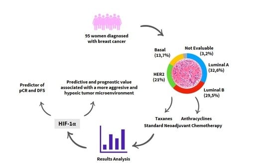
:Simple Summary
Abstract
1. Introduction
2. Materials and Methods
2.1. Patients and Treatment Management
2.2. Histology and Response Pathological Evaluation
2.3. Tissue Microarray Construction
2.4. Immunohistochemistry
2.5. Evaluation of Immunohistochemical Staining
2.6. Statistical Analysis
3. Results
3.1. Relation between HIF-1α Expression and pCR
3.2. Relation between HIF-1α Expression and Biological Markers
3.3. Relation between HIF-1α Expression and pATK, pMAK and EGFR
3.4. Predictive Factors of Response to Treatment—Multivariate Analysis
3.5. Sulvival Analysis—Prognostic Markers
4. Discussion
5. Conclusions
Supplementary Materials
Author Contributions
Funding
Institutional Review Board Statement
Informed Consent Statement
Data Availability Statement
Conflicts of Interest
Abbreviations
References
- Shien, T.; Iwata, H. Adjuvant and neoadjuvant therapy for breast cancer. Jpn. J. Clin. Oncol. 2020, 50, 225–229. [Google Scholar] [CrossRef] [PubMed]
- Shuai, Y.; Ma, L. Prognostic value of pathologic complete response and the alteration of breast cancer immunohistochemical biomarkers after neoadjuvant chemotherapy. Pathol. Res. Pract. 2019, 215, 29–33. [Google Scholar] [CrossRef] [PubMed]
- Greenwell, K.; Hussain, L.; Lee, D.; Bramlage, M.; Bills, G.; Mehta, A.; Jackson, A.; Wexelman, B. Complete pathologic response rate to neoadjuvant chemotherapy increases with increasing HER2/CEP17 ratio in HER2 overexpressing breast cancer: Analysis of the National Cancer Database (NCDB). Breast Cancer Res. Treat. 2020, 181, 249–254. [Google Scholar] [CrossRef] [PubMed]
- Al-Masri, M.; Aljalabneh, B.; Al-Najjar, H.; Al-Shamaileh, T. Effect of time to breast cancer surgery after neoadjuvant chemotherapy on survival outcomes. Breast Cancer Res. Treat. 2021, 186, 7–13. [Google Scholar] [CrossRef] [PubMed]
- Cullinane, C.; Shrestha, A.; Al Maksoud, A.; Rothwell, J.; Evoy, D.; Geraghty, J.; McCartan, D.; McDermott, E.W.; Prichard, R. Optimal timing of surgery following breast cancer neoadjuvant chemotherapy: A systematic review and meta-analysis. Eur. J. Surg. Oncol. 2021, 47, 1507–1513. [Google Scholar] [CrossRef] [PubMed]
- Gálvez-Navas, J.M.; Pérez-Ramírez, C.; Ramírez-Tortosa, M.C. [Secreted Frizzled-Related Protein 4 and breast cancer]. Ars. Pharm. 2021, 62, 438–450. [Google Scholar] [CrossRef]
- Pineda-Lancheros, L.E.; Pérez-Ramírez, C.; Sánchez-Martín, A.; Gálvez-Navas, J.M.; Martínez-Martínez, F.; Ramírez-Tortosa, M.C.; Jiménez-Morales, A. Impact of Genetic Polymorphisms on the Metabolic Pathway of Vitamin D and Survival in Non-Small Cell Lung Cancer. Nutrients 2021, 13, 3783. [Google Scholar] [CrossRef] [PubMed]
- Vaupel, P.; Multhoff, G. Hypoxia-/HIF-1alpha-Driven Factors of the Tumor Microenvironment Impeding Antitumor Immune Responses and Promoting Malignant Progression. Adv. Exp. Med. Biol. 2018, 1072, 171–175. [Google Scholar] [CrossRef]
- Zhang, T.; Suo, C.; Zheng, C.; Zhang, H. Hypoxia and Metabolism in Metastasis. Adv. Exp. Med. Biol. 2019, 1136, 87–95. [Google Scholar] [CrossRef]
- Zhang, Z.; Yao, L.; Yang, J.; Wang, Z.; Du, G. PI3K/Akt and HIF-1 signaling pathway in hypoxia-ischemia (Review). Mol. Med. Rep. 2018, 18, 3547–3554. [Google Scholar] [CrossRef]
- Masoud, G.N.; Li, W. HIF-1α pathway: Role, regulation and intervention for cancer therapy. Acta Pharm. Sin. B 2015, 5, 378–389. [Google Scholar] [CrossRef] [PubMed] [Green Version]
- Wee, P.; Wang, Z. Epidermal Growth Factor Receptor Cell Proliferation Signaling Pathways. Cancers 2017, 9, 52. [Google Scholar] [CrossRef] [PubMed] [Green Version]
- Shamis, S.A.K.; McMillan, D.C.; Edwards, J. The relationship between hypoxia-inducible factor 1α (HIF-1α) and patient survival in breast cancer: Systematic review and meta-analysis. Crit. Rev. Oncol. Hematol. 2021, 159, 103231. [Google Scholar] [CrossRef] [PubMed]
- Sanchez-Munoz, A.; Duenas-Garcia, R.; Jaen-Morago, A.; Carrasco, E.; Chacon, I.; Garcia-Tapiador, A.M.; Ortega-Granados, A.L.; Martínez-Ortega, E.; Ribelles, N.; Fernández-Navarro, M.; et al. Is it posible to increase pCR in the neoadjuvant treatment with dose-dense/sequential combination?: Results from a phase II Trial combining epirubicin and cyclophosphamide followed by paclitaxel and gemtamicine +/− trastuzumab in stage II and III breast cancer patients. Am. J. Clin. Oncol. 2010, 33, 432–437. [Google Scholar] [CrossRef]
- Schneeweiss, A.; Lauschner, I.; Ruiz, A.; Guerrero, A.; Sanchez-Rovira, P.; Segui, M.A.; Goerke, K.; Wolf, M.; Manikhas, A.G.; Wacker, J.; et al. Doxorubicin/pemetrexed followed by docetaxel as neoadjuvant treatment for early-stage breast cancer: A randomized phase II trial. Clin. Breast Cancer 2007, 7, 555–558. [Google Scholar] [CrossRef]
- Oluogun, W.A.; Adedokun, K.A.; Oyenike, M.A.; Adeyeba, O.A. Histological classification, grading, staging, and prognostic indexing of female breast cancer in an African population: A 10-years restrospective study. Int. J. Health Sci. 2019, 13, 3–9. [Google Scholar]
- Ogston, K.N.; Miller, I.D.; Payne, S.; Hutcheon, A.H.; Sarkar, T.K.; Smith, I.; Schofield, A.; Heys, S.D. A new histological grading system to assess response of breast cancer to primary chemotherapy: Prognostic significance and sulrvival. Breast 2003, 12, 320–327. [Google Scholar] [CrossRef]
- Konoken, J.; Bubendorf, L.; Kallioniemi, A.; Barlund, M.; Schraml, P.; Leighton, S.; Torhorst, J.; Mihatsch, M.J.; Sauter, G.; Kallioniemi, O.P. Tissue microarrays for high-throughput molecular profiling of tumor specimens. Nat. Med. 1998, 4, 844–847. [Google Scholar] [CrossRef]
- Zhu, Q.; Ademuyiwa, F.O.; Young, C.; Appleton, C.; Covington, M.F.; Ma, C.; Sanati, S.; Hagemann, I.S.; Mostafa, A.; Uddin KM, S.; et al. Early Assessment Window for Predicting Breast Cancer Neoadjuvant Therapy using Biomarkers, Ultrasound, and Diffuse Optical Tomography. Breast Cancer Res. Treat. 2021, 188, 615–630. [Google Scholar] [CrossRef]
- Avci, N.; Deligonul, A.; Tolunay, S.; Cubukcu, E.; Olmez, O.F.; Ulas, A. Neoadjuvant chemotherapy-induced changes in immunohistochemucal expression of estrogen receptor, progesterone receptor, HER2, and Ki-67 in patients with breast cancer. J. BUON 2015, 20, 45–49. [Google Scholar]
- Sánchez-Muñoz, A.; Plata-Fernández, Y.M.; Fernández, M.; Jaén-Morago, A.; Fernández-Navarro, M.; de la Torre-Cabrera, C.; Ramírez-Tortosa, C.; Lomas-Garrido, M.; Llácer, C.; Navarro-Pérez, V.; et al. The role of immunohistochemistry in breast cancer patients treated with neoadjuvant chemotherapy: And old tool with an enduring prognostic value. Clin. Breast Cancer 2013, 13, 46–52. [Google Scholar] [CrossRef] [PubMed]
- Wolff, A.C.; Hammond, M.E.H.; Allison, K.H.; Harvey, B.E.; Mangu, P.B.; Bartlett, J.M.S.; Bilous, M.; Ellis, I.O.; Fitzgibbons, P.; Hanna, W.; et al. Human epidermal growth factor receptor 2 testing in breast cancer: American Society of Clinical Oncology/College of American Pathologists clinical practice guideline focused update. Arch. Pathol. Lab. Med. 2018, 142, 1364–1382. [Google Scholar] [CrossRef] [PubMed]
- Vermeulen, M.A.; van Deurzen, C.H.; Schroder, C.P.; Matens, J.W.; van Diest, P.J. Expression of hipoxia-induced proteins in ductal carcinoma in situ invasive cancer of the male breast. J. Clin. Pathol. 2020, 73, 204–208. [Google Scholar] [CrossRef] [PubMed]
- Iyikesici, M.S.; Basaran, G.; Dane, F.; Ekenel, M.; Yumuk, P.F.; Cabuk, D.; Ekenel, M.; Yukum, P.F.; Cabuk, D.; Teomete, M.; et al. Associations between clinicopathological prognostic factors and pAkt, pMAPK and topoisomerase II expression in breast cancer. Int. J. Clin. Exp. Med. 2014, 7, 1459–1464. [Google Scholar] [PubMed]
- Sauerbrei, W.; Taube, S.E.; McShane, L.M.; Cavenagh, M.M.; Altman, D.G. Reporting Recommendations for Tumor Market Prognostic Studies (REMARK): An Abridged Explanation and Elaboration. J. Nat. Cancer Inst. 2018, 110, 803–811. [Google Scholar] [CrossRef]
- Zhang, J.; Zhang, S.; Gao, S.; Ma, Y.; Tan, X.; Kang, Y.; Ren, W. HIF-1α, TWIST-1 and ITGB-1, associated with Tumor Stiffness, as Novel Predictive Markers for the Pathological Response to Neoadjuvant Chemotherapy in Breast Cancer. Cancer Manag. Res. 2020, 12, 2209–2222. [Google Scholar] [CrossRef] [Green Version]
- Nie, C.; Lv, H.; Bie, L.; Hou, H.; Chen, X. Hypoxia-inducible factor 1-alpha expression correlated with response to neoadjuvant chemotherapy in women with breast cancer. Medicine 2018, 97, e13551. [Google Scholar] [CrossRef]
- Tiezzi, D.G.; Clagnan, W.S.; Mandarano, L.R.; de Sousa, C.B.; Marana, H.R.; Tiezzi, M.G.; de Andrade, J.M. Expression of aldehyde dehydrogenase after neoadjuvant chemotherapy is associated with expression of hypoxia-inducible factors 1 and 2 alpha and predicts prognosis in locally advanced breast cancer. Clinics 2013, 68, 592–598. [Google Scholar] [CrossRef]
- Yamamoto, Y.; Ibusuki, M.; Okumura, Y.; Kawasoe, T.; Kai, K.; Iyama, K.; Iwase, H. Hypoxia-inducible factor 1 alpha is closely linked to an aggressive phenotype in breast cancer. Breast Cancer Res Treat. 2008, 110, 465–475. [Google Scholar] [CrossRef]
- Generali, D.; Berruti, A.; Brizzi, M.P.; Campo, L.; Bonardi, S.; Wigfield, S.; Bersiga, A.; Allevi, G.; Milani, M.; Aguggini, S.; et al. Hypoxia-inducible factor-1 alpha expression predicts a poor response to primary chemoendocrine therapy and disease-free survival in primary human breast cancer. Clin. Cancer Res. 2006, 12, 4562–4568. [Google Scholar] [CrossRef] [Green Version]
- Generali, D.; Buffa, F.M.; Berruti, A.; Brizzi, M.P.; Campo, L.; Bonardi, S.; Bersiga, A.; Allevi, G.; Milani, M.; Aguggini, S.; et al. Phosphorylated ER alpha, HIF-1 alpha, and MAPK signaling as predictors of primary endocrine treatment response and resistance in patients with breast cancer. J. Clin. Oncol. 2009, 27, 227–234. [Google Scholar] [CrossRef] [PubMed]
- Sanchez-Rovira, P.; Anton, A.; Barnadas, A.; Velasco, A.; Lomas, M.; Rodriguez-Pinilla, M.; Ramírez, J.L.; Ramírez, C.; Ríos, M.J.; Castellá, E.; et al. Classical markers like ER and Ki-67, but also surviving and pERK, could be involved in the pathological response to gemcitabine, Adriamycin and paclitaxel (GAT) in locally advanced breast cancer patients: Results from the GEICAM/2002-01 phase II study. Clin. Transl. Oncol. 2012, 14, 430–436. [Google Scholar] [CrossRef] [PubMed]
- Foldi, J.; Silber, A.; Reisenbichler, E.; Singh, K.; Fishbach, N.; Persico, J.; Adelson, K.; Katoch, A.; Horowitz, N.; Lannin, D.; et al. Neoadjuvant durvalumab plus weekly nab-paclitaxel and dose-dense doxorubicin/cyclophosphamide in triple-negative breast cancer. Npj Breast Cancer 2021, 7, 9. [Google Scholar] [CrossRef] [PubMed]
- Wong, W.; Brogi, E.; Reis-Filho, J.S.; Plitas, G.; Robson, M.; Norton, L.; Morrow, M.; Wen, H.Y. Poor response to neoadjuvant chemotherapy in metaplastic breast carcinoma. Npj Breast Cancer 2021, 7, 96. [Google Scholar] [CrossRef]
- Miglietta, F.; Dieci, M.V.; Griguolo, G.; Guarneri, V. Neoadjuvant approach as a platform for treatment personalization: Focus on HER2-positive and triple-negative breast cancer. Cancer Treat. Rev. 2021, 98, 102222. [Google Scholar] [CrossRef]
- Campbell, E.J.; Dachs, G.U.; Morrin, H.R.; Davey, V.C.; Robinson, B.A.; Vissers, M.C.M. Activation of the hypoxia pathway in breast cancer tissue and patient survival are inversely associated with tumor ascorbate levels. BCM Cancer 2019, 19, 307. [Google Scholar] [CrossRef] [PubMed]
- Zhao, Z.; Mu, H.; Li, Y.; Liu, Y.; Zou, J.; Zhu, Y. Clinicopathological and prognostic value of hipoxia-inducible factor-1α in breast cancer: A meta-analysis including 5177 patients. Clin. Trans. Oncol. 2020, 22, 1892–19086. [Google Scholar] [CrossRef]
- Infantino, V.; Santarsiero, A.; Convertini, P.; Todisco, S.; Iacobazzi, V. Cancer Cell Metabolism in Hypoxia: Role of HIF-1 as Key Regulator and Therapeutic Target. Int. J. Mol. Sci. 2021, 22, 5703. [Google Scholar] [CrossRef]
- Sajnani, K.; Islam, F.; Smith, R.A.; Gopalan, V.; King-Yin Lam, A. Genetic alterations in Krebs cycle and its impact on cancer pathogenesis. Biochimie 2017, 135, 164–172. [Google Scholar] [CrossRef]
- Jögi, A.; Ehinger, A.; Hartman, L.; Alkner, S. Expression of HIF-1α is related to a poor prognosis and tamoxifen resistance in contralateral breast cancer. PLoS ONE 2019, 14, e0226150. [Google Scholar] [CrossRef] [Green Version]
- Bullen, J.W.; Tchernyshyov, I.; Holewinski, R.J.; DeVine, L.; Wu, F.; Venkatraman, V.; Kass, D.L.; Cole, R.N.; Van Eyk, J.; Semenza, G.L. Protein kinase A-dependent phosphorylation stimulates the transcriptional activity of hypoxia-inducible factor 1. Sci. Signal. 2016, 9, ra56. [Google Scholar] [CrossRef] [PubMed] [Green Version]
- Hielscher, A.; Gerecht, S. Hypoxia and free radicals: Role in tumor progression and the use of engineering-based platforms to address these relationships. Free Radic. Biol. Med. 2015, 79, 281–291. [Google Scholar] [CrossRef] [PubMed]
- Vera-Ramírez, L.; Sanchez-Rovira, P.; Ramírez-Tortosa, M.C.; Ramírez-Tortosa, C.L.; Granados-Principal, S.; Fernández-Navarro, M.; Lorente, J.A.; Quiles, J.L. Does chemotherapy-induced oxidative stress improve the survival rates of breast cancer patients? ARS 2011, 15, 903–909. [Google Scholar] [CrossRef] [PubMed]
- Gort, E.H.; Groot, A.J.; Derks van de Ven, T.L.; van der Groep, P.; Verlaan, I.; van Laar, T.; van Diest, P.J.; van der Wall, E.; Shvarts, A. Hypoxia-inducible factor-1 alpha expression requires PI 3-kinase activity and correlates with Akt1 phosphorylation in invasive breast carcinomas. Oncogene 2006, 25, 6123–6127. [Google Scholar] [CrossRef] [PubMed] [Green Version]
- Kronblad, A.; Hedenfalk, I.; Nisson, E.; Pahlman, S.; Landberg, G. ERK1/2 inhibition increases antiestrogen treatment efficacy by interfering with hypoxia-induced downregulation of ER alpha: A combination therapy potentially targeting hypoxic and dormant tumor cells. Oncogene 2005, 24, 6835–6841. [Google Scholar] [CrossRef] [PubMed]
- Hsu, C.-W.; Huang, R.; Khuc, T.; Shou, D.; Bullock, J.; Grooby, S.; Griffin, S.; Zou, C.; Little, A.; Astley, H.; et al. Identification of approved and investigational drugs that inhibit hypoxia-inducible factor-1 signalling. Oncotarget 2016, 7, 8172–8183. [Google Scholar] [CrossRef] [PubMed] [Green Version]
- Yang, S.X.; Constantino, J.P.; Kim, C.; Mamounas, E.P.; Nguyen, D.; Jeong, J.H.; Wolmark, N.; Kidwell, K.; Paik, S.; Swain, S.M. Akt phosphorylation as Ser473 predicts benefits of paclitaxel chemotherapy in node-positive breast cancer. J. Clin. Oncol. 2010, 28, 2974–2981. [Google Scholar] [CrossRef] [PubMed]


| Characteristics | Number of Cases (%) |
|---|---|
| Age at diagnosis (years) | |
| <40 | 19 (20%) |
| 40–49 | 38 (40%) |
| 50–59 | 18 (18.9%) |
| >60 | 20 (21.1%) |
| Mean | 20 (21.1%) |
| Range | 27–74 |
| Pretreatment tumor size (cm) | |
| 1–1.9 | 4 (4.2%) |
| 2–2.9 | 22 (23.2%) |
| 3–3.9 | 22 (23.2%) |
| 4–4.9 | 17 (17.9%) |
| >4.9 | 23 (24.2%) |
| Not measurable | 7 (7.4%) |
| Histological type | |
| Ductal infiltrating | 72 (75.8%) |
| Lobular infiltrating | 9 (9.5%) |
| Inflammatory | 6 (6.3%) |
| Mucinous | 6 (6.3%) |
| Mixed | 2 (2.1%) |
| Histological grade | |
| 1 | 20 (21%) |
| 2 | 37 (39%) |
| 3 | 34 (35.8%) |
| Not evaluable | 4 (4.2%) |
| Clinical TNM at diagnosis | |
| T | |
| T1 | 6 (6.3%) |
| T2 | 62 (65.3%) |
| T3 | 16 (16.8%) |
| T4 | 8 (8.4%) |
| Tx | 3 (3.2%) |
| N | |
| N0 | 34 (35.8%) |
| N1 | 46 (48.4%) |
| N2 | 15 (15.8%) |
| N3 | 0 (0%) |
| Nx | 0 (0%) |
| M | |
| M0 | 95 (100%) |
| M1 | 0 (0%) |
| Clinical stage | |
| IIA | 32 (33.7%) |
| IIB | 31 (32.6%) |
| IIIA | 21 (21.1%) |
| IIIB | 8 (8.4%) |
| Not evaluable | 3 (3.2%) |
| ER | |
| ≥10% | 69 (72.6%) |
| <10% | 25 (26.3%) |
| Not evaluable | 1 (1.1%) |
| Count ≥ 3 | 71 (74.7%) |
| Count < 3 | 23 (24.2%) |
| Not evaluable | 1 (1.1%) |
| PR | |
| ≥10% | 54 (56.8%) |
| <10% | 40 (42.1%) |
| Not evaluable | 1 (1.1%) |
| Count ≥ 3 | 58 (61.1%) |
| Count < 3 | 36 (37.1%) |
| Not evaluable | 1 (1.1%) |
| HER2 | |
| Positive | 20 (21.1%) |
| Negative | 72 (75.8%) |
| Not evaluable | 3 (3.1%) |
| Ki-67 | |
| ≥20% | 38 (40%) |
| <20% | 56 (58.9%) |
| Not evaluable | 1 (1.1%) |
| Phenotype | |
| Basal | 13 (13.7%) |
| HER2 | 20 (21%) |
| Luminal A | 31 (32.6%) |
| Luminal B | 28 (29.5%) |
| Not evaluable | 3 (3.2%) |
| Type of surgery | |
| Conservative | 38 (40%) |
| Not conservative | 56 (58.9%) |
| Not evaluable | 1 (1.1%) |
| Pathological response (M&P) | |
| Grade 1 | 8 (8.4%) |
| Grade 2 | 22 (23.1%) |
| Grade 3 | 28 (29.5%) |
| Grade 4 | 17 (17.9%) |
| Grade 5 (pCR) | 20 (21.1%) |
| Number of Cases (%) | p | ||
|---|---|---|---|
| pCR | No pCR | ||
| HIF-1α < 5% | 6 (33.3%) | 47 (67.1%) | 0.014 a |
| HIF-1α ≥ 5% | 12 (66.7%) | 23 (32.9%) | |
| HIF-1α% | 18 (20.5%) | 70 (79.5%) | 0.017 b |
| x = 10.42; SD = 9.53 | x = 5.05; SD = 7.52 |
| Variable | HIF-1α < 5% | HIF-1α ≥ 5% | p | |
|---|---|---|---|---|
| Grade 1 | 15 (83.3%) | 3 (16.7%) | 0.015 a | |
| Grade 2 | 22 (62.8%) | 13 (37.2%) | ||
| Grade 3 | 13 (41.9%) | 18 (58.1%) | ||
| Ki-67 < 20% | 29 (80.6%) | 7 (19.4%) | 0.001 b | |
| Ki-67 ≥ 20% | 23 (45.1%) | 28(54.9%) | ||
| HER2 − | 43 (63.2%) | 25 (36.8%) | 0.593 b | |
| HER2 + | 10 (55.6%) | 8 (44.4%) | ||
| ER < 10% | 10 (43.5%) | 13 (56.5%) | 0.080 b | |
| ER ≥ 10% | 43 (67.2%) | 21 (32.8%) | ||
| ER count < 3 | 9 (42.9%) | 12 (57.1%) | 0.072 b | |
| ER count ≥ 3 | 44 (66.7%) | 22 (33.3%) | ||
| PR < 10% | 18 (48.6%) | 19 (51.4%) | 0.049 b | |
| PR ≥ 10% | 35 (70%) | 15 (30%) | ||
| PR count < 3 | 16 (47.1%) | 18 (52.9%) | 0.044 b | |
| PR count ≥ 3 | 37 (69.8%) | 16 (30.2%) | ||
| Phenotype | Basal | 5 (41.7%) | 7 (58.3%) | |
| HER2 | 10 (55.6%) | 8 (44.4%) | ||
| Luminal A | 26 (86.7%) | 4 (13.3%) | 0.005 b | |
| Luminal B | 12 (46.2%) | 14 (53.8%) |
| DFS | OS | |||||
|---|---|---|---|---|---|---|
| HR | 95% CI | p-Value | HR | 95% CI | p-Value | |
| Grade 1 vs. 2 vs. 3 | - | - | 0.560 | - | - | 0.977 |
| Ki-67 ≥ 20% vs. <20% | 0.8 | 1.8–35.5 | 0.006 | 0.260 | ||
| ER ≥ 10% vs. <10% | - | - | 0.072 | 3.2 | 1.3–7.7 | 0.008 |
| pCR vs. no pCR | 0.2 | 0.0–0.6 | 0.009 | - | - | 0.06 |
| HIF-1α ≥ 5% vs. <5% | 2.5 | 1.0–6.2 | 0.047 | - | - | 0.295 |
| pAKT ≥ 10% vs. <10% | 2.4 | 1.0–5.9 | 0.039 | 2.4 | 1.0–5.7 | 0.046 |
Publisher’s Note: MDPI stays neutral with regard to jurisdictional claims in published maps and institutional affiliations. |
© 2022 by the authors. Licensee MDPI, Basel, Switzerland. This article is an open access article distributed under the terms and conditions of the Creative Commons Attribution (CC BY) license (https://creativecommons.org/licenses/by/4.0/).
Share and Cite
Ramírez-Tortosa, C.L.; Alonso-Calderón, R.; Gálvez-Navas, J.M.; Pérez-Ramírez, C.; Quiles, J.L.; Sánchez-Rovira, P.; Jiménez-Morales, A.; Ramírez-Tortosa, M. Hypoxia-Inducible Factor-1 Alpha Expression Is Predictive of Pathological Complete Response in Patients with Breast Cancer Receiving Neoadjuvant Chemotherapy. Cancers 2022, 14, 5393. https://doi.org/10.3390/cancers14215393
Ramírez-Tortosa CL, Alonso-Calderón R, Gálvez-Navas JM, Pérez-Ramírez C, Quiles JL, Sánchez-Rovira P, Jiménez-Morales A, Ramírez-Tortosa M. Hypoxia-Inducible Factor-1 Alpha Expression Is Predictive of Pathological Complete Response in Patients with Breast Cancer Receiving Neoadjuvant Chemotherapy. Cancers. 2022; 14(21):5393. https://doi.org/10.3390/cancers14215393
Chicago/Turabian StyleRamírez-Tortosa, César L., Rubén Alonso-Calderón, José María Gálvez-Navas, Cristina Pérez-Ramírez, José Luis Quiles, Pedro Sánchez-Rovira, Alberto Jiménez-Morales, and MCarmen Ramírez-Tortosa. 2022. "Hypoxia-Inducible Factor-1 Alpha Expression Is Predictive of Pathological Complete Response in Patients with Breast Cancer Receiving Neoadjuvant Chemotherapy" Cancers 14, no. 21: 5393. https://doi.org/10.3390/cancers14215393