Article

Layer-by-Layer Films of Silsesquioxane and Nickel(II) Tetrasulphophthalocyanine as Glucose Oxidase Platform Immobilization: Amperometric Determination of Glucose in Kombucha Beverages

by
João Paulo Winiarski
1,*,
Douglas José de Melo
2,
Edson Roberto Santana
1,
Cleverson Siqueira Santos
3,
Cliciane Guadalupe de Jesus
4,
Sérgio Toshio Fujiwara
4,†,
Karen Wohnrath
4 and
Christiana Andrade Pessôa
4
1
Department of Chemistry, Federal University of Santa Catarina, Florianópolis 88040-900, SC, Brazil
2
Department of Chemistry, Federal University of Paraná, Curitiba 81531-980, PR, Brazil
3
Federal Institute of Farroupilha, Campus Alegrete, Alegrete 97555-000, RS, Brazil
4
Department of Chemistry, State University of Ponta Grossa, Ponta Grossa 84030-900, PR, Brazil
*
Author to whom correspondence should be addressed.
In memoriam.
Chemosensors 2023, 11(6), 346; https://doi.org/10.3390/chemosensors11060346
Submission received: 9 May 2023 / Revised: 6 June 2023 / Accepted: 12 June 2023 / Published: 14 June 2023

Abstract

:
This paper describes the development of a novel glucose biosensor through the layer-by-layer technique (LbL). The self-assembled architectures were composed of a positive-charged silsesquioxane polyelectrolyte, 3-n-propylpyridinium silsesquioxane chloride (SiPy+Cl), nickel (II) tetrassulphophthalocyanine (NiTsPc), and a conductive surface of FTO (fluor tin oxide). The construction of the biosensor was influenced by the isoelectric point (pI) of the glucose oxidase enzyme (GOx), which allowed electrostatic interaction between the outer layer of the silsesquioxane film and the enzyme. The architecture of modified electrode GOx/(SiPy+Cl/NiTsPc)5.5/FTO was confirmed by UV-Vis, FTIR, and chronoamperometry techniques using different immobilization methods of GOx. Among the studied methods, a higher variation of current was observed for the modified electrode formed by mixed LbL films of SiPy+Cl and NiTsPc and the enzyme immobilized by drop coating. The stability and reproducibility of the biosensor were verified when the last layer containing the enzyme was coated with 0.2% Nafion® polymer. Under these conditions, a linear response for glucose was obtained in the concentration range of 0.2 to 1.6 mmol L−1 (R2 = 0.991) with a limit of detection of 0.022 mmol L−1. The proposed biosensor was applied to quantify glucose in two different samples of kombucha juices with accuracy, allowing the glucose content of the healthy beverages to be estimated.

1. Introduction

Kombucha is a healthy beverage that is the result of tea fermentation by adding starter cultures such as yeast and acetic acid bacteria. Sucrose is the most commonly added sugar in kombucha tea production [1]. Depending on the temperature of fermentation, sucrose content decreases, which is an expected result of the conversion of fructose and glucose by yeast cells [2,3]. An indirect way to measure the yeast’s hydrolysis of sucrose into glucose and fructose by Invertase is to control the content of glucose produced by bacteria, and a simple device, such as an electrochemical biosensor, may present an alternative to controlling quality in routine analysis. Applegate et al. developed an analysis of kombucha using three different techniques for verifying the changes in sugar content during fermentation of kombucha beverages. The authors demonstrated the accuracy of glucometers for quantifying glucose concentrations in kombucha samples [4]. This directive offers the advantages of developing novel biosensors for glucose quantification in fermented kombucha samples.
Amperometric glucose biosensors based on the immobilization of glucose oxidase on the electrode surfaces have attracted considerable interest due to their advantages in terms of simplicity, short response time, high sensitivity, and excellent selectivity [5,6]. Nanotechnology has relevance in biosensing since it can provide alternative ways to immobilize biomolecules, whose activity must be preserved for long periods [7]. Immobilization of enzymes on the transducer surface is an important and critical step in the design of biosensors [8]. These biomolecules must maintain their structure and function when immobilized to retain their biological activity, in addition to remaining tightly bound to the surface and not being desorbed during the use of the biosensor [9]. In addition, the configuration of the electrodes and materials is crucial for the immobilization of the bioreceptors on the electrode surface [10].
A large number of methods have been proposed to immobilize enzymes on electrode surfaces, concomitantly preserving enzymatic activity and designing efficient electron-transfer pathways between the immobilized enzyme and electrode surface, preventing unspecific side reactions [11,12,13]. Among them, the mostly commonly applied are self-assembled monolayers [14], Langmuir–Blodgett films [15], sol-gel methods [16,17], and LbL films [18,19,20,21,22].
As far as biosensing is concerned, the advantages of the LbL technique lie in the easy control of thickness and possible tuning of molecular architectures to yield tailored sensing units [23]. There are specific methods to immobilize enzymes on the electrode surface combined with the LbL technique. 3-n-propylpyridinium silsesquioxane chloride (SiPy+Cl) and its analogues are hybrid organic–inorganic materials that possess highly advantageous properties in biosensing such as biocompatibility and high chemical stability [24,25]. Nickel(II) phthalocyanine–tetrasulfonic acid tetrasodium salt (Nitsch) is an inorganic complex that possesses valuable characteristics in electrochemical sensors [26,27], such as a good thin film former, a conjugated π system that can add electronic properties to the system, and the anionic nature of the complex that makes it accessible for adsorption in cationic materials.
One of the strategies to improve the performance and lifetime of the biosensor is the application of Nafion®. This polymer has been extensively used in the construction of biosensors since it presents a series of advantages, such as high chemical stability and biocompatibility [28,29].
In this paper, the glucose oxidase enzyme (GOx) was immobilized on the nanoarchitecture (SiPy+Cl/NiTsPc)5.5/FTO by different methods: adsorption and cross-linking, both using the coating surface by dropping the GOx on its surface or dipping the modified electrode in a solution of GOx. The biosensor based on the adsorption of GOx onto the modified electrode that showed a major current response was used to quantify glucose in kombucha’s beverage samples. The quantification of glucose content in kombucha’s beverages is an alternative to measuring the hydrolysis of sucrose from the fermented beverage.

2. Materials and Methods

2.1. Enzyme and Chemicals

All reagents were of analytical-grade purity. Glucose oxidase (EC 1.1.3.4 type VII, from Aspergillus niger, having 100–250 units g−1 of activity, isoelectric point at pH 4.2), nickel(II) phthalocyanine-tetrasulfonic acid tetrasodium salt (NiTsPc), Nafion®, D(+)—glucose, and glutaraldehyde (GA) were purchased from Sigma-Aldrich (St. Louis, MO, USA). The polyelectrolyte 3-n-propylpyridinium chloride silsesquioxane (SiPy+Cl) was synthesized as described in the literature using pyridine as a ligand [30]. The fluorine-tin oxide, one-side coated on a glass substrate (FTO), was purchased from Flexitec with Rs = 10–20 Ωm. The dimensions of the FTO substrate were length: 1.1 cm, width: 0.7 cm, and thickness: 0.1 cm.

2.2. Film Assembly

The LbL films were assembled onto FTO-coated glass (fluorine-tin oxide, one-side coated on glass). Before modification, the FTO substrate was cleaned by immersion in a mixture of H2O:H2O2:NH4OH (10:1:1) and heating. After that, it was cleaned with CHCl3 followed by isopropyl alcohol below the boiling point. The (SiPy+Cl/NiTsPc)n LbL film, where n = number of bilayers, was carried out as follows: FTO electrodes were treated with an aqueous solution of 2.0 mg mL−1 SiPy+Cl (pH 5.2) for 5 min to form a positively charged surface, then the electrode was dipped in a negatively charged 2.0 mg mL−1 NiTsPc solution (pH 5.2) for 5 min. After deposition of each layer, the film was washed with distilled water and air-dried. This procedure was repeated until five bilayers were formed. In order to form a positive layer to immobilize the GOx, a new layer of SiPy+Cl was deposited on the surface [29]. After five bilayers formed, (SiPy+Cl/NiTsPc)5/FTO, the electrode was immersed for another 5 min in the SiPy+Cl solution, forming (SiPy+Cl/NiTsPc)5.5/FTO LbL film.

2.3. Enzyme Immobilization on (SiPy+Cl/NiTsPc)5.5/FTO Modified Electrode

Different immobilization methods of the GOx enzyme on the electrode surface were studied: adsorption and cross-linking, both using the coating surface (SiPy+Cl/NiTsPc)5.5/FTO by dropping the GOx on its surface or dipping the modified electrode in a solution of GOx. In addition to this, alternative methods using glutaraldehyde as a cross-linking agent were also used. For a better understanding, these methods were named cross-linking-dip coating and cross linking-drop coating.
In the drop-coating method, 40 µL of 2 mg mL−1 enzyme (25 U) was dropped on the (SiPy+Cl/NiTsPc)5.5/FTO and stored at 4 °C. In the dip-coating method, the modified electrode was submerged in a GOx solution at the same concentration as in the previous method for 24 h at 4 °C. The GOx solution was prepared in PBS buffer at pH 5.5.
In the cross-linking method by drop-coating (cross-linking-drop coating), 10 µL of a GOx solution was dropped on the (SiPy+Cl/NiTsPc)5.5/FTO and dried at room temperature (23 °C). Then, 10 µL of glutaraldehyde (GA) (1:1 v/v) were dropped on the GOx layer and dried at 4 °C. The electrode obtained by the cross-linking dip-coating method was made by the immersion of (SiPy+Cl/NiTsPc)5.5/FTO in a mixed solution of GA and GOx for 24 h. All the modified electrodes, denoted as GOx/(SiPy+Cl/NiTsPc)5.5/FTO, were stored in a PBS buffer of 0.1 mol L−1, pH 7.0, at 4 °C.
The Nafion/GOx/(SiPy+Cl/NiTsPc)5.5/FTO electrode was performed by dropping 10 µL of 0.1, 0.2, or 0.3% (v/v) of 5% Nafion® solution in ethanol on the outer layer coated with GOx, drying at room temperature, and storing at 4 °C. Scheme 1 shows the preparation of self-assembly LbL films and the biosensor construction.

2.4. Characterization of the Biosensors Obtained by Different Techniques

The UV-Vis spectra of (SiPy+Cl/NiTsPc)5.5/FTO, GOx/(SiPy+Cl/NiTsPc)5.5/FTO and GOx/FTO films were measured with a MultiSpec-1501 spectrophotometer (Shimadzu, Japan). Infrared spectra of these films were collected as the average of 128 scans, with a resolution of 2 cm−1, in the range from 4000 to 400 cm−1 in the transmission mode, using a FTIR-8400 spectrophotometer (Shimadzu, Kyoto, Japan). All measurements of FTIR were performed on a Si substrate.
The electrochemical experiments were carried out with a PGSTAT 30 potentiostat (Metrohm Autolab, Utrecht, The Netherlands), controlled by a personal computer using GPES version 4.9 software using a conventional three-electrode cell. Chronoamperometric measurements were performed with Ag/AgCl (KCl 3.0 mol L−1) and platinum wire as reference and auxiliary electrodes, respectively. FTO modified with Nafion/GOx/(SiPy+Cl/NiTsPc)5.5 films were used as working electrodes in an electrochemical cell with PBS buffer 0.1 mol L−1 pH 7.0 as a supporting electrolyte.

2.5. Determination of Glucose in Kombucha Beverages Samples

The standard addition method was used to evaluate the glucose concentrations in the samples. The proposed electrode was applied to the determination of glucose in two beverage samples of kombucha acquired in a local market in Florianópolis, Brazil. For the determination of glucose in beverages, 9.50 mL of supporting electrolyte and 500 µL of the sample were used in an electrochemical cell. The chronoamperometric analysis was performed with different volume additions of a 0.1 mol L−1 glucose solution into an electrochemical cell using the Nafion/GOx/(SiPy+Cl/NiTsPc)5.5/FTO biosensor.

3. Results and Discussion

3.1. Characterization of GOx/(SiPy+Cl/NiTsPc)5.5 Films

The nanoarchitectured film (SiPy+Cl/NiTsPc)5.5 presents itself as biocompatible for immobilization of the GOx enzyme, as described in similar thin films [29]. Since enzyme activity is dependent on the ionization state of the amino acids in the active site, pH values play an important role in maintaining the proper conformation of an enzyme. The pH value optimum of GOx varies from 5.0 to 7.0 [31], at which the enzyme presents major activity for glucose. However, enzymatic biosensors that present only biomolecules immobilized by physical adsorption can lose activity over time because desorption may occur. The surface layer contains SiPy+Cl with a positive charge at pH 5.2 [32], and the GOx enzyme has an isoelectric point of 4.2 [33,34]. Consequently, the GOx at pH 5.5 is negatively charged. Therefore, the immobilization will occur through electrostatic interactions while maintaining GOx the native conformation. The formation of the electrode with the architecture GOx/(SiPy+Cl/NiTsPc)5.5/FTO was monitored by UV-Vis spectroscopy, as shown in Figure 1A,B.
As observed in Figure 1A, the polyelectrolyte SiPy+Cl does not display any absorption bands in the range of 300 to 800 nm, whereas the aqueous solution of NiTsPc shows its characteristic absorption bands. The band with major energy at 335 nm (B band or Soret) is related to the π-π* transition of the macrocycle ring of the phthalocyanine structure associated with the transition a2u → eg orbitals [35]. At 620 and 675 nm, it is assigned to its dimeric and monomeric species, respectively, due to its transitions between HOMO and LUMO orbitals attributed to the interactions between Ni and axial ligands of the phthalocyanine structures, represented by the transition of a1u → eg orbitals [36]. For (SiPy+Cl/NiTsPc)5.5/FTO LbL film, a slight shift of Q bands at 614 and 664 nm indicates the incorporation of NiTsPc on LbL film.
The UV-Vis spectrum of GOx solution exhibited two characteristic bands at 375 and 465 nm attributed to π-π* transitions along the three cycles of the isoalloxazine ring of FAD, characteristic of the oxidized form of flavin7 groups [37]. Since the enzyme was dropped onto the (SiPy+Cl/NiTsPc)5.5/FTO, a weak band at 459 nm was observed, which can be assigned to the characteristic band of the oxidized form of the flavin7 group in protein structure [38], indicating that the GOx was immobilized onto the LbL film. The band in the region of 375 nm related to the neat GOx (Figure 1A) was supposed to be overlapped by the Soret band of NiTsPc in GOx/(SiPy+Cl/NiTsPc)5.5/FTO, as seen in Figure 1B. The position of the Gox band is in good agreement with the work by Zhou et al., in which GOx was co-immobilized with Au and graphene to further modify a GCE [37]. The shifting of the bands to lower wavelengths observed in LbL film in relation to the precursor solutions is attributed to a very strong interaction when SiPy+Cl, NiTsPc, and GOx are sandwiched in the platform [39].
FTIR spectra of the Nafion®/GOx/(SiPy+Cl/NiTsPc)35.5/Si, native GOx/Si, and Nafion®/Si films are shown in Figure 2. The absorption band for native GOx (Figure 2a) at 3284 cm−1 is assigned to the N–H stretching, and the bands at 1654 and 1537 cm−1 are attributed to amide I (the C=O stretching vibrations of the peptide bond groups) and amide II (the N–H in-plane bending and C–N stretching modes of the polypeptide chains) bands [40,41,42]. The spectrum of Nafion® membrane (Figure 2c) showed bands at 1205 (C–F asymmetric stretching), 1147 (C–F asymmetric stretching), 1056 (S=O symmetric stretching), and 981 cm−1 (symmetric stretching C–O–C) [43]. The Nafion®/GOx/(SiPy+Cl/NiTsPc)5.5 spectrum (Figure 2b) also shows two characteristic adsorption bands at 1647 and 1524 cm−1, suggesting that GOx has been successfully immobilized on the LbL film. The presence of these two bands is indicative that the bioactivity and secondary structure of the GOx enzyme are maintained after its immobilization on the LbL film. Moreover, two bands at 1226 and 1153 cm−1 related to Nafion® membrane were also observed.

3.2. Studies of Different Methods of GOx Immobilization on LbL Modified Electrodes

Enzyme immobilization on the electrode surface is one of the most important factors to be considered in the construction of amperometric biosensors [44,45,46,47]. Different immobilization methods of the GOx enzyme on the electrode surface were studied: the conventional dip and drop-coating methods and using glutaraldehyde as a cross-linking agent, which are based on electrostatic interaction and covalent bonding, respectively. These methods were performed using the coating surface ((SiPy+Cl/NiTsPc)5.5/FTO) by dropping the GOx on its surface or dipping the modified electrode in a solution containing the GOx in the absence or presence of glutaraldehyde. The aim of this study is to evaluate the immobilization procedure of the GOx enzyme on the (SiPy+Cl/NiTsPc)5.5 LbL film using the chronoamperometric technique. The current as a function of different glucose concentrations was monitored to compare each immobilization technique (Figure 3). It was verified that the drop-coating method (Figure 3—curve a) shows a higher variation of current and a chronoamperometric profile (results not shown) compared to the other three methods. It can be inferred that the last layer of polycation (SiPy+Cl) on the film architecture provides a strong interaction with GOx since it is negatively charged at pH 5.2 (above the isoelectric point of GOx at pH 4.2) [29]. The reason why inner layers should not contain GOx is that in this case the enzyme is tightly packed in the layers, thus hampering catalytic activity by blocking glucose diffusion and electron transfer during the redox process [48,49]. Therefore, when GOx was deposited electrostatically by the dip coating method on the LbL film, it did not show any significant current response. This may be attributed to the low amount of enzyme immobilized, so the few active centers present are saturated with low quantities of glucose, resulting in a significant decrease in the current signal. For the methods of dip and drop coating, when used in combination with glutaraldehyde, a considerable decrease in the electrode response is observed. This behavior is related to the fact that GA promotes cross-linking reactions that may lead to deep structural changes of the protein and block its active center, causing a decrease in activity [50].
One of the problems associated with electrochemical biosensors is the leaching of the enzyme from the substrate during the electrochemical measurements [51]. Therefore, the repeatability of the electrode obtained by the drop-coating method was evaluated. It was verified that after the first chronoamperometric measurement with the same electrode, the current response decreased significantly and the enzyme had been leached from the electrode to the solution (results not shown). In order to prevent this and to evaluate the repeatability of the biosensor, Nafion® polymer was used to recover the GOx layer and protect the enzyme on the electrode surface to improve its stability, even though this strategy affects the sensitivity of the electrode with the incorporation of this polymer. In the literature, it has been reported that Nafion® polymer is used as a membrane to recover electrodes. In addition, it works as an anti-interference barrier on the enzymatic electrodes [52,53]. Miao et al. reported a glucose biosensor based on gold nanoparticles hosted on nanocomposites of polyvinylpyrrolidone and polyaniline on GCE. The immobilization of GOx on the topmost electrode was filled with Nafion® dropped on the whole surface, which improved the operational and storage stability of the biosensor [54].
In order to avoid the loss of activity of the enzyme, the ideal concentration of Nafion® to recover the GOx on the last layer of the electrode was optimized. The concentrations of 0.1, 0.2, and 0.3% (v/v) of Nafion® were investigated. Figure 4 shows the calibration plot of different concentrations of Nafion® used on the GOx/(SiPy+Cl/NiTsPc)5.5/FTO modified electrode.
Although the chronoamperometric of the electrode with 0.1% Nafion® on the outer layer shows the major variation of the current, GOx still leaches from the film after three successive measurements, as indicated by no amperometric response. The response of 0.3% shows lower current variation, demonstrating the blocking of the analyte by the biomembrane. The choice of 0.2% Nafion® concentration is attributed to a good chronoamperometric profile with good signal compared to two others studied. Furthermore, the stability of this electrode is enhanced compared with the first one (as can be seen in the last section). Therefore, for the next studies, the Nafion® concentration was kept at 0.2%. A very thin film of Nafion® is satisfactory to offer minimal obstruction to the diffusion of the analyte to the electrode surface while at the same time preventing the desorption of biomolecules from the electrode surface [55,56].

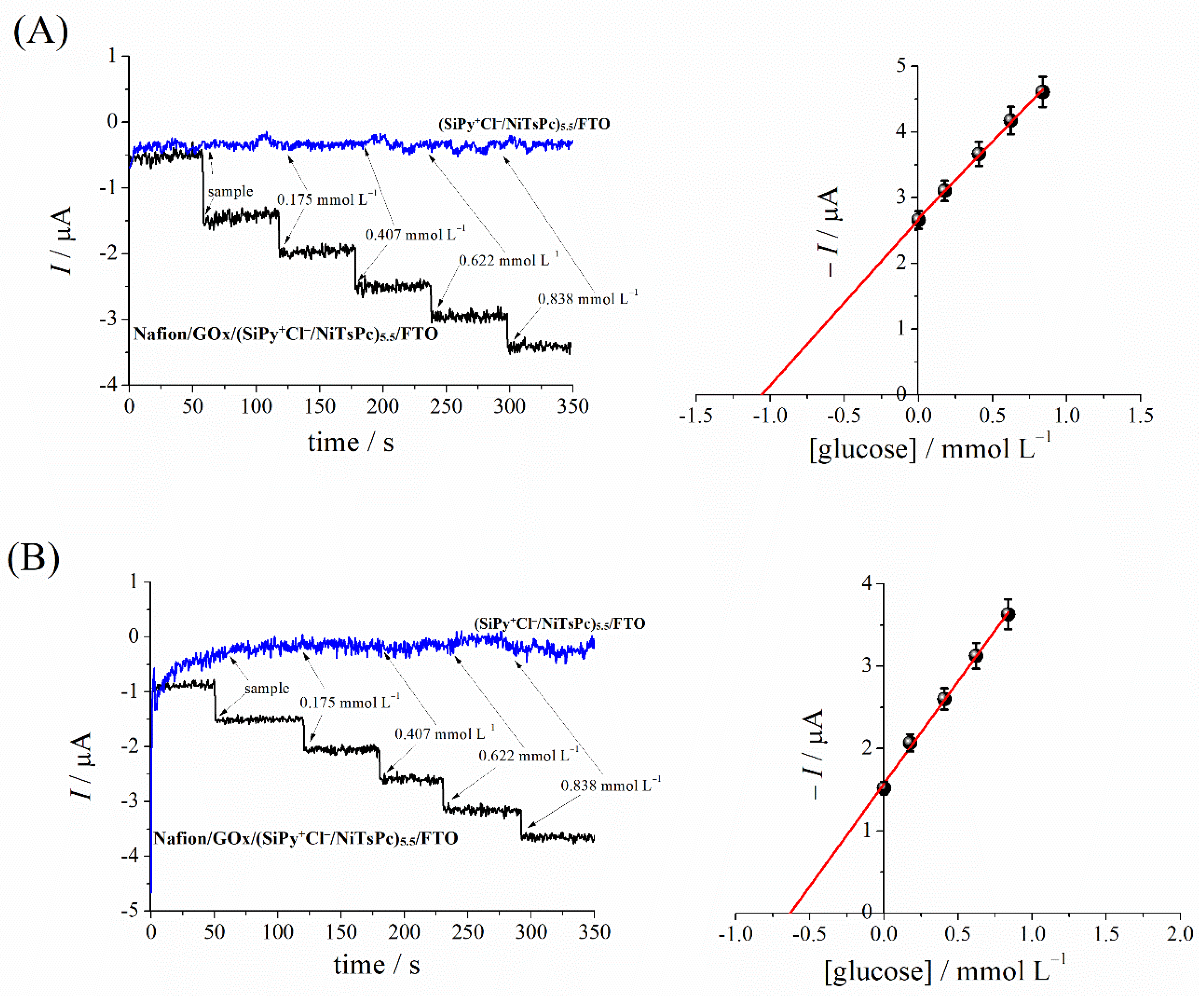
3.3. Electrochemical Performance of the Glucose Biosensor Nafion/GOx/(SiPy+Cl/NiTsPc)5.5/FTO

Figure 5 shows the typical chronoamperometric response curves of Nafion/GOx/(SiPy+Cl/NiTsPc)5.5/FTO on successive additions of glucose to the PBS at 0.1 mol L−1 at −0.1 V. The response increased linearly with the increase in glucose concentration, ranging from 0.2 to 1.6 mmol L−1 (R2 = 0.993). The limits of detection and quantification (LOD and LOQ) were calculated according to LOD = 3 × Sb/B and LOQ = 10 × Sb/B, where Sb is the standard deviation of the intercept and B is the slope of the calibration plot [57]. The LOD and LOQ values obtained were 0.022 mmol L−1 and 0.074 mmol L−1, respectively.
The analytical methods reported for the quantification of glucose in kombucha samples are mostly chromatographic or spectrophotometric. In that regard, the LOD obtained with the Nafion/GOx/(SiPy+Cl/NiTsPc)5.5/FTO is between the values obtained by other studies using LbL electrode systems, as can be seen in Table 1.
The reaction of glucose with the catalytic site of the GOx enzyme generates H2O2 that is electrochemically reduced to −0.1 V, as can be seen in Scheme 2.
Table 1 lists some modified LbL glucose biosensors with some analytical parameters for glucose determination reported in the literature.
The proposed biosensor shows good performance compared to other electrode architectures, as summarized in Table 1. As one can see, the estimated LOD value is quite promising, showing that the proposed method based on Nafion/GOx/(SiPy+Cl/NiTsPc)5.5/FTO is suitable for real-sample routine analysis protocols.

3.4. Reproducibility and Storage Stability of Nafion/GOx/(SiPy+Cl/NiTsPc)5.5/FTO

The reproducibility of the biosensor was estimated from the responses of 0.38 and 0.64 mmol L−1 glucose (PBS, pH 7.0) using three different electrodes prepared by the drop-coating method. The results revealed that the biosensor has satisfactory reproducibility (RSD = 0.38 and 1.26%, respectively). The stability of the Nafion/GOx/(SiPy+Cl/NiTsPc)5.5/FTO biosensor has been monitored by the amperometric response for 0.38 and 0.64 mmol L−1 glucose for a period of 23 days, maintaining the biosensor in a PBS solution for storage. The biosensor retained between 92 and 95% of its original response when stored in refrigerated conditions (4 °C). These results indicated that the biosensor exhibits higher storage stability.

3.5. Determination of Glucose Content in Kombucha Beverages

The proposed biosensor was applied to the determination of glucose in two different commercial kombucha juices (Figure 6). The glucose determination was performed with the standard addition method. In sample 1 (kombucha with strawberry and hibiscus, Figure 6A), the estimated concentration was 22.8 mmol L−1 (4.11 g L−1) and in sample 2 (kombucha pineapple with mint, Figure 6B), the concentration was 12.62 mmol L−1 (2.27 g L−1). It was verified that the fabrication days of sample 1 differed by four days from sample 2, considering the same manufacturer. It can be inferred that the hydrolysis of sucrose keeps occurring throughout the day. Similar results were observed in Neffe-Skocińska et al.’s studies of kombucha’s fermentation on different days (from zero to ten days of fermentation—1.4–37.7 g L−1 ± 0.15) [2]. In addition, we have tested the proposed sensor (SiPy+Cl/NiTsPc)5.5/FTO for the same applications. As can be seen in Figure 6A,B, there is no response for glucose additions, letting us know that GOx is primordial for glucose sensing in the electrochemical platform (SiPy+Cl/NiTsPc)5.5/FTO.
These results agree with other published studies using other analytical techniques for the determination of sucrose fermentation in kombucha samples. Kallel et al. verified the biochemistry of kombucha samples using high performance anion exchange chromatography with pulsed amperometric detection (HPAEC-PAD) and observed the appearance of glucose and fructose with increasing fermentation time. After that, the authors verified that the glucose disappearance was more important than that of fructose [63].

4. Conclusions

We have shown that a simple self-assembly of positively charged silsesquioxane inorganic polymers and anionic nickel(II) tetrasulphophtalocyanine complex formation films can effectively act as a support for GOx under accessible conditions. Since the immobilization of the enzyme is a crucial step in order to obtain a stable device, these conditions were evaluated. It was observed that the simple drop-coating method without any cross-linking agent presented higher current values and that Nafion® in low concentrations improved the biosensor’s stability and sensitivity. Therefore, a novel sensory platform for glucose determination was successfully constructed, and the results had appreciable sensitivity, stability, and detection limits applicable to fermented kombucha samples. These findings will help determine glucose in other relevant food matrices and samples.

Author Contributions

Conceptualization, J.P.W. and D.J.d.M.; methodology, J.P.W. and D.J.d.M.; validation, J.P.W. and D.J.d.M.; investigation, J.P.W. and D.J.d.M.; formal analysis, J.P.W., D.J.d.M., C.G.d.J. and C.S.S.; writing—original draft preparation, J.P.W., C.G.d.J., S.T.F., C.S.S., K.W. and C.A.P. writing—review and editing, J.P.W., E.R.S., S.T.F., C.S.S., K.W. and C.A.P.; supervision, S.T.F., K.W. and C.A.P.; project administration, S.T.F., K.W. and C.A.P. All authors have read and agreed to the published version of the manuscript.

Funding

This research was funded by Conselho Nacional de Desenvolvimento Científico e Tecnológico (CNPq) and Coordenação de Aperfeiçoamento de Pessoal de Nível Superior (Capes), finance code 001.

Data Availability Statement

Data are available from the authors under reasonable request.

Acknowledgments

The authors are thankful to the Brazilian government agencies: CNPq (Conselho Nacional de Desenvolvimento Científico e Tecnológico) and CAPES (Coordenação de Aperfeiçoamento de Pessoal de Nível Superior).

Conflicts of Interest

The authors declare no conflict of interest.

References

  1. Jayabalan, R.; Malbaša, R.V.; Lončar, E.S.; Vitas, J.S.; Sathishkumar, M. A Review on Kombucha Tea-Microbiology, Composition, Fermentation, Beneficial Effects, Toxicity, and Tea Fungus. Compr. Rev. Food Sci. Food Saf. 2014, 13, 538–550. [Google Scholar] [CrossRef] [PubMed]
  2. Neffe-Skocińska, K.; Sionek, B.; Ścibisz, I.; Kołożyn-Krajewska, D. Contenido de Ácido y Efectos de Las Condiciones de Fermentación En Las Propiedades Fisicoquímicas, Microbiológicas y Sensoriales de Bebidas de Té de Kombucha. CYTA—J. Food 2017, 15, 601–607. [Google Scholar] [CrossRef] [Green Version]
  3. Villarreal-Soto, S.A.; Beaufort, S.; Bouajila, J.; Souchard, J.-P.; Taillandier, P. Understanding Kombucha Tea Fermentation: A Review. J. Food Sci. 2018, 83, 580–588. [Google Scholar] [CrossRef] [PubMed]
  4. Applegate, K.B.; Cheek, P.R.; Inlow, J.K. Analysis of Kombucha to Teach Biochemical Concepts and Techniques to Undergraduate Students. Biochem. Mol. Biol. Educ. 2019, 47, 459–467. [Google Scholar] [CrossRef] [PubMed]
  5. Bollella, P. Enzyme-Based Amperometric Biosensors: 60 Years Later … Quo Vadis? Anal. Chim. Acta 2022, 1234, 340517. [Google Scholar] [CrossRef] [PubMed]
  6. Liu, Y.; Yue, W.; Cui, Y. Development of an Amperometric Biosensor on a Toothbrush for Glucose. Sens. Actuators Rep. 2023, 5, 100133. [Google Scholar] [CrossRef]
  7. Nemiwal, M.; Zhang, T.C.; Kumar, D. Enzyme Immobilized Nanomaterials as Electrochemical Biosensors for Detection of Biomolecules. Enzyme Microb. Technol. 2022, 156, 110006. [Google Scholar] [CrossRef] [PubMed]
  8. Theyagarajan, K.; Kim, Y.J. Recent Developments in the Design and Fabrication of Electrochemical Biosensors Using Functional Materials and Molecules. Biosensors 2023, 13, 424. [Google Scholar] [CrossRef]
  9. Sassolas, A.; Blum, L.J.; Leca-Bouvier, B.D. Immobilization Strategies to Develop Enzymatic Biosensors. Biotechnol. Adv. 2012, 30, 489–511. [Google Scholar] [CrossRef] [PubMed]
  10. Liébana, S.; Drago, G.A. Bioconjugation and Stabilisation of Biomolecules in Biosensors. Essays Biochem. 2016, 60, 59–68. [Google Scholar] [CrossRef] [PubMed] [Green Version]
  11. Ashraf, G.; Chen, W.; Asif, M.; Aziz, A.; Zhong, Z.T.; Iftikhar, T.; Zhao, Y.D. Topical Advancements in Electrochemical and Optical Signal Amplification for Biomolecules Detection: A Comparison. Mater. Today Chem. 2022, 26, 101119. [Google Scholar] [CrossRef]
  12. Zhao, H.; Chen, T.; Wu, T.; Xie, L.; Ma, Y.; Sha, J. Strategy Based on Multiplexed Brush Architectures for Regulating the Spatiotemporal Immobilization of Biomolecules. Biomater. Adv. 2022, 141, 213092. [Google Scholar] [CrossRef]
  13. Chen, N.; Chang, B.; Shi, N.; Yan, W.; Lu, F.; Liu, F. Cross-Linked Enzyme Aggregates Immobilization: Preparation, Characterization, and Applications. Crit. Rev. Biotechnol. 2023, 43, 369–383. [Google Scholar] [CrossRef]
  14. Santos, C.S.; Mossanha, R.; Pessôa, C.A. Biosensor for Carbaryl Based on Gold Modified with PAMAM-G4 Dendrimer. J. Appl. Electrochem. 2015, 45, 325–334. [Google Scholar] [CrossRef]
  15. Dopierała, K.; Kołodziejczak-Radzimska, A.; Prochaska, K.; Jesionowski, T. Immobilization of Lipase in Langmuir—Blogett Film of Cubic Silsesquioxane on the Surface of Zirconium Dioxide. Appl. Surf. Sci. 2022, 573, 151184. [Google Scholar] [CrossRef]
  16. Rex, A.; dos Santos, J.H.Z. The Use of Sol–Gel Processes in the Development of Supported Catalysts. J. Sol-Gel Sci. Technol. 2023, 105, 30–49. [Google Scholar] [CrossRef]
  17. Vesoloski, J.F.; Todero, A.S.; Macieski, R.J.; de Oliveira Pereira, F.; Dallago, R.M.; Mignoni, M.L. Immobilization of Lipase from Candida Antarctica B (CALB) by Sol–Gel Technique Using Rice Husk Ash as Silic Source and Ionic Liquid as Additive. Appl. Biochem. Biotechnol. 2022, 194, 6270–6286. [Google Scholar] [CrossRef]
  18. Hemin, F.U.; Oxidase, G.; Yoshida, K.; Kashimura, Y.; Kamijo, T.; Ono, T.; Dairaku, T. Decomposition of Glucose-Sensitive Layer-by-Layer Films Using Hemin, DNA, and Glucose Oxidase. Polymers 2020, 12, 319. [Google Scholar]
  19. Scotto, J.; Piccinini, E.; von Bilderling, C.; Coria-Oriundo, L.L.; Battaglini, F.; Knoll, W.; Marmisolle, W.A.; Azzaroni, O. Flexible Conducting Platforms Based on PEDOT and Graphite Nanosheets for Electrochemical Biosensing Applications. Appl. Surf. Sci. 2020, 525, 146440. [Google Scholar] [CrossRef]
  20. David, M.; Barsan, M.M.; Brett, C.M.A.; Florescu, M. Improved Glucose Label-Free Biosensor with Layer-by-Layer Architecture and Conducting Polymer Poly(3,4-Ethylenedioxythiophene). Sens. Actuators B Chem. 2018, 255, 3227–3234. [Google Scholar] [CrossRef]
  21. Guan, H.; Gong, D.; Song, Y.; Han, B.; Zhang, N. Biosensor Composed of Integrated Glucose Oxidase with Liposome Microreactors/Chitosan Nanocomposite for Amperometric Glucose Sensing. Colloids Surfaces A Physicochem. Eng. Asp. 2019, 574, 260–267. [Google Scholar] [CrossRef]
  22. Ma, J.; Yuan, J.; Xu, Y.; Jiang, Y.; Bai, W.; Zheng, J. Ultrasensitive Electrochemical Determination of Bisphenol A in Food Samples Based on a Strategy for Activity Enhancement of Enzyme: Layer-by-Layer Self-Assembly of Tyrosinase between Two-Dimensional Porphyrin Metal–Organic Framework Nanofilms. Chem. Eng. J. 2022, 446, 137001. [Google Scholar] [CrossRef]
  23. Liu, T.; Yin, Y.; Yang, Y.; Russell, T.P.; Shi, S. Layer-by-Layer Engineered All-Liquid Microfluidic Chips for Enzyme Immobilization. Adv. Mater. 2022, 34, 2105386. [Google Scholar] [CrossRef] [PubMed]
  24. Crocomo, P.Z.; Winiarski, J.P.; de Barros, M.R.; Latocheski, E.; Nagurniak, G.R.; Parreira, R.L.T.; Siebert, D.A.; Micke, G.A.; Magosso, H.A.; Jost, C.L. Silver Nanoparticles-Silsesquioxane Nanomaterial Applied to the Determination of 4-Nitrophenol as a Biomarker. Electroanalysis 2019, 31, 2319–2329. [Google Scholar] [CrossRef]
  25. de Barros, M.R.; Bittencourt, O.R.; Crocomo, P.Z.; Mafra, G.; Carasek, E.; Magosso, H.A.; Jost, C.L.; Winiarski, J.P. Adsorption of Hazardous and Noxious 4-Nitrophenol by a Silsesquioxane Organic-Inorganic Hybrid Material. J. Sol-Gel Sci. Technol. 2021, 99, 402–412. [Google Scholar] [CrossRef]
  26. Ribeiro, I.A.L.; Yotsumoto-Neto, S.; Dos Santos, W.T.P.; Fernandes, R.N.; Goulart, M.O.F.; Damos, F.S.; Luz, R.D.C.S. Improved NADH Electroanalysis on Nickel(II) Phthalocyanine Tetrasulfonic Acid/Calf Thymus Deoxyribonucleic Acid/Reduced Graphene Oxide Composite. J. Braz. Chem. Soc. 2017, 28, 1768–1778. [Google Scholar] [CrossRef]
  27. Ribicki, A.C.; Chemin, B.G.; Van Haandel, V.J.; Winiarski, J.P.; de Castro Rozada, T.; Pessoa, C.A.; Estrada, R.A.; Fiorin, B.C.; Fujiwara, S.T. Sol Gel Synthesis of 3-n-Propyl(4-Aminomethyl)Pyridinium Silsesquioxane Chloride and the Enhanced Electrocatalytic Activity of LbL Films. J. Sol-Gel Sci. Technol. 2018, 87, 216–229. [Google Scholar] [CrossRef]
  28. Ghosh, T.; Sarkar, P.; Turner, A.P.F. A Novel Third Generation Uric Acid Biosensor Using Uricase Electro-Activated with Ferrocene on a Nafion Coated Glassy Carbon Electrode. Bioelectrochemistry 2015, 102, 1–9. [Google Scholar] [CrossRef] [PubMed]
  29. De Jesus, C.G.; Lima, D.; Dos Santos, V.; Wohnrath, K.; Pessôa, C.A. Glucose Biosensor Based on the Highly Efficient Immobilization of Glucose Oxidase on Layer-by-Layer Films of Silsesquioxane Polyelectrolyte. Sens. Actuators B Chem. 2013, 186, 44–51. [Google Scholar] [CrossRef]
  30. Goularte, R.B.; Winiarski, J.P.; Latocheski, E.; Jost, C.L. Novel Analytical Sensing Strategy Using a Palladium Nanomaterial-Based Electrode for Nimesulide Electrochemical Reduction. J. Electroanal. Chem. 2022, 920, 116622. [Google Scholar] [CrossRef]
  31. Bankar, S.B.; Bule, M.V.; Singhal, R.S.; Ananthanarayan, L. Glucose Oxidase—An Overview. Biotechnol. Adv. 2009, 27, 489–501. [Google Scholar] [CrossRef]
  32. de Campos Intema, R.; Wrobel, E.C.; Fujiwara, S.T.; Garcia, J.R.; Pessôa, C.A.; Wohnrath, K. The Effect of Surfactants in the Silsesquioxane Solution for LbL Films Assembly. J. Mater. Sci. 2017, 52, 7647–7663. [Google Scholar] [CrossRef]
  33. Palanisamy, S.; Ezhil Vilian, A.T.; Chen, S.M. Direct Electrochemistry of Glucose Oxidase at Reduced Graphene Oxide/Zinc Oxide Composite Modified Electrode for Glucose Sensor. Int. J. Electrochem. Sci. 2012, 7, 2153–2163. [Google Scholar]
  34. Wang, H.; Ohnuki, H.; Endo, H.; Izumi, M. Impedimetric and Amperometric Bifunctional Glucose Biosensor Based on Hybrid Organic-Inorganic Thin Films. Bioelectrochemistry 2015, 101, 1–7. [Google Scholar] [CrossRef] [PubMed]
  35. Nakamura, S.; Flamini, A.; Fares, V.; Adachi, M. On the Extraordinary Spectral Similarity of Nickel(II) Phthalocyanine and Bis(.Beta.-Diiminotetracyanopyrrolizinato)Nickel(II). J. Phys. Chem. 1992, 96, 8351–8356. [Google Scholar] [CrossRef]
  36. Grobosch, M.; Schmidt, C.; Kraus, R.; Knupfer, M. Electronic Properties of Transition Metal Phthalocyanines: The Impact of the Central Metal Atom (D5-D10). Org. Electron. 2010, 11, 1483–1488. [Google Scholar] [CrossRef]
  37. Zhou, K.; Zhu, Y.; Yang, X.; Li, C. Electrocatalytic Oxidation of Glucose by the Glucose Oxidase Immobilized in Graphene-Au-Nafion Biocomposite. Electroanalysis 2010, 22, 259–264. [Google Scholar] [CrossRef]
  38. Galhardo, K.S.; Torresi, R.M.; De Torresi, S.I.C. Improving the Performance of a Glucose Biosensor Using an Ionic Liquid for Enzyme Immobilization. on the Chemical Interaction between the Biomolecule, the Ionic Liquid and the Cross-Linking Agent. Electrochim. Acta 2012, 73, 123–128. [Google Scholar] [CrossRef]
  39. Siqueira, J.R.; Caseli, L.; Crespilho, F.N.; Zucolotto, V.; Oliveira, O.N. Immobilization of Biomolecules on Nanostructured Films for Biosensing. Biosens. Bioelectron. 2010, 25, 1254–1263. [Google Scholar] [CrossRef] [PubMed]
  40. Villalba, P.; Ram, M.K.; Gomez, H.; Kumar, A.; Bhethanabotla, V.; Kumar, A. GOX-Functionalized Nanodiamond Films for Electrochemical Biosensor. Mater. Sci. Eng. C 2011, 31, 1115–1120. [Google Scholar] [CrossRef]
  41. Portaccio, M.; Della Ventura, B.; Mita, D.G.; Manolova, N.; Stoilova, O.; Rashkov, I.; Lepore, M. FT-IR Microscopy Characterization of Sol-Gel Layers Prior and after Glucose Oxidase Immobilization for Biosensing Applications. J. Sol-Gel Sci. Technol. 2011, 57, 204–211. [Google Scholar] [CrossRef]
  42. Liu, H.; Hu, N. Study on Direct Electrochemistry of Glucose Oxidase Stabilized by Cross-Linking and Immobilized in Silica Nanoparticle Films. Electroanalysis 2007, 19, 884–892. [Google Scholar] [CrossRef]
  43. Liang, Z.; Chen, W.; Liu, J.; Wang, S.; Zhou, Z.; Li, W.; Sun, G.; Xin, Q. FT-IR Study of the Microstructure of Nafion® Membrane. J. Memb. Sci. 2004, 233, 39–44. [Google Scholar] [CrossRef]
  44. Gupta, U.; Gupta, V.; Arun, R.K.; Chanda, N. Recent Advances in Enzymatic Biosensors for Point-of-Care Detection of Biomolecules. Biotechnol. Bioeng. 2022, 119, 3393–3407. [Google Scholar] [CrossRef]
  45. Shen, F.; Arshi, S.; Magner, E.; Ulstrup, J.; Xiao, X. One-Step Electrochemical Approach of Enzyme Immobilization for Bioelectrochemical Applications. Synth. Met. 2022, 291, 117205. [Google Scholar] [CrossRef]
  46. Kadam, A.A.; Saratale, G.D.; Ghodake, G.S.; Saratale, R.G.; Shahzad, A.; Magotra, V.K.; Kumar, M.; Palem, R.R.; Sung, J.S. Recent Advances in the Development of Laccase-Based Biosensors via Nano-Immobilization Techniques. Chemosensors 2022, 10, 58. [Google Scholar] [CrossRef]
  47. Maghraby, Y.R.; El-Shabasy, R.M.; Ibrahim, A.H.; Azzazy, H.M.E.S. Enzyme Immobilization Technologies and Industrial Applications. ACS Omega 2023, 8, 5184–5196. [Google Scholar] [CrossRef] [PubMed]
  48. Caseli, L.; dos Santos, D.S.; Foschini, M.; Gonçalves, D.; Oliveira, O.N. Control of Catalytic Activity of Glucose Oxidase in Layer-by-Layer Films of Chitosan and Glucose Oxidase. Mater. Sci. Eng. C 2007, 27, 1108–1110. [Google Scholar] [CrossRef]
  49. Caseli, L.; dos Santos, D.S.; Foschini, M.; Gonçalves, D.; Oliveira, O.N. The Effect of the Layer Structure on the Activity of Immobilized Enzymes in Ultrathin Films. J. Colloid Interface Sci. 2006, 303, 326–331. [Google Scholar] [CrossRef] [PubMed]
  50. López-Gallego, F.; Betancor, L.; Mateo, C.; Hidalgo, A.; Alonso-Morales, N.; Dellamora-Ortiz, G.; Guisán, J.M.; Fernández-Lafuente, R. Enzyme Stabilization by Glutaraldehyde Crosslinking of Adsorbed Proteins on Aminated Supports. J. Biotechnol. 2005, 119, 70–75. [Google Scholar] [CrossRef]
  51. Komathi, S.; Gopalan, A.I.; Lee, K.P. Fabrication of a Novel Layer-by-Layer Film Based Glucose Biosensor with Compact Arrangement of Multi-Components and Glucose Oxidase. Biosens. Bioelectron. 2009, 24, 3131–3134. [Google Scholar] [CrossRef] [PubMed]
  52. Chinnadayyala, S.R.; Santhosh, M.; Singh, N.K.; Goswami, P. Alcohol Oxidase Protein Mediated In-Situ Synthesized and Stabilized Gold Nanoparticles for Developing Amperometric Alcohol Biosensor. Biosens. Bioelectron. 2015, 69, 155–161. [Google Scholar] [CrossRef] [PubMed]
  53. Liang, B.; Zhang, S.; Lang, Q.; Song, J.; Han, L.; Liu, A. Amperometric L-Glutamate Biosensor Based on Bacterial Cell-Surface Displayed Glutamate Dehydrogenase. Anal. Chim. Acta 2015, 884, 83–89. [Google Scholar] [CrossRef] [PubMed]
  54. Miao, Z.; Wang, P.; Zhong, A.M.; Yang, M.; Xu, Q.; Hao, S.; Hu, X. Development of a Glucose Biosensor Based on Electrodeposited Gold Nanoparticles-Polyvinylpyrrolidone-Polyaniline Nanocomposites. J. Electroanal. Chem. 2015, 756, 153–160. [Google Scholar] [CrossRef]
  55. García-González, R.; Fernández-Abedul, M.T.; Costa-García, A. Nafion® Modified-Screen Printed Gold Electrodes and Their Carbon Nanostructuration for Electrochemical Sensors Applications. Talanta 2013, 107, 376–381. [Google Scholar] [CrossRef]
  56. Eastman, S.A.; Kim, S.; Page, K.A.; Rowe, B.W.; Kang, S.; Soles, C.L.; Yager, K.G. Effect of Confinement on Structure, Water Solubility, and Water Transport in Nafion Thin Films. Macromolecules 2012, 45, 7920–7930. [Google Scholar] [CrossRef]
  57. Thomson, M. Recommendations for the Definition, Estimation and Use of the Detection Limit. Analyst 1987, 112, 199–204. [Google Scholar] [CrossRef]
  58. Zhang, Y.Q.; Fan, Y.J.; Cheng, L.; Fan, L.L.; Wang, Z.Y.; Zhong, J.P.; Wu, L.N.; Shen, X.C.; Shi, Z.J. A Novel Glucose Biosensor Based on the Immobilization of Glucose Oxidase on Layer-by-Layer Assembly Film of Copper Phthalocyanine Functionalized Graphene. Electrochim. Acta 2013, 104, 178–184. [Google Scholar] [CrossRef]
  59. Wang, Y.; Wei, W.; Liu, X.; Zeng, X. Carbon Nanotube/Chitosan/Gold Nanoparticles-Based Glucose Biosensor Prepared by a Layer-by-Layer Technique. Mater. Sci. Eng. C 2009, 29, 50–54. [Google Scholar] [CrossRef]
  60. Sun, Y.; Wang, H.; Sun, C. Amperometric Glucose Biosensor Based on Layer-by-Layer Covalent Attachment of AMWNTs and IO4--Oxidized GOx. Biosens. Bioelectron. 2008, 24, 22–28. [Google Scholar] [CrossRef]
  61. Mascagni, D.B.T.; Miyazaki, C.M.; da Cruz, N.C.; de Moraes, M.L.; Riul, A.; Ferreira, M. Layer-by-Layer Assembly of Functionalized Reduced Graphene Oxide for Direct Electrochemistry and Glucose Detection. Mater. Sci. Eng. C 2016, 68, 739–745. [Google Scholar] [CrossRef] [PubMed] [Green Version]
  62. Barsan, M.M.; David, M.; Florescu, M.; Ţugulea, L.; Brett, C.M.A. A New Self-Assembled Layer-by-Layer Glucose Biosensor Based on Chitosan Biopolymer Entrapped Enzyme with Nitrogen Doped Graphene. Bioelectrochemistry 2014, 99, 46–52. [Google Scholar] [CrossRef] [PubMed]
  63. Kallel, L.; Desseaux, V.; Hamdi, M.; Stocker, P.; Ajandouz, E.H. Insights into the Fermentation Biochemistry of Kombucha Teas and Potential Impacts of Kombucha Drinking on Starch Digestion. Food Res. Int. 2012, 49, 226–232. [Google Scholar] [CrossRef]
Scheme 1. Steps of glucose biosensor preparation.
Chemosensors 11 00346 sch001
Figure 1. UV-Vis spectra of the (A) aqueous solutions of (____) SiPy+Cl, (……) GOx and (----) NiTsPc; (B) (____) (SiPy+Cl/NiTsPc)5.5/FTO and (----) GOx/(SiPy+Cl/NiTsPc)5.5/FTO LbL films. Experimental conditions: [SiPy+Cl] = 2.0 mg mL−1; [NiTsPc] = 2.0 mg mL−1; [GOx] 2.0 mg mL−1; pH = 5.5. For the aqueous solutions, a cell with an optical path of 1.0 cm was used.
Chemosensors 11 00346 g001
Figure 2. FTIR spectra for (a) cast glucose oxidase film, (b) Nafion/GOx/(SiPy+Cl/NiTsPc)5.5 film, and (c) cast Nafion® membrane.
Chemosensors 11 00346 g002
Figure 3. Calibration plot for modified electrodes prepared by different methods of GOx immobilization. (a) drop-coating; (b) dip-coating; (c) drop-coating cross-linking with GA; and (d) dip-coating cross-linking with GA. Experimental conditions: Electrolyte PBS pH 7.0, working potential of −0.1 V with successive additions of 0.01 mol L−1 glucose.
Chemosensors 11 00346 g003
Figure 4. Calibration plot of different concentrations of Nafion® on the outer layer of the GOx/(SiPy+Cl/NiTsPc)5.5/FTO. Electrolyte PBS 0.1 mol L−1, pH 7.0, at an applied potential of −0.1 V with successive additions of 0.01 mol L−1 glucose.
Chemosensors 11 00346 g004
Figure 5. Chronoamperometric profile of Nafion/GOx/(SiPy+Cl/NiTsPc)5.5/FTO. Electrolyte PBS, 0.1 mol L−1, pH 7.0, applied potential of −0.1 V with successive additions of 0.01 mol L−1 glucose. The inset is the corresponding calibration curve (n = 3).
Chemosensors 11 00346 g005
Scheme 2. Indirect electrochemical detection of glucose using Nafion/GOx/(SiPy+Cl/NiTsPc)5.5/FTO by reduction of H2O2 generated in the enzymatic reaction.
Chemosensors 11 00346 sch002
Figure 6. Chronoamperometric response of kombucha beverages and standard addition of glucose from (A) kombucha juice—sample 1 and (B) kombucha juice—sample 2. Experimental conditions: Applied potential: −0.1 V vs. Ag/AgCl; PBS 0.1 mol L−1 using Nafion/GOx/(SiPy+Cl/NiTsPc)5.5/FTO and (SiPy+Cl/NiTsPc)5.5/FTO.
Chemosensors 11 00346 g006
Table 1. Comparison between different glucose oxidase LbL-modified electrodes.
LbL Modified ElectrodeElectrochemical TechniqueApplied PotentialLinear Range
mmol L−1
LOD
mmol L−1
Ref.
Nafion/GOx/(SiPy+Cl/CuTsPc)2.5/FTOAMP−0.1 V vs. Ag/AgCl1.0–10.00.16[29]
GOx/Nafion/(LbL)3.5/ABS/GCECV-0.1–8.00.05[58]
GOx/(CNT/CS/GNp)8/GCEAMP+0.6 V vs. SCE0.006–5.00.003[59]
(GOx/AMWNTs)4/CA/AuAMP−0.3 vs. SCE0.1–7.00.008[60]
(GPDDA-GOx)2/(GPDDA/GPSS)1//ITOAMP−0.3 vs. SCE0.14–0.950.134[61]
(CS/GLM)7/GCECV--0.01–10.00.00132[21]
CS+(NG+GOx)/PSS/CS+(NG+GOx)/AuQCAMP−0.2 vs. Ag/AgCl0.2–1.80.064[62]
Nafion/GOx/(SiPy+Cl/NiTsPc)5.5/FTOAMP−0.1 vs. Ag/AgCl0.2–1.60.022This work
AMP—amperometry; CV—cyclic voltammetry; GCE—glassy carbon electrode; ABS—sulfanilic acid; ITO—indium tin oxide; CNT—carbon nanotube; GNp—gold nanoparticles; CA—cystamine dihydrochloride; AMWNTs—amino-terminated multiwalled carbon nanotubes; GPDDA—poly(diallyldimethylammonium chloride); GPSS—poly (styrene sulfonated); CS—chitosan; GLM—glucose oxidase liposome microreactor; AuQC—gold quartz crystal; NG—nitrogen-doped graphene; PSS—polystyrenesulfonate.
Disclaimer/Publisher’s Note: The statements, opinions and data contained in all publications are solely those of the individual author(s) and contributor(s) and not of MDPI and/or the editor(s). MDPI and/or the editor(s) disclaim responsibility for any injury to people or property resulting from any ideas, methods, instructions or products referred to in the content.

