Novel Ecogenic Plasmonic Biohybrids as Multifunctional Bioactive Coatings

 ,
,  , , , , and
Abstract
:1. Introduction
2. Materials and Methods
2.1. Materials
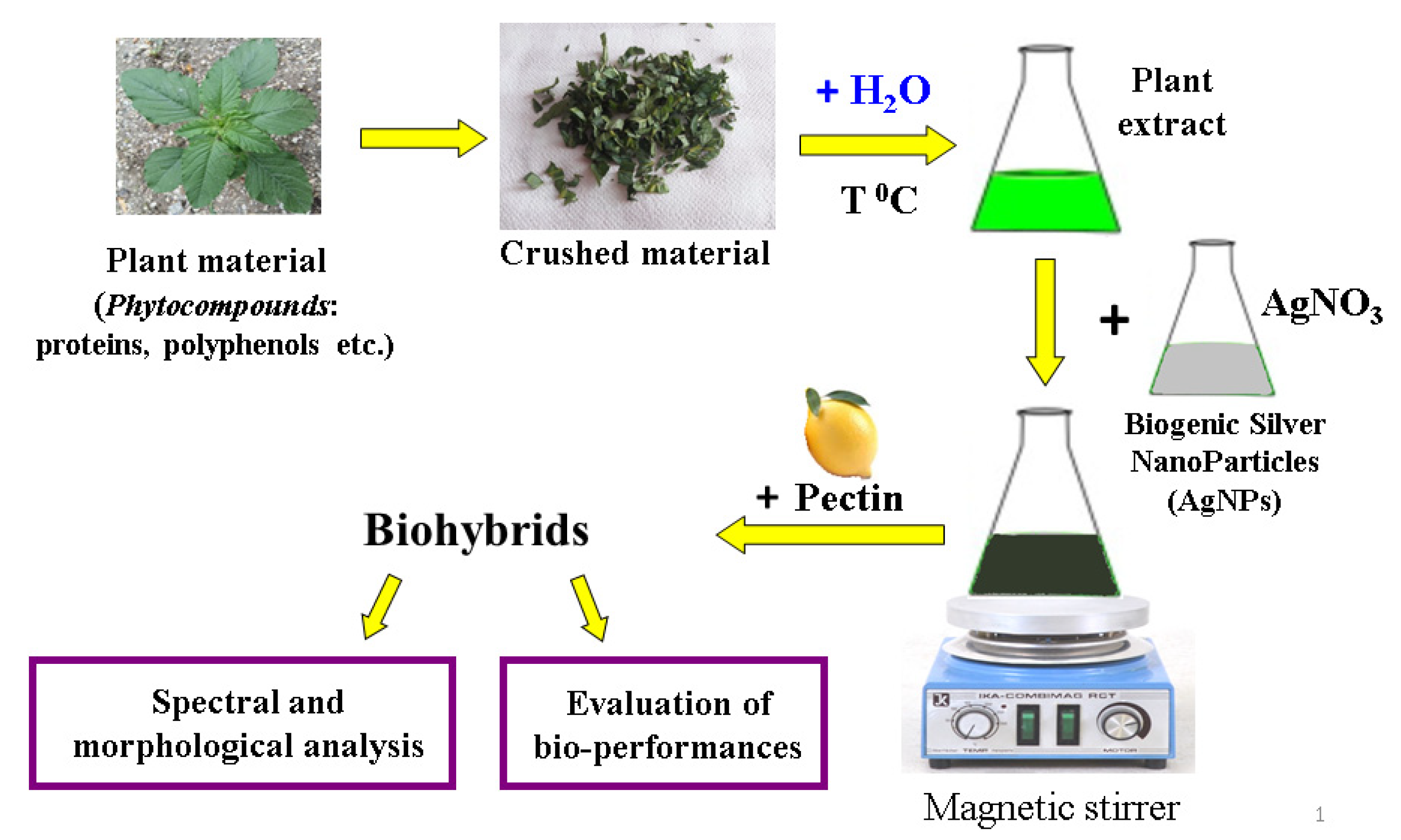
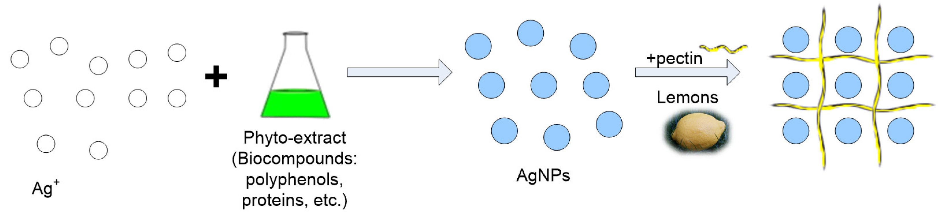
2.2. Biosynthesis of Ecogenic Plasmonic Biohybrids
2.2.1. Preparation of Phytogenic Silver Nanoparticles
2.2.2. Preparation of Ecogenic Plasmonic Biohybrids
2.3. Physico-Chemical and Biological Characterization of Developed Biohybrids
2.3.1. Spectral, Structural, and Morphological Analysis
2.3.2. Biological Characterization of Developed Biohybrid Materials
- A0 is the absorbance of the blank (3 mL of ABTS●+ diluted solution and 2 mL of distilled water);
- As is the absorbance of the samples (3 mL ABTS●+ diluted solution, 1 mL AgNPs/ biohybrids +1 mL of distilled water).
3. Results
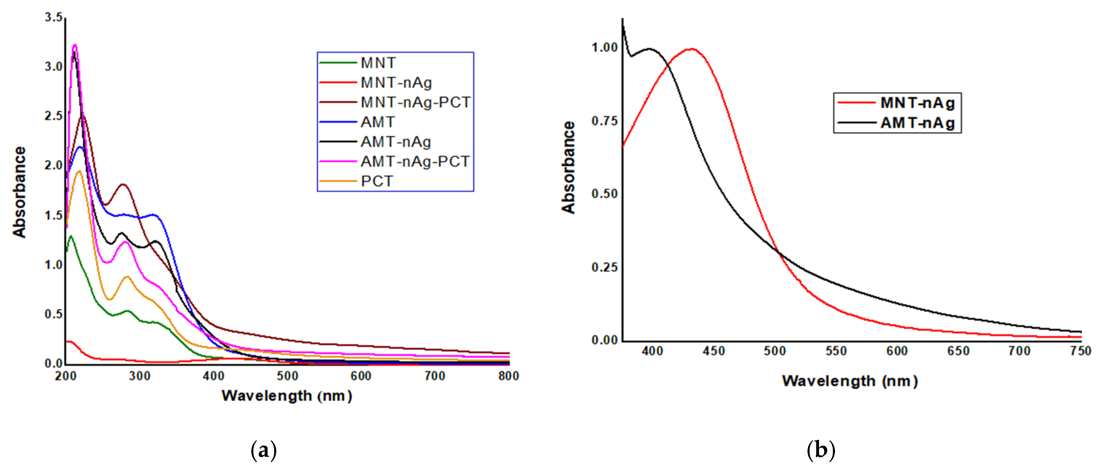
3.1. Optical Characterization
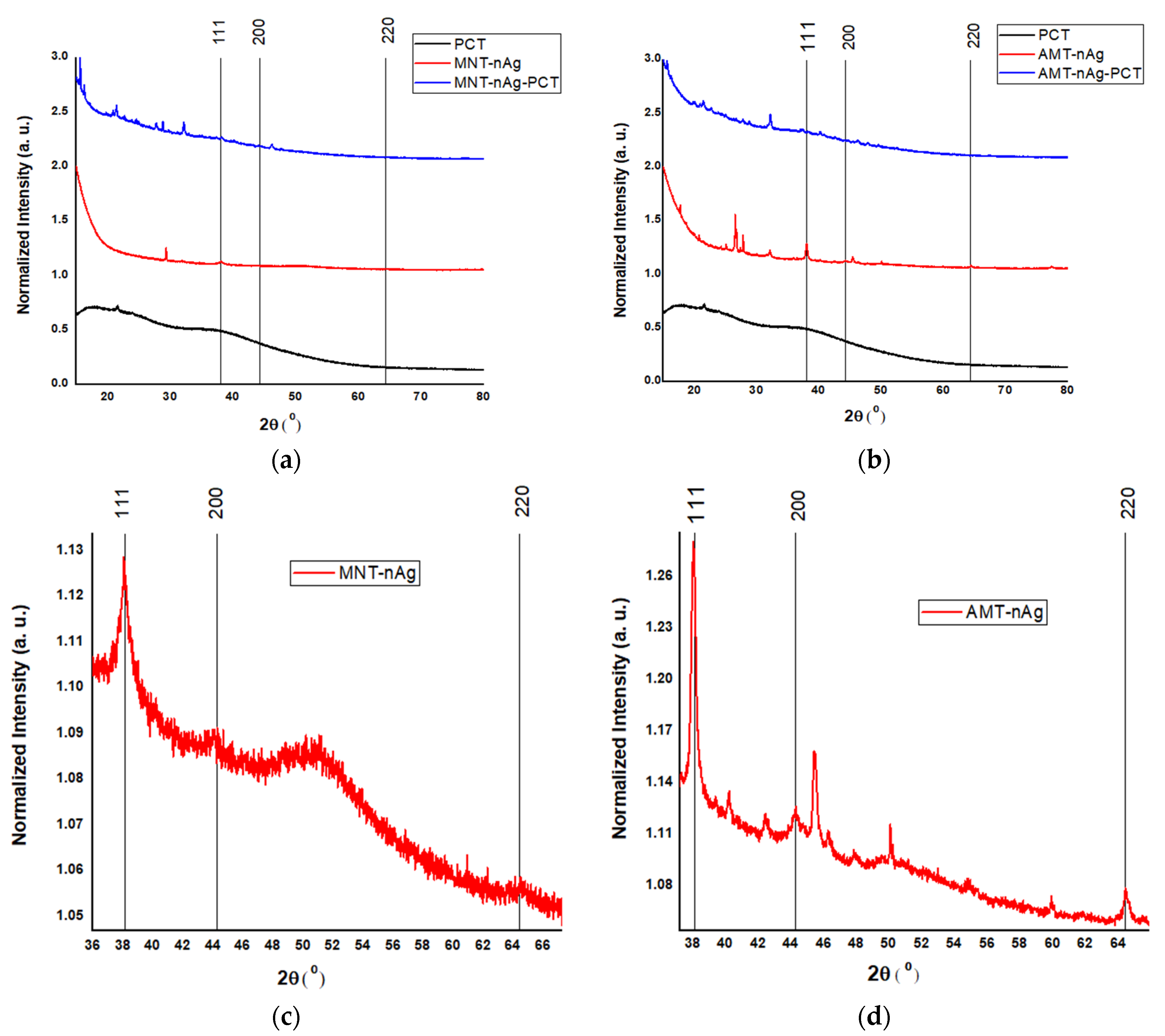
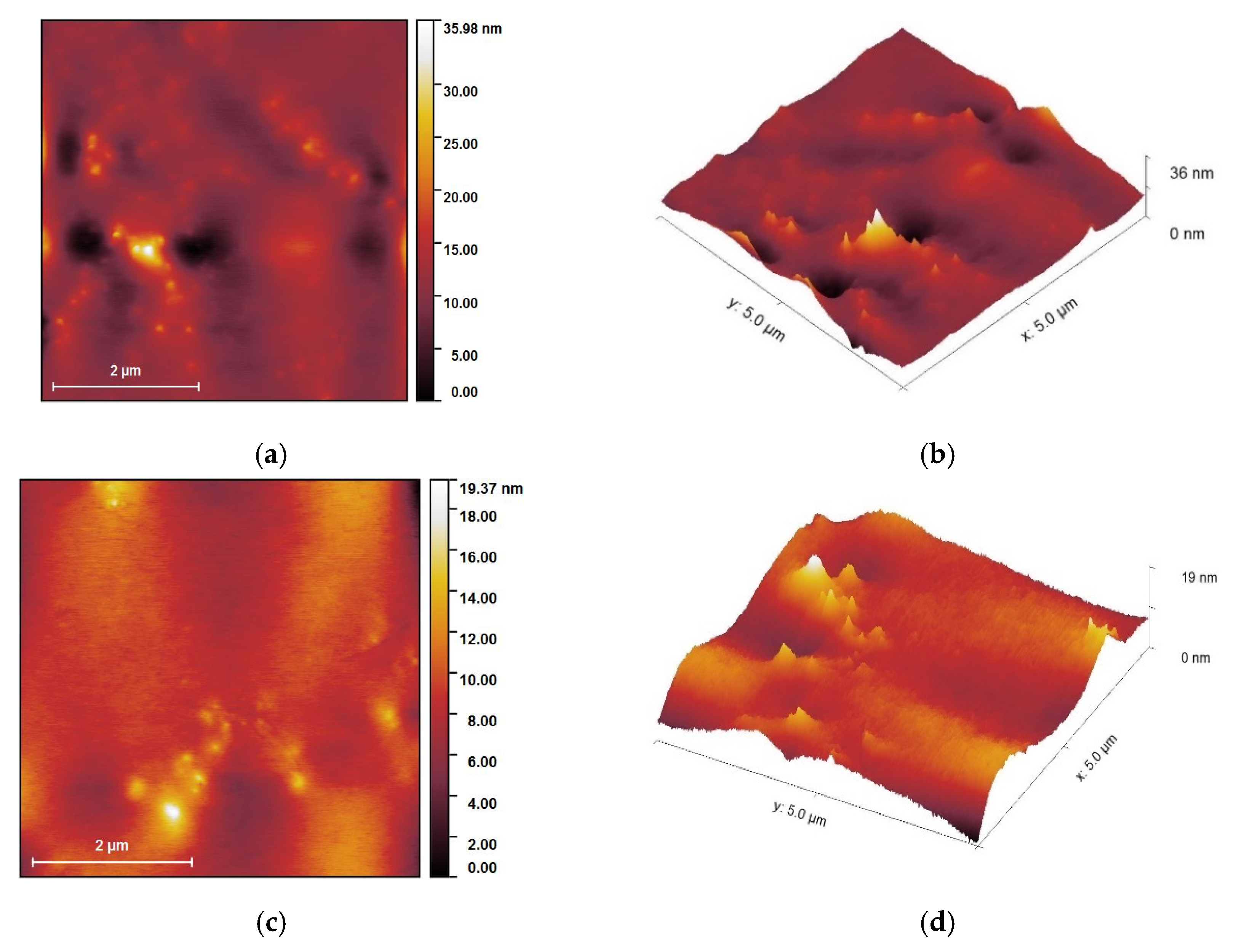
3.2. Structural Investigation of the Samples
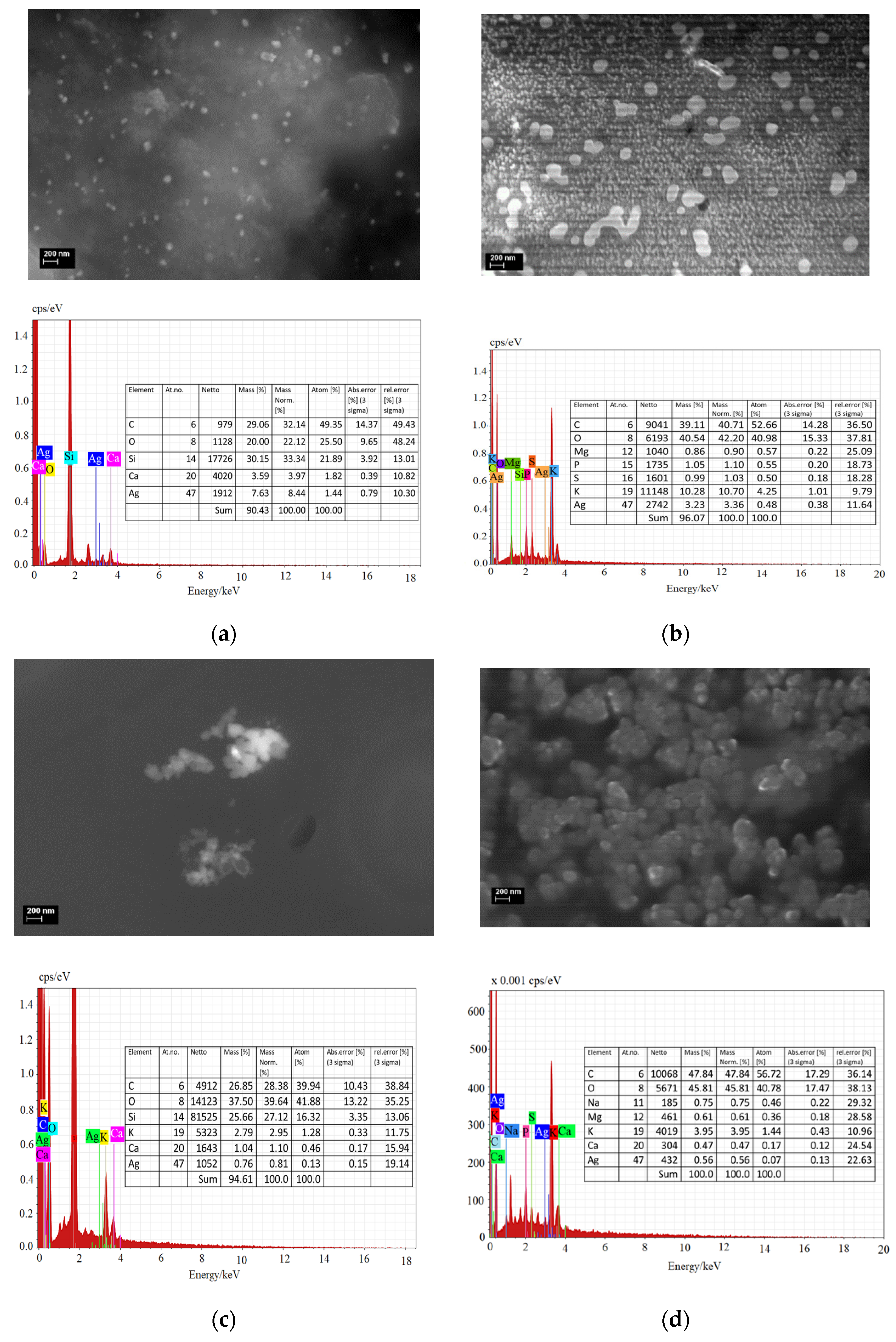
3.3. Elemental Composition of the Samples
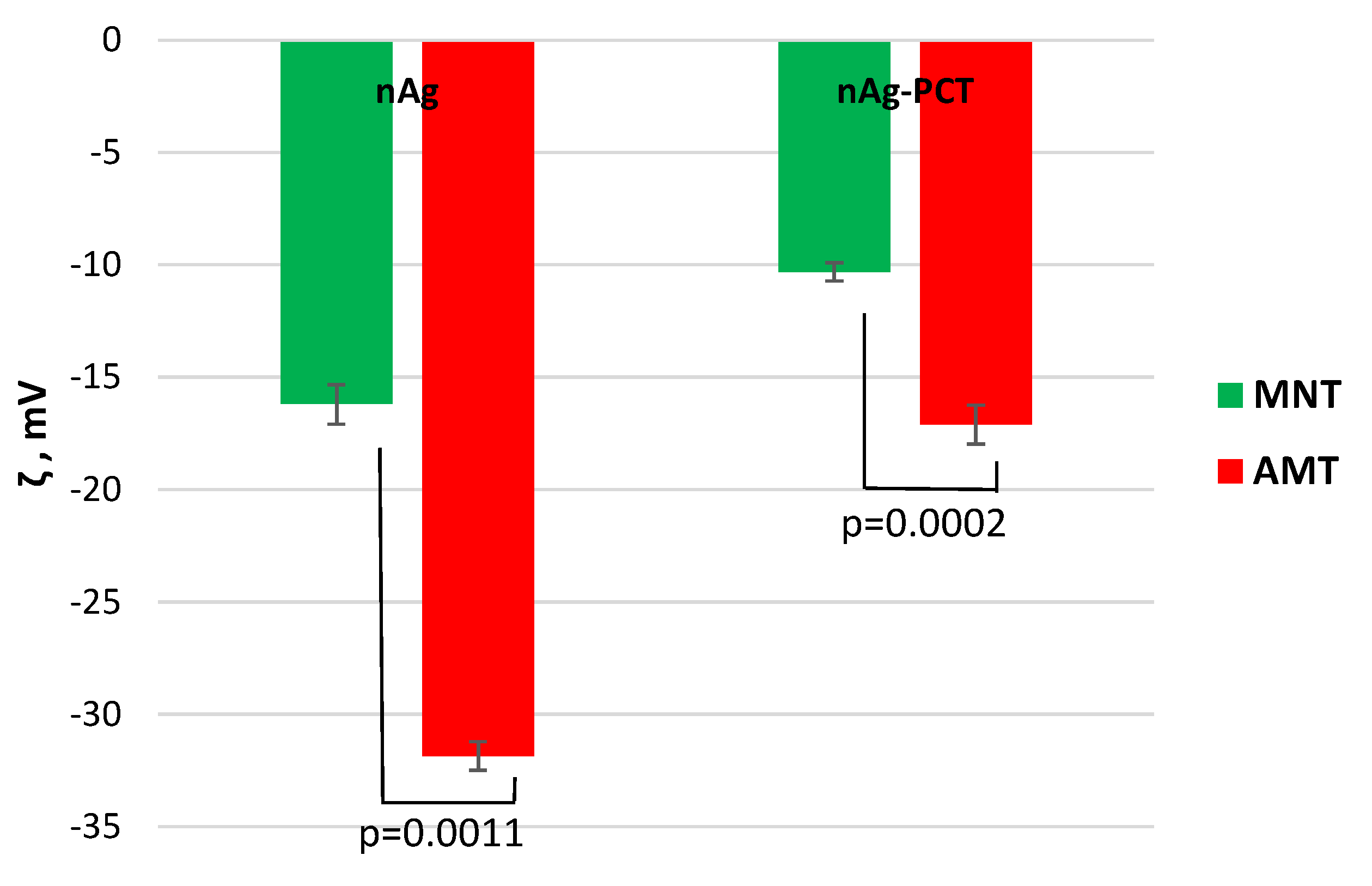
3.4. Evaluation of Zeta Potential of the Pectin-Coated Materials
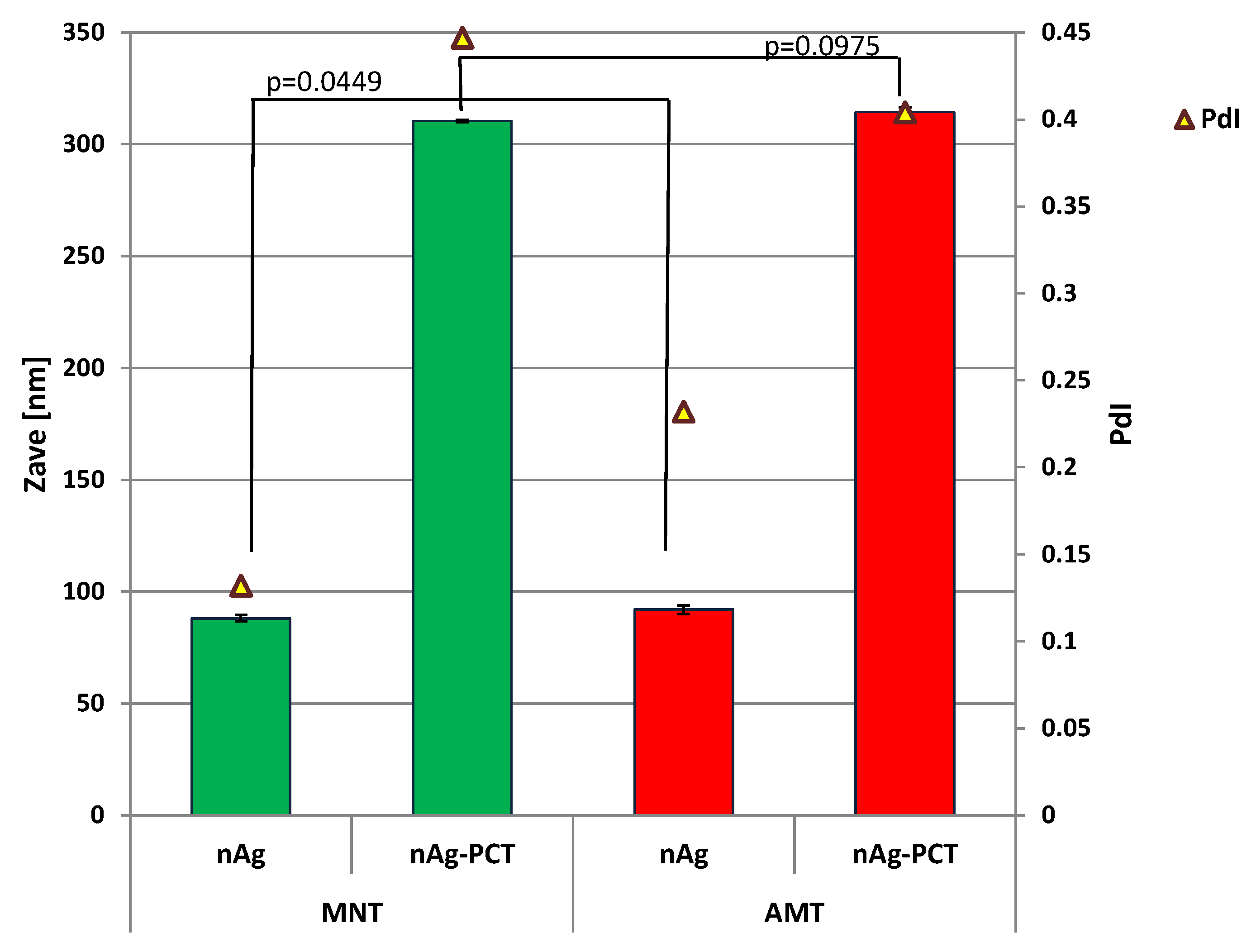
3.5. Size and Morphological Studies of Pectin-Coated Materials
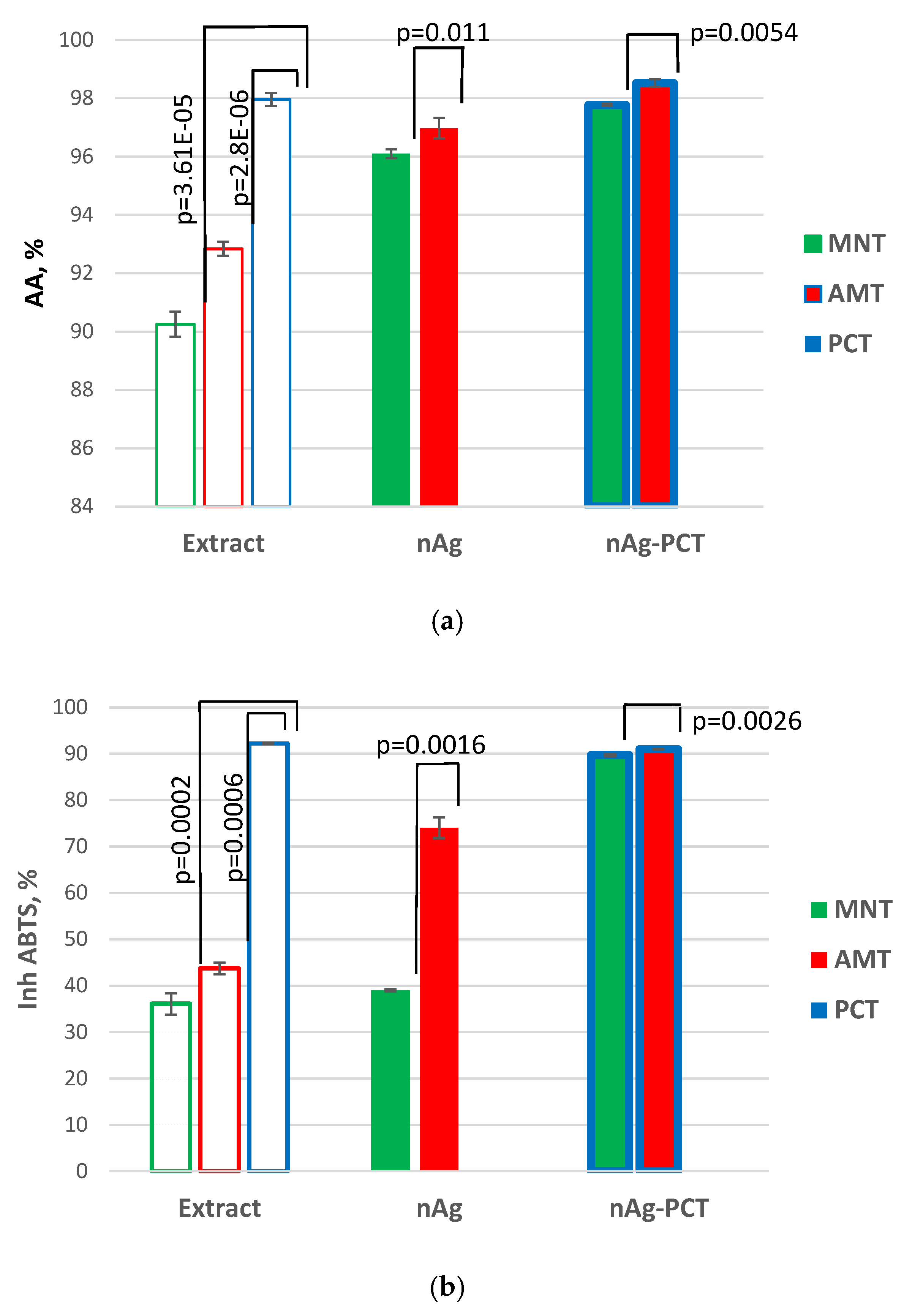
3.6. Evaluation of Bioactivities of Developed Materials
4. Discussion
5. Conclusions
Author Contributions
Funding
Conflicts of Interest
References
- Barbinta-Patrascu, M.E.; Badea, N.; Ungureanu, C.; Iordache, S.M.; Constantin, M.; Purcar, V.; Pirvu, C.; Rau, I. Eco-biophysical aspects on nanosilver bio-generated from Citrus reticulata peels, as potential bio-pesticide for controlling pathogens and wetland plants in aquatic media. J. Nanomater. 2017, 2017, 4214017. [Google Scholar] [CrossRef] [Green Version]
- Fierascu, I.; Fierascu, I.C.; Dinu-Pirvu, C.E.; Fierascu, R.C.; Anuta, V.; Velescu, B.S.; Jinga, M.; Jinga, V. A Short overview of recent developments on antimicrobial coatings based on phytosynthesized metal nanoparticles. Coatings 2019, 9, 787. [Google Scholar] [CrossRef] [Green Version]
- Barbinta-Patrascu, M.E.; Badea, N.; Bacalum, M.; Ungureanu, C.; Suica-Bunghez, I.R.; Iordache, S.M.; Pirvu, C.; Zgura, I.; Maraloiu, V.A. 3D hybrid structures based on biomimetic membranes and Caryophyllus aromaticus—“Green” synthesized nano-silver with improved bioperformances. Mater. Sci. Eng. C 2019, 101, 120–137. [Google Scholar] [CrossRef] [PubMed]
- Barbinta-Patrascu, M.E.; Badea, N.; Bacalum, M.; Antohe, S. Novel bio-friendly nanomaterials based on artificial cell membranes, chitosan and silver nanoparticles phytogenerated from Eugenia caryophyllata buds: Eco-synthesis, characterization and evaluation of bioactivities. Rom. Rep. Phys. 2020, 72, 601. [Google Scholar] [CrossRef] [Green Version]
- Barbinta-Patrascu, M.E.; Ungureanu, C.; Iordache, S.M.; Bunghez, I.R.; Badea, N.; Rau, I. Green silver nanobioarchitectures with amplified antioxidant and antimicrobial properties. J. Mater. Chem. B 2014, 2, 3221–3231. [Google Scholar] [CrossRef]
- Khan, S.U.; Saleh, T.A.; Wahab, A.; Khan, M.H.U.; Khan, D.; Khan, W.U.; Rahim, A.; Kamal, S.; Khan, F.U.; Fahad, S. Nanosilver: New ageless and versatile biomedical therapeutic scaffold. Int. J. Nanomed. 2018, 13, 733–762. [Google Scholar] [CrossRef] [Green Version]
- Valdés, A.; Burgos, N.; Jiménez, A.; Garrigós, M.C. Natural pectin polysaccharides as edible coatings. Coatings 2015, 5, 865–886. [Google Scholar] [CrossRef] [Green Version]
- Muñoz-Bonilla, A.; Echeverria, C.; Sonseca, A.; Arrieta, M.P.; Fernández-García, M. Bio-based polymers with antimicrobial properties towards sustainable development. Materials 2019, 12, 641. [Google Scholar] [CrossRef] [Green Version]
- Treviño-Garza, M.Z.; Correa-Cerón, R.C.; Ortiz-Lechuga, E.G.; Solís-Arévalo, K.K.; Castillo-Hernández, S.L.; Gallardo-Rivera, C.T.; Niño, K.A. Effect of linseed (linum usitatissimum) mucilage and chitosan edible coatings on quality and shelf-life of fresh-cut cantaloupe (cucumis melo). Coatings 2019, 9, 368. [Google Scholar] [CrossRef] [Green Version]
- Maftoonazad, N.; Ramaswamy, H.S. Application and evaluation of a pectin-based edible coating process for quality change kinetics and shelf-life extension of lime fruit (citrus aurantifolium). Coatings 2019, 9, 285. [Google Scholar] [CrossRef] [Green Version]
- Zahran, M.K.; Ahmed, H.B.; El-Rafie, M.H. Facile size-regulated synthesis of silver nanoparticles using pectin. Carbohydr. Polym. 2014, 111, 971–978. [Google Scholar] [CrossRef] [PubMed]
- Balachandran, Y.L.; Girija, S.; Selvakumar, R.; Tongpim, S.; Gutleb, A.C.; Suriyanarayanan, S. differently environment stable bio-silver nanoparticles: Study on their optical enhancing and antibacterial properties. PLoS ONE 2013, 8, e77043. [Google Scholar] [CrossRef] [PubMed]
- Barbinta-Patrascu, M.E.; Constantin, M.; Badea, N.; Ungureanu, C.; Iordache, S.M.; Purcar, V.; Antohe, S. Tangerine-Generated Silver—Silica Bioactive Materials. Rom. J. Phys. 2019, 64, 701. [Google Scholar]
- M’hiri, N.; Ioannou, I.; Ghoul, M.; Mihoubi Boudhrioua, N. Phytochemical characteristics of citrus peel and effect of conventional and nonconventional processing on phenolic compounds: A review. Food Rev. Int. 2017, 33, 587–619. [Google Scholar] [CrossRef]
- Alu’datt, M.H.; Rababah, T.; Alhamad, M.N.; Gammoh, S.; Al-Mahasneh, M.A.; Tranchant, C.C.; Rawshdeh, M. Chapter 15—Pharmaceutical, nutraceutical and therapeutic properties of selected wild medicinal plants: Thyme, spearmint, and rosemary. In Therapeutic, Probiotic, and Unconventional Foods; Academic Press: Cambridge, MA, USA, 2018; pp. 275–290. [Google Scholar]
- Bahrami-Teimoori, B.; Nikparast, Y.; Hojatianfar, M.; Akhlaghi, M.; Ghorbani, R.; Pourianfar, H.R. Characterisation and antifungal activity of silver nanoparticles biologically synthesised by Amaranthus retroflexus leaf extract. J. Exp. Nanosci. 2017, 12, 129–139. [Google Scholar] [CrossRef] [Green Version]
- Barbinta-Patrascu, M.E.; Bunghez, I.R.; Iordache, S.M.; Badea, N.; Fierascu, R.C.; Ion, R.M. Antioxidant properties of biohybrids based on liposomes and sage silver nanoparticles. J. Nanosci. Nanotechnol. 2013, 13, 2051–2060. [Google Scholar] [CrossRef] [PubMed]
- Ruano, P.; Delgado, L.L.; Picco, S.; Villegas, L.; Tonelli, F.; Aguilera Merlo, M.E.; Rigau, J.; Diaz, D.; Masuelli, M. Extraction and characterization of pectins from peels of criolla oranges (Citrus sinensis): Experimental reviews. In Pectins—Extraction, Purification, Characterization and Applications; IntechOpen: London, UK, 2019; pp. 1–44. [Google Scholar]
- Barbinta-Patrascu, M.E. Biohybrids based on DNA and bio-inspired lipid membranes: Design and characterization. Optoelectron. Adv. Mater. Rapid Commun. 2019, 13, 546–550. [Google Scholar]
- Barbinta-Patrascu, M.E.; Badea, N.; Ţugulea, L.; Giurginca, M.; Meghea, A. Oxidative stress simulation on artificial membranes—chemiluminescent studies. Rev. Chim. 2008, 59, 834–837. [Google Scholar]
- Ansari, M.A.; Khan, H.M.; Khan, A.A.; Malik, A.; Sultan, A.; Shahid, M.; Shujatullah, F.; Azam, A. Evaluation of antibacterial activity of silver nanoparticles magainst MSSA and MSRA on isolates from skin infections. Bio. Med. 2011, 3, 141–146. [Google Scholar]
- NCCLS M7-A6. Methods for Dilution Antimicrobial Susceptibility Test for Bacteria that Grow Aerobically, 6th ed.; Clinical and Laboratory Standards Institute: Wayne, PA, USA, 2003; ISBN 1-56235-486-4. [Google Scholar]
- Hugo, W.B.; Russel, A.D. Pharmaceutical Microbiology, 6th ed.; Blackwell Science: London, UK, 1998; pp. 248–253. [Google Scholar]
- Valgas, C.; De Souza, S.M.; Smânia, E.F.A.; Smânia, A., Jr. Screening methods to determine antibacterial activity of natural products. Braz. J. Microbiol. 2007, 38, 369–380. [Google Scholar] [CrossRef] [Green Version]
- Barbinta-Patrascu, M.E.; Ungureanu, C.; Iordache, S.M.; Iordache, A.M.; Bunghez, I.R.; Ghiurea, M.; Badea, N.; Fierascu, R.C.; Stamatin, I. Eco-designed biohybrids based on liposomes, mint—nanosilver and carbon nanotubes for antioxidant and antimicrobial coating. Mat. Sci. Eng. C 2014, 39, 177–185. [Google Scholar] [CrossRef] [PubMed]
- Rajendran, R.; Balakumar, C.; Kalaivani, J.; Sivakumar, R. Dyeability and antimicrobial properties of cotton fabrics finished with Punica granatum extracts. J. Text. Appar. Technol. Manag. 2011, 7, 1–12. [Google Scholar]
- Grecu, M.; Novac, G.; Ionita, D.; Ungureanu, C. Incorporation of tobramycin biomimetic in hydroxyapatite coating on CoCrMo alloy and its antimicrobial activity. Rev. Chim. 2011, 62, 352–356. [Google Scholar]
- Beggs, C.B.; Noakes, C.J.; Sleigh, P.A.; Fletcher, L.A.; Kerr, K.G. Methodology for determining the susceptibility of airborne microorganisms to irradiation by an upper-room UVGI system. J. Aerosol. Sci. 2006, 37, 885–902. [Google Scholar] [CrossRef] [Green Version]
- Wilfinger, W.W.; Mackey, K.; Chomczynski, P. Effect of pH and Ionic Strength on the Spectrophotometric Assessment of Nucleic Acid Purity. BioTechniques 1997, 22, 474–481. [Google Scholar] [CrossRef]
- Kaijanen, L.; Paakkunainen, M.; Pietarinen, S.; Jernström, E.; Reinikainen, S.-P. Ultraviolet detection of monosaccharides: Multiple wavelength strategy to evaluate results after capillary zone electrophoretic separation. Int. J. Electrochem. Sci. 2015, 10, 2950–2961. [Google Scholar]
- Wang, F.; Ma, Y.; Liu, Y.; Cui, Z.; Ying, X.; Zhang, F.; Linhardt, R.J. A simple strategy for the separation and purification of water-soluble polysaccharides from the fresh Spirulina platensis. Sep. Sci. Technol. 2017, 52, 456–466. [Google Scholar] [CrossRef]
- Barbinta-Patrascu, M.E.; Badea, N.; Ungureanu, C.; Ispas, A. Photophysical aspects regarding the effects of Paeonia officinalis flower extract on DNA molecule labelled with methylene blue. Optoelectron. Adv. Mat. Rapid Commun. 2019, 13, 131–135. [Google Scholar]
- Coates, J. Interpretation of Infrared Spectra, a Practical Approach. In Encyclopedia of Analytical Chemistry; Meyers, R.A., Ed.; John Wiley & Sons Ltd.: Chichester, UK, 2000. [Google Scholar]
- Kanmani, P.; Dhivya, E.; Aravind, J.; Kumaresan, K. Extraction and analysis of pectin from citrus peels: Augmenting the yield from citrus limon using statistical experimental design. Iran. J. Energy Environ. 2014, 5, 303–312. [Google Scholar] [CrossRef]
- Mébarki, M.; Hachem, K.; Faugeron-Girard, C.; Mezemaze, R.H.; Kaid-Harche, M. Extraction and analysis of the parietal polysaccharides of acorn pericarps from Quercus trees. Polímeros 2019, 29, 3. [Google Scholar] [CrossRef] [Green Version]
- Vedhanayagama, M.; Nidhinb, M.; Duraipandya, N.; Naresha, N.D.; Jaganathana, G.; Ranganathana, M.; Kirana, M.S.; Narayanc, S.; Naira, B.U.; Sreeram, K.J. Role of nanoparticle size in self-assemble processes of collagen for tissue engineering application. Int. J. Biol. Macromol. 2017, 99, 655–664. [Google Scholar] [CrossRef] [PubMed]
- Giosafatto, C.V.L.; Sabbah, M.; Al-Asmar, A.; Esposito, M.; Sanchez, A.; Santana, R.V.; Cammarota, M.; Mariniello, L.; Di Pierro, P.; Porta, R. Effect of mesoporous silica nanoparticles on glycerol-plasticized anionic and cationic polysaccharide edible films. Coatings 2019, 9, 172. [Google Scholar] [CrossRef] [Green Version]
- Khorrami, S.; Zarrabi, A.; Khaleghi, M.; Danaei, M.; Mozafari, M.R. Selective cytotoxicity of green synthesized silver nanoparticles against the MCF-7 tumor cell line and their enhanced antioxidant and antimicrobial properties. Int. J. Nanomed. 2018, 13, 8013–8024. [Google Scholar] [CrossRef] [PubMed] [Green Version]
- Ajitha, B.; Reddy, Y.A.K.; Reddy, P.S. Green synthesis and characterization of silver nanoparticles using Lantana camara leaf extract. Mater. Sci. Eng. C 2015, 49, 373–381. [Google Scholar] [CrossRef]
- Barbinta-Patrascu, M.E.; Badea, N.; Ungureanu, C.; Constantin, M.; Pirvu, C.; Rau, I. Silver-based biohybrids “green” synthesized from Chelidonium majus L. Opt. Mat. 2016, 56, 94–99. [Google Scholar] [CrossRef]
- Shankar, H.; Karthiga, P.; Swarnalatha, K.; Rajkuma, K. Green synthesis of silver nanoparticles using Capsicum frutescence and its intensified activity against E. coli. Resour. Effic. Technol. 2017, 3, 303–308. [Google Scholar] [CrossRef]
- Raut, R.W.; Lakkakula, J.R.; Kolekar, N.S.; Mendhulkar, V.D.; Kashid, S.B. Phytosynthesis of silver nanoparticle using Gliricidia Sepium. Curr. Nanosci. 2009, 5, 117–122. [Google Scholar]
- Ávila-Morales, G.; Montes de Oca-Vásquez, G.; Alvarado-Marchena, L.; Pereira-Reyes, R.; Hernández-Miranda, M.; Gonzalez-Paz, R.; Vega-Baudrit, J.R. Biosynthesis of Silver Nanoparticles Using Mint Leaf Extract (Mentha piperita) and Their Antibacterial Activity. Adv. Sci. Eng. Med. 2017, 9, 1–10. [Google Scholar]
- Jauregui-Gomez, D.; Bermejo-Gallardo, O.M.; Moreno-Medrano, E.M.; Perez-Garcia, M.G.; Ceja, I.; Soto, V.; Carvajal-Ramos, F.; Gutierrez-Becerra, A. Freeze-drying storage method based on pectin for gold nanoparticles. Nanomater. Nanotechnol. 2017, 7, 1–6. [Google Scholar] [CrossRef]
- Chicea, D. Nanoparticles and nanoparticle aggregates sizing by DLS and AFM. Optoelectron. Adv. Mat. Rapid Commun. 2010, 4, 1310–1315. [Google Scholar]
- Chicea, D. Using AFM topography measurements in nanoparticle sizing. Rom. Rep. Phys. 2014, 66, 778–787. [Google Scholar]
- Minzanova, S.T.; Mironov, V.F.; Arkhipova, D.M.; Khabibullina, A.V.; Mironova, L.G.; Zakirova, Y.M.; Milyukov, V.A. Biological activity and pharmacological application of pectic polysaccharides: A review. Polymers 2018, 10, 1407. [Google Scholar] [CrossRef] [PubMed] [Green Version]
- Ro, Y.K.; Kim, H.; Jang, S.B.; Lee, H.J.; Chakma, S.; Jeong, J.H.; Lee, J. Antioxidative activity of pectin and its stabilizing effect on retinyl palmitate. Korean J. Physiol. Pharmacol. 2013, 17, 197–201. [Google Scholar] [CrossRef] [PubMed]
- Pallavicini, P.; Arciola, C.R.; Bertoglio, F.; Curtosi, S.; Dacarro, G.; D’Agostino, A.; Ferrari, F.; Merli, D.; Milanese, C.; Rossi, S.; et al. Silver nanoparticles synthesized and coated with pectin: An ideal compromise for antibacterial and anti-biofilm action combined with wound-healing properties. J. Colloid Interface Sci. 2017, 498, 271–281. [Google Scholar] [CrossRef] [PubMed]
- Hileuskayaa, K.; Ladutskab, A.; Kulikouskaya, V.; Kraskouski, A.; Novik, G.; Kozerozhets, I.; Kozlovskiy, A.; Agabekov, V. ‘Green’ approach for obtaining stable pectin-capped silver nanoparticles: Physico-chemical characterization and antibacterial activity. Colloids Surf. A 2020, 585, 124–141. [Google Scholar] [CrossRef]
- Ferreyra Maillard, A.P.V.; Dalmasso, P.R.; Lopez de Mishima, B.A.; Hollmann, A. Interaction of green silver nanoparticles with model membranes: Possible role in the antibacterial activity. Colloids Surf. B Biointerfaces 2018, 171, 320–326. [Google Scholar] [CrossRef]
- Písaříková, B.; Kráčmar, S.; Herzig, I. Amino acid contents and biological value of protein in various amaranth species. Czech J. Anim. Sci. 2005, 50, 169–174. [Google Scholar] [CrossRef] [Green Version]
- Hutton, C.A.; Southwood, T.J.; Turner, J.J. Inhibitors of lysine biosynthesis as antibacterial agents. Mini Rev. Med. Chem. 2003, 3, 115–127. [Google Scholar] [CrossRef]


| No. | Sample Description | Sample Code |
|---|---|---|
| 1 | Vegetal aqueous extract from leaves of mint (Mentha piperita) | MNT |
| 2 | Vegetal aqueous extract from leaves of pigweed (Amaranthus retroflexus) | AMT |
| 3 | Pectin extracted from lemon peels | PCT |
| 4 | AgNPs phyto synthesized from vegetal aqueous extract from leaves of mint (Mentha piperita) | MNT-nAg |
| 5 | AgNPs phyto synthesized from vegetal aqueous extract from leaves of pigweed (Amaranthus retroflexus) | AMT-nAg |
| 6 | Biohybrid generated from MNT-nAg and pectin | MNT-nAg-PCT |
| 7 | Biohybrid generated from AMT-nAg and pectin | AMT-nAg-PCT |
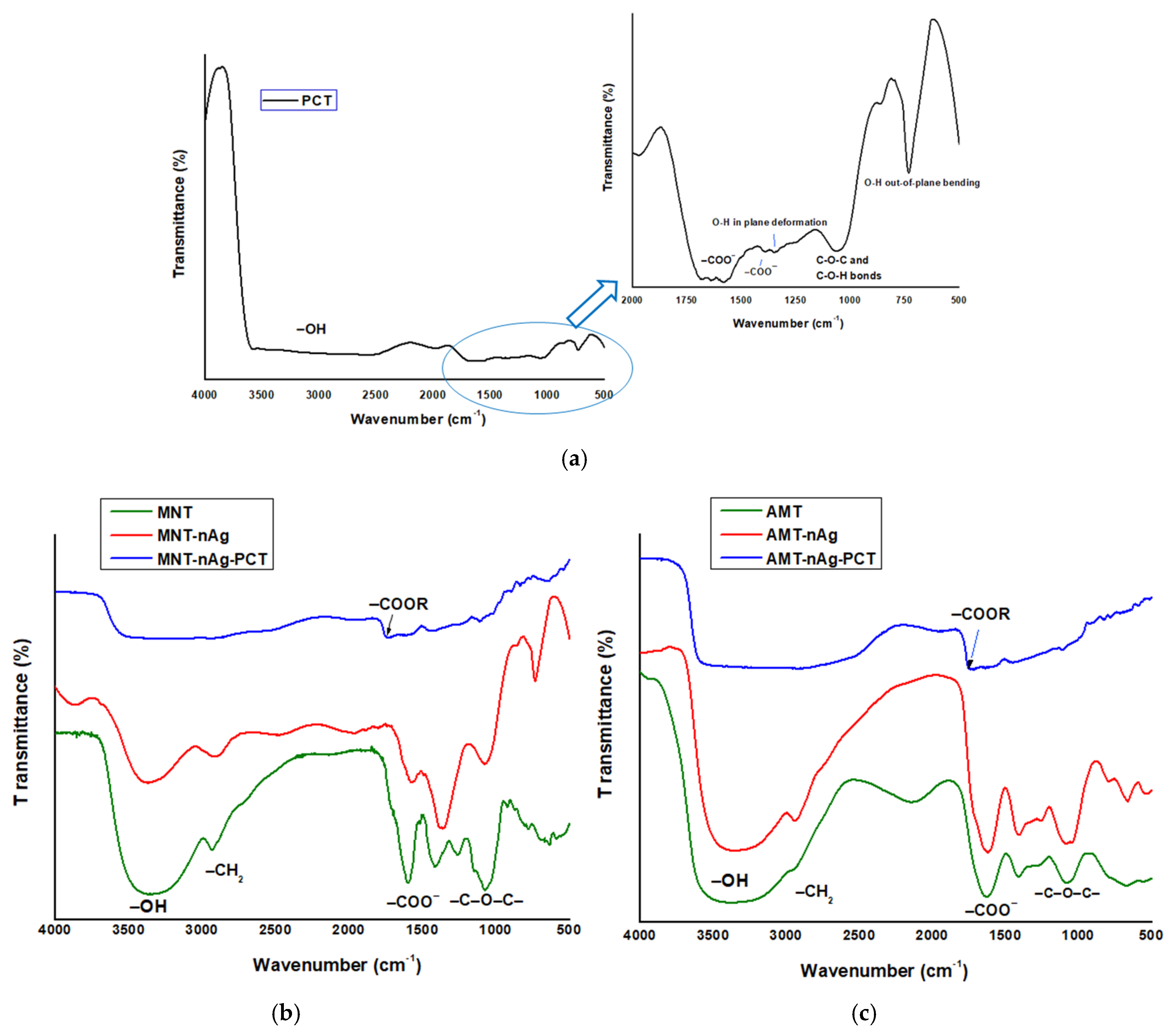
| Sample Code | FT-IR Bands (cm−1) | Assignment | Ref. |
|---|---|---|---|
| MNT/AMT | 3355/3391 (intense, broad band) | Bending and stretching vibrations of hydroxyl groups in polysaccharides, alcohols, and phenolic compounds and to N–H stretching vibrations | [3] |
| 2925/2949 | C–H stretching vibration | [33] | |
| 1611/1630 | Amide I, arising due to carbonyl stretch in proteins | [25] | |
| 1077/1076 (medium broad band) | Antisymmetric stretching of C–O group of polysaccharides and/or chlorophyll | [12] | |
| PCT | 3604,3598 (small peak) | Nonbonded hydroxyl groups | [33] |
| 3500–2500 (very broad band) | Very broad band overlapping the hydrogen-bonded O–H (the bending and stretching vibrations of hydroxyl groups in polysaccharide) and C–H stretching vibration in the frequency 2830–2695 cm−1 shown as carbohydrate ring | [25], [33], [34] | |
| 1715 | Band attributed both to the carboxylic acid and to the ester groups | [33] | |
| 1631,1578 | Carboxylate groups (–COO–) | [33] | |
| 1371 (weak) | O–H in plane deformation | [13] | |
| 1221/1240 (very weak, broad) | Vibrations of the –C–O–C– and –C–O–H bonds present in polysaccharide structures | [33], [35] | |
| 1050 (sharp band) | –C–O–C– ether linkage of pectin | [1], [36] | |
| 830 (weak, sharp peak) | Hydrogen-bonded O–H out-of-plane bending | [33] | |
| MNT-nAg/AMT-nAg | 3345/3363 (intense, very broad band) | This band indicates the presence of hydroxyl groups on the surface of nanoparticles. | [3] |
| 2933/2935 (weak, sharp band) | Alkyls C–H stretching vibration | [33] | |
| 1620/1622 (strong sharp band) | Amide I, arising due to carbonyl (–C=O) stretch in proteins | [25] | |
| 1073/1075 (weak band) | Stretching vibration to –C–O–C– groups of polysaccharides | [12] | |
| MNT-nAg-PCT/AMT-nAg-PCT | 3519–2905/3571–2515 (strong broad band) | Stretching vibration of O–H groups that interact by H bonding (O–H–O), the major contributors to this band being polysaccharides and polyphenolic compounds) | [13] |
| 1738/1747 | –C=O stretching of esterified carboxylic groups (–COOCH3) | [37] | |
| 1634/1582 (this band weakened) | Carboxylate groups (–COO–) | [33] | |
| 1448/1448 | C–H asymmetric bend of methyl group of pectin | [33] | |
| 1109/1105 | ν (CO), ν (CC) ring of polysaccharides, and pectin | [1] |
| Concentration of AgNPs, (µg/mL). | Escherichia Coli | |||||||||||
|---|---|---|---|---|---|---|---|---|---|---|---|---|
| 400 | 200 | 100 | 50 | 25 | 12.5 | 6.25 | 3.125 | 1.56 | 0.78 | 0.39 | 0.195 | |
| MNT-nAg-PCT | S | S | S | R | R | R | R | R | R | R | R | R |
| AMT-nAg-PCT | S | S | S | S | R | R | R | R | R | R | R | R |
| Specimen | CFUs, Escherichia Coli | Bactericidal Ratio (R)% | Susceptibility Constant (Z Value) mL/μg |
|---|---|---|---|
| MNT | 671 ± 4.5 | NBR | NBR |
| MNT-nAg | 93 ± 7.5 | 86 | 0.01976 |
| MNT-nAg-PCT | 82 ± 1.52 | 87.7 | 0.02102 |
| AMT | 648 ± 4.5 | NBR | NBR |
| AMT-nAg | 84 ± 2.5 | 87 | 0.0408 |
| AMT-nAg-PCT | 77 ± 1.5 | 88 | 0.0426 |
| Sample | Photographs of Petri Dishes Inoculated with Samples | Inhibition Zone, IZ (mm) |
|---|---|---|
| PCT |  | 12 ± 0.32 |
| MNT-nAg |  | 21 ± 0.46 |
| AMT-nAg |  | 25 ± 0.26 |
| MNT-nAg-PCT |  | 35 ± 0.58 |
| AMT-nAg-PCT |  | 39 ± 0.62 |
© 2020 by the authors. Licensee MDPI, Basel, Switzerland. This article is an open access article distributed under the terms and conditions of the Creative Commons Attribution (CC BY) license (http://creativecommons.org/licenses/by/4.0/).
Share and Cite
Barbinta-Patrascu, M.E.; Ungureanu, C.; Badea, N.; Bacalum, M.; Lazea-Stoyanova, A.; Zgura, I.; Negrila, C.; Enculescu, M.; Burnei, C. Novel Ecogenic Plasmonic Biohybrids as Multifunctional Bioactive Coatings. Coatings 2020, 10, 659. https://doi.org/10.3390/coatings10070659
Barbinta-Patrascu ME, Ungureanu C, Badea N, Bacalum M, Lazea-Stoyanova A, Zgura I, Negrila C, Enculescu M, Burnei C. Novel Ecogenic Plasmonic Biohybrids as Multifunctional Bioactive Coatings. Coatings. 2020; 10(7):659. https://doi.org/10.3390/coatings10070659
Chicago/Turabian StyleBarbinta-Patrascu, Marcela Elisabeta, Camelia Ungureanu, Nicoleta Badea, Mihaela Bacalum, Andrada Lazea-Stoyanova, Irina Zgura, Catalin Negrila, Monica Enculescu, and Cristian Burnei. 2020. "Novel Ecogenic Plasmonic Biohybrids as Multifunctional Bioactive Coatings" Coatings 10, no. 7: 659. https://doi.org/10.3390/coatings10070659