
Influence of the Biological Medium on the Properties of Magnesium Doped Hydroxyapatite Composite Coatings
by
 , and
, and
Daniela Predoi
1, 
Steluta Carmen Ciobanu
1,*,
Simona Liliana Iconaru
1 and
Mihai Valentin Predoi
2,* 1
National Institute of Materials Physics, Atomistilor Street, No. 405A, P.O. Box MG 07, 077125 Ilfov, Romania
2
Department of Mechanics, University Politehnica of Bucharest, BN 002, 313 Splaiul Independentei, District 6, 060042 Bucharest, Romania
*
Authors to whom correspondence should be addressed.
Coatings 2023, 13(2), 409; https://doi.org/10.3390/coatings13020409
Submission received: 31 December 2022
/
Revised: 8 February 2023
/
Accepted: 9 February 2023
/
Published: 11 February 2023
(This article belongs to the Special Issue Hydroxyapatite Based Coatings for Biomedical Applications)
Abstract
:In this paper, the stability of magnesium-doped hydroxyapatite/chitosan (MHC) suspension obtained with the sol-gel approach was evaluated using nondestructive ultrasound measurements. The MHC coatings obtained by the spin-coating technique were characterized before and after immersion for 7 and 14 days, respectively, in Dulbecco’s modified eagle medium (DMEM) by scanning electron microscopy, equipped with an EDAX detector. Also, the functional groups present on the MHC coatings surface were analyzed with the aid of attenuated total reflection (ATR) Fourier transform infrared (FTIR) spectroscopy. The surface microstructure was evaluated using two commentary techniques, namely atomic force microscopy (AFM) and metallographic microscopy (MM). The influence of immersion in DMEM on the biological properties was studied with in vitro studies using primary osteoblast and HCT-8 cell lines. Our results revealed that both surface morphology and chemical composition of the MHC coatings allow rapid development of a new apatite layer on their surface after immersion in DMEM. Preliminary in vitro biological studies underlined the noncytotoxic effect of the studied samples on the proliferation of primary osteoblast and HCT-8 cell lines, which makes them a promising candidate for applications in fields such as orthopedics or dentistry. The antifungal assay of the MHC coatings was assessed using Candida albicans ATCC 10231 and their results showed a good inhibitory effect. The coatings made on the basis of the MHC composite could contribute to increasing the degree of success of implants by decreasing the risk of infections and postoperative inflammation.
1. Introduction
In recent years, a constant decrease in the efficiency of usual antibacterial agents has been noticed. Thus, the treatment of infectious diseases has become a challenge for the medical community [1]. One of the most important directions of medical research is represented by the development of new antimicrobial agents and new strategies highly efficient in “the fight” against bacterial resistance. The antibacterial properties of magnesium, such as the ability to alter bacterial adherence and biofilm formation, have been reported in previous studies [2,3,4]. Magnesium salt solutions have been used since 1915 as an antimicrobial agent for cleaning wounds [5].
On the other hand, in living organisms, magnesium ions play an essential role, being involved in numerous enzymatic processes [5,6,7]. For example, the development of bacterial biofilm on the prostheses surface after an implantation represents an important health problem, which leads to significant additional costs and a temporary decrease in the patient’s quality of life [8]. Weak osteointegration, mechanical stability and degradation rate are some of the most relevant limitations of using inert metal implants [8,9]. Therefore, in this context, the use of a bioactive coating for metallic implants could represent an efficient approach that would lead to a global increase in an implant’s properties [9].
One of the most often used biomaterials for repairing (filling) bone defects/covering implants is hydroxyapatite (HAp), mainly due to its very good osteoconductivity and osteoinductivity. At the same time, HAp represents the principal inorganic part of teeth and bone tissue. Previous studies highlighted that the HAp structure possesses the ability to easily incorporate various types of ions (magnesium, europium, samarium, silver, cerium, etc.) [10]. The presence of foreign ions in the HAp structure gives it superior biological and physicochemical properties, which makes doped HAp suitable for various applications, including in the biomedical field [11,12,13,14,15].
In their work, S. Banerjee et al. proved that the impregnation of porous hydroxy-apatite with Ag, Au and Cu metallic ions leads to the obtaining of biomaterials with strong antimicrobial properties [12]. More than that, the incorporation in the HAp structure of ions that are found naturally (as trace elements) in natural bone may induce an enhancement of structural, compositional and biological properties and even of the mechanical ones of Hap [13]. Other studies performed by R. Ahmadi et al. showed that covering the surface of Ti6Al4V substrate with HAp/TiO2/Ag nanocomposite coating leads to enhanced corrosion resistance, good antimicrobial activity and good biocompatibility with the MG63 cell line [14]. Furthermore, T. Bazin et al. in their work, highlighted the biocompatibility of copper-doped hydroxyapatite with MC3T3-E1 cell line attachment and growth [16].
At the same time, it is well known that postoperative implant-related infections are one of the most commonly encountered problems that can be the cause of implant failure [17]. The most common microorganism involved in implant-related infections is the fungal strain, Candida albicans (C. albicans). Studies have shown that the adherence and development of fungal cells on an implant’s surface can be prevented by using antimicrobial layers [18,19,20,21,22]. Previously, the antifungal activity of titanium implants covered with chitosan was highlighted by Hallmann L. et al. [23]. Moreover, the study conducted by Iconaru S.L. et al. [24] showed that magnesium-doped hydroxyapatite in a chitosan matrix composite coating possesses good antifungal activity against C. albicans. Thus, it is important that the covering layers of implants possess good antimicrobial properties that could counter microbial adhesion.
Chitosan (a natural polysaccharide) is one of the most renewable biomaterials with superior biological properties, such as biocompatibility, non-toxicity, biodegradability, antitumor and antimicrobial activity, etc. [25]. Due to these unique characteristics, chitosan has been used in various applications in the medical field, which include wound dressing, drug delivery, dentistry, etc. [25]. If we take into consideration all these aspects, we can say that the development of biomaterials based on hydroxyapatite doped with magnesium in a chitosan matrix is of interest for applications in the medical field. At the same time, there were reported studies that showed that cell culture media (Roswell Park Memorial Institute Medium (RPMI), Dulbecco’s modified eagle medium (DMEM), etc.) could be used to obtain gold nanoparticles with controlled size and shape [26]. More than that, in order to evaluate the degradation behavior, in their work, Bo Li and collaborators [27] immersed different types of layers (based on MgO, HAp and Mg(OH)2) in saline solution for several time intervals (up to 90 days). Their findings highlighted the fact that degradation is the main cause of the thinning of the layers and that this is influenced by their composition [27]. In the meantime, they showed that after 90 days of immersion in saline solution on the coating’s surface, the presence of cracks on the layer’s surface was noticed [27].
The aim of this study was to evaluate for the first time the influence of DMEM (Dulbecco’s Modified Eagle Medium) on the surface morphology, chemical composition and biological properties of magnesium-doped hydroxyapatite/chitosan (MHC) composite thin films. The MHC suspensions were obtained using the sol-gel method. Then, the MHC suspension was used for the immersion of the Si substrate in order to obtain the MHC composite thin films. The nondestructive ultrasound measurements were used in order to evaluate the MHC suspension stability. The morphology and chemical composition of both the MHC suspension and composite thin films were studied using scanning electron microscopy (SEM). Furthermore, the modification of the surface microstructure of the MHC composite thin films was evaluated with the aid of atomic force microscopy (AFM) and metallographic microscopy (MM). The presence of the typical vibrational bands in the MHC samples was investigated with the aid of attenuated total reflection (ATR) Fourier transform infrared (FTIR) spectroscopy. The cytotoxicity of the MHC composite thin films was investigated using primary osteoblast and HCT-8 cell lines. The preliminary results of the in vitro biological assay underlined that the MHC samples analyzed after 24 and 72 h of incubation exhibited no toxic effect against the osteoblast and HCT-8 cell lines. Also, the antifungal assay of the MHC coatings was assessed using the Candida albicans ATCC 10231 fungal strain.
2. Materials and Methods
2.1. Materials
For the obtaining of the magnesium-doped hydroxyapatite/chitosan (MHC, Ca10−xMgx(PO4)6(OH)2, with xMg = 0.07) suspension, the sol-gel method was involved and the following precursors were used: magnesium nitrate hexahydrate (Mg(NO₃)₂·6H₂O; Alpha Aesar, Kandel, Germany); chitosan (C6H11NO4; Sigma Aldrich, St. Louis, MO, USA) calcium nitrate tetrahydrate, (Ca(NO3)2∙4H2O; Sigma Aldrich, St. Louis, MO, USA); diammonium hydrogen phosphate ((NH4)2HPO4; Sigma Aldrich, St. Louis, MO, USA). More than that, ammonium hydroxide (NH4OH, 25% NH3 in H2O (T)), ethanol absolute (C2H5OH) purchased from Sigma Aldrich (St. Louis, MO, USA) and bidistilled water were utilized for the obtaining of the MHC sample.
2.2. Synthesis of Magnesium-Doped Hydroxyapatite/Chitosan (MHC) Suspension
The synthesis of magnesium-doped hydroxyapatite/chitosan (MHC) was previously described [28]. Briefly, the sol-gel synthesis of the MHC suspension was made by keeping constant the Ca/P molar ratio at 1.67 and the pH value at 11. The sol-gel was made at 100 °C, under continuous stirring for 4 h. The obtained magnesium-doped hydroxyapatite suspension was centrifuged and added slowly into a chitosan solution (2%). In the end, the MHC gel was mixed for 6 h in an ultrasound bath and then used to obtain the MHC composite thin films.
2.3. Development of MHC Composite Thin Films
The deposition of the MHC composite thin films on the Si substrate (Siegert Wafer GmbH, Aachen, Germany) was made using the spin-coating method. Prior to the deposition process, the substrate was washed with acetone. Thus, the MHC composite thin films were obtained by dropping 0.5 mL of the MHC suspension on the Si substrate surface with the aid of a syringe. The deposition conditions were described in detail in our previous studies [28,29]. The MHC deposition process on the Si substrate was repeated three consecutive times. Finally, the MHC composite thin films were treated at 500 °C for 2 h.
The obtained MHC composite thin films were immersed in DMEM (Sigma Aldrich, St. Louis, MO, USA) for 7 and 14 days at 37 ± 0.5 °C in an incubator (GFL 4010, GFL Gesellschaft für Labortechnik mbH, Burgwedel, Germany). DMEM was renewed daily. At the end of each immersion period, the sample was washed with bidistilled water and then kept in a desiccator.
The MHC composite thin films obtained after 7 days of immersion in DMEM were referred to as MHC_7D and the samples immersed for 14 days were referred to as MHC_14D.
2.4. Physico-Chemical Characterizations
The stability of the MHC suspension was evaluated using ultrasonic measurements. Therefore, the ultrasound measurements were made in agreement with the protocol reported by Predoi D. et al. [28].
The MHC suspension and thin film morphology and chemical composition studies were conducted using a Hitachi S4500 scanning electron microscope (Hitachi, Tokyo, Japan) equipped with an energy dispersive X-ray (EDX) detection system. The 3D SEM, AFM and metallographic images were achieved using Image J software (Image J 1.51j8) [30]. A drop of MHC suspension was put on a double-sided carbon tape and dried in a vacuum prior to SEM examination. The MHC thin films (before and after immersion in DMEM) were analyzed without being previously covered with gold.
The surface topography of the MHC composite thin films before and after immersion in DMEM was evaluated using AFM studies with an NT-MDT NTEGRA Probe Nano Laboratory instrument (NT-MDT, Moscow, Russia) operated in semi-contact mode. The typical AFM images of the MHC composite thin films were recorded for a surface area of 10 × 10 µm2. The RRMS (roughness parameter) was estimated for the MHC composite thin films (before and after exposure to DMEM) from three zones and presented as the mean ± standard deviation. The AFM data were processed with the aid of Gwidion 2.59 software (Department of Nanometrology, Czech Metrology Institute, Brno, Czech Republic) [31].
Information about the surface morphology of the MHC composite thin films was obtained with the help of an inversed trinocular metallographic microscope OX.2153-PLM, (Euromex, Arnhem, The Netherlands). The MHC images were recorded using the 50× magnification of the metallographic microscope.
The molecular structure features of the MHC suspension and thin films were evaluated by conducting FTIR-ATR studies. Thus, a Jasco FTIR-6600 spectrometer (Easton, MD, USA) operated in ATR mode was used. The FTIR-ATR spectra of the MHC samples were recorded in the 450–4000 cm−1 spectral range, in air.
2.5. Cytotoxicity Assay
The cytotoxicity of the MHC samples was investigated using a primary osteoblast culture (hFOB 1.19) prepared according to the protocol of Gallagher et al. [32] and also the human ileocecal adenocarcinoma (HCT-8) cell line. The cytotoxicity assays were performed as previously described in [33]. All the biological investigations were carried out in triplicate. The statistical analysis was carried out using the t-test and analysis of variance (ANOVA). The difference established between the samples was appreciated to be significant at p < 0.05.
2.6. In Vitro Antifungal Assay
The antifungal properties of the MHC samples were investigated in vitro using the Candida albicans ATCC 10231 fungal strain. The experiments were performed as previously described in [24] and the antifungal properties of the composite layers were determined after 24, 48 and 72 h of incubation with the fungal suspensions. All the experiments were carried out in triplicate and the results were presented as the mean ± SD.
3. Results
In order to achieve uniform coatings using the sol-gel method, an important role is played by the stability of the suspension. The stability of the MHC suspension was evaluated using ultrasonic measurements. The stability evaluation was carried out by investigating the standard suspension resulting from the synthesis process. Double-distilled water was chosen as the reference fluid for evaluating the stability of the MHC suspension.
A volume of 100 mL of the MHC suspension sample was stirred for 30 min at 1000 rot/min in a special cubic container. The container coaxially opposed ultrasonic transducers, each with a central frequency of 25 MHz. Immediately after stopping the stirring, the ultrasonic signals transmitted formed one transducer to the other, which were recorded every 5 s, for a total duration of 5000 s. The amplitudes of the transmitted signals were determined as ratios to the amplitudes in double-distilled water under identical conditions, which were taken as the reference liquid.
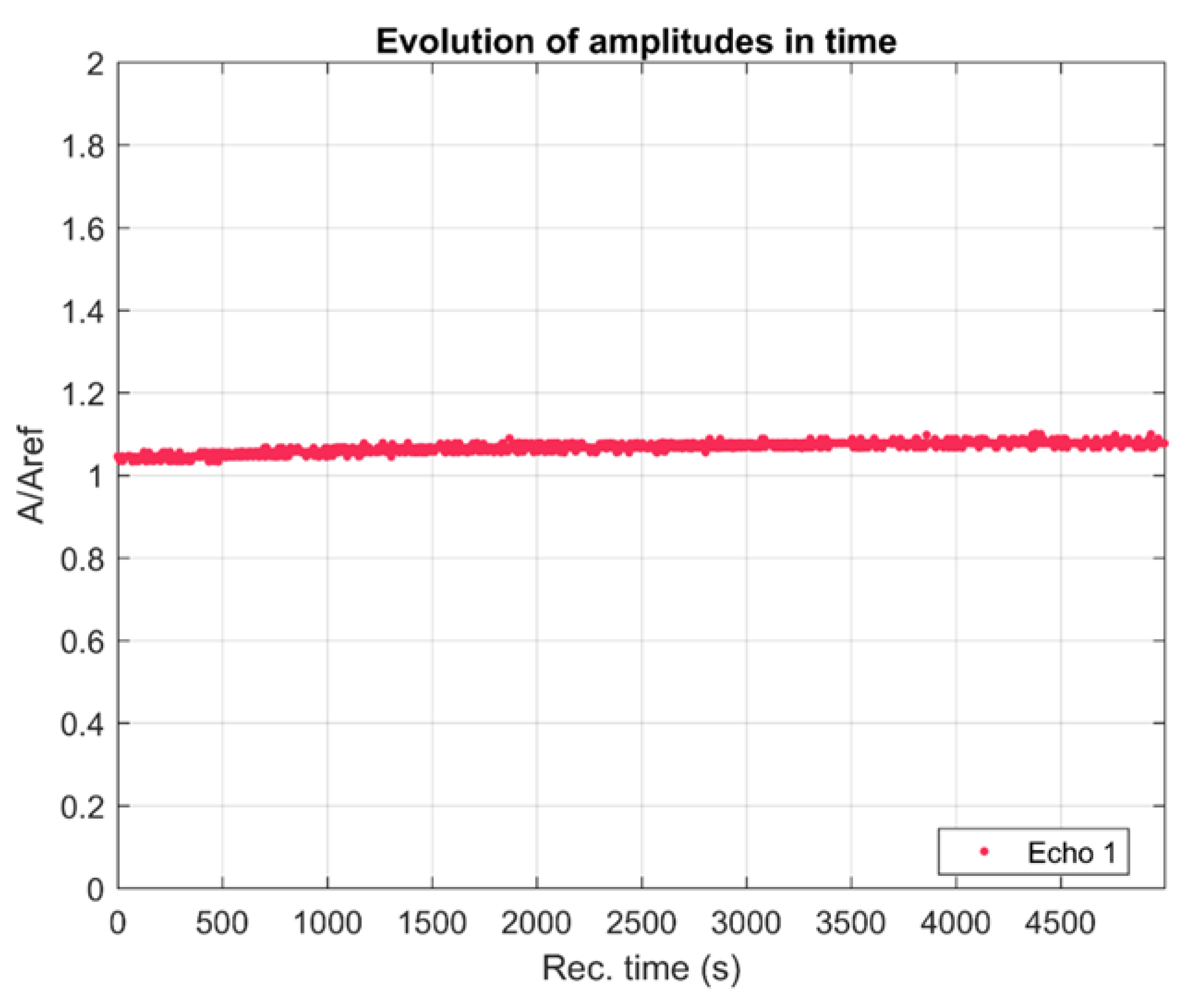
Figure 1 shows the relative amplitudes of the tested sample. The relative amplitudes increased slowly during the testing period.
The ultrasonic signals transmitted through the sample were transformed into their frequency spectra, superposed for all the 1000 recorded signals (Figure 2a). The maximum of all amplitudes was the central frequency of the transducers (25 MHz), which is a general remark. The evolution of the spectral amplitudes was extremely slow and very close to the pattern of the reference liquid (double-distilled water). The spectrum of the spectral amplitude depending on the frequency for the etalon sample (double-distilled water) practically overlapped with the measured spectra associated with the analyzed sample in the range of 23–36 MHz. However, for frequencies below the central frequency, all the spectral amplitudes of the suspension were higher than for the reference liquid. Between 15–36 MHz, the spectral amplitude of the etalon sample had slightly lower values than the spectral amplitudes corresponding to the MHC suspension. This behavior indicates that the MHC nanoparticles in suspension provided a better transmission for this frequency range. These spectra were averaged over the 1000 time records and the attenuation of signals could thus be obtained (Figure 2b). It can be seen that the attenuation was lower than that of water over a wide range of frequencies. At the frequency of 28 MHz, a slightly higher attenuation was observed for the sample analyzed compared to the etalon sample (double-distilled water). The attenuation in this sample was very close to the attenuation in the reference liquid for frequencies above the central frequency, but much smaller as the frequency decreased from the central frequency toward 15 MHz. This fact confirms that the metallic nanoparticles resonate in this frequency range.
The averaged stability parameter , indicated by the slope of the interpolation line in Figure 1, after the initial 300 s, is S = 2.29 × 10⁻⁵ (1/s), represents good stability. In general, the attenuation at every frequency slowly diminished in time (Figure 2c) after a more rapid evolution during the first 100 s. In the first 100 s, a rapid decrease in attenuation was observed up to the frequency of 25 MHz. This behavior could be due to the precipitation of larger particles (formed after aggregation). After this rapid decrease that occurred in the first 100 s, it was observed that in the time interval 100–5000 s, the decrease was very slow, practically imperceptible. For the frequency range 15–22 MHz, the attenuation was negative and decreased in time, influenced by the increasing relative concentration of the MHC nanoparticles in suspension during the sedimentation process. For the higher frequencies (25, 28, 32, 35 MHz) the attenuation was positive. The lowest attenuations are explained by the presence of larger MHC nanoparticles in suspension, which then settled down and the smaller nanoparticle absorbed a higher amount of ultrasonic energy but slowly decreasing in time. This behavior (between 100–5000s) is due to uniform particles with sizes within a narrow range that remained in suspension throughout the experiment. Following these studies, we could say that the suspension presented two populations. Thus, we can talk about a small population of aggregated nanoparticles that tended to precipitate and a population of nanoparticles that was the majority and that remained stable throughout the experiment.
The stability was evaluated both for the standard solution (double-distilled water) and for the MHC suspension. The responses for the MHC suspension were evaluated by comparison with double-distilled water (known to be the most stable suspension). The obtained values for the stability parameter clearly highlighted the stability of the MHC suspension in relation to double-distilled water.
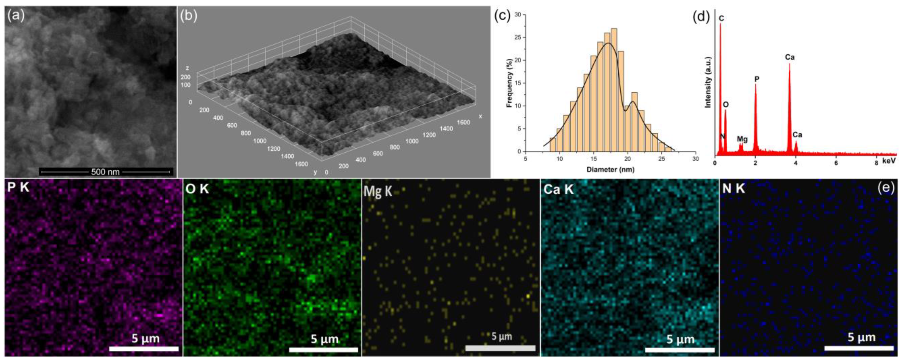
The 2D and 3D SEM images of the MHC suspensions are presented in Figure 3a,b. The results of the SEM studies conducted on the MHC suspensions underline that the sus-pensions contained agglomerated nanoparticles with an ellipsoidal morphology (Figure 3a,b). The mean particle size (Figure 3c) estimated by the SEM studies indicated the presence of particles with an average size of approximately 18 nm. The presence of nanoparticles with an average size of approximately 21 nm was also noticed. These results are in good agreement with the results obtained by the ultrasound measurements (the results that suggested the presence of two populations of particles). It is assumed that the larger particles are the effect of an agglomeration of smaller particles.
The chemical composition of the MHC suspension was evaluated by EDX analysis (Figure 3d). The EDS analysis showed that the MHC suspension was composed of abundant concentrations of calcium (Ca), phosphorus (P) and oxygen (O). Carbon (C), nitrogen (N) and magnesium (Mg) were also observed in quantities that highlight a good process of obtaining the suspensions used for the preparation of the layers. No additional maxima could be noticed in the EDX spectra of the MHC suspension, a fact that proves the purity of the analyzed suspension. Furthermore, the elemental distribution map results (Figure 3e) revealed the uniform distribution of the constituent elements. Each chemical element (N, Ca, P, O and Mg) was well distributed in the sample, which shows that the samples were homogeneous.
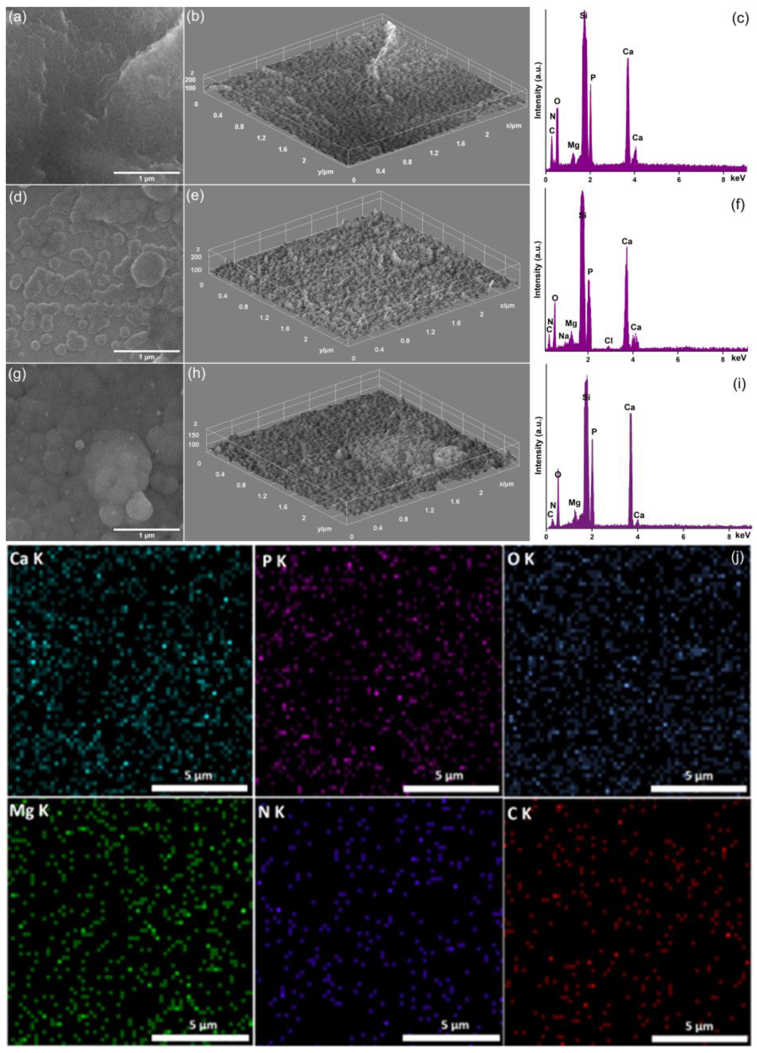
Figure 4a–j shows the surface morphology of the MHC composite thin films (2D SEM images and their 3D representation) together with that of the EDX spectra, obtained before and after exposure to DMEM solution for 7 and 14 days. In both the 2D and 3D SEM images, it can be noticed that the surface of the MHC composite thin films was smooth and continuous without noticing the presence of surface defects. In the case of MHC_7D, after 7 days of DMEM solution, the presence of various nanoparticles on the thin film surface could be noticed, which indicates the formation of a new apatitic layer through biomimetic mineralization. Moreover, on the surface of the MHC_7D sample, the absence of cracks or other surface defects could be noticed. The 2D and 3D SEM images (Figure 4d–e) showed that after 7 days, the formation of the new apatite layer was not uniform on the MHC surface. Also, it could be observed that the new layer consisted mainly of spherical clustered regions (grains). The formation of a new and continuous apatitic coating on the MHC surface was noticed after 14 days of immersion in DMEM solution (Figure 4g,h). Therefore, the smooth surface of the as-deposited MHC thin films was covered by a new apatitic layer.
More than that, the EDX analysis conducted on the MHC (Figure 4c), MHC_7D (Figure 4f) and MHC_14D (Figure 4i) composite thin films indicated the presence of calcium (Ca), phosphorus (P), magnesium (Mg), oxygen (O), carbon (C) and nitrogen (N) on the surface of the biomineralized layers. On the other hand, an increase of Ca and P in the case of the MHC_14D samples was observed compared to the MHC_7D samples. No other maxima could be observed in any of the EDS spectra, which suggests the purity of the studied thin films.
The elemental distribution maps obtained for the MHC_14D composite thin films are depicted in Figure 4j. It could be noticed that the signals characteristic to the main chemical elements (Ca, C, P, O, Mg and N) that are found in the surface composition of the new biomineralized coating were evenly distributed. This feature indicates the homogeneity of the composition across the MHC_14D thin film surface. Therefore, our results are in accordance with the results of the previous studies [34,35,36,37,38,39].
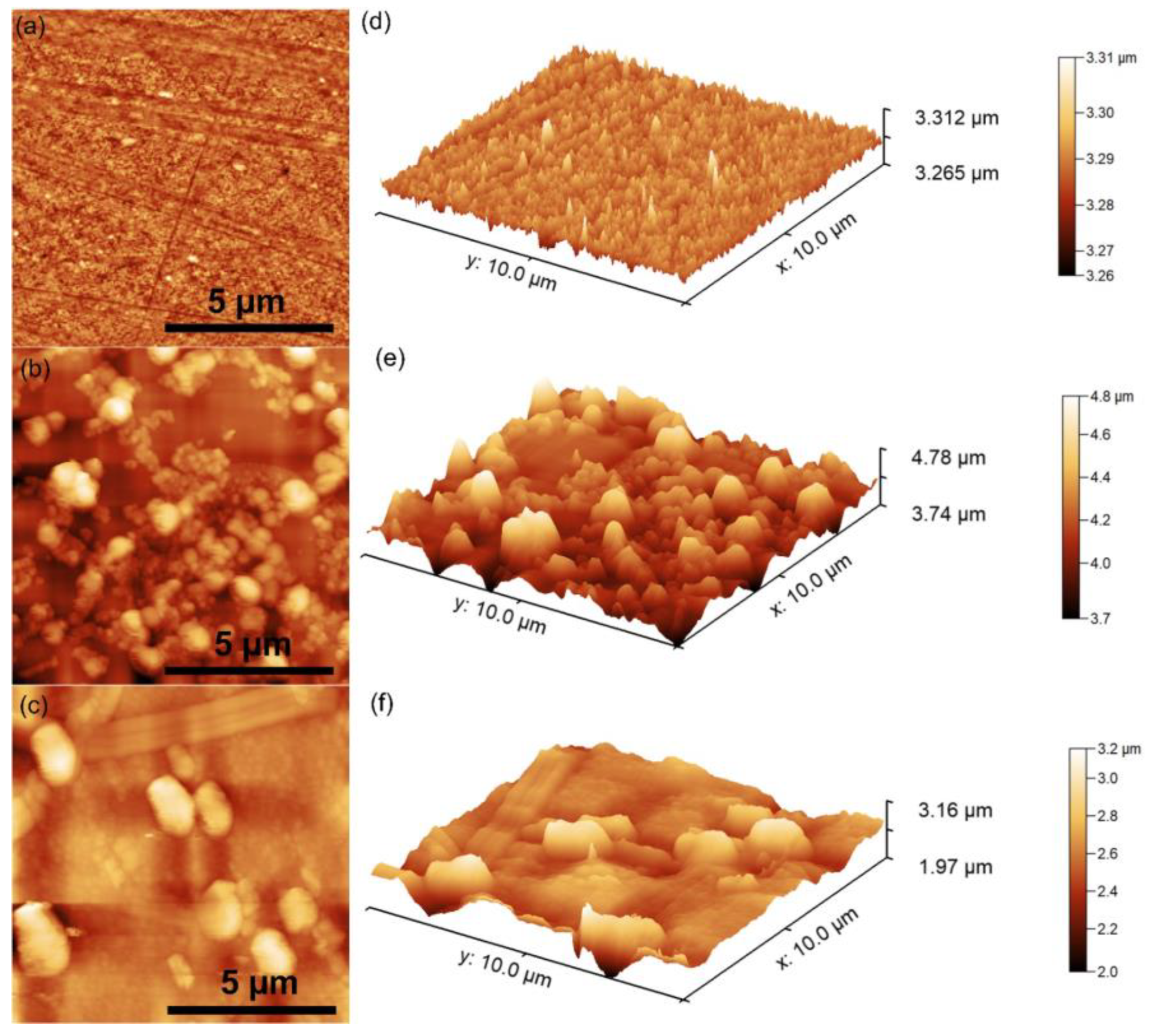
The surface topography of the MHC thin films before and after immersion in DMEM was evaluated by AFM measurements. The 2D images together with the 3D image are presented in Figure 5.
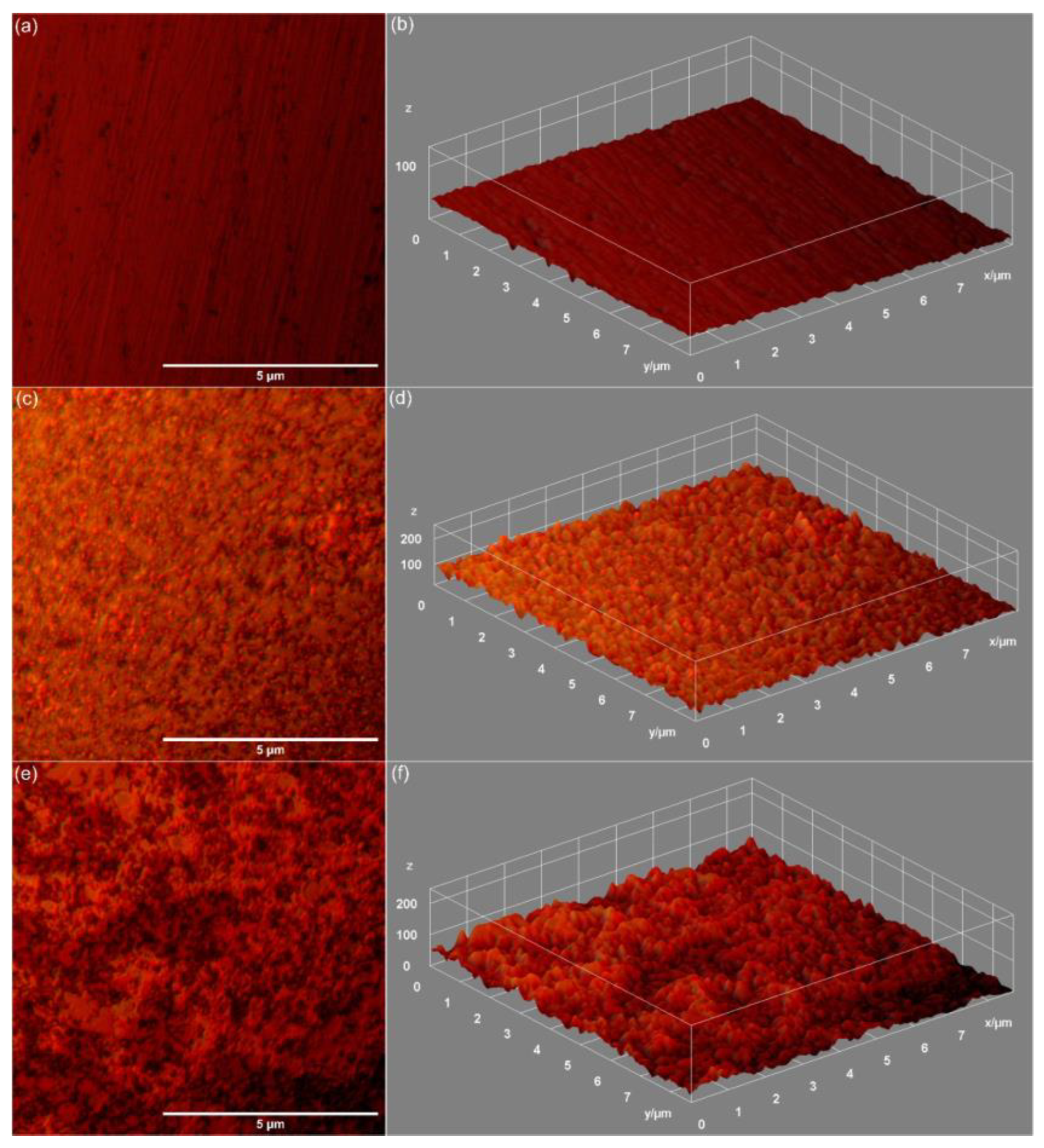
The RRMS roughness parameter values were 3.931 nm (MHC), 143.6 nm (MHC_7D) and 124.3 nm (MHC_14D). These results suggest that the surface roughness increased significantly after immersion in DMEM. For the MHC thin films, the AFM topographies obtained before their immersion in DMEM showed the presence of a continuous layer on the substrate surface and, at the same time, the fact that they were smooth.
After immersion for 7 and 14 days, a modification of the surface morphology was noted, most probably due to the formation of the new apatite-like layer. Even under these conditions, the presence of cracks or other defects on the surface of the MHC layers was not distinguished.
Preliminary data regarding the surface features were obtained by metallographic microscopy (MM). In Figure 6, the typical MM images recorded for the MHC (a,b), MHC_7D (c,d) and MHC_14D (e,f) composite thin films are shown. The formation of the apatitic layer after 7 days of immersion and their homogeneous spread on the MHC coatings can be seen in Figure 6c–f. The MM data support the results obtained by SEM and AFM assessments.
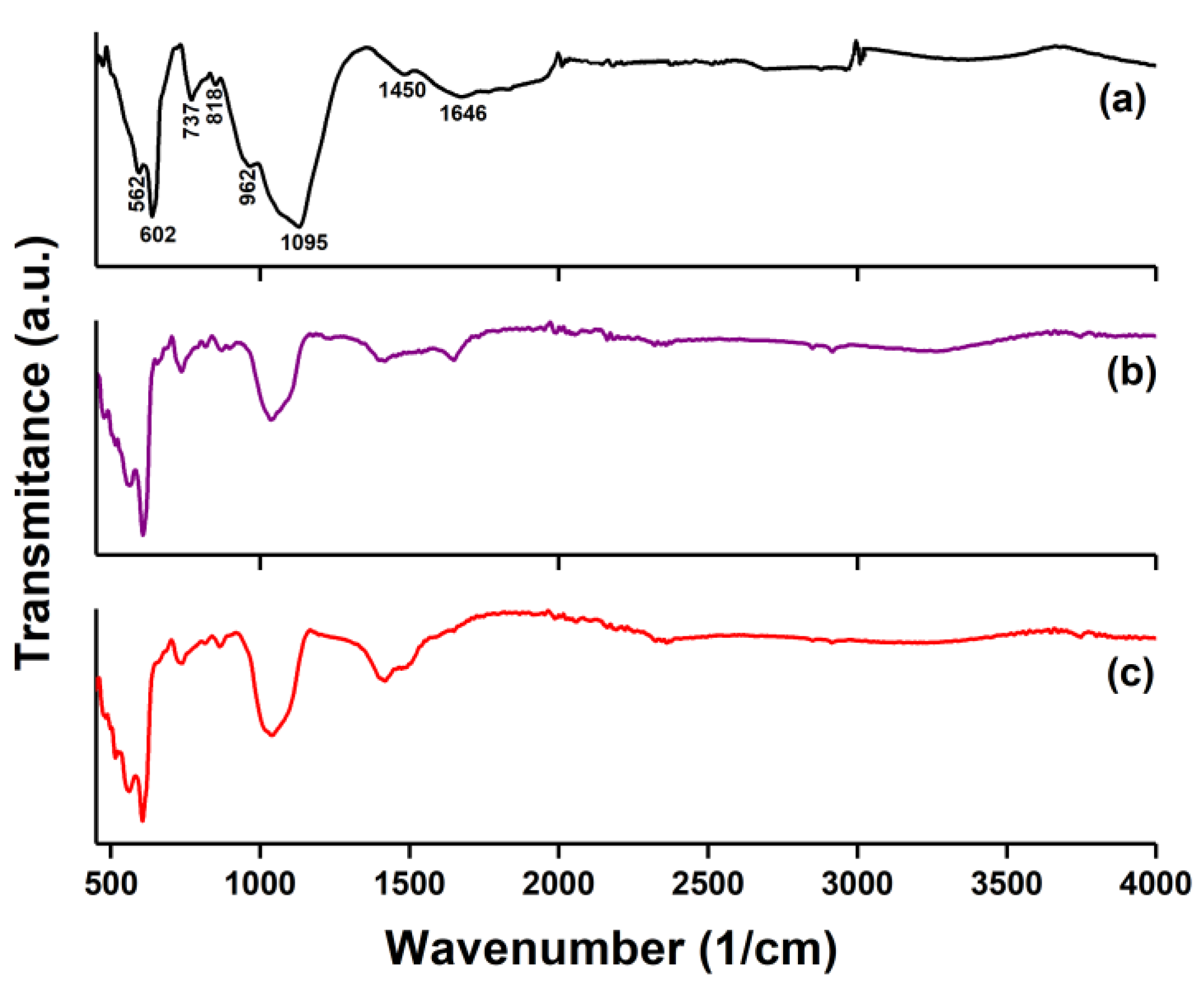
Figure 7a–c reveals the ATR-FTIR spectra obtained on the MHC, MHC_7D and MHC_14D composite thin films.
In the case of the MHC sample, in the ATR-FTIR spectra the presence of bands specific to hydroxyapatite and chitosan structure [40] could be observed. The maxima found at 562 cm−1 belonged to ν4 vibration of phosphate (PO43−). Other peaks specific to the Hap structure could be noticed at 602, 962 and 1095 cm−1, all being assigned to the presence of phosphate (PO43−) groups [40,41]. According to studies reported by Predoi D. et al. [40], the peaks from the 1500–1300 cm−1 can be assigned to C-H and C-O vibration, indicating the chitosan presence in the MHC sample.
The immersion in DMEM solution of the MHC sample induced the appearance of the broad carbonate vibrational bands in the ATR-FTIR spectra in the 1300–1650 cm−1 spectral range [41]. The presence of carbonate groups vibration together with the phosphate vibration peaks indicated that the apatitic compounds with embedded carbonates were deposited on the MHC surface. The slight decrease in the peak intensity noticed in the case of the MHC_7D suggests a low crystallinity degree of the newly formed apatitic layer [38,41].
The results of the SEM studies reported by Sutha S. et al. [42] on Mg-doped hydroxyapatite/chitosan coatings highlighted that, after 21 days of immersion in SBF solution, the presence of a white precipitate was observed on the surface of the analyzed samples, which indicates the formation of a new apatite layer. At the same time, the presence of carbonate, phosphate and hydroxyl functional groups in the FTIR spectra obtained for the graphite-based calcium phosphate/chitosan coatings after immersion in SBF solution for 8 weeks was noted by Zhang J. et al. [43]. Taking into account all these aspects and the results of our studies, we can say that after immersion in the DMEM, the layers based on magnesium-doped hydroxyapatite/chitosan possessed improved properties that are comparable to those of the layers immersed in SBF.
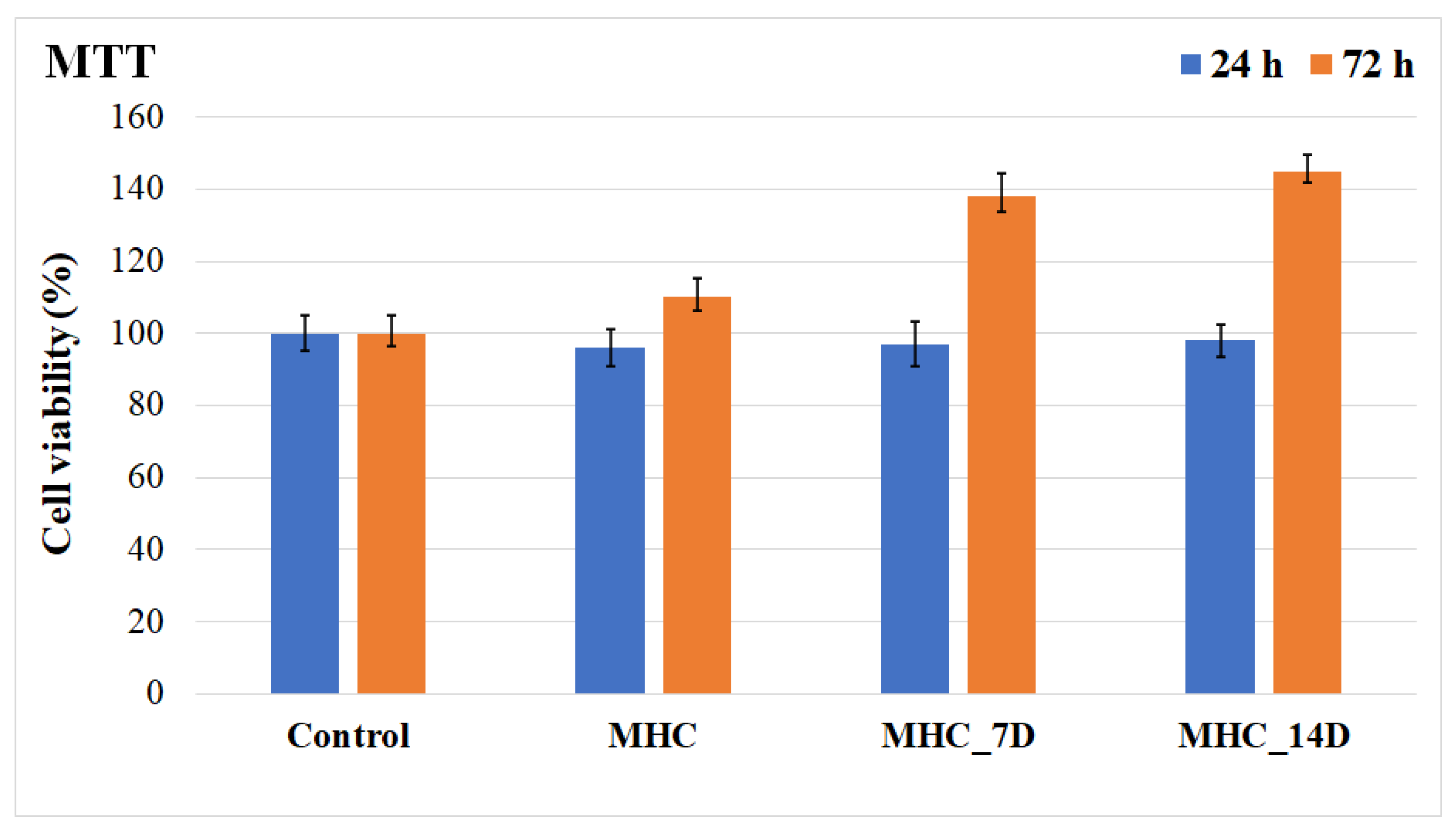
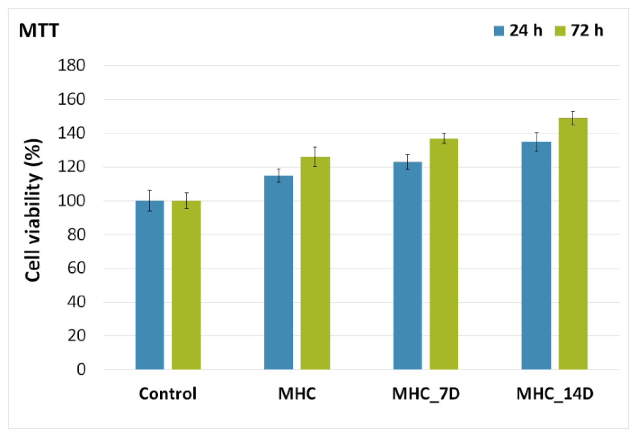
The cytotoxicity of MHC, MHC_7D and MHC_14D was assessed using primary human osteoblast cells, hFOB 1.19, obtained from the upper part of a patient’s femur following the protocol described by Gallagher et al. [32]. The results of the MTT assay, which shows the hFOB 1.19 cell viability after being incubated for 24 h with the MHC, MHC_7D and MHC_14D layers, are depicted in Figure 8. In addition, a free hFOB 1.19 cell culture was also grown and used as a control. The experiments were performed in triplicate and the obtained data were presented as the mean ± SD. More than that, a statistical analysis was performed using a t-test and all the p calculated values were p < 0.05. The results of the in vitro assay depicted that all the tested MHC composite layers presented very good biocompatibility compared with the control cell culture after 24 h of incubation. More than that, the results of the MTT cell viability assay highlighted that after 24 h of incubation, the osteoblast cell viability was above 96%. Moreover, the MTT studies emphasized that the MHC composite layers immersed in DMEM for 7 and 14 days showed an increase in cell viability compared to both the control cell culture and MHC composite layers. Furthermore, the cell viability of the hFOB 1.19 cells was also investigated after 72 h of incubation with the MHC, MHC_7D and MHC_14D composite layers. The results obtained for the cell viability of the hFOB 1.19 cells after 72 h of incubation revealed that the composite layers exhibited strong biocompatibility properties and that they promoted the hFOB 1.19 cell proliferation and adhesion. The cell viability of the cells after 72 h of incubation had higher values in the case of the cells incubated with the composite layers than the value obtained for control cells. More than that, the cell viability increase was influenced by the time that the MHC composite layers were immersed in DMEM. Therefore, the results showed that immersion in DMEM conferred the layers better surface properties that were responsible for the promotion of hFOB 1.19 cell proliferation and adhesion. These results are in good agreement with previously reported studies regarding the biocompatibility and cytotoxicity of magnesium-doped hydroxyapatite-based materials [29,40,44,45,46,47,48,49].
In order to acquire additional information regarding the influence of DMEM on the biological properties of the MHC composite layers, fluorescence micrographs of the hFOB 1.19 cells after 24 h of incubation with MHC, MHC_7D and MHC_14D are presented in Figure 9. The images depicted in Figure 9 highlight that the MHC, MHC_7D and MHC_14D composite layers analyzed after 24 h of incubation exhibited no toxic effect against the osteoblast cells. More than that, the fluorescence micrographs emphasized that the morphology of the hFOB 1.19 cells suffered no significant changes. More than that, the fluorescence images show that the cells started to connect to each other and to achieve an early-stage extra-cellular matrix even after 24 h of incubation. This was observed for all the tested composite layers.
In addition, for the MHC composite layers previously immersed in DMEM for 7 and 14 days, the fluorescence images emphasized that the cells were also prone to adhere and spread on top of each other, converging slowly to well-flattened confluent layers of cells that will eventually cover the entire surface. More than that, the optical visualization emphasized that after 72 h of incubation, the cell spread across the entire surface of the composite layers. Furthermore, after 72 h of incubation, the fluorescence micrographs showed that the osteoblast cells were organized in confluent layers spread out on the surface of the composite layers. These phenomena could be attributed to the changes in the surface morphology of the MHC due to the immersion in DMEM [50,51]. Bodhak et al. [50], in their study on the “Role of surface charge and wettability on early-stage mineralization and bone cell–materials interactions of polarized hydroxyapatite”, demonstrated that, by increasing the surface charge also, they got an increase of the wettability and surface energy of polarized HAp surfaces for both SBF and DMEM. Moreover, their studies also revealed that the increase of the surface energy due to immersion in the medium led to a better hFOB cell attachment and more adhesion contacts between the cells and the surface. In addition, Clupper et al. [51], in their preliminary study regarding the “in vitro bioactivity of S520 glass fibers and initial assessment of osteoblast attachment”, showed that an osteoblast culture on S520 fiber surface immersed in SBF and DMEM was promising, showing proliferation, nodule formation and mineralization.
A supplementary biocompatibility analysis of the MHC, MHC_7D and MHC_14D composite layers was also performed using the HCT-8 cellular line. The cell viability of the HCT-8 cells after being incubated with MHC, MHC_7D and MHC_14D was assessed after 24 and 72 h of exposure. The data obtained from the MTT assays are depicted in Figure 10. A free HCT-8 cell culture was grown and used as a control. The experiments were performed in triplicate and the obtained data are presented as the mean ± SD. More than that, a statistical analysis was performed using a t-test and all the p calculated values were p < 0.05. The results of the MTT assays emphasize that the MHC, MHC_7D and MHC_14D composite layers did not present any toxicity against the investigated cells after 24 and 72 h of exposure. More than that, the results emphasize that after 72 h of incubation, the cell viability of the HCT-8 cells incubated with the MHC, MHC_7D and MHC_14D composite layers surpassed the cell viability of the control cells. These results are in good agreement with the results obtained on hFOB 1.19 cells and reveal, on the one hand, that the MHC, MHC_7D and MHC_14D composite layers present very good biocompatibility and, on the other hand, that after 72 h, due to their enhanced biological properties, they help to promote the adhesion and proliferation of the HCT-8 cells on their surface.
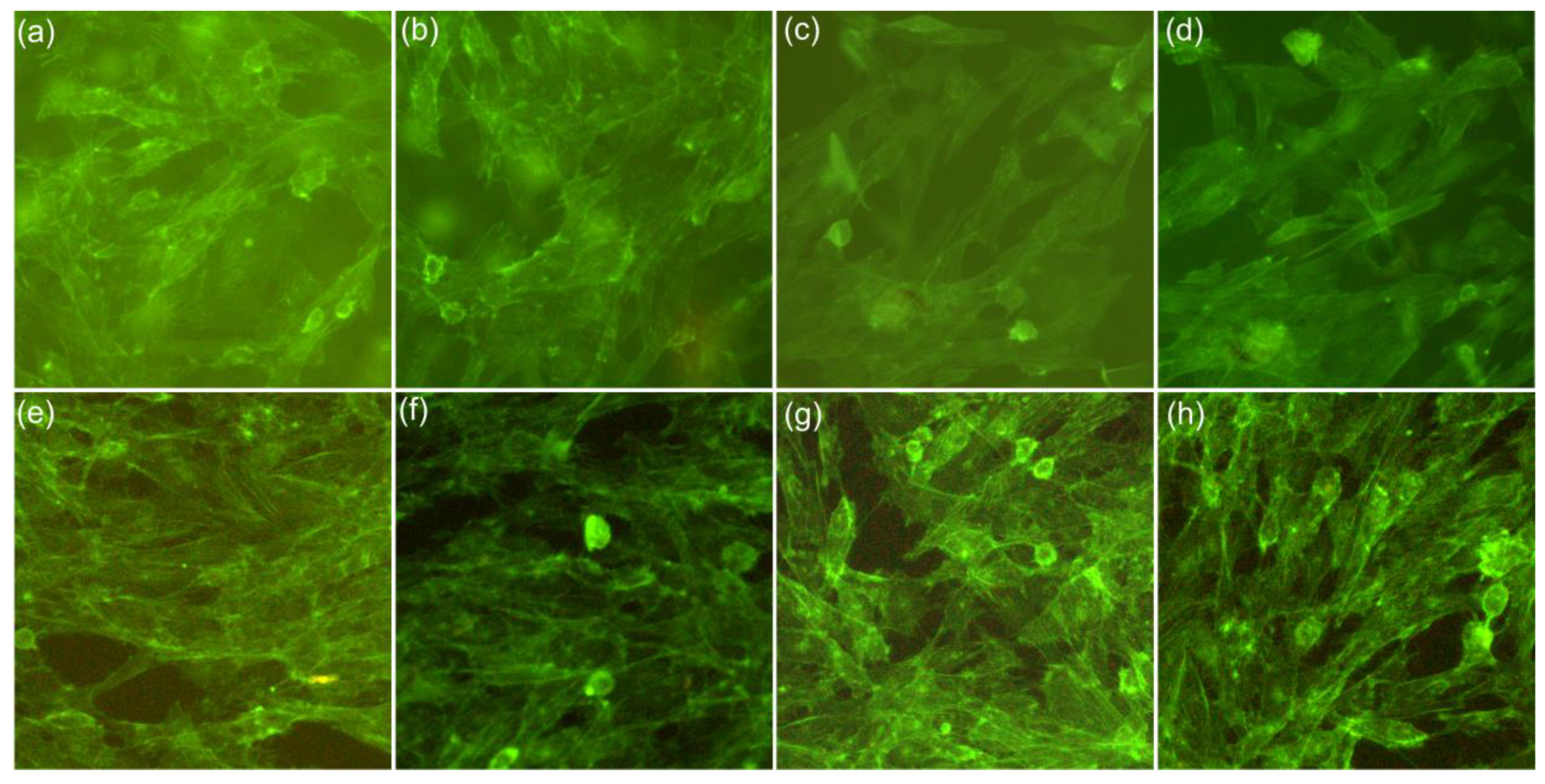
To evaluate the effects of the MHC, MHC_7D and MHC_14D composite layers on the HCT-8 cell morphology, the appearance of the incubated versus the control HCT-8 cells was studied using fluorescence microscopy. The results are depicted in Figure 11a–h. The fluorescence micrographs emphasize that the composite layers exhibited very good biocompatibility towards the investigated cells after 24 and 72 h of incubation compared to the control cells. The fluorescence microscopy images highlight that the control cells appeared as a crowded and disorganized monolayer Figure 11a,e [52,53,54]. However, after 24 and 72 h of incubation with MHC, MHC_7D and MHC_14D composite layers, the images highlight that the cell density was considerably increased, as confirmed by the MTT analysis and that the cells adhered to the surface of the forming confluent layers, uniformly spread across the entire surface of the composite layers. Therefore, these observations demonstrate that the MHC, MHC_7D and MHC_14D composite layers promoted the proliferation and adhesion of HCT-8 cells.
The results of the fluorescence microscopy studies are in agreement with the MTT assays and demonstrate that the MHC, MHC_7D and MHC_14D composite layers did not exhibit any toxic effects on the HCT-8 cell but, on the contrary, they were suitable surfaces for their proliferation and adhesion. More than that, the studies also confirmed that the immersion of the composite layers as well as the immersion period conferred the layers enhanced biological properties that were responsible for the proliferation and attachment of the cells.
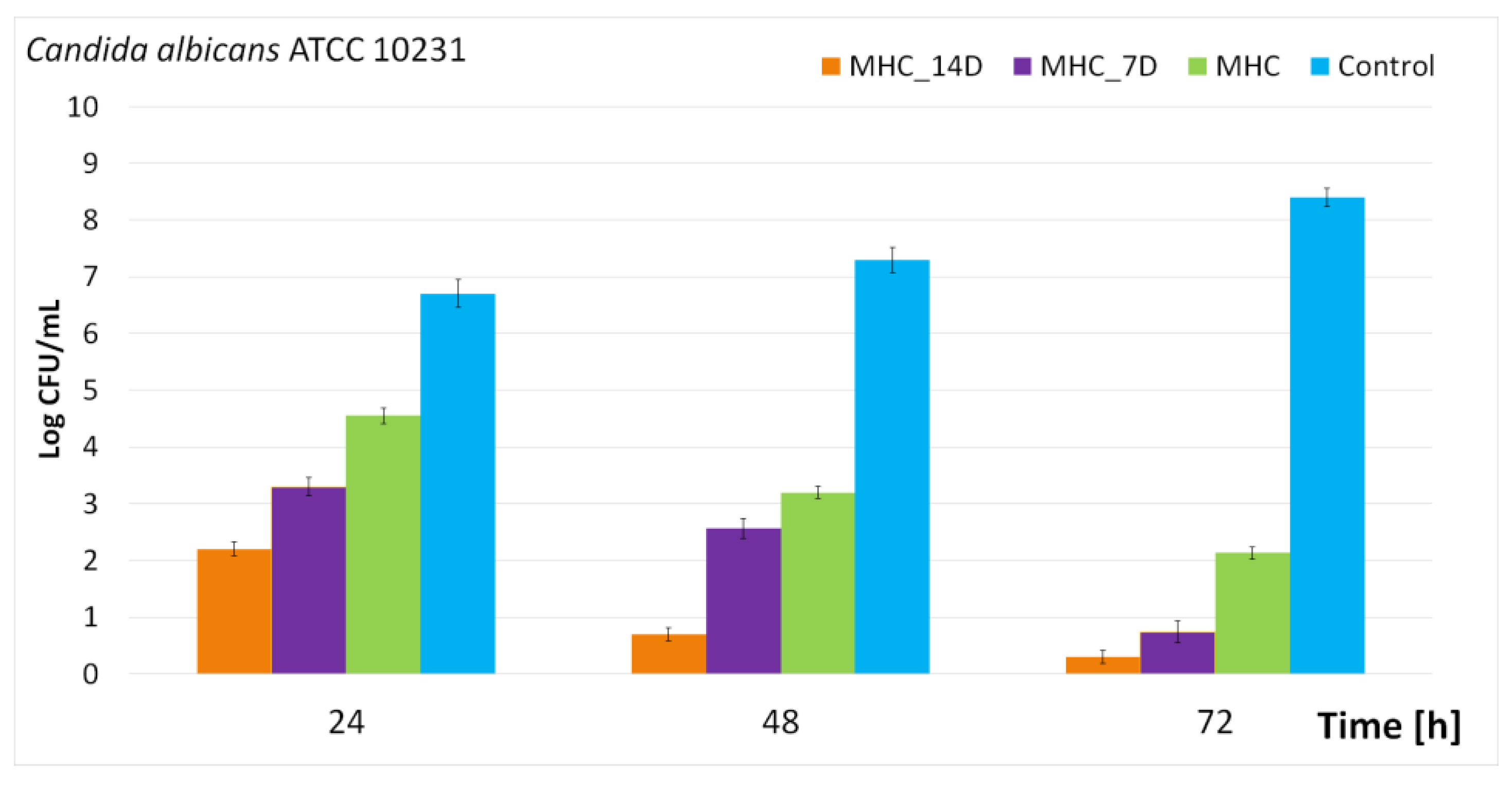
In recent years, due to a considerable increase in the aging population and because of the weak medical advances made in the management of immunocompromised patients, the incidence of fungal infections has increased dramatically [55,56,57]. Due to the fact that C. albicans is a commensal organism that inhabits multiple sites in humans and could become pathogenic, C. albicans is the most predominant cause of fungal infections in humans [55,56,57,58,59,60]. Therefore, in this context, our study also involved the evaluation of the antifungal properties of the MHC, MHC_7D and MHC_14D composite layers. The antifungal properties of the composite layers were determined against C. albicans ATTCC 10231 fungal strains. For this purpose, the layers were put into contact with the fungal suspensions and their activity was assessed after 24, 48 and 72 h. The results of the antifungal assays are presented in Figure 12.
The results of the in vitro antifungal assays demonstrate that all the investigated composite layers exhibited a good inhibitory effect, even after 24 h of incubation, against the development of the fungal cells. Moreover, the data also highlight that the antifungal properties of the MHC, MHC_7D and MHC_14D composite layers were influenced by both incubation time and investigated samples. Therefore, the greater inhibitory effect of the fungal cells was observed after 72 h of incubation for all the investigated samples but, more than that, the greatest antifungal activity was determined to be for the MHC_14D composite layers, which could allow the conclusion that the DMEM immersion time also had an influence on the antifungal properties of the composite layers. The results obtained in this study are in good agreement with previously reported data regarding the antimicrobial activity of composite materials and coatings based on hydroxyapatite with magnesium and chitosan [24,28,29,61,62,63]. The antifungal properties exhibited by the composite’s layers are attributed both to the constituent elements of the suspensions used in the preparation of the layers as well as to the synergies that appeared between the substrate and layers and between the constituent elements of the composite layers [2,28]. In our case, the antifungal activity of the MHC, MHC_7D and MHC_14D composite layers could be attributed to the presence of magnesium ions as well as chitosan. Chitosan is a natural, biodegradable, linear polysaccharide, which has been reported as being non-toxic and exhibiting broad-spectrum antimicrobial activity against Gram-positive bacteria, Gram-negative bacteria and fungi [64,65,66,67]. Even though the exact nature of the mechanisms involved in the antimicrobial activity of the nanoparticles and coatings is not yet fully understood, there are three mechanisms proposed in the case of chitosan. The first mechanism of action is based on the fact that chitosan targets the plasma membrane, which increases the membrane permeability, causing the leakage of the cellular contents that eventually leads to cell death. The second proposed mechanism is that chitosan binds itself to trace elements and interrupts the normal flow of the nutrients necessary for the growth of fungal cells. The third mechanism implies that chitosan could penetrate the wall-cell fungi and inhibit the synthesis of mRNA by binding to the DNA [64,67,68,69]. Furthermore, the results of the antifungal assays emphasize that the changes induced by both the immersion as well as the immersion time of the MHC composite layers in DMEM influenced the inhibitory effects of the composite layers against the development of C. albicans fungal cells. These results strengthen the conclusion that immersion in DMEM conferred the MHC composite layers enhanced biological properties. The results of the biological assays depict that immersion in DMEM for 7 and 14 days of the MHC composite layers resulted in novel composite layers with both enhanced biocompatible properties and antifungal activity. More than that, the results also highlight that the immersion time influenced both the physicochemical as well as the biological properties of the MHC composite layers. The results of this study could be perceived as ground breaking information in the future development of novel biocompatible coatings with antifungal properties for biomedical applications.
As is known, magnesium actively participates in acid-base processes, being present in the biological environment, making a special contribution to bone calcification and decalcification. Moreover, by incorporating Mg2+ ions into HAp/chitosan, an improvement in the biological properties was found. MHC composite thin films can be a serious candidate for applications in bone implants (orthopedic and dental). Taking into account the fact that half of the physiological magnesium is found in bone tissue, having an important functional role in stimulating the growth of new tissue, MHC composite thin films could also have an important role in stimulating the growth of bone tissue after an implant. The present study is in agreement with the recent concerns regarding the use of Mg2+ ions as an additive in the construction of bone biomaterials [70,71]. On the other hand, the presence of chitosan in the studied composite material brought added value as a result of its antibacterial, cicatrizing and anti-inflammatory properties. The studies presented in this paper can contribute to the research carried out for decades [72,73,74] regarding the controlled ionic substitutions of hydroxyapatites. Through controlled ionic substitutions, HAp will have a composition closer to that of natural bone by increasing bioactivity and bone formation capacity. Considering the fact that Bigi et al. [75] demonstrated in 1992 that Mg2+ can promote the formation of new bone mineral nuclei that are very active in the newly formed bone tissue, the present study can actively contribute to the realization of implantable surfaces covered with MHC composite. On the other hand, Boanini et al. [76] showed that Mg2+ ions are regulators of osteoblast cells with effective potential also in the case of osteoporotic bones. Therefore, the use of MHC suspension with good stability allows us to obtain MHC composite layers with improved morphological, structural and biological properties. Thus, the coatings made using this composite will have major contributions to the success of the implants because, in addition to very good biocompatibility and the major contribution to bone regeneration, this biocomposite has anti-inflammatory, antimicrobial and cicatrizing properties that reduce the risk of postoperative infection and implant rejection.
Last but not least, we could say that, as a result of their complex properties, the MHC layers open favorable perspectives for the development of high-performance medical devices with complex biological properties (increased bioactivity, osteointegrability, anti-inflammatory, antimicrobial and cicatrizing), thus preventing postoperative complications.
These preliminary results are of the utmost importance for future advances in tailoring wettability and charge polarity of various surfaces in order to promote a faster apatite nucleation and a better bone cell ingrowth process, which could be the basis of the development of novel materials for implant coatings that can stimulate faster healing.
4. Conclusions
The aim of this study was to evaluate for the first time the influence of DMEM (Dulbecco’s Modified Eagle Medium) on the surface morphology and chemical composition of magnesium-doped hydroxyapatite/chitosan (MHC) composite thin films. The sol-gel method was used for the development of the MHC suspensions. The MHC composite thin films were obtained using the spin-coating method. The MHC suspension stability was studied by performing ultrasound measurements. The results of the SEM and EDX measurements conducted on the MHC composite thin films reveal the formation of an apatitic layer on the samples surface. Furthermore, the modification of the surface microstructure of the MHC composite thin films was evaluated and with the aid of atomic force microscopy (AFM) and metallographic microscopy (MM). The ATR-FTIR data underline the presence of the typical vibrational bands on the surface of MHC samples. The cytotoxicity of the MHC composite thin films was investigated using primary osteoblast and HCT-8 cell culture. The preliminary data obtained after the in vitro biological assay prove the non-toxic effect of the MHC samples against the osteoblast and HCT-8 cells. The good antifungal activity of the MHC composite coatings was also proved by in vitro assays using the Candida albicans ATCC 10231 fungal strain. Therefore, MHC could be suitable candidates for covering implants, giving them improved biological properties, which would ultimately lead to a decrease in the number of implant replacement surgeries, thus improving the quality of life of patients.
Author Contributions
Conceptualization, D.P., S.C.C. and M.V.P.; methodology, D.P.; software, M.V.P.; validation, D.P., S.C.C., S.L.I. and M.V.P.; formal analysis, S.C.C.; investigation, S.C.C. and S.L.I.; resources, D.P. and M.V.P.; data curation, D.P.; writing—original draft preparation, D.P., S.C.C., S.L.I. and M.V.P.; writing—review and editing, D.P., S.C.C., S.L.I. and M.V.P.; visualization, D.P., S.C.C., S.L.I. and M.V.P.; supervision, S.C.C. and M.V.P.; project administration, S.C.C. and M.V.P.; funding acquisition, D.P. All authors have read and agreed to the published version of the manuscript.
Funding
This work was supported by the Romanian Ministry of Research and Innovation through the PN-III-P2-2.1-PED-2019-1375, contract number 331PED⁄2020. Also, this research was supported by Contract No. T-IS 251801/04.05.2018 and Scientific Research Contract Nr.1/4.06.2020.
Institutional Review Board Statement
Not applicable.
Informed Consent Statement
Not applicable.
Data Availability Statement
Data available on demand from the corresponding authors.
Acknowledgments
The authors would like to thank Monica Luminita Badea for assistance with the biological assays.
Conflicts of Interest
The authors declare no conflict of interest. The funders had no role in the design of the study; in the collection, analyses or interpretation of data; in the writing of the manuscript; or in the decision to publish the results.
References
- Peetsch, A.; Greulich, C.; Braun, D.; Stroetges, C.; Rehage, H.; Siebers, B.; Köller, M.; Epple, M. Silver-doped calcium phosphate nanoparticles: Synthesis, characterization, and toxic effects toward mammalian and prokaryotic cells. Colloids Surf. B 2013, 102, 724–729. [Google Scholar] [CrossRef] [PubMed]
- Iconaru, S.L.; Predoi, M.V.; Motelica-Heino, M.; Predoi, D.; Buton, N.; Megier, C.; Stan, G.E. Dextran-Thyme Magnesium-Doped Hydroxyapatite Composite Antimicrobial Coatings. Coatings 2020, 10, 57. [Google Scholar] [CrossRef]
- Predoi, D.; Iconaru, S.L.; Predoi, M.V.; Motelica-Heino, M.; Buton, N.; Megier, C. Obtaining and Characterizing Thin Layers of Magnesium Doped Hydroxyapatite by Dip Coating Procedure. Coatings 2020, 10, 510. [Google Scholar] [CrossRef]
- Demishtein, K.; Reifen, R.; Shemesh, M. Antimicrobial properties of magnesium open opportunities to develop healthier food. Nutrients 2019, 11, 2363. [Google Scholar] [CrossRef]
- Delbet, P. Politique Préventive du Cancer: Cytophylaxie; Denoël: Paris, France, 1944. [Google Scholar]
- Glasdam, S.M.; Glasdam, S.; Peters, G.H. The Importance of Magnesium in the Human Body: A Systematic Literature Review. Adv. Clin. Chem. 2016, 73, 169–193. [Google Scholar] [CrossRef]
- De Baaij, J.H.; Hoenderop, J.G.; Bindels, R.J. Magnesium in man: Implications for health and disease. Physiol. Rev. 2015, 95, 1–46. [Google Scholar] [CrossRef] [PubMed]
- Davidson, D.J.; Spratt, D.; Liddle, A.D. Implant materials and prosthetic joint infection: The battle with the biofilm. EFORT Open Rev. 2019, 4, 633–639. [Google Scholar] [CrossRef]
- Montazerian, M.; Hosseinzadeh, F.; Migneco, C.; Fook, M.V.; Baino, F. Bioceramic coatings on metallic implants: An overview. Ceram. Int. 2022, 48, 8987–9005. [Google Scholar] [CrossRef]
- Ressler, A.; Žužić, A.; Ivanišević, I.; Kamboj, N.; Ivanković, H. Ionic substituted hydroxyapatite for bone regeneration applications: A review. Open Ceram. 2021, 6, 100122. [Google Scholar] [CrossRef]
- Jaafar, A.; Hecker, C.; Árki, P.; Joseph, Y. Sol-Gel Derived Hydroxyapatite Coatings for Titanium Implants: A Review. Bioengineering 2020, 7, 127. [Google Scholar] [CrossRef]
- Banerjee, S.; Bagchi, B.; Bhandary, S.; Kool, A.; Hoque, N.A.; Thakur, P.; Das, S. A facile vacuum assisted synthesis of nanoparticle impregnated hydroxyapatite composites having excellent antimicrobial properties and biocompatibility. Ceram. Int. 2018, 44, 1066–1077. [Google Scholar] [CrossRef]
- Ratha, I.; Datta, P.; Balla, V.K.; Nandi, S.K.; Kundu, B. Effect of doping in hydroxyapatite as coating material on biomedical implants by plasma spraying method: A review. Ceram. Int. 2021, 47, 4426–4445. [Google Scholar] [CrossRef]
- Ahmadi, R.; Asadpourchallou, N.; Kaleji, B.K. In vitro study: Evaluation of mechanical behavior, corrosion resistance, antibacterial properties and biocompatibility of HAp/TiO2/Ag coating on Ti6Al4V/TiO2 substrate. Surf. Interfaces 2021, 24, 101072. [Google Scholar] [CrossRef]
- Sidane, D.; Rammal, H.; Beljebbar, A.; Gangloff, S.C.; Chicot, D.; Velard, F.; Khireddine, H.; Montagne, A.; Kerdjoudj, H. Biocompatibility of sol-gel hydroxyapatite-titania composite and bilayer coatings. Mater. Sci. Eng. C 2017, 72, 650–658. [Google Scholar] [CrossRef] [PubMed]
- Bazin, T.; Magnaudeix, A.; Mayet, R.; Carles, P.; Julien, I.; Demourgues, A.; Gaudon, M.; Champion, E. Sintering and biocompatibility of copper-doped hydroxyapatite bioceramics. Ceram. Int. 2021, 47, 13644–13654. [Google Scholar] [CrossRef]
- Chidambaranathan, A.S.; Mohandoss, K.; Balasubramaniam, M.K. Comparative Evaluation of Antifungal Effect of Titanium, Zirconium and Aluminium Nanoparticles Coated Titanium Plates Against C. albicans. J. Clin. Diagn. Res. 2016, 10, ZC56–ZC59. [Google Scholar] [CrossRef]
- Nasrallah, D.A.; Ibrahim, M.A. Enhancement of physico-chemical, optical, dielectric and antimicrobial properties of polyvinyl alcohol/carboxymethyl cellulose blend films by addition of silver doped hydroxyapatite nanoparticles. J. Polym. Res. 2022, 29, 86. [Google Scholar] [CrossRef]
- Tambone, E.; Marchetti, A.; Ceresa, C.; Piccoli, F.; Anesi, A.; Nollo, G.; Caola, I.; Bosetti, M.; Fracchia, L.; Ghensi, P.; et al. Counter-Acting Candida albicans-Staphylococcus aureus Mixed Biofilm on Titanium Implants Using Microbial Biosurfactants. Polymers 2021, 13, 2420. [Google Scholar] [CrossRef]
- Bürgers, R.; Hahnel, S.; Reichert, T.E.; Rosentritt, M.; Behr, M.; Gerlach, T.; Handel, G.; Gosau, M. Adhesion of Candida albicans to various dental implant surfaces and the influence of salivary pellicle proteins. Acta Biomater. 2010, 6, 2307–2313. [Google Scholar] [CrossRef]
- Vargas-Blanco, D.; Lynn, A.; Rosch, J.; Noreldin, R.; Salerni, A.; Lambert, C.; Rao, R.P. A pre-therapeutic coating for medical devices that prevents the attachment of Candida albicans. Ann. Clin. Microbiol. Antimicrob. 2017, 16, 41. [Google Scholar] [CrossRef] [PubMed] [Green Version]
- Farrag, H.A.; El-Hendawy, H.H.; El-Tablawy, S.Y.; Nora, F.H. Prevention of Adhesion and Surface Growth of Orthopedic Implant Microbial Infection by Surface Modification Using Antibiotics and Irradiated Hydroxyapatite. Silicon 2019, 11, 2333–2343. [Google Scholar] [CrossRef]
- Hallmann, L.; Gerngroß, M.D. Chitosan and its application in dental implantology. J. Stomatol. Oral. Maxillofac. Surg. 2022, 123, e701–e707. [Google Scholar] [CrossRef]
- Iconaru, S.L.; Ciobanu, C.S.; Predoi, G.; Rokosz, K.; Chifiriuc, M.C.; Bleotu, C.; Stanciu, G.; Hristu, R.; Raaen, S.; Raita, S.M.; et al. Biological and Physico-Chemical Properties of Composite Layers Based on Magnesium-Doped Hydroxyapatite in Chitosan Matrix. Micromachines 2022, 13, 1574. [Google Scholar] [CrossRef]
- Kravanja, G.; Primožič, M.; Knez, Ž.; Leitgeb, M. Chitosan-Based (Nano)Materials for Novel Biomedical Applications. Molecules 2019, 24, 1960. [Google Scholar] [CrossRef] [PubMed]
- Anil, K.S.; Veena, V.; Bahrudeen, S.H.; Sureshkumar, R.; Natarajan, S. Medium constituents mediated engineering for size and shape tuning of gold nanocrystallites. J. Ind. Eng. Chem. 2017, 51, 288–294. [Google Scholar] [CrossRef]
- Li, B.; Han, Y.; Qi, K. Formation mechanism, degradation behavior, and cytocompatibility of a nanorod-shaped HA and pore-sealed MgO bilayer coating on magnesium. ACS Appl. Mater. Interfaces 2014, 6, 18258–18274. [Google Scholar] [CrossRef]
- Predoi, D.; Ciobanu, C.S.; Iconaru, S.L.; Predoi, S.A.; Chifiriuc, M.C.; Raaen, S.; Badea, M.L.; Rokosz, K. Impact of Gamma Irradiation on the Properties of Magnesium-Doped Hydroxyapatite in Chitosan Matrix. Materials 2022, 15, 5372. [Google Scholar] [CrossRef]
- ImageJ. Available online: http://imagej.nih.gov/ij (accessed on 10 November 2022).
- Gwyddion. Available online: http://gwyddion.net/ (accessed on 30 November 2022).
- Gallagher, A.J.; Gundle, R.; Beresford, N.J. Isolation and culture of bone forming cells (osteoblasts) from human bone. Hum. Cell Cult. Protoc. 1996, 2, 233–263. [Google Scholar]
- Predoi, D.; Iconaru, S.L.; Predoi, M.V. Bioceramic Layers with Antifungal Properties. Coatings 2018, 8, 276. [Google Scholar] [CrossRef]
- Ciobanu, C.S.; Iconaru, S.L.; Predoi, D.; Trușcă, R.-D.; Prodan, A.M.; Groza, A.; Chifiri-uc, M.C.; Beuran, M. Fabrication of Novel Chitosan–Hydroxyapatite Nanostructured Thin Films for Biomedical Applications. Coatings 2021, 11, 1561. [Google Scholar] [CrossRef]
- Stojanović, S.; Mitić, Ž.; Miljković, M.; Rajković, J.; Trajanović, M.; Najman, S. SEM-EDX analysis of BIO-OSS® granules after incubation in cell culture medium. In Proceedings of the III Advanced Ceramics and Applications Conference; Atlantis Press: Paris, France, 2016; pp. 259–264. [Google Scholar] [CrossRef]
- John, Ł.; Bałtrukiewicz, M.; Sobota, P.; Brykner, R.; Cwynar-Zając, Ł.; Dzięgiel, P. Non-cytotoxic organic–inorganic hybrid bioscaffolds: An efficient bedding for rapid growth of bone-like apatite and cell proliferation. Mater. Sci. Eng. C 2012, 32, 1849–1858. [Google Scholar] [CrossRef]
- Bodhak, S.; Bose, S.; Bandyopadhyay, A. Electrically polarized HAp-coated Ti: In vitro bone cell-material interactions. Acta Biomater. 2010, 6, 641–651. [Google Scholar] [CrossRef]
- Vranceanu, D.M.; Parau, A.C.; Cotrut, C.M.; Kiss, A.E.; Constantin, L.R.; Braic, V.; Vladescu, A. In vitro evaluation of Ag doped hydroxyapatite coatings in acellular media. Ceram. Int. 2019, 45, 11050–11061. [Google Scholar] [CrossRef]
- Ahmadkhaniha, D.; Fedel, M.; Sohi, M.H.; Hanzaki, A.Z.; Deflorian, F. Corrosion behavior of magnesium and magnesium–hydroxyapatite composite fabricated by friction stir processing in Dulbecco’s phosphate buffered saline. Corros. Sci. 2016, 104, 319–329. [Google Scholar] [CrossRef]
- Wagener, V.; Virtanen, S. Protective layer formation on magnesium in cell culture medium. Mater. Sci. Eng. C 2016, 63, 341–351. [Google Scholar] [CrossRef] [PubMed]
- Predoi, D.; Ciobanu, C.S.; Iconaru, S.L.; Raaen, S.; Badea, M.L.; Rokosz, K. Physicochemical and Biological Evaluation of Chitosan-Coated Magnesium-Doped Hydroxyapatite Composite Layers Obtained by Vacuum Deposition. Coatings 2022, 12, 702. [Google Scholar] [CrossRef]
- Vlădescu, A.; Pârâu, A.; Pană, I.; Cotruț, C.M.; Constantin, L.R.; Braic, V.; Vrânceanu, D.M. In Vitro Activity Assays of Sputtered HAp Coatings with SiC Addition in Various Simulated Biological Fluids. Coatings 2019, 9, 389. [Google Scholar] [CrossRef]
- Sutha, S.; Dhineshbabu, N.R.; Prabhu, M.; Rajendran, V. Mg-doped hydrox-yapatite/chitosan composite coated 316l stainless steel implants for biomedical appli-cations. J. Nanosci. Nanotechnol. 2015, 15, 4178–4187. [Google Scholar] [CrossRef]
- Zhang, J.; Dai, C.; Wei, J.; Wen, Z.; Zhang, S.; Lin, L. Calcium phos-phate/chitosan composite coating: Effect of different concentrations of Mg2+ in the m-SBF on its bioactivity. Appl. Surf. Sci. 2013, 280, 256–262. [Google Scholar] [CrossRef]
- Yamasaki, Y.; Yoshida, Y.; Okazaki, M.; Shimazu, A.; Kubo, T.; Akagawa, Y.; Uchida, T. Action of FGMgCO3Ap–collagen composite in promoting bone formation. Biomaterials 2003, 24, 4913–4920. [Google Scholar] [CrossRef]
- Zhao, S.F.; Jiang, Q.H.; Peel, S.; Wang, X.X.; He, F.M. Efects of magnesium-substituted nanohydroxyapatite coating on implant osseointegration. Clin. Oral Implant. Res. 2013, 24, 34–41. [Google Scholar] [CrossRef]
- Yamasaki, Y.; Yoshida, Y.; Okazaki, M.; Shimazu, A.; Uchida, T.; Kubo, T.; Akagawa, Y.; Hamada, Y.; Takahashi, J.; Matsuura, N. Synthesis of functionally graded MgCO3 apatite accelerating osteoblast adhesion. J. Biomed. Mater. Res. 2002, 62, 99–105. [Google Scholar] [CrossRef]
- Landi, E.; Logroscino, G.; Proietti, L.; Tampieri, A.; Sandri, M.; Sprio, S. Biomimetic Mg-substituted hydroxyapatite: From synthesis to in vivo behaviour. J. Mater. Sci. Mater. Med. 2008, 19, 239–247. [Google Scholar] [CrossRef]
- Cai, Y.L.; Zhang, J.J.; Zhang, S.; Venkatraman, S.S.; Zeng, X.T.; Du, H.J.; Mondal, D. Osteoblastic cell response on fluoridated hydroxyapatite coatings: The efect of magnesium incorporation. Biomed. Mater. 2010, 5, 054114. [Google Scholar] [CrossRef]
- Jonasova, L.; Muller, F.A.; Helebrant, A.; Strnad, J.; Greil, P. Biomimetic apatite formation on chemically treated titanium. Biomaterials 2004, 25, 1187–1194. [Google Scholar] [CrossRef]
- Bodhak, S.; Bose, S.; Bandyopadhyay, A. Role of surface charge and wettability on early stage mineralization and bone cell-materials interactions of polarized hydroxyapatite. Acta Biomater. 2009, 5, 2178–2188. [Google Scholar] [CrossRef]
- Clupper, D.C.; Gough, J.E.; Hall, M.M.; Clare, A.G.; LaCourse, W.C.; Hench, L.L. In vitro bioactivity of S520 glass fibers and initial assessment of osteoblast attachment. J. Biomed. Mater. Res. A 2003, 67, 285–294. [Google Scholar] [CrossRef] [PubMed]
- Li, Q.; Wang, X.; Shen, A.; Zhang, Y.; Chen, Y.; Sferra, T.J.; Lin, J.; Peng, J. Hedyotis diffusa Willd overcomes 5-fluorouracil resistance in human colorectal cancer HCT-8/5-FU cells by downregulating the expression of P-glycoprotein and ATP-binding casette subfamily G member 2. Exp. Ther. Med. 2015, 10, 1845–1850. [Google Scholar] [CrossRef] [PubMed]
- Desir, S.; Wong, P.; Turbyville, T.; Chen, D.; Shetty, M.; Clark, C.; Zhai, E.; Romin, Y.; Manova-Todorova, K.; Starr, T.K.; et al. Intercellular Transfer of Oncogenic KRAS via Tunneling Nanotubes Introduces Intracellular Mutational Het-erogeneity in Colon Cancer Cells. Cancers 2019, 11, 892. [Google Scholar] [CrossRef] [PubMed]
- Xu, L.N.; Lu, B.N.; Hu, M.M.; Xu, Y.W.; Han, X.; Qi, Y.; Peng, J.Y. Mechanisms involved in the cytotoxic effects of berberine on human colon cancer HCT-8 cells. Biocell 2012, 36, 113–120. [Google Scholar]
- Cassone, A.; Cauda, R. Candida and candidiasis in HIV-infected patients: Where com-mensalism, opportunistic behavior and frank pathogenicity lose their borders. AIDS 2012, 26, 1457–1472. [Google Scholar] [CrossRef] [PubMed]
- Dall, T.M.; Gallo, P.D.; Chakrabarti, R.; West, T.; Semilla, A.P.; Storm, M.V. An aging population and growing disease burden will require a large and specialized health care workforce by 2025. Health Aff. 2013, 32, 2013–2020. [Google Scholar] [CrossRef]
- Papon, N.; Courdavault, V.; Clastre, M.; Bennett, R.J. Emerging and emerged pathogenic Candida species: Beyond the Candida albicans paradigm. PLoS Pathog. 2013, 9, e1003550. [Google Scholar] [CrossRef]
- Martin, M.V. The use of fluconazole and itraconazole in the treatment of Candida albicans infections: A review. J. Antimicrob. Chemoth. 1999, 44, 429–437. [Google Scholar] [CrossRef] [PubMed]
- Weiner, L.M.; Webb, A.K.; Limbago, B.; Dudeck, M.A.; Patel, J.; Kallen, A.J.; Edwards, J.R.; Sievert, D.M. Antimicrobial-resistant pathogens associated with health care associated infections: Summary of data reported to the National Healthcare Safety Network at the Centers for Disease Control and Prevention, 2011-2014. Infect. Cont. Hosp. Epidemol. 2016, 37, 1288–1301. [Google Scholar] [CrossRef]
- Kullberg, B.J.; Arendrup, M.C. Invasive candidiasis. N. Engl. J. Med. 2015, 373, 1445–1456. [Google Scholar] [CrossRef] [PubMed]
- Rabea, E.I.; Badawy, M.E.; Stevens, C.V.; Smagghe, G.; Steurbaut, W. Chitosan as Anti-microbial Agent: Applications and Mode of Action. Biomacromolecules 2003, 4, 1457–1465. [Google Scholar] [CrossRef]
- Confederat, L.G.; Tuchilus, C.G.; Dragan, M.; Sha’at, M.; Dragostin, O.M. Preparation and Antimicrobial Activity of Chitosan and Its Derivatives: A Concise Review. Molecules 2021, 26, 3694. [Google Scholar] [CrossRef] [PubMed]
- Hans, S.; Fatima, Z.; Ahmad, A.; Hameed, S. Magnesium impairs Candida albicans immune evasion by reduced hyphal damage, enhanced β-glucan exposure and altered vacuole homeostasis. PLoS ONE 2022, 17, e0270676. [Google Scholar] [CrossRef] [PubMed]
- Cheung, R.C.; Ng, T.B.; Wong, J.H.; Chan, W.Y. Chitosan: An update on potential biomedical and pharmaceutical applications. Mar. Drugs 2015, 13, 5156–5186. [Google Scholar] [CrossRef]
- Pena, A.; Sanchez, N.S.; Calahorra, M. Effects of chitosan on Candida albicans: Conditions for its antifungal activity. Biomed. Res. Int. 2013, 2013, 527549. [Google Scholar] [CrossRef] [Green Version]
- Kendra, D.F.; Hadwiger, L.A. Characterization of the smallest chitosan oligomer that is maximally antifungal to Fusarium Solani and elicits pisatin formation in How chitosan, a DNA-complexing carbohydrate activates genes associated with disease resistance in peasPisum sativum. Exp. Mycol. 1984, 8, 276–281. [Google Scholar] [CrossRef]
- Sudarshan, N.R.; Hoover, D.G.; Knorr, D. Antibacterial action of chitosan. Food Biotechnol. 1992, 6, 257–272. [Google Scholar] [CrossRef]
- Raafat, D.; von Bargen, K.; Haas, A.; Sahl, H.G. Insights into the mode of action of chitosan as an antibacterial compound. Appl. Environ. Microbiol. 2008, 74, 3764–3773. [Google Scholar] [CrossRef] [PubMed]
- Lopez-Moya, F.; Suarez-Fernandez, M.; Lopez-Llorca, L.V. Molecular Mechanisms of Chitosan Interactions with Fungi and Plants. Int. J. Mol. Sci. 2019, 20, 332. [Google Scholar] [CrossRef]
- Xiong, G.; Nie, Y.; Ji, D.; Li, J.; Li, C.; Li, W.; Zhu, Y.; Luo, H.; Wan, Y. Characterization of biomedical hydroxyapatite/magnesium composites prepared by powder metallurgy assisted with microwave sintering. Curr. Appl. Phys. 2016, 16, 830–836. [Google Scholar] [CrossRef]
- Staiger, M.P.; Pietak, A.M.; Huadmai, J.; Dias, G. Magnesium and its alloys as orthopedic biomaterials: A review. Biomaterials 2006, 27, 1728–1734. [Google Scholar] [CrossRef]
- Ballardini, A.; Montesi, M.; Panseri, S.; Vandini, A.; Balboni, P.G.; Tampieri, A.; Sprio, S. New hydroxyapatite nanophases with enhanced osteogenic and anti-bacterial activity. J. Biomed. Mater. Res. A 2018, 106, 521–530. [Google Scholar] [CrossRef] [PubMed]
- Cazalbou, S.; Eichert, D.; Ranz, X.; Drouet, C.; Combes, C.; Harmand, M.F.; Rey, C. Ion exchanges in apatites for biomedical application. J. Mater. Sci. Mater. Med. 2005, 16, 405–409. [Google Scholar] [CrossRef] [PubMed]
- Sprio, S.; Tampieri, A.; Landi, E.; Sandri, M.; Martorana, S.; Celotti, G.; Logroscino, G. Physico-chemical properties and solubility behaviour of multi-substituted hydroxyapatite powders containing silicon. Mater. Sci. Eng. C 2008, 28, 179–187. [Google Scholar] [CrossRef]
- Bigi, A.; Foresti, E.; Gregorini, R.; Ripamonti, A.; Roveri, N.; Shah, J.S. The role of magnesium on the structure of biological apatites. Calcif. Tissue Intern. 1992, 50, 439–444. [Google Scholar] [CrossRef] [PubMed]
- Boanini, E.; Gazzano, M.; Bigi, A. Ionic substitutions in calcium phosphates synthesized at low temperature. Acta Biomater. 2010, 6, 1882–1894. [Google Scholar] [CrossRef] [PubMed]
Figure 1.
Evolution in time of the ultrasonic signal relative amplitudes through the MHC suspension.
Figure 1.
Evolution in time of the ultrasonic signal relative amplitudes through the MHC suspension.

Figure 2.
Frequency spectra of all recorded signals and of the reference liquid and the tested suspension (a); attenuation of ultrasonic signals vs. frequency for this suspension (b); attenuation of ultrasonic signals vs. time for this suspension (c).
Figure 2.
Frequency spectra of all recorded signals and of the reference liquid and the tested suspension (a); attenuation of ultrasonic signals vs. frequency for this suspension (b); attenuation of ultrasonic signals vs. time for this suspension (c).

Figure 3.
2D (a) and 3D (b) SEM images of MHC suspensions. Particle size distribution (c), EDX (d) and elemental mapping (e) of MHC suspensions.
Figure 3.
2D (a) and 3D (b) SEM images of MHC suspensions. Particle size distribution (c), EDX (d) and elemental mapping (e) of MHC suspensions.

Figure 4.
2D and 3D SEM images of MHC (a,b), MHC_7D (d,e) and MHC_14D (g,h) composite thin films. The EDX spectra obtained for MHC (c), MHC_7D (f) and MHC_14D (i) samples. The EDX mapping of MHC_14D composite thin films (j).
Figure 4.
2D and 3D SEM images of MHC (a,b), MHC_7D (d,e) and MHC_14D (g,h) composite thin films. The EDX spectra obtained for MHC (c), MHC_7D (f) and MHC_14D (i) samples. The EDX mapping of MHC_14D composite thin films (j).

Figure 5.
2D and 3D AFM images of MHC (a,d), MHC_7D (b,e) and MHC_14D (c,f) composite thin films.

Figure 6.
2D and 3D metallographic microscopy images of MHC (a,b), MHC_7D (c,d) and MHC_14D (e,f) composite thin films.
Figure 6.
2D and 3D metallographic microscopy images of MHC (a,b), MHC_7D (c,d) and MHC_14D (e,f) composite thin films.

Figure 7.
Typical FTIR spectra of MHC (a), MHC_7D (b) and MHC_14D (c) composite thin films.

Figure 8.
MTT assay of the viability of primary human osteoblast cells (hFOB 1.19) incubated with MHC, MHC_7D and MHC_14D for 24 and 72 h. The results are represented as the mean ± standard error. The data were statistically analyzed using paired and two-sample t-tests for means, with p ≤ 0.05 accepted as statistically significant.

Figure 9.
Fluorescence micrographs of osteoblasts cells after incubation with MHC (b,f), MHC_7D (c,g) and MHC_14D (d,h) for 24 h (a–d) and 72 h (e–h); (a,e)—control cells.
Figure 9.
Fluorescence micrographs of osteoblasts cells after incubation with MHC (b,f), MHC_7D (c,g) and MHC_14D (d,h) for 24 h (a–d) and 72 h (e–h); (a,e)—control cells.

Figure 10.
MTT assay of the viability of HCT-8 cells incubated with MHC, MHC_7D and MHC_14D for 24 and 72 h. The results are represented as the mean ± standard error. The data were statistically analyzed using paired and two-sample t-tests for means, with p ≤ 0.05 accepted as statistically significant.

Figure 11.
Fluorescence micrographs of HCT-8 cells after incubation with MHC (b,f), MHC_7D (c,g) and MHC_14D (d,h) for 24 h (a–d) and 72 h(e–h); (a,e)—control cells.
Figure 11.
Fluorescence micrographs of HCT-8 cells after incubation with MHC (b,f), MHC_7D (c,g) and MHC_14D (d,h) for 24 h (a–d) and 72 h(e–h); (a,e)—control cells.

Figure 12.
The graphical representation of CFU/mL of MHC, MHC_7D and MHC_14D composite layers after 24, 48 and 72 h of exposure to Candida albicans ATCC 10231.
Figure 12.
The graphical representation of CFU/mL of MHC, MHC_7D and MHC_14D composite layers after 24, 48 and 72 h of exposure to Candida albicans ATCC 10231.

Disclaimer/Publisher’s Note: The statements, opinions and data contained in all publications are solely those of the individual author(s) and contributor(s) and not of MDPI and/or the editor(s). MDPI and/or the editor(s) disclaim responsibility for any injury to people or property resulting from any ideas, methods, instructions or products referred to in the content. |
© 2023 by the authors. Licensee MDPI, Basel, Switzerland. This article is an open access article distributed under the terms and conditions of the Creative Commons Attribution (CC BY) license (https://creativecommons.org/licenses/by/4.0/).
Share and Cite
MDPI and ACS Style
Predoi, D.; Ciobanu, S.C.; Iconaru, S.L.; Predoi, M.V. Influence of the Biological Medium on the Properties of Magnesium Doped Hydroxyapatite Composite Coatings. Coatings 2023, 13, 409. https://doi.org/10.3390/coatings13020409
AMA Style
Predoi D, Ciobanu SC, Iconaru SL, Predoi MV. Influence of the Biological Medium on the Properties of Magnesium Doped Hydroxyapatite Composite Coatings. Coatings. 2023; 13(2):409. https://doi.org/10.3390/coatings13020409
Chicago/Turabian StylePredoi, Daniela, Steluta Carmen Ciobanu, Simona Liliana Iconaru, and Mihai Valentin Predoi. 2023. "Influence of the Biological Medium on the Properties of Magnesium Doped Hydroxyapatite Composite Coatings" Coatings 13, no. 2: 409. https://doi.org/10.3390/coatings13020409
Note that from the first issue of 2016, this journal uses article numbers instead of page numbers. See further details here.