Article

Plasma or Urine Neutrophil Gelatinase-Associated Lipocalin (NGAL): Which Is Better at Detecting Chronic Kidney Damage in Type 2 Diabetes?

1
Department of Health Sciences, University “Magna Græcia” of Catanzaro, 88100 Catanzaro, Italy
2
Unit of Endocrinology, Azienda Ospedaliera Mater-Domini, 88100 Catanzaro, Italy
3
Unit of Nephrology, Azienda Ospedaliera Mater-Domini, 88100 Catanzaro, Italy
4
Department of Clinical and Experimental Medicine, University “Magna Græcia” of Catanzaro, 88100 Catanzaro, Italy
*
Author to whom correspondence should be addressed.
Endocrines 2022, 3(2), 175-186; https://doi.org/10.3390/endocrines3020016
Submission received: 4 March 2022 / Revised: 22 March 2022 / Accepted: 1 April 2022 / Published: 6 April 2022
(This article belongs to the Section Obesity, Diabetes Mellitus and Metabolic Syndrome)

Abstract

:
Background and study aims—Albuminuria, defined as an enhanced urine albumin/creatinine ratio (ACR) on a spot sample, is a validated biomarker of glomerular damage. However, it cannot always detect early renal failures in patients with type 2 diabetes (T2D), thus prompting the search for more sensitive and specific parameters. Herein, we investigated the differential role of plasma and urine neutrophil-gelatinase-associated lipocalin (NGALp,—NGALu) for the detection of diabetic kidney disease (DKD). Methods—Traditional glomerular (serum creatinine, cystatin C, ACR) damage biomarkers were evaluated in 84 patients with T2D and in 21 metabolically healthy controls. Diabetic patients were stratified into four groups based on T2D duration (less or more than 5 years) and presence and severity of DKD (early- or advanced-stage), as defined by the ACR and estimated glomerular filtration rate (eGFR). NGALp and NGALu were determined by ELISA methodology and compared among groups. Results—There was no difference in NGALp and NGALu levels between the metabolically healthy individuals and the age-matched, newly diagnosed diabetic patients in the absence of DKD. However, in contrast to NGALu, NGALp was found to be substantially increased in patients with long-standing diabetes without biochemical evidence of DKD, closely mirroring the modest, but still accelerated, decline in the eGFR typical of this chronic dysmetabolic condition, and remained overexpressed throughout the stages of DKD progression. Increased NGALu levels were, instead, rather specific in patients with biochemical evidence of DKD (i.e., marked by increased albuminuria), regardless of T2D duration. Spearman’s correlation and regression analyses showed that patient age and T2D duration could exert a strong positive impact exclusively on NGALp concentrations (ρ = 0.419, p < 0.001 for age; ρ = 0.581, p < 0.001 for T2D), and none on NGALu. Furthermore, receiver operating characteristic (ROC) analysis showed the best performance of NGALp compared to NGALu for the detection of DKD (AUC = 0.817 for NGALp, AUC = 0.711 for NGALu). Conclusions—Our data suggest a different pathophysiological and predictive role for urine and plasma NGAL in the context of T2D and DKD.

1. Introduction

Chronic kidney disease (CKD) is a common condition that is estimated to affect over 9% of people worldwide [1]. Similarly, type 2 diabetes mellitus (T2D) is of an epidemic scale, with current global prevalence estimates of 463 million individuals [2]. In about half of patients on renal replacement therapy (i.e., peritoneal dialysis and hemodialysis) due to an end-stage renal disease, T2D is constantly recognized as the primary cause of their kidney failure. Further, up to 30–40% of T2D patients are reported to develop CKD, and the combination of diagnoses, commonly referred to as diabetic kidney disease (DKD) in the absence of alternative causes of renal damage [3], increases their risk of cardiovascular events and related mortality [4]. It is expected that early identification of diabetic patients with DKD should allow the optimization of medical management, so that the adverse outcomes of kidney failure would be prevented or, at least, delayed. At present, the “gold standard” glomerular injury marker for DKD is albuminuria, which, in routine clinical diabetes practice, is preferentially measured as the albumin/creatinine ratio (ACR) on spot urine in spite of the albumin excretion rate (AER) over a 24 h period [5]. However, regardless of the method used to measure urine albumin, this marker shows low sensitivity in detecting early changes in renal function in patients with T2D [6], as approximately 20% of the normoalbuminuric individuals show a progression to renal disease [7]. Biopsy data have demonstrated that both tubulointerstitial changes and glomerular injury are involved in the pathogenesis of T2D-related kidney damage [8,9], and this recent notion has fostered the search for more sensitive and specific markers of tubulointerstitial pathology to detect DKD in its early stages and overcome the limits of albuminuria. Among them, neutrophil-gelatinase-associated lipocalin (NGAL or lipocalin 2), a small (25-kDa) acute-phase protein belonging to the lipocalin family [10], displays the greatest potential. NGAL is an essential component of the antimicrobial innate immune system which predominantly acts by preventing bacterial iron acquisition via siderophores during the early stages of infection [11]. It is also an early marker for granulocyte cell differentiation that modulates several neutrophil functions [12,13]. In addition, NGAL has been reported to stimulate nephrogenesis and to enhance the conversion of mesenchymal cells into kidney epithelial cells [14]. In fact, in kidney tubules, NGAL production increases in response to several noxious stimuli, such as ischemia–reperfusion damage, suggesting a major role of this protein in tubular regeneration and repair [14,15]. Recently, NGAL has been validated as a useful biomarker for the diagnosis of acute kidney injury (AKI) [16,17], as well as tubular necrosis and tubulointerstitial nephropathy [18]. NGAL levels are markedly elevated in children and adults affected by AKI, anticipating the rise of creatinine by 24 to 48 h [19,20]. On the contrary, to date, we have few and conflicting results on the validity of NGAL as a marker of chronic renal damage. According to some of these, NGAL may be a useful diagnostic and prognostic biomarker in certain CKD subgroups and an important independent predictor of cardiovascular events [21,22]. Furthermore, NGAL is a circulating biomarker closely related with hyperglycemia, insulin resistance and obesity [23], all of which are important risk factors for a T2D-related kidney injury.
Physiologically, the fraction excreted in the urine (NGALu) is approximately equal to 0.1–0.2% of the circulating NGAL (NGALp), as it is rapidly filtered by the glomerulus and efficiently reabsorbed by the proximal tubules [24]. Urine and plasma NGAL levels are generally well-correlated with each other, whatever the cause of NGAL increasing. However, particularly high urine NGAL levels can be found in tubular damage or urothelial carcinomas because this protein is directly released into the urine [25]. Additionally, recent correlation studies in patients with T2D, stratified by a decline in renal function and intensity of albuminuria, have documented some differences among NGALp and NGALu, as only NGALu was found to be associated with glycemic control and T2D duration, whereas increased NGALp concentrations were specifically linked with systemic markers of chronic low-grade inflammation, which is typical of T2D, as well as of several CKD entities [26]. If recent studies have highlighted that the degree and progression of renal dysfunction in DKD might be more closely associated with hyperglycemia-induced tubulointerstitial injury than with the severity of lesions in the glomerular compartment, the precise molecular mechanisms linking T2D to the development of DKD are still not fully elucidated [27]. Among the proteins involved in the pathogenesis of tubular damage, NGAL has been proposed as a candidate biomarker for the early evaluation of renal impairment in patients with T2D [26]. Currently, the American Diabetes Association (ADA) and National Kidney Foundation—Kidney Disease Outcomes Quality Initiative (NKF-KDOQI) guidelines recommend the use of glomerular injury biomarkers such as albuminuria and the estimated glomerular filtration rate (eGFR), calculated with validated formulas, such as those proposed by the Chronic Kidney Disease Epidemiology Collaboration (CKD-EPI), for the diagnosis, staging and follow-up of DKD [28,29]. Nevertheless, the complexity of the pathogenetic mechanisms of renal failure in DKD suggests the necessity of a multiple-test strategy, counting both glomerular and tubulointerstitial parameters. However, uncertainties surround the diagnostic performance of emerging biomarkers such as NGAL, and it is not clear if its plasma or urine levels would retain the same significance.
The aim of the study was to investigate the clinical relevance of both NGALp and NGALu as biomarkers for the detection of DKD in patients with T2D.

2. Materials and Methods

2.1. Study Participants

For this case–control observational study, we enrolled 84 consecutive patients with T2D and 21 metabolically healthy, non-obese patients attending the Endocrinology and Nephrology Operative Units at University Hospital “Mater-Domini” (Catanzaro, Italy) for routine diagnostic work-up between January and December 2020. Diabetic participants were stratified into four group as per T2D duration (less or more than 5 years) and presence and severity of DKD. This common complication of T2D was defined as per ADA 2020 Standard of Care [30] criteria, using serum creatinine and Cystatin C to estimate the eGFR (using a validated CKD-EPI equation) and the ACR to evaluate albuminuria. Moderately increased albuminuria (ACR between 30 and 300 mg/g or A2 urine albumin category [28]) in first morning urine and/or an eGFR <60 mL/min/1.73 m2 on least two occasions was considered as the objective evidence of T2D-related kidney disease. Stages of DKD were classified according to the NKF KDOQI guidelines [29]. In particular, patients with an eGFR ≥90 mL/min/1.73 m2 and A2 albuminuria (stage 1 KDOQI) or an eGFR 60–89.9 mL/min/1.73 m2 with A2 albuminuria (stage 2 KDOQI) were considered as affected by an early-stage DKD, whereas patients with eGFR values of 45–59.9 mL/min/1.73 m2 (stage 3A KDOQI), 30–44.9 mL/min/1.73 m2 (stage 3B KDOQI) and 15–29.9 mL/min/1.73 m2 (stage 4 KDOQI) were considered as experiencing an advanced-stage of disease, irrespective of urine albumin (A1-A3 albuminuria). Patients with CKD of different etiologies other than T2D, end-stage renal disease (stage 5 KDOQI with an eGFR <15 mL/min/1.73 m2) or nephrotic-range albuminuria (≥2200 mg/24 h), anemia, ongoing inflammatory conditions marked by leukocytosis and/or an erythrocyte sedimentation rate >30 mm/h, poorly controlled hyperglycemia (glycated hemoglobin, HbA1c > 8%), uncontrolled hypertension or history of acute cardiovascular events, using antidiabetic medications potentially affecting NGAL determinations [31,32], active solid and hematological malignancies, untreated thyroid dysfunctions or severe obesity (as defined by a body mass index, BMI, above 35 kg/m2) potentially affecting measures of serum creatinine, cystatin C and eGFR estimates [33,34] were excluded from participating in this study. The diagnosis of DKD was not confirmed by renal biopsy, as our patients did not have clinical indications for this procedure [35].

2.2. Biochemical Assessments

Blood collection was carried out after 12–14 h of fasting with EDTA tubes by approved venipuncture techniques from qualified staff. Routine analyses, including blood count, fasting glucose, creatinine, urine creatinine, urine albumin and HbA1c, were performed from fresh samples, whereas appropriately sized aliquots of plasma-EDTA, serum and urine were frozen at −80° for subsequent laboratory determinations (plasma and urine NGAL, cystatin C). The first morning urine was collected and centrifuged before freezing. Blood count analysis was performed using ADVIA 2120i (Siemens, Healthcare, Erlangen, Germany); fasting blood glucose (FBG), serum creatinine and urine creatinine were measured on Cobas 6000 (Roche Diagnostics, Basel, Switzerland) through the dedicated kits (Roche Diagnostics, Basel, Switzerland); and HbA1c was analyzed on Premier Hb9210™ (A. Menarini Diagnostics, Florence, Italy). All the above-mentioned assays were carried out according to the manufacturers’ instructions. Plasma and urine NGAL levels were evaluated by Rapid ELISA Kit (Bioporto, Hellerup, Denmark) on the automated Triturus analyzer (Grifols, Barcelona, Spain). The amount of bounded NGAL was detected with a horseradish-peroxidase (HRP)-conjugated monoclonal antibody directed towards the 25-kDa glycosylated protein. Serum levels of cystatin C and urine albumin were evaluated by quantitative immunonephelometric assay (particle-enhanced immuno-nephelometric N Latex Cystatin C kit and N Antiserum to human Albumin Kit) through the BN II System (Siemens Healthcare, Erlangen, Germany).

2.3. Statistical Analysis

All continuous variables were expressed as median and interquartile range (IQR). Differences in continuous traits among study groups were addressed by the non-parametric Mann–Whitney U test, whereas proportions of qualitative traits were compared with Fisher’s exact test by considering each set of unpaired data separately. The Spearman’s rank correlation coefficient ρ was used to express the strength of association between NGALp, NGALu and other clinical and biochemical traits indicative of the glycometabolic status and renal impairment in patients with T2D. Then, linear regression analyses with appropriate covariate adjustment and receiver operating characteristic (ROC) curve were used to assess the discriminative capacity of NGALp and NGALu in detecting DKD and reductions in the eGFR calculated with traditional glomerular biomarkers (ACR, serum creatinine, serum cystatin C). The assumption of normal distribution in linear regression analysis was satisfied by visual check of quantile–quantile plots of residuals. Statistical analysis was performed with JASP Graphical Statistical Software Version 0.14.1 (University of Amsterdam, Amsterdam, NL) based on R Stats packages. A significance level of p < 0.05 was set for a type I error in all analyses.

3. Results

3.1. Clinical and Biochemical Features of the Study Participants

As shown in Table 1, summarizing the clinical and biochemical features of participants, we conducted a case–control study on a total cohort of 105 middle-aged and young-old individuals (40 females) divided into five groups: Group (A), metabolically healthy control subjects (N = 21) who were used as a reference for NGAL determinations; Group (B), newly diagnosed T2D patients without evidence of DKD (N = 27); Group (C), patients with long-standing T2D without evidence of DKD (N = 21); Group (D), diabetic patients with early-stage DKD (N = 17); and Group (E) diabetic patients with late-stage DKD (N = 19). From the intergroup analysis, significant differences were found for both continuous and qualitative traits. Patients with DKD were significantly older than control individuals in Group A and Group B and had a higher proportion of smokers, thus supporting the etiological role of these traditional cardiovascular risk factors (i.e., aging, smoking) in the progression of DKD [36]. Furthermore, we could observe a reduced representation of women in the advanced-stage DKD group, in spite of a higher representation of these in the early-stage DKD group (Table 1). Sex and gender disparities in the prevalence and progression of diabetic complications are frequent findings in contemporary endocrinology and nephrology research [37]. CKD has been reported to affect more women than men, but there is a clear tendency towards a more rapid progression to kidney failure in male individuals [38], which is in line with our observation. Additionally, almost all patients with T2D and overt (proteinuric) renal damage were affected by diabetic retinopathy, which is often the first microvascular complication of diabetes to appear as a direct consequence of disease duration and hyperglycemia [39]. Concerning the traditional markers of glomerular damage, we could observe a gradual increase in the ACR, serum creatinine and cystatin C (and inevitably a decline in the eGFR modeled on these variables) from the metabolically healthy controls compared to the newly diagnosed and long-standing patients with T2D and diabetic patients advancing to more severe DKD stages, thereby reflecting the inclusion criteria of this study and confirming the appropriate selection of case and control groups. Intriguingly, we could also evidence a parallel, gradual increase in both NGALp and NGALu concentrations, although with some discrepancy between the two trends (Table 1).
Both NGALp and NGALu levels remained unchanged between the metabolically healthy individuals (Group A) and the age-matched, newly diagnosed T2D patients without objective clinical/biochemical evidence of DKD (Group B). However, in contrast to NGALu, NGALp was found to be substantially increased in patients with long-standing T2D without DKD (Group C), closely mirroring the modest, but still accelerated, decline in the eGFR typical of prolonged hyperglycemic, hypertensive and dyslipidemic states in aging individuals [40], and remained overexpressed throughout the stages of DKD progression. NGALu levels increased more specifically in patients with objective clinical/biochemical evidence of DKD, fundamentally expressed by increased albuminuria (Group D and Group E), with respect to healthy controls and newly diagnosed T2D patients (Table 1 and Figure 1).

3.2. Associations of NGALp and NGALu with Indicators of Renal Function and Glycometabolic Status

Next, to better address the differences between NGALp and NGALu and their association with clinical and biochemical parameters indicative of the glycometabolic status and renal function in patients with T2D, we performed a series of correlation analyses. As shown in Table 2, NGALp and NGALu were weakly but positively correlated with each other (ρ = 0.271, p = 0.016). However, the positive associations of NGAL with conventional glomerular parameters of T2D-related kidney damage (i.e., serum creatinine, cystatin C and ACR) were significantly stronger when plasma measurements were considered with respect to urine determinations. Neither NGALp nor NGALu were found to be correlated with BMI or FBG in our diabetic patient cohort, but a moderate positive association with HbA1c, a proxy indicator of the mean glycemic control over the last 3 months [41], was found for both. Finally, patient age and duration of T2D exerted a strong positive impact exclusively on NGALp concentrations (ρ = 0.419, p < 0.001 for age; ρ = 0.581, p < 0.001 for duration of T2D) and none on NGALu, thus supporting the recent notion that distinct pathological mechanisms, possibly unrelated to the kidney, could influence circulating NGAL overproduction and excretion in T2D [42] (Table 2).
Taking into consideration the emerging role of circulating NGAL fractions as independent indices of CKD progression and severity in a variety of primary glomerulopathies and glomerulonephritis/vasculitis [43], and in order to explore the potential relationship of this novel biomarker with T2D-induced, chronic glomerular damage, we performed linear regression analyses using the eGFR (calculated by combining serum creatinine and cystatin C) as the dependent variable and NGALp and NGALu as the explanatory factors (Table 3). We observed that increasing NGALp and NGALu concentrations were both associated with progressive reductions in the eGFR, even after adjusting for potential confounders, such as age, gender, HbA1c and T2D duration. However, as demonstrated by the higher absolute values of standardized β coefficients (Table 3), the effect was stronger in predictive linear models of eGFR decline based on NGALp than in those based on NGALu.

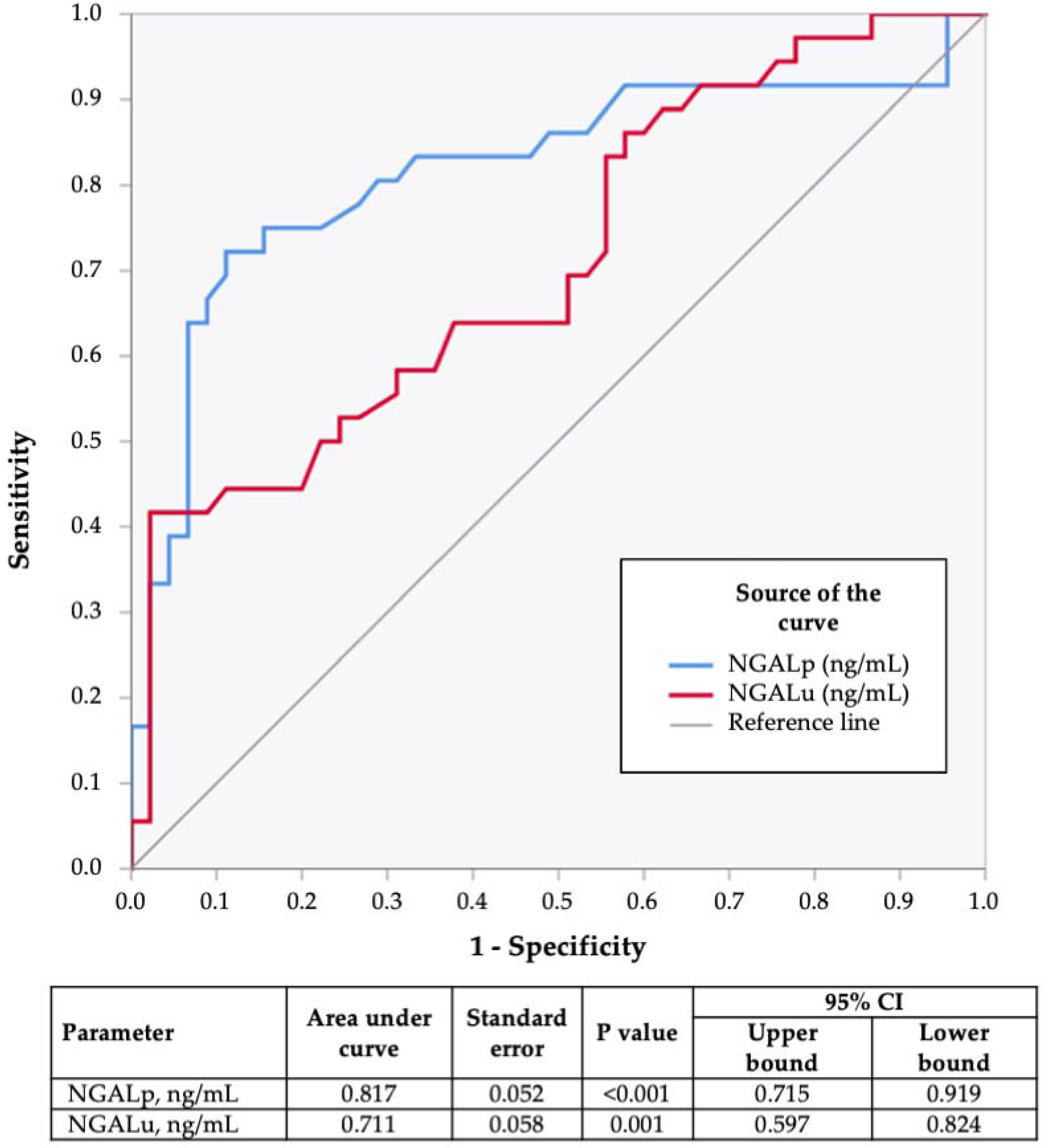
3.3. ROC Curve Analysis

At the end of the study, ROC curves were generated to assess the performance of the two circulating NGAL fractions in detecting DKD. As shown in Figure 2, both NGALp and NGALu showed an excellent discriminative capacity in the classification of patients with DKD (either early- or advanced-stage) from those still possessing good renal function. However, in consideration of the higher AUC (0.817 vs. 0.711) among the two biomarkers, it would seem that NGALp should be more reliable for the identification of T2D-induced kidney damage, at least in our routine clinical diabetes scenario.

4. Discussion

The present case–control study investigated the differential performance of plasma and urine NGAL in the detection of T2D-related chronic kidney damage, usually assessed by measuring albuminuria and the eGFR in routine clinical diabetes practice. While robust data on the diagnostic and prognostic values of NGAL in AKI are constantly coming out [17,20], the same cannot be said for CKD, as published analyses have often rendered conflicting results, and the true nature of the relationship between circulating NGAL measures and renal histopathology in DKD is still unclear. In a recent prospective cohort study on a heterogeneous, real-life patient population affected by different entities of CKD, NGALu did not correlate with the baseline eGFR and ACR or predict renal function deterioration over time [43]. As a pure tubular injury biomarker, NGALu might not be able to detect the glomerular aspects of a chronic renal disease with a low degree of accompanying active tubulointerstitial damage or inflammation [43]. In support of this assumption, in the subgroups of patients with biopsy-proven active glomerulonephritis or vasculitis as the primary etiology of CKD, NGALu could successfully predict eGFR loss and anticipate the prognosis of the disease [43,44].
In the course of DKD, morphological changes can be found in almost all nephron structures, including the glomerulus (i.e., thickening and denuding of the glomerular basement membrane, mesangial volume expansion, glomerulosclerosis), renal tubules (i.e., thickening and wrinkling of tubular basement membranes, tubular atrophy) and interstitium (i.e., interstitial widening with fibrosis and mild inflammation, arteriolosclerosis) [45], so the use of a multiple-test strategy, based on both glomerular (i.e., ACR, serum creatinine, cystatin C) and tubulointerstitial damage biomarkers, might be preferable for a timely, non-invasive, diagnosis of this condition [46]. However, as per standard NKF- KDOQI definitions, DKD is detected based on albuminuria (of at least the A2 category) and/or reduced eGFR [29], and markers of tubular damage are currently not a part of routine assessment.
Cross-sectional data from the National Health and Nutrition Examination Survey (NHANES) have indicated that prevalence of DKD among diabetic people in the U.S. has remained stable during the last three decades, though the prevalence of an impaired eGFR (defined as an eGFR <60 mL/min/1.73 m2) has increased, whereas that of persistent albuminuria has decreased [47]. These opposite trends in the two main DKD manifestations may reflect changes that have occurred in the natural history of DKD, which, in many patients with T2D, no longer follows the classic, sequential five-stage course but has more atypical presentations [48]. In particular, in patients with T2D, eGFR loss may start well before albuminuria has developed [48], which is in line with what we observed in our cohort. Non-albuminuric DKD has even been postulated to represent a distinct phenotype of DKD, with macroangiopathy instead of microangiopathy as the prevailing underlying histopathology [48]. This would explain the weak association between diabetic retinopathy and early-stage renal injury (marked by a modest decline in eGFR and/or mild albuminuria), which is instead common in patients with albuminuria of moderate–severe intensity, as reported in several investigations [48,49], including the present one. As evidenced in our own findings, in addition to hyperglycemia, T2D patients with DKD present other cardiovascular risk factors, including hypertension, dyslipidemia, smoking habits, overweight and aging itself, all contributing to progressive renal failure and arteriolosclerosis, though to a varying extent in each individual [48].
More than a simple potential diagnostic biomarker, in recent years, mechanistic evidence has demonstrated a direct involvement of NGAL in renal protective mechanisms against active tubular injury [50,51]. This notion has prompted us to hypothesize that the specific increase in NGALu levels observed in our patients with DKD are probably due to a greater release from kidney epithelial cells and might represent an adaptive mechanism against tubulointerstitial stress induced by hyperglycemia and/or defective renal oxygenation, which is common in T2D [52]. Indeed, based on our results, urine NGAL increases in T2D seem to be more closely related to a greater in situ production by the injured proximal tubular epithelial cells and might be a less sensitive index of the overall kidney function (including glomerular filtering ability) than plasma NGAL. Furthermore, being closely linked with a chronic, low-grade systemic proinflammatory state [53], T2D is supposed to induce increased NGAL production in extra-renal tissues [42], including the arterial wall and the vascular system [54]. NGALp levels are considerably higher in diabetic patients at high risk for acute cardiovascular events with respect to non-diabetic individuals, and this may reflect the burden of endothelial dysfunction, correlating with locally produced NGAL within atherosclerotic plaques by infiltrating leukocytes [54]. The progressive increase in circulating NGAL observed in the late course of T2D and throughout the stages of DKD may indicate a generalized vascular damage of progressively increased severity, and thus a higher risk of target organ complications. Beside a greater systemic atherosclerotic burden, however, we cannot exclude the possibility that NGALp increases at a later stage of diabetic disease could be due to impaired renal clearance of circulating NGAL, caused by a decline in the eGFR [55]. Indeed, strong negative correlations between NGALp and the eGFR, and validated endogenous markers of renal clearance (serum creatinine, cystatin C), have been evidenced in our results.
To the best of our knowledge, this is the first report of its kind including a healthy control group and clinically well-characterized, real-life, ambulatory patients progressing throughout the different stages of T2D and DKD courses. Possible confounding factors in statistical analysis and interpretation of results have been avoided, since patients using antidiabetic drugs directly affecting the nephron proximal tubule function (i.e., SGLT-2 inhibitors [31,52,56]) or the renal production of NGAL in response to toxic stimuli (i.e., pioglitazone [32]) have been excluded. Additionally, patients with severe comorbidities affecting available measures of renal clearance (i.e., serum creatinine, cystatin C, eGFR) have been prevented from participating in this study. However, our work is not void of limitations, which are mostly related to the relatively low number of participants, the cross-sectional setting and the lack of biopsy data for histopathological correlations, precluding any firm conclusions from being drawn. Larger studies are warranted to further corroborate our findings.

5. Conclusions

Our data suggest a different pathophysiological and diagnostic role for urine and plasma NGAL in the context of T2D and DKD: NGALu may be a more specific marker of active renal tubular epithelial damage and tubulointerstitial inflammation, whereas NGALp may be more indicative of the renal (and possibly also extra-renal) vasculature state, including glomerular filtration ability.

Author Contributions

Conceptualization, M.G., D.P.F. and A.B.; investigation, V.T. and P.C.; data curation, M.G., E.C. and M.M.; writing—original draft preparation, M.G. and M.M.; writing—review and editing, E.C., A.S., D.P.F. and A.B.; project administration, D.P.F. and A.B. All authors have read and agreed to the published version of the manuscript.

Funding

This publication is co-financed with the support of the European Commission, FESR FSE 2014–2020 and Regione Calabria. The European Commission and Regione Calabria’s supports for the production of this publication do not constitute an endorsement of the contents, which reflect the views of the authors only, and cannot be held responsible for any use which may be made of the information contained therein.

Institutional Review Board Statement

The study was conducted in accordance with the Declaration of Helsinki and approved by the Ethics Committee of Regione Calabria, Sezione Area Centro, Catanzaro, Italy (protocol code n. 116, 14 May 2015).

Informed Consent Statement

Informed consent was obtained from all subjects involved in the study.

Data Availability Statement

Data supporting the reported results are available from the corresponding author upon reasonable request.

Conflicts of Interest

The authors declare no conflict of interest.

References

  1. GBD Chronic Kidney Disease Collaboration. Global, regional, and national burden of chronic kidney disease, 1990–2017: A systematic analysis for the Global Burden of Disease Study 2017. Lancet 2020, 395, 709–733. [Google Scholar] [CrossRef] [Green Version]
  2. Khan, M.A.B.; Hashim, M.J.; King, J.K.; Govender, R.D.; Mustafa, H.; Al Kaabi, J. Epidemiology of Type 2 Diabetes—Global Burden of Disease and Forecasted Trends. J. Epidemiol. Glob. Health 2020, 10, 107–111. [Google Scholar] [CrossRef] [PubMed] [Green Version]
  3. National Institute of Diabetes and Digestive and Kidney Diseases. Kidney Disease Statistics for the United States. Available online: https://www.niddk.nih.gov/health-information/health-statistics/kidney-disease (accessed on 25 February 2022).
  4. Wu, T.; McGrath, K.C.; Death, A.K. Cardiovascular disease in diabetic nephropathy patients: Cell adhesion molecules as potential markers? Vasc. Health Risk Manag. 2005, 1, 309–316. [Google Scholar] [CrossRef] [PubMed]
  5. Younes, N.; Cleary, P.A.; Steffes, M.W.; de Boer, I.H.; Molitch, M.E.; Rutledge, B.N.; Lachin, J.M.; Dahms, W.; DCCT/EDIC Research Group. Comparison of urinary albumin-creatinine ratio and albumin excretion rate in the Diabetes Control and Complications Trial/Epidemiology of Diabetes Interventions and Complications study. Clin. J. Am. Soc. Nephrol. 2010, 5, 1235–1242. [Google Scholar] [CrossRef] [PubMed] [Green Version]
  6. Klimontov, V.V.; Korbut, A.I. Albuminuric and non-albuminuric patterns of chronic kidney disease in type 2 diabetes. Diabetes Metab. Syndr. 2019, 13, 474–479. [Google Scholar] [CrossRef]
  7. Rigalleau, V.; Lasseur, C.; Raffaitin, C.; Beauvieux, M.C.; Barthe, N.; Chauveau, P.; Combe, C.; Gin, H. Normoalbuminuric renal-insufficient diabetic patients: A lower-risk group. Diabetes Care 2007, 30, 2034–2039. [Google Scholar] [CrossRef] [Green Version]
  8. Bonventre, J.V. Can we target tubular damage to prevent renal function decline in diabetes? Semin. Nephrol. 2012, 32, 452–462. [Google Scholar] [CrossRef] [Green Version]
  9. Slyne, J.; Slattery, C.; McMorrow, T.; Ryan, M.P. New developments concerning the proximal tubule in diabetic nephropathy: In Vitro models and mechanisms. Nephrol. Dial. Transplant. 2015, 30 (Suppl. 4), iv60–iv67. [Google Scholar] [CrossRef] [Green Version]
  10. Kjeldsen, L.; Johnsen, A.H.; Sengeløv, H.; Borregaard, N. Isolation and primary structure of NGAL, a novel protein associated with human neutrophil gelatinase. J. Biol. Chem. 1993, 268, 10425–10432. [Google Scholar] [CrossRef]
  11. Goetz, D.H.; Holmes, M.A.; Borregaard, N.; Bluhm, M.E.; Raymond, K.N.; Strong, R.K. The neutrophil lipocalin NGAL is a bacteriostatic agent that interferes with siderophore-mediated iron acquisition. Mol. Cell 2002, 10, 1033–1043. [Google Scholar] [CrossRef]
  12. Kjeldsen, L.; Bainton, D.F.; Sengeløv, H.; Borregaard, N. Identification of neutrophil gelatinase-associated lipocalin as a novel matrix protein of specific granules in human neutrophils. Blood 1994, 83, 799–807. [Google Scholar] [CrossRef] [PubMed] [Green Version]
  13. Chakraborty, S.; Kaur, S.; Guha, S.; Batra, S.K. The multifaceted roles of neutrophil gelatinase associated lipocalin (NGAL) in inflammation and cancer. Biochim. Biophys. Acta 2012, 1826, 129–169. [Google Scholar] [CrossRef] [PubMed] [Green Version]
  14. Mishra, J.; Ma, Q.; Prada, A.; Mitsnefes, M.; Zahedi, K.; Yang, J.; Barasch, J.; Devarajan, P. Identification of neutrophil gelatinase-associated lipocalin as a novel early urinary biomarker for ischemic renal injury. J. Am. Soc. Nephrol. 2003, 14, 2534–2543. [Google Scholar] [CrossRef] [Green Version]
  15. Mori, K.; Lee, H.T.; Rapoport, D.; Drexler, I.R.; Foster, K.; Yang, J.; Schmidt-Ott, K.M.; Chen, X.; Li, J.Y.; Weiss, S.; et al. Endocytic delivery of lipocalin-siderophore-iron complex rescues the kidney from ischemia-reperfusion injury. J. Clin. Investig. 2005, 115, 610–621. [Google Scholar] [CrossRef] [PubMed]
  16. Clerico, A.; Galli, C.; Fortunato, A.; Ronco, C. Neutrophil gelatinase-associated lipocalin (NGAL) as biomarker of acute kidney injury: A review of the laboratory characteristics and clinical evidences. Clin. Chem. Lab. Med. 2012, 50, 1505–1517. [Google Scholar] [CrossRef] [PubMed] [Green Version]
  17. Leditzke, K.; Wagner, M.E.H.; Neunaber, C.; Clausen, J.D.; Winkelmann, M. Neutrophil Gelatinase-associated Lipocalin Predicts Post-traumatic Acute Kidney Injury in Severely Injured Patients. In Vivo 2021, 35, 2755–2762. [Google Scholar] [CrossRef]
  18. Ding, H.; He, Y.; Li, K.; Yang, J.; Li, X.; Lu, R.; Gao, W. Urinary neutrophil gelatinase-associated lipocalin (NGAL) is an early biomarker for renal tubulointerstitial injury in IgA nephropathy. Clin. Immunol. 2007, 123, 227–234. [Google Scholar] [CrossRef]
  19. Parravicini, E. The clinical utility of urinary neutrophil gelatinase-associated lipocalin in the neonatal ICU. Curr. Opin. Pediatr. 2010, 22, 146–150. [Google Scholar] [CrossRef]
  20. Shang, W.; Wang, Z. The Update of NGAL in Acute Kidney Injury. Curr. Protein Pept. Sci. 2017, 18, 1211–1217. [Google Scholar] [CrossRef]
  21. Bolignano, D.; Lacquaniti, A.; Coppolino, G.; Donato, V.; Campo, S.; Fazio, M.R.; Nicocia, G.; Buemi, M. Neutrophil gelatinase-associated lipocalin (NGAL) and progression of chronic kidney disease. Clin. J. Am. Soc. Nephrol. 2009, 4, 337–344. [Google Scholar] [CrossRef] [Green Version]
  22. Hasegawa, M.; Ishii, J.; Kitagawa, F.; Takahashi, H.; Sugiyama, K.; Tada, M.; Kanayama, K.; Takahashi, K.; Hayashi, H.; Koide, S.; et al. Plasma Neutrophil Gelatinase-Associated Lipocalin as a Predictor of Cardiovascular Events in Patients with Chronic Kidney Disease. Biomed. Res. Int. 2016, 2016, 8761475. [Google Scholar] [CrossRef] [PubMed] [Green Version]
  23. Wang, Y.; Lam, K.S.; Kraegen, E.W.; Sweeney, G.; Zhang, J.; Tso, A.W.; Chow, W.S.; Wat, N.M.; Xu, J.Y.; Hoo, R.L.; et al. Lipocalin-2 is an inflammatory marker closely associated with obesity, insulin resistance, and hyperglycemia in humans. Clin. Chem. 2007, 53, 34–41. [Google Scholar] [CrossRef] [PubMed] [Green Version]
  24. Helanova, K.; Spinar, J.; Parenica, J. Diagnostic and prognostic utility of neutrophil gelatinase-associated lipocalin (NGAL) in patients with cardiovascular diseases-review. Kidney Blood Press. Res. 2014, 39, 623–629. [Google Scholar] [CrossRef]
  25. Candido, S.; Di Maso, M.; Serraino, D.; McCubrey, J.A.; Bortolus, R.; Zanin, M.; Battiston, M.; Salemi, R.; Libra, M.; Polesel, J. Diagnostic value of neutrophil gelatinase-associated lipocalin/matrix metalloproteinase-9 pathway in transitional cell carcinoma of the bladder. Tumor Biol. 2016, 37, 9855–9863. [Google Scholar] [CrossRef]
  26. Żyłka, A.; Dumnicka, P.; Kuśnierz-Cabala, B.; Gala-Błądzińska, A.; Ceranowicz, P.; Kucharz, J.; Ząbek-Adamska, A.; Maziarz, B.; Drożdż, R.; Kuźniewski, M. Markers of Glomerular and Tubular Damage in the Early Stage of Kidney Disease in Type 2 Diabetic Patients. Mediat. Inflamm. 2018, 2018, 7659243. [Google Scholar] [CrossRef] [PubMed] [Green Version]
  27. Tang, S.C.; Lai, K.N. The pathogenic role of the renal proximal tubular cell in diabetic nephropathy. Nephrol. Dial. Transplant. 2012, 27, 3049–3056. [Google Scholar] [CrossRef] [Green Version]
  28. Tuttle, K.R.; Bakris, G.L.; Bilous, R.W.; Chiang, J.L.; de Boer, I.H.; Goldstein-Fuchs, J.; Hirsch, I.B.; Kalantar-Zadeh, K.; Narva, A.S.; Navaneethan, S.D.; et al. Diabetic kidney disease: A report from an ADA Consensus Conference. Diabetes Care 2014, 37, 2864–2883. [Google Scholar] [CrossRef] [Green Version]
  29. National Kidney Foundation. KDOQI Clinical Practice Guideline for Diabetes and CKD: 2012 Update. Am. J. Kidney Dis. 2012, 60, 850–886, Erratum in Am. J. Kidney Dis. 2013, 61, 1049. [Google Scholar] [CrossRef]
  30. American Diabetes Association. 11. Microvascular Complications and Foot Care: Standards of Medical Care in Diabetes-2020. Diabetes Care 2020, 43, S135–S151. [Google Scholar] [CrossRef] [Green Version]
  31. Darawshi, S.; Yaseen, H.; Gorelik, Y.; Faor, C.; Szalat, A.; Abassi, Z.; Heyman, S.N.; Khamaisi, M. Biomarker evidence for distal tubular damage but cortical sparing in hospitalized diabetic patients with acute kidney injury (AKI) while on SGLT2 inhibitors. Ren. Fail. 2020, 42, 836–844. [Google Scholar] [CrossRef]
  32. Chen, W.; Xi, X.; Zhang, S.; Zou, C.; Kuang, R.; Ye, Z.; Huang, Y.; Hu, H. Pioglitazone Protects Against Renal Ischemia-Reperfusion Injury via the AMP-Activated Protein Kinase-Regulated Autophagy Pathway. Front. Pharmacol. 2018, 9, 851. [Google Scholar] [CrossRef] [PubMed] [Green Version]
  33. Greco, M.; Foti, D.P.; Aversa, A.; Fuiano, G.; Brunetti, A.; Simeoni, M. Cystatin C, a Controversial Biomarker in Hypothyroid Patients under Levothyroxine Therapy: THYRenal, a Pilot Cohort Observational Study. J. Clin. Med. 2020, 9, 2958. [Google Scholar] [CrossRef] [PubMed]
  34. Kittiskulnam, P.; Tiskajornsiri, K.; Katavetin, P.; Chaiwatanarat, T.; Eiam-Ong, S.; Praditpornsilpa, K. The failure of glomerular filtration rate estimating equations among obese population. PLoS ONE 2020, 15, e0242447. [Google Scholar] [CrossRef] [PubMed]
  35. Crocerossa, F.; Fiori, C.; Capitanio, U.; Minervini, A.; Carbonara, U.; Pandolfo, S.D.; Loizzo, D.; Eun, D.D.; Larcher, A.; Mari, A.; et al. Estimated Glomerular Filtration Rate Decline at 1 Year after Minimally Invasive Partial Nephrectomy: A Multimodel Comparison of Predictors. Eur. Urol. Open Sci. 2022, 38, 52–59. [Google Scholar] [CrossRef]
  36. Provenzano, M.; Serra, R.; Michael, A.; Bolignano, D.; Coppolino, G.; Ielapi, N.; Serraino, G.F.; Mastroroberto, P.; Locatelli, F.; De Nicola, L.; et al. Smoking habit as a risk amplifier in chronic kidney disease patients. Sci. Rep. 2021, 11, 14778. [Google Scholar] [CrossRef] [PubMed]
  37. Brunetti, A.; Aversa, A. Editorial overview: ‘Caring for diabetes in its complexity: From targetable metabolic-organ crosstalk to novel drug interactions’. Curr. Opin. Pharmacol. 2022, 63, 102185. [Google Scholar] [CrossRef]
  38. Carrero, J.J.; Hecking, M.; Chesnaye, N.C.; Jager, K.J. Sex and gender disparities in the epidemiology and outcomes of chronic kidney disease. Nat. Rev. Nephrol. 2018, 14, 151–164. [Google Scholar] [CrossRef]
  39. Greco, M.; Chiefari, E.; Accattato, F.; Corigliano, D.M.; Arcidiacono, B.; Mirabelli, M.; Liguori, R.; Brunetti, F.S.; Pullano, S.A.; Scorcia, V.; et al. MicroRNA-1281 as a Novel Circulating Biomarker in Patients With Diabetic Retinopathy. Front. Endocrinol. 2020, 11, 528. [Google Scholar] [CrossRef]
  40. Sheen, Y.J.; Sheu, W.H. Risks of rapid decline renal function in patients with type 2 diabetes. World J. Diabetes 2014, 5, 835–846. [Google Scholar] [CrossRef]
  41. Iuliano, S.; Greco, E.A.; Mirabelli, M.; Chiefari, E.; Caroleo, P.; Puccio, L.; Giuliano, S.; Foti, D.P.; Brunetti, A.; Aversa, A. Predicting the response to SGLT-2 inhibitors as add-on therapy to multiple day injection insulin with glycated albumin: A pilot study. Minerva Endocrinol. 2022; Epub ahead of printing. [Google Scholar] [CrossRef]
  42. Aslanhan, E.; Ojalvo, D.; Özsenel, E.B.; Ucak Basat, S.; Borlu, F. Association of neutrophil-gelatinase-associated lipocalin with microvascular complications in patients with type 2 diabetes: A cross-sectional study. Cardiovasc. Endocrinol. Metab. 2019, 8, 82–87. [Google Scholar] [CrossRef] [PubMed]
  43. Seibert, F.S.; Sitz, M.; Passfall, J.; Haesner, M.; Laschinski, P.; Buhl, M.; Bauer, F.; Babel, N.; Pagonas, N.; Westhoff, T.H. Prognostic Value of Urinary Calprotectin, NGAL and KIM-1 in Chronic Kidney Disease. Kidney Blood Press. Res. 2018, 43, 1255–1262. [Google Scholar] [CrossRef] [PubMed]
  44. Coppolino, G.; Comi, N.; Bolignano, D.; Patella, G.; Comi, A.; Provenzano, M.; Rivoli, L.; Andreucci, M.; Fuiano, G. Urinary Neutrophil Gelatinase-Associated Lipocalin (NGAL) Predicts Renal Function Decline in Patients with Glomerular Diseases. Front. Cell Dev. Biol. 2020, 8, 336. [Google Scholar] [CrossRef]
  45. Alicic, R.Z.; Rooney, M.T.; Tuttle, K.R. Diabetic Kidney Disease: Challenges, Progress, and Possibilities. Clin. J. Am. Soc. Nephrol. 2017, 12, 2032–2045. [Google Scholar] [CrossRef] [PubMed]
  46. Abbasi, F.; Moosaie, F.; Khaloo, P.; Dehghani Firouzabadi, F.; Fatemi Abhari, S.M.; Atainia, B.; Ardeshir, M.; Nakhjavani, M.; Esteghamati, A. Neutrophil Gelatinase-Associated Lipocalin and Retinol-Binding Protein-4 as Biomarkers for Diabetic Kidney Disease. Kidney Blood Press. Res. 2020, 45, 222–232. [Google Scholar] [CrossRef] [PubMed]
  47. de Boer, I.H.; Rue, T.C.; Hall, Y.N.; Heagerty, P.J.; Weiss, N.S.; Himmelfarb, J. Temporal trends in the prevalence of diabetic kidney disease in the United States. JAMA 2011, 305, 2532–2539. [Google Scholar] [CrossRef] [PubMed]
  48. Penno, G.; Solini, A.; Orsi, E.; Bonora, E.; Fondelli, C.; Trevisan, R.; Vedovato, M.; Cavalot, F.; Lamacchia, O.; Scardapane, M.; et al. Non-albuminuric renal impairment is a strong predictor of mortality in individuals with type 2 diabetes: The Renal Insufficiency and Cardiovascular Events (RIACE) Italian multicentre study. Diabetologia 2018, 61, 2277–2289. [Google Scholar] [CrossRef] [Green Version]
  49. Park, H.C.; Lee, Y.K.; Cho, A.; Han, C.H.; Noh, J.W.; Shin, Y.J.; Bae, S.H.; Kim, H. Diabetic retinopathy is a prognostic factor for progression of chronic kidney disease in the patients with type 2 diabetes mellitus. PLoS ONE 2019, 14, e0220506. [Google Scholar] [CrossRef] [Green Version]
  50. Han, M.; Li, Y.; Wen, D.; Liu, M.; Ma, Y.; Cong, B. NGAL protects against endotoxin-induced renal tubular cell damage by suppressing apoptosis. BMC Nephrol. 2018, 19, 168. [Google Scholar] [CrossRef]
  51. Liu, X.; Zhao, X.; Duan, X.; Wang, X.; Wang, T.; Feng, S.; Zhang, H.; Chen, C.; Li, G. Knockout of NGAL aggravates tubulointerstitial injury in a mouse model of diabetic nephropathy by enhancing oxidative stress and fibrosis. Exp. Ther. Med. 2021, 21, 321. [Google Scholar] [CrossRef]
  52. Packer, M. Mechanisms Leading to Differential Hypoxia-Inducible Factor Signaling in the Diabetic Kidney: Modulation by SGLT2 Inhibitors and Hypoxia Mimetics. Am. J. Kidney Dis. 2021, 77, 280–286. [Google Scholar] [CrossRef] [PubMed]
  53. Arcidiacono, B.; Chiefari, E.; Foryst-Ludwig, A.; Currò, G.; Navarra, G.; Brunetti, F.S.; Mirabelli, M.; Corigliano, D.M.; Kintscher, U.; Britti, D.; et al. Obesity-related hypoxia via miR-128 decreases insulin-receptor expression in human and mouse adipose tissue promoting systemic insulin resistance. EBioMedicine 2020, 59, 102912. [Google Scholar] [CrossRef] [PubMed]
  54. Eilenberg, W.; Stojkovic, S.; Piechota-Polanczyk, A.; Kaider, A.; Kozakowski, N.; Weninger, W.J.; Nanobachvili, J.; Wojta, J.; Huk, I.; Demyanets, S.; et al. Neutrophil gelatinase associated lipocalin (NGAL) is elevated in type 2 diabetics with carotid artery stenosis and reduced under metformin treatment. Cardiovasc. Diabetol. 2017, 16, 98. [Google Scholar] [CrossRef] [PubMed]
  55. McIlroy, D.R.; Wagener, G.; Lee, H.T. Neutrophil gelatinase-associated lipocalin and acute kidney injury after cardiac surgery: The effect of baseline renal function on diagnostic performance. Clin. J. Am. Soc. Nephrol. 2010, 5, 211–219. [Google Scholar] [CrossRef] [PubMed] [Green Version]
  56. Mirabelli, M.; Chiefari, E.; Caroleo, P.; Vero, R.; Brunetti, F.S.; Corigliano, D.M.; Arcidiacono, B.; Foti, D.P.; Puccio, L.; Brunetti, A. Long-Term Effectiveness and Safety of SGLT-2 Inhibitors in an Italian Cohort of Patients with Type 2 Diabetes Mellitus. J. Diabetes Res. 2019, 2019, 3971060. [Google Scholar] [CrossRef] [PubMed] [Green Version]
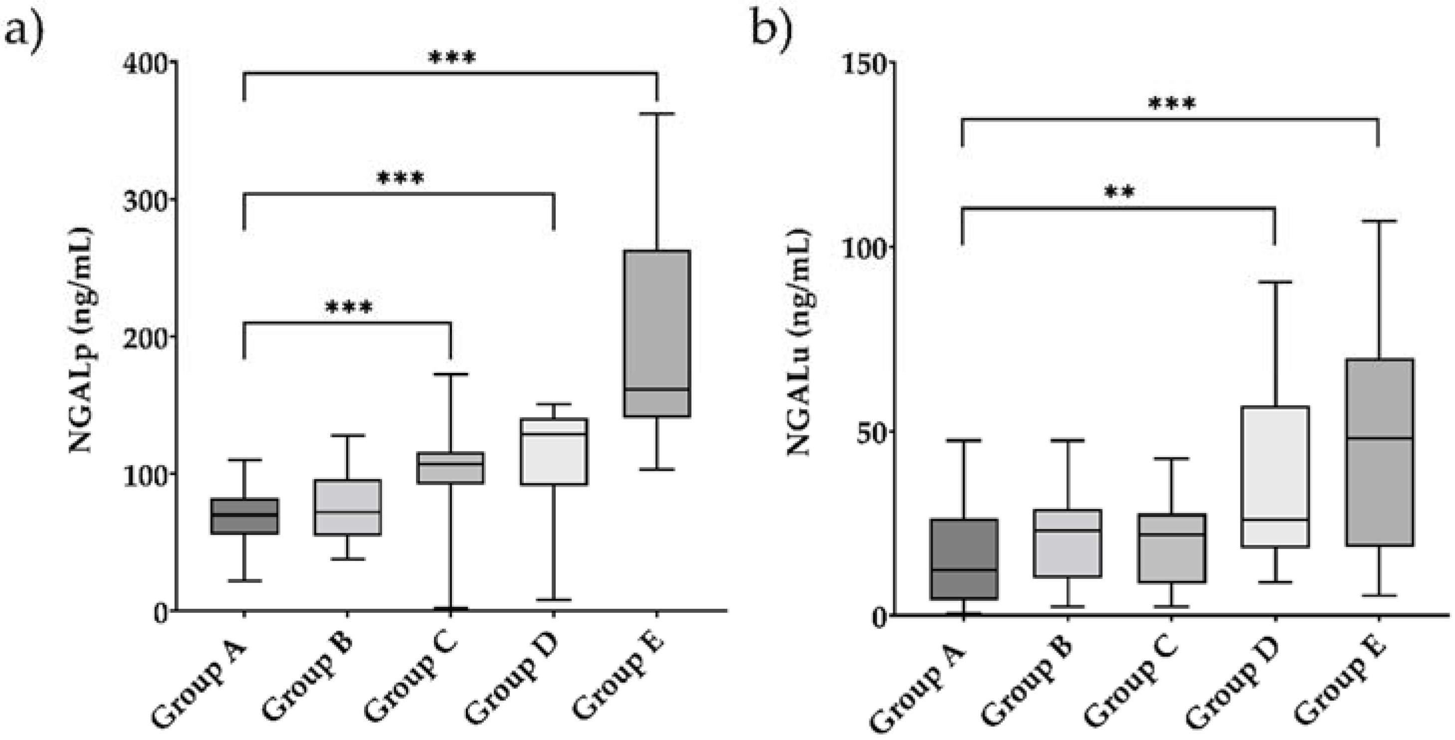
Figure 1. Plasma NGAL (a) and urine NGAL (b) levels in study population. ** p < 0.05; *** p < 0.001.
Endocrines 03 00016 g001
Figure 2. Receiver operating characteristic (ROC) analysis showing the performance of NGALp and NGALu for the detection of DKD.
Endocrines 03 00016 g002
Table 1. Characteristics of the study population according to the presence or absence of DKD, DKD severity and duration of T2D.
CharacteristicsGroup A
Healthy Controls
(N = 21)
Group B
Newly Diagnosed Diabetic Patients without DKD
(N = 27)
Group C
Long-Standing Diabetic Patients without DKD
(N = 21)
Group D
Diabetic Patients with Early-Stage DKD
(N = 17)
Group E
Diabetic Patients with Advanced-Stage
DKD
(N = 19)
A vs. BA vs. CA vs. DA vs. EB vs. CB vs. DB vs. EC vs. DC vs. ED vs. E
Age, years53
(49–55)
57
(51–60)
65
(58–71)
70
(61–75)
67
(60–70)
0.054<0.001<0.001<0.001<0.001<0.001<0.0010.3830.9500.220
EthnicityCaucasianCaucasianCaucasianCaucasianCaucasian__________
Female gender, N14 (66.7%)10 (37%)3 (14.2%)11 (64.7%)2 (10.5%)0.0490.0011.00<0.0010.1070.1210.0850.0021.000.001
BMI, kg/m224.8
(23.3–28.0)
27.3
(25.0–30.8)
25.9
(24.0–28.9)
26.8
(25–31.6)
28.6
(25.4–31.2)
0.1000.3580.0780.0140.1941.000.4540.3080.0920.601
T2D duration, years_1
(0.5–3)
10
(7–20)
6
(2.5–14.0)
16
(13–31)
____<0.001<0.001<0.0010.1900.045<0.001
Hypertension, N0 (0%)9 (33.3%)16 (76.2%)11 (64.7%)19 (100%)0.002<0.001<0.001<0.0010.0040.063<0.0010.4900.0480.006
Dyslipidemia, N0 (0%)7 (25.9%)15 (71.4%)12 (70.6%)15 (78.9%)0.013<0.001<0.001<0.0010.0030.0050.1261.000.7210.706
Smoking StatusCurrent-smoker, N0 (0%)0 (0%)1 (4.8%)1 (5.9%)7 (36.8%)__________
Ex-smoker, N0 (0%)0 (0%)0 (0%)0 (0%)4 (21.1%)__________
Diabetic Retinopathy, N_0 (0%)1 (4.8%)0 (0%)18 (94.7%)____0.4371.00<0.0011.00<0.001<0.001
FBG, mg/dL89
(87–92)
130
(116–150)
137
(120–151)
151
(127–165)
150
(124–173)
<0.001<0.001<0.001<0.0010.5260.0710.0600.1790.2080.839
HbA1c, %5.2
(5.1–5.4)
6.3
(5.9–6.7)
7
(6.6–7.4)
6.5
(6.0–7.8)
7.5
(6.9–7.9)
<0.001<0.001<0.001<0.0010.0010.148<0.0010.3670.1020.069
NGALp, ng/mL70
(55–82)
72
(55–96)
107
(92–116)
129
(91–140)
164
(143–278)
0.411<0.001<0.001<0.0010.0020.001<0.0010.196<0.0010.001
NGALu, ng/mL12.2
(4.1–26.4)
23
(11.2–29.6)
22
(8.6–27.6)
27.5
(18.8–66.3)
60
(19–79)
0.0930.2700.001<0.0010.5570.0560.0180.0220.0090.616
Serum creatinine, mg/dL0.8
(0.65–0.85)
0.8
(0.7–0.9)
0.9
(0.8–1.0)
0.94
(0.70–1.09)
1.8
(1.2–2.5)
0.258<0.0010.068<0.0010.1410.126<0.0010.706<0.001<0.001
Cystatin C, mg/dL0.76
(0.72–0.81)
0.8
(0.71–0.95)
0.92
(0.81–1.0)
1.2
(1.0–1.37)
1.83
(1.4–2.3)
0.1510.285<0.001<0.0010.017<0.001<0.0010.001<0.0010.001
eGFR, ml/min/1.73 m2102
(93–108)
95.9
(83.3–105.0)
87.3
(73.6–99.9)
66.5
(57.1–70.2)
38.3
(24.2–52.8)
0.254<0.001<0.001<0.0010.043<0.001<0.001<0.001<0.0010.001
ACR, mg/g creatinine<10.7 * 10.7 *
(11–12)
10.7 *
(11–11)
41
(30.7–257)
520
(400–1360)
0.2701.000.026<0.0010.9710.006<0.0010.006<0.001<0.001
Data are presented as median (IQR) or N (%). p values are calculated using the non-parametric Mann–Whitney U test or Fisher’s exact test, as appropriate. * Urine albumin was below detection and quantification limits in most samples. DKD, diabetic kidney disease; T2D, type 2 diabetes; BMI, body mass index; FBG, fasting blood glucose; HbA1c, glycated hemoglobin; NGALp, plasma neutrophil gelatinase-associated lipocalin; NGALu, urine neutrophil gelatinase-associated lipocalin; eGFR, estimated glomerular filtration rate; ACR, albumin/creatinine ratio. Bold numbers denote statistical significance at the p < 0.05 level.
Table 2. Spearman’s correlation analysis of plasma and urine NGAL with DKD indices and glycometabolic parameters in patients with T2D (N = 84).
ParameterNGALu Serum Creatinine Cystatin C ACRFBGHbA1cBMIT2D DurationAge
NGALp (ng/mL)Spearman’s ρ0.2710.5210.6960.6000.1840.261−0.0270.5810.419
p value0.016<0.001<0.001<0.0010.0980.0190.810<0.001<0.001
NGALu (ng/mL)Spearman’s ρ_0.2340.3100.3620.0270.2670.0350.1410.097
p value_0.0370.0050.0020.8120.0170.7580.2110.393
Bold values denote statistical significance at the p < 0.05 level.
Table 3. Linear regression analysis showing the associations between NGALp, NGALu and eGFR (estimated by CKD EPI formula combining serum creatinine and cystatin C) in patients with T2D (N = 84), adjusting for potential confounders.
ParameterUnstandardized B (Standard Error)Standardized βp Value
NGALp−0.194 (0.028)−0.612<0.001
NGALp *−0.160 (−0.026)−0.504<0.001
NGALp **−0.138 (−0.026)−0.446<0.001
NGALu−0.142 (0.052)−0.2970.007
NGALu *−0.100 (0.044)−0.2100.024
NGALu **−0.100 (0.039)−0.2130.012
* Age and gender were added as covariates. ** Age, gender and duration of T2D and HbA1c were added as covariates. Bold values denote statistical significance at the p < 0.05 level.
Publisher’s Note: MDPI stays neutral with regard to jurisdictional claims in published maps and institutional affiliations.

