Article

Src Cooperates with Oncogenic Ras in Tumourigenesis via the JNK and PI3K Pathways in Drosophila epithelial Tissue

by
Carole L.C. Poon
1,2,*,†,
Anthony M. Brumby
1,3 and
Helena E. Richardson
1,2,3,4,*
1
Cell Cycle and Development lab, Peter MacCallum Cancer Centre, Melbourne, VIC 3002, Australia
2
Department of Biochemistry and Molecular Biology, University of Melbourne, Melbourne, VIC 3010, Australia
3
Department of Anatomy and Cell Biology, University of Melbourne, Melbourne, VIC 3010, Australia
4
Department of Biochemistry and Genetics, La Trobe Institute of Molecular Science, La Trobe University, Melbourne, VIC 3086, Australia
*
Authors to whom correspondence should be addressed.
Current address: Sir Peter MacCallum Department of Oncology, University of Melbourne and Cell Growth and Proliferation laboratory, Peter MacCallum Cancer Centre, Melbourne, VIC 3010, Australia.
Int. J. Mol. Sci. 2018, 19(6), 1585; https://doi.org/10.3390/ijms19061585
Submission received: 19 April 2018 / Revised: 15 May 2018 / Accepted: 23 May 2018 / Published: 27 May 2018
(This article belongs to the Special Issue Drosophila Model and Human Disease)

Abstract

:
The Ras oncogene (Rat Sarcoma oncogene, a small GTPase) is a key driver of human cancer, however alone it is insufficient to produce malignancy, due to the induction of cell cycle arrest or senescence. In a Drosophila melanogaster genetic screen for genes that cooperate with oncogenic Ras (bearing the RasV12 mutation, or RasACT), we identified the Drosophila Src (Sarcoma virus oncogene) family non-receptor tyrosine protein kinase genes, Src42A and Src64B, as promoting increased hyperplasia in a whole epithelial tissue context in the Drosophila eye. Moreover, overexpression of Src cooperated with RasACT in epithelial cell clones to drive neoplastic tumourigenesis. We found that Src overexpression alone activated the Jun N-terminal Kinase (JNK) signalling pathway to promote actin cytoskeletal and cell polarity defects and drive apoptosis, whereas, in cooperation with RasACT, JNK led to a loss of differentiation and an invasive phenotype. Src + RasACT cooperative tumourigenesis was dependent on JNK as well as Phosphoinositide 3-Kinase (PI3K) signalling, suggesting that targeting these pathways might provide novel therapeutic opportunities in cancers dependent on Src and Ras signalling.

Graphical Abstract

1. Introduction

The Src (Sarcoma virus oncogene) family of non-receptor tyrosine protein kinases are highly conserved and comprise nine members in vertebrates: Src, Fyn (oncogene related to Src, Fgr, Yes), Yes (Yamaguchi sarcoma virus oncogene), Blk (B Lymphoid Tyrosine Kinase), Yrk (Yes-related kinase), Fgr, Hck (hemopoietic cell kinase), Lck (lymphocyte-specific protein tyrosine kinase) and Lyn (v-yes-1 Yamaguchi sarcoma viral related oncogene homolog) [1]. Of these, Src, Fyn and Yes are ubiquitously expressed in tissues and the remaining members are restricted to specific cell types [2]. Src family kinases have pleiotropic functions including intracellular signalling, actin remodelling, cell adhesion and apoptosis [1,3,4,5]. Despite extensive analysis in cell culture and mouse models, the precise role of Src kinases during tumourigenesis in vivo is yet to be clearly defined. Aberrant Src activity is strongly associated with human tumour development [6], and, in analysis of human tumour samples, increased Src activity arises from an activating mutation at the inhibitory C-terminal tyrosine residue [7]. However, other studies indicate that elevated Src activity is due to increased protein expression and increased kinase activity that enhance tyrosine phosphorylation of substrates [6,8,9,10,11,12,13,14,15,16].
Although there is a clear correlation for increased Src function in human cancer, there are discrepancies in the literature regarding the influence of overactivated Src at different stages of tumour development. Aberrant Src activation correlates with advanced cancer development and is associated with tumour characteristics, such as increased invasiveness and metastasis [6,7,15,17]. However, other studies suggest that Src may be required earlier in tumour development. Increased Src activity is observed in samples sourced from low-grade human bladder tumour samples compared with a low Src activity in high grade samples [11]. Interestingly, metastatic cell lines that possess elevated Src kinase activity are more sensitive to receptor tyrosine kinase (RTK) signalling [18], suggesting that Src may require other cooperative events.
Indeed, c-Src (cellular-Src proto-oncogene) cooperates with the epidermal growth factor (EGF) receptor (EGFR) in murine fibroblast cell lines [19], and downstream of EGFR signalling with activated (oncogenic) mutations in the Ras (Rat Sarcoma oncogene) small-GTPase [20,21]. Oncogenic Ras mutations (such as RasV12) locks Ras in the GTP-bound activated state resulting in constitutive signalling through the MAPK (Mitogen activated protein kinase) pathway [22,23]. The cooperative interaction of Src with EGFR is characterised by increased DNA synthesis and colony formation in soft agar in vitro and increased tumour incidence in vivo when c-Src- and EGFR-expressing cells are transplanted into nude mice [19]. The cooperation is also reflected in three-dimensional cell culture where overexpression of c-Src and EGFR in human epithelial cell lines results in disruption to acinar architecture and mislocalisation of polarity markers resulting in potentiation of invasion, migration and anchorage-independent growth [24]. Additionally, combinatorial treatment with inhibitors of EGFR (Gefitinib) and Src (AZD0530) in human head and neck squamous cell carcinoma cell lines show greater reduction of growth and invasion compared with treatment with a single compound [25]. With the high correlation of EGFR and Src expression in primary human colon cancer cells [18] and mammary breast tumours [13], and oncogenic Ras, RasV12, and Src in other human cancer cell lines [20,21], these observations suggest that the contributions of both EGFR-Ras and Src are important in cooperative tumourigenesis.
Interestingly, the requirement of Src in tumourigenesis appears to be context dependent. In vitro, c-Src expression alone cannot transform cells without cooperating partners [26,27,28,29], whilst. in an in vivo mouse model, c-Src expression is sufficient to initiate tumour formation [30]. The ubiquitously expressed Src family member, Yes, can activate Ras-MAPK signalling, unlike c-Src in colorectal cancer cells [31], and therefore may require alternate cooperative partners to c-Src. On the other hand, another ubiquitously-expressed Src family kinase, Fyn, is induced by Ras-MAPK signalling and required for the mesenchymal phenotype or invasive behaviour of Ras-driven breast and skin cancer cells [32,33]. These context-dependent functions of Src family members in cancer suggests that analysis of overexpressed or activated Src within a simple in vivo biological context may reveal functions of Src kinases, either alone or with a cooperating partner, that are not readily discerned using in vitro systems or in vivo knockout models. In the vinegar fly, Drosophila melanogaster, the two Src family homologues, Src42A and Src64B, are highly conserved in sequence and domain structure with vertebrate c-Src (an overall identity of 61% and 49%, respectively). Thus, in comparison to the nine Src kinases identified in vertebrates [1], Drosophila provides an opportunity to study the role of Src function in vivo as there is less complication from functional compensation from multiple Src family members such as observed in mouse knockout models [34,35]. Given the different biological responses when Src is expressed in vitro or in vivo, a whole animal model of tumourigenesis, such as in Drosophila, provides an opportunity to investigate the in vivo role of Src kinases and its effectors in the development of cancer.
Previous studies have analysed the role of the two Drosophila Src kinase family members, Src42A and Src64B, in different settings in vivo. In the Drosophila developing eye, ectopic expression of wild-type Src64B results in a disorganised (rough) eye phenotype, due to supernumerary R7 cells [36], although overexpression of wild-type Src42A does not have a discernible effect [37]. Furthermore, expression of C-terminally truncated Src42A or Src64B, rendering constitutive activation, resulted in a more pronounced phenotype [36,37]. Analysis in the Drosophila eye revealed that overexpression of Src resulted in different phenotypes dependent on expression level, with strong overexpression resulting in reduced eye size due to increased proliferation accompanied by elevated cell death [38]. However, lower levels of Src activation using a mutation in a negative regulator of Src, C-terminal Src-related kinase (Csk), resulted in tissue overgrowth [39]. Csk loss induced overproliferation in the eye epithelium, even within regions of differentiation, suggesting that cells are unable to exit the cell cycle [40,41]. Genetic analysis revealed that the Csk mutant overgrowth phenotype was suppressed by mutations in Src42A and Src64B, as well as the downstream Src kinase, Btk29A (Tec29) [40]. Furthermore, knockdown of Jun N-terminal Kinase (JNK) and the STAT92E transcription factor suppressed the Csk mutant overgrowth phenotype [40]. Furthermore, other studies have revealed that downstream of Src signalling, the impairment of the conserved Hippo negative tissue growth control and tumour suppressor pathway [42,43] is important for Src-induced tissue overgrowth [44,45].
In another context, discrete expression of activated Src42A in the embryo results in apoptosis and migration of cells away from the expression domain [46]. A migratory phenotype is also observed with Csk-deficient cells in the wing epithelium, where cells are excluded basally from the epithelia, migrating through the extracellular matrix and eventually undergoing apoptosis [47]. This phenotype was only observed at the borders between wild-type and mutant cells, and required input from E-cadherin, p120-catenin, RhoA, JNK and matrix metalloproteinases (MMP1/2) [47,48]. This effect is also observed upon Src42A overexpression along the dorsal-ventral boundary in the wing epithelium, which was dependent on JNK activation regulated by the E2 ubiquitin ligase Bendless-dUev1a [49]. The JNK pathway is also a key effector of Src in dorsal closure (the process of epithelial sheet migration to close the dorsal epidermis during embryogenesis), where activation of JNK signalling partially suppresses the dorsal open phenotype associated with Src42A Tec29 double mutant flies [50]. Thus, in Src-mediated cell migration/invasion, JNK activation is a key effector of the invasive phenotype [47,48,49,50].
We investigated the role of the Drosophila Src kinases in cooperative tumourigenesis with activated (oncogenic) Ras (RasV12 or RasACT) in the fly eye epithelium. Characterisation of Src42A and Src64B reveals both dose- and context-dependent effects. Src expression alone results in increased cell death, a loss of cell polarity and disruption to F-actin organisation, but in itself is not sufficient to promote tumour formation. Significantly, overexpression of Drosophila Src genes cooperate with RasACT in eye disc clones, resulting in neoplastic overgrowth characterised by tissue overgrowth, increased clonal tissue size, loss of differentiation, disrupted F-actin organisation and cell polarity, and invasive clonal phenotypes leading to larval lethality. This cooperation requires the contributions of the Raf as well as the Phosphoinositide 3-Kinase (PI3K) effector pathways of Ras. Src activates JNK to promote apoptosis and defects in F-actin, however, when RasACT is coexpressed, JNK pathway signalling contributes to inhibition of differentiation, clonal overgrowth and invasive phenotypes associated with Src + RasACT neoplastic overgrowth. Given the strong correlation of aberrant Src function with EGFR-Ras activation in human cancers, the finding that JNK and PI3K are critical mediators of Src–Ras cooperative tumourigenesis may provide specific targets for cancer therapy.

2. Results

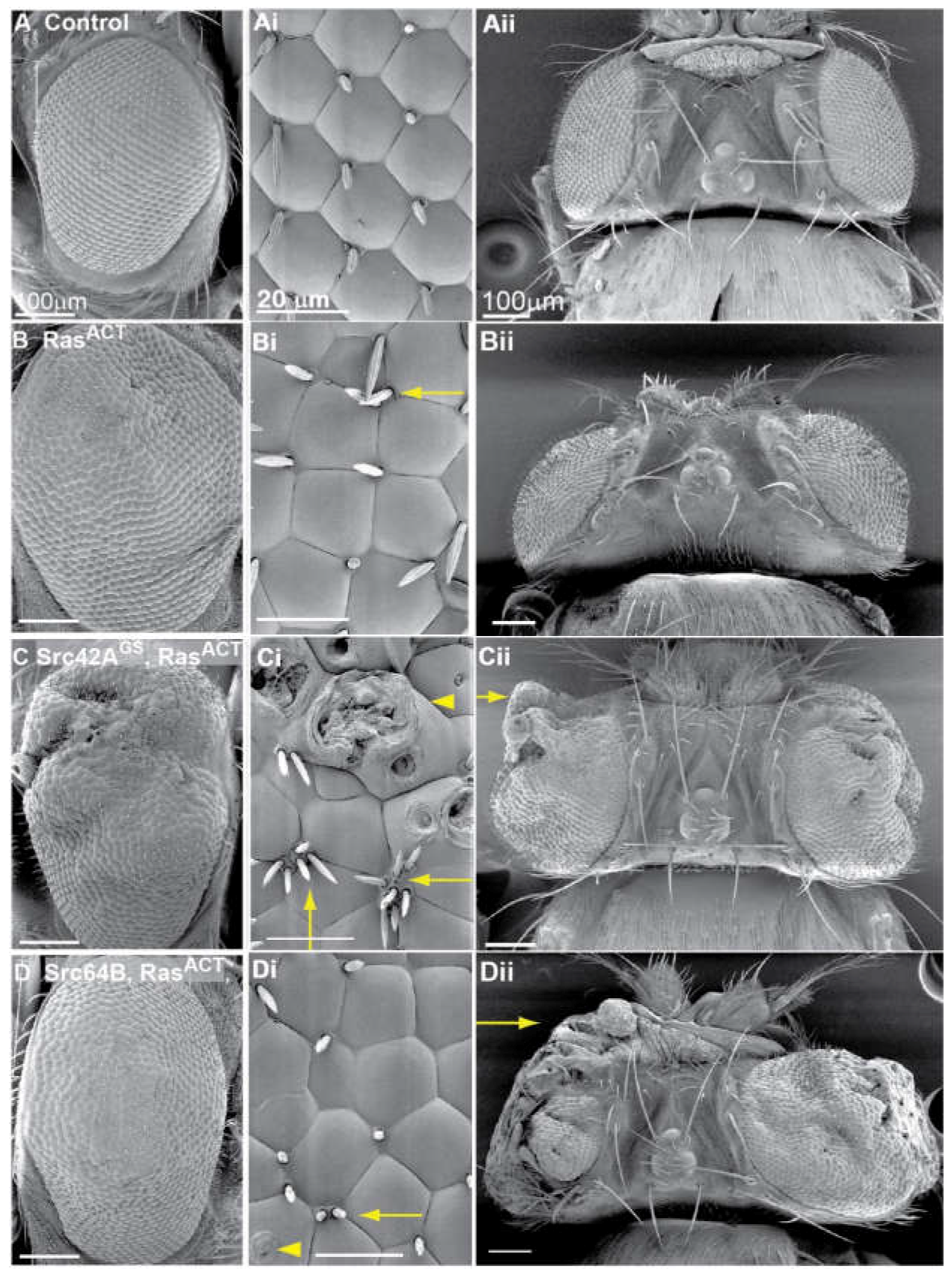
2.1. Src42A Overexpression Enhances the Eyeless-Driven RasACT Hyperplastic Eye Phenotype

In a genetic screen designed to identify novel enhancers of RasACT, a GS (Gene Search) line, GS11049, that overexpresses Src42A (Src42AGS) was identified as an enhancer of the hyperplastic eyeless (ey)-GAL4, UAS (Upsteam Activating Sequence for GAL4) -RasACT (ey > RasACT) adult eye phenotype [51]. Expression of Src42AGS with ey-GAL4 (ey > Src42AGS, Figure S1B) resulted in a normal eye phenotype relative to the control (Figure 1A and Figure S1A, and Table 1). ey > RasACT expression resulted in a mild hyperplastic rough eye phenotype (Figure 1B and Figure S1K). This phenotype was also characterised by disruption to the ommatidial array, ectopic bristles and misshapen ommaditia (Figure 1Bi). In contrast, expression of Src42AGS with ey > RasACT enhanced the hyperplastic RasACT eye phenotype resulting in overgrowth of the adult eye (Figure 1C and Figure S1L, and Table 1). Ultrastructural analysis by scanning electron micrographs showed morphological defects in ommatidia organisation, ectopic bristles within one vertex and blistering of ommatidia, most likely due to cone cell defects (Figure 1Ci,Cii). These effects were more severe than observed for ey > RasACT alone (Figure 1B). The dorsal view (Figure 1Cii) showed enhanced outgrowth of ey > RasACT + Src42AGS. The resulting Src42AGS + RasACT overgrown phenotype was more pronounced in male flies than females.
To validate the interaction identified in the screen, an independent UAS line expressing wild-type Src42A was tested. ey-GAL4-driven expression of Src42A alone did not appreciably affect the adult eye (Figure S1C and Table 1), consistent with the ey > Src42AGS phenotype and previous observations [37]. However, unlike Src42AGS, coexpression of Src42A with ey > RasACT did not enhance the hyperplastic RasACT eye phenotype (Figure S1M and Table 1). Expression of a second wild-type Src42A line, Src42AEY08937 (containing a P element insertion, EPgy2 [52]) with ey > RasACT also did not enhance the hyperplastic RasACT eye phenotype (data not shown). Since previous studies in Drosophila have revealed a dosage-dependent response to Src expression [37,38,39], it is possible that positional effects of the UAS or EYgy2 integration site [52,53] may affect expression levels Src, which could explain why they did not enhance the ey > RasACT phenotype. To further analyse the effects of overexpression of Src42AGS and the independent UAS-Src42A line, an alternative driver was adopted. GMR (Glass Multimer Reporter)-GAL4 drives expression in differentiating cells beginning in the morphogenetic furrow [54,55] in comparison with ey-GAL4, which drives expression from stage 15 during embryogenesis and predominates in cycling cells during third instar larval stage [56,57]. GMR > Src42AGS adult eyes were glazed with loss of pigmentation in the central ommatidia (Figure S2B). In contrast, GMR > Src42A adult eyes had a small, glassy stripe in the posterior region of a rough adult eye (Figure S2C). The glassy phenotype was less severe in GMR > Src42A than GMR > Src42AGS adult eyes, suggesting that Src42A may not be as potent as Src42AGS in activation of Src signalling.
To examine Src activity and protein expression levels in the wild-type Src42A lines, Western blot analysis was carried out on equally loaded amount of protein lysates from third instar larval heads expressing hsp70 (heat-shock inducible promoter)-GAL4 induced Src42AGS or the UAS-Src42A transgene (lanes c and d, respectively, Figure S2D). An antibody raised specifically to Drosophila Src42A [58] detected a protein corresponding to endogenous Src42A at an approximate molecular weight of 60 kDa in wild-type lysates (Figure S2D, lane a: no heat shock, lane b: with heat shock). Upon heat shock induction, Src42AGS lysates and the UAS-Src42A transgene lysates showed 2.1-fold and 1.7-fold increase in expression of Src42A, respectively, in comparison to the heat-shocked negative control (Figure S2). The same Western blot was also probed with a phospho-Src (pSrc) antibody, which recognises a conserved phosphorylated Tyrosine residue in the kinase domain and indicates an active Src protein (Figure S2D). Endogenous basal Src activity is evident in negative control lanes (Figure S2D). Upon expression of Src42AGS or the UAS-Src42A transgene, the level of pSrc is increased by 2.5-fold and 1.8-fold, respectively, compared to the negative control (Figure S2D). Taken together, Western blot analysis revealed that expression of Src42AGS resulted in higher protein levels and autophosphorylated Src compared with UAS-Src42A, suggesting that Src42AGS was more potent than the UAS-Src42A transgene in activation of Src signalling. The increased levels of Src expression and activity may explain why Src42AGS, but not the UAS-Src42A transgene, was able to enhance the RasACT hyperplastic eye phenotype.
The C-terminal tail is a crucial regulatory component of all Src family kinases. A conserved Tyrosine residue in this region (Tyr527 in chickens and Tyr530 in humans) when phosphorylated by C-terminal Src-like kinase (Csk) C-terminal Src-like kinase (Csk) and/or Csk-Homologous-Kinase (Chk) mediates Src inhibition [59]. Upon phosphorylation, the kinase folds into a closed, inactive conformation by binding between the C-terminal tail and the SH2 domain, with concomitant binding between the kinase domain and the SH3 domain. This mode of regulation is critical to Src function, most notably illustrated by the v-Src (viral-Src) mutation, which is rendered constitutively active by truncation of this region [27,60]. Here, malignant potential is enhanced since Src protein is no longer restrained by intramolecular protein interactions. To examine the effect of constitutively active Src, a gain-of-function mutation of Src42 (Src42AACT) was utilised, which bears a C-terminal truncation that removes the inhibitory regulatory region resulting in unattenuated Src signalling [37]. Expression of UAS-Src42AACT alone, using ey-GAL4, resulted in a range of adult eye sizes from a reduced eye, a split eye, or a completely absent eye, where cuticle and/or ectopic hairs replaced some areas of the eye field (representative image, Figure S2D and Table 1). The range of phenotypes observed can be attributed to the inherent variability observed with the ey-GAL4 driver [51]. These effects suggest that Src42AACT was more potent in activation of Src signalling than the wild-type Src42A transgene tested and, consistent with this idea, expression of Src42AACT with the GMR-GAL4 driver resulted in pupal lethality. However, coexpression of Src42AACT + RasACT with ey-GAL4 did not enhance the hyperplastic RasACT eye phenotype (Figure S1N and Table 1). There was some overgrowth in the dorsal region of the eye but the ventral region appeared reduced (Figure S1N). The phenotypes were more pronounced in males than females, and fewer males eclosed than female adults (1 male in 10 Src42AACT + RasACT eclosed adults) suggesting a degree of lethality. Altogether, these analyses are consistent with previous observations in Drosophila that Src expression and activation results in a dosage-dependent response [38,39].

2.2. Src64B Overexpression Also Enhances the ey > RasACT Hyperplastic Eye Phenotype

A GS line overexpressing Src64B, GS9875, was identified in the genetic screen as a mild enhancer of ey > RasACT [51]. An independent UAS-Src64B line was also tested for cooperation with ey > RasACT. Expression of Src64B alone with ey-GAL4 resulted in a range of adult eye sizes, from a smaller eye to no eye (representative image Figure S1E and Table 1). Additionally, severe defects were also present in the head region with excessive cuticle and ectopic antennae. The reduced ey > Src64B adult eye (Figure S1E) was comparable to ey > Src42AACT (Figure S1D), suggesting that Src64B strongly activates Src signalling. Consistent with this, stronger expression of Src64B, using GMR > Src64B, resulted in pupal lethality.
Coexpression of Src64B with ey > RasACT enhanced the RasACT hyperplastic eye phenotype characterised by disorganised ommatidia, ectopic bristles and a larger eye (Figure 1D and Figure S1O). Dorsal views demonstrated the severe disruption to adult eye structures (Figure 1Dii, arrow, and Table 1) and highlighted the enhanced outgrowth of the ey > RasACT + Src64B phenotype. These effects were more pronounced in males and resulted in a degree of lethality. This phenotype was comparable to that observed in the RasACT screen with Src42AGS [51]. Thus, overexpression or activation of Src42A and overexpression of Src64B cooperate with oncogenic Ras in inducing tissue overgrowth (summarised in Table 1).

2.3. RasACT Signalling Contributes more than just Survival Signals in Cooperation with Src

The Ras pathway is a central hub for cell signalling and in Drosophila has been shown to regulate cellular functions including specification, proliferation, growth and cell survival [61,62,63,64,65,66,67]. Although Ras has a multitude of effectors by which it can influence these processes, the Raf-MAPK cascade has predominantly been implicated in many of these functions. For example, Ras-mediated Raf-MAPK signalling results in ectopic proliferation and hyperplastic growth [62], and also promotes cell survival [66,67].
Given the small eye phenotype upon expression of the stronger Src lines (Figure S1D,E) and the previous roles in proliferation and apoptosis ascribed to Src (for example, in [38]), we tested whether the role of RasACT in cooperation with Src family kinases was merely to mediate protection from cell death. Firstly, to mimic the function of Ras in promoting cell survival, the cell death inhibitor, the baculovirus protein p35, which acts as a substrate for effector caspases and thereby inhibits apoptosis [68,69], was used. Coexpression of p35 with Src42AACT or Src64B via the ey-GAL4 driver partially rescued the small adult eye phenotype (Figure S1I,J, respectively, and Table 1), although the eye field was still smaller than control (Figure S1A) or p35 expressed alone (Figure S1E). There was no obvious difference in the adult eye size of Src42AGS or Src42A upon coexpression with p35 (Figure S1G,H, respectively, and Table 1) compared with Src42AGS or Src42A expressed alone (Figure S1B,C, respectively, and Table 1). The suppression of the Src42AACT or Src64B small eye phenotype by p35 expression suggests that Src expression promotes apoptosis. However, since only a partial suppression was observed, it suggests that, in addition to anti-apoptotic cues, RasACT is contributing other functions that cooperate with Src expression.

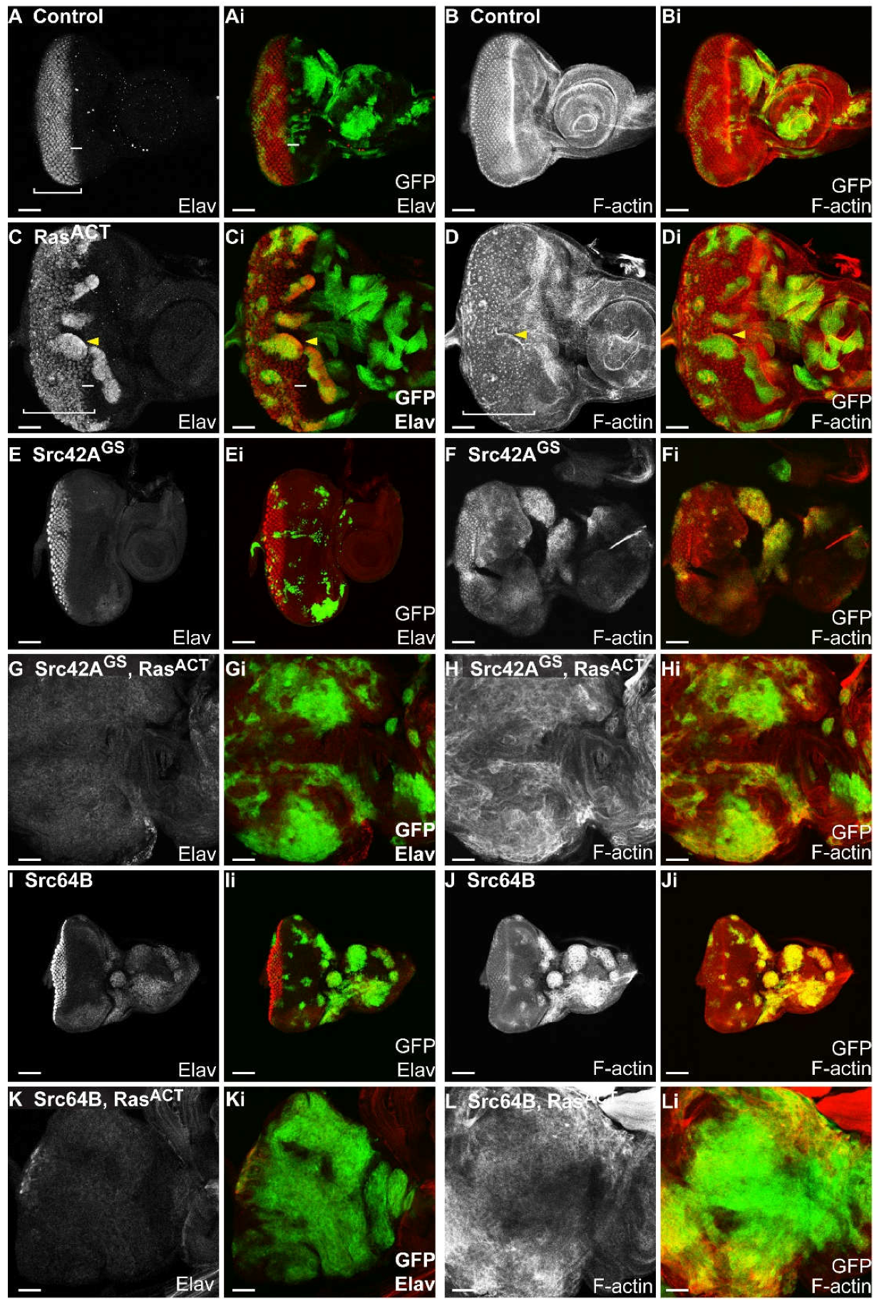
2.4. Src and RasACT Cooperate to form Overgrown Neoplastic Tumours in the Eye Epithelium

To further investigate the cooperative interaction between Src family kinases and activated Ras during tumour development, we utilized the ey-FLP (Flippase) MARCM (Mosaic Analysis with a Repressible Cell Marker) system to generate clones in the developing eye epithelium [70]. Src transgenes were coexpressed with RasACT in clones to determine whether neoplastic overgrowth would occur, similar to that previously observed with mutants in the cell polarity genes, scribbled (scrib), disc large (dlg) and lethal-2-giant larvae (lgl) [71,72,73]. Three Src42A lines were co-expressed with RasACT in eye disc clones: the original candidate identified, Src42AGS; a second wild-type allele, UAS-Src42A; and the activated form, UAS-Src42AACT. The second Src family member Src64B was also tested with RasACT in clones. To establish whether expression of Src with RasACT in clones resulted in neoplastic overgrowth, mosaic eye discs were analysed for changes in clonal tissue size, differentiation and F-actin organisation.
Expression of RasACT alone in mosaic eye discs resulted in clones with rounded borders (Figure 2C,D) similar to that observed previously [64] compared to the jagged edges of clones in control discs (Figure 2A,B). Compared with the regular array of photoreceptors in the posterior region in the control mosaic eye disc (Figure 2A), expression of RasACT resulted in ectopic differentiation within clonal tissue (Figure 2C) located just anterior to the morphogenetic furrow (bar, Figure 2C). Spacing between ommatidial clusters was irregular in RasACT-expressing mosaic eye discs (Figure 2C,D). The tissue was folded resulting in an apparent enrichment of F-actin at the borders between wild-type and clonal tissue (Figure 2D). These observations correlate with previous clonal analysis of RasACT in the eye disc that showed ectopic differentiation and rounded borders [51,64,74]. The expression of RasACT in eye disc clones resulted in pupal lethality.
Expression of Src42AGS (Figure 2E,F) or Src42AACT (Figure S3D) resulted in dramatically reduced Src-expressing clonal size compared to control mosaic eye discs (Figure 2A,B). In contrast, expression of the weaker wild-type Src42A transgene (Figure S3A) resulted in similarly sized clonal tissue to the control (Figure 2A,B). Despite the differences in clonal tissue size, expression of the three Src42A transgenes resulted in similar mosaic adult eyes to each other and to the control (Figure S5L,N), indicating the possibility that mutant tissue was eliminated by the adult stage. Src64B mosaic eye discs contained large clonal clusters that predominantly localised to the anterior of the eye disc or the antennal disc (Figure 2I). The expression of Src42AGS or Src64B in eye disc clones also delayed development resulting in adults that eclosed 1–2 days after their control counterparts. In Src64B mosaic animals, black masses were observed throughout the body of larvae and adults. These black masses are likely to be melanotic tumours arising from an immune response. Melanotic tumours have previously been observed in Drosophila larvae in the context of loss of a caspase protein, Drosophila Caspase-1 (dcp) [75], as well as haemopoietic defects upon overproliferation, arising from activation of the JAK/STAT pathway [76,77].
Coexpression of Src42AGS + RasACT in clones (Figure 2G,H and Table 2) resulted in tissue overgrowth, where the eye antennal disc was greatly enlarged compared with wild-type (Figure 2A,B), RasACT (Figure 2C,D) or Src42AGS (Figure 2E,F) control eye discs. Compared to the flat, planar shape of control mosaic eye discs, Src42AGS + RasACT mosaic eye discs formed a three-dimensional amorphous mass of tissue (Figure 2G,H). During late third instar stages, the eye and antennal structures were no longer distinguishable (Figure 2G,H). These tissues fused together and, in rare instances, the eye-antennal imaginal disc also fused with the brain lobe. Although Src42AGS + RasACT mosaic eye discs were larger than the controls, Src42AGS + RasACT clonal tissue did not predominate in the eye disc, suggesting that non-cell autonomous overgrowth was also occurring (Figure 2G,H). There was also a loss of differentiation, marked by Elav, within the eye disc in both clonal and surrounding wild-type tissue (Figure 2G). Src42AGS + RasACT clonal tissue showed higher accumulation of F-actin and an overall disruption to F-actin organisation within the eye tissue was observed (Figure 2H). Furthermore, Src42AGS + RasACT larvae were larger in size than controls and reached third instar later at Day 6–7, rather than at Day 5 as observed for controls, with melanotic masses in the abdomen and subsequent lethality during late third instar. Taken together, the overgrowth, loss of differentiation, and altered cell morphology of clonal tissue suggests that expression of Src42AGS + RasACT in eye disc clones induces neoplastic tumour formation. Similar effects were also observed with expression of UAS-Src42A with RasACT (Figure S3B,C), however differentiation was still observed in the apical section of wild-type tissue although the patterning was disrupted (Figure S3B), and differentiating clonal and wild-type tissue was also observed aberrantly in basal sections (Figure S3C).
Expression of Src42AACT + RasACT resulted in overgrowth of the eye antennal disc (Figure S3E) with severe disruption to its planar structure and tissue morphology in comparison to wild-type (Figure 2A,B), Src42AACT (Figure S3D) or RasACT (Figure 2C,D) controls. In contrast to either of the Src42A lines expressed with RasACT, the activated Src42AACT + RasACT GFP-marked clonal tissue comprised the majority of the eye imaginal disc (Figure S3E). Clones formed in rounded clusters (Figure S3E) and large clones predominantly localised to the basal part of the epithelium. Differentiation was observed in small regions of wild-type tissue, but within Src42AACT + RasACT clonal tissue, differentiation was greatly reduced (Figure S3Ei). In Src42AACT + RasACT clones, F-actin levels were enriched and F-actin organisation was severely disrupted compared to adjacent wild-type tissue (Figure S3Eii,Eiii). The normal ommatidial clusters in the posterior region could no longer be distinguished. As observed for expression of Src42AGS + RasACT (Figure 2G,H) or Src42A + RasACT (Figure S3B,C), expression of Src42AACT + RasACT resulted in delayed development and lethality at late third instar. In addition to the tissue overgrowth observed in the eye disc, GFP-positive tissue was observed in the brain lobes of Src42AGS (Figure 3C), Src42A or Src42AACT transgenes (Figure S3F,G) coexpressed with RasACT. In the brain lobe, clonal tissue expressing Src42AGS alone also had a protrusive clonal morphology (Figure 3B), but this phenotype was enhanced upon further expression of RasACT (Figure 3C).
Coexpression of Src64B + RasACT in eye disc clones also resulted in enhanced overgrowth of the eye-antennal imaginal disc (Figure 2K,L and Table 3). Src64B + RasACT clonal tissue encompassed most of the eye imaginal disc and clonal mutant size was increased compared to wild-type control (Figure 2A,B), RasACT (Figure 2C,D) or Src64B mosaic eye discs (Figure 2I,J). Differentiation was absent in both clonal and wild-type tissue, although there were rare examples of differentiated cells in wild-type tissue (Figure 2K,L). F-actin levels were enriched within clones and F-actin organisation was disrupted in the eye tissue (Figure 2L). Src64B + RasACT larvae developed more slowly than control counterparts, reaching third instar at Day 6 or 7, compared with Day 5 for controls, and subsequently died during late third instar. Unlike that observed for Src42A transgenes + RasACT, expression of Src64B alone (Figure 3D) or Src64B + RasACT did not result in clones with protrusive morphology in the brain lobe (Figure 3E).
In summary, in the clonal system, expression of Src42A or Src64B cooperated with RasACT to result in neoplastic overgrowth (summarised in Table 2 and Table 3). Src + RasACT mosaic eye discs were characterised by overgrowth of the eye antennal tissue, an increase in clonal tissue, loss of differentiation and disruption to F-actin organisation. The cooperation observed between Drosophila Src genes and RasACT in the clonal system therefore validates the ey > RasACT screen [51] for identifying cooperating partners in tumourigenesis.

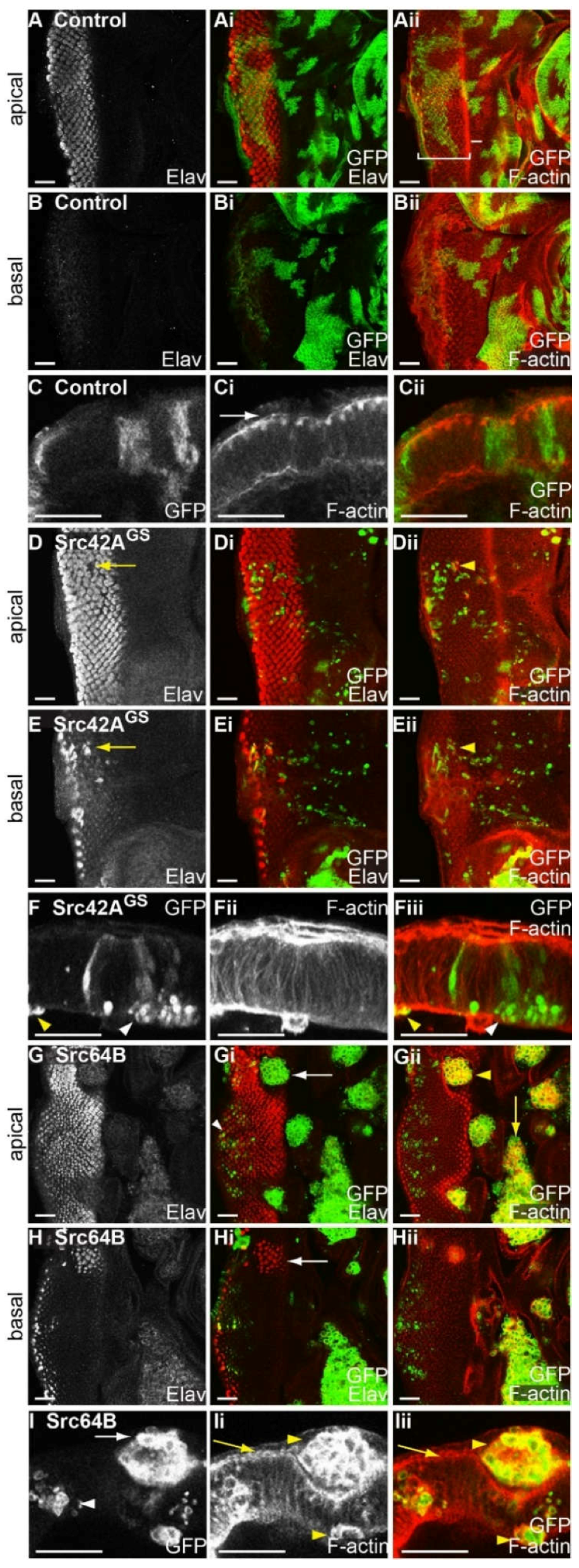
2.5. Expression of Drosophila Src42A and Src64B Results in Distinct Effects in Eye Epithelial Clones

Since all Src transgenes tested were confirmed to cooperate with RasACT in the clonal system, the expression of Src alone in eye disc clones was characterised for effects on differentiation and F-actin organisation. Compared with control mosaic eye discs (Figure 4A–C), Src42AGS clones were greatly reduced in size (Figure 4D–F). Differentiation occurred normally in both wild-type and clonal tissue in the mosaic eye disc, however small gaps were observed in apical sections of Src42AGS-expressing clonal tissue (Figure 4D, yellow arrow). Basal sections of the same eye disc indicated the normally apical photoreceptor nuclei were inappropriately located at the base of the epithelium suggesting that cells within Src42AGS-expressing clones may be shorter than the adjacent wild-type tissue (Figure 4E, arrow). An accumulation of punctate F-actin was observed apically, but not basally, immediately surrounding these gaps in differentiation (yellow arrowhead, Figure 4Dii). This suggested that the adjacent wild-type tissue could be folding in towards the shorter Src42AGS clone. Cross sections of Src42AGS-expressing mosaic eye discs indicated that the small round clones localised basally, sometimes in clusters (Figure 4F), rather than spanning the apical/basal axis of the epithelium as in control discs (Figure 4C). Some Src42AGS-expressing mosaic eye discs contained clonal tissue clustered beneath the differentiating epithelium. Other than in association with the differentiation gaps, F-actin was generally unaffected in Src42AGS-expressing eye disc clones, however, rare small round cells were enriched for F-actin (Figure 4F,Fiii, yellow arrowhead).
Expression of Src64B in mosaic eye discs resulted in clonal tissue with rounded borders that were excluded from the epithelium (Figure 4G,H). Rounded clones were located around the morphogenetic furrow and in the anterior (Figure 4G, arrow), and larger clones were observed between the eye and the antennal disc (Figure 4Gii, yellow arrow). The apically-localised, rounded Src64B-expressing clones did not differentiate (Figure 4Gi, arrow). However, underneath the rounded clones, differentiation occurred normally in the wild-type tissue, as well as in small clones in the epithelium proper (Figure 4Gi, arrowhead). To examine cell shape, Src64B mosaic eye discs were stained for F-actin. Planar views (Figure 4Gii,Hii) and cross sections (Figure 4I) of Src64B-expressing clones showed an increase in F-actin (Figure 4Gii,Ii,Iii, yellow arrowheads). Wild-type tissue showed apically enriched F-actin (Figure 4I, yellow arrow) as observed in controls (Figure 4Ci arrow). However, the rounded cells within Src64B-expressing clones were outlined by increased F-actin (Figure 4Gii, yellow arrowhead, Figure 4Ii,Iii, yellow arrowheads). Generally, Src64B-expressing clones were located above the differentiating epithelium (Figure 4I, arrow) and did not span the apical/basal axis of the epithelium (Figure 4I), as observed in control mosaic eye discs (Figure 4C). However, smaller clones were also observed within the epithelium proper (Figure 4I, white arrowhead). Thus, a comparison between Src42A and Src64B expression in eye disc clones has revealed distinct phenotypic consequences.

2.6. Expression of Src64B in Eye Disc Clones Results in A Loss of Cell Polarity

The rounded morphology of cells in Src42AGS and Src64B clonal tissue/clusters suggests a loss of cell polarity and, indeed, Src has an established role in regulation of cell adhesion components [46]. To further analyse the localisation of cell polarity components at a cellular level in Src-expressing clones, Src64B-expressing mosaic eye discs were stained with the adherens junctions marker E-cadherin [78], Discs large (Dlg) which marks septate junctions [78] and subapical proteins, Bazooka (Baz [79]) and atypical protein kinase C (aPKC [79]) (Figure S4). In control mosaic eye discs, E-cadherin was localised near the apical surface (Figure S4A). Expression of Src64B resulted in diffuse E-cadherin localisation within clonal tissue (Figure S4B, white arrow). Larger clones showed E-cadherin at the cell surface, but this was not always uniform (Figure S4Bii, arrow, and S4Cii) and while most small, rounded clones within the epithelia did not show aberrant E-cadherin localisation, some smaller clones within the epithelia were aberrantly outlined with E-cadherin (Figure S4Bii, arrowhead).
In cross-sections of control mosaic eye discs, aPKC (Figure S4D) and Baz (Figure S4Fii) localised to the subapical region, and Dlg (Figure S4Fi) to the septate junction. In Src64B-expressing clones within the epithelium proper, aPKC (Figure S4Ei, arrow), Baz (Figure S4Gii, arrow) and Dlg (Figure S4Gi, arrow) were correctly localised in smaller clones that were generally restricted to the posterior region of the eye disc. However, in the large round Src64B-expressing clones that were excluded from the epithelium, the subapical markers, aPKC and Baz, were mislocalised (Figure S4Ei, arrowhead and Figure S4Gii). The septate junction protein, Dlg, appeared diffuse in Src64B-expressing clonal tissue, although in some cells, a distinct enrichment of Dlg was observed in the cellular cortex (Figure S4Gi, arrowhead). Thus, Src64B-expressing large, rounded clonal tissue showed mislocalisation of components of the adherens junctions, septate junctions and subapical complex, which are normally associated with the plasma membrane but become diffuse within the cells of the mutant clone. In contrast, in smaller Src64B-expressing clones that span the apical/basal axis of the epithelium, the localisation of cell adhesion components was unaffected relative to the adjacent wild-type tissue.

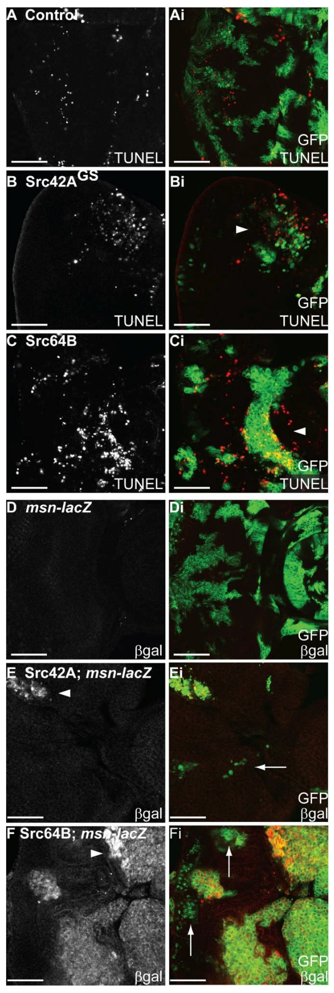
2.7. Expression of Src in Eye Epithelial Clones Promotes Cell Death, but Does Not Reduce Cell Proliferation

In the Drosophila eye epithelium, Src expression promotes both proliferative and pro-apoptosis signals [38]. To determine the effect of Src expression on S phases, BrdU incorporation assays were performed on mosaic eye discs. Expression of Src42AGS (Figure S5G) or Src64B (Figure S5I) did not discernibly affect the pattern of S phases in either mutant clones or surrounding wild-type tissue in the vicinity of the second mitotic wave or in the posterior of the eye epithelium, where differentiation occurs. In rare instances, larger Src42AGS-expressing clones showed increased S phases anterior to the morphogenetic furrow.
Src-expressing mosaic eye discs were assessed for alterations to apoptosis by terminal deoxynucleotidyl transferase dUTP nick end labelling (TUNEL) assays. In comparison to control mosaic discs (Figure 5A), expression of Src42AGS (Figure 5B) or Src64B (Figure 5C) resulted in a higher number of TUNEL-positive cells that appeared both within and around the clones (Figure 5Bi,Ci, arrowheads). Thus, the expression of Src42AGS or Src64B in mosaic eye discs resulted in induction of cell death. Since no discernible effect on S phases were observed in mosaic eye discs expressing Src42AGS or Src64B, it is likely that the reduced clonal size of Src-expressing clones in the eye disc is due to an increase in cell death rather than an inhibition of cell proliferation.
To assess the consequences of blocking cell death in Src-expressing clones, the p35 caspase inhibitor transgene [68] was coexpressed with Src42AGS or Src64B in eye disc clones and the effect on clonal tissue size, differentiation and F-actin organisation was examined. Src42AGS + p35 clonal tissue size (Figure S5C and Table 2) was only marginally larger than Src42AGS-expressing clones (Figure S5B) and remained smaller than control (Figure 2A,B) or p35-expressing clones (Figure S5A). Clonal expression of Src42AGS + p35 in clones disrupted the overall morphology of eye tissue resulting in an eye disc that was undulated (Figure S5C) compared with the wild-type control (Figure 2A,B), p35 (Figure S5A) or Src42AGS mosaic eye discs (Figure S5B). This change in tissue morphology correlated with the disrupted patterning of differentiating cells. F-actin staining showed that it was the wild-type tissue, rather than Src42AGS + p35 clones that was overgrown and folded (Figure S5C), suggesting non-cell-autonomous overgrowth effects. F-actin was enriched in Src42AGS + p35 clones (Figure S5C). Similar effects were observed for Src64B + p35 mosaic eye discs: Src64B + p35 clonal tissue (Figure S5E and Table 3) was not noticeably larger than that of Src64B-expression alone (Figure S5D), but the pattern of differentiation was disrupted and the morphology of the eye disc was no longer planar, due to the distortion of the surrounding wild-type tissue (Figure S5E), suggesting non-cell autonomous tissue growth. This non-cell autonomous effect is similar to that observed by “undead” cells, where apoptosis is initiated but prevented by p35 expression, leading to the secretion of morphogens and non-cell autonomous proliferation [80].
Indeed, BrdU incorporation assays showed that coexpression of Src42AGS + p35 (Figure S5H and Table 2) or Src64B + p35 in clones (Figure S5J and Table 3) resulted in an increase in S phases in the surrounding wild-type tissue. S phases were generally not observed in clones expressing Src42A + p35 (Figure S5H) or Src64B + p35 (Figure S5J), relative to p35-expressing control eye epithelium (Figure S5F). In comparison with the normal Src42AGS mosaic adult eyes (Figure S5L), Src42AGS + p35 mosaic adult eyes were folded and overgrown (Figure S5M and Table 2), reflecting the ectopic proliferation observed during third instar. Whilst expression of Src64B resulted in adult flies with normal eyes (Figure S5N), Src64B + p35 expression in clones resulted in lethality during the third instar larval stage (Figure S5O and Table 3), suggesting that uncontrolled cell proliferation might have impaired metamorphosis.

2.8. Expression of Src in Eye Disc Clones Promotes JNK Pathway Signalling and Activity

It has been shown that Src regulates JNK signalling during dorsal closure [50] and activation of Src, indirectly via loss of Csk, requires JNK signalling in cell proliferation, apoptosis and cell migration [40,47]. JNK signalling has also been shown to mediate cell death signals in both mammalian and Drosophila studies [81,82,83]. Thus, to determine if JNK signalling was activated in Src-expressing clones in the eye epithelium, JNK pathway activity was assessed using the misshapen-lacZ (msn-lacZ) JNK pathway enhancer trap [84]. In control mosaic eye discs, JNK pathway activation reported by msn-lacZ was undetectable (Figure 5D). Expression of Src42AGS (Figure 5E, arrowhead) or Src64B (Figure 5F, arrowhead) in clones resulted in upregulation of msn-lacZ in clonal tissue, indicating that the JNK pathway was activated. However, smaller clones in Src42AGS- (Figure 5Ei, arrow) or Src64B- (Figure 5Fi, arrow) expressing mosaic eye discs did not show upregulation of the msn-lacZ reporter. Thus, JNK pathway activity is activated in larger Src-expressing clones that lose cell polarity (Figure S4).
To more directly assess JNK activation, Src-expressing mosaic eye discs were stained with a mammalian phospho-specific JNK (pJNK) antibody that cross-reacts with Drosophila JNK [50,85,86]. Compared with the low pJNK signal in control mosaic eye discs (Figure S6A), Src42AGS-expressing clones showed an increase in pJNK signal (Figure S6B, arrowhead, with coexpression of p35 to increase clonal tissue size). Similarly, Src64B-expressing mosaic eye discs showed a strong upregulation of pJNK in clonal tissue (Figure S6C, arrowhead) in comparison to control mosaic eye antennal discs (Figure S6A). As observed with the msn-lacZ reporter (Figure 5E,F), pJNK was not discernible in small clones of either Src42AGS + p35- or Src64B-expressing mosaic eye discs ((Figure S6Bi,Ci) arrow), although it was detected in large Src42AGS + p35- or Src64B-expressing clones (Figure S6Bi,Ci and Table 2 and Table 3).

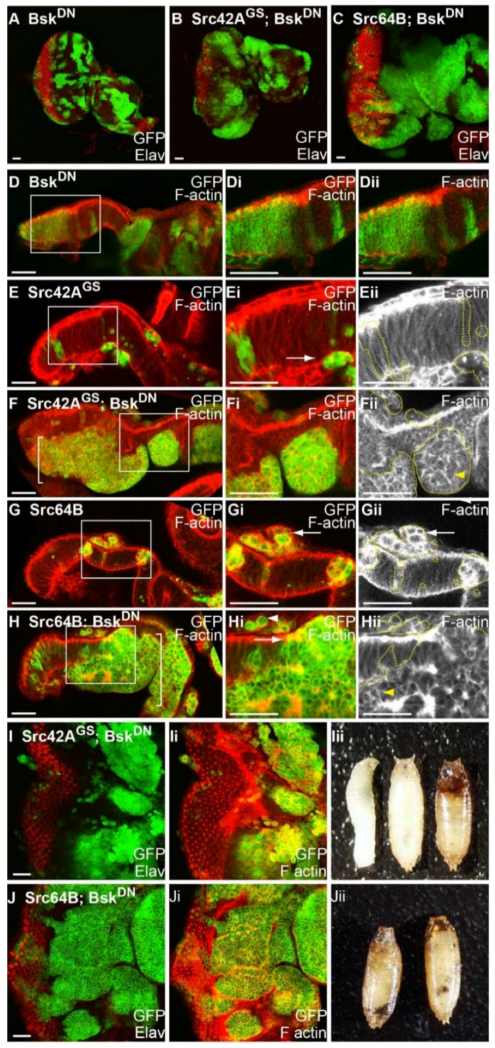
2.9. Blocking JNK Increases Clone Viability of Src-Expressing Clones, Reduces F-actin Accumulation and Results in Basal Extrusion

Since the JNK pathway is activated in Src-expressing clones, we used a dominant negative, kinase dead, form of Drosophila JNK basket, (bskDN) [87], to investigate whether blocking JNK signalling could suppress the Src overexpression clonal phenotype. BskDN has been used extensively to inhibit JNK pathway signalling, and functions similarly or more potently than loss-of-function alleles or other approaches to reduce JNK activity [51,71,88,89,90]. In comparison to the control (Figure 2A,B), mosaic eye discs expressing bskDN showed no discernible effects on differentiation (Figure 6A) or actin organisation (Figure 6D) in apical or basal sections during larval eye development, and bskDN-expressing mosaic adult eyes were comparable to control adult eyes. To test whether loss of JNK could suppress Src clonal phenotypes, Src42AGS or Src64B were coexpressed with bskDN in mosaic eye discs and examined for alterations in clone size, and markers of differentiation (Elav) and F-actin organisation (phalloidin). Expression of bskDN in Src42AGS- (Figure 6B and Table 2) or Src64B- (Figure 6C and Table 3) expressing clones resulted in increased clone viability compared with clonal expression of Src42AGS (Figure 2E,F) or Src64B (Figure 2I,J) alone. Cross-sections of clones coexpressing Src42AGS + bskDN (Figure 6F) or Src64B + bskDN (Figure 6H) revealed that clonal tissue was dramatically increased in size and localised to the basal part of the epithelium compared to Src42AGS- or Src64B-expressed alone in mosaic eye discs, which showed only small Src42AGS-expressing clones residing in the basal part of the epithelium (Figure 6E,G). These large clonal clusters were characterised by smooth borders and contained rounded cells (Figure 6F,H), with enrichment of cortical F-actin in the large clonal clusters (Figure 6Fii,Hii, yellow arrowheads), relative to bskDN control eye discs (Figure 6D). Additionally, smaller clones of Src42AGS- or Src64B- + bskDN-expressing cells were observed within the epithelium and above the apical surface of the epithelium (Figure 6F,H), as were also observed with Src42AGS- or Src64B-expressing cells (Figure 6E,G). Cells in Src64B-expressing clones showed an enrichment of F-actin around the cell cortex (Figure 6Gi,Gii, arrow), whereas F-actin in Src64B + bskDN cells appear to have reduced cortical staining (Figure 6Gi, arrow). Further, in regions of the larger Src64B + bskDN clones that border wild-type tissue, F-actin appears to be apically enriched similar to that of adjacent wild-type cells (Figure 6Hi, white arrowhead). These observations suggest that Src64B-mediated JNK activation may normally promote F-actin polymerisation leading to enriched F-actin in clonal tissue. Although patterning was disrupted, differentiated cells were still observed in Src42AGS + bskDN (Figure 6I) and Src64B + bskDN mosaic eye discs (Figure 6J), however, the Src + bskDN larva did not develop to adulthood, with lethality occurring during late larval/pupal stages (Figure 6Iii,Jii). Black melanotic masses were sometimes observed in Src64B + bskDN pupae (Figure 6Jii and Table 3). Altogether, these data show that blocking the JNK pathway increases viability of Src-expressing clones and suppresses F-actin accumulation, suggesting that JNK acts downstream of Src to induce cell death and F-actin polymerisation consistent with other studies in the Drosophila wing epithelium [88,89], and eye epithelium [44].

2.10. JNK is Activated in Src + RasACT Neoplastic Overgrowth, and Blocking JNK Results in Partial Suppression of the Overgrowth and Differentiation Defects

Given the requirement for JNK signalling in Src-expressing clones shown here and previously [50], and the role of JNK signalling in cooperative interactions observed between cell polarity mutants and Ras-driven tumourigenesis [51,71,74,90,91,92,93,94,95,96], it is conceivable that JNK activity could be upregulated and/or required for the cooperative overgrowth between Src + RasACT-induced neoplastic overgrowth. Therefore, to assess whether JNK signalling was active in Src + RasACT clonal tissue, the msn-lacZ reporter was utilised as a readout for JNK pathway activation. Compared with control and Src42AGS mosaic eye discs (Figure 5D–F), expression of Src42AGS + RasACT in eye disc clones results in upregulation of the msn-lacZ reporter within most clonal cells (Figure 7A and Table 2). Thus, JNK signalling is upregulated in Src and RasACT expressing tissue, consistent with it also playing a role in tumourigenesis in this setting.
To determine whether blocking the JNK pathway could alter Src + RasACT clonal neoplastic overgrowth, bskDN was coexpressed in Src + RasACT eye disc clones, and mosaic eye discs were analysed for changes in clonal tissue size (marked by GFP expression), cell morphology (F-actin marked by phalloidin) and differentiation (marked by Elav). Expression of bskDN in RasACT-expressing clones (Figure 7B) resulted in ectopic differentiation, similar to that observed upon expression of RasACT alone (Figure 2C,D). Similar to that observed for RasACT mosaic eye discs, expression of RasACT + bskDN resulted in pupal lethality. Whereas Src42AGS + RasACT (Figure 7C) or Src64B + RasACT (Figure 7D) expression in mosaic eye discs resulted in dramatic tissue overgrowth, striking suppression of overgrowth was observed upon blocking JNK signalling by coexpression of bskDN with Src42AGS + RasACT (Figure 7E and Table 2) or Src64B + RasACT (Figure 7F and Table 3) in mosaic eye discs. The eye antennal disc had normal tissue morphology with recognisable shape and structure (Figure 7E,F). Higher magnification views revealed that F-actin was enriched cortically in clonal tissue and that tissue morphology of the eye disc was still disrupted (Figure 7G,Hii,Hiii). In contrast to Src overexpression with RasACT, where clonal tissue was observed in the brain lobes (Figure 3C,E), co-expression of bskDN resulted in reduced clonal tissue observed in the brain lobes of Src + RasACT mosaic larvae (Figure 7E,F). Whilst Src42AGS + RasACT clones exhibited protrusive morphology (Figure 3C), this was no longer observed upon bskDN expression (Figure 7E and Table 2). Moreover, the loss of differentiation observed in Src + RasACT mosaic eye discs (Figure 2G,H,K,L) was partially suppressed when JNK was blocked by expression of bskDN (Figure 7G,H and Table 2). Differentiation occurred in both wild-type and Src + RasACT + bskDN clonal tissue (Figure 7G,Hi), although some cells were incorrectly basally localised. However, in comparison to control mosaic eye discs (Figure 4A,B), differentiation was not restored completely to normal in Src + RasACT + bskDN mosaic eye discs. Thus, expression of Src + RasACT in eye disc clones results in robust upregulation of JNK pathway (as measured by JNK pathway enhancer trap, msn-lacZ), and blocking the JNK pathway partially suppressed tissue overgrowth of the eye-antennal disc, restored the morphology of the eye-antennal disc and partially restored differentiation to the Src + RasACT-expressing clones. Altogether, these results show that JNK activation is required for Src + RasACT cooperative, neoplastic overgrowth.

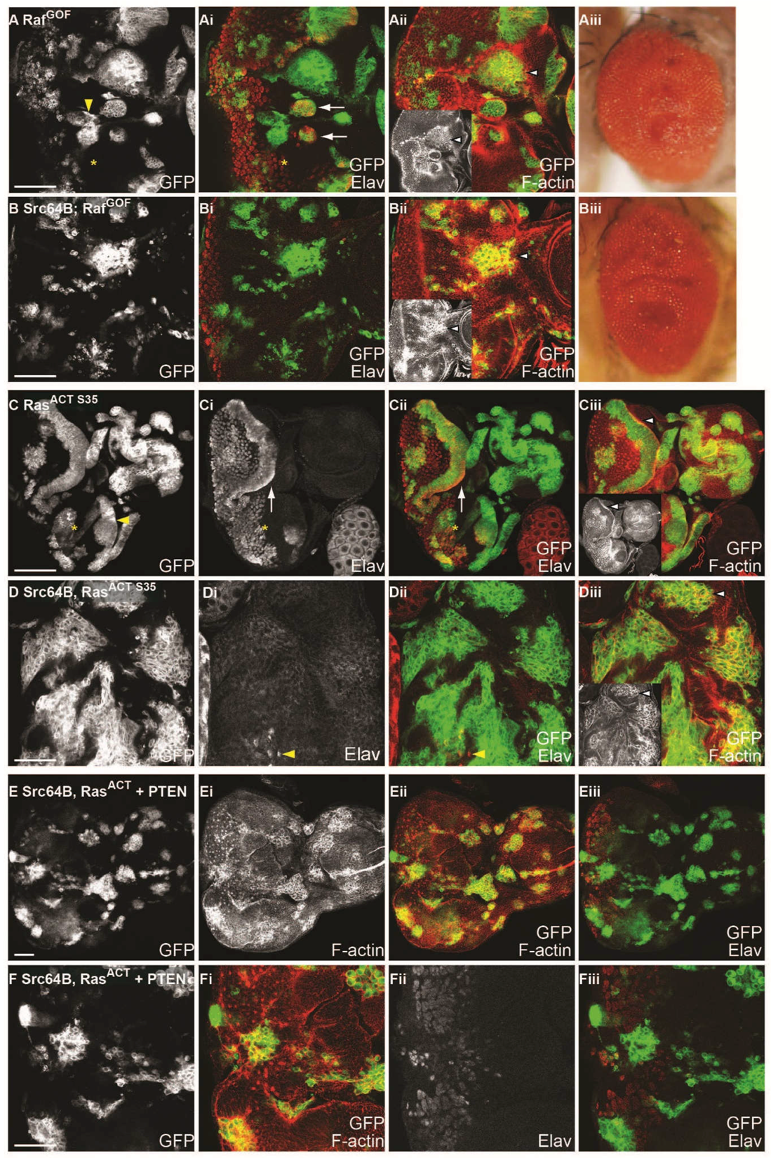
2.11. Ras-Raf-MAPK and Ras-PI3K Pathways are Required with Src for Cooperative Tumourigenesis
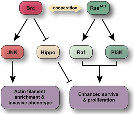
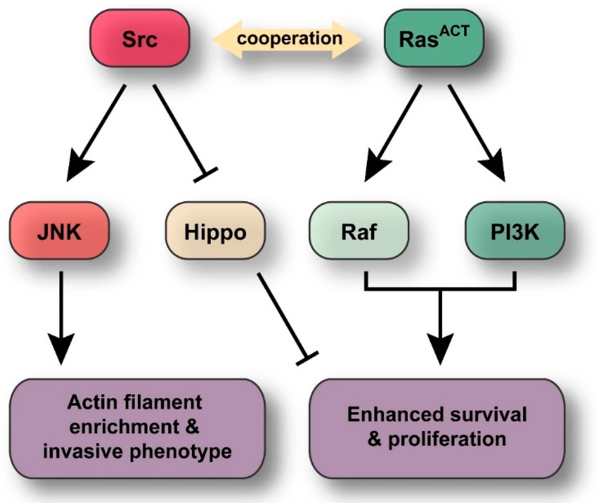
Ras conveys its signals by many effectors, of which the most well-known are Raf-mitogen-activated protein kinase (MAPK), Phosphoinositide 3-Kinase (PI3K) and Ral pathways [23,97], however in Drosophila, RasACT-mediated tissue growth effects were mediated by Raf-MAPK and PI3K (Dp110/PI3K92E) effectors [62,64,98]. In cooperation with scrib mutant however, only activated Raf (RafGOF) [99] is able to phenocopy the effects of Ras function to result in neoplastic overgrowth [72,100]. This requirement of Raf-MAPK is likely to be specific as expression of other Ras effectors PI3K or Ral were unable to recapitulate the effects of RasACT in scrib mutant clones [72].
To test whether Src kinases similarly cooperate with Raf signalling in clonal analysis, Src42AGS or Src64B were coexpressed with an amino-terminal truncation allele of Raf, RafGOF, which renders constitutive activation of Raf signalling [99]. As observed in RasACT mosaic eye discs, expression of RafGOF resulted in rounded clones with smooth borders and precocious differentiation anterior to the morphogenetic furrow (Figure 8Ai). RafGOF mosaic eye discs exhibited disrupted tissue morphology resulting in an enrichment of F-actin at the borders between wild-type and mutant clonal tissue (Figure 8Aii). Unlike RasACT, clonal expression of RafGOF did not lead to pupal lethality but resulted in adult flies with folded, overgrown eye tissue (Figure 8Aiii). To determine whether Raf signalling could phenocopy RasACT in cooperation with Drosophila Src kinases, RafGOF was coexpressed with Src42A or Src64B in mosaic eye discs. Surprisingly, cooperative overgrowth was not observed; instead, expression of Src42AGS + RafGOF or Src64B + RafGOF (Figure 8Bi and Table 4) resulted in rounded clones that did not differentiate, although some clones appear smaller than that of clones expressing RafGOF alone. Thus, although Src expression inhibits Raf-induced differentiation, Src was unable to cooperate with RafGOF. F-actin was enriched in Src64B + RafGOF clonal tissue (Figure 8Bii and inset, arrowhead) compared to clones expressing RafGOF alone (Figure 8Aii and inset, arrowhead). Adult eyes expressing Src42AGS + RafGOF or Src64B + RafGOF (Figure 8Biii) were rough and folded, comparable to the hyperplastic RafGOF adult eye phenotype (Figure 8Aiii). Thus, these data suggest that Raf signalling is not sufficient to phenocopy Ras in cooperation with Src family kinases, and other Ras effectors may be required for neoplastic overgrowth.
Since activation of the Raf-MAPK cascade alone was not sufficient to cooperate with Src family kinases, the RasACT-S35 effector domain mutant [62] was utilised to test the contribution of Raf signalling. The RasACT-S35 mutant preferentially signals to the Raf-MAPK pathway and has been characterised in both mammals and Drosophila [61,62,64,101]. Specifically, previous analysis in Drosophila eye and wing discs has demonstrated that RasACT-S35 favours Raf-MAPK and is less potent than RasACT in recruitment of a PI3K reporter [62,64]. Firstly, we tested the effect of expression of RasACT-S35 alone in mosaic eye discs by examining differentiation and F-actin organisation. RasACT-S35 mosaic eye discs were characterised by rounded clones with smooth borders (Figure 8C, yellow arrowhead) and ectopic differentiation anterior to the morphogenetic furrow (Figure 8C, asterisk). Due to tissue misfolding of RasACT-S35 mosaic eye tissue, cells are abnormally arranged, but F-actin appears to be apically localised (Figure 8Ciii and inset, white arrowhead). These effects were similar to that observed in RasACT mosaic eye discs (Figure 2B), and, like RasACT, expression of RasACT-S35 resulted in pupal lethality.
To determine whether Src expression could cooperate with RasACT-S35, Src42AGS or Src64B were coexpressed with RasACT-S35 in mosaic eye discs. Expression of Src42AGS + RasACT-S35 or Src64B + RasACT-S35 (Figure 8D) in mosaic eye discs resulted in overgrowth of clonal tissue (Figure 8D, GFP-marked, and Table 4). There was a loss of differentiation in both wild-type and clonal tissue, although on rare occasions, differentiation occurred in wild-type tissue (Figure 8Di,Dii, yellow arrowhead). F-actin was enriched within clonal tissue and F-actin organisation was disrupted (Figure 8Diii and inset, white arrowhead). Distinct from Src64B + RasACT-expressing mosaic eye discs where clonal tissue largely predominates in the eye disc (Figure 2K,L), Src64B + RasACT-S35 clonal tissue, while still over-represented, did not overtake the whole eye disc (Figure 8D). This suggested that RasACT-S35 is less potent than RasACT in cooperation with Src. Thus, while expression of activated Raf was not sufficient for cooperation with Src, expression of RasACT-S35, which preferentially but not exclusively signals via Raf, was able to cooperate with Src, albeit to a lesser extent than RasACT. These data suggest that Raf signalling is required for neoplastic overgrowth in concert with Src overexpression, however, other Ras effectors are also likely to be required.
To test the whether the PI3K pathway, an effector of RasACT in tissue growth control [64], was required for Src + RasACT tumourigenesis, we co-expressed PTEN (which antagonises the activity of PI3K) or a dominant-negative version of PI3K (Dp110DN) in Src64B +RasACT clones in the eye-antennal epithelium (Figure 8E,F and Figure S7). Expression of PTEN alone did not affect the clone size (Figure S7A) compared to wild-type (Figure 2A,B). However, expression of PTEN with RasACT or Src64B (Figure S7B,C) mildly reduced clonal size relative to RasACT or Src64B alone (Figure 2C,D,I,J). Strikingly, co-expression of PTEN in Src64B + RasACT eye disc clones dramatically reduced tumour growth and restored differentiation in most clones (Figure 8E,F and Table 4), compared to Src64B + RasACT tumours (Figure 2K,L). To more directly assess the requirement of PI3K activity, we co-expressed of a dominant negative allele of the Phosphoinositide 3-Kinase PI3K gene, Dp110/PI3K92E(Dp110DN) in Src64B + RasACT eye disc clones also reduced tumour size (Figure S7G,H and Table 4), although not as potently as with PTEN (Figure 8E,F). Thus, consistent with the data showing that RafGOF expression alone were insufficient to cooperate with Src (Figure 8A,B), we show that PI3K activity plays an important function in Src64B + RasACT tumourigenesis.

3. Discussion

In this study, we have identified the Drosophila Src kinase genes, Src42A and Src64B, as cooperating genes with activated Ras in tumourigenesis in the eye-antennal epithelial tissue. Src42A and Src64B were identified in a genetic screen as enhancers of the ey > RasACT hyperplastic adult eye phenotype. Importantly, in a clonal context, the Drosophila Src kinases were also able to cooperate with RasACT resulting in neoplastic overgrowth of the eye-antennal epithelium (Figure 9). This cooperative tumourigenesis is characterised by tissue overgrowth, increased clonal tissue, loss of differentiation, disruption to F-actin organisation, larval lethality and an invasive clonal phenotype. Src expressed alone in eye disc clones results in increased apoptosis, a loss of cell polarity and disruption to F-actin organisation, but was not sufficient alone to promote tumour formation. We show that JNK signalling acts downstream of Src to promote cell death and increased accumulation of F-actin. In the context of Src + RasACT tumours, the JNK pathway contributes to the inhibition of differentiation, clonal overgrowth and invasive phenotypes associated with Src + RasACT neoplastic overgrowth. Moreover, we show that the PI3K pathway is critical for the cooperation of Src with RasACT in tumourigenesis. Altogether, our findings provide insight into the mechanism by which Src and Ras signalling cooperate in tumourigenesis, which may provide new avenues for the treatment of human cancer.
Others have provided indirect evidence that Src signalling can cooperate with RasACT in a clonal context [39]. However, in this previous study, RasACT was expressed in Csk-deficient clones, mimicking Src activation, to result in tissue overgrowth and delayed development [39]. Since it is unclear whether Csk may act solely through Src kinases [41], our study has provided evidence that Src is able to cooperate with RasACT in tumourigenesis in the eye-antennal epithelium. Furthermore, additional expression of Src64B with RasACT in Csk-mutant clones resulted in an enhancement of this phenotype [39]. This correlates with our observations using the GMR-GAL4 system that Src expression can elicit a dose-dependent response. Although expression of all three Src42A overexpression lines tested cooperated with RasACT, there were differences observed. There was little differentiation observed in Src42AACT + RasACT mosaic eye discs, whereas expression of wild type Src42A + RasACT enabled some differentiation, albeit aberrantly, to occur in the mosaic eye disc.
While Src is known to affect many proteins that modulate actin dynamics [44,88,89,102,103], blocking JNK in Src-expressing clones suppresses the F-actin enrichment that is associated with Src-expressed alone. This shows that Src may also act via JNK, at least in part, to mediate changes in F-actin organization. In fact, JNK upregulates transcripts of the Drosophila actin-binding protein, profilin (Chickadee, Chic) [104], which promotes the formation of F-actin [105]. The loss of chic enhances the hep (Drosophila JNKK) mutant dorsal open phenotype [104], and ectopic actin polymerization occurs in both hep and chic mutants suggesting that these may be acting via a common pathway to regulate cytoskeletal rearrangements [104]. Moreover, a positive feedback loop has been revealed to exist between JNK signalling and actin cytoskeletal regulators [89]. Furthermore, a link between JNK and integrins, which can promote actin assembly [106], has been described: JNK promotes expression of βPS integrin (encoded by myospheroid, mys) and αPS3 integrin (encoded by scab, scb), and the loss of mys and scb results in a similar dorsal open phenotype due to loss of JNK [107]. Therefore, in addition to the established function of Src in direct regulation of actin dynamics, additional signalling via the JNK pathway may also contribute to regulation of cell shape or cell adhesion via its effects on actin regulation. Additionally, F-actin reorganization can promote cell proliferation through inhibiting the Hippo pathway [93,108,109,110,111,112,113]. Furthermore, whilst JNK activity promotes cell death and activates the Hippo pathway, in the presence of Ras signalling, JNK signalling instead leads to Hippo pathway impairment via increased actin polymerization [114]. Src64B can also affect Hippo pathway signalling more directly through the actin cytoskeletal regulators, Rac1 and Diaphanous, which together with Src64B-induced Ras-MAPK signalling drives actin polymerisation, and when JNK signalling is impaired promotes tissue overgrowth in eye disc clones [44].
We show here that, in Src + RasACT clones, the activation of JNK promotes overall tissue growth, inhibition of differentiation and migratory-like phenotypes. In Src + RasACT cooperation, Ras most likely functions to suppress JNK-mediated cell death by inhibiting the apoptosis inducer, Hid [66,67,115], thereby revealing other functions of JNK signalling, such as the promotion of cell migration and inhibition of differentiation. Src expression in the embryo induces cell migration [46], and in corroboration with the potential role of JNK in migratory-like phenotypes in Src + RasACT tumours, the loss of JNK was shown to suppress the migratory effects arising from Src activation [116].
The finding that JNK is a critical component in the cooperation between Src + RasACT, correlates with previous analysis of Drosophila cooperative tumourigenesis with cell polarity or actin cytoskeletal regulators [51,71,72,74,90,91,92,93,94,100]. JNK is activated in RasACT + scrib, dlg or lgl mutant tumours, and also promotes tissue growth and invasive phenotypes. Expression profiling has revealed a large number of JNK targets that affect cell differentiation in RasACT + scrib mutant eye-antennal epithelial tissue [92,94,95,96], or in scrib homozygous wing epithelial tissue [117]. Moreover, JNK and Yorkie (Yki, a co-transcription factor inhibited by the Hippo pathway) mediated-upregulation of secreted factor dILP8 (Drosophila Insulin-Like Peptide 8), which inhibits the Ecdysone steroid hormone production from the prothoracic gland, results in the delayed larval-pupal transition caused by imaginal disc neoplastic tumours [94,118,119,120]. It is likely that similar mechanisms are induced in Src + RasACT tumours to result in differentiation defects and the developmental delay at the larval stage. In mammalian systems, a role for JNK in inhibition of differentiation has been reported [121,122]. JNK also plays a role in cell transformation induced by coexpression of c-myc (cellular-myc proto-oncogene) and RasV12 in mouse embryonic fibroblasts [123], although, these studies were carried out in vitro, and the precise effect of JNK signalling in c-myc and RasV12-mediated oncogenic cooperation is unclear. However, based on the findings in Drosophila tumour models described here and previously [71,72,90,100], inhibiting JNK signalling may restore differentiation and suppress the malignant overgrowth and invasive characteristics in many human tumours. Indeed, in bRAF (b-RAF proto-oncogene)-driven melanomas, JNK-cJun (cellular-Jun proto-oncogene) signalling has been revealed to contribute to tumour progression, suggesting that blocking JNK signalling may be of therapeutic benefit in at least some cancer types [124,125,126]. However, although JNK clearly plays an important role in Src + RasACT tumourigenesis, the activation of JNK signalling with RasACT does not result in as aggressive tumours as with Src + RasACT [51], which might be due to the contribution of Src signalling to Hippo pathway impairment [44] or to possible effects of Src on the activation of Myosin II activity and actinomyosin cell contractility, cell shape changes and tissue growth [74].
Our discovery that RafGOF was not sufficient to phenocopy RasACT in cooperation with Src was surprising, given that Raf expression is able to phenocopy Ras in cooperation with scrib mutant [72,100] and RhoGEF2 [74]. Interestingly, we found that in the context of Src and RasACT cooperative tumourigenesis, the PI3K pathway is likely to play a critical role alongside contribution from Raf-MAPK signalling. Indeed, dlg RasACT tumours have compromised PI3K signalling and knockdown of PI3K pathway signalling is synthetically lethal to tumourigenesis [127]. Therefore, although scrib RasACT and RhoGEF2 RasACT tumours do not depend on Ras-driven PI3K signalling, it is possible that they are still sensitive to its depletion. The reason Src + RasACT tumourigenesis is dependent on PI3K signalling remains to be determined. One possible mechanism might relate to the importance of PI3K-mTOR (mechanistic Target of Rapamycin) signalling in blocking autophagy, a catabolic pathway that leads to the degradation of cellular components to produce energy [128]. Recently, polarity-impaired Ras-driven cancers have been shown to be dependent on induction of autophagy in neighbouring wild-type cells [129], which suggests that the PI3K-mTOR pathway might be important in this non-cell autonomous mechanism in tumour development. Additionally, the PI3K-mTOR-S6K pathway has been revealed to be critical in modulating metabolism from oxidative phosphorylation to aerobic glycolysis, which is important for neoplastic tumour progression [130]. Further studies will be required to determine if PI3K signalling may be important in regulating such biological processes in the Src + RasACT or other cooperative tumour models. Altogether, our work along with these other studies revealing the requirement of PI3K signalling in tumourigenesis in Drosophila models, suggests that targeting PI3K signalling might provide a novel therapeutic approach for Src-overexpressing or polarity-impaired Ras-driven cancers.
Although the Src proto-oncogene is associated with cancer [131], its precise role in tumour development and the significance of the contributions of its many downstream effectors to tumourigenesis remains unclear. Furthermore, given the strong correlation of elevated Ras protein expression in human tumours [132], examining the mechanism of cooperation between these key oncogenes may allow more precise targeting of critical signalling components, such as the JNK, Raf-MAPK and PI3K pathways, for improved therapies and better patient outcomes. Therefore, this Drosophila clonal model system has provided a robust in vivo setting in which to investigate Src function in cooperation with RasACT, and potentially could be utilized to gain further insight to other cooperative interactions identified in human disease.

4. Materials and Methods

4.1. Drosophila Stocks

Transgenes were overexpressed using ey-GAL4 [56], GMR-GAL4 [133] and hsp70-GAL4 [134]. The MARCM system [70,135] was used to generate mosaic eye tissue using w ey-FLP1, UAS-mCD8-GFP; tub-GAL4 FRT82B tub-GAL80. Transgenic fly stocks employed were: Src42AGS [136], UAS-Src42A [50], UAS-Src42AACT [50], UAS-Src64B (R. Cagan), Src42AEY08937 [52], UAS-RasACT [61], UAS-RafGOF [99], UAS-bskDN [87], UAS-p35 [68], UAS-PTEN [137] and UAS-Dp110DN [138]. ey-GAL4 analysis using p35 and RasACT were carried out using recombinants generated on the second chromosome carrying ey-GAL4 and UAS-p35 or ey-GAL4 and UAS-RasACT.

4.2. Immunohistochemistry

Third instar larval eye imaginal discs were dissected and fixed in 6% (w/v) paraformaldehyde/HEPES for 20 min. Antibodies used were mouse anti-Elav (Developmental Studies Hybridoma Bank, 1:5), mouse anti-BrdU (Becton-Dickinson, Franklin Lakes, NJ, USA, 1:50) and rabbit anti-β-galactosidase (Rockland, 1:400), anti-E-cadherin (DSHB, 1:50), anti-aPKC-ζ (Upstate Biotechnology Inc., Lake Placid, NY, USA, 1:1000), anti-Discs Large (Dlg) (DSHB, 1:20), anti-Bazooka (Baz) [139], 1:200) and anti-GFP (Invitrogen, Carlsbad, CA, USA, 1:1000). F-actin was detected with phalloidin-tetramethylrhodamine isothiocyanate (TRITC; Sigma, St. Louis, MO, USA, 0.3 mM). For detection of apoptotic cells, a TUNEL assay (TMR, Roche, Basel, Switzerland) was conducted on eye imaginal discs that were permeabilized overnight at 4 °C in 0.3% (v/v) Triton X-100 in PBS according to manufacturer’s instructions. For the detection of S phase cells, a 1 h BrdU pulse at 25 °C was followed by 1 h fixation, immunodetection using anti-GFP antibody, further fixation, acid treatment using 0.1 M HCl, followed by detection of the BrdU epitope. All fluorescent labelled samples were captured by confocal microscopy (BioRad MRC 1000; Kidlington, Oxford, UK)) using Lasersharp 2000 software (Micro-Scientific, Gurnee, IL USA). Data were processed using Confocal Assistant (Purdue University Cytometry Laboratories, IN, USA) and Adobe Photoshop CS2 software (Adobe Systems, San Jose, CA, USA).

4.3. Adult Eye Imaging

Adult eyes were viewed under a light dissecting microscope and images captured on an Olympus DP11 camera, and data were processed using Adobe Photoshop CS2. For scanning electron microscope images, Adult flies were placed in a screw-cap tube in 25% (v/v) acetone for 1 h nutation at room temperature, followed by 50% (v/v) acetone for 2–3 h, 75% (v/v) acetone for a further 3 h and then finally placed in 100% acetone. Adult flies were critical point dried on a Balters CPD 030 Critical Point Dryer and coated with gold particles in an Edwards 6150B Gold Sputter Coater (kindly carried out by Simon Crawford, Department of Botany, University of Melbourne). Images were recorded on a Phillips XL30 FEG Field Emission Electron Microscope (Amsterdam, Netherlands).

4.4. Western Analysis

Protein expression was induced in third instar larvae by 1 h heat shock at 37 °C where hsp70-GAL4 was used to drive expression of UAS-Src42A, UAS-Src42AGS or no transgene control (w1118). After 1 h recovery at 25 °C, larval eye tissue was dissected and lysed in NTEN buffer with fresh protease inhibitor. Protein lysates were subjected to SDS-PAGE and immunoblotted with anti-phosphorylated Src (against the autophosphorylated Tyrosine residue in the kinase domain, indicating active Src (anti-pSrc, Biosource, 1:1000)), anti-Drosophila Src42A to detect expression levels (α-Src42A, [58], 1:1000), and anti-tubulin to indicate protein loading (anti-tubulin, Calbiochem, 1:10,000). Protein bands on Western blots were quantified using Image J (National Institutes of Health, Bethesda, MA, USA).

Supplementary Materials

Supplementary materials can be found at https://www.mdpi.com/1422-0067/19/6/1585/s1.

Author Contributions

C.L.C.P., A.M.B. and H.E.R. conceived and designed the experiments; C.L.C.P. performed the experiments; C.L.C.P., A.M.B. and H.E.R. analysed the data; and C.L.C.P. and H.E.R. wrote the paper.

Acknowledgments

We thank all members of the lab for help and advice, and Peter Burke for maintaining the fly stocks. Thanks to Kathryn Guthridge, Leonie Quinn, Linda Parsons, Patrick Humbert and Geraldine O’Neill (The Children’s Hospital Westmead, Sydney) for useful discussions on the results and interpretations. We acknowledge Sarah Ellis and the Microscope Core Facility, Peter MacCallum Cancer Centre and Simon Crawford at the Department of Botany, University of Melbourne for assistance with the SEM images. We are appreciative to the fly community for supplying reagents and Flybase for their wealth of information. This work was supported by NIH-R21 grant (1R21CA098997-01) to H.E.R., and a Wellcome Trust Senior Research Fellowship and National Health and Medical Research Council (NHMRC) Fellowships to H.E.R. (#299842, #454700), as well as funds from the Peter MacCallum Cancer Centre and La Trobe Institute of Molecular Science and La Trobe University to H.E.R. A.M.B. was supported by an NIH-R21 grant (1R21CA098997-01) and an NHMRC grant (#350396). C.L.C.P. was supported by a Melbourne Research Scholarship from the University of Melbourne and currently by an NHMRC grant (#1142469).

Conflicts of Interest

The authors declare no conflict of interest. The founding sponsors had no role in the design of the study; in the collection, analyses, or interpretation of data; in the writing of the manuscript, and in the decision to publish the results.

Abbreviations

BazBazooka
CskC-terminal Src-like kinase
DlgDiscs-large
EGFEpidermal growth factor
EGFREpidermal growth factor receptor
JNKJun N-terminal Kinase
MMPMetalloproteinase
MAPKMitogen-activated protein kinase
PI3KPhosphoInoitide 3-kinase

References

  1. Frame, M.C. Src in cancer: Deregulation and consequences for cell behaviour. Biochim. Biophys. Acta Rev. Cancer 2002, 1602, 114–130. [Google Scholar] [CrossRef]
  2. Thomas, S.M.; Brugge, J.S. Cellular functions regulated by Src family kinases. Annu. Rev. Cell Dev. Biol. 1997, 13, 513–609. [Google Scholar] [CrossRef] [PubMed]
  3. Martin, G.S. The hunting of the Src. Nat. Rev. Mol. Cell Biol 2001, 2, 467–475. [Google Scholar] [CrossRef] [PubMed]
  4. Yeatman, T.J. A renaissance for Src. Nat. Rev. Cancer 2004, 4, 470–480. [Google Scholar] [CrossRef] [PubMed]
  5. Frame, M.C. Newest findings on the oldest oncogene; how activated Src does it. J. Cell Sci 2004, 117, 989–998. [Google Scholar] [PubMed]
  6. Talamonti, M.S.; Roh, M.S.; Curley, S.A.; Gallick, G.E. Increase in activity and level of pp60c-Src in progressive stages of human colorectal cancer. J. Clin. Investig. 1993, 91, 53–60. [Google Scholar] [CrossRef] [PubMed]
  7. Irby, R.B.; Mao, W.; Coppola, D.; Kang, J.; Loubeau, J.M.; Trudeau, W.; Karl, R.; Fujita, D.J.; Jove, R.; Yeatman, T.J. Activating Src mutation in a subset of advanced human colon cancers. Nat. Genet. 1999, 21, 187–190. [Google Scholar] [CrossRef] [PubMed]
  8. Rosen, N.; Bolen, J.B.; Schwartz, A.M.; Cohen, P.; DeSeau, V.; Israel, M.A. Analysis of pp60c-Src protein kinase activity in human tumor cell lines and tissues. J. Biol. Chem. 1986, 261, 13754–13759. [Google Scholar] [PubMed]
  9. Barnekow, A.; Schartl, M. Comparative studies on the Src proto-oncogene and its gene product pp60c-Src in normal and neoplastic tissues of lower vertebrates. Comp. Biochem. Physiol. B Comp. Biochem. 1987, 87, 663–670. [Google Scholar] [CrossRef]
  10. Cartwright, C.A.; Kamps, M.P.; Meisler, A.I.; Pipas, J.M.; Eckhart, W. Pp60c-Src activation in human colon carcinoma. J. Clin. Investig. 1989, 83, 2025–2033. [Google Scholar] [CrossRef] [PubMed]
  11. Fanning, P.; Bulovas, K.; Saini, K.S.; Libertino, J.A.; Joyce, A.D.; Summerhayes, I.C. Elevated expression of pp60c-Src in low grade human bladder carcinoma. Cancer Res. 1992, 52, 1457–1462. [Google Scholar] [PubMed]
  12. Barnekow, A.; Paul, E.; Schartl, M. Expression of the c-Src protooncogene in human skin tumors. Cancer Res. 1987, 47, 235–240. [Google Scholar] [PubMed]
  13. Muthuswamy, S.K.; Muller, W.J. Activation of Src family kinases in neu-induced mammary tumors correlates with their association with distinct sets of tyrosine phosphorylated proteins in vivo. Oncogene 1995, 11, 1801–1810. [Google Scholar] [PubMed]
  14. Mao, W.; Irby, R.; Coppola, D.; Fu, L.; Wloch, M.; Turner, J.; Yu, H.; Garcia, R.; Jove, R.; Yeatman, T.J. Activation of c-Src by receptor tyrosine kinases in human colon cancer cells with high metastatic potential. Oncogene 1997, 15, 3083–3090. [Google Scholar] [CrossRef] [PubMed]
  15. Wiener, J.R.; Windham, T.C.; Estrella, V.C.; Parikh, N.U.; Thall, P.F.; Deavers, M.T.; Bast, R.C.; Mills, G.B.; Gallick, G.E. Activated Src protein tyrosine kinase is overexpressed in late-stage human ovarian cancers. Gynecol. Oncol. 2003, 88, 73–79. [Google Scholar] [CrossRef] [PubMed]
  16. Mandal, M.; Myers, J.N.; Lippman, S.M.; Johnson, F.M.; Williams, M.D.; Rayala, S.; Ohshiro, K.; Rosenthal, D.I.; Weber, R.S.; Gallick, G.E.; et al. Epithelial to mesenchymal transition in head and neck squamous carcinoma: Association of Src activation with E-cadherin down-regulation, vimentin expression, and aggressive tumor features. Cancer 2008, 112, 2088–2100. [Google Scholar] [CrossRef] [PubMed]
  17. Boyer, B.; Bourgeois, Y.; Poupon, M.F. Src kinase contributes to the metastatic spread of carcinoma cells. Oncogene 2002, 21, 2347–2356. [Google Scholar] [CrossRef] [PubMed]
  18. Irby, R.; Mao, W.; Coppola, D.; Jove, R.; Gamero, A.; Cuthbertson, D.; Fujita, D.J.; Yeatman, T.J. Overexpression of normal c-Src in poorly metastatic human colon cancer cells enhances primary tumor growth but not metastatic potential. Cell Growth Differ. 1997, 8, 1287–1295. [Google Scholar] [PubMed]
  19. Maa, M.C.; Leu, T.H.; McCarley, D.J.; Schatzman, R.C.; Parsons, S.J. Potentiation of epidermal growth factor receptor-mediated oncogenesis by c-Src: Implications for the etiology of multiple human cancers. Proc. Natl. Acad. Sci. USA 1995, 92, 6981–6985. [Google Scholar] [CrossRef] [PubMed]
  20. Chan, P.C.; Chen, H.C. P120Rasgap-mediated activation of c-Src is critical for oncogenic Ras to induce tumor invasion. Cancer Res. 2012, 72, 2405–2415. [Google Scholar] [CrossRef] [PubMed]
  21. Shields, D.J.; Murphy, E.A.; Desgrosellier, J.S.; Mielgo, A.; Lau, S.K.; Barnes, L.A.; Lesperance, J.; Huang, M.; Schmedt, C.; Tarin, D.; et al. Oncogenic Ras/Src cooperativity in pancreatic neoplasia. Oncogene 2011, 30, 2123–2134. [Google Scholar] [CrossRef] [PubMed]
  22. Fernandez-Medarde, A.; Santos, E. Ras in cancer and developmental diseases. Genes Cancer 2011, 2, 344–358. [Google Scholar] [CrossRef] [PubMed]
  23. Malumbres, M.; Barbacid, M. Ras oncogenes: The first 30 years. Nat. Rev. Cancer 2003, 3, 459–465. [Google Scholar] [CrossRef] [PubMed]
  24. Dimri, M.; Naramura, M.; Duan, L.; Chen, J.; Ortega-Cava, C.; Chen, G.; Goswami, R.; Fernandes, N.; Gao, Q.; Dimri, G.P.; et al. Modeling breast cancer-associated c-Src and Egfr overexpression in human mecs: C-Src and Egfr cooperatively promote aberrant three-dimensional acinar structure and invasive behavior. Cancer Res. 2007, 67, 4164–4172. [Google Scholar] [CrossRef] [PubMed]
  25. Koppikar, P.; Choi, S.H.; Egloff, A.M.; Cai, Q.; Suzuki, S.; Freilino, M.; Nozawa, H.; Thomas, S.M.; Gooding, W.E.; SiEgfried, J.M.; et al. Combined inhibition of c-Src and epidermal growth factor receptor abrogates growth and invasion of head and neck squamous cell carcinoma. Clin. Cancer Res. 2008, 14, 4284–4291. [Google Scholar] [CrossRef] [PubMed]
  26. Iba, H.; Takeya, T.; Cross, F.R.; Hanafusa, T.; Hanafusa, H. Rous sarcoma virus variants that carry the cellular Src gene instead of the viral Src gene cannot transform chicken embryo fibroblasts. Proc. Natl. Acad. Sci. USA 1984, 81, 4424–4428. [Google Scholar] [CrossRef] [PubMed]
  27. Parker, R.C.; Varmus, H.E.; Michael Bishop, J. Expression of v-Src and chicken c-Src in rat cells demonstrates qualitative differences between pp60v-Src and pp60c-Src. Cell 1984, 37, 131–139. [Google Scholar] [CrossRef]
  28. Shalloway, D.; Johnson, P.J.; Freed, E.O.; Coulter, D.; Flood, W.A. Transformation of nih 3t3 cells by cotransfection with c-Src and nuclear oncogenes. Mol. Cell. Biol. 1987, 7, 3582–3590. [Google Scholar] [CrossRef] [PubMed]
  29. Ishizawar, R.C.; Tice, D.A.; Karaoli, T.; Parsons, S.J. The c terminus of c-Src inhibits breast tumor cell growth by a kinase-independent mechanism. J. Biol. Chem. 2004, 279, 23773–23781. [Google Scholar] [CrossRef] [PubMed]
  30. Kline, C.L.B.; Jackson, R.; Engelman, R.; Pledger, W.J.; Yeatman, T.J.; Irby, R.B. Src kinase induces tumor formation in the c-SRC C57BL/6 mouse. Int. J. Cancer 2008, 122, 2665–2673. [Google Scholar] [CrossRef] [PubMed]
  31. Dubois, F.; Leroy, C.; Simon, V.; Benistant, C.; Roche, S. Yes oncogenic activity is specified by its SH4 domain and regulates Ras/MAPK signaling in colon carcinoma cells. Am. J. Cancer Res. 2015, 5, 1972–1987. [Google Scholar] [PubMed]
  32. Fenton, S.E.; Hutchens, K.A.; Denning, M.F. Targeting Fyn in Ras-transformed cells induces F-actin to promote adherens junction-mediated cell-cell adhesion. Mol. Carcinog. 2015, 54, 1181–1193. [Google Scholar] [CrossRef] [PubMed]
  33. Yadav, V.; Denning, M.F. Fyn is induced by Ras/PI3K/Akt signaling and is required for enhanced invasion/migration. Mol. Carcinog. 2011, 50, 346–352. [Google Scholar] [CrossRef] [PubMed]
  34. Soriano, P.; Montgomery, C.; Geske, R.; Bradley, A. Targeted disruption of the c-Src proto-oncogene leads to osteopetrosis in mice. Cell 1991, 64, 693–702. [Google Scholar] [CrossRef]
  35. Stein, P.L.; Vogel, H.; Soriano, P. Combined deficiencies of Src, Fyn, and yes tyrosine kinases in mutant mice. Genes Dev. 1994, 8, 1999–2007. [Google Scholar] [CrossRef] [PubMed]
  36. Kussick, S.J.; Basler, K.; Cooper, J.A. Ras1-dependent signaling by ectopically-expressed Drosophila Src gene product in the embryo and developing eye. Oncogene 1993, 8, 2791–2803. [Google Scholar] [PubMed]
  37. Takahashi, F.; Endo, S.; Kojima, T.; Saigo, K. Regulation of cell-cell contacts in developing Drosophila eyes by dSrc41, a new, close relative of vertebrate c-Src. Genes Dev. 1996, 10, 1645–1656. [Google Scholar] [CrossRef] [PubMed]
  38. Pedraza, L.G.; Stewart, R.A.; Li, D.M.; Xu, T. Drosophila Src-family kinases function with Csk to regulate cell proliferation and apoptosis. Oncogene 2004, 23, 4754–4762. [Google Scholar] [CrossRef] [PubMed]
  39. Vidal, M.; Warner, S.; Read, R.; Cagan, R.L. Differing Src signaling levels have distinct outcomes in Drosophila. Cancer Res. 2007, 67, 10278–10285. [Google Scholar] [CrossRef] [PubMed]
  40. Read, R.D.; Bach, E.A.; Cagan, R.L. Drosophila c-terminal Src kinase negatively regulates organ growth and cell proliferation through inhibition of the Src, jun N-terminal kinase, and stat pathways. Mol. Cell. Biol. 2004, 24, 6676–6689. [Google Scholar] [CrossRef] [PubMed]
  41. Stewart, R.A.; Li, D.M.; Huang, H.; Xu, T. A genetic screen for modifiers of the lats tumor suppressor gene identifies c-terminal Src kinase as a regulator of cell proliferation in Drosophila. Oncogene 2003, 22, 6436–6444. [Google Scholar] [CrossRef] [PubMed]
  42. Pfleger, C.M. The Hippo pathway: A master regulatory network important in development and dysregulated in disease. Curr. Top. Dev. Biol. 2017, 123, 181–228. [Google Scholar] [PubMed]
  43. Harvey, K.F.; Zhang, X.; Thomas, D.M. The Hippo pathway and human cancer. Nat. Rev. Cancer 2013, 13, 246–257. [Google Scholar] [CrossRef] [PubMed]
  44. Enomoto, M.; Igaki, T. Src controls tumorigenesis via JNK-dependent regulation of the Hippo pathway in Drosophila. EMBO Rep. 2013, 14, 65–72. [Google Scholar] [CrossRef] [PubMed]
  45. Kwon, H.J.; Waghmare, I.; Verghese, S.; Singh, A.; Singh, A.; Kango-Singh, M. Drosophila C-terminal Src kinase regulates growth via the Hippo signaling pathway. Dev. Biol. 2015, 397, 67–76. [Google Scholar] [CrossRef] [PubMed]
  46. Takahashi, M.; Takahashi, F.; Ui-Tei, K.; Kojima, T.; Saigo, K. Requirements of genetic interactions between Src42a, armadillo and shotgun, a gene encoding E-cadherin, for normal development in Drosophila. Development 2005, 132, 2547–2559. [Google Scholar] [CrossRef] [PubMed]
  47. Vidal, M.; Larson, D.E.; Cagan, R.L. Csk-deficient boundary cells are eliminated from normal Drosophila epithelia by exclusion, migration, and apoptosis. Dev. Cell. 2006, 10, 33–44. [Google Scholar] [CrossRef] [PubMed]
  48. Vidal, M.; Salavaggione, L.; Ylagan, L.; Wilkins, M.; Watson, M.; Weilbaecher, K.; Cagan, R. A role for the epithelial microenvironment at tumor boundaries: Evidence from Drosophila and human squamous cell carcinomas. Am. J. Pathol. 2010, 176, 3007–3014. [Google Scholar] [CrossRef] [PubMed]
  49. Ma, X.; Shao, Y.; Zheng, H.; Li, M.; Li, W.; Xue, L. Src42a modulates tumor invasion and cell death via ben/dUev1a-mediated JNK activation in Drosophila. Cell Death Dis. 2013, 4, e864. [Google Scholar] [CrossRef] [PubMed]
  50. Tateno, M.; Nishida, Y.; Adachi-Yamada, T. Regulation of JNK by Src during Drosophila development. Science 2000, 287, 324–327. [Google Scholar] [CrossRef] [PubMed]
  51. Brumby, A.M.; Goulding, K.R.; Schlosser, T.; Loi, S.; Galea, R.; Khoo, P.; Bolden, J.E.; Aigaki, T.; Humbert, P.O.; Richardson, H.E. Identification of novel Ras-cooperating oncogenes in Drosophila melanogaster: A RhoGEF/Rho-family/JNK pathway is a central driver of tumorigenesis. Genetics 2011, 188, 105–125. [Google Scholar] [CrossRef] [PubMed]
  52. Bellen, H.J.; Levis, R.W.; Liao, G.; He, Y.; Carlson, J.W.; Tsang, G.; Evans-Holm, M.; Hiesinger, P.R.; Schulze, K.L.; Rubin, G.M.; et al. The BDGP gene disruption project: Single Transposon Insertions Associated With 40% of Drosophila Genes. Genetics 2004, 167, 761–781. [Google Scholar] [CrossRef] [PubMed]
  53. Venken, K.J.T.; Bellen, H.J. Transgenesis upgrades for Drosophila melanogaster. Development 2007, 134, 3571–3584. [Google Scholar] [CrossRef] [PubMed]
  54. Ellis, M.C.; O’Neill, E.M.; Rubin, G.M. Expression of Drosophila glass protein and evidence for negative regulation of its activity in non-neuronal cells by another DNA-binding protein. Development 1993, 119, 855–865. [Google Scholar] [PubMed]
  55. Freeman, M. Reiterative use of the EGF receptor triggers differentiation of all cell types in the Drosophila eye. Cell 1996, 87, 651–660. [Google Scholar] [CrossRef]
  56. Hazelett, D.J.; Bourouis, M.; Walldorf, U.; Treisman, J.E. Decapentaplegic and wingless are regulated by eyes absent and eyegone and interact to direct the pattern of retinal differentiation in the eye disc. Development 1998, 125, 3741–3751. [Google Scholar] [PubMed]
  57. Hauck, B.; Gehring, W.J.; Walldorf, U. Functional analysis of an eye specific enhancer of the eyeless gene in Drosophila. Proc. Natl. Acad. Sci. USA 1999, 96, 564–569. [Google Scholar] [CrossRef] [PubMed]
  58. Muda, M.; Worby, C.A.; Simonson-Leff, N.; Clemens, J.C.; Dixon, J.E. Use of double-stranded rna-mediated interference to determine the substrates of protein tyrosine kinases and phosphatases. Biochem. J. 2002, 366, 73–77. [Google Scholar] [CrossRef] [PubMed]
  59. Cooper, J.A.; Gould, K.L.; Cartwright, C.A.; Hunter, T. Tyr527 is phosphorylated in pp60c-Src: Implications for regulation. Science 1986, 231, 1431–1434. [Google Scholar] [CrossRef] [PubMed]
  60. Jove, R.; Hanafusa, H. Cell transformation by the viral Src oncogene. Annu. Rev. Cell Biol 1987, 3, 31–56. [Google Scholar] [CrossRef] [PubMed]
  61. Halfar, K.; Rommel, C.; Stocker, H.; Hafen, E. Ras controls growth, survival and differentiation in the Drosophila eye by different thresholds of map kinase activity. Development 2001, 128, 1687–1696. [Google Scholar] [PubMed]
  62. Karim, F.D.; Rubin, G.M. Ectopic expression of activated Ras1 induces hyperplastic growth and increased cell death in Drosophila imaginal tissues. Development 1998, 125, 1–9. [Google Scholar] [PubMed]
  63. O’Keefe, D.D.; Prober, D.A.; Moyle, P.S.; Rickoll, W.L.; Edgar, B.A. Egfr/Ras signaling regulates DE-cadherin/shotgun localization to control vein morphogenesis in the Drosophila wing. Dev. Biol. 2007, 311, 25–39. [Google Scholar] [CrossRef] [PubMed]
  64. Prober, D.A.; Edgar, B.A. Interactions between Ras1, dMyc, and dPI3K signaling in the developing Drosophila wing. Genes Dev. 2002, 16, 2286–2299. [Google Scholar] [CrossRef] [PubMed]
  65. Yang, L.; Baker, N.E. Role of the Egfr/Ras/Raf pathway in specification of photoreceptor cells in the Drosophila retina. Development 2001, 128, 1183–1191. [Google Scholar] [PubMed]
  66. Bergmann, A.; Agapite, J.; McCall, K.; Steller, H. The Drosophila gene hid is a direct molecular target of Ras-dependent survival signaling. Cell 1998, 95, 331–341. [Google Scholar] [CrossRef]
  67. Kurada, P.; White, K. Ras promotes cell survival in Drosophila by downregulating hid expression. Cell 1998, 95, 319–329. [Google Scholar] [CrossRef]
  68. Hay, B.; Wolff, T.; Rubin, G. Expression of baculovirus p35 prevents cell death in Drosophila. Development 1994, 120, 2121–2129. [Google Scholar] [PubMed]
  69. Meier, P.; Silke, J.; Leevers, S.J.; Evan, G.I. The Drosophila caspase DRONC is regulated by DIAP1. EMBO J. 2000, 19, 598–611. [Google Scholar] [CrossRef] [PubMed]
  70. Lee, T.; Luo, L. Mosaic analysis with a repressible cell marker for studies of gene function in neuronal morphogenesis. Neuron 1999, 22, 451–461. [Google Scholar] [CrossRef]
  71. Igaki, T.; Pagliarini, R.A.; Xu, T. Loss of cell polarity drives tumor growth and invasion through JNK activation in Drosophila. Curr. Biol. 2006, 16, 1139–1146. [Google Scholar] [CrossRef] [PubMed]
  72. Brumby, A.M.; Richardson, H.E. Scribble mutants cooperate with oncogenic Ras or notch to cause neoplastic overgrowth in Drosophila. EMBO J. 2003, 22, 5769–5779. [Google Scholar] [CrossRef] [PubMed]
  73. Pagliarini, R.A.; Xu, T. A genetic screen in Drosophila for metastatic behavior. Science 2003, 302, 1227–1231. [Google Scholar] [CrossRef] [PubMed]
  74. Khoo, P.; Allan, K.; Willoughby, L.; Brumby, A.M.; Richardson, H.E. In Drosophila, RhoGEF2 cooperates with activated Ras in tumorigenesis through a pathway involving Rho1-Rok-Myosin-II and JNK signalling. Dis. Model Mech. 2013, 6, 661–678. [Google Scholar] [CrossRef] [PubMed]
  75. Song, Z.; McCall, K.; Steller, H. Dcp-1, a Drosophila cell death protease essential for development. Science 1997, 275, 536–540. [Google Scholar] [CrossRef] [PubMed]
  76. Luo, H.; Hanratty, W.P.; Dearolf, C.R. An amino acid substitution in the Drosophila hoptum-l jak kinase causes leukemia-like hematopoietic defects. EMBO J. 1995, 14, 1412–1420. [Google Scholar] [PubMed]
  77. Arbouzova, N.I.; Zeidler, M.P. Jak/stat signalling in Drosophila: Insights into conserved regulatory and cellular functions. Development 2006, 133, 2605–2616. [Google Scholar] [CrossRef] [PubMed]
  78. Woods, D.F.; Wu, J.-W.; Bryant, P.J. Localization of proteins to the apico-lateral junctions of Drosophila epithelia. Dev. Genet. 1997, 20, 111–118. [Google Scholar] [CrossRef]
  79. Harris, T.J.C.; Peifer, M. The positioning and segregation of apical cues during epithelial polarity establishment in Drosophila. J. Cell Biol. 2005, 170, 813–823. [Google Scholar] [CrossRef] [PubMed]
  80. Martín, F.A.; Perez-Garijo, A.; Morata, G. Apoptosis in Drosophila: Compensatory proliferation and undead cells. Int. J. Dev. Biol. 2009, 53, 1341–1347. [Google Scholar] [CrossRef] [PubMed]
  81. Adachi-Yamada, T.; Nakamura, M.; Irie, K.; Tomoyasu, Y.; Sano, Y.; Mori, E.; Goto, S.; Ueno, N.; Nishida, Y.; Matsumoto, K. P38 mitogen-activated protein kinase can be involved in transforming growth factor beta superfamily signal transduction in Drosophila wing morphogenesis. Mol. Cell. Biol. 1999, 19, 2322–2329. [Google Scholar] [CrossRef] [PubMed]
  82. Davis, R.J. Signal transduction by the JNK group of map kinases. Cell 2000, 103, 239–252. [Google Scholar] [CrossRef]
  83. Moreno, E.; Yan, M.; Basler, K. Evolution of TNF signaling mechanisms: JNK-dependent apoptosis triggered by eiger, the Drosophila homolog of the TNF superfamily. Curr. Biol. 2002, 12, 1263–1268. [Google Scholar] [CrossRef] [Green Version]
  84. Galko, M.J.; KRasnow, M.A. Cellular and genetic analysis of wound healing in Drosophila larvae. PLOS Biol. 2004, 2, e239. [Google Scholar] [CrossRef] [PubMed] [Green Version]
  85. Riesgo-Escovar, J.R.; Jenni, M.; Fritz, A.; Hafen, E. The Drosophila jun-N-terminal kinase is required for cell morphogenesis but not for djun-dependent cell fate specification in the eye. Genes Dev. 1996, 10, 2759–2768. [Google Scholar] [CrossRef] [PubMed]
  86. Sluss, H.K.; Han, Z.; Barrett, T.; Davis, R.J.; Ip, Y.T. A JNK signal transduction pathway that mediates morphogenesis and an immune response in Drosophila. Genes Dev. 1996, 10, 2745–2758. [Google Scholar] [CrossRef] [PubMed]
  87. Weber, U.; Paricio, N.; Mlodzik, M. Jun mediates frizzled-induced R3/R4 cell fate distinction and planar polarity determination in the Drosophila eye. Development 2000, 127, 3619–3629. [Google Scholar] [PubMed]
  88. Fernandez, B.; Jezowska, B.; Janody, F. Drosophila actin-capping protein limits JNK activation by the Src proto-oncogene. Oncogene 2014, 33, 2027–2039. [Google Scholar] [CrossRef] [PubMed]
  89. Rudrapatna, V.A.; Bangi, E.; Cagan, R.L. A JNK-Rho-actin remodeling positive feedback network directs Src-driven invasion. Oncogene 2014, 33, 2801–2806. [Google Scholar] [CrossRef] [PubMed]
  90. Uhlirova, M.; Bohmann, D. JNK- and Fos-regulated Mmp1 expression cooperates with Ras to induce invasive tumors in Drosophila. EMBO J. 2006, 25, 5294–5304. [Google Scholar] [CrossRef] [PubMed]
  91. Leong, G.R.; Goulding, K.R.; Amin, N.; Richardson, H.E.; Brumby, A.M. Scribble mutants promote APKC and JNK-dependent epithelial neoplasia independently of crumbs. BMC Biol. 2009, 7, 62. [Google Scholar] [CrossRef] [PubMed]
  92. Doggett, K.; Turkel, N.; Willoughby, L.F.; Ellul, J.; Murray, M.J.; Richardson, H.E.; Brumby, A.M. BTB-zinc finger oncogenes are required for Ras and Notch-driven tumorigenesis in Drosophila. PLoS ONE 2015, 10, e0132987. [Google Scholar] [CrossRef] [PubMed]
  93. Külshammer, E.; Uhlirova, M. The actin cross-linker Filamin/Cheerio mediates tumor malignancy downstream of JNK signaling. J. Cell Sci. 2013, 126, 927–938. [Google Scholar] [CrossRef] [PubMed]
  94. Külshammer, E.; Mundorf, J.; Kilinc, M.; Frommolt, P.; Wagle, P.; Uhlirova, M. Interplay among Drosophila transcription factors Ets21c, Fos and Ftz-F1 drives JNK-mediated tumor malignancy. Dis. Models Mech. 2015, 8, 1279–1293. [Google Scholar] [CrossRef] [PubMed]
  95. Davie, K.; Jacobs, J.; Atkins, M.; Potier, D.; Christiaens, V.; Halder, G.; Aerts, S. Discovery of transcription factors and regulatory regions driving in vivo tumor development by ATAC-seq and FAIRE-seq open chromatin profiling. PLoS Genet. 2015, 11, e1004994. [Google Scholar] [CrossRef] [PubMed] [Green Version]
  96. Atkins, M.; Potier, D.; Romanelli, L.; Jacobs, J.; Mach, J.; Hamaratoglu, F.; Aerts, S.; Halder, G. An ectopic network of transcription factors regulated by Hippo signaling drives growth and invasion of a malignant tumor model. Curr. Biol. 2016, 26, 2101–2113. [Google Scholar] [CrossRef] [PubMed]
  97. Mott, H.R.; Owen, D. Structures of Ras superfamily effector complexes: What have we learnt in two decades? Crit. Rev. Biochem. Mol. Biol. 2015, 50, 85–133. [Google Scholar] [CrossRef] [PubMed]
  98. Rubin, G.; Chang, H.; Karim, F.; Laverty, T.; Michaud, N.; Morrison, D.; Rebay, I.; Tang, A.; Therrien, M.; Wassarman, D. Signal Transduction Downstream from Ras in Drosophila; Cold Spring Harbor Laboratory Press: Cold Spring Harbor, NY, USA, 1997; pp. 347–352. [Google Scholar]
  99. Brand, A.H.; Perrimon, N. Raf acts downstream of the EGF receptor to determine dorsoventral polarity during Drosophila oogenesis. Genes Dev. 1994, 8, 629–639. [Google Scholar] [CrossRef] [PubMed]
  100. Uhlirova, M.; Jasper, H.; Bohmann, D. Non-cell-autonomous induction of tissue overgrowth by JNK/Ras cooperation in a Drosophila tumor model. Proc. Natl. Acad. Sci. USA 2005, 102, 13123–13128. [Google Scholar] [CrossRef] [PubMed]
  101. White, M.A.; Nicolette, C.; Minden, A.; Polverino, A.; Van Aelst, L.; Karin, M.; Wigler, M.H. Multiple Ras functions can contribute to mammalian cell transformation. Cell 1995, 80, 533–541. [Google Scholar] [CrossRef]
  102. Kelso, R.J.; Hudson, A.M.; Cooley, L. Drosophila kelch regulates actin organization via Src64-dependent tyrosine phosphorylation. J. Cell Biol. 2002, 156, 703–713. [Google Scholar] [CrossRef] [PubMed]
  103. Satoh, S.; Tominaga, T. mDia-interacting protein acts downstream of Rho-mDia and modifies Src activation and stress fiber formation. J. Biol. Chem. 2001, 276, 39290–39294. [Google Scholar] [CrossRef] [PubMed]
  104. Jasper, H.; Benes, V.; Schwager, C.; Sauer, S.; Clauder-Münster, S.; Ansorge, W.; Bohmann, D. The genomic response of the Drosophila embryo to JNK signaling. Dev. Cell 2001, 1, 579–586. [Google Scholar] [CrossRef]
  105. Benlali, A.; DRaskovic, I.; Hazelett, D.J.; Treisman, J.E. Act up controls actin polymerization to alter cell shape and restrict hedgehog signaling in the Drosophila eye disc. Cell 2000, 101, 271–281. [Google Scholar] [CrossRef]
  106. DeMali, K.A.; Wennerberg, K.; Burridge, K. Integrin signaling to the actin cytoskeleton. Curr. Opin. Cell Biol. 2003, 15, 572–582. [Google Scholar] [CrossRef]
  107. Homsy, J.G.; Jasper, H.; Peralta, X.G.; Wu, H.; Kiehart, D.P.; Bohmann, D. JNK signaling coordinates integrin and actin functions during Drosophila embryogenesis. Dev. Dyn. 2006, 235, 427–434. [Google Scholar] [CrossRef] [PubMed]
  108. Gaspar, P.; Holder, M.V.; Aerne, B.L.; Janody, F.; Tapon, N. Zyxin antagonizes the ferm protein expanded to couple F-actin and yorkie-dependent organ growth. Curr. Biol. 2015, 25, 679–689. [Google Scholar] [CrossRef] [PubMed]
  109. Deng, H.; Wang, W.; Yu, J.; Zheng, Y.; Qing, Y.; Pan, D. Spectrin regulates Hippo signaling by modulating cortical actoMyosin activity. eLife 2015, 4, e06567. [Google Scholar] [CrossRef] [PubMed]
  110. Dent, L.G.; Poon, C.L.C.; Zhang, X.; Degoutin, J.L.; Tipping, M.; Veraksa, A.; Harvey, K.F. The GTPase regulatory proteins pix and git control tissue growth via the Hippo pathway. Curr. Biol. 2015, 25, 124–130. [Google Scholar] [CrossRef] [PubMed]
  111. Wong, K.K.L.; Li, W.; An, Y.; Duan, Y.; Li, Z.; Kang, Y.; Yan, Y. Β-spectrin regulates the Hippo signaling pathway and modulates the basal actin network. J. Biol. Chem. 2015, 290, 6397–6407. [Google Scholar] [CrossRef] [PubMed]
  112. Sansores-Garcia, L.; Bossuyt, W.; Wada, K.-I.; Yonemura, S.; Tao, C.; Sasaki, H.; Halder, G. Modulating F-actin organization induces organ growth by affecting the Hippo pathway. EMBO J. 2011, 30, 2325–2335. [Google Scholar] [CrossRef] [PubMed]
  113. Fernández, B.G.; Gaspar, P.; Brás-Pereira, C.; Jezowska, B.; Rebelo, S.R.; Janody, F. Actin-capping protein and the Hippo pathway regulate F-actin and tissue growth in Drosophila. Development 2011, 138, 2337–2346. [Google Scholar] [CrossRef] [PubMed]
  114. Enomoto, M.; Kizawa, D.; Ohsawa, S.; Igaki, T. JNK signaling is converted from anti- to pro-tumor pathway by Ras-mediated switch of warts activity. Dev. Biol. 2015, 403, 162–171. [Google Scholar] [CrossRef] [PubMed]
  115. Wu, Y.; Zhuang, Y.; Han, M.; Xu, T.; Deng, K. Ras promotes cell survival by antagonizing both JNK and hid signals in the Drosophila eye. BMC Dev. Biol. 2009, 9, 53. [Google Scholar] [CrossRef] [PubMed]
  116. Vidal, M.; Cagan, R.L. Drosophila models for cancer research. Curr. Opin. Genet. Dev. 2006, 16, 10–16. [Google Scholar] [CrossRef] [PubMed]
  117. Bunker, B.D.; Nellimoottil, T.T.; Boileau, R.M.; Classen, A.K.; Bilder, D. The transcriptional response to tumorigenic polarity loss in Drosophila. eLife 2015, 4, e03189. [Google Scholar] [CrossRef] [PubMed]
  118. Colombani, J.; Andersen, D.S.; Leopold, P. Secreted peptide dilp8 coordinates Drosophila tissue growth with developmental timing. Science 2012, 336, 582–585. [Google Scholar] [CrossRef] [PubMed]
  119. Garelli, A.; Gontijo, A.M.; Miguela, V.; Caparros, E.; Dominguez, M. Imaginal discs secrete insulin-like peptide 8 to mediate plasticity of growth and maturation. Science 2012, 336, 579–582. [Google Scholar] [CrossRef] [PubMed]
  120. Boone, E.; Colombani, J.; Andersen, D.S.; Léopold, P. The Hippo signalling pathway coordinates organ growth and limits developmental variability by controlling dilp8 expression. Nat. Commun. 2016, 7, 13505. [Google Scholar] [CrossRef] [PubMed]
  121. Alter, J.; Rozentzweig, D.; Bengal, E. Inhibition of myoblast differentiation by tumor necrosis factor α is mediated by c-Jun N-terminal kinase 1 and leukemia inhibitory factor. J. Biol. Chem. 2008, 283, 23224–23234. [Google Scholar] [CrossRef] [PubMed]
  122. Gazel, A.; Banno, T.; Walsh, R.; Blumenberg, M. Inhibition of JNK promotes differentiation of epidermal keratinocytes. J. Biol. Chem. 2006, 281, 20530–20541. [Google Scholar] [CrossRef] [PubMed]
  123. Wada, T.; Stepniak, E.; Hui, L.; Leibbrandt, A.; Katada, T.; Nishina, H.; Wagner, E.F.; Penninger, J.M. Antagonistic control of cell fates by JNK and p38-MAPK signaling. Cell. Death Differ. 2008, 15, 89–93. [Google Scholar] [CrossRef] [PubMed]
  124. Ramsdale, R.; Jorissen, R.N.; Li, F.Z.; Al-Obaidi, S.; Ward, T.; Sheppard, K.E.; Bukczynska, P.E.; Young, R.J.; Boyle, S.E.; Shackleton, M. The transcription cofactor c-Jun mediates phenotype switching and bRaf inhibitor resistance in melanoma. Sci. Signal. 2015, 8, ra82. [Google Scholar] [CrossRef] [PubMed]
  125. Ferrao, P.T. Taking out the JNK: A window of opportunity to improve cancer therapy. Mol. Cell. Oncol. 2016, 3, e1128515. [Google Scholar] [CrossRef] [PubMed]
  126. Wagner, E.F.; Nebreda, Á.R. Signal integration by JNK and p38 MAPK pathways in cancer development. Nat. Rev. Cancer 2009, 9, 537. [Google Scholar] [CrossRef] [PubMed]
  127. Willecke, M.; Toggweiler, J.; Basler, K. Loss of PI3K blocks cell-cycle progression in a Drosophila tumor model. Oncogene 2011, 30, 4067–4074. [Google Scholar] [CrossRef] [PubMed]
  128. Anding, A.L.; Baehrecke, E.H. Chapter three—Autophagy in cell life and cell death. In Current Topics in Developmental Biology; Steller, H., Ed.; Academic Press: Cambridge, MA, USA, 2015; Volume 114, pp. 67–91. [Google Scholar]
  129. Katheder, N.S.; Khezri, R.; O’Farrell, F.; Schultz, S.W.; Jain, A.; Rahman, M.M.; Schink, K.O.; Theodossiou, T.A.; Johansen, T.; Juhász, G.; et al. Microenvironmental autophagy promotes tumour growth. Nature 2017, 541, 417–420. [Google Scholar] [CrossRef] [PubMed]
  130. Wang, C.W.; Purkayastha, A.; Jones, K.T.; Thaker, S.K.; Banerjee, U. In vivo genetic dissection of tumor growth and the warburg effect. Elife 2016, 5, e18126. [Google Scholar] [CrossRef] [PubMed]
  131. Summy, J.M.; Gallick, G.E. Src family kinases in tumor progression and metastasis. Cancer Metastasis Rev. 2003, 22, 337–358. [Google Scholar] [CrossRef] [PubMed]
  132. Bos, J.L. Ras oncogenes in human cancer: A review. Cancer Res. 1989, 49, 4682–4689. [Google Scholar] [PubMed]
  133. Hay, B.A.; Maile, R.; Rubin, G.M. P element insertion-dependent gene activation in the Drosophila eye. Proc. Natl. Acad. Sci. USA 1997, 94, 5195–5200. [Google Scholar] [CrossRef] [PubMed]
  134. Ruberte, E.; Marty, T.; Nellen, D.; Affolter, M.; Basler, K. An absolute requirement for both the type II and type I receptors, punt and thick veins, for dpp signaling in vivo. Cell 1995, 80, 889–897. [Google Scholar] [CrossRef] [Green Version]
  135. Lee, T.; Luo, L. Mosaic analysis with a repressible cell marker (marcm) for Drosophila neural development. Trends Neurosci. 2001, 24, 251–254. [Google Scholar] [CrossRef]
  136. Toba, G.; Ohsako, T.; Miyata, N.; Ohtsuka, T.; Seong, K.H.; Aigaki, T. The gene search system. A method for efficient detection and rapid molecular identification of genes in Drosophila melanogaster. Genetics 1999, 151, 725–737. [Google Scholar] [PubMed]
  137. Gao, X.; Neufeld, T.P.; Pan, D. Drosophila PTEN regulates cell growth and proliferation through PI3K-dependent and -independent pathways. Dev. Biol. 2000, 221, 404–418. [Google Scholar] [CrossRef] [PubMed]
  138. Leevers, S.J.; Weinkove, D.; MacDougall, L.K.; Hafen, E.; Waterfield, M.D. The Drosophila phosphoinositide 3-kinase dp110 promotes cell growth. EMBO J. 1996, 15, 6584–6594. [Google Scholar] [PubMed]
  139. Wodarz, A.; Ramrath, A.; Kuchinke, U.; Knust, E. Bazooka provides an apical cue for inscuteable localization in Drosophila neuroblasts. Nature 1999, 402, 544. [Google Scholar] [CrossRef] [PubMed]
Figure 1. Src enhances hyperplastic ey > RasACT adult eye phenotype. Scanning electron micrographs of male adult flies. Lateral view (first column, 100 μM scale bar) and higher magnification of ommatidia (i, second column, 20 μM scale bar) with posterior to the left. Dorsal view was imaged from a separate example (ii, third column, 100 μM scale bar). Genotypes: (A) Control: ey-GAL4/+; (B) RasACT: ey-GAL4, UAS-RasACT/+; (C) Src42AGS, RasACT: ey-GAL4, UAS-RasACT/GS11049; and (D) Src64B, RasACT: ey-GAL4, UAS-RasACT/UAS-Src64B. Relative to the control (A), ey > RasACT (B) adult eye were enlarged with irregular ommatidial organisation and ectopic bristles (arrow, Bi). Coexpression of Src42AGS with RasACT (C) enhanced the RasACT hyperplastic eye phenotype resulting in enlarged, folded eyes. Adult eyes showed misplaced and ectopic bristles (sometimes in the same vertex, arrows, Ci), blistering of ommatidia (arrowhead, Ci), and protrusion of the overgrown eye from the head (arrow, Cii). Src64B expression also enhanced the ey > RasACT hyperplastic phenotype resulting in an enlarged adult eye characterised by enhanced overgrowth and aberrant cuticle (D) with ectopic and misplaced bristles (arrow, Di), blistering of ommaditia (arrowhead, Di), which was less severe in female adults (D,Di) compared to severe disruption to eye morphology in male adults (arrow, Dii).
Ijms 19 01585 g001
Figure 2. Src and RasACT cooperate in clonal analysis of eye disc clones. Confocal images, planar views, of third instar eye-antennal imaginal discs with posterior to the left, in this and subsequent eye antennal imaginal disc figures. GFP (Green Fluorescent Protein) marked clones (green in merged images) were generated using ey-FLP MARCM in this and subsequent figures. Elav marks differentiated cells (red in merged images) and rhodamine-phalloidin visualises F-actin to mark cell outlines (red in merged images). Scale bar, 50 μM. The small bars in A,C indicate the morphogenetic furrow, and the brackets in A,C,D indicate the differentiated region of the eye disc. Genotypes: (A,B) Control: ey-FLP1, UAS-mCD8-GFP/+; +/+; tub-GAL4 FRT82B tub-GAL80/FRT82B; (C,D) RasACT: ey-FLP1, UAS-mCD8-GFP/+; UAS-RasACT/+; tub-GAL4 FRT82B tub-GAL80/FRT82B; (E,F) Src42AGS: ey-FLP1, UAS-mCD8-GFP/+; GS11049/+; tub-GAL4 FRT82B tub-GAL80/FRT82B; (G,H) Src42AGS, RasACT: ey-FLP1, UAS-mCD8-GFP/+; GS11049, UAS-RasACT/+; tub-GAL4 FRT82B tub-GAL80/FRT82B; (I,J) Src64B: ey-FLP1, UAS-mCD8-GFP/+; UAS-Src64B/+; tub-GAL4 FRT82B tub-GAL80/FRT82B; and (K,L) Src64B, RasACT: ey-FLP1, UAS-mCD8-GFP/+; UAS-Src64B, UAS-RasACT/+; tub-GAL4 FRT82B tub-GAL80/FRT82B. In comparison to control eye discs (A,B), expression of RasACT resulted in clones with rounded borders (arrowhead, GFP, C) and ectopic differentiation within clonal tissue (arrowhead, C) located just anterior to the morphogenetic furrow (small bar, C). Tissue morphology was disrupted, resulting in an enrichment of F-actin in the wild-type tissue surrounding rounded clones (yellow arrowheads, D), and a disruption in the regular array of photoreceptors and ommatidial clusters (bracket, D). Compared to clones in control (A,B), RasACT (C,D), Src42AGS (E,F) or Src64B (I,J) mosaic eye discs, expression of Src42AGS + RasACT (G,H) or Src64B + RasACT (K,L) in mosaic eye discs resulted in clone and tissue overgrowth. Src42AGS + RasACT clonal tissue was overgrown (G,H), but did not overtake the eye disc. In contrast, Src64B + RasACT clonal tissue consistently comprised most of the eye imaginal disc tissue (K,L) in comparison to RasACT (C,D) or Src64B clones (I,J). Coexpression of Src42AGS + RasACT (G,H) or Src64B + RasACT (K,L) resulted in a loss of differentiation (G,K, respectively), and disruption of F-actin organisation and accumulation of F-actin in clonal tissue (H,L, respectively). Cell morphology was altered and normal photoreceptors (as outlined by F-actin) could not be distinguished (H,L).
Ijms 19 01585 g002
Figure 3. Expression of Src42AGS + RasACT results in protrusive morphology of eye disc clones. Confocal images of eye discs attached to the brain lobes. Clones are marked by GFP (green in merged images). Rhodamine-phalloidin visualises F-actin (red in merged images). Scale bar, 100 μM. The white dashed circle indicates the brain lobe region. Genotypes: (A) Control: ey-FLP1, UAS-mCD8-GFP/+; +/+; tub-GAL4 FRT82B tub-GAL80/FRT82B; (B) Src42AGS: ey-FLP1, UAS-mCD8-GFP/+; GS11049/+; tub-GAL4 FRT82B tub-GAL80/FRT82B; (C) Src42AGS, RasACT: ey-FLP1, UAS-mCD8-GFP/+; GS11049, UAS-RasACT/+; tub-GAL4 FRT82B tub-GAL80/FRT82B; (D) Src64B: ey-FLP1, UAS-mCD8-GFP/+; UAS-Src64B/+; tub-GAL4 FRT82B tub-GAL80/FRT82B; and (E) Src64B, RasACT: ey-FLP1, UAS-mCD8-GFP/+; UAS-Src64B, UAS-RasACT/+; tub-GAL4 FRT82B tub-GAL80/FRT82B. In comparison to control eye discs and brain lobes (A), expression of Src42AGS resulted in reduced clonal tissue in the eye disc and clonal tissue with protrusive morphology in the brain lobe (B). Src42AGS + RasACT-expressing clones in the brain lobe (br, C) were observed to have an enhanced protrusive morphology compared to brain lobes adjacent to mosaic eye discs expressing Src42AGS alone (br, B). Expression of Src64B resulted in small clones in the eye disc (ey, D) and adjacent brain lobe (br, D). Expression of Src64B + RasACT resulted in large clones in the eye disc and in the brain lobe (br, E).
Ijms 19 01585 g003
Figure 4. Expression of Drosophila Src genes results in distinct effects in eye disc clones. Confocal images, planar views or cross sections, of eye-antennal discs: (C,F,I) posterior to the left. Clones are marked by GFP (green in merged images). Elav marks differentiated cells (in A,B,D,E,G,H as marked, red in merged images, ii) and rhodamine-phalloidin visualises F-actin to mark cell outlines (in A,B,D,E,G,H as marked, red in merged images, ii). Apical and basal sections in planar views as marked. Cross sections (C,F,I) represent side mounted eye discs and were oriented with apical to the top, basal to the bottom, and posterior to the left. Cross sections were stained with rhodamine-phalloidin to visualise F-actin (red in merged images). Scale bar, 25 μM. Genotypes: (AC) Control: ey-FLP1, UAS-mCD8-GFP/+; +/+; tub-GAL4 FRT82B tub-GAL80/FRT82B; (DF) Src42AGS: ey-FLP1, UAS-mCD8-GFP/+; GS11049/+; tub-GAL4 FRT82B tub-GAL80/FRT82B; and (GI) Src64B: ey-FLP1, UAS-mCD8-GFP/+; UAS-Src64B/+; tub-GAL4 FRT82B tub-GAL80/FRT82B. Compared to control mosaic eye discs (A), expression of Src42AGS in eye disc clones (DF) resulted in greatly reduced clone size (GFP in merged images, D,Ei,Eii). Control eye discs show differentiated cells (Elav) at the apical region of the epithelial cell (compare apical section, Ai, to basal section, Bi). However, in Src42AGS mosaic eye discs, small gaps in the differentiation pattern in the apical section of the eye disc were observed (arrow, D), which corresponded to clonal tissue where the normally apical Elav staining was now basally located (arrow, E). F-actin was enriched in the apical section immediately surrounding these gaps (yellow arrowhead, Dii) but not in the basal section (yellow arrowhead, Eii). Src42AGS-expressing clones contained rounded cells that localised to the basal part of the epithelium, sometimes in clusters (white arrowhead, F). F-actin was enriched at the apical surface of the epithelium and was generally unperturbed in basal clonal tissue, as observed for controls (A), although rare cells within Src42AGS clones were enriched for F-actin (yellow arrowheads, F,Fiii). Expression of Src64B in eye disc clones (GI) resulted in rounded clones located around the morphogenetic furrow and in the anterior (white arrow, Gi), which localised discretely above the differentiating epithelium (white arrow, I). Note the large apically located cluster (white arrow, I) and smaller clusters within the columnar epithelium itself (white arrowhead, I). Larger clones were observed in between the eye and antennal imaginal disc (yellow arrow, Gii). In wild-type tissue of Src64B mosaic eye discs, F-actin was concentrated at the apical surface (yellow arrow, Ii,Iii), however, in Src64B-expressing clonal tissue, F-actin was increased in clonal tissue and outlined the rounded cells within clonal clusters (yellow arrowheads, Ii) in comparison to adjacent wild-type tissue that showed apically enriched F-actin (yellow arrow, Ii).
Ijms 19 01585 g004
Figure 5. Overexpression of Src42AGS or Src64B results in increased cell death in eye disc clones and JNK pathway activation. Planar confocal images of eye discs: (AC) TUNEL assay to analyse cell death (red in merged images); and (DF) βgal antibody detection measures transcription of the msn-lacZ enhancer trap (red in merged images). Clones are marked by GFP (green in merged images). Scale bar, 50 μM. Genotypes: (A) Control: ey-FLP1, UAS-mCD8-GFP; +/+; tub-GAL4 FRT82B tub-GAL80/FRT82B; (B) Src42AGS: ey-FLP1, UAS-mCD8-GFP/+; GS11049/+; tub-GAL4 FRT82B tub-GAL80/FRT82B; (C) Src64B: ey-FLP1, UAS-mCD8-GFP/+; UAS-Src64B/+; tub-GAL4 FRT82B tub-GAL80/FRT82B; (D) msn-lacZ control: ey-FLP1, UAS-mCD8-GFP/+; +/+; tub-GAL4 FRT82B tub-GAL80/msn-lacZ FRT82B; (E) Src42AGS; msn-lacZ: ey-FLP1, UAS-mCD8-GFP/+; GS11049/+; tub-GAL4 FRT82B tub-GAL80/msn-lacZ FRT82B; and (F) Src64B; msn-lacZ: ey-FLP1, UAS-mCD8-GFP/+; UAS-Src64B/+; tub-GAL4 FRT82B tub-GAL80/msn-lacZ FRT82B. In comparison to control mosaic discs (A), expression of Src42AGS (B) or Src64B (C) resulted in an increased number of TUNEL positive cells. The apoptotic cells appeared around and within Src42AGS (arrowhead, Bi) and Src64B-expressing clones (arrowhead, Ci). JNK pathway activation, as measured by the msn-lacZ enhancer trap (DF), in control mosaic eye imaginal discs was not noticeable in the disc proper (D), although characteristic staining of subretinal glial cells was observed (not shown). Src42AGS (arrowhead, E) or Src64B (arrowhead, F) mosaic eye discs showed upregulation of βgal protein representing msn-lacZ reporter expression. Smaller clones in Src42AGS (arrow, Ei) or Src64B (arrow, Fi) mosaic eye discs did not show upregulation of the msn-lacZ reporter.
Ijms 19 01585 g005
Figure 6. Blocking JNK results in increased clone viability and basally localised clonal tissue. Confocal images, planar views or cross-sections, of eye-antennal discs: Clones are marked by GFP (green in merged images). Elav marks differentiated cells (red in merged images AC,I,J) and rhodamine-phalloidin visualises F-actin to mark cell outlines (in DH as marked, red in merged images, ii; red in merged images, Ii,Ji). Light micrographs Iii,Jii. (AD) Planar views (EH) Cross-sections, (I,J) Planar views scale bar, 25 μM. Genotypes: (A,D) BskDN: ey-FLP1, UAS-mCD8-GFP/+; +/+; tub-GAL4 FRT82B tub-GAL80/FRT82B UAS-bskDN; (B,F,J) Src42AGS; BskDN: ey-FLP1, UAS-mCD8-GFP/+; GS11049/+; tub-GAL4 FRT82B tub-GAL80/FRT82B UAS-bskDN; (C,H,I) Src64B; BskDN: ey-FLP1, UAS-mCD8-GFP/+; UAS-Src64B/+; tub-GAL4 FRT82B tub-GAL80/FRT82B UAS-bskDN (E) Src42AGS: ey-FLP1, UAS-mCD8-GFP/+; GS11049/+; tub-GAL4 FRT82B tub-GAL80/FRT82B; and (G) Src64B: ey-FLP1, UAS-mCD8-GFP/+; UAS-Src64B/+; tub-GAL4 FRT82B tub-GAL80/FRT82B. Eye clones expressing bskDN demonstrated similar patterns of differentiation (red, A) compared to wild-type controls (Figure 2A). Expression of bskDN in Src42AGS (B) or Src64B (C) clones resulted in increased clone viability compared with expression of Src42AGS (Figure 2E) or Src64B alone (Figure 2I). Cross sections indicate F-actin was enriched at the apical surface in bskDN mosaic eye discs (E), comparable to control mosaic eye discs (Figure 4C). Expression of Src42AGS in mosaic eye discs (F) resulted in reduced-sized clones that localised to the basal part of the epithelium (arrow, Ei). In comparison, coexpression of Src42AGS + BskDN resulted in increased clonal tissue size (F). These clones had smooth borders and localised in the basal part of the epithelium (bracket, F). Smaller clones were observed within the epithelium and above the apical surface (arrowhead, Fii). Cells within the clones were rounded, as outlined by membrane bound GFP (Fi,Fii). F-actin was cortically enriched in the centre of large clonal clusters (yellow arrowhead, Fii). Src64B-expressing clones form rounded, discrete clusters (arrow, Gi,Gii) that were enriched for F-actin (arrow, Gi,Gii) in comparison with surrounding wild-type tissue. Cells within clones were rounded and outlined with F-actin (Gii). Coexpression of Src64B + bskDN (H) resulted in larger clones that were basally localised (bracket, H). Smaller clones were also observed just above the apical surface of the eye disc (white arrowhead, Hi). In clones that immediately abut wild-type tissue, clonal cells had apically enriched F-actin (white arrow, Hi) similar to that observed in adjacent wild-type tissue. F-actin staining outlines the cell cortex (yellow arrow, Hii). In Src42AGS + bskDN (I) and Src64B + bskDN (J) mosaic eye discs, differentiation occurred, although the patterning was disrupted (I,J). F-actin was increased in clones and outlined cells within clonal tissue (Ii,Ji) and lethality occurred during late larval/pupal stages (Iii,Jii); melanotic masses were observed in Src64B + bskDN pupae (pupae on right, Jii).
Ijms 19 01585 g006
Figure 7. JNK pathway is activated in Src + RasACT eye disc clones, and blocking JNK signalling in Src + RasACT clones suppresses clonal tissue overgrowth and restores differentiation. Confocal images, planar views, of eye-antennal discs. Clones are marked by expression of GFP (green in merged images). βgal antibody detection measures transcription of the msn-lacZ enhancer trap (red in merged images). Elav marks differentiated cells (in B,G,H as marked, red in merged images, i) and rhodamine-phalloidin visualises F-actin to mark cell outlines (in CH as marked, red in merged images, CiFi and Giii,Hiii). (A) Scale bar, 50 μM; (BF) Scale bar, 100 μM; and (G,H) Scale bar, 50 μM. Genotypes: (A) Src42AGS, RasACT; msn-lacZ: ey-FLP1, UAS-mCD8-GFP/+; GS11049, UAS-RasACT/+; tub-GAL4 FRT82B tub-GAL80/msn-lacZ FRT82B; (B) RasACT; BskDN: ey-FLP1, UAS-mCD8-GFP/+; UAS-RasACT/+; tub-GAL4 FRT82B tub-GAL80/FRT82B UAS-bskDN; (C) Src42AGS, RasACT: ey-FLP1, UAS-mCD8-GFP/+; GS11049, UAS-RasACT/+; tub-GAL4 FRT82B tub-GAL80/FRT82B; (D) Src64B, RasACT: ey-FLP1, UAS-mCD8-GFP/+; UAS-Src64B, UAS-RasACT/+; tub-GAL4 FRT82B tub-GAL80/FRT82B; (E,G) Src42AGS, RasACT; BskDN: ey-FLP1, UAS-mCD8-GFP/+; GS11049, UAS-RasACT/+; tub-GAL4 FRT82B tub-GAL80/FRT82B UAS-bskDN; and (F,H) Src64B, RasACT; BskDN: ey-FLP1, UAS-mCD8-GFP/+; UAS-Src64B, UAS-RasACT/+; tub-GAL4 FRT82B tub-GAL80/FRT82B UAS-bskDN. Src42AGS + RasACT clones show upregulation of the msn-lacZ reporter (A). Expression of RasACT + bskDN in clones (B) resulted in ectopic differentiation anterior to the morphogenetic furrow (white arrow, B), comparable to that of expression of RasACT alone (Figure 2C). Expression of Src42AGS + RasACT (C) or Src64B + RasACT (D) in mosaic eye discs resulted in tissue overgrowth with indistinguishable eye or antennal tissue. Coexpression of bskDN with Src42AGS + RasACT (E) or Src64B + RasACT (F) resulted in partial suppression of overgrowth, and eye imaginal discs regained normal shape and morphology. Protrusive clones were not observed within brain lobes. Differentiation defects observed in Src42AGS + RasACT clones (Figure 2G) and Src64B + RasACT eye disc clones (Figure 2K) were suppressed upon expression of bskDN (G,H, respectively). Differentiation occurred in both wild-type and clonal tissue (Gi,Hi), although as indicated by the gaps in staining, some cells were incorrectly localised basally. Tissue morphology was disrupted and F-actin was increased in clonal tissue (Gii,Giii,Hii,Hiii).
Ijms 19 01585 g007
Figure 8. Src + RasACT cooperative overgrowth requires the contribution of Raf and PI3K signalling. Planar confocal images of eye-antennal discs. Clones are marked by expression of GFP (green in merged images). Asterisk marks the morphogenetic furrow, Elav marks differentiated cells (in AF as marked, red in merged images: Ai,Bi,Cii,Dii,Eiii,Fiii) and rhodamine-phalloidin visualises F-actin to mark cell outlines (in AF as marked, red in merged images, Aii,Bii,Ciii,Diii,Eii,Fi and grey in insets of Aii,Bii,Ciii and Diii). Scale bar, 50 μM. Genotypes: (A) RafGOF: ey-FLP1, UAS-mCD8-GFP/+; +/+; tub-GAL4 FRT82B tub-GAL80/FRT82B UAS-RafGOF; (B) Src64B; RafGOF: ey-FLP1, UAS-mCD8-GFP/+; UAS-Src64B/+; tub-GAL4 FRT82B tub-GAL80/FRT82B UAS-RafGOF; (C) RasACT S35: ey-FLP1, UAS-mCD8-GFP/+; +/+; tub-GAL4 FRT82B tub-GAL80/FRT82B UAS-RasACT S35; (D) Src64B; RasACT S35: ey-FLP1, UAS-mCD8-GFP/+; UAS-Src64B/+; tub-GAL4 FRT82B tub-GAL80/FRT82B UAS-RasACT S35; and (E,F) Src64B, RasACT/PTEN: ey-FLP1, UAS-mCD8-GFP/+; UAS-Src64B, UAS-RasACT/UAS-PTEN; tub-GAL4 FRT82B tub-GAL80/FRT82B. In control mosaic eye discs, differentiated cells were restricted to the posterior region of the eye (refer to Figure 4A). Expression of RafGOF (A) resulted in ectopic differentiation (arrow, Ai) in rounded clones (yellow arrowhead) anterior to the morphogenetic furrow (asterisk). The resulting adult eyes had a folded, slightly overgrown phenotype (Aiii). Coexpression of Src64B + RafGOF (B) produced some rounded clones with reduced differentiation (Bi). Compared to RafGOF alone (A), no ectopic differentiation was observed in Src64B + RafGOF clones (Bi) and clonal tissue were smaller in size compared to RafGOF (A). F-actin was enriched in Src64B + RafGOF clones (Bii, arrowhead) compared to clones expressing RafGOF alone (Aii, arrowhead). Src64B + RafGOF (Biii) adult eyes had a rough and folded phenotype comparable to RafGOF adult eyes (Aiii). Expression of RasACT-S35 in mosaic eye discs resulted in ectopic differentiation (arrow, Ci,Cii) in clones immediately anterior to the morphogenetic furrow (asterisk) as observed in RasACT mosaic eye discs (Figure 2C). RasACT-S35 expressing ells are abnormally arranged due to tissue misfolding, but F-actin appears to be apically localised (white arrowhead, C, Ciii and inset). Expression of RasACT-S35 resulted in pupal lethality. Coexpression of Src64B + RasACT-S35 (D) resulted in tissue overgrowth where GFP-marked clonal tissue represented more than half of the eye disc. There was a general loss of differentiation in mosaic eye discs expressing Src64B + RasACT-S35 (Di), although a very small number of cells were observed to differentiate (yellow arrowhead, Di,Dii). Actin organisation was disrupted and F-actin levels were increased within clonal tissue (white arrowhead, Diii and inset). Expression of Src64B + RasACT in eye disc clones resulted in over-representation of GFP-marked clonal tissue (Figure 2K,L). Coexpression of PTEN in Src64B + RasACT eye disc clones resulted in reduced GFP-marked clonal tissue (E), which was enriched for F-actin compared with surrounding wild-type tissue (Ei,Eii,Fi), although differentiation was not restored (Eiii,Fii,Fiii).
Ijms 19 01585 g008
Figure 9. Model for the cooperation of Src and oncogenic Ras in tumourigenesis. Coexpression of Src and oncogenic Ras (RasV12/ACT) results in cooperative tumourigenesis in Drosophila epithelia, resulting in invasive, overgrown tumours characterised by loss of differentiation and F-actin accumulation. Upon Src expression, JNK pathway signalling is activated and the Hippo pathway is inhibited, whilst downstream of oncogenic Ras, activation of Raf and PI3K is required for cooperative tumourigenesis with Src.
Ijms 19 01585 g009
Table 1. Summary of adult eye phenotypes produced by Src transgene expression using ey-GAL4.
TransgeneControlSrc42AGSSrc42ASrc42AACTSrc64B
GAL4 driver
ey>Wild-typeWild-typeWild-typeRange of rough eye phenotypes, from very small to reduced eye sizeRange of rough eye phenotypes, from no eye to smaller reduced size
ey > p35Wild-typeWild-typeWild-typeRough, reduced eye sizeRough, reduced eye size
ey > RasACTHyperplastic overgrowthEnhanced hyperplastic overgrowthNo enhanced overgrowthEnhanced growth in dorsal region, reduced in ventral regionEnhanced hyperplastic overgrowth
Table 2. Summary of phenotypes of ey-FLP MARCM third instar eye clones expressing Src42AGS with the indicated transgenes.
TransgeneSrc42AGS +
Phenotype Controlp35bskDNRasACTRasACT bskDN
Clone sizeSmall clones; increased cell deathSmall clones, with increased cell proliferation in adjacent wild-type clonesIncreased clone size, and basal overgrowthLarge clones, wild type tissue presentReduced clonal overgrowth; restored tissue morphology
DifferentiationNormalNormal but disrupted organisationNormal but disrupted organisationReducedRestored
F actinNormal, some clones show enriched F-actinAccumulationCortical localisationAccumulationCortical localisation
Protrusive morphologyYesNANAEnhancedSuppressed
JNK (Jun N-terminal kinase) pathway reporterNAIncreasedNAIncreasedNA
Adult phenotype1–2 day delay in adult eclosion, eye phenotype comparable to controlOvergrown eye tissueLethal at late larval/early pupal stageLethal at late L3, with melanotic massesLethal at late larval/early pupal stage
NA = not assessed.
Table 3. Summary of phenotypes of ey-FLP MARCM third instar eye clones expressing Src64B with the indicated transgenes.
TransgeneSrc64B +
Phenotype Controlp35bskDNRasACTRasACT + bskDN
Clone sizeSmall clones within epithelium; Discrete, rounded clones excluded from epithelia proper; increased cell deathSmall clones, clones, with increased cell proliferation in adjacent wild-type clonesIncreased clone size, basal over-growthLarge clones that out compete wild -type tissueReduced clone size; restored overall tissue morphology
DifferentiationNormal differentiation in small clones; no differentiation in clones located apical to eye disc properNormal but disrupted organisationNormal but disrupted organisation, with some differentiated cells localised basallyReducedPartially restored
F-actinAccumulationAccumulationReduced corticallyAccumulationEnriched cortically
Protrusive morphologyNANANAIncreasedDecreased
JNK pathway reporterIncreasedNANAIncreasedNA
Adult phenotype1–2 day delay in adult eclosion, eye phenotype comparable to controlLarval lethalLethal at late larval/early pupal stage, with melanotic massesLarval lethalLethal at late larval/early pupal stage
NA = not assessed.
Table 4. Summary of phenotypes of ey-FLP MARCM third instar eye clones expressing Src64B with indicated transgenes.
TransgeneSrc64B +
Phenotype RafGOFRasACT S35RasACT + PTENRasACT + Dp110DN
Clone sizeNo cooperative overgrowthLarge clonesReduced clone size compared to Src + RasACTReduced clone size compared to Src + RasACT
DifferentiationSuppressed ectopic RafGOF differentiationReducedReducedReduced
F-actinEnrichment at clone bordersAccumulationAccumulationAccumulation

