Article

Interaction of Chrysin and Its Main Conjugated Metabolites Chrysin-7-Sulfate and Chrysin-7-Glucuronide with Serum Albumin

1
Department of Pharmacology, University of Pécs, Faculty of Pharmacy, Szigeti út 12, H-7624 Pécs, Hungary
2
János Szentágothai Research Center, University of Pécs, Ifjúság útja 20, H-7624 Pécs, Hungary
3
Department of Pharmacology and Pharmacotherapy, University of Pécs, Medical School, Szigeti út 12, H-7624 Pécs, Hungary
4
Department of Pharmaceutical Chemistry, University of Pécs, Faculty of Pharmacy, Rókus utca 2, H-7624 Pécs, Hungary
5
Department of Organic and Pharmacological Chemistry, University of Pécs, Medical School, Honvéd utca 1, H-7624 Pécs, Hungary
*
Author to whom correspondence should be addressed.
Int. J. Mol. Sci. 2018, 19(12), 4073; https://doi.org/10.3390/ijms19124073
Submission received: 12 November 2018 / Revised: 10 December 2018 / Accepted: 14 December 2018 / Published: 17 December 2018
(This article belongs to the Special Issue Polyphenols: Nutrition, Physiology, Metabolism and Health Benefits)

Abstract

:
Chrysin (5,7-dihydroxyflavone) is a flavonoid aglycone, which is found in nature and in several dietary supplements. During the biotransformation of chrysin, its conjugated metabolites chrysin-7-sulfate (C7S) and chrysin-7-glucuronide (C7G) are formed. Despite the fact that these conjugates appear in the circulation at much higher concentrations than chrysin, their interactions with serum albumin have not been reported. In this study, the complex formation of chrysin, C7S, and C7G with human (HSA) and bovine (BSA) serum albumins was investigated employing fluorescence spectroscopic, ultrafiltration, and modeling studies. Our major observations/conclusions are as follows: (1) Compared to chrysin, C7S binds with a threefold higher affinity to HSA, while C7G binds with a threefold lower affinity; (2) the albumin-binding of chrysin, C7S, and C7G did not show any large species differences regarding HSA and BSA; (3) tested flavonoids likely occupy Sudlow’s Site I in HSA; (4) C7S causes significant displacement of Sudlow’s Site I ligands, exerting an even stronger displacing ability than the parent compound chrysin. Considering the above-listed observations, the high intake of chrysin (e.g., through the consumption of dietary supplements with high chrysin contents) may interfere with the albumin-binding of several drugs, mainly due to the strong interaction of C7S with HSA.

1. Introduction

Flavonoids are polyphenolic compounds, which are widely distributed in the plant kingdom. Flavonoids can exert several beneficial effects in the body [1]; however, they can also interact with serum albumin [2,3], biotransformation enzymes [4], and transport proteins [5], leading to their ability to affect the pharmacokinetics of drugs [6]. Nowadays, many dietary supplements contain extremely high doses of flavonoids (ranging from several hundreds to thousands of milligrams), resulting in their unusually high appearance in the blood (and likely in tissues) [7]. After oral consumption/administration, flavonoids commonly undergo biotransformation; therefore, their naturally occurring structure may not have significant oral bioavailability [8]. Therefore, mainly the metabolites appear at high concentrations in the circulation.
Chrysin (5,7-dihydroxyflavone; Figure 1) is a flavonoid aglycone, which occurs in propolis, honey, flowers, mushrooms, and fruits [9,10,11]. Based on in vitro studies, chrysin inhibits the aromatase enzyme (which converts androstenedione to estrone and testosterone to estradiol) [12,13]; therefore, chrysin is widely used as a dietary supplement to maintain testosterone levels. Ciftci et al. [14] suggest that the oral consumption of chrysin (50 mg/kg per day) exerts positive reproductive effects and might be useful for the treatment of male infertility. As previous studies described, chrysin exerts a neuroprotective effect and alleviates neuro-inflammation and depression in mice [15,16]. In diabetic rats, orally administered chrysin normalized glucose and insulin levels [17]. Furthermore, chrysin showed anti-inflammatory [18,19] and antioxidant effects in rats [20]. Despite the absence of human clinical evidences, chrysin-containing dietary supplements are advertised to treat anxiety, inflammation, gout, erectile dysfunction, baldness, and even cancer (see Supplementary Material). The oral bioavailability of chrysin is low, due to its poor aqueous solubility and significant presystemic elimination in enterocytes and hepatocytes [7,21]. As a result of its biotransformation, conjugated metabolites are formed (Figure 1): The two dominant products are chrysin-7-sulfate (C7S) and chrysin-7-glucuronide (C7G) in humans and in mice [7,22]. After the oral administration of 20 mg/kg chrysin to mice, the Cmax of chrysin was only 10 nmol/L, while 160 and 130 nmol/L peak plasma concentrations of C7S and C7G were quantified, respectively [22]. In another study, a 400 mg dose of chrysin was administered orally to healthy human volunteers, after which C7S reached approximately 30-fold higher AUC0–∞ values compared with chrysin (420–4220 ng·mL−1·h vs. 3–16 ng·mL−1·h, respectively) [7]. Based on previous studies, chrysin is a potent inhibitor of some biotransformation enzymes (e.g., CYP3A4, CYP2C9, and xanthine oxidase) and is also able to affect drug transporters (e.g., P-glycoprotein) [5,23,24].
Human serum albumin (HSA) is the most abundant plasma protein [25,26]. Drugs and other xenobiotics commonly occupy one of the two major binding sites of HSA, namely Sudlow’s Site I and Sudlow’s Site II. Site I is an apolar cavity in Subdomain IIA [26]. It contains the only tryptophan residue of HSA (Trp-214), and specific ligands include warfarin and furosemide. Site II is located in Subdomain IIIA; e.g., naproxen and ibuprofen occupy this binding site with high affinity [26]. Displacement of a drug from HSA leads to the elevated free (not protein-bound) plasma concentration of the drug, which can affect its tissue uptake and/or elimination [25]. Chrysin binds to HSA with high affinity (K is in the 105–106 L/mol range), occupying Site I or Site II as its high-affinity binding site on HSA [2,27,28]. However, based on our current knowledge, the interactions of C7S and C7G with serum albumin have not been reported, despite the fact that these are the dominant metabolites in the circulation.
In this study, the complex formation of chrysin, C7S, and C7G with HSA was investigated. Binding constants of formed flavonoid–albumin complexes were determined based on fluorescence quenching. Binding sites of chrysin, C7S, and C7G in HSA were examined using site markers of Sudlow’s Site I (warfarin and ochratoxin A) and Sudlow’s Site II (naproxen) by steady-state fluorescence spectroscopy, fluorescence anisotropy, ultrafiltration, and molecular modeling. This study demonstrates that both C7S and C7G form stable complexes with HSA. C7S binds with even greater affinity to HSA compared to chrysin, and it exerts stronger displacing ability vs. Sudlow’s Site I ligands than the parent compound. Considering the above-listed observations, a high intake of chrysin may interfere with the albumin-binding of some drugs, mainly due to the strong interaction of C7S with HSA.

2. Results

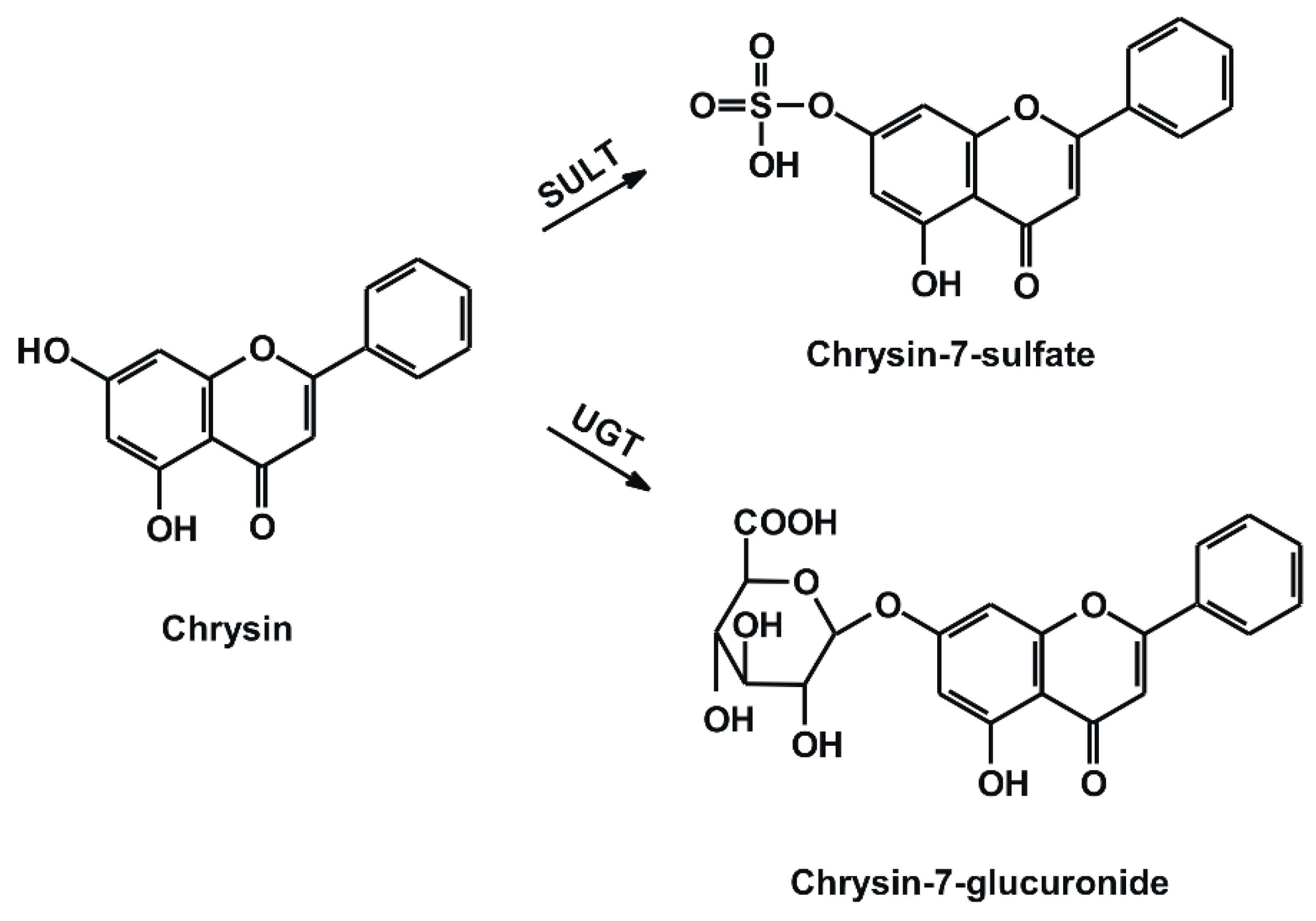
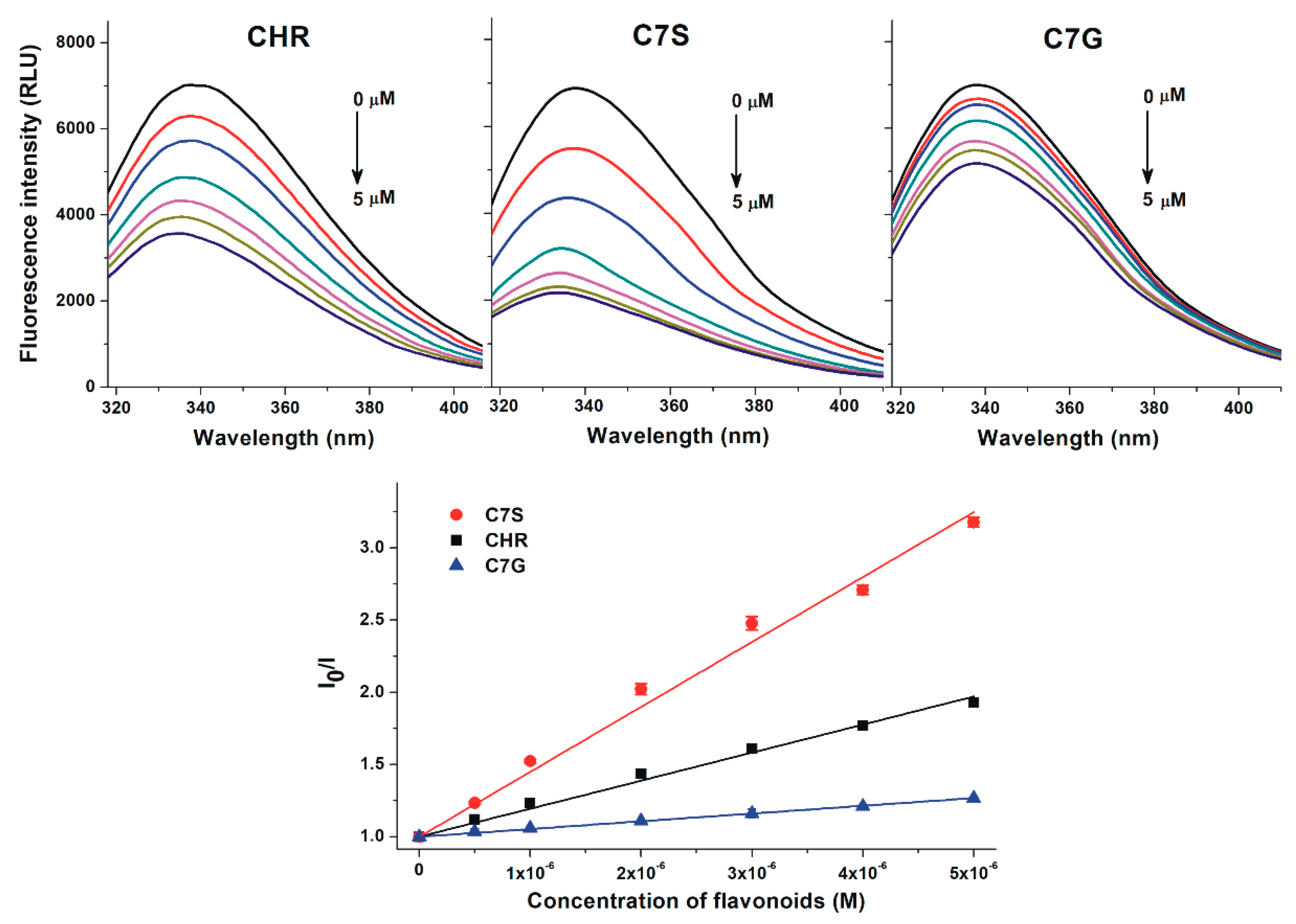
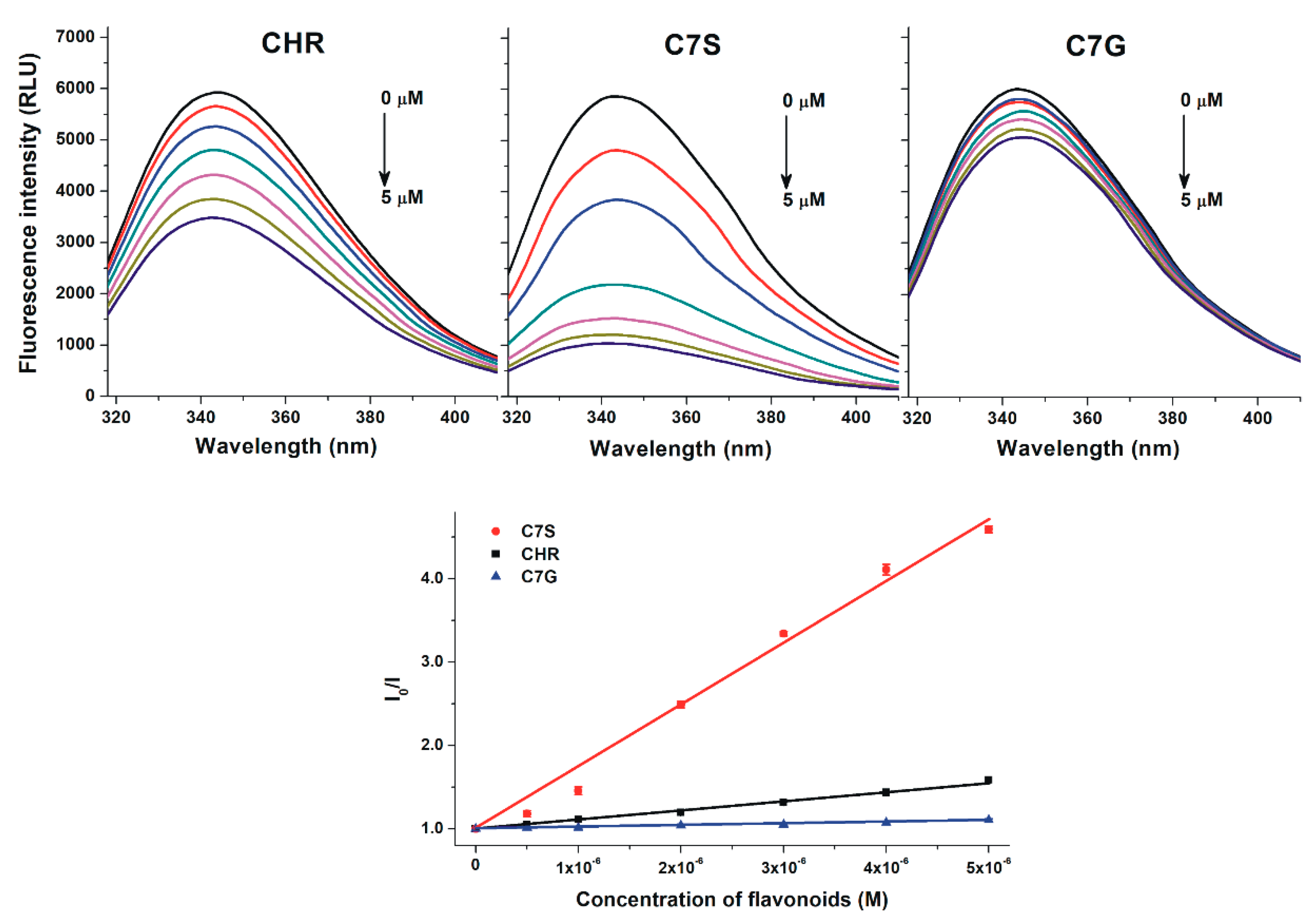
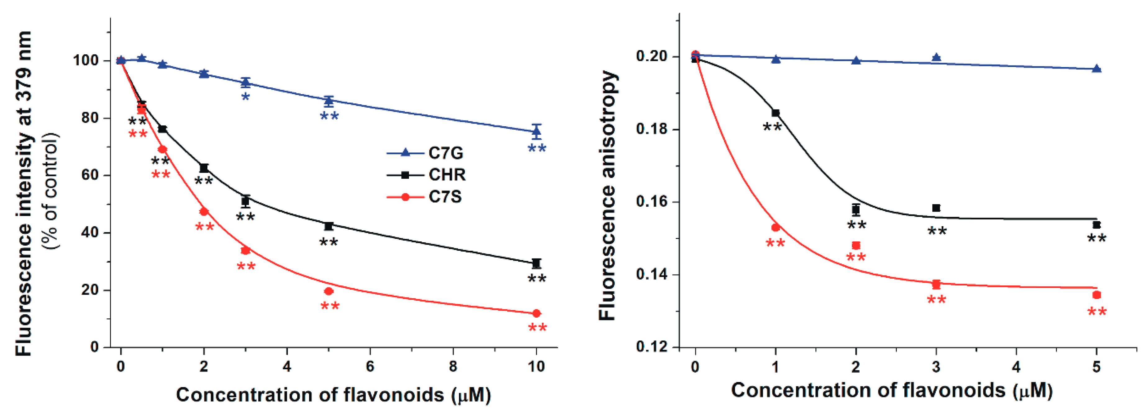
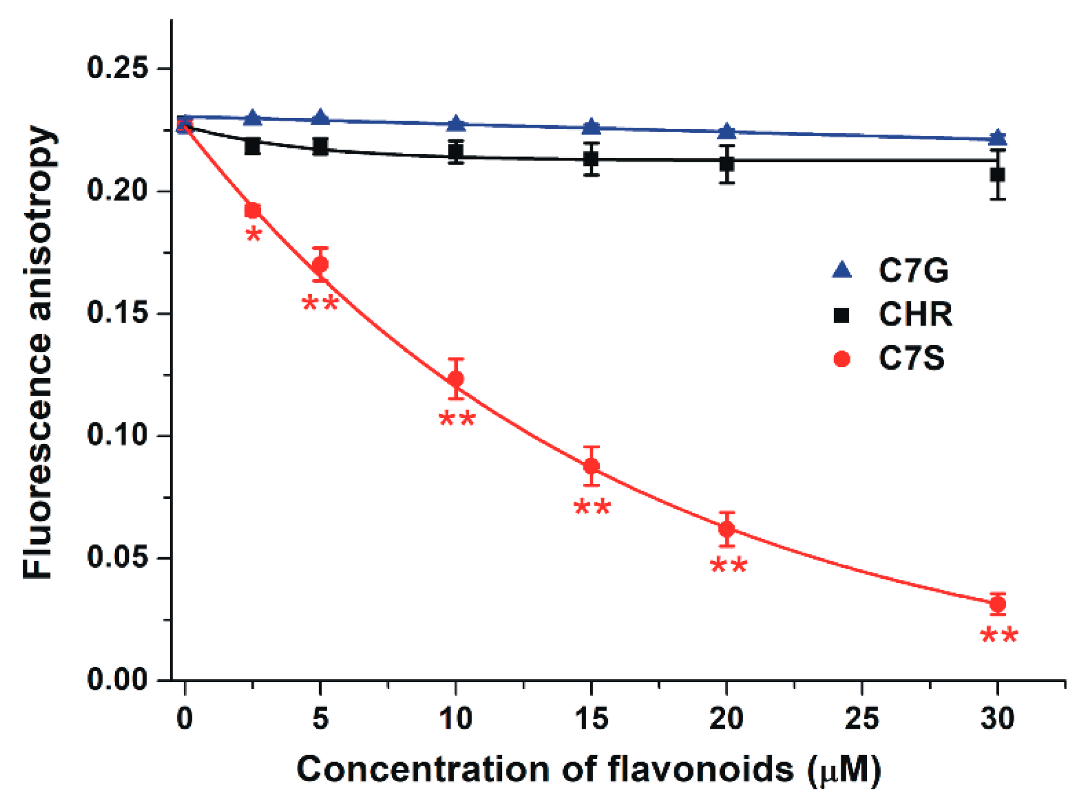
2.1. Interaction of Chrysin, C7S, and C7G with Human and Bovine Serum Albumins

To investigate the binding affinity of chrysin, C7S, and C7G toward HSA and bovine serum albumin (BSA), fluorescence quenching experiments were performed. BSA is commonly applied as a model protein to examine albumin–ligand interactions (because it is cheaper and structurally similar to HSA) [29]. In a concentration-dependent fashion, chrysin and its metabolites induced a significant decrease in the fluorescence emission signal of HSA at 340 nm (Figure 2, top; λex = 295 nm). C7G caused weaker, and C7S produced stronger, quenching effects on HSA than the parent compound. Stern–Volmer plots of flavonoid–HSA interactions show a good linearity (Figure 2, bottom). Chrysin and its metabolites induced very similar quenching effects on BSA compared to HSA (Figure 3). Stern–Volmer quenching constants (Ksv) and binding constants (K) were calculated based on the graphical application of the Stern–Volmer equation and evaluated by Hyperquad2006 software, respectively. Table 1 demonstrates the decimal logarithmic values of Ksv and K for flavonoid–albumin complexes. Both albumins formed the most stable complexes with C7S followed by chrysin and C7G. The stability of C7S–HSA and C7G–HSA complexes are approximately threefold higher and threefold lower vs. the chrysin–HSA complex, respectively. Affinities of the individual flavonoid–HSA and flavonoid–BSA complexes are similar; however, chrysin and C7G bind to HSA stronger than they do to BSA, while C7S forms a more stable complex with BSA (Table 1).

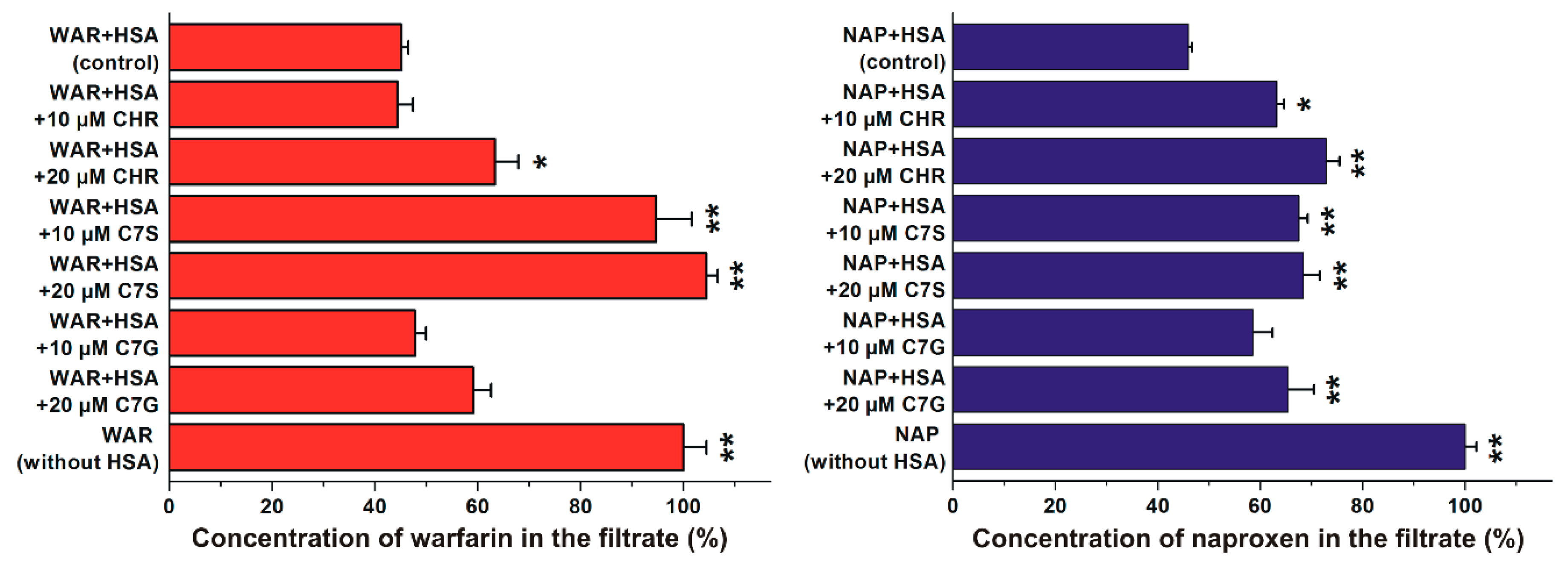
2.2. Effects of Flavonoids on Warfarin–HSA and Naproxen–HSA Interactions Based on Ultrafiltration Studies

In the following experiments, the displacing ability of chrysin, C7S, and C7G vs. the Site I ligand warfarin and the Site II ligand naproxen was evaluated based on ultrafiltration experiments. Interestingly, chrysin and its metabolites influenced the filtered fractions of both warfarin and naproxen. Under the applied conditions, C7G did not significantly change the concentration of filtered warfarin, while only the higher concentration of chrysin (20 μM) induced a statistically significant (albeit slight) increase of warfarin in the filtrate (Figure 4, left). However, even a 10 μM concentration of C7S caused a remarkable increase in warfarin in the filtrate. Furthermore, both concentrations of chrysin and C7S induced a statistically significant elevation of naproxen concentrations in the filtrate, while only a higher concentration of C7G produced a significant effect (Figure 4, right).

2.3. Effects of Chrysin and Its Metabolites on the Albumin-Binding of Warfarin Based on Fluorescence Spectroscopic Studies

In order to test the displacement of the Sudlow’s Site I ligand warfarin from HSA by flavonoids, increasing concentrations of chrysin and its metabolites were added to the warfarin–HSA complex. Displacement of warfarin from HSA results in a strong decrease in its fluorescence [30,31]. Our results demonstrate that each flavonoid significantly decreased the emission signal of warfarin at 379 nm (Figure 5, left): C7S induced the strongest decrease, followed by chrysin, while C7G showed only a slight (but statistically significant) effect.
In another model, fluorescence anisotropy was employed to test the effects of chrysin, C7S, and C7G on the albumin-binding of warfarin. Anisotropy gives information regarding the rotational freedom of a molecule. Our results demonstrate that C7G failed to affect the fluorescence anisotropy of warfarin, while chrysin and mainly C7S induced a strong decrease in anisotropy values (Figure 5, right).

2.4. Effects of Chrysin and Its Metabolites on the Albumin-Binding of Ochratoxin A

The mycotoxin ochratoxin A binds to HSA with very high affinity, occupying Sudlow’s Site I as a binding site [32,33]. Therefore, to confirm the involvement of Site I as the binding site of flavonoids tested, the displacement of ochratoxin A from HSA by chrysin, C7S, and C7G was examined by fluorescence anisotropy. Even high concentrations (2.5–30 μM vs. 1 μM ochratoxin A) of C7G and chrysin failed to affect the anisotropy of ochratoxin A; however, C7S induced a concentration-dependent decrease in anisotropy values (Figure 6).

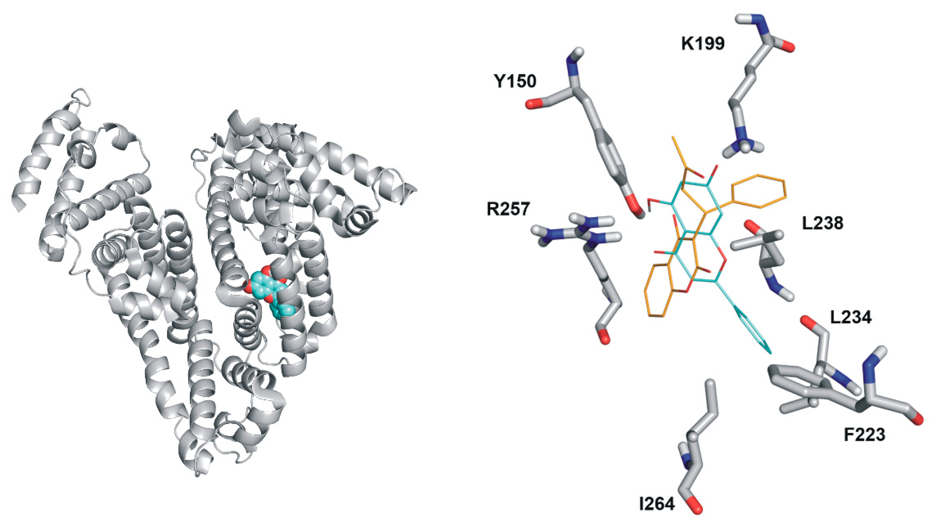
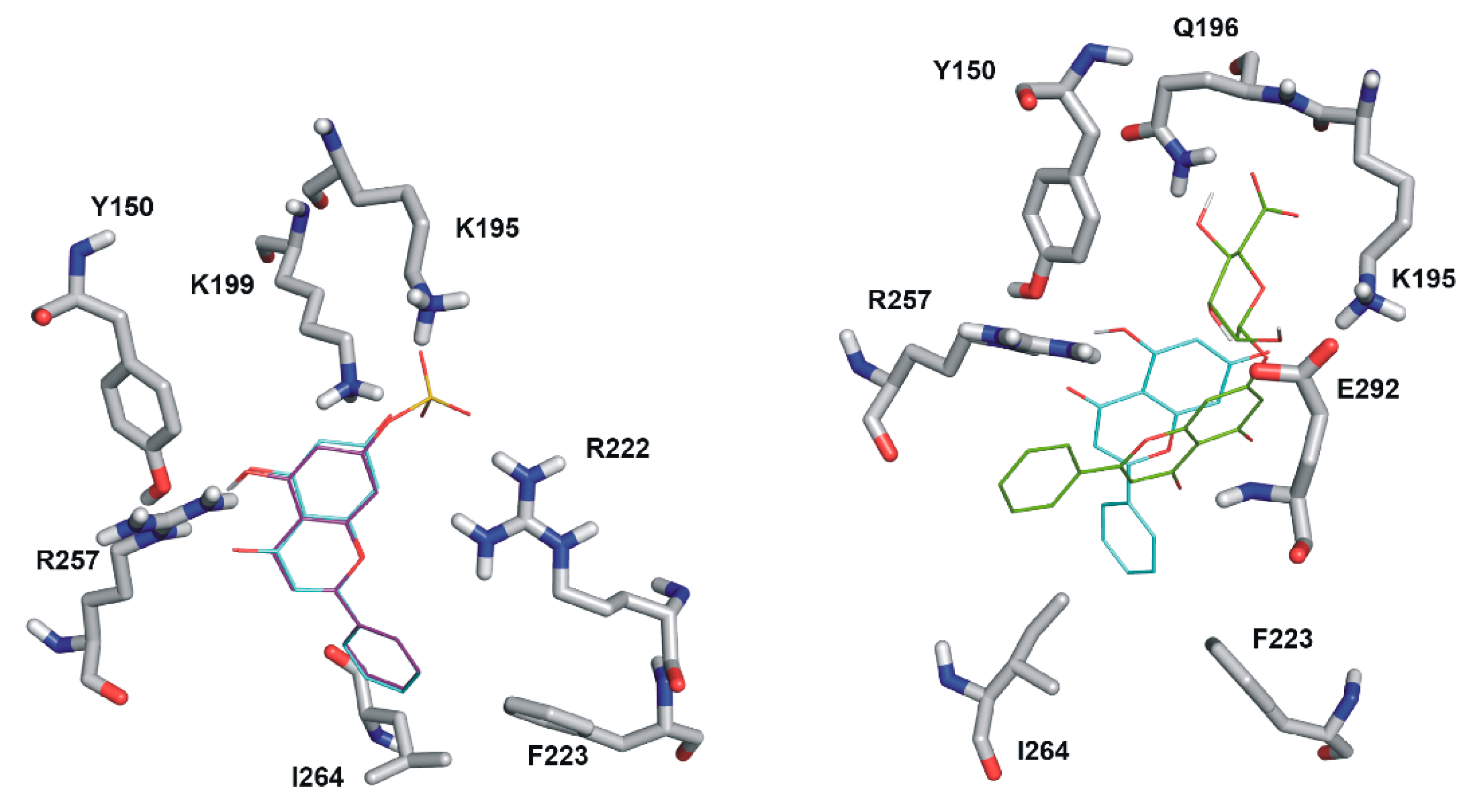
2.5. Modeling Studies

The binding positions of chrysin, C7S, and C7G in Sudlow’s Site I were investigated employing focused docking calculations with Autodock 4. Ranking of binding positions was performed by the binding energy calculated with the AutoDock 4 scoring function. Docking results show that the binding site of chrysin on HSA significantly overlaps with the binding site of the Sudlow’s Site I ligand warfarin (Figure 7). The first rank positions of chrysin and C7S show large similarities (Figure 8, left). The chrysin–HSA interaction is stabilized by the salt bridge with K199 and by hydrogen bonds with R257 and Y150. C7S occupies Site I in a very similar position than chrysin, but the formed complex is further stabilized by salt bridges with K195 and R222. The binding position of C7G is different (Figure 7, left) compared to chrysin and C7S. The position of glucuronic acid moiety is fixed by hydrogen bonds with K195, Q196, and E292.

3. Discussion

Chrysin, C7S, and C7G induced the significant decrease in the fluorescence signal of HSA (Figure 2, top), suggesting the formation of flavonoid–HSA complexes. Since emission signals were corrected with absorbance values of flavonoids at the excitation and emission wavelengths used (see in 4.2), we can exclude that the decrease in fluorescence resulting from the inner-filter effect. Using 295 nm as an excitation wavelength, the emitted light of HSA was exerted mainly by its only tryptophan moiety (Trp-214), which is located in Sudlow’s Site I [26,32,33]. Because flavonoids caused the significant quenching of Trp-214 even at a relatively low concentration (0.5 μmol/L), it is reasonable to hypothesize that the binding site of flavonoids needs to be located close to the tryptophan residue (e.g., in Site I). Both the good linearity of Stern–Volmer plots (Figure 2, bottom) and the evaluation of data with the Hyperquad software suggest the 1:1 stoichiometry of complex formation. KSV and K values showed good correlations (Table 1). Binding constant of the chrysin–HSA complex is in good agreement with the previously reported values [2,27,28]. Sometimes significant species differences in albumin-binding can be noticed [29,34]; however, the stability of the formed HSA and BSA complexes of chrysin (and chrysin conjugates) did not show large differences, as has also been described regarding the basic flavone structure by Xiao et al. [2]. Like previous investigations with quercetin and its sulfate metabolites [30,35,36,37], our results highlight that sulfate metabolites of flavonoids are able to bind to albumin with similar or even higher affinities than the aglycones. Considering these observations, it is reasonable to hypothesize that sulfate metabolites may also exert similar or higher effects on other proteins (e.g., biotransformation enzymes or transporters) than flavonoid aglycones, as has been reported regarding the interactions of quercetin-3’-sulfate with CYP2C9, CYP3A4, OAT1, or GLUT [30,38,39].
In order to test the binding sites of flavonoids in HSA as well as to investigate their displacing ability vs. drugs, site markers were applied. Since previous investigations suggest the possible involvement of Site I or Site II regarding chrysin–HSA interaction [2,28], the displacing effects of chrysin, C7S, and C7G were evaluated vs. warfarin (Site I) and naproxen (Site II) based on ultrafiltration studies. Because albumin is a large macromolecule (66.5 kDa), HSA and HSA-bound ligand molecules cannot pass through the filter unit with a 30 kDa molecular weight cut-off value. Therefore, albumin significantly decreases the concentrations of the site markers in the filtrate (Figure 4). In both cases (warfarin and naproxen), the same concentrations of the site markers (1 μmol/L both) were applied in the presence of HSA concentrations that induce an approximately 60% decrease in the site marker concentrations in the filtrate. Thus, the flavonoid-induced increase in filtered site marker concentrations suggests their displacement from HSA. C7G did not cause significant changes and chrysin induced slight (but statistically significant) interaction only at the higher concentration applied (20 μmol/L); however, C7S inflicted remarkable effects at both concentrations, resulting in practically the complete displacement of bound warfarin molecules from HSA (Figure 4, left). Chrysin, C7S, and C7G induced statistically significant (but only slight) displacement of the Site II marker naproxen (Figure 4, right). Based on these observations, it is reasonable to hypothesize that the high-affinity binding site of C7S is located in Site I (subdomain IIA), while the lower displacing ability of chrysin and C7G likely resulted from their lower affinity toward HSA. This hypothesis is also supported by the strong quenching effects of flavonoids on HSA, because the Trp-214 moiety is located in Subdomain IIA. Furthermore, chrysin and its conjugates may allosterically influence the albumin-binding of the Site II ligand naproxen as well.
To confirm our hypothesis that the examined flavonoids occupy Site I in HSA, further experiments were performed. Effects of chrysin, C7S, and C7G on the albumin-binding of warfarin were examined based on steady-state fluorescence spectroscopy and fluorescence anisotropy. The fluorescence emission intensity of HSA-bound warfarin is approximately 20-fold higher compared to free warfarin [30,31]. Therefore, the concentration-dependent reduction of the fluorescence signal of warfarin in the presence of flavonoids (Figure 5, left) suggests the displacement of warfarin from HSA. The displacing ability of flavonoids shows the same order than their binding affinity toward HSA: C7S > chrysin > C7G. In the following experiment, the effect of flavonoids on the fluorescence anisotropy of warfarin was examined. Warfarin, as a small molecule, has a large rotational freedom and consequently a relatively low anisotropy value. However, during its interaction with the HSA, its rotational freedom strongly decreases while its anisotropy value significantly increases. Therefore, the displacement of a small fluorophore from the protein leads to the decrease in its fluorescence anisotropy [31,40]. Anisotropy measurements also suggest the strongest displacement of warfarin by C7S, while a weaker and only a slight impact was noticed regarding chrysin and C7G, respectively (Figure 5, right). Thus, spectroscopic measurements confirmed our results derived from ultrafiltration studies.
To further support these results, the interactions of chrysin, C7S, and C7G were investigated with ochratoxin A. Ochratoxin A occupies Site I in the albumin [32,33], during which it forms extremely stable non-covalent complexes with HSA (logK = 7.4–7.6) [29,41]. In these experiments, only C7S was able to significantly decrease the fluorescence anisotropy of ochratoxin A (Figure 6), suggesting that the sulfate metabolite of chrysin can disrupt even this high-affinity interaction. Chrysin and C7G binds with lower affinity to HSA than C7S, which may explain why these flavonoids failed to displace ochratoxin A.
Finally, modeling studies were employed to explore the reason of the different binding affinities of chrysin, C7S, and C7G toward HSA. Our results suggest a similar binding position of chrysin and C7S (Figure 8). The sulfate group of C7S forms further two salt bridges with K195 and R222, which results in more stable complexes of C7S with HSA compared to chrysin. While the different binding position of C7G in Site I may explain its lower binding affinity compared to chrysin and C7S.
Several chrysin-containing dietary supplements are available on the Internet, which contain extremely high doses of chrysin (e.g., 500 mg or even higher dose/formulation unit) (see Supplementary Materials). The consumption of these supplements can result in the appearance of high total chrysin (chrysin and its metabolites) concentrations in the circulation, as has been reported regarding quercetin [42,43]. Since mainly the conjugated metabolites of chrysin appear at high concentrations in the circulation, C7S–HSA and C7G–HSA interactions may have high importance. C7S is the dominant metabolite of chrysin in the human circulation [7]. As it is demonstrated, compared with the parent compound, C7S binds with a higher affinity to HSA and exerts higher displacing ability vs. Sudlow’s Site I ligands. Based on spectroscopic and ultrafiltration studies, C7S has a similar or even higher effect on the albumin-binding of warfarin than quercetin or quercetin-3’-sulfate [30]. Furthermore, the displacement of ochratoxin A by C7S in our experimental model also underlines that C7S is a powerful competitor of Site I ligands. Like chrysin or quercetin-3’-sulfate [23,30,38], C7S is likely able to interact with biotransformation enzymes and transporters that may exacerbate the pharmacokinetic interactions of chrysin. However, to confirm this hypothesis, further investigations are needed.
In summary, interactions of chrysin, C7S, and C7G with human and bovine serum albumins were examined. Binding constants show the formation of stable flavonoid–albumin complexes. C7S binds to HSA with a three-fold higher affinity compared with chrysin, while C7G binds with a threefold lower affinity. Based on site marker experiments and modeling studies, tested compounds occupy Sudlow’s Site I in HSA; however, the binding position of C7G differs from chrysin or C7S. Interestingly, high concentrations of chrysin and its metabolites were also able to influence the albumin-binding of the Sudlow’s Site II ligand naproxen. Considering our observations, the high intake of chrysin (e.g., through the consumption of dietary supplements with high chrysin content) may interfere with the albumin-binding of several drugs, mainly due to the strong interaction of C7S with HSA.

4. Materials and Methods

4.1. Reagents

Chrysin, racemic warfarin, naproxen, ochratoxin A, human serum albumin (HSA), and bovine serum albumin (BSA) were purchased from Sigma-Aldrich (Waltham, MA, USA). Chyrsin-7-sulfate (C7S) was synthetized based on a method described regarding the sulfation of baicalein [44]. Chrysin-7-glucuronide (C7G) was obtained from Carbosynth (Compton, Berkshire, UK). Chrysin and its metabolites were dissolved in dimethyl sulfoxide (DMSO; Reanal, Budapest, Hungary, spectroscopic grade), and stock solutions (2000 μM each) were stored and protected from light at −20 °C.

4.2. Spectroscopic Measurements

Fluorescence spectroscopic measurements were carried out employing a Hitachi F-4500 fluorimeter (Tokyo, Japan). Absorption spectra of flavonoids were recorded using a HALO DB-20 UV-Vis spectrophotometer (Dynamica, London, UK). Spectroscopic measurements were performed in phosphate-buffered saline (pH 7.4; PBS: 8.0 g/L NaCl, 0.2 g/L KCl, 1.81 g/L Na2HPO4·2H2O, and 0.24 g/L KH2PO4) at room temperature, in the presence of air.
The complex formation of flavonoids with albumin was tested with both HSA and BSA. Binding constants of flavonoid–albumin complexes were determined by fluorescence quenching, during which emission spectra of albumins (2 µmol/L) were recorded in the presence of increasing flavonoid concentrations (0.0, 0.5, 1.0, 2.0, 3.0, 4.0 and 5.0 µmol/L; λexc = 295 nm). Quenching studies were evaluated based on the graphical application of the Stern–Volmer equation [31,34]. To eliminate the potential inner filter effects of flavonoids, fluorescence intensities were corrected as described [31,34].
Binding constants (K) of flavonoid–albumin complexes were also evaluated by non-linear fitting employing the Hyperquad2006 program package (Protonic Software, Leeds, UK) as described elsewhere [34,37]. Binding constants and stoichiometry were quantified based on the model with the lowest standard deviation.
Displacement of warfarin (site marker of Sudlow’s Site I) from HSA was examined based on the previously reported model [30,31]. Increasing concentrations of chrysin, C7S, and C7G (0.0, 0.5, 1.0, 2.0, 3.0, 5.0, and 10 µmol/L) were added to standard concentrations of warfarin and HSA (1.0 and 3.5 µmol/L, respectively). Thereafter, fluorescence emission spectra were recorded using 317 nm as excitation wavelength.
To confirm further the displacement of warfarin from HSA by chrysin, C7S, and C7G, fluorescence anisotropy measurements were also performed. Increasing concentrations of flavonoids (0.0, 1.0, 2.0, 5.0, and 10 µmol/L) were added to warfarin and HSA (1.0 and 2.0 µmol/L, respectively). Fluorescence anisotropy values were determined (λex = 317 nm; λem = 379 nm) as described [31].
Our previously described fluorescence anisotropy-based model [45] was employed to examine the displacing ability of the test compounds vs. ochratoxin A (another ligand of Sudlow’s Site I). Increasing concentrations of flavonoids (0.0, 2.5, 5.0, 10, 15, 20, and 30 µmol/L) were added to ochratoxin A and HSA (1.0 and 1.5 µmol/L, respectively), after which anisotropy values were determined at 393 and 446 nm excitation and emission wavelengths, respectively (wavelength maxima of albumin-bound ochratoxin A).

4.3. Ultrafiltration Experiments

Effects of chrysin, C7S, and C7G on the albumin-binding of Sudow’s Site I (warfarin) and Site II (naproxen) markers were tested by ultrafiltration. In these experiments, Pall MicrosepTM Advance Centrifugal Devices (Pall Corporation, Ann Arbor, MI, USA) (from VWR) with a 30 kDa molecular weight cut-off value were employed, as described elsewhere [46]. To examine the displacement of warfarin by flavonoids, samples contained warfarin and HSA (1.0 and 5.0 µmol/L, respectively) in the absence and presence of flavonoids (10 or 20 µmol/L). Furthermore, to test the displacing ability of chrysin and its metabolites vs. naproxen, flavonoids (10 or 20 µmol/L) were added to naproxen and HSA (1.0 and 1.7 µmol/L, respectively). Each sample was prepared in PBS (pH 7.4). Before ultrafiltration, filter units were washed once with 3 mL of water, and twice with 3 mL of PBS (to completely eliminate glycerol from the filters). Thereafter, samples (2.5 mL each) were driven through the filter units by centrifugation at 7500 g and 25 °C for 10 min (fixed angle rotor). Concentrations of warfarin and naproxen in the filtrate were quantified with HPLC (see details in Section 4.4).

4.4. HPLC Analyses

The HPLC system used to quantify warfarin and naproxen was equipped with a pump (model 510, Waters, Milford, MA, USA) and an injector (Rheodyne 7125, Rheodyne, Berkeley, CA, USA) with a 20 µL sample loop. Warfarin and naproxen were analyzed with a fluorescence detector (Jasco FP-920, Reanal) and an UV detector (Waters 486), respectively. Data were evaluated using Millennium Chromatography Manager (Waters).
Warfarin was analyzed based on the previously reported method without modifications [46]. Methanol (VWR), acetonitrile (VWR), and sodium phosphate buffer (20 mM, pH 7.0) (25:5:70 v/v%) were applied in the mobile phase with 1 mL/min flow rate during the isocratic elution. Samples were driven through a guard column (Phenomenex SecurityGuardTM Catridge C18 4.0 × 3.0 mm) linked to an analytical column (Nova-Pak C18 150 × 3.9 mm, 4 μm). Separation and analysis were performed at room temperature, during which warfarin was detected by fluorescence employing 310 and 390 nm as excitation and emission wavelengths, respectively.
Naproxen was quantified based on the previously reported method without modifications [46]. Acetonitrile and sodium acetate buffer (6.9 mM, pH 4.0) (50:50 v/v%) were applied in the mobile phase with a 1 mL/min flow rate during the isocratic elution. Samples were driven through a guard column (Phenomenex SecurityGuardTM Catridge C18 4.0 × 3.0 mm) linked to an analytical column (Phenomenex Gemini C18 150 × 4.6 mm, 3 µm). Separation and analysis were performed at room temperature, during which naproxen was detected by absorbance at 230 nm.

4.5. Modeling Studies

Docking calculations were performed using the AutoDock 4.2 program package [47]. Ligand 3D structures were downloaded from pubchem [48], and the protonation and charge states of ligands were calculated at pH 7.4 by msketch [49]. Energy-minimization of molecules was performed by the semi-empirical quantum chemistry program package, MOPAC. The geometries were optimized at a 0.001 gradient norm and subjected to subsequent force calculations using PM7 parameterization. In all cases, the force constant matrices were positive definite. The apo structure of HSA (PDB code 1a06) was used as a target of docking. The Gasteiger–Marsilli partial charges [50] were added to both ligand and target atoms and a Kollman united atom representation was applied for groups with non-polar bonds. For BD, the grid box was centered on the center of Sudlow’s Site I. A grid map with a box size of 90 × 90 × 90 points and 0.375 Å spacing was calculated by AutoGrid 4. During the focused docking calculations on Sudlow’s Site I, the grid box of 90 × 90 × 90 was centered on 35.000 31.825 37.000. In all calculations, the number of docking runs was set to 10 numbers of energy evaluations and generations were 20 million. Ligand conformations that resulted from the docking runs were ordered by the corresponding calculated ∆G values and clustered using a tolerance of 1.75 Å distance between cluster members. Conformations with the lowest binding energy within a cluster were selected as representatives.

4.6. Statistics

Data demonstrate the means (±SEM) from at least three independent experiments. During statistical analyses, the one-way ANOVA test (IBS SPP Statistics Version 21) was applied, and the level of significance was set at p < 0.05 and p < 0.01.

Supplementary Materials

Supplementary materials can be found at https://www.mdpi.com/1422-0067/19/12/4073/s1.

Author Contributions

Conceptualization, M.P.; Formal analysis, V.M., E.F.-N., G.S., C.H., B.L., and S.K.-M.; Funding acquisition, C.H. and M.P.; Investigation, V.M., E.F.-N., G.S., C.H., B.L., S.K.-M., B.B., and M.P.; Methodology, V.M., E.F.-N., C.H., and M.P.; Resources, B.B.; Writing—original draft, V.M., C.H., and M.P. Spectroscopic studies and Stern–Volmer evaluations were performed by V.M. and M.P. V.M. and E.F-N. carried out ultrafiltration studies and the related HPLC analyses. B.L. and S.K-M. performed the Hyperquad evaluations based on spectroscopic data. G.S. and C.H. carried out the modeling studies. B.B. synthetized chrysin-7-sulfate. All authors have read, edited, and approved the final version of the paper.

Funding

The project was founded by the European Union and co-financed by the European Social Fund (EFOP-3.6.1.-16-2016-00004). This work was supported by the Hungarian National Research, Development and Innovation Office (K123836: G.S. and C.H.; FK124331: B.B.).

Acknowledgments

The authors thank István Schweibert for his excellent assistance in the experimental work. The project was supported by the ÚNKP-18-3 and ÚNKP-18-4 New National Excellence Program of the Ministry of Human Capacities and by the University of Pécs in the frame of Pharmaceutical Talent Centre program. Financial support of the GINOP-2.3.2-15-2016-00049 grant is highly appreciated (S.K.). We acknowledge the grant of computer time from CSCS Swiss National Supercomputing Centre, and the Governmental Information Technology Development Agency, Hungary. We acknowledge that the results of this research have been achieved using the DECI resource Archer based in the UK at the National Supercomputing Service with support from the PRACE (Partnership for Advanced Computing in Europe) aisbl (Association internationale sans but lucratif). The University of Pécs is acknowledged for a support by the 17886-4/23018/FEKUTSTRAT excellence grant.

Conflicts of Interest

The authors declare no conflict of interest.

Abbreviations

BSAbovine serum albumin
C7Gchrysin-7-glucuronide
C7Schrysin-7-sulfate
CHRchrysin
CYPcytochrom P450 enzymes
GLUTglucose transporter
HPLChigh-performance liquid chromatography
HSAhuman serum albumin
OAT1organic anion transporter 1
PBSphosphate-buffered saline
SULTsulfotransferase
UGTuridine 5′-diphospho-glucuronosyltransferase

References

  1. Havsteen, B.H. The biochemistry and medical significance of the flavonoids. Pharmacol. Ther. 2002, 96, 67–202. [Google Scholar] [CrossRef]
  2. Xiao, J.; Cao, H.; Wang, Y.; Yamamoto, K.; Wei, X. Structure-affinity relationship of flavones on binding to serum albumins: Effect of hydroxyl groups on ring A. Mol. Nutr. Food Res. 2010, 54, 253–260. [Google Scholar] [CrossRef] [PubMed]
  3. Liu, Q.; Chen, T.T.; Chao, H. Flavonoids inhibiting glycation of bovine serum albumin: Affinity-activity relationship. Chem. Pap. 2015, 69, 409–415. [Google Scholar] [CrossRef]
  4. Korobkova, E.A. Effect of Natural Polyphenols on CYP Metabolism: Implications for Diseases. Chem. Res. Toxicol. 2015, 28, 1359–1390. [Google Scholar] [CrossRef]
  5. Alvarez, A.I.; Real, R.; Pérez, M.; Mendoza, G.; Prieto, J.G.; Merino, G. Modulation of the activity of ABC transporters (P-glycoprotein, MRP2, BCRP) by flavonoids and drug response. J. Pharm. Sci. 2010, 99, 598–617. [Google Scholar] [CrossRef] [PubMed]
  6. Cermak, R.; Wolffram, S. The potential of flavonoids to influence drug metabolism and pharmacokinetics by local gastrointestinal mechanisms. Curr. Drug Metab. 2006, 7, 729–744. [Google Scholar] [CrossRef] [PubMed]
  7. Walle, T.; Otake, Y.; Brubaker, J.A.; Walle, U.K.; Halushka, P.V. Disposition and metabolism of the flavonoid chrysin in normal volunteers. Br. J. Clin. Pharmacol. 2001, 51, 143–146. [Google Scholar] [CrossRef] [PubMed] [Green Version]
  8. Lambert, J.D.; Sang, S.; Lu, A.Y.H.; Yang, C.S. Metabolism of Dietary Polyphenols and Possible Interactions of Drugs. Curr. Drug Metab. 2007, 8, 499–507. [Google Scholar] [CrossRef]
  9. Siess, M.H.; Le Bon, A.M.; Canivenc-Lavier, M.C.; Amiot, M.J.; Sabatier, S.; Aubert, S.Y.; Suschetet, M. Flavonoids of Honey and Propolis: Characterization and Effects on Hepatic Drug-Metabolizing Enzymes and Benzo [a]pyrene-DNA Binding in Rats. J. Agric. Food Chem. 1996, 44, 2297–2301. [Google Scholar] [CrossRef]
  10. Anandhi, R.; Annadurai, T.; Anitha, T.S.; Muralidharan, A.R.; Najmunnisha, K.; Nachiappan, V.; Thomas, P.A.; Geraldine, P. Antihypercholesterolemic and antioxidative effects of an extract of the oyster mushroom, Pleurotus ostreatus, and its major constituent, chrysin, in Triton WR-1339-induced hypercholesterolemic rats. J. Physiol. Biochem. 2013, 69, 313–323. [Google Scholar] [CrossRef]
  11. Chen, F.; Li, H.L.; Tan, Y.F.; Li, Y.H.; Lai, W.Y.; Guan, W.W.; Zhang, J.Q.; Zhao, Y.S.; Qin, Z.M. Identification of known chemicals and their metabolites from Alpinia oxyphylla fruit extract in rat plasma using liquid chromatography/tandem mass spectrometry (LC–MS/MS) with selected reaction monitoring. J. Pharm. Biomed. Anal. 2014, 97, 166–177. [Google Scholar] [CrossRef] [PubMed]
  12. Kao, J.C.; Zhou, C.; Sherman, M.; Laughton, C.A.; Chen, S. Molecular basis of the inhibition of human aromatase (estrogen synthetase) by flavone and isoflavone phytoestrogens: A site-directed mutagenesis study. Environ. Health Perspect. 1998, 106, 85–92. [Google Scholar] [CrossRef] [PubMed]
  13. Moon, Y.J.; Wang, X.; Morris, M.E. Dietary flavonoids: Effects on xenobiotic and carcinogen metabolism. Toxicol. Vitro 2006, 20, 187–210. [Google Scholar] [CrossRef]
  14. Ciftci, O.; Ozdemir, I.; Aydin, M.; Beytur, A. Beneficial effects of chrysin on the reproductive system of adult male rats. Andrologia 2012, 44, 181–186. [Google Scholar] [CrossRef] [PubMed]
  15. Nabavi, S.F.; Braidy, N.; Habtemariam, S.; Orhan, I.E.; Daglia, M.; Manayi, A.; Gortzi, O.; Nabavi, S.M. Neuroprotective effects of chrysin: From chemistry to medicine. Neurochem. Int. 2015, 90, 224–231. [Google Scholar] [CrossRef] [PubMed]
  16. Filho, C.B.; Jesse, C.R.; Donato, F.; Fabbro, L.D.; de Gomes, M.G.; Goes, A.T.R.; Souza, L.C.; Boeira, S.P. Chrysin promotes attenuation of depressive-like behavior and hippocampal dysfunction resulting from olfactory bulbectomy in mice. Chem. Biol. Interact. 2016, 260, 154–162. [Google Scholar] [CrossRef] [PubMed]
  17. Satyanarayana, K.; Sravanthi, K.; Shaker, I.A.; Ponnulakshmi, R.; Selvaraj, J. Role of chrysin on expression of insulin signaling molecules. J. Ayurveda Integr. Med. 2015, 6, 248–258. [Google Scholar] [CrossRef]
  18. Cho, H.; Yun, C.W.; Park, W.K.; Kong, J.Y.; Kim, K.S.; Park, Y.; Lee, S.; Kim, B.K. Modulation of the activity of pro-inflammatory enzymes, COX-2 and iNOS, by chrysin derivatives. Pharmacol. Res. 2004, 49, 37–43. [Google Scholar] [CrossRef]
  19. Xiao, J.; Zhai, H.; Yao, Y.; Wang, C.; Jiang, W.; Zhang, C.; Simard, A.R.; Zhang, R.; Hao, J. Chrysin attenuates experimental autoimmune neuritis by suppressing immuno-inflammatory responses. Neuroscience 2014, 262, 156–164. [Google Scholar] [CrossRef]
  20. Pushpavalli, G.; Kalaiarasi, P.; Veeramani, C.; Pugalendi, K.V. Effect of chrysin on hepatoprotective and antioxidant status in d-galactosamine-induced hepatitis in rats. Eur. J. Pharmacol. 2010, 631, 36–41. [Google Scholar] [CrossRef]
  21. Galijatovic, A.; Otake, Y.; Walle, U.K.; Walle, T. Extensive metabolism of the flavonoid chrysin by human Caco-2 and Hep G2 cells. Xenobiotica 1999, 29, 1241–1256. [Google Scholar] [CrossRef] [PubMed]
  22. Ge, S.; Gao, S.; Yin, T.; Hu, M. Determination of Pharmacokinetics of Chrysin and Its Conjugates in Wild-Type FVB and Bcrp1 Knockout Mice Using a Validated LC-MS/MS Method. J. Agric. Food Chem. 2015, 63, 2902–2910. [Google Scholar] [CrossRef] [PubMed]
  23. Kimura, Y.; Ito, H.; Ohnishi, R.; Hatano, T. Inhibitory effects of polyphenols on human cytochrome P450 3A4 and 2C9 activity. Food Chem. Toxicol. 2010, 48, 429–435. [Google Scholar] [CrossRef] [PubMed]
  24. Pingili, R.B.; Pawar, A.K.; Challa, S.R. Systemic exposure of Paracetamol (acetaminophen) was enhanced by quercetin and chrysin co-administration in Wistar rats and in vitro model: Risk of liver toxicity. Drug Dev. Ind. Pharm. 2015, 41, 1793–1800. [Google Scholar] [CrossRef] [PubMed]
  25. Schmidt, S.; Gonzalez, D.; Derendorf, H. Significance of protein binding in pharmacokinetics and pharmacodynamics. J. Pharm. Sci. 2010, 99, 1107–1122. [Google Scholar] [CrossRef] [PubMed]
  26. Fanali, G.; Di Masi, A.; Trezza, V.; Marino, M.; Fasano, M.; Ascenzi, P. Human serum albumin: From bench to bedside. Mol. Asp. Med. 2012, 33, 209–290. [Google Scholar] [CrossRef]
  27. Zhang, G.; Chen, X.; Guo, J.; Wang, J. Spectroscopic investigation of the interaction between chrysin and bovine serum albumin. J. Mol. Struct. 2010, 921, 346–351. [Google Scholar] [CrossRef]
  28. Tu, B.; Chen, Z.F.; Liu, Z.J.; Li, R.R.; Ouyang, Y.; Hu, Y.J. Study of the structure-activity relationship of flavonoids based on their interaction with human serum albumin. RSC Adv. 2015, 5, 73290–73300. [Google Scholar] [CrossRef]
  29. Poór, M.; Li, Y.; Matisz, G.; Kiss, L.; Kunsági-Máté, S.; Kőszegi, T. Quantitation of species differences in albumin-ligand interactions for bovine, human and rat serum albumins, using fluorescence spectroscopy: A test case with some Sudlow’s site I ligands. J. Lumin. 2014, 145, 767–773. [Google Scholar] [CrossRef]
  30. Poór, M.; Boda, G.; Needs, P.W.; Kroon, P.A.; Lemli, B.; Bencsik, T. Interaction of quercetin and its metabolites with warfarin: Displacement of warfarin from human serum albumin and inhibition of CYP2C9. Biomed. Pharmacother. 2017, 88, 574–581. [Google Scholar] [CrossRef]
  31. Poór, M.; Boda, G.; Mohos, V.; Kuzma, M.; Bálint, M.; Hetényi, C.; Bencsik, T. Pharmacokinetic interaction of diosmetin and silibinin with other drugs: Inhibiton of CYP2C9-mediated biotransformation and displacement from serul albumin. Biomed. Pharmacother. 2018, 102, 912–921. [Google Scholar] [CrossRef] [PubMed]
  32. Il’ichev, I.V.; Perry, J.L.; Simon, J.D. Interaction of Ochratoxin A with Human Serum Albumin. A Common Binding Site of Ochratoxin A and Warfarin in Subdomain IIA. J. Phys. Chem. B 2002, 106, 460–465. [Google Scholar] [CrossRef]
  33. Il’ichev, Y.V.; Perry, J.L.; Rüker, F.; Dockal, M.; Simon, J.D. Interaction of ochratoxin A with human serum albumin. Binding sites localized by competitive interactions with the native protein and its recombinant fragments. Chem. Biol. Interact. 2002, 141, 275–293. [Google Scholar] [CrossRef]
  34. Faisal, Z.; Lemli, B.; Szerencsés, D.; Kunsági-Máté, S.; Bálint, M.; Hetényi, C.; Kuzma, M.; Mayer, M.; Poór, M. Interactions of zearalenone and its reduced metabolites α-zearalenol and β-zearalenol with serum albumins: Species differences, binding sites, and thermodynamics. Mycotoxin Res. 2018, 34, 269–278. [Google Scholar] [CrossRef] [PubMed]
  35. Dangles, O.; Dufour, C.; Manach, C.; Morand, C.; Remesy, C. Binding of flavonoids to plasma proteins. Methods Enzymol. 2001, 335, 319–333. [Google Scholar] [CrossRef] [PubMed]
  36. Janisch, K.M.; Williamson, G.; Needs, P.; Plumb, G.W. Properties of quercetin conjugates: Modulation of LDL oxidation and binding to human serum albumin. Free Radic. Res. 2004, 38, 877–884. [Google Scholar] [CrossRef] [PubMed]
  37. Poór, M.; Boda, G.; Kunsági-Máté, S.; Needs, P.W.; Kroon, P.A.; Lemli, B. Fluorescence spectroscopic evaluation of the interactions of quercetin, isorhamnetin, and quercetin-3′-sulfate with different albumins. J. Lumin. 2018, 194, 156–163. [Google Scholar] [CrossRef]
  38. Miron, A.; Aprotosoaie, A.C.; Trifan, A.; Xiao, J. Flavonoids as modulators of metabolic enzymes and drug transporters. Ann. N. Y. Acad. Sci. 2017, 1398, 152–167. [Google Scholar] [CrossRef] [PubMed]
  39. Csepregi, R.; Temesfői, V.; Sali, N.; Poór, M.; Needs, P.W.; Kroon, P.A.; Kőszegi, T. A one-step extraction and luminescence assay for quantifying glucose and ATP levels in cultured HepG2 cells. Int. J. Mol. Sci. 2018, 19, 2670. [Google Scholar] [CrossRef] [PubMed]
  40. Zhang, G.; Wang, L.; Pan, J. Probing the binding of the flavonoid diosmetin to human serum albumin by multispectroscopic techniques. J. Agric. Food Chem. 2012, 60, 2721–2729. [Google Scholar] [CrossRef] [PubMed]
  41. Poór, M.; Kunsági-Máté, S.; Bencsik, T.; Petrik, J.; Vladimir-Knezevic, S.; Kőszegi, T. Flavonoid aglycones can compete with Ochratoxin A for human serum albumin: A new possible mode of action. Int. J. Biol. Macromol. 2012, 51, 279–283. [Google Scholar] [CrossRef] [PubMed]
  42. Conquer, J.A.; Maiani, G.; Azzini, E.; Raguzzini, A.; Holub, B.J. Supplementation with quercetin markedly increases plasma quercetin concentration without effect on selected risk factors for heart disease in healthy subjects. J. Nutr. 1998, 128, 593–597. [Google Scholar] [CrossRef] [PubMed]
  43. Heinz, S.A.; Henson, D.A.; Nieman, D.C.; Austin, M.D.; Jin, F. A 12-week supplementation with quercetin does not affect natural killer cell activity, granulocyte oxidative burst activity or granulocyte phagocytosis in female human subjects. Br. J. Nutr. 2010, 104, 849–857. [Google Scholar] [CrossRef] [PubMed] [Green Version]
  44. Huang, W.H.; Lee, A.R.; Yang, C.H. Antioxidative and anti-inflammatory activities of polyhydroxyflavonoids of Scutellaria baicalensis GEORGI. Biosci. Biotechnol. Biochem. 2006, 70, 2371–2380. [Google Scholar] [CrossRef] [PubMed]
  45. Poór, M.; Lemli, B.; Bálint, M.; Hetényi, C.; Sali, N.; Kőszegi, T.; Kunsági-Máté, S. Interaction of citrinin with human serum albumin. Toxins 2015, 7, 5155–5166. [Google Scholar] [CrossRef] [PubMed]
  46. Mohos, V.; Bencsik, T.; Boda, G.; Fliszár-Nyúl, E.; Lemli, B.; Kunsági-Máté, S.; Poór, M. Interactions of casticin, ipriflavone, and resveratrol with serum albumin and their inhibitory effects on CYP2C9 and CYP3A4 enzymes. Biomed. Pharmacother. 2018, 107, 777–784. [Google Scholar] [CrossRef] [PubMed]
  47. Morris, G.M.; Huey, R.; Lindstrom, W.; Sanner, M.F.; Belew, R.K.; Goodsell, D.S.; Olson, A.J. Autodock4 and AutoDockTools4: Automated docking with selective receptor flexiblity. J. Comput. Chem. 2009, 16, 2785–2791. [Google Scholar] [CrossRef]
  48. Kim, S.; Thiessen, P.A.; Bolton, E.E.; Chen, J.; Fu, G.; Gindulyte, A.; Han, L.; He, J.; He, S.; Shoemaker, B.A.; et al. PubChem Substance and Compound databases. Nucleic Acids Res. 2016, 44, D1202–D1213. [Google Scholar] [CrossRef]
  49. Csizmadia, F. Java Applets and Modules Supporting Chemical Database Handling from Web Browsers. J. Chem. Inf. Comput. Sci. 2000, 40, 323–324. [Google Scholar] [CrossRef]
  50. Gasteiger, J.; Marsili, M. Iterative partial equalization of orbital electronegativity-a rapid access to atomic charges. Tetrahedron 1980, 36, 3219–3228. [Google Scholar] [CrossRef]
Figure 1. Chemical structures of chrysin, chrysin-7-sulfate, and chrysin-7-glucuronide (SULT: sulfotransferase; UGT: uridine 5′-diphospho-glucuronosyltransferase).
Ijms 19 04073 g001
Figure 2. Top graphs: Fluorescence quenching effects of chrysin (CHR; left), chrysin-7-sulfate (C7S; middle), and chrysin-7-glucuronide (C7G; right) on HSA (2 µmol/L) in PBS (pH 7.4; λex = 295 nm, λem = 340 nm). Bottom graph: Stern–Volmer plots of flavonoid–HSA complexes.
Ijms 19 04073 g002
Figure 3. Top graphs: Fluorescence quenching effects of chrysin (CHR; left), chrysin-7-sulfate (C7S; middle), and chrysin-7-glucuronide (C7G; right) on BSA (2 µmol/L) in PBS (pH 7.4; λex = 295 nm, λem = 340 nm). Bottom graph: Stern–Volmer plots of flavonoid–BSA complexes.
Ijms 19 04073 g003
Figure 4. Concentrations of warfarin (left) and naproxen (right) in the filtrate. Left: Before ultrafiltration, samples contained 1 µmol/L warfarin (WAR) with 5 µmol/L HSA in the absence and presence of 10 or 20 μM flavonoid concentrations in PBS (pH 7.4). Right: Before ultrafiltration, samples contained 1 µmol/L naproxen (NAP) with 1.7 µmol/L HSA in the absence and presence of 10 or 20 μM flavonoid concentrations in PBS (pH 7.4). (* p < 0.05, * *p < 0.01; CHR: chrysin; C7S: chrysin-7-sulfate; C7G: chrysin-7 glucuronide).
Ijms 19 04073 g004
Figure 5. Left: A concentration-dependent decrease in the fluorescence emission intensity of the warfarin–HSA complex (1.0 and 3.5 µmol/L of warfarin and HSA, respectively) in the presence of increasing flavonoid concentrations (0.0, 0.5, 1.0, 2.0, 3.0, 5.0, and 10 µmol/L). Right: Fluorescence anisotropy values of the warfarin–HSA complex (1.0 and 2.0 µmol/L of warfarin and HSA, respectively) in the presence of increasing concentrations of chrysin, C7S, or C7G (0.0, 1.0, 2.0, 3.0, and 5.0 µmol/L). (λex = 317 nm, λem = 379 nm; * p < 0.05, ** p < 0.01; CHR: chrysin; C7S: chrysin-7-sulfate; C7G: chrysin-7 glucuronide).
Ijms 19 04073 g005
Figure 6. Fluorescence anisotropy of the ochratoxin A–HSA complex (1.0 and 1.5 µmol/L of ochratoxin A and HSA, respectively) in the presence of increasing concentrations of flavonoids (0.0, 2.5, 5.0, 10, 15, 20, and 30 µmol/L) in PBS (pH 7.4; λex = 393 nm; λem = 446 nm; * p < 0.05, ** p < 0.01; CHR: chrysin; C7S: chrysin-7-sulfate; C7G: chrysin-7 glucuronide).
Ijms 19 04073 g006
Figure 7. Left: Chrysin (cyan spheres) occupies Sudlow’s Site I in HSA (represented with grey cartoon). Right: Rank1 binding positions of chrysin (cyan lines) and warfarin (orange lines; reference structure) in the HSA molecule.
Ijms 19 04073 g007
Figure 8. Left: Rank1 binding position of C7S (purple lines) vs. chrysin (cyan lines). Right: Rank1 binding position of C7G (green lines) compared to chrysin (cyan lines).
Ijms 19 04073 g008
Table 1. Stability of flavonoid–albumin complexes. Decimal logarithmic values (±SEM) of Stern–Volmer quenching constants (Ksv, unit: L/mol) and binding constants (K, unit: L/mol).
ComplexlogKSVlogK
Chrysin–HSA5.25 ± 0.025.41 ± 0.01
Chrysin-7-sulfate–HSA5.61 ± 0.035.88 ± 0.02
Chrysin-7-glucuronide–HSA4.71 ± 0.034.89 ± 0.00
Chrysin–BSA5.03 ± 0.065.20 ± 0.00
Chrysin-7-sulfate–BSA5.86 ± 0.046.20 ± 0.01
Chrysin-7-glucuronide–BSA4.34 ± 0.034.63 ± 0.01

