A Novel Dental Sealant Containing Dimethylaminohexadecyl Methacrylate Suppresses the Cariogenic Pathogenicity of Streptococcus mutans Biofilms

 , ,
, , 
Abstract
:
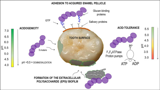
1. Introduction
2. Results
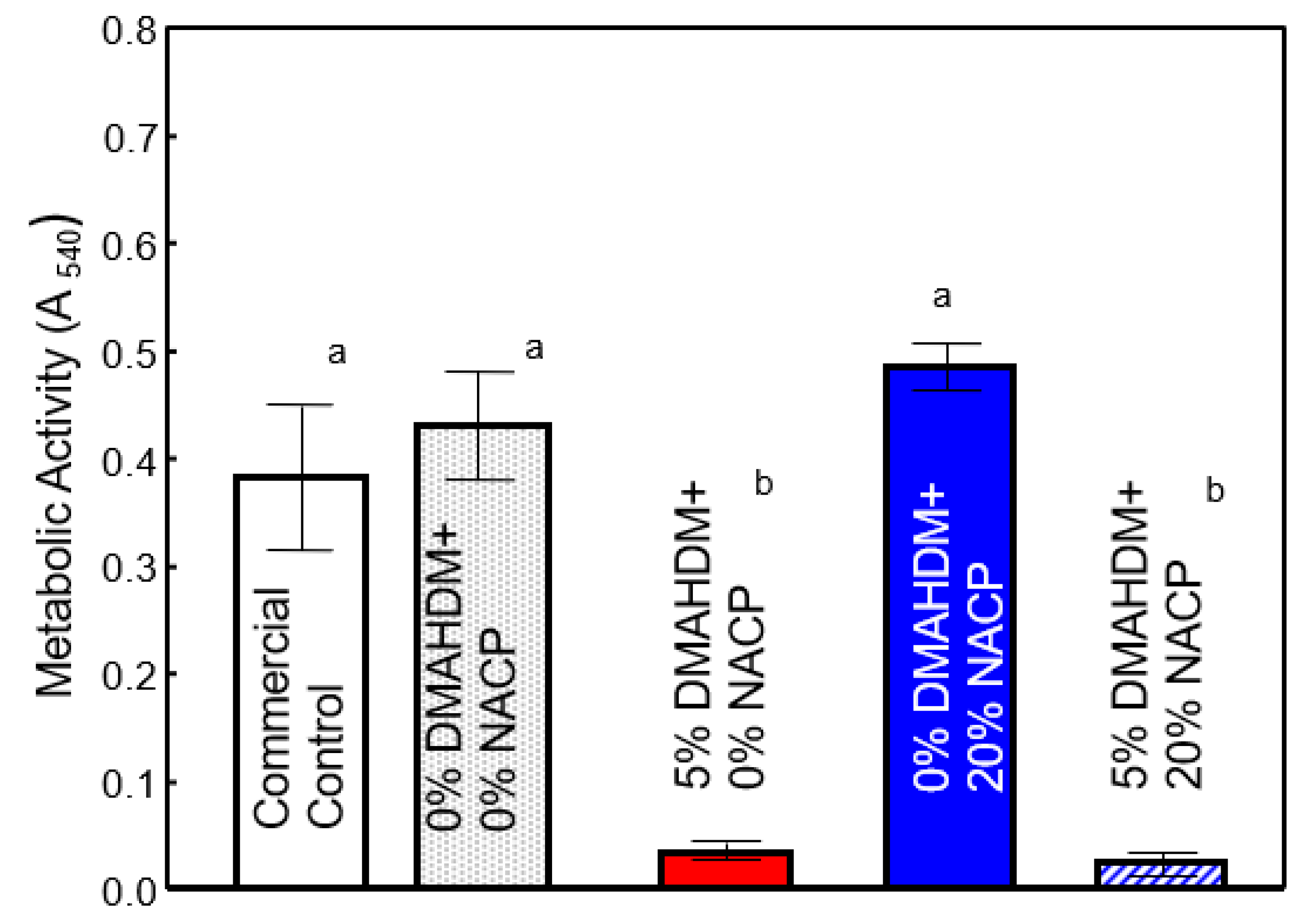
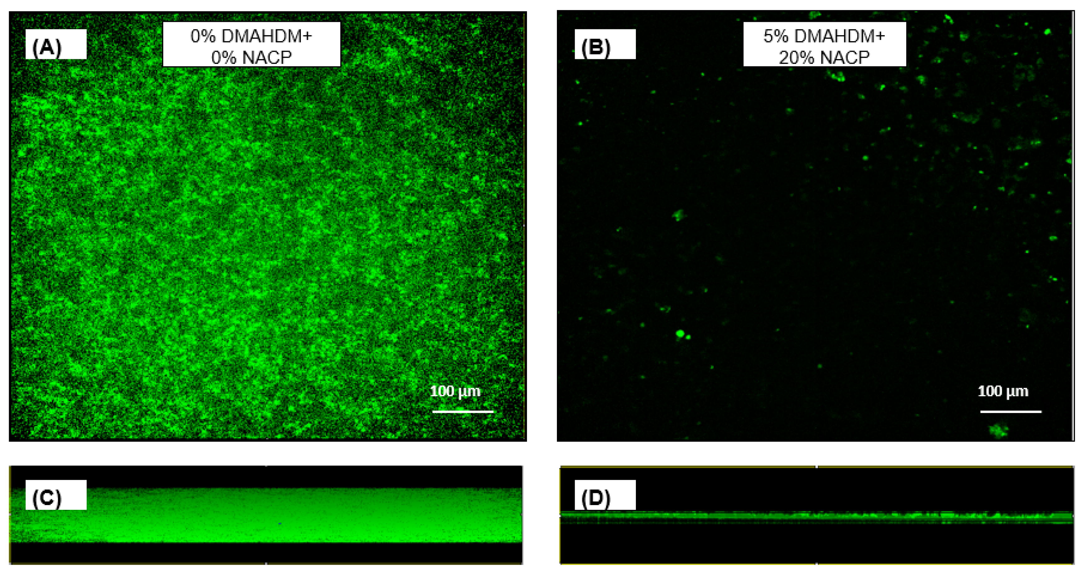
2.1. DMAHDM Reduced Biofilm Formation
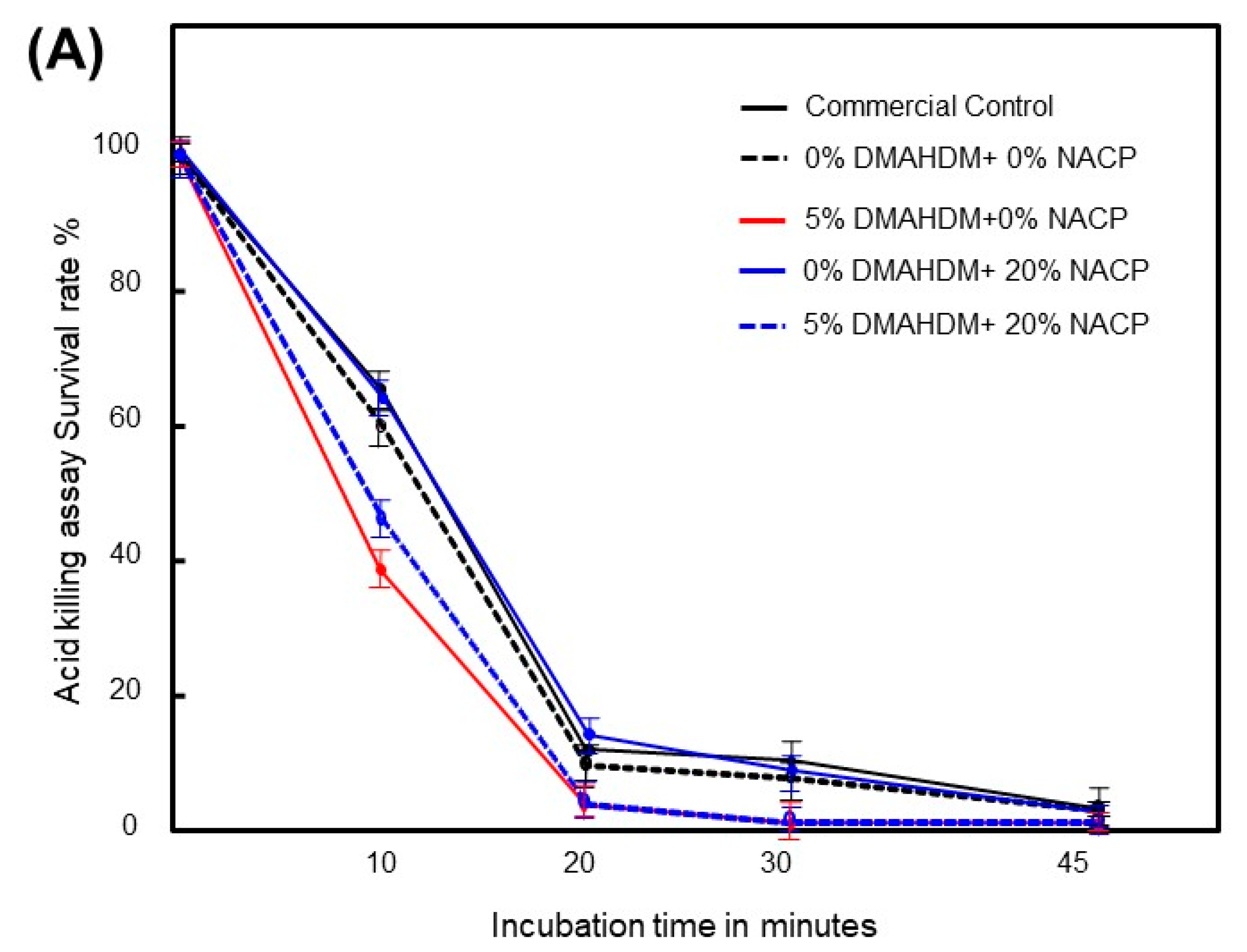
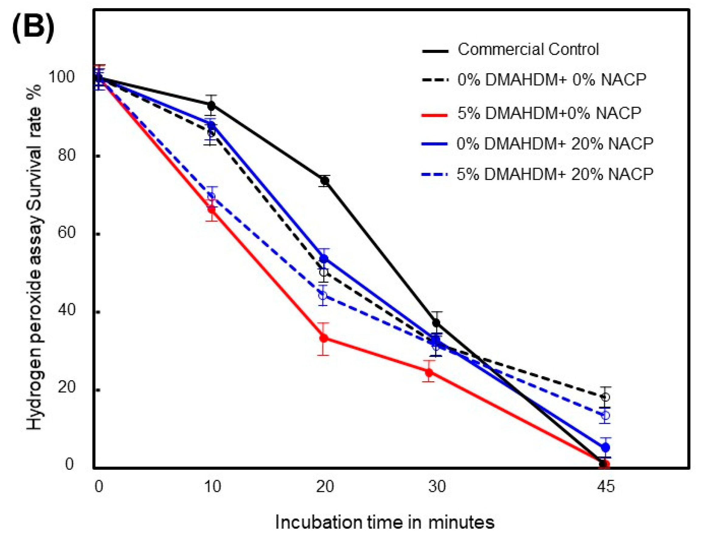
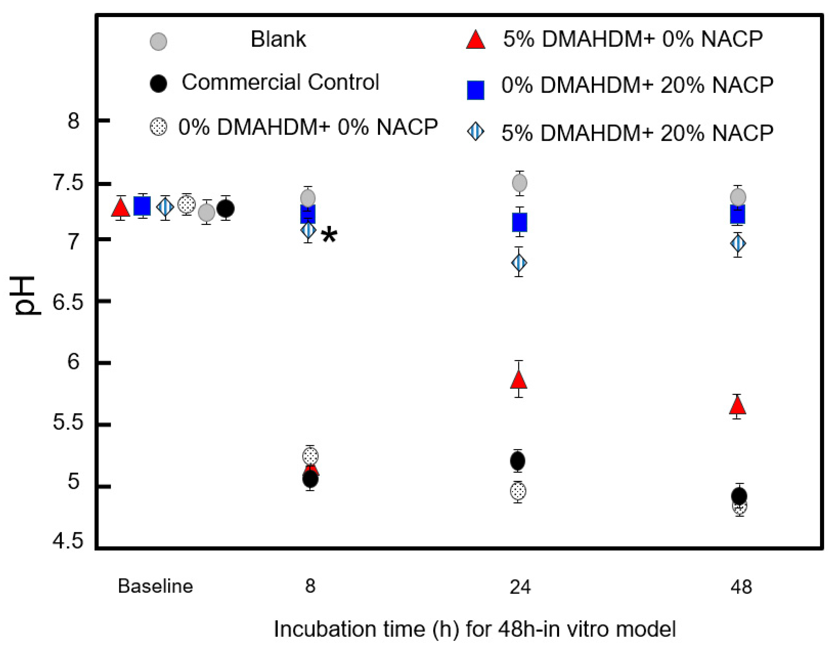
2.2. DMAHDM Alone and in Combination with NACP Alters Acid and Oxygen Tolerance of S. mutans
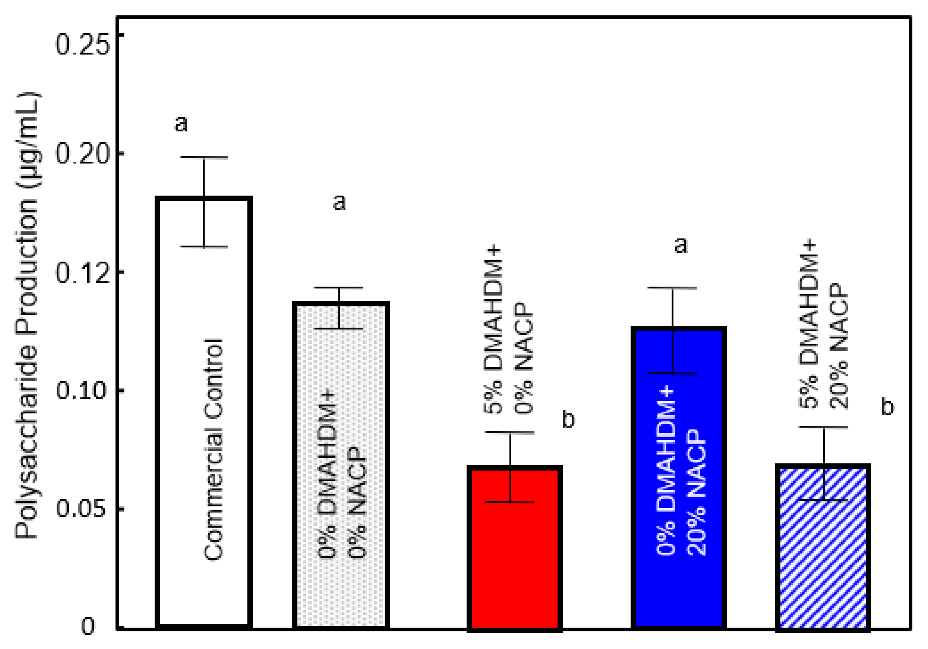
2.3. Attenuation of Exopolysaccharide Produced by S. mutans
3. Discussion
4. Materials and Methods
4.1. Development of Dental Resin Sealant Formulations
- Commercial High-viscosity Sealant/Flowable Composite control termed “Commercial Control” (Virtuoso, DenMat, Lompoc, CA, USA).
- Experimental Control Sealant termed “0% DMAHDM + 0% NACP; Experimental Control” (50% PEHB + 0% DMAHDM + 50% Glass + 0% NACP).
- Experimental Sealant termed “5% DMAHDM + 0% NACP” (45% PEHB + 5% DMAHDM + 50% Glass + 0% NACP).
- Experimental Sealant termed “0% DMAHDM + 20% NACP” (50% PEHB + 0% DMAHDM + 30% Glass + 20% NACP).
- Experimental Sealant termed “5% DMAHDM + 20% NACP” (45% PEHB + 5% DMAHDM + 30% Glass + 20% NACP).
4.2. Sample Preparation
4.3. S. mutans Biofilm Formation
4.4. Colony-Forming Unit Counts
4.5. Metabolic Activity
4.6. Acid Stress and Oxygen Stress Tolerance
4.7. Polysaccharide Production
4.8. Acid-Neutralizing Activity
4.9. Lactic Acid Production
4.10. Confocal Laser Scanning Microscopy
4.11. Statistical Analysis
4.12. Disclaimer
5. Conclusions
Author Contributions
Funding
Acknowledgments
Conflicts of Interest
References
- Manton, D.J. Child dental caries—A global problem of inequality. EClinicalMedicine 2018, 1, 3–4. [Google Scholar] [CrossRef] [PubMed]
- WHO. Sugars and Dental Caries. Available online: http://www.who.int/oral_health/publications/sugars-dental-caries-keyfacts/en/ (accessed on 24 February 2019).
- Papageorgiou, S.N.; Dimitraki, D.; Kotsanos, N.; Bekes, K.; van Waes, H. Performance of pit and fissure sealants according to tooth characteristics: A systematic review and meta-analysis. J. Dent. 2017, 66, 8–17. [Google Scholar] [CrossRef] [PubMed] [Green Version]
- Wright, J.T.; Crall, J.J.; Fontana, M.; Gillette, E.J.; Nový, B.B.; Dhar, V.; Donly, K.; Hewlett, E.R.; Quinonez, R.B.; Chaffin, J.; et al. Evidence-based clinical practice guideline for the use of pit-and-fissure sealants: A report of the American dental association and the American academy of pediatric dentistry. J. Am. Dent. Assoc. 2016, 147, 672–682. [Google Scholar] [CrossRef] [PubMed]
- Alves, L.S.; Giongo, F.C.M.D.S.; Mua, B.; Martins, V.B.; Barbachan e Silva, B.; Qvist, V.; Maltz, M. A randomized clinical trial on the sealing of occlusal carious lesions: 3–4-year results. Braz. Oral Res. 2017, 31, e44. [Google Scholar] [CrossRef] [PubMed]
- Fontana, M.; Platt, J.A.; Eckert, G.J.; González-Cabezas, C.; Yoder, K.; Zero, D.T.; Ando, M.; Soto-Rojas, A.E.; Peters, M.C. Monitoring of sound and carious surfaces under sealants over 44 months. J. Dent. Res. 2014, 93, 1070–1075. [Google Scholar] [CrossRef] [PubMed]
- Cheng, Y.; Feng, G.; Moraru, C.I. Micro-and nanotopography sensitive bacterial attachment mechanisms: A review. Front. Microbiol. 2019, 10. [Google Scholar] [CrossRef] [PubMed]
- Melo, M.A.S.; Weir, M.D.; Li, F.; Cheng, L.; Zhang, K.; Xu, H.H.K. Control of biofilm at the tooth-restoration bonding interface: A question for antibacterial monomers? A critical review. Rev. Adhes. Adhes. 2017, 5, 303–324. [Google Scholar] [CrossRef]
- Wang, L.; Xie, X.; Imazato, S.; Weir, M.D.; Reynolds, M.A.; Xu, H.H.K. A protein-repellent and antibacterial nanocomposite for class-V restorations to inhibit periodontitis-related pathogens. Mater. Sci. Eng. C 2016, 67, 702–710. [Google Scholar] [CrossRef]
- Inácio, Â.S.; Domingues, N.S.; Nunes, A.; Martins, P.T.; Moreno, M.J.; Estronca, L.M.; Fernandes, R.; Moreno, A.J.M.; Borrego, M.J.; Gomes, J.P.; et al. Quaternary ammonium surfactant structure determines selective toxicity towards bacteria: mechanisms of action and clinical implications in antibacterial prophylaxis. J. Antimicrob. Chemother. 2016, 71, 641–654. [Google Scholar] [CrossRef]
- Melo, M.A.; Orrego, S.; Weir, M.D.; Xu, H.H.K.; Arola, D.D. Designing multiagent dental materials for enhanced resistance to biofilm damage at the bonded interface. ACS Appl. Mater. Interfac. 2016, 8, 11779–11787. [Google Scholar] [CrossRef]
- Melo, M.A.S.; Guedes, S.F.F.; Xu, H.H.K.; Rodrigues, L.K.A. Nanotechnology-based restorative materials for dental caries management. Trends Biotechnol. 2013, 31, 459–467. [Google Scholar] [CrossRef] [PubMed] [Green Version]
- Kuramitsu, H.K.; Wang, B.Y. The whole is greater than the sum of its parts: dental plaque bacterial interactions can affect the virulence properties of cariogenic Streptococcus mutans. Am. J. Dent. 2011, 24, 153–154. [Google Scholar] [PubMed]
- Lin, W.; Yuan, D.; Deng, Z.; Niu, B.; Chen, Q. The cellular and molecular mechanism of glutaraldehyde-didecyldimethylammonium bromide as a disinfectant against Candida albicans. J. Appl. Microbiol. 2019, 126, 102–112. [Google Scholar] [CrossRef] [PubMed]
- Li, J.; Zhao, L.; Wu, Y.; Rajoka, M.S.R. Insights on the ultra high antibacterial activity of positionally substituted 2′-O-hydroxypropyl trimethyl ammonium chloride chitosan: A joint interaction of -NH2 and -N + (CH3)3 with bacterial cell wall. Colloids Surf. B Biointerf. 2019, 173, 429–436. [Google Scholar] [CrossRef] [PubMed]
- Ibrahim, M.S.; AlQarni, F.D.; Al-Dulaijan, Y.A.; Weir, M.D.; Oates, T.W.; Xu, H.H.K.; Melo, M.A.S. Tuning nano-amorphous calcium phosphate content in novel rechargeable Antibacterial dental sealant. Materials 2018, 11, 1544. [Google Scholar] [CrossRef] [PubMed]
- Xiao, J.; Klein, M.I.; Falsetta, M.L.; Lu, B.; Delahunty, C.M.; Yates, J.R.; Heydorn, A.; Koo, H. The exopolysaccharide matrix modulates the interaction between 3D architecture and virulence of a mixed-species oral biofilm. PLoS Pathog. 2012, 8, e1002623. [Google Scholar] [CrossRef]
- Klein, M.I.; Hwang, G.; Santos, P.H.S.; Campanella, O.H.; Koo, H. Streptococcus mutans-derived extracellular matrix in cariogenic oral biofilms. Front. Cell. Infect. Microbiol. 2015, 5, 10. [Google Scholar] [CrossRef]
- Valdez, R.M.A.; Duque, C.; Caiaffa, K.S.; dos Santos, V.R.; de Aguiar Loesch, M.L.; Colombo, N.H.; Arthur, R.A.; de Negrini, T.C.; Boriollo, M.F.G.; Delbem, A.C.B. Genotypic diversity and phenotypic traits of Streptococcus mutans isolates and their relation to severity of early childhood caries. BMC Oral Health 2017, 17, 115. [Google Scholar] [CrossRef]
- Krzyściak, W.; Jurczak, A.; Kościelniak, D.; Bystrowska, B.; Skalniak, A. The virulence of Streptococcus mutans and the ability to form biofilms. Eur. J. Clin. Microbiol. Infect. Dis. 2014, 33, 499–515. [Google Scholar] [CrossRef]
- Michels, H.T.; Keevil, C.W.; Salgado, C.D.; Schmidt, M.G. From laboratory research to a clinical trial. HERD 2015, 9, 64–79. [Google Scholar] [CrossRef]
- Wu, J.; Zhou, H.; Weir, M.D.; Melo, M.A.S.; Levine, E.D.; Xu, H.H.K. Effect of dimethylaminohexadecyl methacrylate mass fraction on fracture toughness and antibacterial properties of CaP nanocomposite. J. Dent. 2015, 43, 1539–1546. [Google Scholar] [CrossRef] [PubMed] [Green Version]
- Zhou, H.; Li, F.; Weir, M.D.; Xu, H.H.K. Dental plaque microcosm response to bonding agents containing quaternary ammonium methacrylates with different chain lengths and charge densities. J. Dent. 2013, 41, 1122–1131. [Google Scholar] [CrossRef] [PubMed] [Green Version]
- Baker, J.L.; Faustoferri, R.C.; Quivey, R.G. Acid-adaptive mechanisms of Streptococcus mutans-the more we know, the more we don’t. Mol. Oral Microbiol. 2017, 32, 107–117. [Google Scholar] [CrossRef] [PubMed]
- Len, A.C.L.; Harty, D.W.S.; Jacques, N.A. Stress-responsive proteins are upregulated in Streptococcus mutans during acid tolerance. Microbiology 2004, 150, 1339–1351. [Google Scholar] [CrossRef] [PubMed]
- Kajfasz, J.K.; Rivera-Ramos, I.; Abranches, J.; Martinez, A.R.; Rosalen, P.L.; Derr, A.M.; Quivey, R.G.; Lemos, J.A. Two Spx proteins modulate stress tolerance, survival, and virulence in Streptococcus mutans. J. Bacteriol. 2010, 192, 2546–2556. [Google Scholar] [CrossRef] [PubMed]
- Kajfasz, J.K.; Ganguly, T.; Hardin, E.L.; Abranches, J.; Lemos, J.A. Transcriptome responses of Streptococcus mutans to peroxide stress: Identification of novel antioxidant pathways regulated by Spx. Sci. Rep. 2017, 7, 16018. [Google Scholar] [CrossRef] [PubMed]
- Hwang, G.; Klein, M.I.; Koo, H. Analysis of the mechanical stability and surface detachment of mature Streptococcus mutans biofilms by applying a range of external shear forces. Biofouling 2014, 30, 1079–1091. [Google Scholar] [CrossRef]
- Sun, L.; Chow, L.C.; Frukhtbeyn, S.A.; Bonevich, J.E. Preparation and properties of nanoparticles of calcium phosphates with various Ca/P ratios. J. Res. Natl. Inst. Stand. Technol. 2010, 115, 243–255. [Google Scholar] [CrossRef]
- Melo, M.A.S.; Weir, M.D.; Rodrigues, L.K.A.; Xu, H.H.K. Novel calcium phosphate nanocomposite with caries-inhibition in a human in situ model. Dent. Mater. 2013, 29, 231–240. [Google Scholar] [CrossRef]
- Weir, M.D.; Chow, L.C.; Xu, H.H.K. Remineralization of demineralized enamel via calcium phosphate nanocomposite. J. Dent. Res. 2012, 91, 979–984. [Google Scholar] [CrossRef]
- Abou Neel, E.A.; Aljabo, A.; Strange, A.; Ibrahim, S.; Coathup, M.; Young, A.M.; Bozec, L.; Mudera, V. Demineralization-remineralization dynamics in teeth and bone. Int. J. Nanomed. 2016, 11, 4743–4763. [Google Scholar] [CrossRef] [PubMed]
- Wang, H.; Wang, S.; Cheng, L.; Jiang, Y.; Melo, M.A.S.; Weir, M.D.; Oates, T.W.; Zhou, X.; Xu, H.H.K. Novel dental composite with capability to suppress cariogenic species and promote non-cariogenic species in oral biofilms. Mater. Sci. Eng. C 2019, 94, 587–596. [Google Scholar] [CrossRef] [PubMed]
- Wilkosz, N.; Jamróz, D.; Kopeć, W.; Nakai, K.; Yusa, S.I.; Wytrwal-Sarna, M.; Bednar, J.; Nowakowska, M.; Kepczynski, M. Effect of polycation structure on interaction with lipid membranes. J. Phys. Chem. B 2017, 121, 7318–7326. [Google Scholar] [CrossRef] [PubMed]
- Al-Dulaijan, Y.A.; Cheng, L.; Weir, M.D.; Melo, M.A.S.; Liu, H.; Oates, T.W.; Wang, L.; Xu, H.H.K. Novel rechargeable calcium phosphate nanocomposite with antibacterial activity to suppress biofilm acids and dental caries. J. Dent. 2018, 72, 44–52. [Google Scholar] [CrossRef] [PubMed]
- Wang, S.; Zhou, C.; Ren, B.; Li, X.; Weir, M.D.; Masri, R.M.; Oates, T.W.; Cheng, L.; Xu, H.K.H. Formation of persisters in Streptococcus mutans biofilms induced by antibacterial dental monomer. J. Mater. Sci. Mater. Med. 2017, 28, 178. [Google Scholar] [CrossRef] [PubMed]
- Al-Qarni, F.D.; Tay, F.; Weir, M.D.; Melo, M.A.S.; Sun, J.; Oates, T.W.; Xie, X.; Xu, H.H.K. Protein-repelling adhesive resin containing calcium phosphate nanoparticles with repeated ion-recharge and re-releases. J. Dent. 2018, 78, 91–99. [Google Scholar] [CrossRef] [PubMed]
- Kim, K.; An, J.S.; Lim, B.S.; Ahn, S.J. Effect of bisphenol a glycol methacrylate on virulent properties of Streptococcus mutans UA159. Caries Res. 2019, 53, 84–95. [Google Scholar] [CrossRef]
- Melo, M.A.S.; Cheng, L.; Weir, M.D.; Hsia, R.C.; Rodrigues, L.K.A.; Xu, H.H.K. Novel dental adhesive containing antibacterial agents and calcium phosphate nanoparticles. J. Biomed. Mater. Res. B Appl. Biomater. 2013, 101, 620–629. [Google Scholar] [CrossRef]


© 2019 by the authors. Licensee MDPI, Basel, Switzerland. This article is an open access article distributed under the terms and conditions of the Creative Commons Attribution (CC BY) license (http://creativecommons.org/licenses/by/4.0/).
Share and Cite
Ibrahim, M.S.; Ibrahim, A.S.; Balhaddad, A.A.; Weir, M.D.; Lin, N.J.; Tay, F.R.; Oates, T.W.; Xu, H.H.K.; Melo, M.A.S. A Novel Dental Sealant Containing Dimethylaminohexadecyl Methacrylate Suppresses the Cariogenic Pathogenicity of Streptococcus mutans Biofilms. Int. J. Mol. Sci. 2019, 20, 3491. https://doi.org/10.3390/ijms20143491
Ibrahim MS, Ibrahim AS, Balhaddad AA, Weir MD, Lin NJ, Tay FR, Oates TW, Xu HHK, Melo MAS. A Novel Dental Sealant Containing Dimethylaminohexadecyl Methacrylate Suppresses the Cariogenic Pathogenicity of Streptococcus mutans Biofilms. International Journal of Molecular Sciences. 2019; 20(14):3491. https://doi.org/10.3390/ijms20143491
Chicago/Turabian StyleIbrahim, Maria Salem, Ahmed S. Ibrahim, Abdulrahman A. Balhaddad, Michael D. Weir, Nancy J. Lin, Franklin R. Tay, Thomas W. Oates, Hockin H. K. Xu, and Mary Anne S. Melo. 2019. "A Novel Dental Sealant Containing Dimethylaminohexadecyl Methacrylate Suppresses the Cariogenic Pathogenicity of Streptococcus mutans Biofilms" International Journal of Molecular Sciences 20, no. 14: 3491. https://doi.org/10.3390/ijms20143491