Article

Partial Inhibition of Calcineurin Activity by Rcn2 as a Potential Remedy for Vps13 Deficiency

Institute of Biochemistry and Biophysics Polish Academy of Science, 02-106 Warsaw, Poland
*
Author to whom correspondence should be addressed.
Int. J. Mol. Sci. 2021, 22(3), 1193; https://doi.org/10.3390/ijms22031193
Submission received: 7 October 2020 / Revised: 21 January 2021 / Accepted: 22 January 2021 / Published: 26 January 2021
(This article belongs to the Special Issue Yeast Models and Molecular Mechanisms of Neurodegenerative Diseases)

Abstract

:
Regulation of calcineurin, a Ca2+/calmodulin-regulated phosphatase, is important for the nervous system, and its abnormal activity is associated with various pathologies, including neurodegenerative disorders. In yeast cells lacking the VPS13 gene (vps13Δ), a model of VPS13-linked neurological diseases, we recently demonstrated that calcineurin is activated, and its downregulation reduces the negative effects associated with vps13Δ mutation. Here, we show that overexpression of the RCN2 gene, which encodes a negative regulator of calcineurin, is beneficial for vps13Δ cells. We studied the molecular mechanism underlying this effect through site-directed mutagenesis of RCN2. The interaction of the resulting Rcn2 variants with a MAPK kinase, Slt2, and subunits of calcineurin was tested. We show that Rcn2 binds preferentially to Cmp2, one of two alternative catalytic subunits of calcineurin, and partially inhibits calcineurin. Rcn2 ability to bind to and reduce the activity of calcineurin was important for the suppression. The binding of Rcn2 to Cmp2 requires two motifs in Rcn2: the previously characterized C-terminal motif and a new N-terminal motif that was discovered in this study. Altogether, our findings can help to better understand calcineurin regulation and to develop new therapeutic strategies against neurodegenerative diseases based on modulation of the activity of selected calcineurin isoforms.

1. Introduction

Calcium ions (Ca2+) are important signaling molecules that regulate multiple processes. In neurons, calcium signaling is crucial for neurotransmitter release and for triggering a wide range of changes in neuronal function. Among the different proteins involved are Ca2+ sensors that transduce signals to specific effectors in a cell. One of the best-studied Ca2+-responsive signaling pathways involves a Ca2+/calmodulin-dependent protein phosphatase, calcineurin, which is also known to be important for the immune response [1]. Calcineurin is present in virtually all organisms. The architecture of this phosphatase, which is composed of catalytic and regulatory subunits, is conserved among species. In human cells, there are three different isoforms of the catalytic subunit (α, β, and γ, encoded by PPP3CA, PPP3CB, and PPP3CC genes, respectively) and two regulatory subunits (B1 and B2, encoded by PPP3R1 and PPP3R2 genes, respectively) [2,3]. The activity of calcineurin must be tightly regulated because its abnormal activity is associated with various pathologies in humans, including neurological and psychological disorders [4]. Therefore, calcineurin exists in an autoinhibited state and is activated when Ca2+ concentration is increased, and Ca2+/calmodulin is bound [5]. Thus, calcineurin inhibition may occur through Ca2+ or calmodulin sequestration or, alternatively, by blocking the binding of calmodulin to substrates or activators. The calcineurin recognizes two types of linear motifs in its substrates and regulators, PxIxIT and LxVP, by two binding pockets—one for each motif. PxIxIT-type motif recognition is independent of the presence of the regulatory subunit. The LxVP-type motif binds at the junction of catalytic and regulatory subunits [6]. Thus, blocking the pocket responsible for PxIxIT-type motif binding prevents the interaction of calcineurin with substrates that possess this type of motif without blocking calcineurin activity. By contrast, blocking the binding site for the LxVP-type motif not only interferes with substrate binding but also inhibits calcineurin activity. This site is also used by the immunosuppressants FK506 and cyclosporin A, which block substrate binding to the active p site of phosphatase [6]. They are common in medical use, although they have serious side effects [7,8]. There is a group of proteins, known as the regulators of calcineurin (RCANs) [9], that inhibit phosphatase activity by occupying substrate-binding motifs. In human cells, there are three RCAN-encoding genes, RCAN1 (DSCR1/CALP1/MCIP1/ADAPT78), RCAN2 (DSCR1L1/CALP2/ZAKI-4/MCIP2), and RCAN3 (DSCR1L2⁄CALP3⁄MCIP3) [10,11]. The most prominent feature identified in RCAN family proteins is a serine–proline motif (containing an ISPPXSPP box), which is conserved across species [9]. Moreover, at the N-terminus, weak homology with RNA-binding domains has been detected [10] and, recently, it was shown that this region of RCAN1 binds to RNA [12]. The human RCAN1 gene has potential roles in Down syndrome, Alzheimer’s disease, in the regulation of the immune and inflammatory responses, and in different types of pathological angiogenesis [13]. All mammalian RCANs are expressed in the brain [10,13,14] and are important for brain physiology [15]. Furthermore, calcineurin catalytic subunits are specifically regulated by posttranslational modifications such as phosphorylation or ubiquitination [16,17].
In yeast cells, two alternative catalytic subunits of calcineurin are encoded by either CNA1 or CMP2/CNA2 genes [18], and only one regulatory subunit is encoded by CNB1 [19]. There are also two RCANs: Rcn1 and Rcn2. The RCN2 gene that encodes the Rcn2 protein was identified in a genetic screen for endogenous inhibitors of calcineurin [20]. Rcn2 shares structural and functional similarities to Rcn1 and RCAN proteins in other yeasts and is more distantly related to RCANs in mammals [9]. Active calcineurin dephosphorylates numerous factors that promote cell survival. In yeast, the best-investigated example is the transcription factor Crz1 [21], which is regulated similarly to the NFAT (nuclear factor of activated T cells) transcription factor in mammalian cells [6,22]. Furthermore, calcineurin inhibits a plasma membrane Ca2+ channel and a vacuolar H+/Ca2+ exchanger, both of which participate in the control cytosolic Ca2+ levels [23]. In yeast cells, proper regulation of calcineurin is important for the response to environmental stress, during which cytoplasmic Ca2+ levels are increased and for survival during long-term endoplasmic reticulum (ER) stress [24,25,26,27]. However, calcineurin is not important under standard growth conditions but becomes essential in cells lacking certain proteins involved in the maintenance of cell wall integrity, such as Slt2, Fks1, or Pkc1 [28]. Calcineurin activity is upregulated in yeast models of neurodegenerative diseases, including Parkinson’s disease [29]. Recently, we showed that calcineurin activity is increased in a yeast mutant lacking the Vps13 protein [30], indicating the link of Vps13 with calcium signaling.
The human genome contains four VPS13 genes, hVPS13A-D, which encode proteins associated with the neurological disorders: chorea-acanthocytosis (ChAc) [31], Cohen syndrome [32], early-onset Parkinsonism [33], and spastic ataxia and paraplegia [34,35], respectively. VPS13 genes are conserved among eukaryotes, and proteins of the Vps13 family show the highest homology in the N- and C-termini [36]. Vps13 proteins are proposed to be lipid transfer proteins based on in vitro studies showing that the N-terminus of yeast Vps13 is able to transfer lipids between liposomes [37]. This is also supported by a structural study of the N-terminal part of the Vps13 protein from Chaetomium thermophilum [38]. The high homology of the N-terminal part of all Vps13 proteins [36] suggests that they have a common function. However, the phenotypes caused by their dysfunction differ, which is indicative of differences in their regulation and/or localization. Indeed, differences in the localization of hVps13A and hVps13C proteins to membrane contact sites (MCS) have been described [37]. In yeast cells, the Vps13 protein localizes to various MCSs depending on the growth conditions [39,40,41]. Therefore, the localization of Vps13 proteins must be regulated by certain signals. Recently, it has been reported that the APT1 domains of hVps13A and yeast Vps13 bind signaling phosphoinositides and that this binding is differentially regulated by Ca2+ [42].
Noteworthy, a decrease in the level of Orai1 protein, the subunit of a channel responsible for Ca2+ entry into the cell, was found in fibroblasts of ChAc patients, resulting in a decrease in the influx of Ca2+ ions [43,44]. However, the synaptic hyperexcitability observed in ChAc neurons is not linked to changes in Ca2+ signaling but to changes in actin polymerization since actin influences the release of synaptic neurotransmitters [45]. More information about the function and regulation of Vps13 proteins, and the consequences of their defect, can be obtained using simple model organisms, such as yeast [46].
Since yeast cells have only one VPS13 gene, which encodes a protein similar to all human Vps13 proteins, yeast can be used to study the effect of human VPS13 gene mutations on cell physiology. This has already been successfully carried out to study the effect of mutations in hVPS13A and hVPS13D genes in terms of human diseases [30,40,47,48]. Recently, we demonstrated that in yeast cells lacking the VPS13 gene (vps13Δ), the phosphorylation of MAPK kinase, the Slt2 protein, is increased and that calcium signaling is activated [30]. Moreover, we indicated that downregulation of this activity by Ca2+ chelation, administration of FK506, or calmodulin sequestration could mitigate the negative effects of vps13Δ mutation [30]. A more comprehensive understanding of the components, functions, and molecular mechanism of the Ca2+/calcineurin signaling cascade in yeast models of diseases will provide insights into pathological interactions and facilitate the development of novel therapeutic approaches.
In this work, we show that overexpression of the RCN2 gene, which encodes a negative regulator of calcineurin, is beneficial for vps13Δ mutant cells, and we studied the molecular mechanisms underlying RCN2 action. The results of site-directed mutagenesis, two-hybrid interaction studies, and phenotypic analysis show that suppression does not involve the interaction of Rcn2 protein with Slt2 kinase but involves binding with one of the calcineurin catalytic subunits, the Cmp2 protein. These results demonstrate that effective suppression of vps13 mutant phenotypes can be obtained by inhibiting a subset of calcineurin complexes containing a particular isoform of a catalytic subunit, and this finding could be used in the development of new strategies to treat numerous neurodegenerative diseases.

2. Results

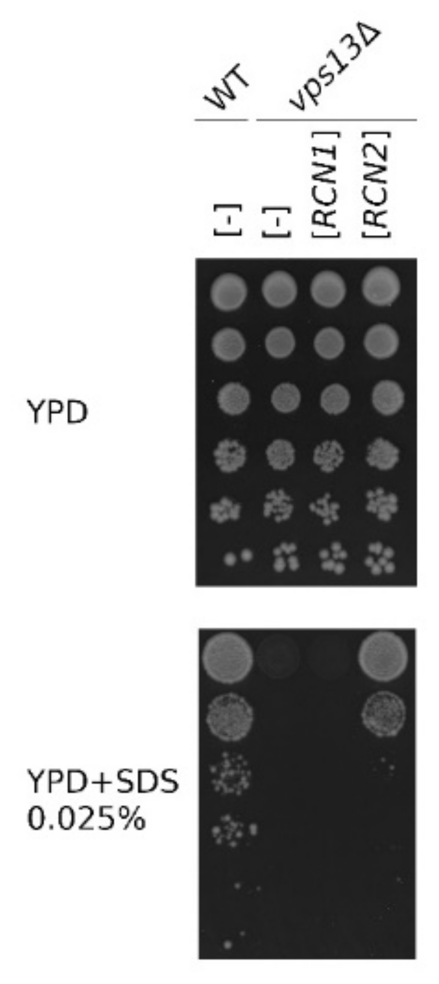
2.1. The RCN2 Gene is a Multicopy Suppressor of vps13Δ SDS Hypersensitivity

A vps13 mutants exhibit several phenotypes: they show defects in protein transport [49,50,51], sporulation [52], and actin cytoskeleton organization [47]. Our studies indicate the alteration of calcium homeostasis in vps13Δ cells and a functional connection between Vps13 and calcium signaling [30]. To this list, we recently added a new phenotype: hypersensitivity to low concentrations of a commonly used amphiphilic detergent, sodium dodecyl sulfate (SDS), which we used in a genetic screen for multicopy suppressors of the vps13-I2749R mutation. This screen resulted in the identification of fragments of the MYO3 gene (MYO3-N) and of the FET4 gene [30,53] as suppressors, and both were also effective in vps13Δ cells. In this screen, we also found that a plasmid containing the RCN2 gene, which encodes a negative regulator of calcineurin, restored the growth of the vps13Δ mutant in the presence of SDS (Figure 1). The paralogous RCN1 gene, which encodes the other calcineurin regulator, has not been isolated; thus, we directly tested whether the overexpression of RCN1 [54] could restore the growth of vps13Δ cells on SDS medium. In contrast to RCN2, overexpressed RCN1 was not active as a suppressor (Figure 1). These results support the view that specific downregulation of calcium signaling, including that caused by Rcn2, is beneficial for vps13Δ mutant cells.

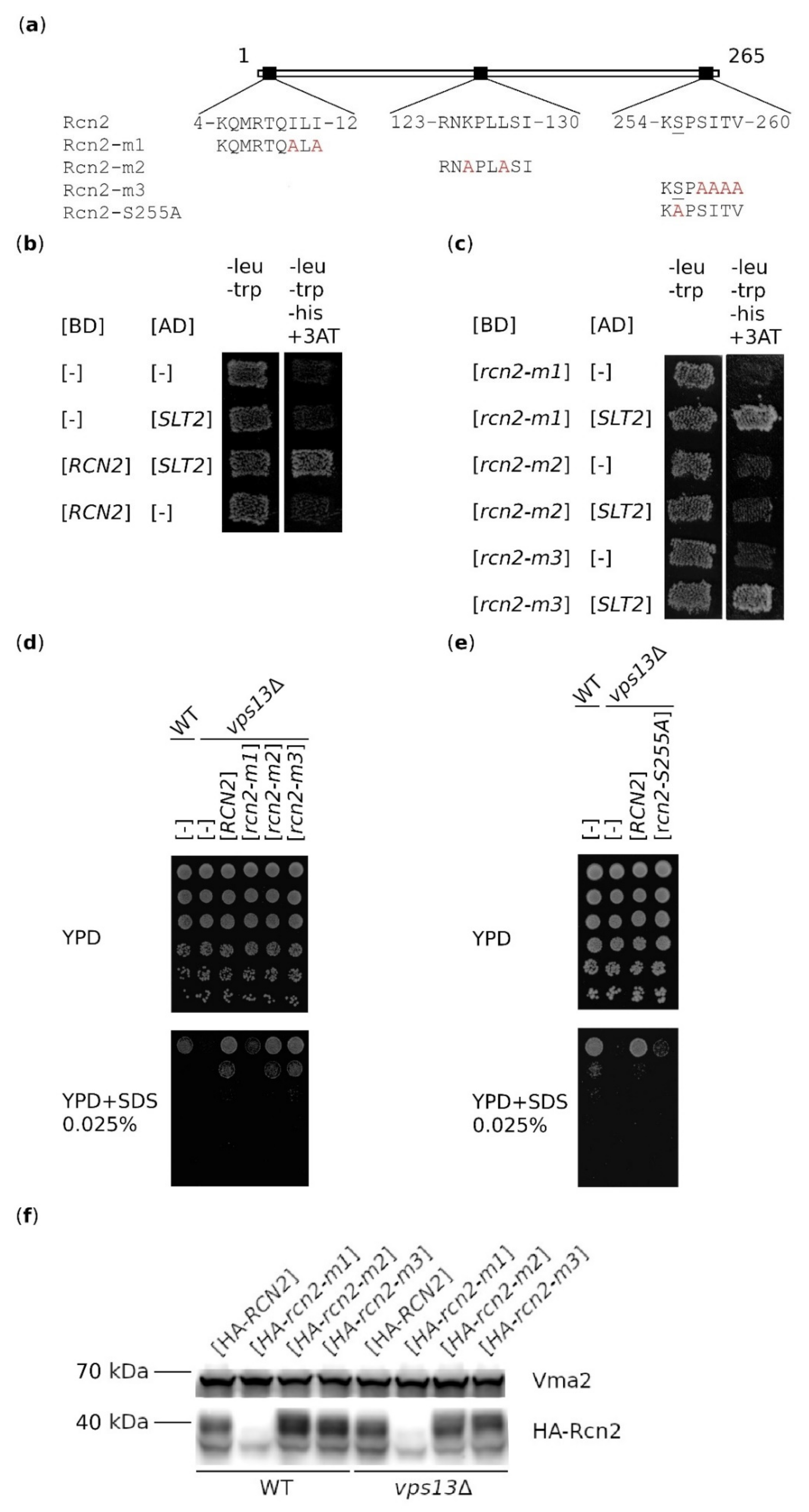
2.2. Binding of Rcn2 to Slt2 Kinase is Not Important for Suppression

Due to the fact that Rcn2 is a substrate of Slt2 kinase [55] and that the vps13Δ mutant shows increased Slt2 activity upon SDS treatment [30], we speculated that the mechanism of suppression could rely on Rcn2–Slt2-binding and Rcn2 phosphorylation. To test this hypothesis, we evaluated the importance of Rcn2 interaction with Slt2 and specifically of serine amino acid residue (aa) S255, which is phosphorylated by Slt2 [55], in the RCN2-based suppression of vps13Δ. The consensus of the motif recognized by Slt2 kinase in its target proteins is (R/K)1−2 − (X)2−6 − Φ − X − Φ), where X denotes any aa and Φ denotes a hydrophobic aa [56]. There are three such motifs in Rcn2, which are potential Slt2-binding sites (Figure 2a). We analyzed the effects of rcn2-m1, -m2 and -m3 mutations, each of which causes 3–4 aa substitutions for alanine (Figure 2a). The interaction between Slt2 and wild-type or mutant Rcn2 proteins was tested using a two-hybrid system. As shown in Figure 2b, the co-expression of RCN2 and SLT2 resulted in restored growth on media without leucine, tryptophan and histidine, indicating the interaction of wild-type Rcn2 protein with Slt2, while the interaction was lost between Rcn2-m2 and Slt2. These substitutions did not result in the lack of expression of BD-rcn2-m2 (Figure S1). This shows that aa substitutions in the Rcn2-m2 protein are in the region important for Slt2-binding.
The mutation causing substitution of the S255 residue, which is phosphorylated by Slt2, for alanine was previously shown to result in the greatest reduction in Rcn2 phosphorylation [55]. This mutation was, therefore, introduced into RCN2 (Figure 2a). In the next step, we tested how all introduced mutations affect the RCN2-based suppression of vps13Δ SDS hypersensitivity. The rcn2-m2 and rcn2-m3 alleles were effective as suppressors (Figure 2d), although the protein encoded by rcn2-m2 was not able to bind Slt2 (Figure 2c). By contrast, the rcn2-m1 and rcn2-S255A alleles (Figure 2d,e) lost the ability to support the growth of the vps13Δ mutant when overexpressed. This result could be explained by the introduction of rcn2-m mutations causing instability of the mutant protein or abolishing Rcn2 function in a way that is important for its suppressor activity. To distinguish between these two possibilities, the cellular levels of hemagglutinin (HA)-tagged versions of wild-type and mutant Rcn2 proteins were compared. Previous studies of the rcn2-S255A mutation found that it did not affect the level of the Rcn2 protein, so this was not tested in our study [55]. As shown in Figure 2f, the HA-Rcn2 protein migrated as multiple bands, ranging from about 37 to 55 kDa, even though the estimated molecular mass of the tagged version is about 32.5 kDa. The tagged version HA-Rcn2 is probably not functional because its overproduction did not reverse the SDS sensitivity of the vps13Δ mutant (Figure S2). The observed slower migrating bands could include forms that are post-translationally modified (phosphorylated, ubiquitinated) to mark HA-Rcn2 for degradation. The Western blot analysis revealed that the HA-Rcn2-m1 protein is produced but at a low level. In the case of HA-Rcn2-m2, the pattern of bands was slightly different compared with that of HA-Rcn2 and HA-Rcn-m3. Thus, it is possible that a lack of Slt2-binding affects Rcn2 phosphorylation and/or other posttranslational modifications. The level and modification pattern of the HA-Rcn2-m3 protein were similar to those of HA-Rcn2. These results indicate that HA-Rcn2 is a highly modified protein and that the N-terminal part of Rcn2 is important for its stability and functionality. In summary, these results show that the lack of interaction between Slt2 and Rcn2 does not interfere with the suppression of vps13Δ mutant phenotypes, but the presence of the S255 residue and, therefore, phosphorylation of this residue is necessary. The inability of rcn2-m1 to suppress vps13Δ could be due to a low level of Rcn2-m1, but it is also possible that the N-terminal motif of Rcn2 containing isoleucine residues I10 and I12 is specifically important.
Since the mechanism of suppression is not based on Slt2–Rcn2 interaction, we considered the second possibility that Rcn2 acts as a negative regulator of calcineurin and that downregulation of this phosphatase activity is the basis for suppression.

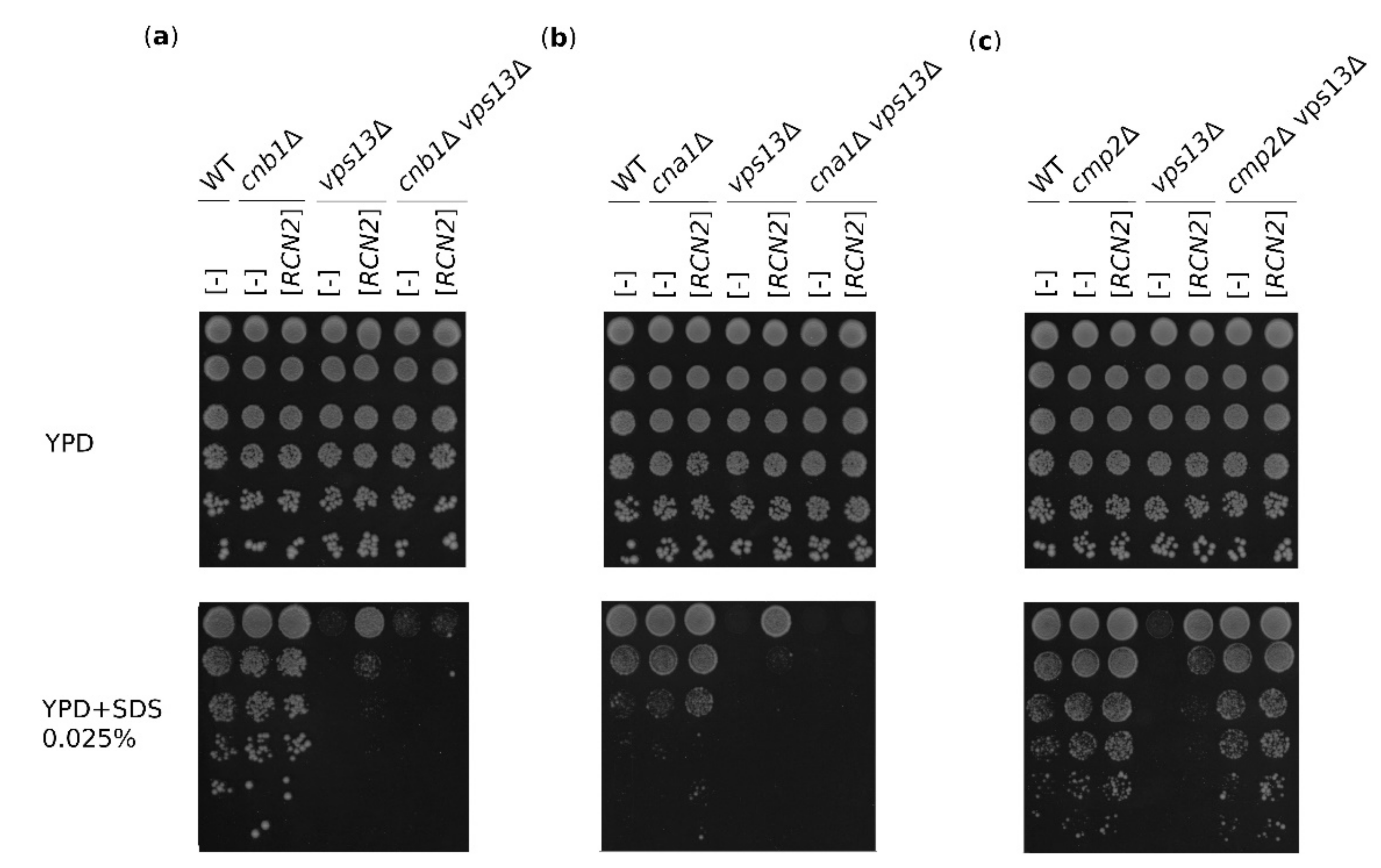
2.3. The Suppression of vps13Δ by RCN2 Requires Calcineurin

RCANs are also involved in calcineurin-independent processes, such as binding mRNA and affecting its stability [12,13], so we tested whether Rcn2-based suppression requires calcineurin presence and/or activity. For this reason, the double mutants cnb1Δ vps13Δ, cna1Δ vps13Δ, and cmp2Δ vps13Δ were constructed that lacked either a regulatory (Cnb1) or catalytic (Cna1 or Cmp2) subunit. These mutants were transformed with an RCN2-bearing plasmid, and their growth on SDS-containing media was tested. The double cnb1Δ vps13Δ mutant was hypersensitive to SDS, and RCN2 overexpression did not cause suppression (Figure 3a). Similarly, in the cna1Δ vps13Δ double mutant, there was no RCN2-based suppression (Figure 3b). Because the phosphatase is inactive in the cnb1Δ strain [19], the lack of suppression in the cnb1Δ vps13Δ mutant indicates that functional calcineurin is required. Interestingly, the cmp2Δ mutation caused cells not to be SDS-sensitive, and the double mutant cmp2Δ vps13Δ grew as single cmp2Δ (Figure 3c). Thus, the cmp2Δ mutation is epistatic to vps13Δ. Altogether, these results indicate that in the presence of SDS stress, the vps13Δ mutation triggers changes in the cell that can be overcome by the reduction of Cmp2 activity. This could be obtained by RCN2 overexpression or deletion of the CMP2 gene, but the active Cna1–Cnb1 phosphatase complexes are required.

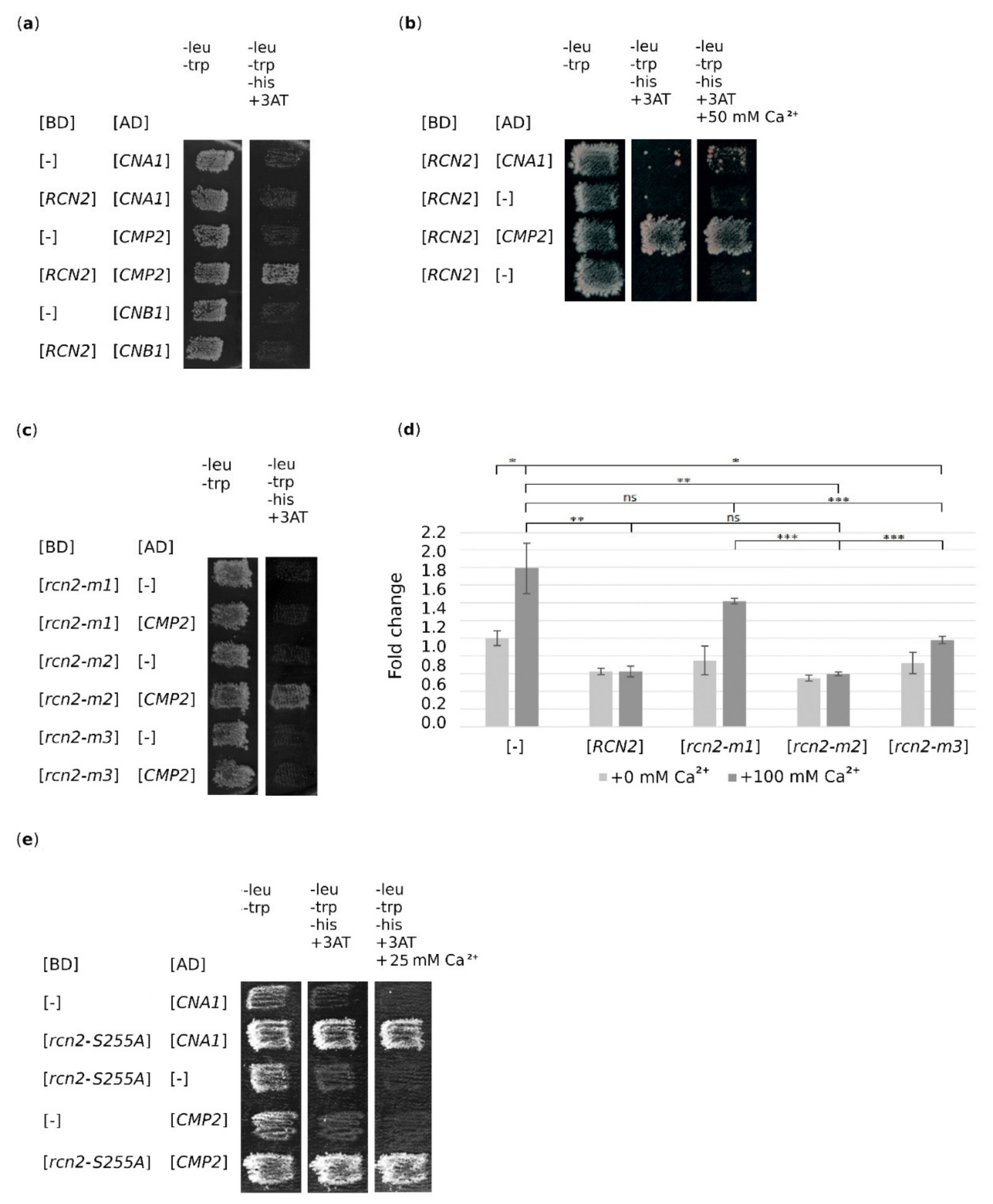
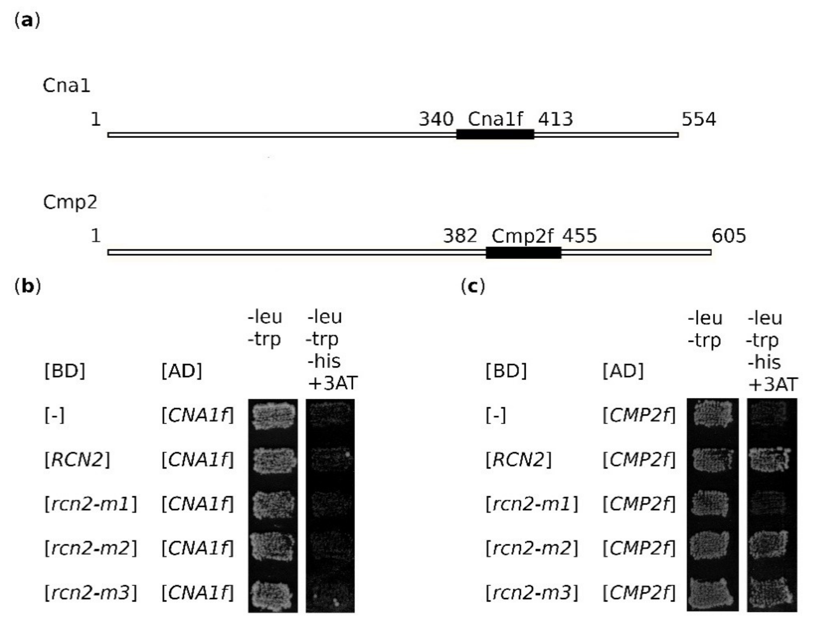
2.4. Both rcn2-m1 and rcn2-m3 Mutations Abolish the Binding of Rcn2 to Catalytic Subunit of Calcineurin, Cmp2

We noticed that mutations in the rcn2-m3 allele caused changes in the PSITVN sequence (aa 256–261) previously identified in the calcineurin binding site, described as PxIxIT-type [6,9], but did not abolish suppression. To clarify the mechanism by which RCN2 suppresses vps13Δ mutant phenotypes, we tested the ability of mutant Rcn2 versions to bind to calcineurin subunits. By using the two-hybrid system, we tested whether we could detect the binding of Rcn2 to both phosphatase catalytic subunits, Cna1 and Cmp2 and, additionally, to the phosphatase regulatory subunit Cnb1. Using this approach, we found that Rcn2 bound to Cmp2, but not to Cna1 or Cnb1 (Figure 4a). As a control, we tested the interaction of Cna1 and Cmp2 with calmodulin (Cmd1), and we observed both interactions, as expected (Figure S3). The observed lack of interaction of Rcn2 with Cna1 was surprising, so we checked whether the addition of Ca2+ to the medium allowed the interaction of Rcn2 with Cna1. Indeed, in the presence of Ca2+ and after a long time (>7 days), the interaction was visible (Figure 4b), suggesting that the interaction of Rcn2 with Cna1 is probably weak and/or controlled. We also found that both Rcn2-m1 and Rcn2-m3 lost the ability to bind Cmp2 (Figure 4c) while Rcn2-m2 interacted with Cmp2 similarly to wild-type Rcn2. Thus, the interaction of Rcn2 with calcineurin required two different motifs of Rcn2: one at the N-terminus and the second, the PIxIxIT-type motif (aa 256–261) at the C-terminus. Moreover, the presence of this last motif alone was not sufficient to support interaction.

2.5. Partial Inhibition of Calcineurin Activity is Necessary for Suppression

To better understand the mechanism of vps13Δ suppression by Rcn2, we examined the influence of RCN2 mutant gene overexpression on calcineurin activity. To compare the activity of calcineurin, we used a reporter system that comprised a plasmid containing a CDRE regulatory element fused to the lacZ open reading frame encoding β-galactosidase [21]. Upon calcineurin activation, the expression of CDRE element-driven sequences is upregulated, for example, in response to an increase in cytoplasmic calcium levels, resulting in an increased β-galactosidase activity. In our experimental settings, β-galactosidase activity was measured in cell extracts derived from transformants of the wild-type strain bearing empty vector or plasmids with different RCN2 alleles. The transformants were grown in the absence or after the addition of extra calcium ions to induce calcineurin activity. The results demonstrate that the activity of calcineurin was slightly reduced by the overexpression of wild-type RCN2 (p < 0.05) with an almost equivalent reduction by the expression of tested rcn2 versions when cells were grown without the addition of calcium ions to the medium. As expected, when calcium ions were added to the culture of cells transformed with an empty vector, calcineurin activity increased (Figure 4d). The overexpression of the RCN2 or rcn2-m2 allele significantly prevented the activation of calcineurin under these conditions. The overexpression of the rcn2-m1 allele did not affect calcineurin activity. The effect of the rcn2-m3 mutation on Rcn2 action was minor compared with that of the rcn2-m1 mutation but was significant. This indicates that two mutations, rcn2-m1 and rcn2–m3, affect Rcn2 function as a calcineurin inhibitor; thus, at least two motifs in the Rcn2 protein determine its inhibitory activity toward calcineurin. One is the well-known calcineurin-binding motif PSITVN (aa 256–261) at the C-terminus. As revealed by studying Rcn2-m1, the second element that influences Rcn2-binding to calcineurin is found at the N-terminus, and this last element is necessary for the suppression of vps13Δ. Because the rcn2-S255A mutation also impairs the ability of Rcn2 to suppress vps13Δ and the resulting amino acid residue substitution is in the calcineurin-binding motif PSITVN (aa 256–261), therefore, we tested if this influences the interaction with Cna1 and Cmp2 in the presence and in the absence of Ca2+. Surprisingly, Rcn2-S255A interacted well with both alternative catalytic subunits (Figure 4e). This result supports the earlier conclusion that inhibition of only Cmp2-based activity is necessary for suppression.

2.6. Rcn2 Binds to the Cmp2 Fragment Corresponding to aa 382–455

Based on our results, we suspected that a second calcineurin-binding site might be present in the N-terminus of Rcn2. The Rcn1 inhibitor has two types of calcineurin-binding sites: the PxIxIT-type and LxVP-type [6]. Even though the calcineurin-binding site of the LxVP-type is not present in Rcn2, we speculated that Rcn2 might interact using an N-terminal motif that was inactivated in Rcn2-m1. We noticed that the sequence TQILIT (aa 9–14) in the Rcn2 protein is close to the PxIxIT consensus sequence, with only the P residue missing. For this reason, we investigated whether Rcn2 binds to the region of calcineurin catalytic subunits responsible for the binding of substrates. Fragments of Cna1 (Cna1f; aa 340–413) and Cmp2 (Cmp2f; aa 382–455) encompassing the sequence responsible for the binding of the PxIxIT-type motif (Figure 5a) were tested for interaction with Rcn2 using the two-hybrid system. The analysis revealed that Rcn2 and Rcn2-m2 and -m3 proteins do not interact with Cna1f (Figure 5b), but they do interact with Cmp2f, while Rcn2-m1 does not interact with either (Figure 5c). This result suggests that the N-terminal part of Rcn2 can indeed support binding to calcineurin even when the C-terminal PxIxIT-type motif (aa 256–261) is abolished. The second Cmp2-binding determinant of Rcn2 interacts with this phosphatase, although its aa sequence does not perfectly fit the consensus. Interestingly, the C-terminal PxIxIT-type motif of Rcn2 (aa 256–261) is not sufficient to support Cmp2-binding when the N-terminal motif is absent (rcn2-m1, Figure 5c). This result shows that the specificity of Rcn2 for binding the Cmp2 phosphatase subunit is maintained even when only a fragment of this protein is analyzed. In summary, Rcn2 binds a subset of calcineurin complexes, that is, those containing Cmp2, using two motifs—the already-described PxIxIT-type motif (aa 256–261) and the newly characterized motif, which encompasses aa 9–14 (TQILIT) at the N-terminus.

3. Discussion

Ca2+-ion signaling is fundamental for neuron function. Ca2+ fluxes across the plasma membrane and between intracellular compartments in response to different stimuli and calcium regulates several processes, such as neurite outgrowth, synaptogenesis, synaptic transmission, plasticity and cell survival. Vps13 proteins are also important for neuronal cell physiology and mutations in VPS13 genes that cause genetic neurodegenerative disorders in humans. Using the yeast model of VPS13-related disorders, we have shown that calcineurin activity is potentiated in cells lacking the Vps13 protein and that the calmodulin/calcineurin signaling pathway could be a target for drug intervention in VPS13-dependent disorders. However, for the effective modulation of calcium signaling via the inhibition of calcineurin activity, the drug FK506 or the calcium chelator EGTA must be limited to a very specific concentration [30]. Here, using yeast, we found a different way to partially reduce the activity of calcineurin in vps13Δ cells and overcome the associated defects. We demonstrate that this could be achieved by overexpression of the RCN2 gene encoding Rcn2 protein, a calcineurin inhibitor that binds preferentially to Cmp2, one of two alternative calcineurin catalytic subunits. Moreover, our studies reveal that binding between Rcn2 and Cmp2 requires two motifs in Rcn2: the already-known motif at the C-terminus and the newly characterized motif at the N-terminus. Because the Ca2+ signaling pathways are preserved from yeast to humans, we assume that our findings may be helpful for patients suffering from VPS13-dependent diseases or other neurodegenerative diseases characterized by high calcineurin activity, such as Parkinson’s disease.
Using yeast vps13Δ mutant cells, a model of ChAc and other VPS13-dependent diseases, we demonstrated that calmodulin–calcineurin signaling is a suitable target for drug intervention [30]. Similarly, for Parkinson’s disease, reduction of calcineurin activity was shown to mitigate alpha-synuclein toxicity in both yeast and mouse models [29]. Although drugs that reduce calcineurin activity, such as cyclosporin A and FK506, are known and have been used for years as immunosuppressants after transplantation, their use has many side effects on the nervous system, such as recurrent headaches, tremors, seizures, and even a decrease in cognitive function or changes in brain structure [8]. These symptoms put the long-term use of immunosuppressant drugs into question, especially for neurological disorders. This is also because both insufficient and excessive phosphatase activity has an adverse effect, as demonstrated in vps13 mutant cells by us [30] or in alpha-synuclein-producing yeast cells by others [29]. Thus, further research is needed to discover alternative methods for the reduction of calcineurin activity without causing neurotoxicity, and studies in yeast can be very helpful [57]. Our multicopy suppressor screen was designed to find ways to circumvent the effects of the vps13-I2749R mutation, equivalent to a hvps13A-I2771R mutation found in ChAc patients [47], identified the RCN2 gene encoding a negative regulator of calcineurin and gave us a hint for further study.
We tested two hypotheses about the molecular mechanism of Rcn2 action in the vps13Δ mutant. The first hypothesis assumes that overproduced Rcn2 binds and sequesters Slt2 and that this reduces Slt2 kinase activity toward other substrates, such as the plasma membrane high-affinity Ca2+ influx system comprising Cch1 and Mid1 proteins. As a consequence, decreased phosphorylation of the channel reduces its activity and calcium influx into the cell [27]. Our finding that the overexpression of the rcn2-m2 allele, which encodes a protein variant unable to bind Slt2, is still able to suppress vps13Δ, argues against this hypothesis. In the same line is the finding that the homologous RCN1 gene, which encodes the Rcn1 protein and also has potential Slt2-binding sites, did not rescue vps13Δ growth on SDS media. Although the study of the rcn2-S255A allele indicates that the S255 residue, an acceptor residue for Slt2-dependent phosphorylation, is important for the suppression of the vps13Δ mutant, it could be modified by another kinase instead. This can be concluded from the finding that the rcn2-m2 allele, which encodes a protein that is not able to bind Slt2, is an efficient multicopy suppressor of vps13Δ. However, phosphorylation of residue S255 by unknown kinase is crucial for the regulation of Rcn2 interaction with alternative catalytic subunits of calcineurin.
The second most probable possibility for the mechanism of the suppression of vps13 is that Rcn2 simply acts as a negative regulator of calcineurin. In fact, in studying various rcn2 mutant alleles, we found that the ability to partially inhibit calcineurin is correlated with suppression. Using genetic analysis, we found that the CNA1 and CNB1 genes are required for RCN2 suppression of vps13Δ and, importantly, that cmp2Δ alone is a suppressor of vps13Δ. Further analysis showed that Rcn2 interacts preferentially with a subset of calcineurin complexes, namely, those containing Cmp2 as a catalytic subunit. The weak interaction with Cna1 was only observed when cells were grown in the presence of additional Ca2+. This is in contrast to the earlier result of a co-immunoprecipitation experiment in which Rcn2 was found to interact equally well with both Cna1 and Cmp2 subunits [58]. The selective binding and inhibiting of the Cmp2 catalytic subunit (but not to Cna1), which is required for suppression, suggests the existence of a difference in specificity or regulation between Cmp2- and Cna1-containing calcineurin complexes. It was previously shown that the presence of the CMP2 gene is more important than CNA1 in the ER stress response [59]. It is worth noting that the activity of calcineurin is only partially inhibited by Rcn2, which is in agreement with the view that it might inhibit only a subset of calcineurin complexes. The observed selective interaction of Rcn2 with Cmp2 supports the hypothesis that the selective inhibition of Cmp2-based complexes is important for the survival of the vps13Δ mutant under stress conditions. This finding is additionally supported by the fact that Rcn2-S255A, which interacts with Cna1, lost the ability to suppress. In mammals, it is known that specific catalytic subunits of calcineurin also differ in their substrate specificity [60] and regulation. For example, it was shown that for T cell activation, calcineurin activity could be selectively regulated by ubiquitination and deubiquitination depending on the catalytic subunit [16]. The conclusion, similar to ours—that different RCANs have the potential to interact and regulate different calcineurin subunits isoforms—comes from studies in rodents in which three catalytic calcineurin subunits (CnA)—Aα, Aβ, and Aγ [61,62,63,64] have been shown to have different distributions in the brain corresponding to that of individual RCAN proteins [65]. Moreover, in humans, the distribution of calcineurin subunits is tissue-specific, with Aα and Aβ isoforms being ubiquitously expressed in a variety of tissues and Aγ restricted to the brain and testis [60]. Thus, it is possible that inhibiting one of the calcineurin isoforms in patients suffering from ChAc or other neurodegenerative diseases may help to overcome pathologies without the side effects connected to the inhibition of all isoforms by FK506.
The requirement for selective and only partial inhibition of phosphatase activity is also supported by the fact that RCN1, which encodes the other calcineurin regulator present in yeast cells, was not isolated in our screen and, when overexpressed, did not suppress the SDS hypersensitivity of the vps13Δ mutant. In fact, Rcn1 differs from Rcn2 in several ways. It interacts with Cna1 [6] and has the ability to activate calcineurin [9,66], but, most importantly, Rcn1 has two different types of calcineurin-binding motifs, the PxIxIT-type and LxVP-type, while in Rcn2, the LxVP-type motif is missing [58]. The presence of two phosphatase-interacting motifs in Rcn1 means that the binding is dual. This was demonstrated for RCAN1 or A238L from the African swine fever virus. Their motifs, similar to those in NFAT, occupy the PxIxIT-binding site on Cna, and the amino acid chain then turns back to allow the second motif to encompass the LxVP-binding pocket on the Cna/Cnb interface [67,68]. We demonstrate here that Rcn2 probably similarly interacts with Cmp2 using a dual-binding mode. This is suggested by the fact that Rcn-m3 is able to interact with Cmp2f, but not with Cmp2. Thus, the scenario is that Rcn2 first requires a C-terminal PSITVN motif (PxIxIT-type; aa 256–261) to bind Cmp2 and then the N-terminally located second motif. However, the difference is that the second motif in Rcn2 is not LxVP-like. This suggests that either Rcn2 possesses a structural calcineurin-binding motif in which part of the interacting surface is located at the C-terminus and the second part at the N-terminus, or there are two binding sites, and their binding occurs sequentially. The distinction between these two presented scenarios requires further study. In vitro studies have indicated that a synthetic peptide derived from the C-terminal part of Rcn2, the PSITVN motif, effectively competes with the PVIVIT peptide for binding to human calcineurin [9], which supports the idea of the presence of two independent binding motifs. This result also suggests that it should be possible to use a peptide derived from the N-terminal part of yeast Rcn2 to inhibit human calcineurin. Our present work indicates that this peptide may have a very desirable property. It has the potential for selective inhibition of only a subset of calcineurin and does not completely block calcineurin activity. For this reason, it should be less toxic for cells than immunosuppressant drugs such as FK506, similar to other tested peptides derived from human RCAN or the protein inhibitor A238L from the African swine fever virus [68].
Our studies are important from a clinical perspective. Here, we show a way to control calcium signaling more selectively than by the administration of calcineurin chemical inhibitors. Our present study suggests that new peptides with inhibitory activity toward calcineurin could be designed as an alternative to those investigated so far [68,69] and be specific to a subset of calcineurin complexes. Moreover, comparable changes observed on a molecular level, such as defects in vesicular trafficking, endocytosis, and the actin cytoskeleton, and increased calcineurin activity in yeast Parkinson’s alpha-synuclein model [29] and VPS13-deficiency model [46], which includes early-onset Parkinsonism, suggest that a similar approach can be taken for the treatment of several diseases.

4. Materials and Methods

4.1. Strains, Media, and Growth Conditions

For cloning and plasmid propagation, E.coli DH5α was used. The yeast strains used in this study are listed in Table S1 (Table S1). The strains KJK190 and KJK191 were constructed by integration of the vps13::URA3 cassette amplified by PCR into the genomic DNA of the KJK181A strain. Strains BYcna1Δ and BYcmp2Δ were transformed with the PCR product, and transformants were selected on SC-Ura plates. Integrations were confirmed by PCR of genomic DNA. These strains were used to test the SDS sensitivity phenotype and the influence of RCN2 overexpression on cell growth.
The media used were YPD complete medium (1% yeast extract, 2% peptone, 2% glucose) or synthetic SC medium (0.067% yeast nitrogen base without amino acids, 2% glucose) with supplements (adenine, uracil, amino acids) as indicated.
For growth tests, transformants were grown overnight in liquid media, and cultures were diluted to an optical density OD600 of ~1. Five-fold serial dilutions of cells were spotted on solid YPD with or without the addition of SDS (Sigma-Aldrich, St Louis, MO, USA) media, as described in the figure legends. Plates were incubated at 30 °C. The sensitivity of vps13Δ mutant cells to SDS is variable and depends on several factors, such as the medium lot, the lot of the SDS stock solution, and various physical factors; therefore, in each growth experiment, several SDS concentrations were tested, and one representative result is shown. For two-hybrid analysis, the PJ69-4A strain [70] was used, and respective transformants were grown on SC-Trp-Leu plates, replicated on SC-Trp-Leu-His supplemented with 1.5 mM 3-aminotriazole (Sigma-Aldrich) or SC-Trp-Leu-His supplemented with 1.5 mM 3-aminotriazole and 25 or 50 mM CaCl2. Plates were incubated at 30 °C for 3–10 days.

4.2. Plasmids, Mutagenesis, and Screen for Multicopy Suppressors of vps13 Mutations

All plasmids used in this study are listed in Table S2. Screening for multicopy suppressors of the vps13-I2749R SDS hypersensitivity phenotype is described in Soczewka et al. 2019 [30]. A plasmid with a DNA fragment containing the RCN2 gene, which was isolated as a multicopy suppressor from the pFL44-based genome library [71], was used to amplify RCN2 by PCR. Next, the amplified fragment was transferred to the YEp181lac plasmid [72] using BamHI and EcoRI restriction enzymes to obtain YEplac181-RCN2.
The SLT2, CNA1, CMP2, and CNB1 genes were amplified by PCR using BY4741 genomic DNA as a template with appropriate primers (Table S3) and were cloned into pGBT9 and/or pGAD424 vectors. Mutant rcn2-m1, rcn2-m2 and rcn2-m3 alleles were obtained using one-step mutagenesis with appropriate primers. The construct YEp181lac-3HA-RCN2 contains a 3HA-tag coding sequence followed by the full-length RCN2 gene. It is a derivative of the YEp181lac-KXS-RCN2 plasmid in which KpnI (K), XhoI (X), and SacI (S) restriction sites were introduced directly in front of the RCN2 ATG sequence in YEp181lac-RCN2 by generating two types of overlapping PCR products (A and B) using Phusion High Fidelity DNA polymerase and the following pairs of primers. For the type A product, RCN2_For and L486KXS were used; for the B product, U486KXS and RCN2_Rev were used. Both products (A and B) were purified and used as templates for the next round of PCR reactions with the use of RCN2_For and RCN2_Rev primers. The final products were digested with BamHI and EcoRI, and the restriction fragments were transferred to the YEplac181-RCN2 plasmid, exchanging the original sequence for the introduced sequence with KpnI, XhoI, and SacI restriction sites immediately before the ATG codon. The 3HA encoding sequence was amplified from the plasmid pET9dSUMO [73] with primers 3HA-U50 and 3HA-L130 flanked by KpnI and SacI restriction sites. The PCR product was digested with KpnI and SacI and cloned into YEp181lac-KXS-RCN2. All PCR products were sequenced after cloning.

4.3. β-Galactosidase Activity Assay

β-Galactosidase activity was analyzed as previously described [74]. For this assay, cells were transformed with the pAMS363 plasmid bearing CDRE-LacZ fusion and either an empty plasmid or a plasmid bearing one of the RCN2 alleles. Transformants were grown overnight in SC-Ura-Leu and then diluted and divided into two independent cultures, one of which was supplemented with CaCl2 to a final concentration of 100 mM. The cells were grown for another 4 h, and extracts were prepared using glass beads. In each of the three experiments, at least three independent transformants of the respective yeast strains were inoculated, and their extracts were assayed in duplicate.

4.4. Protein Extracts and Western Blot Analysis

Yeast cells were grown at 30 °C in SC-Leu to the logarithmic phase. Protein extracts were prepared by alkaline lysis [75]. Samples were analyzed by standard SDS–PAGE followed by Western blotting using a mouse monoclonal anti-HA epitope (BioLegend, San Diego, CA, USA) and Vma2 (Thermo Fisher Scientific, Waltham, MA, USA) or the polyclonal anti-GAL4 DNA-BD rabbit antibody (Sigma-Aldrich) followed by secondary anti-mouse or anti-rabbit IgG horseradish peroxidase (HRP)-conjugated antibodies (Dako, Glostrup, Denmark). The signal was detected by enhanced chemiluminescence (Millipore, Darmstadt, Germany).

Supplementary Materials

Supplementary Materials can be found at https://www.mdpi.com/1422-0067/22/3/1193/s1.

Author Contributions

Conceptualization, J.K. and T.Z.; methodology, P.W., M.S.; validation, J.K., P.W.; formal analysis, J.K., P.W.; investigation, P.W., J.K., P.S., M.S.; resources, P.W., J.K., P.S., M.S.; data curation, P.W., J.K.; writing—original draft preparation, J.K.; writing—review and editing, J.K., T.Z., P.W., P.S., M.S.; visualization, P.W.; supervision, J.K.; project administration, T.Z., J.K.; funding acquisition, T.Z. All authors have read and agreed to the published version of the manuscript.

Funding

This research was funded by National Science Centre Poland, grant number UMO-2015/19/B/NZ3/01515 to T.Z., the APC was funded by MDPI.

Institutional Review Board Statement

Not applicable.

Informed Consent Statement

Not applicable.

Data Availability Statement

Data available on request from corresponding author due to privacy.

Acknowledgments

We would like to thank R. Kucharczyk (IBB PAN, Warsaw, Poland) and M. Cyert (Stanford University, Stanford, CA, USA) for plasmids.

Conflicts of Interest

The authors declare no conflict of interest. The funders had no role in the design of the study; in the collection, analyses, or interpretation of data; in the writing of the manuscript, or in the decision to publish the results.

References

  1. Rao, A.; Luo, C.; Hogan, P.G. Transcription factors of the nfat family: Regulation and Function. Annu. Rev. Immunol. 1997, 15, 707–747. [Google Scholar] [CrossRef] [PubMed]
  2. Wang, M.; Yi, H.; Guerini, D.; Klee, C.; McBride, O. Calcineurin A alpha (PPP3CA), calcineurin A beta (PPP3CB) and calcineurin B (PPP3R1) are located on human chromosomes 4, 10q21→q22 and 2p16→p15 respectively. Cytogenet. Genome Res. 1996, 72, 236–241. [Google Scholar] [CrossRef] [Green Version]
  3. Liu, L.; Zhang, J.; Yuan, J.; Dang, Y.; Yang, C.; Chen, X.; Xu, J.; Yu, L. Characterization of a human regulatory subunit of protein phosphatase 3 gene (PPP3RL) expressed specifically in testis. Mol. Biol. Rep. 2005, 32, 41–45. [Google Scholar] [CrossRef] [PubMed]
  4. Saraf, J.; Bhattacharya, P.; Kalia, K.; Borah, A.; Sarmah, D.; Kaur, H.; Dave, K.R.; Yavagal, D.R. A Friend or Foe: Calcineurin across the Gamut of Neurological Disorders. ACS Central Sci. 2018, 4, 805–819. [Google Scholar] [CrossRef] [PubMed]
  5. Klee, C.B.; Ren, H.; Wang, X. Regulation of the Calmodulin-stimulated Protein Phosphatase, Calcineurin. J. Biol. Chem. 1998, 273, 13367–13370. [Google Scholar] [CrossRef] [Green Version]
  6. Rodríguez, A.; Roy, J.; Martínez-Martínez, S.; López-Maderuelo, M.D.; Niño-Moreno, P.; Ortí, L.; Pantoja-Uceda, D.; Pineda-Lucena, A.; Cyert, M.S.; Redondo, J.M. A Conserved Docking Surface on Calcineurin Mediates Interaction with Substrates and Immunosuppressants. Mol. Cell 2009, 33, 616–626. [Google Scholar] [CrossRef] [Green Version]
  7. Liu, J. FK506 and cyclosporin, molecular probes for studying intracellular signal transduction. Immunol. Today 1993, 14, 290–295. [Google Scholar] [CrossRef]
  8. Bechstein, W.O. Neurotoxicity of calcineurin inhibitors: Impact and clinical management. Transpl. Int. 2000, 13, 313–326. [Google Scholar] [CrossRef]
  9. Mehta, S.; Li, H.; Hogan, P.G.; Cunningham, K.W. Domain Architecture of the Regulators of Calcineurin (RCANs) and Identification of a Divergent RCAN in Yeast. Mol. Cell. Biol. 2009, 29, 2777–2793. [Google Scholar] [CrossRef] [Green Version]
  10. Strippoli, P.; Lenzi, L.; Petrini, M.; Carinci, P.; Zannotti, M. A New Gene Family Including DSCR1 (Down Syndrome Candidate Region 1) and ZAKI-4: Characterization from Yeast to Human and Identification of DSCR1-like 2, a Novel Human Member (DSCR1L2). Genomics 2000, 64, 252–263. [Google Scholar] [CrossRef]
  11. Candelas, E.S.; Farré, D.; Aranguren-Ibáñez, Á.; Martínez-Høyer, S.; Pérez-Riba, M. The Vertebrate RCAN Gene Family: Novel Insights into Evolution, Structure and Regulation. PLoS ONE 2014, 9, e85539. [Google Scholar] [CrossRef] [Green Version]
  12. Yun, Y.; Zhang, Y.; Zhang, C.; Huang, L.; Tan, S.; Wang, P.; Vilariño-Güell, C.; Song, W.; Sun, X. Regulator of calcineurin 1 is a novel RNA-binding protein to regulate neuronal apoptosis. Mol. Psychiatry 2019, 1–15. [Google Scholar] [CrossRef] [PubMed]
  13. Lee, S.K.; Ahnn, J. Regulator of Calcineurin (RCAN): Beyond down Syndrome Critical Region. Mol. Cells 2020, 43, 671–685. [Google Scholar] [CrossRef] [PubMed]
  14. Fuentes, J.-J.; A Pritchard, M.; Planas, A.M.; Bosch, A.; Ferrer, I.; Estivill, X. A new human gene from the Down syndrome critical region encodes a proline-rich protein highly expressed in fetal brain and heart. Hum. Mol. Genet. 1995, 4, 1935–1944. [Google Scholar] [CrossRef] [PubMed]
  15. Sanna, B.; Brandt, E.B.; Kaiser, R.A.; Pfluger, P.T.; Witt, S.A.; Kimball, T.R.; Van Rooij, E.; De Windt, L.J.; Rothenberg, M.E.; Tschöp, M.; et al. Modulatory calcineurin-interacting proteins 1 and 2 function as calcineurin facilitators in vivo. Proc. Natl. Acad. Sci. USA 2006, 103, 7327–7332. [Google Scholar] [CrossRef] [Green Version]
  16. Zhang, Y.; Liu, R.-B.; Cao, Q.; Fan, K.-Q.; Huang, L.-J.; Yu, J.-S.; Gao, Z.-J.; Huang, T.; Zhong, J.-Y.; Mao, X.-T.; et al. USP16-mediated deubiquitination of calcineurin A controls peripheral T cell maintenance. J. Clin. Investig. 2019, 129, 2856–2871. [Google Scholar] [CrossRef] [PubMed] [Green Version]
  17. Kafadar, K.A.; Zhu, H.; Snyder, M.; Cyert, M.S. Negative regulation of calcineurin signaling by Hrr25p, a yeast homolog of casein kinase I. Genes Dev. 2003, 17, 2698–2708. [Google Scholar] [CrossRef] [Green Version]
  18. Cyert, M.S.; Kunisawa, R.; Kaim, D.; Thorner, J. Yeast has homologs (CNA1 and CNA2 gene products) of mammalian calcineurin, a calmodulin-regulated phosphoprotein phosphatase. Proc. Natl. Acad. Sci. USA 1991, 88, 7376–7380. [Google Scholar] [CrossRef] [Green Version]
  19. Cyert, M.S.; Thorner, J. Regulatory subunit (CNB1 gene product) of yeast Ca2+/calmodulin-dependent phosphoprotein phosphatases is required for adaptation to pheromone. Mol. Cell. Biol. 1992, 12, 3460–3469. [Google Scholar] [CrossRef] [Green Version]
  20. Kingsbury, T.J.; Cunningham, K.W. A conserved family of calcineurin regulators. Genes Dev. 2000, 14, 1595–1604. [Google Scholar]
  21. Stathopoulos, A.M.; Cyert, M.S. Calcineurin acts through the CRZ1/TCN1-encoded transcription factor to regulate gene expression in yeast. Genes Dev. 1997, 11, 3432–3444. [Google Scholar] [CrossRef] [PubMed] [Green Version]
  22. Roy, J.; Li, H.; Hogan, P.G.; Cyert, M.S. A Conserved Docking Site Modulates Substrate Affinity for Calcineurin, Signaling Output, and In Vivo Function. Mol. Cell 2007, 25, 889–901. [Google Scholar] [CrossRef] [Green Version]
  23. Espeso, E.A. The CRaZy Calcium Cycle. Adv. Exp. Med. Biol. 2016, 169–186. [Google Scholar]
  24. Batiza, A.F.; Schulz, T.; Masson, P.H. Yeast Respond to Hypotonic Shock with a Calcium Pulse. J. Biol. Chem. 1996, 271, 23357–23362. [Google Scholar] [CrossRef] [PubMed] [Green Version]
  25. Denis, V.; Cyert, M.S. Internal Ca2+ release in yeast is triggered by hypertonic shock and mediated by a TRP channel homologue. J. Cell Biol. 2002, 156, 29–34. [Google Scholar] [CrossRef] [PubMed]
  26. Courchesne, W.E.; Vlasek, C.; Klukovich, R.; Coffee, S. Ethanol induces calcium influx via the Cch1-Mid1 transporter in Saccharomyces cerevisiae. Arch. Microbiol. 2011, 193, 323–334. [Google Scholar] [CrossRef]
  27. Bonilla, M.; Cunningham, K.W. Mitogen-activated Protein Kinase Stimulation of Ca2+ Signaling Is Required for Survival of Endoplasmic Reticulum Stress in Yeast. Mol. Biol. Cell 2003, 14, 4296–4305. [Google Scholar] [CrossRef] [PubMed] [Green Version]
  28. Garrett-Engele, P.; Moilanen, B.; Cyert, M.S. Calcineurin, the Ca2+/calmodulin-dependent protein phosphatase, is essential in yeast mutants with cell integrity defects and in mutants that lack a functional vacuolar H(+)-ATPase. Mol. Cell. Biol. 1995, 15, 4103–4114. [Google Scholar] [CrossRef] [PubMed] [Green Version]
  29. Caraveo, G.; Soste, M.; Cappelleti, V.; Fanning, S.; Van Rossum, D.B.; Whitesell, L.; Huang, Y.; Chung, C.Y.; Baru, V.; Zaichick, S.; et al. FKBP12 contributes to α-synuclein toxicity by regulating the calcineurin-dependent phosphoproteome. Proc. Natl. Acad. Sci. USA 2017, 114, E11313–E11322. [Google Scholar] [CrossRef] [Green Version]
  30. Soczewka, P.; Kolakowski, D.; Rooij, I.S.-D.; Rzepnikowska, W.; Ayscough, K.R.; Kaminska, J.; Żołądek, T. Yeast-model-based study identified myosin- and calcium-dependent calmodulin signalling as a potential target for drug intervention in chorea-acanthocytosis. Dis. Model. Mech. 2019, 12, dmm036830. [Google Scholar] [CrossRef] [Green Version]
  31. Rampoldi, L.; Dobson-Stone, C.; Rubio, J.P.; Danek, A.; Chalmers, R.M.; Wood, N.W.; Verellen, C.; Ferrer, X.; Malandrini, A.; Fabrizi, G.M.; et al. A conserved sorting-associated protein is mutant in chorea-acanthocytosis. Nat. Genet. 2001, 28, 119–120. [Google Scholar] [CrossRef] [PubMed]
  32. Kolehmainen, J.; Black, G.C.; Saarinen, A.; Chandler, K.; Clayton-Smith, J.; Träskelin, A.L.; Perveen, R.; Kivitie-Kallio, S.; Norio, R.; Warburg, M.; et al. Cohen syndrome is caused by mutations in a novel gene, COH1, encoding a transmembrane protein with a presumed role in vesicle-mediated sorting and intracellular protein transport. Am. J. Hum. Genet 2003, 72, 1359–1369. [Google Scholar] [CrossRef] [PubMed] [Green Version]
  33. Lesage, S.; Drouet, V.; Majounie, E.; Deramecourt, V.; Jacoupy, M.; Nicolas, A.; Cormier-Dequaire, F.; Hassoun, S.M.; Pujol, C.; Ciura, S.; et al. Loss of VPS13C Function in Autosomal-Recessive Parkinsonism Causes Mitochondrial Dysfunction and Increases PINK1/Parkin-Dependent Mitophagy. Am. J. Hum. Genet. 2016, 98, 500–513. [Google Scholar] [CrossRef] [Green Version]
  34. Gauthier, J.; Meijer, I.A.; Lessel, D.; Mencacci, N.E.; Krainc, D.; Hempel, M.; Tsiakas, K.; Prokisch, H.; Rossignol, E.; Helm, M.H.; et al. Recessive mutations in VPS13D cause childhood onset movement disorders. Ann. Neurol. 2018, 83, 1089–1095. [Google Scholar] [CrossRef] [PubMed]
  35. Seong, E.; Insolera, R.; Dulovic, M.; Kamsteeg, E.; Trinh, J.; Brüggemann, N.; Sandford, E.; Li, S.; Ozel, A.B.; Li, J.; et al. Mutations in VPS13D lead to a new recessive ataxia with spasticity and mitochondrial defects. Ann. Neurol. 2018, 83, 1075–1088. [Google Scholar] [CrossRef]
  36. Velayos-Baeza, A.; Vettori, A.; Copley, R.R.; Dobson-Stone, C.; Monaco, A.P. Analysis of the human VPS13 gene family. Genomics 2004, 84, 536–549. [Google Scholar] [CrossRef]
  37. Kumar, N.; Leonzino, M.; Hancock-Cerutti, W.; Horenkamp, F.A.; Li, P.; Lees, J.A.; Wheeler, H.; Reinisch, K.M.; De Camilli, P. VPS13A and VPS13C are lipid transport proteins differentially localized at ER contact sites. J. Cell Biol. 2018, 217, 3625–3639. [Google Scholar] [CrossRef] [Green Version]
  38. Li, P.; Lees, J.A.; Lusk, C.P.; Reinisch, K.M. Cryo-EM reconstruction of a VPS13 fragment reveals a long groove to channel lipids between membranes. J. Cell Biol. 2020, 219, e202001161. [Google Scholar] [CrossRef]
  39. Lang, A.B.; Peter, A.T.J.; Walter, P.; Kornmann, B. ER–mitochondrial junctions can be bypassed by dominant mutations in the endosomal protein Vps13. J. Cell Biol. 2015, 210, 883–890. [Google Scholar] [CrossRef] [Green Version]
  40. Park, J.-S.; Thorsness, M.K.; Policastro, R.; McGoldrick, L.L.; Hollingsworth, N.M.; Thorsness, P.E.; Neiman, A.M. Yeast Vps13 promotes mitochondrial function and is localized at membrane contact sites. Mol. Biol. Cell 2016, 27, 2435–2449. [Google Scholar] [CrossRef] [Green Version]
  41. Peter, A.T.J.; Herrmann, B.; Antunes, D.; Rapaport, D.; Dimmer, K.S.; Kornmann, B. Vps13-Mcp1 interact at vacuole–mitochondria interfaces and bypass ER–mitochondria contact sites. J. Cell Biol. 2017, 216, 3219–3229. [Google Scholar] [CrossRef] [PubMed] [Green Version]
  42. Kolakowski, D.; Kaminska, J.; Zoladek, T. The binding of the APT1 domains to phosphoinositides is regulated by metal ions in vitro. Biochim. Biophys. Acta (BBA) Biomembr. 2020, 1862, 183349. [Google Scholar] [CrossRef] [PubMed]
  43. Yu, W.; Honisch, S.; Schmidt, S.; Yan, J.; Schmid, E.; Alkahtani, S.; AlKahtane, A.A.; Alarifi, S.; Stournaras, C.; Lang, F. Chorein Sensitive Orai1 Expression and Store Operated Ca2+ Entry in Rhabdomyosarcoma Cells. Cell. Physiol. Biochem. 2016, 40, 1141–1152. [Google Scholar] [CrossRef]
  44. Pelzl, L.; Elsir, B.; Sahu, I.; Bissinger, R.; Singh, Y.; Sukkar, B.; Honisch, S.; Schoels, L.; Jemaà, M.; Lang, E.; et al. Lithium Sensitivity of Store Operated Ca2+ Entry and Survival of Fibroblasts Isolated from Chorea-Acanthocytosis Patients. Cell. Physiol. Biochem. 2017, 42, 2066–2077. [Google Scholar] [CrossRef] [Green Version]
  45. Stanslowsky, N.; Reinhardt, P.; Glass, H.; Kalmbach, N.; Naujock, M.; Hensel, N.; Lübben, V.; Pal, A.; Venneri, A.; Lupo, F.; et al. Neuronal Dysfunction in iPSC-Derived Medium Spiny Neurons from Chorea-Acanthocytosis Patients Is Reversed by Src Kinase Inhibition and F-Actin Stabilization. J. Neurosci. 2016, 36, 12027–12043. [Google Scholar] [CrossRef] [Green Version]
  46. Rzepnikowska, W.; Flis, K.; Muñoz-Braceras, S.; Menezes, R.; Escalante, R.; Żołądek, T. Yeast and other lower eukaryotic organisms for studies of Vps13 proteins in health and disease. Traffic 2017, 18, 711–719. [Google Scholar] [CrossRef] [Green Version]
  47. Rzepnikowska, W.; Flis, K.; Kaminska, J.; Grynberg, M.; Urbanek, A.; Ayscough, K.R.; Żołądek, T. Amino acid substitution equivalent to human chorea-acanthocytosis I2771R in yeast Vps13 protein affects its binding to phosphatidylinositol 3-phosphate. Hum. Mol. Genet. 2017, 26, 1497–1510. [Google Scholar] [CrossRef] [Green Version]
  48. Dziurdzik, S.K.; Bean, B.D.M.; Davey, M.; Conibear, E. A VPS13D spastic ataxia mutation disrupts the conserved adaptor-binding site in yeast Vps13. Hum. Mol. Genet. 2020, 29, 635–648. [Google Scholar] [CrossRef] [Green Version]
  49. De, M.; Oleskie, A.N.; Ayyash, M.; Dutta, S.; Mancour, L.; Abazeed, M.; Brace, E.J.; Skiniotis, G.; Fuller, R.S. The Vps13p–Cdc31p complex is directly required for TGN late endosome transport and TGN homotypic fusion. J. Cell Biol. 2017, 216, 425–439. [Google Scholar] [CrossRef]
  50. Brickner, J.H.; Fuller, R.S. SOI1 encodes a novel, conserved protein that promotes TGN-endosomal cycling of Kex2p and other membrane proteins by modulating the function of two TGN localization signals. J. Cell Biol. 1997, 139, 23–36. [Google Scholar] [CrossRef]
  51. Redding, K.; Brickner, J.H.; Marschall, L.G.; Nichols, J.W.; Fuller, R.S. Allele-specific suppression of a defective trans-Golgi network (TGN) localization signal in Kex2p identifies three genes involved in localization of TGN transmembrane proteins. Mol. Cell. Biol. 1996, 16, 6208–6217. [Google Scholar] [CrossRef] [PubMed] [Green Version]
  52. Park, J.-S.; Neiman, A.M. VPS13 regulates membrane morphogenesis during sporulation in Saccharomyces cerevisiae. J. Cell Sci. 2012, 125, 3004–3011. [Google Scholar] [CrossRef] [Green Version]
  53. Soczewka, P.; Flis, K.; Tribouillard-Tanvier, D.; Di Rago, J.-P.; Santos, C.N.; Menezes, R.; Kaminska, J.; Żołądek, T. Flavonoids as Potential Drugs for VPS13-Dependent Rare Neurodegenerative Diseases. Genes 2020, 11, 828. [Google Scholar] [CrossRef] [PubMed]
  54. Kucharczyk, R.; Dupre, S.; Avaro, S.; Haguenauer-Tsapis, R.; Słonimski, P.P.; Rytka, J. The novel protein Ccz1p required for vacuolar assembly in Saccharomyces cerevisiae functions in the same transport pathway as Ypt7p. J. Cell Sci. 2000, 113, 4301–4311. [Google Scholar]
  55. Alonso-Rodríguez, E.; Fernández-Piñar, P.; Sacristán-Reviriego, A.; Molina, M.; Martín, H. An Analog-sensitive Version of the Protein Kinase Slt2 Allows Identification of Novel Targets of the Yeast Cell Wall Integrity Pathway. J. Biol. Chem. 2016, 291, 5461–5472. [Google Scholar] [CrossRef] [Green Version]
  56. Levin, D.E. Regulation of Cell Wall Biogenesis in Saccharomyces cerevisiae: The Cell Wall Integrity Signaling Pathway. Genetics 2011, 189, 1145–1175. [Google Scholar] [CrossRef] [Green Version]
  57. Yoshida, J.; Okawa, Y.; Oyama, T.; Shimoda, N.; Uesugi, S.; Takagi, H.; Ito, Y.; Kimura, K. Inhibition of Calcineurin and Glycogen Synthase Kinase-3β by Ricinoleic Acid Derived from Castor Oil. Lipids 2020, 55, 89–99. [Google Scholar] [CrossRef]
  58. Goldman, A.; Roy, J.; Bodenmiller, B.; Wanka, S.; Landry, C.R.; Aebersold, R.; Cyert, M.S. The Calcineurin Signaling Network Evolves via Conserved Kinase-Phosphatase Modules that Transcend Substrate Identity. Mol. Cell 2014, 55, 422–435. [Google Scholar] [CrossRef] [Green Version]
  59. Mizuno, T.; Nakamura, M.; Irie, K. Induction of Ptp2 and Cmp2 protein phosphatases is crucial for the adaptive response to ER stress in Saccharomyces cerevisiae. Sci. Rep. 2018, 8, 13078. [Google Scholar] [CrossRef]
  60. Francis, C.; Bai, Y. Differential expression of cyclosporine A-Induced calcineurin isoform-specific matrix metalloproteinase 9 (MMP-9) in renal fibroblasts. Biochem. Biophys. Res. Commun. 2018, 503, 2549–2554. [Google Scholar] [CrossRef]
  61. Takaishi, T.; Saito, N.; Kuno, T.; Tanaka, C. Differential distribution of the mRNA encoding two isoforms of the catalytic subunit of calcineurin in the rat brain. Biochem. Biophys. Res. Commun. 1991, 174, 393–398. [Google Scholar] [CrossRef]
  62. Kuno, T.; Mukai, H.; Ito, A.; Chang, C.-D.; Kishima, K.; Saito, N.; Tanaka, C. Distinct Cellular Expression of Calcineurin Aɑ and Aβ in Rat Brain. J. Neurochem. 1992, 58, 1643–1651. [Google Scholar] [CrossRef] [PubMed]
  63. Usuda, N.; Arai, H.; Sasaki, H.; Hanai, T.; Nagata, T.; Muramatsu, T.; Kincaid, R.L.; Higuchi, S. Differential subcellular localization of neural isoforms of the catalytic subunit of calmodulin-dependent protein phosphatase (calcineurin) in central nervous system neurons: Immunohistochemistry on formalin-fixed paraffin sections employing antigen retrieval by microwave irradiation. J. Histochem. Cytochem. 1996, 44, 13–18. [Google Scholar] [CrossRef]
  64. Eastwood, S.L.; Salih, T.; Harrison, P.J. Differential expression of calcineurin A subunit mRNA isoforms during rat hippocampal and cerebellar development. Eur. J. Neurosci. 2005, 22, 3017–3024. [Google Scholar] [CrossRef]
  65. Porta, S.; Martí, E.; De La Luna, S.; Arbones, M.L. Differential expression of members of the RCAN family of calcineurin regulators suggests selective functions for these proteins in the brain. Eur. J. Neurosci. 2007, 26, 1213–1226. [Google Scholar] [CrossRef]
  66. Hilioti, Z.; Gallagher, D.A.; Low-Nam, S.T.; Ramaswamy, P.; Gajer, P.; Kingsbury, T.J.; Birchwood, C.J.; Levchenko, A.; Cunningham, K.W. GSK-3 kinases enhance calcineurin signaling by phosphorylation of RCNs. Genes Dev. 2004, 18, 35–47. [Google Scholar] [CrossRef] [Green Version]
  67. Grigoriu, S.; Bond, R.; Cossio, P.; Chen, J.A.; Ly, N.; Hummer, G.; Page, R.; Cyert, M.S.; Peti, W. The Molecular Mechanism of Substrate Engagement and Immunosuppressant Inhibition of Calcineurin. PLoS Biol. 2013, 11, e1001492. [Google Scholar] [CrossRef] [Green Version]
  68. Wang, L.; Cheng, N.; Wang, P.; Li, J.; Jia, A.; Li, W.; Zhang, N.; Yin, Y.; Tong, L.; Wei, Q.; et al. A novel peptide exerts potent immunosuppression by blocking the two-site interaction of NFAT with calcineurin. J. Biol. Chem. 2020, 295, 2760–2770. [Google Scholar] [CrossRef]
  69. Noguchi, H.; Sugimoto, K.; Miyagi-Shiohira, C.; Nakashima, Y.; Kobayashi, N.; Saitoh, I.; Watanabe, M.; Noguchi, Y. RCAN-11R peptide provides immunosuppression for fully mismatched islet allografts in mice. Sci. Rep. 2017, 7, 3043. [Google Scholar] [CrossRef] [Green Version]
  70. James, P.; Halladay, J.; Craig, E.A. Genomic libraries and a host strain designed for highly efficient two-hybrid selection in yeast. Genetics 1996, 144, 1425–1436. [Google Scholar] [CrossRef]
  71. Stettler, S.; Chiannilkulchai, N.; Denmat, S.H.-L.; Lalo, D.; Lacroute, F.; Sentenac, A.; Thuriaux, P. A general suppressor of RNA polymerase I, II and III mutations in Saccharomyces cerevisiae. Mol. Genet. Genom. 1993, 239, 169–176. [Google Scholar] [CrossRef] [PubMed]
  72. Gietz, R.; Akio, S. New yeast-Escherichia coli shuttle vectors constructed with in vitro mutagenized yeast genes lacking six-base pair restriction sites. Gene 1988, 74, 527–534. [Google Scholar] [CrossRef]
  73. Budhiraja, R.; Hermkes, R.; Müller, S.; Schmidt, J.; Colby, T.; Panigrahi, K.; Coupland, G.; Bachmair, A. Substrates Related to Chromatin and to RNA-Dependent Processes Are Modified by Arabidopsis SUMO Isoforms That Differ in a Conserved Residue with Influence on Desumoylation. Plant Physiol. 2009, 149, 1529–1540. [Google Scholar] [CrossRef] [PubMed] [Green Version]
  74. Kaminska, J.; Wysocka-Kapcińska, M.; Rooij, I.S.-D.; Rytka, J.; Żołądek, T. Pan1p, an actin cytoskeleton-associated protein, is required for growth of yeast on oleate medium. Exp. Cell Res. 2005, 310, 482–492. [Google Scholar] [CrossRef] [PubMed]
  75. Yaffe, M.P.; Schatz, G. Two nuclear mutations that block mitochondrial protein import in yeast. Proc. Natl. Acad. Sci. USA 1984, 81, 4819–4823. [Google Scholar] [CrossRef] [PubMed] [Green Version]
Figure 1. RCN2, but not RCN1, is a multicopy suppressor of the sodium dodecyl sulfate (SDS) hypersensitivity phenotype of the vps13Δ mutant. Serial dilutions of wild-type and vps13Δ strains transformed with empty vector [-] or vector bearing RCN2 or RCN1 genes were plated on YPD and YPD + SDS media. Plates were incubated for 3 days.
Ijms 22 01193 g001
Figure 2. Rcn2-binding to Slt2 is not important for suppression of vps13Δ SDS hypersensitivity, in contrast to serine residue S255 and the motif at the N-terminus of Rcn2. (a) Schematic representation of Rcn2 indicating three potential Slt2-binding sites. Amino acid residues substituted in mutant proteins, Rcn2-m1, Rcn2-m2, Rcn2-m3, and Rcn2-S255A, are in red. S255 is underlined. (b) Rcn2 and Slt2 interact in the two-hybrid system. (c) Rcn2-m2 does not interact with Slt2. BD, DNA-binding domain; AD, activating domain; 3AT, 3-aminotriazole. rcn2-m1 (d) and rcn2-S255A (e) are not able to suppress the hypersensitivity of vps13Δ to SDS. Serial dilution of wild-type and vps13Δ strains transformed with empty vector [-] or vector bearing the RCN2 gene were plated on YPD and YPD + SDS. Plates were incubated for 3 days. (f) rcn2-m1 is less efficiently expressed. Western blot analysis of HA-Rcn2 and HA-Rcn2-m1, -m2 and -m3 levels in wild-type and vps13Δ mutant cells. The tagged versions of Rcn2 were detected with the anti-HA antibody. The levels of vacuolar protein Vma2 were used as a loading control.
Ijms 22 01193 g002
Figure 3. The suppression of vps13Δ mutant SDS hypersensitivity by RCN2 requires active calcineurin. The growth of wild-type and cnb1Δ, vps13Δ, and cnb1Δ vps13Δ mutants (a), wild-type, cna1Δ, vps13Δ, and cna1Δ vps13Δ (b), and wild-type, cmp2Δ, vps13Δ, and cmp21Δ vps13Δ mutants (c) transformed with empty vector [-] or vector bearing the RCN2 gene on YPD and YPD + SDS medium. Plates were incubated for 3 days.
Ijms 22 01193 g003
Figure 4. Rcn2 requires two motifs for the binding of Cmp2, and they are both required to efficiently inhibit calcineurin. (a) Rcn2 interacts with Cmp2, but not with Cna1 and Cnb1 in the two-hybrid system. (b) Addition of Ca2+ enables the interaction of Rcn2 with Cna1 in the two-hybrid system. (c) The rcn2-m1 and rcn2-m3 mutations abolished the interaction of Rcn2 with Cmp2. (d) Calcineurin is activated by calcium, and this effect is abolished by RCN2, but not by rcn2-m1 overexpression. Calcineurin activity was assayed in wild-type cells transformed with empty plasmid [-] or plasmid bearing the indicated alleles of RCN2, which were grown in standard growth conditions (+ 0 mM Ca2+) or with the addition of calcium ions (+ 100 mM Ca2+). The graph presents means with error bars indicating SD. Results were analyzed using two-tailed Student’s t-test (n = 3, p < 0.0005 ***; <0.005 **; <0.05 *; ns, non-significant). (e) Rcn2-S255A interacts with both Cna1 and Cmp2 in the presence and in the absence of Ca2+.
Ijms 22 01193 g004
Figure 5. Rcn2 binds to the fragment of Cmp2 involved in the binding of the PXIXIT-type motif in substrates, but not to an analogous region of Cna1. (a) Schematic presentation of Cna1 and Cmp2 proteins with the indicated fragments (Cna1f; aa 340–413 and Cmp2f; aa 382–455) used to study interaction with Rcn2. (b) Rcn2 and Cna1f do not interact in the two-hybrid system. (c) Rcn2 and Cmp2f interact in the two-hybrid system. BD, DNA-binding domain; AD, activating domain.
Ijms 22 01193 g005
Publisher’s Note: MDPI stays neutral with regard to jurisdictional claims in published maps and institutional affiliations.

