Article

Metalloproteinases 1 and 3 as Potential Biomarkers in Breast Cancer Development

by
Angela Ximena Argote Camacho
1,
Amanda Rocío González Ramírez
2,
Alejandro José Pérez Alonso
3,
Juan David Rejón García
4,
María Auxiliadora Olivares Urbano
5,
Pablo Torné Poyatos
6,
Sandra Ríos Arrabal
5,* and
María Isabel Núñez
5,7,8,*
1
Department of Surgery, Clínico San Cecilio University Hospital, 18012 Granada, Spain
2
Bio-Health Research Foundation of Eastern Andalusia—Alejandro Otero (FIBAO), 18012 Granada, Spain
3
Department of Surgery, Virgen de las Nieves University Hospital, 18014 Granada, Spain
4
Andalusian Tumour Bank Network, 18012 Granada, Spain
5
Department of Radiology and Physical Medicine, University of Granada, 18012 Granada, Spain
6
Department of Surgery and Its Specialties, University of Granada, 18012 Granada, Spain
7
Institute of Biopathology and Regenerative Medicine (IBIMER), University of Granada, 18016 Granada, Spain
8
Biosanitary Research Institute, ibs.Granada, 18012 Granada, Spain
*
Authors to whom correspondence should be addressed.
Int. J. Mol. Sci. 2021, 22(16), 9012; https://doi.org/10.3390/ijms22169012
Submission received: 7 July 2021 / Revised: 15 August 2021 / Accepted: 17 August 2021 / Published: 20 August 2021
(This article belongs to the Special Issue Proteases: Role and Function in Cancer)

Abstract

:
Breast cancer continues to be one of the main causes of morbidity and mortality globally and was the leading cause of cancer death in women in Spain in 2020. Early diagnosis is one of the most effective methods to lower the incidence and mortality rates of breast cancer. The human metalloproteinases (MMP) mainly function as proteolytic enzymes degrading the extracellular matrix and plays important roles in most steps of breast tumorigenesis. This retrospective cohort study shows the immunohistochemical expression levels of MMP-1, MMP-2, MMP-3, and MMP-9 in 154 women with breast cancer and 42 women without tumor disease. The samples of breast tissue are assessed using several tissue matrices (TMA). The percentages of staining (≤50%–>50%) and intensity levels of staining (weak, moderate, or intense) are considered. The immunohistochemical expression of the MMP-1-intensity (p = 0.043) and MMP-3 percentage (p = 0.018) and intensity, (p = 0.025) present statistically significant associations with the variable group (control–case); therefore, expression in the tumor tissue samples of these MMPs may be related to the development of breast cancer. The relationships between these MMPs and some clinicopathological factors in breast cancer are also evaluated but no correlation is found. These results suggest the use of MMP-1 and MMP-3 as potential biomarkers of breast cancer diagnosis.

1. Introduction

There were an estimated 19.3 million new cases of cancer (18.1 million excluding non-melanoma skin cancer) and almost 10.0 million deaths from cancer (9.9 million excluding non-melanoma skin cancer) worldwide in 2020 [1], with the most commonly diagnosed cancers worldwide being female breast cancer (11.7%), followed by lung (11.4%), colorectal (10.0%), prostate (7.3%), and stomach (5.6%) cancers. Lung cancer remained the leading cause of cancer death, with an estimated 1.8 million deaths (18%), followed by colorectal (9.4%), liver (8.3%), stomach (7.7%), and female breast (6.9%) cancers; however, the COVID-19 pandemic is known to have affected the number of cancer diagnoses in many countries, so the actual number of cancers diagnosed in 2020 will likely have been lower. Global estimates also indicate that the number of new cases will increase in the next two decades to 30.2 million new cases per year by 2040 [2]. The overall impact of COVID-19 on cancer deaths due to delays in diagnosis has been reported [3,4]. Particularly, the use of immune checkpoint inhibitors on cancer management has been analyzed by some authors [5].
Female breast cancer (BC) has now surpassed lung cancer as the leading cause of global cancer incidence in 2020, with an estimated 2.3 million new cases, representing 11.7% of all cancer cases. It is the fifth leading cause of cancer mortality worldwide, with 685,000 deaths. Among women, breast cancer accounts for 1 in 4 cancer cases and for 1 in 6 cancer deaths, ranking first for incidence in most countries [2]. In 2021, BC will be the most frequent tumor diagnosed in women in Spain, with a total of 33,375 new cases [6].
BC is the fourth leading cause of cancer death in Spain in both sexes, but the first cause of death in women in Spain in 2020 due to cancer. The mortality rate of this pathology is lower than the incidence, due to its high prevalence; Of the 32,953 patients diagnosed with breast cancer in 2020 in Spain, only 5.8% died (1911), so its prevalence at 5 years is 144,233 women [2,3].
Long years of research have demonstrated the fundamental role played by proteases in embryonic development, in the repair and remodeling of tissues, as well as in the processes of invasion by malignant cells that lead to infiltration and metastasis, properties that they influence the malignancy of cancer [7,8].
Metalloproteinases (MMP) are a family of zinc-dependent endopeptidases secreted by tumor and stromal cells, which participate in the degradation of the extracellular matrix (ECM) and the barriers of the basement membrane [9]. Their activity is regulated by specific inhibitors known as tissue inhibitors of metalloproteinases, called TIMPs [10]. Currently, 24 members of the MMP family have been described in humans, which are classified into subfamilies according to their structure, substrate specificity, and proteolytic function, including collagenases, gelatinases, stromelysins, matrilysins, metalloelastase, enamelysin, membrane metalloproteases (MT-MMPs), and other MMPs [11,12,13]. The activity of the MMPs is regulated physiologically in a meticulous way to avoid the interruption of the architecture of the tissue [14,15]; however, this activity seems to be uncontrolled in cancer, since different studies have shown increased levels of several MMPs in various cancerous tissues, including breast tumors [16,17]. Some studies have also shown significant associations between tumor aggressiveness and elevated MMP expression. For example, distant metastases from BC have been correlated with high levels of multiple MMPs, including MMP-1, MMP-2, MMP-7, MMP-9, MMP-11, and MMP-13 [18]. Upregulation of several MMPs has also been associated with poor outcome in BC [19].
It has been shown that some members of the family of MMPs promote tumor growth, angiogenesis, epithelial–mesenchymal transition (EMT), and premetastatic niche formation in cancer patients, as is the case with MMP-1, MMP-2, MMP-3, and MMP-9 [20,21,22]. The balance between MMPs and their tissue inhibitors (TIMPs) plays a crucial role in cancer progression and metastasis [23]. Our previous work has shown the involvement of MMP-3, MMP-9, TIMP-3, and TIMP-4 in response to radiotherapy in breast cancer patients, suggesting their utility as potential prognostic and predictive biomarkers for this pathology [24]. In breast cancer, several strategies for the development of inhibitors with therapeutic potential targeting the MMPs were discussed by Radisky et al. [11,19].
MMP-1, also known as collagenase-1 or interstitial collagenase, has a gene locus on chromosome 11q22.3, i.e., MMP-1 is coded on the q arm of chromosome 11. MMP-1 is part of the family of collagenases and is able to degrade interstitial collagens I, II, and III, resulting in denatured collagen or gelatin, and its upregulated expression status has been detected among several kinds of malignant tumors [25,26,27]. As with many other MMPs, the levels of MMP-1 are very low in most cells under physiological conditions but are upregulated in inflammatory conditions and autoimmune disease. MMP-1 is synthesized by normal cells such as macrophages, fibroblasts, and dendritic cells, and in turn is responsible for promoting cell growth. It is the only MMP capable of degrading all types of collagen in the mammary gland and plays a key role in the degradation of stromal fibers in several diseases, including BC [28].
MMP-2 and MMP-9, also called gelatinases A and B, respectively, direct their proteolytic activity to the degradation of denatured interstitial collagen or gelatin, as well as to collagen types IV and V of the basement membrane. MMP-2 has a gene locus on chromosome 16q13-q21 and is physiologically expressed by the stromal cells of most tissues. MMP-9 is also a type IV collagenase that has a gene locus on chromosome 20q11.2-q13.1. MMP-9 is produced by a variety of cells, including epithelial cells, fibroblasts, keratinocytes, osteoblasts, dendritic cells, macrophages, granulocytes, and T-cells [29]; however, its expression can be induced in cases of tissue remodeling, such as embryonic development, scarring, or growth, as well as tumor invasion [30,31]. Stimulation of angiogenesis is an additional role assigned to MMP-9, through its control over the availability of vascular endothelial growth factor (VEGF), which is essential for tumor neovascularization. MMP-9 is synthesized by inflammatory cells, which stimulate angiogenesis by releasing sequestered VEGF, allowing its interaction with VEGF receptors [32].
It is known that metastatic BC cells prefer certain organs to establish secondary tumors, such as bone or lung [33,34]; therefore, the dissemination of cancer cells is not a random mechanism, but rather it seems to require the formation of a receptive environment, the so-called premetastatic niche. MMP-2 also participates in this important step of the carcinogenesis process [35].
MMP-3, also known as stromelysin-1, has a gene locus on chromosome 11q22.3. Structurally, MMP-3 possesses some unique characteristics and participates in the breakdown of the adherent junctions mediated by E-cadherin, which means that the tumor cells lose contact with the surrounding cells, promoting the invasion capacity of the tumor cells. This MMP promotes the epithelial–mesenchymal transition, a process associated with structural and functional changes in the epithelial cells that allow their migration through the basement membrane [36,37]. MMP-3 degrades collagen types II, IV, and IX, as well as a variety of proteoglycans, elastin, fibronectin, and laminin. MMP-3 may also activate other MMPs necessary for tissue remodeling including, MMP-1, MMP-7, and MMP-9. MMP-3 has been detected in the nuclei of cultured chondrocytic cells and in normal and osteoarthritic chondrocytes in vivo, as well as in the nuclei of hepatocytes.
Studies carried out on human breast cancers have reported that the stromal fibroblasts that surrounded the tumor cells, not the tumor cells themselves, are responsible for producing the stromelysins [38].
We hypothesized that the MMP expression pattern could be a potential biomarker of BC diagnosis; thus, the aim of this retrospective cohort study was to analyze the immunohistochemical expression of MMP 1, MMP-2, MMP-3, and MMP-9 in tumoral and non-tumoral breast tissues to identify potential tumor markers for BC. The results suggest that MMP-1 and MMP-3 might be associated with BC development, highlighting the need for further functional analysis of their role in breast cancer.

2. Results

2.1. Clinicopathological Features of Patients

Clinical and pathological data for the studied BC cases are presented in Table 1. A total of 154 women were included in the case group, while 42 women were included in the control group. Clinical and epidemiological characteristics were only investigated for the case group. Descriptive analysis of the study population (cases) showed that the mean age of the women in the study was 63.34 ± 15.30 years; the dates in which BC was diagnosed were between 2015 and 2020, with a clear predominance of BC prevalence in 2017, with a total of 54 patients or 35.1%. Of the 154 patients with cancer, it was found that 53 women had some form of associated risk factor, among which tobacco and obesity showed high percentages of 35.8% and 30.2%, respectively, while 3.8% of patients had a family history of cancer. Of the 100% of the cases, 69.3% the tumors were located in the left breast with involvement of several quadrants. The most prevalent histological type of mammary carcinoma was infiltrating ductal carcinoma (CDI) at 87.7%. It was also observed that 54.2% of our patients had lymph node involvement and 73.5% had positive hormone receptors (estrogen and progestogens). The levels of tumor extension were classified using the TNM system, which in turn were grouped into tumor stages for prognostic evaluation. In total, 46% of the women were classified as stage II (EIIA-EIIB) and only 4.4% were classified as stage IV. Most patients with BC received a combination of surgery (CX) with chemotherapy (QT) or radiotherapy (RT) as their medical treatment, while 27.3% were treated only with CX; of these women who were diagnosed and treated, 10.1% suffered breast tumor recurrence. It was also observed that of the total number of cases, 32% suffered another type of cancer (ovary, endometrium, renal, colon, skin, or stomach); the most common site of tumor invasion was bone tissue at 20%, followed by the lungs at 16.0%. Analyzing the total number of the cases, a global survival rate of 95.6% was found; that is, 131 patients were alive at the end date of the study, with a follow-up patient mortality rate of 4.4% in 2020.

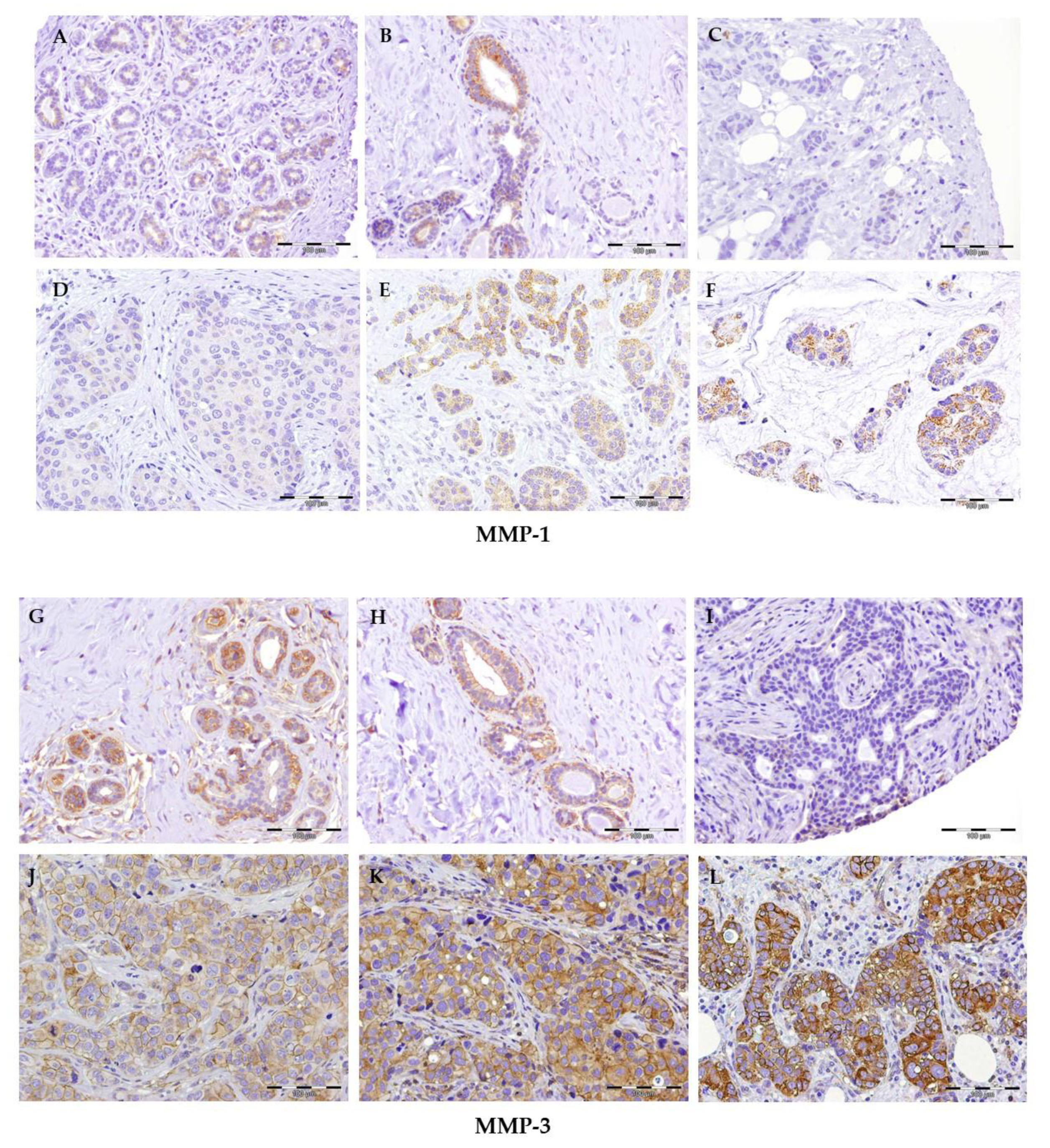
2.2. Immunohistochemical Expression of MMPs

Of the studied population of women (controls-cases), not all of them presented immunohistochemical expression of MMP-1-2-3-9 (percentage-intensity) in the mammary tissue analyzed with and without tumor disease. The study variables expression of MMP-1 intensity and MMP-3 percentage and intensity (Table 2) showed statistically significant associations with the variable group (control–case, p = 0.043 Chi-square, p = 0.018/0.025 Chi-square, respectively). No statistically significant associations were found (p ≥ 0.05) for MMP-2 and MMP-9 with the variable group (cases-controls) (Table 2).
Figure 1 shows the immunostaining of MMP-1 and MMP-3 in the sections of the mammary tissue studied. Staining with anti-MMP-1 was only observed in the cytoplasm of some glandular cells in normal tissue specimens (Figure 1A,B). Reactivity in the cytoplasm of the tumor cells and occasionally in some cells of the stroma was found for tumoral tissues. Four different levels of staining intensity are described (non-staining, weak, moderate, and intense, Figure 1C–F).
Anti-MMP3 antibody in normal breast tissue shows reactivity in the cytoplasm of glandular cells, especially myoepithelial cells, with different levels of intensity. Reactivity is also observed in the vascular endothelium cells and in some cells of the inflammatory infiltrate (Figure 1G,H). Staining is also observed in the cytoplasm of tumor cells. Four different levels of staining intensity are described (non-staining, weak, moderate, and intense, Figure 1I–L). In some cells, the cytoplasmic staining results in membrane enhancement.
We found no relationship between overexpression of MMP-1 and MMP-3 with the clinicopathological characteristics of patients included in this study (age, risk factors, tumor stage, lymph node involvement, treatment, tumor invasion). Nevertheless, of the total number of patients analyzed with cancer, we identified that MMP-1 staining intensity and MMP3 staining percentage and intensity were higher in postmenopause patients, in those with positive hormone receptors, and in the histological ductal carcinoma type (Table 3).

3. Discussion

This study was performed to analyze the immunohistochemical expression of four human specific MMPs in specimens from normal and BC tissue. This study aimed to establish a panel of MMP expression as possible biomarkers for the diagnosis of breast cancer. The present study shows that there is a statistically significant association between the immunohistochemical expression of MMP-1 and MMP-3 in the breast tissue of women suffering BC and their expression in the tissue of patients without tumor disease. Immunostaining of MMP-1 and MMP-3 was higher in early stages of the disease, suggesting the strongest role of both MMPs being at the beginning of BC development.
BC is the most common malignant tumor among women worldwide [39]. In recent years, considerable progress has been made in the early detection of BC, allowing higher survival and cure rates in patients diagnosed with this disease; therefore, novel prognostic indicators are necessary to further improve the prognosis of breast cancer patients.
The tumor microenvironment (TME) is increasingly recognized as a key player in tumor progression and as a promising therapeutic target in breast cancer [40]. The TME is composed of ECM, as well as several cellular elements and soluble factors evolving a network of proteins and signalling molecules that play important roles in breast cancer progression and metastasis [41]. The breast TME is modulated by the ECM and extracellular vesicles [42]. The cancer microenvironment often differs from healthy tissue via ECM degradation of protein concentrations. Major alterations occur in the ECM as breast cancer progresses. The epigenetic machinery plays a central role in generating an immunosuppressive environment for cancer growth. For this reason, epigenetic alterations affecting immune cell function in the tumor microenvironment represent a growing area of investigation [43].
There are distinct multilayered epigenetic mechanisms that regulate MMPs. DNA methylation of the regulatory genes may indirectly affect the expression of MMPs in malignancy. Falzone et al. [44] have described the intragenic methylation as a mechanism responsible for the MMP-9 upregulation in cancer. Nevertheless, some results suggest that elevated or ectopic expression rather than MMP gene methylation-driven silencing might link MMPs with tumorigenesis [45]. On the other hand, microRNAs (miRNAs) regulate the expression and function of extracellular matrix molecules and are often dysregulated in BC [46,47]. Particularly, miRNAs have been a focus of interest in the post-transcriptional regulation of MMPs [48]. Different mechanisms in which miRNAs regulate MMPs have been analyzed in several contexts of tumor invasion, EMT, and ECM remodeling by some authors [49]. Moreover, microRNAs can control breast cancer development, invasion, and migration directly and indirectly through regulating specific MMPs. MiRNAs are also involved in the downregulation of TIMP-1 and TIMP-3 in breast cancer [49]. Additionally, miRNAs target chromatin-remodeling histone deacetylases (HDACs), leading to altered MMP activity. In healthy states, there is a physiological balance between activation and inhibition of proteolytic degradation by expression of TIMPs and MMPs. In cancer states, this balance seems to be disrupted [49]. MiR-21 plays an important role in breast cancer. In this regard, miR-21 was shown to target MMP-3 expression to regulate breast cancer invasion [50], and is found in breast malignancy with high proliferation, advanced-stage, and aggressive phenotypes, such as pregnancy-associated breast cancer [51]. MiR-206 is involved in the downregulations of MMP-2 and MMP-9 [52]. The upregulation of miR-103/107 was shown to be associated with metastasis and poor outcome of breast cancer patients [53]. The downregulation of miR-210 was reported to be inversely correlated with cancer aggressiveness and metastatic capability [54]. Other studies have shown increases of breast cancer cell invasion and migration as well as metastasis associated with higher MMP-9 activity caused by miR-182 regulation. Chu et al. showed that overexpression of miR-519d significantly suppressed proliferation, migration, and metastasis of breast cancer cells by targeting MMP-3, suggesting that the novel molecular correlation between miR-519d and MMP-3 may become a potential therapeutic approach for breast cancer treatment [55].
Extracellular proteinases such as MMP maintain homeostasis of the ECM and are important key players in the tumor microenvironment. MMPs are a subclass of ECM degradation proteins with concentration differences between healthy and cancer tissues. Changes in the ECM and the interactions between cells and the ECM, with a particular focus on MMPs, have been well documented [56]. Additionally, MMP expression alters the rigidity, porosity, and many other characteristics of the ECM, facilitating cell migration and invasion.
MMPs are involved in the multistep processes of EMT and cancer progression; therefore, they have been considered as potential diagnostic and therapeutic biomarkers for several types of cancer [57]. The initiating step for cancer cells to acquire migratory potential is the EMT, which refers to the reprogramming that occurs in genetically and epigenetically modified cells [58]. E-cadherin has been proposed as an EMT indicator and as a direct target for MMP-dependent shedding, suggesting a direct role for MMPs in disassembly of cell junctions [59]. Some authors have described that Wnt1-induced EMT is associated with MMP-3 activation and that this inhibition resulted in repression of EMT characteristics [60]. MMP-3 is responsible for rendering several active proMMPs, and specifically Suzuki and his colleagues reported the transformation of proMMP-1 to the completely active MMP-1 form by MMP-3 [61].
The overexpression of MMP-1 and MMP-3 is associated with the clinicopathological characteristics of several malignancies [62,63]. This study shows no relationships between expression of MMP-1 and MMP-3 and age, histological type, lymph node affectation, treatment, or hormone receptors for BC (Table 3). This is the main limitation of this study. Nevertheless, of the total number of patients analyzed with cancer, we identified that MMP-1 staining intensity and MMP-3 staining percentage and intensity were significantly increased in the cancerous tissues by 62.3% and 67.5%, respectively, compared to the normal mammary tissues. Preclinical studies revealed that overexpression of MMP-1 plays a role in initiating mammary tumorigenesis through breaking down stroma and disseminating growth factors and mitogens for epithelial cells [64]. Abnormal expression of MMP-1 was identified in several types of malignant cancers [65,66], although its expression status and prognostic merit in BC remain unclear. Some studies have found that elevated expression of MMP-1 can promote the local growth and formation of brain metastases by breast cancer cell [67]. High MMP-1 gene expression has also been reported to predict for a lower overall survival rate in invasive breast carcinoma [68] and poorer prognosis in patients treated with systemic therapy [69,70]; thus, the expression of MMP-1 is a significant prognostic indicator and a potential drug target for BC [70].
The active participation of MMPs in the different stages of tumor progression is based on various clinical observations related to the expression of these enzymes in different types of human metastatic cancers, as well as on the matrix proteins that are modified by them [71]. Our results show higher immunohistochemical expression levels for MMP1 intensity and MMP-3 percentage and intensity for early-stage breast cancer (EI, EII). This fact would support the interplay between these two MMPs and the biological roles of MMPs related to different steps of carcinogenesis.
MMP-3 is highly expressed in the mammary gland, where it functions to regulate branching morphogenesis and postlactational involution [72]. On the other hand, MMP-3 provides an example of an MMP that can be either protective or protumorigenic in relation to growth [73].
Some studies have been published on the roles of MMP-1 in BC progression and metastasis [74,75]. Other authors have shown the important roles of MMP-3 in tumor progression and overall survival [76]; however, unlike our study, none of the other studies have measured the expression of four MMPs in the biopsies of cancer patients. Our results show that only the expression of MMP-1 and MMP-3 in tumor tissue could be related to the progression of BC and suggest prioritizing these MMPs as candidates for development of therapeutic strategies in these patients.
Few studies have evaluated the immunohistochemical expression of MMP-2 and MMP-9 in BC and fibroadenoma. Some authors have found significantly higher MMP-2 and MMP-9 protein expression in BC cells than in fibroadenoma [77]. Sampaio et al. [78] showed significantly higher expression of metallotionein-1, a membrane-type 1 MMP, in BC than in fibroadenoma. A study published by Li et al. involved 270 patients with BC and consecutive negative lymph node cases who received radical mastectomy or modified radical mastectomy, concluding that MMP-2 and MMP-9 are unfavorable prognostic factors in BC patients. They might be potential predictive factors for adjuvant systemic therapy [79]. MMP-2 and MMP-9 are also involved in each stage of breast-cancer-to-bone metastasis [80]. For these reasons, MMP-2 and MMP-9 have been considered as reliable biomarkers for the prediction of BC prognosis [54] and for metastasis development [55,56].
In this study, MMP-2 and MMP-9 staining showed no significant differences between case and control groups, while a role of MMP-2 and MMP-9 as biomarkers for the prediction of BC progression and metastasis was not supported. These findings are not usual given that the expression levels of MMP-2 and MMP-9 in BC have been described in different studies, since not only do they exhibit proteolytic activity against basal membrane proteins, which translates into tumor invasion, but also influence tumor growth, angiogenesis, and premetastatic niche formation [81,82,83,84]. It is important to highlight that uneven expression levels of several MMP have been found in BC. This could be due to differences in the commercial companies supplying the primary antibody and the methods of immunohistochemical staining used. Currently, there is no consensus on the threshold for MMP overexpression as assessed by immunohistochemistry. Additionally, the cut-off values for percentages or staining intensity levels may differ between studies, resulting in inconsistent positivity rates and predictive values for MMP overexpression. This may be an important source of heterogeneity and could limit the clinical use of MMP expression for the diagnosis of BC. It is also important to consider publication bias in the analysis of MMP overexpression, since studies with negative results may tend to be unpublished.
MMPs are involved in many biological processes and could be important biomarkers for cardiovascular disease, musculoskeletal disorders, and cancer. It is important to consider that the activities of MMPs may vary during disease due to differences in the proteolytic activities of MMPs towards different substrates [85,86,87]; thus, targeting MMPs is a complex task given that individual MMPs act in different cancers and at distinct stages of cancer progression. Pursuing only MMPs expressed by the specific tumor would be a new step torward personalized medicine. Several MMPs are strongly implicated as promising targets for breast cancer therapy. Considering that the efficacy of the therapy with MMPIs drastically decreases with the progression of the disease, it can be hypothesized that inhibition of MMPs could be effective in limiting tumor progression during its initial phase [12]. Some studies [11,12,19,88] have analyzed different strategies for development of inhibitors with therapeutic potential that are capable of selectively targeting the MMPs most responsible for tumor promotion, with special consideration of the potential of biologics including antibodies and engineered proteins based on the TIMP scaffold. Napoli et al. [12] showed the involvement of MMP-9 in the degradation of ECM and the consequent progression of melanoma, as well as the potential therapeutic implication of both endogenous and exogenous MMPIs for the design of new therapeutic protocols for melanoma patients. Most of the MMPIs evaluated in clinical trials to date have failed, causing major musculoskeletal toxicity and failing to improve clinical outcomes [89,90]. The reason for this could be that these trials studied patients with stage IV disease. New trials should enrol patients with high-risk disease that is not yet clinically or pathologically metastatic. On the other hand, the drug should be given prior to surgery, in the so called “window of opportunity” between the time of diagnosis and surgical excision, or postoperatively in the adjuvant setting. This would help to identify and validate biomarkers of enzymatic inhibition and metastasis as a proxy for clinical success [88]. In this regard, the D-Care study (NCT01077154) investigated denosumab, a drug with a similar MMPI design, in the neoadjuvant or adjuvant setting for patients with stage II or III breast cancer at high risk of recurrence. This study demonstrated that denosumab improves bone-related outcomes for women with high-risk early breast cancer.
Despite recent advances in our knowledge of MMPs, multiple functional aspects of these proteases remain unknown [58]. Therefore, we believe that more studies are needed to confirm any of the hypotheses proposed due to the lack of evidence in the literature on this subject.

4. Materials and Methods

4.1. Patients

This was a retrospective cohort study on 196 elderly patients undergoing breast surgery by the General Surgery Services of the Hospitals associated with the Biobank of Granada, in a period from July 2015 to July 2020. The group of cases was composed of 154 women diagnosed with BC and 42 women without tumor disease belonging to the control group (patients undergoing surgery because of benign breast disease such as fibroadenoma). Written informed consent was obtained from all cases and control subjects involved in the study.
The data and samples were managed through the Biobank for Research of the San Cecil-io-Granada University Hospital, belonging to the National Biobank Network (Project RD09/0076/00148), ensuring the integral treatment of the samples and associated data in accordance with Law 14/2007 of July 3 on Biomedical Research. The study was conducted according to the guidelines of the Declaration of Helsinki and was approved by the Provincial Research Ethics Committee of Granada. The biographical, clinical, and anatomopathological information was obtained only for the group of cases, recording the following data: age, date of diagnosis, risk factors, menopause, affected breast, tumor location, histological type, tumor stage, hormonal receptors, medical treatment established, lymph node involvement, tumor recurrence, metastasis to other organs, and mortality at 5 years.

4.2. Immunocytochemical Staining

Breast tissue samples were obtained from both groups to be included in several tissue matrices (TMA), facilitating the processing, staining and interpretation, and successive titration of antibodies for subsequent immunohistochemical assessment. Inmunohistochemistry was carried out on 3µm TMA sections, fixed in 10% buffered formalin, and embedded in paraffin using both PTLink and AutostainerLink (Dako, Glostrup, Denmark). Antibodies for MMPs were obtained from ABCAM (MMP-1, -2, -9) and ABGENT (MMP-3).
The staining process was carried out simultaneously in all sections stained with the same antibody. Both positive and negative controls (replacing the primary antibody with PBS) were made for each antibody used. If there were published reactivity levels for each of the used antibodies, different tissues were considered as positive controls. Assessment of the staining of MMPs was independently evaluated by two pathologists who were blinded to the patients’ clinicopathologic data. Disagreements were resolved by discussion in a meeting to obtain the results. The staining stratification was established based on two scores: (1) the proportion score representing the fraction of positively stained cells (≤50%–>50%, respectively); (2) the intensity of the staining (weak, moderate, or intense). This assessment allows for a semiquantitative estimate of the expression levels of protein in the tissue section. The two scores were added and the final definition of every section was obtained.

4.3. Statistical Analysis

The statistical program IBM-SPSS V.26.0 (SPSS Inc., Chicago, IL, USA) was used for the statistical analysis. The Shapiro–Wilk test was applied to verify the normality of the quantitative variable (age at diagnosis of the disease). Further, the age variable was categorized by age range (≤50 years, 51–70 years, and >70 years). The results for the categorical variables are expressed in percentages and the quantitative variables are expressed as means ± standard deviations, minimum and maximum values, and 95% CI values. For the bivariate analysis, Chi-square test was used to compare proportions between groups. Fisher’s exact test was used (Table 2) when the validity conditions were not met.

5. Conclusions

The results of this study suggest increased MMP-1 and MMP-3 expression in BC tissue compared to normal breast epithelium tissue. Regarding the association between MMP-1 and MMP-3 expression and other clinicopathological prognostic factors, we could not find significant relationships between the expression of these biomarkers and age, histological type, lymph node affectation, treatment, or hormonal receptors. MMP-1 and MMP-3 are involved in the maintenance of the angiogenic phenotype; thus, inhibition of these proteinases may be of value both in preventing breast cancer and in blocking metastasis of established tumors. As such, the use of MMP inhibitors in patients with early-stage cancer should be considered, as it has mainly been limited to patients with advanced disease to date.

Author Contributions

Conceptualization, A.X.A.C., S.R.A., P.T.P., and M.I.N.; methodology, A.X.A.C. and A.J.P.A.; formal analysis, A.X.A.C. and A.R.G.R.; investigation, J.D.R.G. and M.A.O.U.; data curation, S.R.A. and A.R.G.R.; writing—original draft preparation, A.X.A.C. and A.J.P.A.; writing—review and editing, A.X.A.C., A.J.P.A., and M.I.N.; supervision, M.I.N. and P.T.P.; funding acquisition, M.I.N. All authors have read and agreed to the published version of the manuscript.

Funding

This research was funded by Fundación Progreso Salud, grant number PI-0730-2013, and by ISCIII, grant number PIE16/00045.

Institutional Review Board Statement

The study was conducted according to the guidelines of the Declaration of Helsinki, and approved by the Provincial Research Ethics Committee of Granada (protocol code 72; date of approval: 27 February 2015).

Informed Consent Statement

Informed consent was obtained from all subjects involved in the study.

Data Availability Statement

The data presented in this study are available on request from the corresponding author.

Acknowledgments

We would like to thank the patients and control subject for their participation. We would also like to acknowledge the contribution of Andalusian Tumor Bank Network of Granada.

Conflicts of Interest

The authors declare no conflict of interest.

References

  1. Ferlay, J.; Colombet, M.; Soerjomataram, I.; Parkin, D.M.; Piñeros, M.; Znaor, A.; Bray, F. Cancer statistics for the year 2020: An overview. Int. J. Cancer 2021, 149, 778–789. [Google Scholar] [CrossRef]
  2. Sung, H.; Ferlay, J.; Siegel, R.L.; Laversanne, M.; Soerjomataram, I.; Jemal, A.; Bray, F. Global cancer statistics 2020: GLOBOCAN estimates of incidence and mortality worldwide for 36 cancers in 185 countries. CA Cancer J. Clin. 2021, 71, 209–249. [Google Scholar] [CrossRef] [PubMed]
  3. Maringe, C.; Spicer, J.; Morris, M.; Purushotham, A.; Nolte, E.; Sullivan, R.; Rachet, B.; Aggarwal, A. The impact of the COVID-19. Pandemic on cancer deaths due to delays in diagnosis in England, UK: A national, population-based, modelling study. Lancet Oncol. 2020, 21, 1023–1034. [Google Scholar] [CrossRef]
  4. Momenimovahed, Z.; Salehiniya, H. Delay in the diagnosis of breast cancer during coronavirus pandemic. EXCLI J. 2021, 20, 142–144. [Google Scholar] [PubMed]
  5. Vivarelli, S.; Falzone, L.; Grillo, C.M.; Scandurra, G.; Torino, F.; Libra, M. Cancer Management during COVID-19 Pandemic: Is Immune Checkpoint Inhibitors-Based Immunotherapy Harmful or Beneficial? Cancers. 2020, 12, 2237. [Google Scholar] [CrossRef] [PubMed]
  6. Sociedad Española de Oncología Médica. Las Cifras del Cáncer en España. 2021. Available online: http://www.seom.org (accessed on 26 June 2021).
  7. Lu, P.; Weaver, V.M.; Werb, Z. The extracellular matrix: A dynamic niche in cancer progression. J. Cell Biol. 2012, 196, 395–406. [Google Scholar] [CrossRef]
  8. Deryugina, E.I.; Quigley, J.P. Matrix metalloproteinases and tumor metastasis. Cancer Metastasis Rev. 2006, 25, 9–34. [Google Scholar]
  9. Belkin, A.M.; Akimov, S.S.; Zaritskaya, L.S.; Ratnikov, B.I.; Deryugina, E.I.; Strongin, A.Y. Matrix-dependent proteolysis of surface transglutaminase by membrane-type metalloproteinase regulates cancer cell adhesion and locomotion. J. Biol. Chem. 2001, 276, 18415–18422. [Google Scholar] [CrossRef] [Green Version]
  10. Lambert, E.; Dasse, E.; Haye, B.; Petitfrere, E. TIMPS as multifacial proteins. Crit. Rev. Oncol. Hematol. 2004, 49, 187–198. [Google Scholar] [CrossRef]
  11. Radisky, E.S.; Raeeszadeh-Sarmazdeh, M.; Radisky, D.C. Therapeutic Potential of Matrix Metalloproteinase Inhibition in Breast Cancer. J Cell Biochem. 2017, 118, 3531–3548. [Google Scholar] [CrossRef] [Green Version]
  12. Napoli, S.; Scuderi, C.; Gattuso, G.; Bella, V.D.; Candido, S.; Basile, M.S.; Libra, M.; Falzone, L. Functional Roles of Matrix Metalloproteinases and Their Inhibitors in Melanoma. Cells 2020, 9, 1151. [Google Scholar] [CrossRef]
  13. Tampa, M.; Georgescu, S.R.; Mitran, M.I.; Mitran, C.I.; Matei, C.; Caruntu, A.; Scheau, C.; Nicolae, I.; Matei, A.; Caruntu, C.; et al. Current Perspectives on the Role of Matrix Metalloproteinases in the Pathogenesis of Basal Cell Carcinoma. Biomolecules 2021, 11, 903. [Google Scholar] [CrossRef]
  14. Cascales, M.; Álvarez-Gómez, J.A. Metalloproteinasas, matriz extracelular y caáncer. An. R. Acad. Nac. Farm. 2010, 76, 59–84. [Google Scholar]
  15. Holmbeck, K.; Bianco, P.; Inoue, S.; Billinghurst, R.C.; Wu, W.; Chrysovergis, K.; Yamada, S.; Birkedal-Hansen, H.; Poole, A.R. The metalloproteinase MT1-MMP is required for normal development and maintenance of osteocyte processes in bone. J Cell Sci. 2005, 118, 147–156. [Google Scholar] [CrossRef] [PubMed] [Green Version]
  16. Kajiwara, Y.; Ueno, H.; Hashihuchi, Y.; Shinto, E.; Shimazaki, H.; Mochizuki, H.; Hase, K. Heterogeneity of metalloproteinase expression in colorectal cancer e relation of molecular findings to basic morphology. Anticancer Res. 2011, 31, 1567–1575. [Google Scholar] [PubMed]
  17. Sena, P.; Mariani, F.; Marzona, L.; Benincasa, M.; Ponz de Leon, M.; Palumbo, C.; Roncucciet, L. Matrix metalloproteinases 15 and 19 are stromal regulators of colorectal cancer development from the early stages. Int. J. Oncol. 2012, 41, 260–266. [Google Scholar]
  18. Rydlova, M.; Holubec, L.; Ludvikova, M.; Kalfert, D.; Franekova, J.; Povysil, C.; Ludvikova, M. Biological activity and clinical implications of the matrix metalloproteinases. Anticancer Res. 2008, 28, 1389–1397. [Google Scholar]
  19. Radisky, E.S.; Radisky, D.C. Matrix metalloproteinases as breast cancer drivers and therapeutic targets. Front. Biosci. 2015, 20, 1144–1163. [Google Scholar] [CrossRef]
  20. Kessenbrock, K.; Plaks, V.; Werb, Z. Matrix metalloproteinases: Regulators of the tumor microenvironment. Cell 2010, 141, 52–67. [Google Scholar] [CrossRef] [Green Version]
  21. Du, X.; Wang, S.; Lu, J.; Cao, Y.; Song, N.; Yang, T.; Dong, R.; Zang, L.; Yang, Y.; Wu, T.; et al. Correlation between MMP1-PAR1 axis and clinical outcome of primary gallbladder carcinoma. Jpn. J. Clin. Oncol. 2011, 41, 1086–1093. [Google Scholar] [CrossRef]
  22. Grimm, M.; Lazariotou, M.; Kircher, S.; Stuermer, L.; Reiber, C.; Höfelmayr, A.; Gattenlöhner, S.; Otto, C.; Germer, C.T.; von Rahden, B.H. MMP-1 is a (pre-)invasive factor in Barrett- associated esophageal adenocarcinomas and is associated with positive lymph node status. J. Transl. Med. 2010, 8, 99. [Google Scholar] [CrossRef] [Green Version]
  23. Fan, S.Q.; Wei, Q.T.; Li, M.I.; Zhang, L.Q. Expression and clinical significance of MMP-2, MMp-9, Timp-1 and TIMP-2 in breast carcintoma. Ai Zheng 2003, 22, 968–973. [Google Scholar]
  24. Olivares-Urbano, M.A.; Griñan-Lisón, C.; Zurita, M.; del Moral, R.; Ríos-Arrabal, S.; Artacho-Cordón, F.; Arrebola, J.P.; González, A.R.; León, J.; Marchal, J.A.; et al. Matrix-metalloproteases and TIMPs as prognostic biomarkers in breast cancer patients treated with radiotherapy: A pilot study. J. Cell. Mol. Med. 2020, 24, 139–148. [Google Scholar] [CrossRef] [Green Version]
  25. Ito, T.; Ito, M.; Shiozawa, J.; Naito, S.; Kanematsu, T.; Sekine, I. Expression of the MMP-1 in human pancreatic carcinoma: Relationship with prognostic factor. Mod. Pathol. 1999, 12, 669–674. [Google Scholar]
  26. Egeblad, M.; Werb, Z. New functions for the matrix metalloproteinases in cancer progression. Nat. Rev. Cancer 2002, 2, 161–174. [Google Scholar] [CrossRef]
  27. Stetler-Stevenson, W.G. Matrix metalloproteinases in angiogenesis: A moving target for therapeutic intervention. J. Clin. Investig. 1999, 103, 1237–1241. [Google Scholar] [CrossRef] [PubMed] [Green Version]
  28. Eck, S.M.; Blackburn, J.S.; Schmucker, A.C.; Burrage, P.S.; Brinckerhoff, C.E. Matrix metalloproteinase and G protein coupled receptors: Co-conspirators in the pathogenesis of autoimmune disease and cancer. J Autoimmun. 2009, 33, 214–221. [Google Scholar] [CrossRef] [PubMed] [Green Version]
  29. Köhrmann, A.; Kammerer, U.; Kapp, M.; Dietl, J.; Anacker, J. Expression of matrix metalloproteinases (MMPs) in primary human breast cancer and breast cancer cell lines: New findings and review of the literature. BMC Cancer 2009, 9, 188. [Google Scholar] [CrossRef] [PubMed] [Green Version]
  30. Patel, B.P.; Shah, P.M.; Rawal, U.M.; Desai, A.A.; Shah, S.V.; Rawal, R.M.; Patel, P.S. Activation of MMP-2 and MMP-9 in patients with oral squamous cell carcinoma. J. Surg. Oncol. 2005, 90, 81–88. [Google Scholar] [CrossRef]
  31. Pellikainen, J.M.; Ropponen, K.M.; Kataja, V.V.; Kellokoski, J.K.; Eskelinen, M.J.; Kosma, V.M. Expression of matrix metalloproteinase MMP-2 and MMP-9 in breast cancer with special reference to activator protein-2, HER-2 and prognosis. Clin. Cancer Res. 2004, 10, 7621–7628. [Google Scholar] [CrossRef] [PubMed] [Green Version]
  32. Sparmann, A.; Bar-Sagi, D. Ras-induced interleukin-8 expression plays a critical role in tumor growth and angiogenesis. Cancer Cell 2004, 6, 447–458. [Google Scholar] [CrossRef] [PubMed] [Green Version]
  33. Patel, L.R.; Camacho, D.F.; Shiozawa, Y.; Pienta, K.J.; Taichman, R.S. Mechanisms of cancer cell metastasis to the bone: A multistep process. Future Oncol. 2011, 7, 1285–1297. [Google Scholar] [CrossRef] [Green Version]
  34. Drabsch, Y.; Ten, D.P. TGF-b signaling in breast cancer cell invasion and bone metastasis. J. Mammary Gland Biol. Neoplasia. 2011, 16, 97–108. [Google Scholar] [CrossRef] [Green Version]
  35. Chabottaux, V.; Noel, A. Breast cancer progression: Insights into multifaceted matrix metalloproteinases. Clin. Exp. Metastasis 2007, 24, 647–656. [Google Scholar] [CrossRef] [PubMed]
  36. Noë, V.; Fingleton, B.; Jacobs, K.; Crawford, H.C.; Vermeulen, S.; Steelant, W.; Bruyneel, E.; Matrisian, L.M.; Mareel, M. Release of an invasion promoter E-cadherin fragment by matrilysin and stromelysin-1. J. Cell Sci. 2001, 114, 111–118. [Google Scholar] [CrossRef]
  37. Sternlicht, M.D.; Bissett, M.J.; Werb, Z. The matrix metalloproteinase stromelysin-1 acts as a natural mammary tumor promoter. Oncogene 2000, 19, 1102–1113. [Google Scholar] [CrossRef] [Green Version]
  38. Sato, H.; Takino, T.; Miyamori, H. Roles of membrene-Type matrix metalloproteinase-1 in tumor invasion and metastasis. Cancer Sci. 2005, 96, 212–217. [Google Scholar] [CrossRef] [Green Version]
  39. Siegel, R.L.; Miller, K.D.; Fuchs, H.E.; Jemal, A. Cancer Statistics, 2021. CA Cancer J. Clin. 2021, 71, 7–33. [Google Scholar] [CrossRef]
  40. Artacho-Cordón, F.; Ríos-Arrabal, S.; Lara, P.C.; Artacho-Cordón, A.; Calvente, I.; Núñez, M.I. Matrix metalloproteinases: Potential therapy to prevent the development of second malignancies after breast radiotherapy. Surg. Oncol. 2012, 21, 143–151. [Google Scholar]
  41. Bahcecioglu, G.; Basara, G.; Ellis, B.W.; Ren, X.; Zorlutuna, P. Breast cancer models: Engineering the tumor microenvironment. Acta Biomater. 2020, 106, 1–21. [Google Scholar] [CrossRef]
  42. Yang, J.; Bahcecioglu, G.; Zorlutuna, P. The Extracellular Matrix and Vesicles Modulate the Breast Tumor Microenvironment. Bioengineering 2020, 7, 124. [Google Scholar] [CrossRef]
  43. Lodewijk, I.; Nunes, S.P.; Henrique, R.; Jerónimo, C.; Dueñas, M.; Paramio, J.M. Tackling tumor microenvironment through epigenetic tools to improve cancer immunotherapy. Clin. Epigenetics 2021, 13, 63. [Google Scholar] [CrossRef]
  44. Falzone, L.; Salemi, R.; Travali, S.; Scalisi, A.; McCubrey, J.A.; Candido, S.; Libra, M. MMP-9 overexpression is associated with intragenic hypermethylation of MMP9 gene in melanoma. Aging 2016, 8, 933–944. [Google Scholar] [CrossRef] [Green Version]
  45. Simonova, O.A.; Kuznetsova, E.B.; Tanas, A.S.; Rudenko, V.V.; Poddubskaya, E.V.; Kekeeva, T.V.; Trotsenko, I.D.; Larin, S.S.; Kutsev, S.I.; Zaletaev, D.V.; et al. Abnormal Hypermethylation of CpG Dinucleotides in Promoter Regions of Matrix Metalloproteinases Genes in Breast Cancer and Its Relation to Epigenomic Subtypes and HER2 Overexpression. Biomedicines 2020, 8, 116. [Google Scholar] [CrossRef]
  46. Rutnam, Z.J.; Wight, T.N.; Yang, B.B. miRNAs regulate expression and function of extracellular matrix molecules. Matrix Biol. 2013, 32, 74–85. [Google Scholar] [CrossRef] [Green Version]
  47. Falzone, L.; Grimaldi, M.; Celentano, E.; Augustin, L.S.A.; Libra, M. Identification of Modulated MicroRNAs Associated with Breast Cancer, Diet, and Physical Activity. Cancers 2020, 12, 2555. [Google Scholar] [CrossRef]
  48. Rak, B.; Mehlich, D.; Garbicz, F.; Domosud, Z.; Paskal, W.; Marczewska, J.M.; Włodarski, P.K. Post-transcriptional Regulation of MMP16 and TIMP2 Expression via miR-382, miR-410 and miR-200b in Endometrial Cancer. Cancer Genom. Proteom. 2017, 14, 389–401. [Google Scholar]
  49. Javadian, M.; Gharibi, T.; Shekari, N.; Abdollahpour-Alitappeh, M.; Mohammadi, A.; Hossieni, A.; Mohammadi, H.; Kazemi, T. The role of microRNAs regulating the expression of matrix metalloproteinases (MMPs) in breast cancer development, progression, and metastasis. J. Cell Physiol. 2019, 234, 5399–5412. [Google Scholar] [CrossRef]
  50. Song, B.; Wang, C.; Liu, J.; Wang, X.; Lv, L.; Wei, L.; Xie, L.; Zheng, Y.; Song, X. MicroRNA-21 regulates breast cancer invasion partly by targeting tissue inhibitor of metalloproteinase 3 expression. J. Exp. Clin. Cancer Res. 2010, 29, 29. [Google Scholar] [CrossRef] [Green Version]
  51. Gumireddy, K.; Young, D.D.; Xiong, X.; Hogenesch, J.B.; Huang, Q.; Deiters, A. Small-Molecule Inhibitors of MicroRNA miR-21 Function. Angew. Chem. Int. 2008, 47, 7482–7484. [Google Scholar] [CrossRef] [Green Version]
  52. Liu, H.; Cao, Y.D.; Ye, W.X.; Sun, Y.Y. Effect of microRNA-206 on cytoskeleton remodelling by downregulating Cdc42 in MDA-MB-231 cells. Tumori. 2010, 96, 751–755. [Google Scholar] [CrossRef]
  53. Martello, G.; Rosato, A.; Ferrari, F.; Manfrin, A.; Cordenonsi, M.; Dupont, S.; Enzo, E.; Guzzardo, V.; Rondina, M.; Spruce, T.; et al. A microRNA targeting dicer for metastasis control. Cell 2010, 141, 1195–1207. [Google Scholar] [CrossRef] [Green Version]
  54. Foekens, J.A.; Sieuwerts, A.M.; Smid, M.; Look, M.P.; De Weerd, V.; Boersma, A.W.M.; Klijn, J.G.M.; Wiemer, E.A.C.; Martens, J.W.M. Four miRNAs associated with aggressiveness of lymph node-negative, estrogen receptor-positive human breast cancer. Proc. Natl. Acad. Sci. USA 2008, 105, 13021–13026. [Google Scholar] [CrossRef] [PubMed] [Green Version]
  55. Chu, C.; Liu, X.; Bai, X.; Zhao, T.; Wang, M.; Xu, R.; Li, M.; Hu, Y.; Li, W.; Yang, L.; et al. MiR-519d suppresses breast cancer tumorigenesis and metastasis via targeting MMP3. Int. J. Biol. Sci. 2018, 14, 228–236. [Google Scholar] [CrossRef] [PubMed] [Green Version]
  56. Niland, S.; Eble, J.A. Hold on or Cut? Integrin- and MMP-Mediated Cell-Matrix Interactions in the Tumor Microenvironment. Int. J. Mol. Sci. 2020, 22, 238. [Google Scholar] [CrossRef] [PubMed]
  57. Gialeli, C.; Theocharis, A.D.; Karamanos, N.K. Roles of matrix metalloproteinases in cancer progression and their pharmacological targeting. FEBS J. 2011, 278, 16–27. [Google Scholar] [CrossRef]
  58. Lamouille, S.; Xu, J.; Derynck, R. Molecular mechanisms of epithelial-mesenchymal transition. Nat. Rev. Mol. Cell Biol. 2014, 15, 178–196. [Google Scholar] [CrossRef] [PubMed] [Green Version]
  59. Rodríguez, D.; Morrison, C.J.; Overall, C.M. Matrix metalloproteinases: What do they not do? New substrates and biological roles identified by murine models and proteomics. Biochim. Biophy. Acta. 2010, 1803, 39–54. [Google Scholar] [CrossRef] [PubMed] [Green Version]
  60. Blavier, L.; Lazaryev, A.; Shi, X.H.; Dorey, F.J.; Shackleford, G.M.; DeClerk, Y.A. Stromelysin-1 (MMP-3) is a target and a regulator of Wnt1-induced epithelial-mesenchymal transition (EMT). Cancer Biol. Ther. 2010, 10, 198–208. [Google Scholar] [CrossRef] [PubMed] [Green Version]
  61. Nagase, H.; Ogata, Y.; Suzuki, K.; Enghild, J.J.; Salvesen, G. Substrate specificities and activation mechanisms of matrix metalloproteinases. Biochem. Soc. Trans. 1991, 19, 715–718. [Google Scholar] [CrossRef] [Green Version]
  62. Fisher, C.; Gilbertson-Beadling, S.; Powers, E.A.; Petzlod, G.; Poorman, R.; Mitchell, M.A. Interstitial collagenase is required for angiogenesis in vitro. Dev. Biol. 1994, 162, 499–510. [Google Scholar] [CrossRef]
  63. Jezierska, A.; Motyl, T. Matrix metalloproteinase-2 involvement in breast cancer progression: A mini-review. Med. Sci. Monit. 2009, 15, 32–40. [Google Scholar]
  64. Van’t Veer, L.J.; Dai, H.; van de Vijver, M.J.; He, Y.D.; Hart, A.A.; Mao, M.; Peterse, H.L.; van der Kooy, K.; Marton, M.J.; Witteveen, A.T.; et al. Gene expression profiling predicts clinical outcome of breast cancer. Nature 2002, 415, 530–536. [Google Scholar] [CrossRef] [Green Version]
  65. Roeb, E.; Arndt, M.; Jansen, B.; Schumpelick, V.; Matern, S. Simultaneous determination of matrix metalloproteinase (MMP)-7, MMP-1, -3, and -13 gene expression by multiplex PCR in colorectal carcinomas. Int. J. Colorectal. Dis. 2004, 19, 518–524. [Google Scholar] [CrossRef]
  66. Wieczorek, E.; Reszka, E.; Jablonowski, Z.; Jablonska, E.; Krol, M.B.; Grzegorczyk, A.; Gromadzinska, J.; Sosnowski, M.; Wasowicz, W. Genetic polymorphisms in matrix metalloproteinases (MMPs) and tissue inhibitors of MPs (TIMPs), and bladder cancer susceptibility. BJU Int. 2013, 112, 1207–1214. [Google Scholar] [CrossRef]
  67. Liu, H.; Kato, Y.; Erzinger, S.A.; Galena, M.K.; Qian, Y.; Palmieri, D.; Steeg, P.S.; Price, J.E. The role of MMP-1 in breast cancer growth and metastasis to the brain in a xenograft model. BMC Cancer 2012, 12, 583. [Google Scholar] [CrossRef] [Green Version]
  68. Cheng, S.; Mitsuhiro, T.; Yasuhiro, H.; Toshimichi, A.; Kuramae, T.; Takemoto, N.; Hamada, J.-I.; Miyamoto, M.; Hirano, S.; Kondo, S.; et al. High MMP-1 mRNA expression is a risk factor for disease-free and overall survivals in patients with invasive breast carcinoma. J. Surg. Res. 2008, 146, 104–109. [Google Scholar] [CrossRef]
  69. Shen, C.J.; Kuo, Y.L.; Chen, C.C.; Chen, M.J.; Cheng, Y.M. MMP1 expression is activated by Slug and enhances multi-drug resistance (MDR) in breast cancer. PLoS ONE 2017, 12, e0174487. [Google Scholar] [CrossRef] [Green Version]
  70. Ji, W.; Chenyang, Y.; Demin, L.; Yongxia, C.; Yunlu, J.; Xiaogang, Y.; Hanchu, X.; Wenhe, Z.; Jichun, Z.; Linbo, W. Matrix metalloproteinase-1 expression in breast carcinoma: A marker for unfavorable prognosis. Oncotarget 2017, 8, 91379–91390. [Google Scholar]
  71. Jones, J.L.; Walker, R.A. Control of matrix metalloproteinase activity in cancer. J. Pathol. 1997, 183, 377–379. [Google Scholar] [CrossRef]
  72. Alexander, C.M.; Selvarajan, S.; Mudgett, J.; Werb, Z. Stromelysin-1 regulates adipogenesis during mammary gland involution. J. Cell Biol. 2001, 152, 693–703. [Google Scholar] [CrossRef] [Green Version]
  73. McCawley, L.J.; Crawford, H.C.; King, L.E., Jr.; Mudgett, J.; Matrisian, L.M. A protective role for matrix metalloproteinase-3 in squamous cell carcinoma. Cancer Res. 2004, 64, 6965–6972. [Google Scholar] [CrossRef] [Green Version]
  74. Boström, P.; Söderström, M.; Vahlberg, T.; Söderström, K.O.; Roberts, P.J.; Carpén, O.; Hirsimäki, P. MMP-1 expression has an independent prognostic value in breast cancer. BMC Cancer 2011, 11, 348. [Google Scholar] [CrossRef] [PubMed] [Green Version]
  75. Wang, Q.M.; Lv, L.; Tang, Y.; Zhang, L.; Wang, L.F. MMP-1 is overexpressed in triple-negative breast cancer tissues and the knockdown of MMP-1 expression inhibits tumor cell malignant behaviors in vitro. Oncol. Lett. 2019, 17, 1732–1740. [Google Scholar] [CrossRef] [PubMed] [Green Version]
  76. Mehner, C.; Miller, E.; Nassar, A.; Bamlet, W.R.; Radisky, E.S.; Radisky, D.C. Tumor cell expression of MMP3 as a prognostic factor for poor survival in pancreatic, pulmonary, and mammary carcinoma. Genes Cancer. 2015, 6, 480–489. [Google Scholar] [CrossRef] [Green Version]
  77. Martins, L.M.; de Melo Escorcio Dourado, C.S.; Campos-Verdes, L.M.; Sampaio, F.A.; Revoredo, C.M.S.; Costa-Silva, D.R.; da Conceição Barros-Oliveira, M.; de Jesus Nery Junior, E.; do Rego-Medeiros, L.M.; Gebrim, L.H.; et al. Expression of matrix metalloproteinase 2 and 9 in breast cancer and breast fibroadenoma: A randomized, double-blind study. Oncotarget 2019, 10, 6879–6884. [Google Scholar] [CrossRef] [PubMed]
  78. Sampaio, F.A.; Martins, L.M.; Dourado, C.S.M.E.; Revoredo, C.M.S.; Costa-Silva, D.R.; Oliveira, V.A.; Alves-Ribeiro, F.A.; Silva, B.B.D. A case-control study of Metallothionein-1 expression in breast cancer and breast fibroadenoma. Sci. Rep. 2019, 9, 7407. [Google Scholar] [CrossRef]
  79. Li, H.C.; Cao, D.C.; Liu, Y.; Hou, Y.F.; Wu, J.; Lu, J.S.; Di, G.H.; Liu, G.; Li, F.M.; Ou, Z.L.; et al. Prognostic value of matrix metalloproteinases (MMP-2 and MMP-9) in patients with lymph node-negative breast carcinoma. Breast Cancer Res. Treat. 2004, 88, 75–85. [Google Scholar] [CrossRef]
  80. Tauro, M.; Lynch, C.C. Cutting to the Chase: How Matrix Metalloproteinase-2 Activity Controls Breast-Cancer-to-Bone Metastasis. Cancers 2018, 10, 185. [Google Scholar] [CrossRef] [Green Version]
  81. Mook, O.R.; Frederiks, W.M.; Van Noorden, C.J. The role of gelatinases in colorectal cancer progression and metastasis. Biochim. Biophy. Acta 2004, 1705, 69–89. [Google Scholar] [CrossRef]
  82. Bjorklund, M.; Koivunen, E. Gelatinase-mediated migration and invasion of cancer cells. Biochim. Biophys Acta 2005, 1755, 37–69. [Google Scholar] [CrossRef] [PubMed] [Green Version]
  83. Otani, Y.; Okazaki, I.; Arai, M.; Kameyama, K.; Wada, N.; Maruyama, K.; Yoshino, K.; Kitajima, M.; Hosoda, Y.; Tsuchiya, M. Gene expression of interstitial collagenase (matrix metalloproteinase 1) in gastrointestinal tract cancers. J. Gastroenterol. 1994, 29, 391–397. [Google Scholar] [CrossRef]
  84. Jiang, H.; Li, H. Prognostic values of tumoral MMP2 and MMP9 overexpression in breast cancer: A systematic review and meta-analysis. BMC Cancer 2021, 21, 149. [Google Scholar] [CrossRef]
  85. Roy, R.; Yang, J.; Moses, M.A. Matrix metalloproteinases as novel biomarkers and potential therapeutic targets in human cancer. J. Clin. Oncol. 2009, 27, 5287–5297. [Google Scholar] [CrossRef] [PubMed] [Green Version]
  86. Mohammadizadeh, F.; Bagherian-Dehkordia, M. Relationship between matrix metalloproteinase-9 and some clinicopathological prognostic factors of breast carcinoma. Am. J. Clin. Exp. Immunol. 2021, 10, 17–22. [Google Scholar] [PubMed]
  87. Cui, N.; Hu, M.; Khalil, R.A. Biochemical and Biological Attributes of Matrix Metalloproteinases. Prog. Mol. Biol. Transl. Sci. 2017, 147, 1–73. [Google Scholar]
  88. Winer, A.; Adams, S.; Mignatti, P. Matrix Metalloproteinase Inhibitors in Cancer Therapy: Turning Past Failures Into Future Successes. Mol. Cancer Ther. 2018, 17, 1147–1155. [Google Scholar] [CrossRef] [Green Version]
  89. Coussens, L.M.; Fingleton, B.; Matrisian, L.M. Matrix metalloproteinase inhibitors and cancer: Trials and tribulations. Science 2002, 295, 2387–2392. [Google Scholar] [CrossRef] [PubMed]
  90. Vandenbroucke, R.E.; Libert, C. Is there new hope for therapeutic matrix metalloproteinase inhibition? Nat. Rev. Drug. Discov. 2014, 13, 904–927. [Google Scholar] [CrossRef]
Figure 1. Immunostaining of MMP-1 (ABCAM, (AF)): (A,B) sections of normal breast tissue; (CF), sections of breast carcinoma with different level of staining intensity; (C) absence of staining; (D) weak staining; (E) moderate staining; (F) intense staining. Immunostaining of MMP-3 (AGENT, (GL)): (G,H) sections of normal breast tissue; (IL) sections of breast carcinoma with different levels of staining intensity; (I) absence of staining; (J) weak staining; (K) moderate staining; (L) intense staining. The length of the scale bar is 100 μm.
Ijms 22 09012 g001
Table 1. Descriptive analysis of the study population (cases).
Independent VariablesNo. of PatientsPercentage
Age interval≤50 years4428.6
51–70 years5334.4
>70 years5737.0
Risk factorsFirst-degree family history of BC23.8
Personal history of other tumors611.3
History of benign breast lesions47.5
Smoking1935.8
Obesity1630.2
More than one risk factor611.3
MenopausePremenopause4731.1
Postmenopause10468.9
Affected breastRight breast3930.7
Left breast8869.3
Histological typeInfiltrating ductal carcinoma13587.7
Infiltrating lobulillar carcinoma138.4
Other carcinomas63.9
Lymph node involvementNo7045.8
Yes8354.2
Tumor stageEI:IA-IB2623.0
EII:IIA-IIB5246.0
EIII:IIIA-IIIB-IIIC3026.5
EIV with any TNM54.4
Hormone receptorsEstrogenics and Progestogens +7273.5
Estrogenics and Progestogens −1818.4
TreatmentSurgery + Chemotherapy + Radiotherapy5233.8
Surgery + Chemotherapy4931.8
Surgery + Radiotherapy117.1
Surgery4227.3
Tumor recurrenceNo12589.9
Yes1410.1
Tumor invasionBone520.0
Lung416.0
Brain28.0
Liver14.0
Association with another cancer832.0
Metastasis to more than one organ520.0
Follow-up patientMortality in 2020Dead64.4
Alive13195.6
Table 2. Statistical analysis of interactions between MMP-1, MMP-2, MMP-3, and MMP-9 (percentage and intensity) and the variable groups (case and control).
Study VariablesGroup p
CaseControlTotal
MMP-1 percentage≤50%299380.073
>50%11716133
MMP-1 intensityWeak96221180.043
Moderate53457
Intense5NA5*
MMP-2 percentage≤50%126221480.139
>50%24125
MMP-2 intensityWeak137211580.996
Moderate13215
IntenseNANANA*
MMP-3 percentage≤50%5716730.018
>50%90999
MMP-3 intensityWeak104121160.025
Moderate431356
Intense7NA7*
MMP-9 percentage≤50%146281740.421
>50%NANANA*
MMP-9 intensityWeak146281740.326
ModerateNANANA*
IntenseNANANA*
* Statistical analysis could not be performed with the intense category for MMP-1 and MMP-3 because immunostaining in breast tissue sections was only found in cases and not in controls. The intense category was not found in either normal or tumor breast biopsies for MMP-2 and MMP-9.
Table 3. Statistical analysis between variables MMP-1 and MMP-3 expression and clinical–pathological variables.
MMP-1 IntensityMMP-3 IntensityMMP-3 Percentage
WeakModeratepWeakModeratep≤50%>50%p
Age interval≤50 years32100.1683580.17712310.210
51–70 years312031172028
>70 years332338182531
Tumor stageEI1970.5491950.76610140.978
EII331636142129
EIII19101891017
EIV234123
MenopausePremenopause34110.0643790.11514320.204
Postmenopause604165334157
Lymph node involvementNo42250.73048190.85626410.510
Yes54275524 3049
TumorrecurrenceNo79421.0008735/20.51047751.000
Yes95102 57
Affected breastRight breast2980.06323130.52010260.307
Left breast513462253354
Hormone receptorsEstrogenics and Progestogens +41290.39445260.08522490.348
Estrogenics and Progestogens −13414288
Estrogenics +537135
Risk factorsFirst-degree family history of BC110.916110.056110.151
Personal history of other tumors421515
History of benign breast lesions313113
Smoking108126315
Obesity10614297
More than one risk factor413315
Histological typeInfiltrating ductal carcinoma84470.74891380.27248810.502
Infiltratinglobulillarcarcinoma858567
Othercarcinomas415032
TreatmentSurgery +Chemotherapy+ Radiotherapy31180.98436150.85618330.863
Surgery +Chemotherapy321631131727
Surgery +Radiotherapy749247
Surgery261528131823
TumorinvasionBone410.674400.216220.912
Lung313113
Brain112011
Liver011001
Association with another cancer623535
Metastasis to more than one organ323113
Publisher’s Note: MDPI stays neutral with regard to jurisdictional claims in published maps and institutional affiliations.

